Note: Descriptions are shown in the official language in which they were submitted.
CA 02720124 2014-03-20
ANALYSIS OF ANTIBODY DRUG CONJUGATES BY BEAD-BASED AFFINITY
CAPTURE AND MASS SPECTROMETRY
[00031 FIELD OF THE INVENTION
[00041 The invention relates generally to methods to capture, detect,
analyze, screen,
characterize, and quantitate antibody conjugate compounds, including antibody-
drug
conjugates, and their fragments and metabolites, by mass spectrometry. The
invention also
relates to methods to prepare mass spectrometric samples for pharmacokinetic
and
toxicokinetic studies.
[0005] BACKGROUND OF THE INVENTION
[00061 Antibody-drug conjugates (ADC) are targeted anti-cancer therapeutics
designed to reduce nonspecific toxicities and increase efficacy relative to
conventional small
molecule and antibody cancer chemotherapy. They employ the powerful targeting
ability of
monoclonal antibodies to specifically deliver highly potent, conjugated small
molecule
therapeutics to a cancer cell. To evaluate properties such as pharmacokinetics
and toxicity of
these antibody-drug conjugates, it is useful to be able to characterize and
quantitate them
from plasma, urine, and other biological samples. Additionally, the ability to
quantitate the
free drug (not conjugated to the antibody) in the method from the same sample
and the same
chromatographic injection would also be useful.
[0007] A variety of mass spectrometry techniques have been employed for
identification and quantitation of small molecule therapeutics in
pharmacokinetic studies,
such as: electron impact (El), chemical ionization (CI), desorption chemical
ionization (DC1),
fast atom bombardment (FAB), electrospray ionization (ESI), matrix-assisted
laser
desorption/ionization (MALDI), and tandem mass spectrometry (MS/MS) (Yao et al
(2001)
Jour. of Chrom. B 752:9-16; Royer et al (1995) Rapid Comm. in Mass Spec. 9:495-
502),
including single ion monitoring (SIM) mode of ion selection for deconvolution
(Souppart et
1
CA 02720124 2010-09-29
WO 2009/140242
PCT/US2009/043560
al (2002) Jour. of Chrom. B 774:195-203; Wong eta! (2001) Jour. of Chrom.
765:55-62; Yao
eta! (1998) Jour. of Chrom. B 718:77-85; Abdel-Hamid eta! (2001) Jour. of
Chrom. B
753:401-408; Marques eta! (2001) Jour. of Chrom. 762:87-95). These methods and
instrumentation require the separation of the various analytes from biological
fluids for
sufficient sensitivity. Such purification can be labor-intensive, slow, and
require large
volumes of sample fluids due to the low concentration of the analytes of
interest in samples
such as cell culture medium, human plasma, urine, and bile.
[0008] The direct combination of a separation/isolation/purification front-
end step
coupled with detection/characterization/quantitation by mass spectrometry is
effective for
metabolic studies of complex biological samples. Typically, LC/MS is used for
characterization of antibodies (Martin et al (1997) Cancer Chemother.
Pharmacol. 40:189-
201; WO 03/046571; WO 03/046572), and ELISA is used for quantitation in
biological
matrices (Murray et al (2001) J. Imm. Methods 255:41-56; Kirchner et al (2004)
Clin.
Pharmacokinetics 43(2):83-95). ELISA assays typically are sensitive and
amenable to high-
throughput screens.
[0009] Recent advances in protein analysis by mass spectrometry (MS) are
due to
front-end gas phase ionization and introduction techniques such as
electrospray ionization
(ESI), matrix-assisted laser desorption ionization (MALDI, US 2003/0027216)
and Surface
Enhanced Laser Desorption Ionization (SELDI, US 6020208), as well as
improvements in
instrument sensitivity, resolution, mass accuracy, bioinformatics, and
software data
deconvolution algorithms ("Electrospray Ionization Mass Spectrometry:
Fundamentals,
Instrumentation, and Applications", Cole, R.B., Ed. (1997) Wiley, New York;
"Modern
Protein Chemistry: Practical Aspects", Howard, G.C. and Brown, W.E., Eds.
(2002) CRC
Press, Boca Raton, FL, p. 71-102;). The primary (sequence), secondary, and
tertiary structure
of proteins can be probed and elucidated with MS. Electrospray ionization
(ESI) provides for
the atmospheric pressure ionization (API) of a liquid sample. The electrospray
process
creates highly-charged droplets that, under evaporation, create ions
representative of the
species contained in the solution. An ion-sampling orifice of a mass
spectrometer may be
used to sample these gas phase ions for mass analysis. The response for an
analyte measured
by the mass spectrometer detector is dependent on the concentration of the
analyte in the
fluid and independent of the fluid flow rate.
[0010] Methods to detect and screening antibody-drug conjugates by
Immunoaffinity
membrane (JAM) capture and mass spectrometry have been disclosed (US
2005/0232929).
2
CA 02720124 2010-09-29
WO 2009/140242
PCT/US2009/043560
[0011] SUMMARY
[0012] An aspect of the invention includes methods to detect, screen, and
quantitate
antibody conjugate compounds and compositions, antibodies, and fragments and
metabolites
thereof, by immunoaffinity bead capture, separation, chromatography, and mass
spectrometry. Exemplary methods of mass spectrometry include electrospray
ionization
(ESI), and full scan mass spectrometry (MS).
[0013] Immunoaffinity bead capture may be conducted with streptavidin
coated
paramagnetic beads capitalizing on: (i) the strong streptavidin-biotin
interaction, (ii) high
binding capacity to capture sufficient material for analysis of intact
proteins, (iii) low non-
specific binding, (iv) elution of sample constituents with mass spectrometry-
compatible
solvents, (v) good sample recovery, and (vi) amenability for automation.
[0014] Antibody conjugate compounds of the invention having Formula I:
Ab-(L-D)P
[0015] wherein
[0016] Ab is an antibody;
[0017] D is a maytansinoid or monomethylauristatin drug moiety;
[0018] L is a linker covalently attached to Ab, and covalently attached to
D; and
[0019] p is 1, 2, 3, 4, 5, 6, 7, or 8.
[0020] An aspect of the invention includes a method for detecting antibody-
drug
conjugate compounds comprising:
[0021] (i) providing an antibody-drug conjugate compound having Formula
I;
[0022] (ii) contacting the antibody-drug conjugate compound, and
optionally an
antibody of Formula I where p is 0, or antibody fragments or metabolites
thereof, with a
biological source selected from a mammal, tissue, cell culture, plasma or
serum;
[0023] (iii) collecting a biological sample from the biological source;
[0024] (iv) processing the biological sample to form an analysis sample by
formulating, immobilizing, centrifuging, isolating, digesting, inducing or
preventing blood
cell clotting, hydrolyzing, or purifying to form a processed analysis sample;
[0025] (v) capturing the processed analysis sample on immunoaffinity beads
comprising an antigen specific for the processed analysis sample;
[0026] (vi) eluting the processed analysis sample;
[0027] (vii) applying the eluted analysis sample to a separation media to
effect
separation of more than one sample constituent wherein a separated sample
constituent
3
CA 02720124 2010-09-29
WO 2009/140242
PCT/US2009/043560
comprises an antibody-drug conjugate compound having the Formula I, or
antibody fragment
or metabolite thereof, and where p is 0, 1, 2, 3, 4, 5, 6, 7, or 8; and
[0028] (viii) establishing the mass to charge ratio of one or more
separated sample
constituents by mass spectrometry.
[0029] Another aspect of the invention includes a method for screening a
mixture of
antibody-drug conjugate compounds and to determine the clearance of the
compounds, or
fragments or metabolites thereof, in a mammal, comprising:
[0030] (i) providing a mixture of antibody-drug conjugate compounds
having
Formula I where the mixture optionally comprises an antibody, or fragments or
metabolites
thereof, where p is 0;
[0031] (ii) administering the mixture to a mammal;
[0032] (iii) collecting a blood sample or excretion from the mammal to
which the
mixture has been administered;
[0033] (iv) processing the blood sample or excretion to form an analysis
sample by
formulating, immobilizing, centrifuging, isolating, digesting, inducing or
preventing blood
cell clotting, hydrolyzing, or purifying to form a processed analysis sample;
[0034] (v) capturing the processed analysis sample on an immunoaffinity
bead
comprising an antigen specific for the processed analysis sample;
[0035] (vi) eluting the processed analysis sample;
[0036] (vii) applying the blood sample, excretion or analysis sample to a
separation
media to effect separation of more than one sample constituents wherein a
separated sample
constituent comprises an antibody-drug conjugate compound having the Formula
I, or
antibody fragment or metabolite thereof, and where p is 0, 1, 2, 3, 4, 5, 6,
7, or 8; and
[0037] (viii) establishing the mass to charge ratio of more than one
separated sample
constituents by mass spectrometry.
[0038] The invention may be understood by reference to the following
detailed
description of the exemplary embodiments, taken in conjunction with the
accompanying
drawings, figures, and Examples. The discussion below is descriptive,
illustrative and
exemplary and is not to be taken as limiting the scope defined by any appended
claims.
[0039] BRIEF DESCRIPTION OF THE DRAWINGS
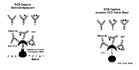
[0040] Figure la shows an illustration of antibodies (MAb) and antibody-
drug
conjugates (ADC) binding to the ECD (extracellular domain) of a biotinylated
ECD protein
which is bound to a streptavidin coated paramagnetic bead in contact with a
magnet.
4
CA 02720124 2010-09-29
WO 2009/140242
PCT/US2009/043560
[0041] Figure lb shows an illustration of antibodies (MAb) and antibody-
drug
conjugates (ADC) binding to the ECD (extracellular domain) of an ECD protein
which is
covalently linked to a bead.
[0042] Figure 2 shows an illustration of antibody-drug conjugates (ADC)
binding to a
biotinylated anti-drug monoclonal antibody (Biotin-Anti Drug MAb) which is
bound to a
streptavidin coated paramagnetic bead in contact with a magnet.
[0043] Figure 3 shows illustrations of cysteine engineered antibody-drug
conjugates,
from the top to the bottom: two MMAE drug moieties located on the light chain -
Thio Hu
Anti HER2 4D5 LC V205C-MC-vc-PAB-MMAE; two MMAE drug moieties located on the
heavy chain ¨ Thio Hu Anti HER2 4D5 HC All8C-MC-vc-PAB-MMAE; two MMAE drug
moieties located on the Fc region of the heavy chain - Thio Hu Anti HER2 4D5
Fc S400C-
MC-vc-PAB-MMAE; and a cysteine engineered antibody ready for conjugation: Thio
Hu
Anti HER2 4D5 Fc S400C.
[0044] Figure 4 shows changes in the drug/antibody ratio (DAR) distribution
for:
(top) light chain (Thio Hu Anti HER2 4D5 LC V205C-MC-vc-PAB-MMAE), and
(bottom)
heavy chain (Thio Hu Anti HER2 4D5 HC Al 18C-MC-vc-PAB-MMAE) ADC variants in
plasma after immunoaffinity ECD modified bead capture and mass spectrometry
characterization from in vitro plasma stability samples collected at 0, 8, 24,
48, and 96 hour
time points. The sample constituents were assigned DAR of 0 (naked antibody),
1 (one MC-
vc-PAB-MMAE drug linker unit) and 2 (two MC-vc-PAB-MMAE drug linker units).
[0045] Figure 5 shows deconvoluted mass spectrometry data of stability of
Thio Hu
Anti HER2 4D5 HC All8C-MC-vc-PAB-MMAE (100 ug/m1 in rat plasma incubated at 37
C) samples collected at 0, 8, 24, 48, and 96 hour time points, as plotted in
Figure 4 (bottom).
The sample constituents were assigned DAR of +0 (naked antibody), +1D (one MC-
vc-PAB-
MMAE drug linker unit) and +2D (two MC-vc-PAB-MMAE drug linker units). The
small
peaks at about 151,000 amu are sample constituents undergoing incomplete
deglycosylation.
[0046] Figure 6 shows deconvoluted mass spectrometry data of stability of
Thio Hu
Anti MUC16 (3A5) HC A118C-MC-vc-PAB-MMAE (100 ug/m1 in rat plasma incubated at
37 C) samples collected at 0, 6, 24, 48, and 96 hour time points. The sample
constituents
were assigned DAR of +0 (naked antibody), +1D (one MC-vc-PAB-MMAE drug linker
unit)
and +2D (two MC-vc-PAB-MMAE drug linker units).
[0047] Figure 7 shows the drug/antibody (DAR) distribution changes with
time in the
rat plasma stability study of Thio Hu Anti MUC16 (3A5) HC All8C-MC-vc-PAB-
MMAE.
[0048] Figure 8 shows deconvoluted mass spectrometry data of stability of
Thio Hu
CA 02720124 2010-09-29
WO 2009/140242
PCT/US2009/043560
Anti MUC16 (3M) HC Al 18C-MC-vc-PAB-MMAE (100 ng/ml incubated at 37 C)
samples in rat, cynomolgus monkey, and human plasma, and Buffer (20 mM
histidine/acetate, 240 mM trehalose, 0.02% polysorbate 20, pH 5.5 with 0.5%
BSA) collected
at the 96 hour time point and captured by rhuMUC16 ECD.
[0049] Figure 9 shows deconvoluted mass spectrometry data of in vivo
kinetics in
cynomolgus monkeys dosed with 38 mg/kg Thio Hu Anti MUC16 (3A5) HC All8C-MC-vc-
PAB-MMAE. The average drug loading was 1.6 MMAE/3A5. About 30% of the dosed
ADC was DAR +1. Plasma samples were collected at 5 min, 6 hr, 24 hr, 72 hr, 6
day, 8 day,
15 day, and 22 day time points, and captured by immunoaffinity ECD modified
bead method.
The sample constituents were assigned DAR of +0 (naked antibody), +1D (one MC-
vc-PAB-
MMAE drug linker unit) and +2D (two MC-vc-PAB-MMAE drug linker units). The
small
peaks at about 149,000 and 150,000 amu are sample constituents undergoing
incomplete
deglycosylation.
[0050] Figure 10a shows a Total ELISA assay format whereby ECD of a
receptor is
immobilized on a solid support for binding to antibody or antibody-drug
conjugate (ADC).
The ADC binds to a F(ab')2 goat anti-human Fc-HRP (horse radish peroxidase )
for
chemiluminescent detection.
[0051] Figure 10b shows a conjugate ELISA assay format whereby an anti-drug
MAb
is immobilized on a solid support for binding to an antibody-drug conjugate
(ADC). The
ADC binds to a biotinylated ECD of a receptor in solution. The complex can
then bind to
streptavidin-horse radish peroxidase (HRP) for chemiluminescent detection.
[0052] Figure 11 shows a comparison of detection of sample constituents by
the
ELISA method and by the immunoaffinity ECD modified bead capture/mass
spectrometry
(MS) method by a plot of the percentage of antibody remaining conjugated to
the drug
moiety in rat plasma samples with time points up to 96 hours.
[0053] DETAILED DESCRIPTION OF THE EXEMPLARY EMBODIMENTS
[0054] Reference will now be made in detail to certain embodiments of the
invention,
examples of which are illustrated in the accompanying structures and formulas.
While the
invention will be described in conjunction with the enumerated embodiments, it
will be
understood that they are not intended to limit the invention to those
embodiments. On the
contrary, the invention is intended to cover all alternatives, modifications,
and equivalents,
which may be included within the scope of the present invention as defined by
the claims.
One skilled in the art will recognize many methods and materials similar or
equivalent to
6
CA 02720124 2010-09-29
WO 2009/140242
PCT/US2009/043560
those described herein, which could be used in the practice of the present
invention. The
present invention is in no way limited to the methods and materials described.
[0055] Unless defined otherwise, technical and scientific terms used herein
have the
same meaning as commonly understood by one of ordinary skill in the art to
which this
invention belongs, and are consistent with: Singleton et al, (1994)
"Dictionary of
Microbiology and Molecular Biology", 2nd Ed., J. Wiley & Sons, New York, NY;
and
Janeway, et al (2001) "Immunobiology", 5th Ed., Garland Publishing, New York.
When
trade names are used herein, applicants intend to independently include the
trade name
product formulation, the generic drug, and the active pharmaceutical
ingredient(s) of the trade
name product.
[0056] DEFINITIONS
[0057] Unless stated otherwise, the following terms and phrases as used
herein are
intended to have the following meanings:
[0058] "Antibody" is used in the broadest sense and specifically covers
monoclonal
antibodies, polyclonal antibodies, multispecific antibodies (e.g., bispecific
antibodies), and
antibody fragments. Antibodies may be murine, human, humanized, chimeric, or
derived
from other species. An antibody is a protein generated by the immune system
that is capable
of recognizing and binding to a specific antigen. (Janeway, et al (2001)
"Immunobiology",
5th Ed., Garland Publishing, New York). A target antigen generally has
numerous binding
sites, also called epitopes, recognized by CDRs on multiple antibodies. Each
antibody that
specifically binds to a different epitope has a different structure. Thus, one
antigen may have
more than one corresponding antibody. Antibody also refers to a full-length
immunoglobulin
molecule or an immunologically active portion of a full-length immunoglobulin
molecule,
i.e., a molecule that contains an antigen binding site that immunospecifically
binds an antigen
of a target of interest or part thereof, such targets including but not
limited to, cancer cell or
cells that produce autoimmune antibodies associated with an autoimmune
disease. The
immunoglobulin disclosed herein can be of any type (e.g., IgG, IgE, IgM, IgD,
and IgA),
class (e.g., IgG 1, IgG2, IgG3, IgG4, IgAl and IgA2) or subclass of
immunoglobulin
molecule. The immunoglobulins can be derived from any species. In one aspect,
however,
the immunoglobulin is of human, murine, or rabbit origin.
[0059] "Antibody fragments" comprise a portion of a full length antibody,
generally
the antigen binding or variable region thereof Examples of antibody fragments
include Fab,
Fab', F(ab')2, and FIT fragments; diabodies; linear antibodies; fragments
produced by a Fab
7
CA 02720124 2010-09-29
WO 2009/140242
PCT/US2009/043560
expression library, anti-idiotypic (anti-Id) antibodies, CDR (complementary
determining
region), ECD (extracellular domain), and epitope-binding fragments of any of
the above
which immunospecifically bind to cancer cell antigens, viral antigens or
microbial antigens,
single-chain antibody molecules; and multispecific antibodies formed from
antibody
fragments.
[0060] An "intact antibody" herein is one comprising a antigen-binding
variable
region VL and VH domains, as well as complete light and heavy chain constant
domains
(CL) and heavy chain constant domains, CHL CH2 and CH3. The constant domains
may be
native sequence constant domains (e.g., human native sequence constant
domains) or amino
acid sequence variant thereof The intact antibody may have one or more
"effector
functions" which refer to those biological activities attributable to the Fc
region (a native
sequence Fc region or amino acid sequence variant Fc region) of an antibody.
Examples of
antibody effector functions include Clq binding; complement dependent
cytotoxicity; Fc
receptor binding; antibody-dependent cell-mediated cytotoxicity (ADCC);
phagocytosis;
down regulation of cell surface receptors (e.g., B cell receptor; BCR), etc.
[0061] "Monoclonal antibody" refers to an antibody obtained from a
population of
substantially homogeneous antibodies, i.e., the individual antibodies
comprising the
population are identical except for possible naturally occurring mutations
that may be present
in minor amounts. Monoclonal antibodies are highly specific, being directed
against a single
antigenic site. Furthermore, in contrast to polyclonal antibody preparations
which include
different antibodies directed against different determinants (epitopes), each
monoclonal
antibody is directed against a single determinant on the antigen. In addition
to their
specificity, the monoclonal antibodies are advantageous in that they may be
synthesized
uncontaminated by other antibodies. The modifier "monoclonal" indicates the
character of
the antibody as being obtained from a substantially homogeneous population of
antibodies,
and is not to be construed as requiring production of the antibody by any
particular method.
For example, the monoclonal antibodies to be used in accordance with the
present invention
may be made by the hybridoma method first described by Kohler et al (1975)
Nature
256:495, or may be made by recombinant DNA methods (see, US 4816567). The
"monoclonal antibodies" may also be isolated from phage antibody libraries
using the
techniques described in Clackson et al (1991) Nature, 352:624-628; Marks et al
(1991) J.
Mol. Biol., 222:581-597; for example.
[0062] The monoclonal antibodies herein specifically include "chimeric"
antibodies
in which a portion of the heavy and/or light chain is identical with or
homologous to
8
CA 02720124 2010-09-29
WO 2009/140242
PCT/US2009/043560
corresponding sequences in antibodies derived from a particular species or
belonging to a
particular antibody class or subclass, while the remainder of the chain(s) is
identical with or
homologous to corresponding sequences in antibodies derived from another
species or
belonging to another antibody class or subclass, as well as fragments of such
antibodies, so
long as they exhibit the desired biological activity (US 4816567; and Morrison
et al (1984)
Proc. NatL Acad. Sci. USA, 81:6851-6855). Chimeric antibodies of interest
herein include
"primatized" antibodies comprising variable domain antigen-binding sequences
derived from
a non-human primate (e.g., Old World Monkey, Ape etc) and human constant
region
sequences.
[0063] "Biological sample" means (i) blood, bile, urine, or feces; (ii)
tissue extract;
and (iii) cell culture media, cell lysate, or cell extract.
[0064] "Biological source" means (i) mammals such as a mouse, a rat, a
rabbit, a dog,
a monkey, or a human; (ii) mammalian tissue; and (iii) cultured cells.
[0065] "Label" means any moiety which can be covalently attached to an
antibody
and that functions to: (i) provide a detectable signal; (ii) interact with a
second label to
modify the detectable signal provided by the first or second label, e.g. FRET
(fluorescence
resonance energy transfer); (iii) stabilize interactions or increase affinity
of binding, with
antigen or ligand; (iv) affect mobility, e.g. electrophoretic mobility, or
cell-permeability, by
charge, hydrophobicity, shape, or other physical parameters, or (v) provide a
capture moiety,
to modulate ligand affinity, antibody/antigen binding, or ionic complexation.
[0066] ANTIBODIES
[0067] The antibody unit (Ab-) of Formula I includes within its scope any
unit of an
antibody (Ab) that binds or reactively associates or complexes with a
receptor, antigen or
other receptive moiety associated with a given target-cell population. An
antibody can be
any protein or protein-like molecule that binds to, complexes with, or reacts
with a moiety of
a cell population sought to be therapeutically or otherwise biologically
modified. In one
aspect, the antibody unit acts to deliver the Drug unit to the particular
target cell population
with which the antibody unit reacts. Such antibodies include, but are not
limited to, large
molecular weight proteins such as, full-length antibodies and antibody
fragments.
[0068] Antibodies which comprise Ab in Formula I antibody-drug conjugates
(ADC)
and which may be useful in the treatment of cancer include, but are not
limited to, antibodies
against tumor-associated antigens (TAA). Such tumor-associated antigens are
known in the
art, and can be prepared for use in generating antibodies using methods and
information
9
CA 02720124 2014-03-20
which are well known in the art. In attempts to discover effective cellular
targets for cancer
diagnosis and therapy, researchers have sought to identify transmembrane or
otherwise
tumor-associated polypeptides that are specifically expressed on the surface
of one or more
particular type(s) of cancer cell as compared to on one or more normal non-
cancerous cell(s).
Often, such tumor-associated polypeptides are more abundantly expressed on the
surface of
the cancer cells as compared to on the surface of the non-cancerous cells. The
identification
of such tumor-associated cell surface antigen polypeptides has given rise to
the ability to
specifically target cancer cells for destruction via antibody-based therapies.
[0069] Examples of TAA include, but are not limited to, TAA (1)-(35) listed
below.
For convenience, information relating to these antigens, all of which are
known in the art, is
listed below and includes names, alternative names, Genbank accession numbers
and primary
reference(s). Tumor-associated antigens targeted by antibodies include all
amino acid
sequence variants and isoforms possessing at least about 70%, 80%, 85%, 90%,
or 95%
sequence identity relative to the sequences identified in the cited
references, or which exhibit
substantially the same biological properties or characteristics as a TAA
having a sequence
found in the cited references. For example, a TAA having a variant sequence
generally is
able to bind specifically to an antibody that binds specifically to the TAA
with the
corresponding sequence listed.
[0070] TUMOR-ASSOCIATED ANTIGENS (1)-(36):
[0071] (1) BMPR1B (bone morphogenetic protein receptor-type IB, Genbank
accession no. NM 001203) ten Dijke,P., et al Science 264 (5155):101-104
(1994), Oncogene
14 (11):1377-1382 (1997)); W02004063362 (Claim 2); W02003042661 (Claim 12);
US2003134790-Al (Page 38-39); W02002102235 (Claim 13; Page 296); W02003055443
(Page 91-92); W0200299122 (Example 2; Page 528-530); W02003029421 (Claim 6);
W02003024392 (Claim 2; Fig 112); W0200298358 (Claim 1; Page 183); W0200254940
(Page 100-101); W0200259377(Page 349-350); W0200230268 (Claim 27; Page 376);
W0200148204 (Example; Fig 4). NP 001194 bone morphogenetic protein receptor,
type 1B
/pid----NP_001194.1 -Cross-references: MIM:603248; NP_001194.1; NM_001203_1
[0072] (2) E16 (LAT1, SLC7A5, Genbank accession no. NM_003486) Biochem.
Biophys. Res. Commun. 255 (2), 283-288 (1999), Nature 395 (6699):288-291
(1998),
Gaugitsch, H.W., et al (1992) J. Biol. Chem. 267 (16):11267-11273);
W02004048938
(Example 2); W02004032842 (Example IV); W02003042661 (Claim 12); W02003016475
CA 02720124 2010-09-29
WO 2009/140242
PCT/US2009/043560
(Claim 1); W0200278524 (Example 2); W0200299074 (Claim 19; Page 127-129);
W0200286443 (Claim 27; Pages 222, 393); W02003003906 (Claim 10; Page 293);
W0200264798 (Claim 33; Page 93-95); W0200014228 (Claim 5; Page 133-136);
US2003224454 (Fig 3); W02003025138 (Claim 12; Page 150); NP_003477 solute
carrier
family 7 (cationic amino acid transporter, y+system), member 5
/pid=NP_003477.3 - Homo
sapiens. Cross-references: MIM:600182; NP 003477.3; NM_015923; NM_003486_1
[0073] (3) STEAP1 (six transmembrane epithelial antigen of prostate,
Genbank
accession no. NM 012449) Cancer Res. 61(15), 5857-5860 (2001), Hubert, R.S.,
et al
(1999) Proc. Natl. Acad. Sci. U.S.A. 96 (25):14523-14528); W02004065577 (Claim
6);
W02004027049 (Fig 1L); EP1394274 (Example 11); W02004016225 (Claim 2);
W02003042661 (Claim 12); US2003157089 (Example 5); US2003185830 (Example 5);
U52003064397 (Fig 2); W0200289747 (Example 5; Page 618-619); W02003022995
(Example 9; Fig 13A, Example 53; Page 173, Example 2; Fig 2A); NP 036581 six
transmembrane epithelial antigen of the prostate. Cross-references:
MIM:604415;
NP 036581.1; NM 012449 1
[0074] (4) 0772P (CA125, MUC16, Genbank accession no. AF361486) J. Biol.
Chem. 276 (29):27371-27375 (2001)); W02004045553 (Claim 14); W0200292836
(Claim
6; Fig 12); W0200283866 (Claim 15; Page 116-121); US2003124140 (Example 16);
US2003091580 (Claim 6); W0200206317 (Claim 6; Page 400-408); Cross-references:
GI:34501467; AAK74120.3; AF361486_1
[0075] (5) MPF (MPF, MSLN, SMR, megakaryocyte potentiating factor,
mesothelin,
Genbank accession no. NM 005823) Yamaguchi, N., et al Biol. Chem. 269 (2), 805-
808
(1994), Proc. Natl. Acad. Sci. U.S.A. 96 (20):11531-11536 (1999), Proc. Natl.
Acad. Sci.
U.S.A. 93 (1):136-140 (1996), J. Biol. Chem. 270 (37):21984-21990 (1995));
W02003101283 (Claim 14); (W02002102235 (Claim 13; Page 287-288); W02002101075
(Claim 4; Page 308-309); W0200271928 (Page 320-321); W09410312 (Page 52-57);
Cross-
references: MIM:601051; NP_005814.2; NM_005823_1
[0076] (6) Napi3b (NAPI-3B, NPTIIb, 5LC34A2, solute carrier family 34
(sodium
phosphate), member 2, type II sodium-dependent phosphate transporter
3b,Genbank
accession no. NM 006424) J. Biol. Chem. 277 (22):19665-19672 (2002), Genomics
62
(2):281-284 (1999), Feild, J.A., et al (1999) Biochem. Biophys. Res. Commun.
258 (3):578-
582); W02004022778 (Claim 2); EP1394274 (Example 11); W02002102235 (Claim 13;
Page 326); EP875569 (Claim 1; Page 17-19); W0200157188 (Claim 20; Page 329);
W02004032842 (Example IV); W0200175177 (Claim 24; Page 139-140); Cross-
references:
11
CA 02720124 2010-09-29
WO 2009/140242
PCT/US2009/043560
MIM:604217; NP 006415.1; NM_006424_1
[0077] (7) Sema 5b (FLJ10372, KIAA1445, Mm.42015, SEMA5B, SEMAG,
Semaphorin 5b Hlog, sema domain, seven thrombospondin repeats (type 1 and type
1-like),
transmembrane domain (TM) and short cytoplasmic domain, (semaphorin) 5B,
Genbank
accession no. AB040878)Nagase T., et al (2000) DNA Res. 7 (2):143-150);
W02004000997
(Claim 1); W02003003984 (Claim 1); W0200206339 (Claim 1; Page 50); W0200188133
(Claim 1; Page 41-43, 48-58); W02003054152 (Claim 20); W02003101400 (Claim
11);
Accession: Q9P283; EMBL; AB040878; BAA95969.1. Genew; HGNC:10737;
[0078] (8) PSCA hlg (2700050C12Rik, C530008016Rik, RIKEN cDNA
2700050C12, RIKEN cDNA 2700050C12 gene, Genbank accession no. AY358628);
US2003129192 (Claim 2); US2004044180 (Claim 12); US2004044179 (Claim 11);
US2003096961 (Claim 11); US2003232056 (Example 5); W02003105758 (Claim 12);
US2003206918 (Example 5); EP1347046 (Claim 1); W02003025148 (Claim 20); Cross-
references: GI:37182378; AAQ88991.1; AY358628_1
[0079] (9) ETBR (Endothelin type B receptor, Genbank accession no.
AY275463);
Nakamuta M., et al Biochem. Biophys. Res. Commun. 177, 34-39, 1991; Ogawa Y.,
et al
Biochem. Biophys. Res. Commun. 178, 248-255, 1991; Arai H., et al Jpn. Circ.
J. 56, 1303-
1307, 1992; Arai H., et al J. Biol. Chem. 268, 3463-3470, 1993; Sakamoto A.,
Yanagisawa
M., et al Biochem. Biophys. Res. Commun. 178, 656-663, 1991; Elshourbagy N.A.,
et al J.
Biol. Chem. 268, 3873-3879, 1993; Haendler B., et al J. Cardiovasc. Pharmacol.
20, sl-S4,
1992; Tsutsumi M., et al Gene 228, 43-49, 1999; Strausberg R.L., et al Proc.
Natl. Acad. Sci.
U.S.A. 99, 16899-16903, 2002; Bourgeois C., et al J. Clin. Endocrinol. Metab.
82, 3116-
3123, 1997; Okamoto Y., et al Biol. Chem. 272, 21589-21596, 1997; Verheij
J.B., et al Am.
J. Med. Genet. 108, 223-225, 2002; Hofstra R.M.W., et al Eur. J. Hum. Genet.
5, 180-185,
1997; Puffenberger E.G., et al Cell 79, 1257-1266, 1994; Attie T., et al, Hum.
Mol. Genet. 4,
2407-2409, 1995; Auricchio A., et al Hum. Mol. Genet. 5:351-354, 1996; Amiel
J., et al
Hum. Mol. Genet. 5, 355-357, 1996; Hofstra R.M.W., et al Nat. Genet. 12, 445-
447, 1996;
Svensson P.J., et al Hum. Genet. 103, 145-148, 1998; Fuchs S., et al Mol. Med.
7, 115-124,
2001; Pingault V., et al (2002) Hum. Genet. 111, 198-206; W02004045516 (Claim
1);
W02004048938 (Example 2); W02004040000 (Claim 151); W02003087768 (Claim 1);
W02003016475 (Claim 1); W02003016475 (Claim 1); W0200261087 (Fig 1);
W02003016494 (Fig 6); W02003025138 (Claim 12; Page 144); W0200198351 (Claim 1;
Page 124-125); EP522868 (Claim 8; Fig 2); W0200177172 (Claim 1; Page 297-299);
US2003109676; US6518404 (Fig 3); U55773223 (Claim la; Col 31-34);
W02004001004;
12
CA 02720124 2010-09-29
WO 2009/140242
PCT/US2009/043560
[0080] (10) MSG783 (RNF124, hypothetical protein FLJ20315, Genbank
accession
no. NM 017763); W02003104275 (Claim 1); W02004046342 (Example 2);
W02003042661 (Claim 12); W02003083074 (Claim 14; Page 61); W02003018621 (Claim
1); W02003024392 (Claim 2; Fig 93); W0200166689 (Example 6); Cross-references:
LocusID:54894; NP 060233.2; NM_017763_1
[0081] (11) STEAP2 (HGNC_8639, IPCA-1, PCANAP1, STAMP1, STEAP2,
STMP, prostate cancer associated gene 1, prostate cancer associated protein 1,
six
transmembrane epithelial antigen of prostate 2, six transmembrane prostate
protein, Genbank
accession no. AF455138) Lab. Invest. 82 (11):1573-1582 (2002)); W02003087306;
US2003064397 (Claim 1; Fig 1); W0200272596 (Claim 13; Page 54-55); W0200172962
(Claim 1; Fig 4B); W02003104270 (Claim 11); W02003104270 (Claim 16);
US2004005598
(Claim 22); W02003042661 (Claim 12); US2003060612 (Claim 12; Fig 10);
W0200226822
(Claim 23; Fig 2); W0200216429 (Claim 12; Fig 10); Cross-references:
GI:22655488;
AAN04080.1; AF455138_1
[0082] (12) TrpM4 (BR22450, FLJ20041, TRPM4, TRPM4B, transient receptor
potential cation channel, subfamily M, member 4, Genbank accession no.
NM_017636) Xu,
X.Z., et al Proc. Natl. Acad. Sci. U.S.A. 98 (19):10692-10697 (2001), Cell 109
(3):397-407
(2002), J. Biol. Chem. 278 (33):30813-30820 (2003)); US2003143557 (Claim 4);
W0200040614 (Claim 14; Page 100-103); W0200210382 (Claim 1; Fig 9A);
W02003042661 (Claim 12); W0200230268 (Claim 27; Page 391); US2003219806 (Claim
4); W0200162794 (Claim 14; Fig 1A-D); Cross-references: MIM:606936;
NP_060106.2;
NM 017636 1
[0083] (13) CRIPTO (CR, CR1, CRGF, CRIPTO, TDGF1, teratocarcinoma-derived
growth factor, Genbank accession no. NP 003203 or NM 003212) Ciccodicola, A.,
et al
EMBO J. 8 (7):1987-1991 (1989), Am. J. Hum. Genet. 49 (3):555-565 (1991));
U52003224411 (Claim 1); W02003083041 (Example 1); W02003034984 (Claim 12);
W0200288170 (Claim 2; Page 52-53); W02003024392 (Claim 2; Fig 58); W0200216413
(Claim 1; Page 94-95, 105); W0200222808 (Claim 2; Fig 1); U55854399 (Example
2; Col
17-18); U55792616 (Fig 2); Cross-references: MIM:187395; NP_003203.1;
NM_003212_1
[0084] (14) CD21 (CR2 (Complement receptor 2) or C3DR (C3d/Epstein Barr
virus
receptor) or Hs.73792 Genbank accession no. M26004) Fujisaku et al (1989) J.
Biol. Chem.
264 (4):2118-2125); Weis J.J., et al J. Exp. Med. 167, 1047-1066, 1988; Moore
M., et al
Proc. Natl. Acad. Sci. U.S.A. 84, 9194-9198, 1987; Barel M., et al Mol.
Immunol. 35, 1025-
1031, 1998; Weis J.J., et al Proc. Natl. Acad. Sci. U.S.A. 83, 5639-5643,
1986; Sinha S.K., et
13
CA 02720124 2010-09-29
WO 2009/140242
PCT/US2009/043560
al (1993) J. Immunol. 150, 5311-5320; W02004045520 (Example 4); US2004005538
(Example 1); W02003062401 (Claim 9); W02004045520 (Example 4); W09102536 (Fig
9.1-9.9); W02004020595 (Claim 1); Accession: P20023; Q13866; Q14212; EMBL;
M26004; AAA35786.1.
[0085] (15) CD79b (CD79B, CD7913, IGb (immunoglobulin-associated beta),
B29,
Genbank accession no. NM 000626 or 11038674) Proc. Natl. Acad. Sci. U.S.A.
(2003) 100
(7):4126-4131, Blood (2002) 100 (9):3068-3076, Muller et al (1992) Eur. J.
Immunol. 22
(6):1621-1625); W02004016225 (claim 2, Fig 140); W02003087768, U52004101874
(claim
1, page 102); W02003062401 (claim 9); W0200278524 (Example 2); US2002150573
(claim
5, page 15); U55644033; W02003048202 (claim 1, pages 306 and 309); WO
99/558658,
U56534482 (claim 13, Fig 17A/B); W0200055351 (claim 11, pages 1145-1146);
Cross-
references: MIM:147245; NP_000617.1; NM_000626_1
[0086] (16) FcRH2 (IFGP4, IRTA4, SPAP1A (5H2 domain containing phosphatase
anchor protein la), SPAP1B, SPAP1C, Genbank accession no. NM 030764) Genome
Res.
13 (10):2265-2270 (2003), Immunogenetics 54 (2):87-95 (2002), Blood 99
(8):2662-2669
(2002), Proc. Natl. Acad. Sci. U.S.A. 98 (17):9772-9777 (2001), Xu, M.J., et
al (2001)
Biochem. Biophys. Res. Commun. 280 (3):768-775; W02004016225 (Claim 2);
W02003077836; W0200138490 (Claim 5; Fig 18D-1-18D-2); W02003097803 (Claim 12);
W02003089624 (Claim 25); Cross-references: MIM:606509; NP_110391.2;
NM_030764_1
(17) HER2 (ErbB2, Genbank accession no. M11730) Coussens L., et al Science
(1985) 230(4730):1132-1139); Yamamoto T., et al Nature 319, 230-234, 1986;
Semba K., et
al Proc. Natl. Acad. Sci. U.S.A. 82, 6497-6501, 1985; Swiercz J.M., et al J.
Cell Biol. 165,
869-880, 2004; Kuhns J.J., et al J. Biol. Chem. 274, 36422-36427, 1999; Cho H.-
S., et al
Nature 421, 756-760, 2003; Ehsani A., et al (1993) Genomics 15, 426-429;
W02004048938
(Example 2); W02004027049 (Fig 11); W02004009622; W02003081210; W02003089904
(Claim 9); W02003016475 (Claim 1); US2003118592; W02003008537 (Claim 1);
W02003055439 (Claim 29; Fig 1A-B); W02003025228 (Claim 37; Fig 5C);
W0200222636
(Example 13; Page 95-107); W0200212341 (Claim 68; Fig 7); W0200213847 (Page 71-
74);
W0200214503 (Page 114-117); W0200153463 (Claim 2; Page 41-46); W0200141787
(Page 15); W0200044899 (Claim 52; Fig 7); W0200020579 (Claim 3; Fig 2);
US5869445
(Claim 3; Co! 31-38); W09630514 (Claim 2; Page 56-61); EP1439393 (Claim 7);
W02004043361 (Claim 7); W02004022709; W0200100244 (Example 3; Fig 4);
Accession:
P04626; EMBL; M11767; AAA35808.1. EMBL; M11761; AAA35808.1. Anti-HER2
14
CA 02720124 2010-09-29
WO 2009/140242
PCT/US2009/043560
antibodies include: HERCEPTINO (trastuzumab, huMAb4D5-8) a full length,
humanized
antiHER2 (MW 145167), trastuzumab F(ab')2 = derived from anti-HER2
enzymatically
(MW 100,000), 4D5 = full-length, murine antiHER2, from hybridoma, rhu4D5 =
transiently
expressed, full-length humanized antibody, rhuFab4D5 = recombinant humanized
Fab (MW
47738), 4D5Fc8 = full-length, murine antiHER2, with mutated FcRn binding
domain,
huMAb4D5-1, huMAb4D5-2, huMAb4D5-3, huMAb4D5-4, huMAb4D5-5, huMAb4D5-6,
huMAb4D5-7 and (trastuzumab).
[0087] (18) NCA (CEACAM6, Genbank accession no. M18728); Barnett T., et al
Genomics 3, 59-66, 1988; Tawaragi Y., et al Biochem. Biophys. Res. Commun.
150, 89-96,
1988; Strausberg R.L., et al Proc. Natl. Acad. Sci. U.S.A. 99:16899-16903,
2002;
W02004063709; EP1439393 (Claim 7); W02004044178 (Example 4); W02004031238;
W02003042661 (Claim 12); W0200278524 (Example 2); W0200286443 (Claim 27; Page
427); W0200260317 (Claim 2); Accession: P40199; Q14920; EMBL; M29541;
AAA59915.1. EMBL; M18728;
[0088] (19) MDP (DPEP1, Genbank accession no. BC017023) Proc. Natl. Acad.
Sci.
U.S.A. 99 (26):16899-16903 (2002)); W02003016475 (Claim 1); W0200264798 (Claim
33;
Page 85-87); JP05003790 (Fig 6-8); W09946284 (Fig 9); Cross-references:
MIM:179780;
AAH17023.1; BC017023_1
[0089] (20) IL2ORa (IL2ORa, ZCYTOR7, Genbank accession no. AF184971);
Clark
H.F., et al Genome Res. 13, 2265-2270, 2003; Mungall A.J., et al Nature 425,
805-811, 2003;
Blumberg H., et al Cell 104, 9-19, 2001; Dumoutier L., et al J. Immunol. 167,
3545-3549,
2001; Parrish-Novak J., et al J. Biol. Chem. 277, 47517-47523, 2002; Pletnev
S., et al (2003)
Biochemistry 42:12617-12624; Sheikh F., et al (2004) J. Immunol. 172, 2006-
2010;
EP1394274 (Example 11); U52004005320 (Example 5); W02003029262 (Page 74-75);
W02003002717 (Claim 2; Page 63); W0200222153 (Page 45-47); U52002042366 (Page
20-
21); W0200146261 (Page 57-59); W0200146232 (Page 63-65); W09837193 (Claim 1;
Page
55-59); Accession: Q9UHF4; Q6UWA9; Q965H8; EMBL; AF184971; AAF01320.1.
[0090] (21) Brevican (BCAN, BEHAB, Genbank accession no. AF229053) Gary
S.C., et al Gene 256, 139-147, 2000; Clark H.F., et al Genome Res. 13, 2265-
2270, 2003;
Strausberg R.L., et al Proc. Natl. Acad. Sci. U.S.A. 99, 16899-16903, 2002;
US2003186372
(Claim 11); US2003186373 (Claim 11); U52003119131 (Claim 1; Fig 52);
US2003119122
(Claim 1; Fig 52); US2003119126 (Claim 1); US2003119121 (Claim 1; Fig 52);
US2003119129 (Claim 1); US2003119130 (Claim 1); US2003119128 (Claim 1; Fig
52);
CA 02720124 2010-09-29
WO 2009/140242
PCT/US2009/043560
US2003119125 (Claim 1); W02003016475 (Claim 1); W0200202634 (Claim 1);
[0091] (22) EphB2R (DRT, ERK, Hek5, EPHT3, Tyro5, Genbank accession no.
NM 004442) Chan, J. and Watt, V.M., Oncogene 6 (6), 1057-1061 (1991) Oncogene
10
(5):897-905 (1995), Annu. Rev. Neurosci. 21:309-345 (1998), Int. Rev. Cytol.
196:177-244
(2000)); W02003042661 (Claim 12); W0200053216 (Claim 1; Page 41); W02004065576
(Claim 1); W02004020583 (Claim 9); W02003004529 (Page 128-132); W0200053216
(Claim 1; Page 42); Cross-references: MIM:600997; NP_004433.2; NM_004442_1
[0092] (23) ASLG659 (B7h, Genbank accession no. AX092328) US20040101899
(Claim 2); W02003104399 (Claim 11); W02004000221 (Fig 3); US2003165504 (Claim
1);
US2003124140 (Example 2); US2003065143 (Fig 60); W02002102235 (Claim 13; Page
299); US2003091580 (Example 2); W0200210187 (Claim 6; Fig 10); W0200194641
(Claim
12; Fig 7b); W0200202624 (Claim 13; Fig 1A-1B); US2002034749 (Claim 54; Page
45-46);
W0200206317 (Example 2; Page 320-321, Claim 34; Page 321-322); W0200271928
(Page
468-469); W0200202587 (Example 1; Fig 1); W0200140269 (Example 3; Pages 190-
192);
W0200036107 (Example 2; Page 205-207); W02004053079 (Claim 12); W02003004989
(Claim 1); W0200271928 (Page 233-234, 452-453); WO 0116318;
[0093] (24) PSCA (Prostate stem cell antigen precursor, Genbank accession
no.
AJ297436) Reiter R.E., et al Proc. Natl. Acad. Sci. U.S.A. 95, 1735-1740,
1998; Gu Z., et al
Oncogene 19, 1288-1296, 2000; Biochem. Biophys. Res. Commun. (2000) 275(3):783-
788;
W02004022709; EP1394274 (Example 11); U52004018553 (Claim 17); W02003008537
(Claim 1); W0200281646 (Claim 1; Page 164); W02003003906 (Claim 10; Page 288);
W0200140309 (Example 1; Fig 17); U52001055751 (Example 1; Fig lb); W0200032752
(Claim 18; Fig 1); W09851805 (Claim 17; Page 97); W09851824 (Claim 10; Page
94);
W09840403 (Claim 2; Fig 1B); Accession: 043653; EMBL; AF043498; AAC39607.1.
[0094] (25) GEDA (Genbank accession No. AY260763); AAP14954 lipoma HMGIC
fusion-partner-like protein /pid=AAP14954.1 - Homo sapiens Species: Homo
sapiens
(human) W02003054152 (Claim 20); W02003000842 (Claim 1); W02003023013 (Example
3, Claim 20); US2003194704 (Claim 45); Cross-references: GI:30102449;
AAP14954.1;
AY260763 1
[0095] (26) BAFF-R (B cell -activating factor receptor, BLyS receptor 3,
BR3,
Genbank accession No. NP 443177.1); NP 443177 BAFF receptor /pid=NP 443177.1 -
Homo sapiens; Thompson, J.S., et al Science 293 (5537), 2108-2111(2001);
W02004058309; W02004011611; W02003045422 (Example; Page 32-33);
W02003014294 (Claim 35; Fig 6B); W02003035846 (Claim 70; Page 615-616);
16
CA 02720124 2010-09-29
WO 2009/140242
PCT/US2009/043560
W0200294852 (Col 136-137); W0200238766 (Claim 3; Page 133); W0200224909
(Example 3; Fig 3); Cross-references: MIM:606269; NP_443177.1; NM_052945_1
[0096] (27) CD22 (B-cell receptor CD22-B isoform, Genbank accession No. NP-
001762.1); Stamenkovic, I. and Seed, B., Nature 345 (6270), 74-77 (1990);
U52003157113;
US2003118592; W02003062401 (Claim 9); W02003072036 (Claim 1; Fig 1);
W0200278524 (Example 2); Cross-references: MIM:107266; NP_001762.1;
NM_001771_1
[0097] (28) CD79a (CD79A, CD79a, immunoglobulin-associated alpha, a B cell-
specific protein that covalently interacts with Ig beta (CD79B) and forms a
complex on the
surface with Ig M molecules, transduces a signal involved in B-cell
differentiation)
PROTEIN SEQUENCE Full mpggpgv...dvqlekp (1..226; 226 aa), pI: 4.84, MW: 25028
TM:
2 [P] Gene Chromosome: 19q13.2, Genbank accession No. NP 001774.10)
W02003088808,
U520030228319; W02003062401 (claim 9); US2002150573 (claim 4, pages 13-14);
W09958658 (claim 13, Fig 16); W09207574 (Fig 1); U55644033; Ha et al (1992) J.
Immunol. 148(5):1526-1531; Mueller et al (1992) Eur. J. Biochem. 22:1621-1625;
Hashimoto et al (1994) Immunogenetics 40(4):287-295; Preud'homme et al (1992)
Clin. Exp.
Immunol. 90(1):141-146; Yu et al (1992) J. Immunol. 148(2) 633-637; Sakaguchi
et al
(1988) EMBO J. 7(11):3457-3464;
[0098] (29) CXCR5 (Burkitt's lymphoma receptor 1, a G protein-coupled
receptor
that is activated by the CXCL13 chemokine, functions in lymphocyte migration
and humoral
defense, plays a role in HIV-2 infection and perhaps development of AIDS,
lymphoma,
myeloma, and leukemia) PROTEIN SEQUENCE Full mnypltl...atslttf (1..372; 372
aa), pI:
8.54 MW: 41959 TM: 7 [P] Gene Chromosome: 11q23.3, Genbank accession No.
NP 001707.1) W02004040000; W02004015426; US2003105292 (Example 2); U565553 39
(Example 2); W0200261087 (Fig 1); W0200157188 (Claim 20, page 269);
W0200172830
(pages 12-13); W0200022129 (Example 1, pages 152-153, Example 2, pages 254-
256);
W09928468 (claim 1, page 38); U55440021 (Example 2, col 49-52); W09428931
(pages
56-58); W09217497 (claim 7, Fig 5); Dobner et al (1992) Eur. J. Immunol.
22:2795-2799;
Barella et al (1995) Biochem. J. 309:773-779;
[0099] (30) HLA-DOB (Beta subunit of MHC class II molecule (Ia antigen)
that
binds peptides and presents them to CD4+ T lymphocytes) PROTEIN SEQUENCE Full
mgsgwvp...yllpqsc (1..273; 273 aa, pI: 6.56 MW: 30820 TM: 1 [P] Gene
Chromosome:
6p21.3, Genbank accession No. NP_002111.1) Tonnelle et al (1985) EMBO J.
4(11):2839-
2847; Jonsson et al (1989) Immunogenetics 29(6):411-413; Beck et al (1992) J.
Mol. Biol.
17
CA 02720124 2010-09-29
WO 2009/140242
PCT/US2009/043560
228:433-441; Strausberg eta! (2002) Proc. Natl. Acad. Sci USA 99:16899-16903;
Servenius
eta! (1987) J. Biol. Chem. 262:8759-8766; Beck eta! (1996) J. Mol. Biol. 255:1-
13; Naruse
et al (2002) Tissue Antigens 59:512-519; W09958658 (claim 13, Fig 15);
US6153408 (Co!
35-38); US5976551 (col 168-170); US6011146 (col 145-146); Kasahara eta! (1989)
Immunogenetics 30(1):66-68; Larhammar et al (1985) J. Biol. Chem.
260(26):14111-14119;
[00100] (31) P2X5 (Purinergic receptor P2X ligand-gated ion channel 5, an
ion
channel gated by extracellular ATP, may be involved in synaptic transmission
and
neurogenesis, deficiency may contribute to the pathophysiology of idiopathic
detrusor
instability) PROTEIN SEQUENCE Full mgqagck...lephrst (1..422; 422 aa), pI:
7.63, MW:
47206 TM: 1 [P] Gene Chromosome: 17p13.3, Genbank accession No. NP 002552.2)
Le et
al (1997) FEBS Lett. 418(1-2):195-199; W02004047749; W02003072035 (claim 10);
Touchman eta! (2000) Genome Res. 10:165-173; W0200222660 (claim 20);
W02003093444 (claim 1); W02003087768 (claim 1); W02003029277 (page 82);
[00101] (32) CD72 (B-cell differentiation antigen CD72, Lyb-2) PROTEIN
SEQUENCE Full maeaity...tafrfpd (1..359; 359 aa), pI: 8.66, MW: 40225 TM: 1
[P] Gene
Chromosome: 9p13.3, Genbank accession No. NP 001773.1) W02004042346 (claim
65);
W02003026493 (pages 51-52, 57-58); W0200075655 (pages 105-106); Von Hoegen
eta!
(1990) J. Immunol. 144(12):4870-4877; Strausberg et al (2002) Proc. Natl.
Acad. Sci USA
99:16899-16903;
[00102] (33) LY64 (Lymphocyte antigen 64 (RP105), type I membrane protein
of the
leucine rich repeat (LRR) family, regulates B-cell activation and apoptosis,
loss of function is
associated with increased disease activity in patients with systemic lupus
erythematosis)
PROTEIN SEQUENCE Full mafdvsc...rwkyqhi (1..661; 661 aa), pI: 6.20, MW: 74147
TM:
1 [P] Gene Chromosome: 5q12, Genbank accession No. NP 005573.1) US2002193567;
W09707198 (claim 11, pages 39-42); Miura eta! (1996) Genomics 38(3):299-304;
Miura et
al (1998) Blood 92:2815-2822; W02003083047; W09744452 (claim 8, pages 57-61);
W0200012130 (pages 24-26);
[00103] (34) FCRH1 (Fc receptor-like protein 1, a putative receptor for the
immunoglobulin Fc domain that contains C2 type Ig-like and ITAM domains, may
have a
role in B-lymphocyte differentiation) PROTEIN SEQUENCE Full mlpr111...vdyedam
(1..429;
429 aa), pI: 5.28, MW: 46925 TM: 1 [P] Gene Chromosome: 1q21-1q22, Genbank
accession
No. NP 443170.1) W02003077836; W0200138490 (claim 6, Fig 18E-1-18-E-2); Davis
eta!
(2001) Proc. Natl. Acad. Sci USA 98(17):9772-9777; W02003089624 (claim 8);
EP1347046
(claim 1); W02003089624 (claim 7);
18
CA 02720124 2010-09-29
WO 2009/140242
PCT/US2009/043560
[00104] (35) IRTA2 (Immunoglobulin superfamily receptor translocation
associated 2,
a putative immunoreceptor with possible roles in B cell development and
lymphomagenesis;
deregulation of the gene by translocation occurs in some B cell malignancies)
PROTEIN
SEQUENCE Full mllwvil...assaphr (1..977; 977 aa), pI: 6.88 MW: 106468 TM: 1
[P] Gene
Chromosome: 1q21, Genbank accession No. NP_112571.1) W02003024392 (claim 2,
Fig
97); Nakayama et al (2000) Biochem. Biophys. Res. Commun. 277(1):124-127;
W02003077836; W0200138490 (claim 3, Fig 18B-1-18B-2)
[00105] (36) TENB2 (TMEFF2, tomoregulin, TPEF, HPP1, TR, putative
transmembrane proteoglycan, related to the EGF/heregulin family of growth
factors and
follistatin); 374 aa, NCBI Accession: AAD55776, AAF91397, AAG49451, NCBI
RefSeq:
NP 057276; NCBI Gene: 23671; OMIM: 605734; SwissProt Q9UIK5; Genbank accession
No. AF179274; AY358907, CAF85723, CQ782436. W02004074320; JP2004113151;
W02003042661; W02003009814; EP1295944 (pages 69-70); W0200230268 (page 329);
W0200190304; U52004249130; U52004022727; W02004063355; U52004197325;
U52003232350; U52004005563; US2003124579; Hone et al (2000) Genomics 67:146-
152;
Uchida et al (1999) Biochem. Biophys. Res. Commun. 266:593-602; Liang et al
(2000)
Cancer Res. 60:4907-12; Glynne-Jones et al (2001) Int J Cancer. Oct
15;94(2):178-84.
[00106] The antibody of the antibody-drug conjugates (ADC) of the invention
may
specifically bind to a receptor encoded by an ErbB gene. The antibody may bind
specifically
to an ErbB receptor selected from EGFR, HER2, HER3 and HER4. The ADC may
specifically bind to the extracellular domain of the HER2 receptor and inhibit
the growth of
tumor cells which overexpress HER2 receptor. HERCEPTINO (trastuzumab)
selectively
binds to the extracellular domain (ECD) of the human epidermal growth factor
receptor2
protein, HER2 (ErbB2) (US 5821337; US 6054297; US 6407213; US 6639055;
Coussens et
al (1985) Science 230:1132-9; Slamon, et al (1989) Science 244:707-12).
Trastuzumab is an
IgG1 kappa antibody that contains human framework regions with the
complementarity-
determining regions (cdr) of a murine antibody (4D5) that binds to HER2.
Trastuzumab
binds to the HER2 antigen and thus inhibits the proliferation of human tumor
cells that
overexpress HER2 (Hudziak RM, et al (1989) Mol Cell Biol 9:1165-72; Lewis GD,
et al
(1993) Cancer Immunol Immunother; 37:255-63; Baselga J, et al (1998) Cancer
Res.
58:2825-2831).
[00107] Antibodies can be labelled, or conjugated with enzymes that
catalyze a
chemical alteration of a chromogenic substrate that can be measured using
various
techniques. For example, the enzyme may catalyze a color change in a
substrate, which can
19
CA 02720124 2010-09-29
WO 2009/140242
PCT/US2009/043560
be measured spectrophotometrically. Alternatively, the enzyme may alter the
fluorescence or
chemiluminescence of the substrate. Techniques for quantifying a change in
fluorescence are
known. The chemiluminescent substrate becomes electronically excited by a
chemical
reaction, such as cleavage of an 0-0 bond of a dioxetane group, and may then
emit light
which can be measured (using a chemiluminometer, for example) or donate energy
to a
fluorescent acceptor. Examples of enzymatic labels include luciferases (e.g.,
firefly
luciferase and bacterial luciferase; US 4737456), luciferin, 2,3-
dihydrophthalazinediones,
malate dehydrogenase, urease, peroxidase such as horseradish peroxidase (HRP),
alkaline
phosphatase (AP), fl-galactosidase, glucoamylase, lysozyme, saccharide
oxidases (e.g.,
glucose oxidase, galactose oxidase, and glucose-6-phosphate dehydrogenase),
heterocyclic
oxidases (such as uricase and xanthine oxidase), lactoperoxidase,
microperoxidase, and the
like. Techniques for conjugating enzymes to antibodies are described in
O'Sullivan et al.,
Methods for the Preparation of Enzyme-Antibody Conjugates for use in Enzyme
Immunoassay, in Methods in Enzym. (ed J. Langone & H. Van Vunakis), Academic
press,
New York, 73:147-166 (1981).
[00108] Examples of enzyme-substrate combinations include, for example: (i)
Horseradish peroxidase (HRP) with hydrogen peroxidase as a substrate, wherein
the
hydrogen peroxidase oxidizes a dye precursor (e.g. orthophenylene diamine
(OPD) or
3,3',5,5'-tetramethyl benzidine hydrochloride (TMB)); (ii) alkaline
phosphatase (AP) with
para-nitrophenyl phosphate as chromogenic substrate; and (iii) fl-D-
galactosidase (13-D-Gal)
with a chromogenic substrate (e.g., p-nitrophenyl-fl-D-galactosidase) or
fluorogenic substrate
4-methylumbelliferyl-13-D-galactosidase. Numerous other enzyme-substrate
combinations
are available to those skilled in the art (US 4275149; US 4318980).
[00109] The label may be indirectly or non-covalently conjugated with the
antibody.
For example, the antibody can be conjugated with biotin and any of the
categories of labels
mentioned above can be conjugated with avidin, including streptavidin, or vice
versa. Biotin
binds selectively to avidin and thus, the label can be conjugated with the
polypeptide variant
in this indirect manner.
[00110] DRUG MOIETIES
[00111] The drug moiety (D) of the Formula I antibody-drug conjugates (ADC)
includes any compound, moiety or group which has a cytotoxic or cytostatic
effect. Drug
moieties include chemotherapeutic agents, which may function as microtubulin
inhibitors,
mitosis inhibitors, topoisomerase inhibitors, or DNA intercalators, and
particularly those
CA 02720124 2010-09-29
WO 2009/140242
PCT/US2009/043560
which are used for cancer therapy. The drug moieties in the Formula I antibody-
drug
conjugates may have other mechanisms of action, and are not limited to any
such
mechanisms.
[00112] The drug moiety (D) of the antibody drug conjugates (ADC) of
Formula I
include maytansinoids having the structure:
H3S (CR2)m-S-
0 N¨
H3C 0 0
CI \N 0
CH30 ilk
0
NO
HO I
CH30 H
[00113] where the wavy line indicates the covalent attachment of the sulfur
atom of D
to a linker (L) of an antibody drug conjugate (ADC). R may independently be H
or a C1¨C6
alkyl. The alkylene chain attaching the amide group to the sulfur atom may be
methyl, ethyl,
or propyl, i.e. m is 1, 2, or 3.
[00114] Maytansine compounds inhibit cell proliferation by inhibiting the
formation of
microtubules during mitosis through inhibition of polymerization of the
microtubulin protein,
tubulin (Remillard et al (1975) Science 189:1002-1005; US 5208020). Maytansine
was
isolated from the east African shrub Maytenus serrata and shown to be 100- to
1000-fold
more cytotoxic than conventional cancer chemotherapeutic agents like
methotrexate,
daunorubicin, and vincristine (US 3896111). Subsequently, it was discovered
that some
microbes also produce maytansinoids, such as maytansinol and C-3 esters of
maytansinol
(US 4151042). Synthetic C-3 esters of maytansinol and analogues of maytansinol
have also
been reported (Kupchan et al., (1978) J. Med. Chem. 21:31-37; Higashide et al.
(1977)
Nature 270:721-722; Kawai et al., 32 Chem. Pharm. (1984) Bull. 3441-3451).
Analogs of
maytansinol from which C-3 esters have been prepared include maytansinol with
modifications on the aromatic ring (e.g. dechloro) or at the C-9, C-14 (e.g.
hydroxylated
methyl group), C-15, C-18, C-20 and C-4,5. The naturally occurring and
synthetic C-3 esters
can be classified into two groups: (a) C-3 esters with simple carboxylic acids
(US 4248870;
US 4265814; US 4308268; US 4308269; US 4309428; US 4317821; US 4322348; and US
4331598), and (b) C-3 esters with derivatives of N-methyl-L-alanine (US
4137230 and US
21
CA 02720124 2010-09-29
WO 2009/140242
PCT/US2009/043560
4260608; and Kawai etal., (1984) Chem. Pharm. Bull. 32:3441-3451). Esters of
group (b)
were found to be much more cytotoxic than esters of group (a).
[00115] As with other drug moieties, all stereoisomers of the maytansinoid
drug
moiety are contemplated for the compounds of the invention, i.e. any
combination of R and S
configurations at the chiral carbons of D. In one embodiment, the maytansinoid
drug moiety
(D) will have the following stereochemistry:
H30 (0R2)m-S-
0 N-
0
H3C 0 0
CI \
N 7 0
õA
CH30 ilik
0
- = N 0
Ho I
CH30 H
[00116] Exemplary embodiments of maytansinoid drug moieties include: DM1,
(CR2)m = CH2CH2; DM3, (CR2)m = CH2CH2CH(CH3); and DM4, (CR2)m = CH2CH2C(CH3)2,
having the structures:
H3C\ CH2CH2S-
0 N4
H3C 0 0
CI \N 7 0
,A\ DM 1
CH30 .
0
- N
a Flo I
CH30 H
22
CA 02720124 2010-09-29
WO 2009/140242 PCT/US2009/043560
CH3
I
CH2CH2C¨S¨
H3S / I
0 N4 H
'¨co
H3C 0 0
CI \
N 7 0
0H30 411Ik DM3
0
- = N 0
I-II5 I
CH30 H
TH3
H3c cH2cH2c_s_
\
0 N4 I
c a CH3
H3C 0 0
CI \
N 7 0
DM4
CH30 =
0
_ = N 0
1-11:5 I
CH30 H
[00117] The drug
moiety (D) of the antibody drug conjugates (ADC) of Formula I also
include dolastatins and their peptidic analogs and derivatives, the
auristatins (US Patent Nos.
5635483; 5780588). Dolastatins and auristatins have been shown to interfere
with
microtubule dynamics, GTP hydrolysis, and nuclear and cellular division (Woyke
et al (2001)
Antimicrob. Agents and Chemother. 45(12):3580-3584) and have anticancer (US
5663149)
and antifungal activity (Pettit et al (1998) Antimicrob. Agents Chemother.
42:2961-2965).
The auristatin peptides, auristatin E (AE) and monomethylauristatin (MMAE),
synthetic
analogs of dolastatin (WO 02/088172), have been conjugated as drug moieties
to: (i) chimeric
monoclonal antibodies cBR96 (specific to Lewis Y on carcinomas); (ii) cAC10
which is
specific to CD30 on hematological malignancies (Klussman, et al (2004)
Bioconjugate
Chemistry 15(4):765-773; Doronina et al (2003) Nature Biotechnology 21(7):778-
784;
Francisco et al (2003) Blood 102(4):1458-1465; US 2004/0018194; (iii) anti-
CD20
antibodies such as rituxan (WO 04/032828) for the treatment of CD20-expressing
cancers
and immune disorders; (iv) anti-EphB2R antibody 2H9 for treatment of
colorectal cancer
23
CA 02720124 2010-09-29
WO 2009/140242
PCT/US2009/043560
(Mao et al (2004) Cancer Research 64(3):781-788); (v) E-selectin antibody
(Bhaskar et al
(2003) Cancer Res. 63:6387-6394); (vi) trastuzumab (HERCEPTINO, US
2005/0238649),
and (vi) anti-CD30 antibodies (WO 03/043583). Variants of auristatin E are
disclosed in US
5767237 and US 6124431, including monomethylauristatin E conjugated to
monoclonal
antibodies (Senter et al, Proceedings of the American Association for Cancer
Research,
Volume 45, Abstract Number 623, presented March 28, 2004). Auristatin analogs
MMAE
and MMAF have been conjugated to various antibodies (US 2005/0238649).
[00118] The monomethylauristatin drug moiety (D) of the antibody-drug
conjugates
(ADC) of Formula I include the auristatin drug moieties MMAE (US 7090843) and
MMAF
(US 2005/0238649). The N-terminus of the MMAE or MMAF drug moiety is
covalently
attached via a linker to a engineered cysteine of the antibody.
0 (r!Fi OH
,
I0 0 0 0
0 MMAE
\/ 0
irXNE1\-111'""ANVNH
0 0 OH MMAF
[00119] Other exemplary auristatin drug moieties include monomethylvaline
compounds having phenylalanine carboxy modifications at the C-terminus of the
pentapeptide auristatin drug moiety (WO 2007/008848) and monomethylvaline
compounds
having phenylalanine sidechain modifications at the C-terminus of the
pentapeptide auristatin
drug moiety (WO 2007/008603).
[00120] LINKERS
[00121] A linker is a bifunctional or multifunctional chemical moiety
comprising a
covalent bond or a chain of atoms that covalently attaches an antibody (Ab) to
a drug moiety
(D) according to Formula I antibody-drug conjugates. Antibody-drug conjugates
(ADC) can
be conveniently prepared using a Linker (L) having reactive functionality for
binding to the
Drug and to the Antibody. A Linker may have an electrophilic group reactive
with a
nucleophilic group present on an antibody, such as thiol or amino. A cysteine
thiol of the
antibody is reactive with an electrophilic group on a Linker and forms a
covalent bond to a
Linker. Useful electrophilic groups include, but are not limited to, maleimide
and
haloacetamide groups. Linkers also include a divalent radical such as an
alkyldiyl, an
24
CA 02720124 2010-09-29
WO 2009/140242
PCT/US2009/043560
arylene, a heteroarylene, moieties such as: ¨(CR2)õ0(CR2)õ¨, repeating units
of alkyloxy (e.g.
polyethylenoxy, PEG, polymethyleneoxy) and alkylamino (e.g. polyethyleneamino,
JeffamineTm); and diacid ester and amides including succinate, succinamide,
diglycolate,
malonate, and caproamide. Useful nucleophilic groups on an antibody include
but are not
limited to, sulfhydryl, hydroxyl and amino groups.
[00122] In another embodiment, a linker reagent or drug-linker reagent has
a reactive
nucleophilic functional group which is reactive with an electrophile present
on an antibody to
form a covalent bond. Useful electrophilic groups on an antibody include, but
are not limited
to, aldehyde and ketone carbonyl groups. Useful nucleophilic groups on a
linker include, but
are not limited to, hydrazide, oxime, amino, hydrazine, thiosemicarbazone,
hydrazine
carboxylate, and arylhydrazide.
[00123] The linker may be composed of one or more linker components.
Exemplary
linker components include 6-maleimidocaproyl ("MC"), maleimidopropanoyl
("MP"), valine-
citrulline ("val-cit" or "vc"), alanine-phenylalanine ("ala-phe" or "af'), p-
aminobenzyloxycarbonyl ("PAB"), N-succinimidyl 4-(2-pyridylthio) pentanoate
("SPP"), N-
succinimidyl 4-(N-maleimidomethyl) cyclohexane-1 carboxylate ("SMCC'), N-
Succinimidyl
(4-iodo-acetyl) aminobenzoate ("STAB"), ethyleneoxy -CH2CH20- as one or more
repeating
units ("E0" or "PEO"). Additional linker components are known in the art and
some are
described herein.
[00124] In one embodiment, the antibody has one or more lysine residues
that can be
chemically modified to introduce one or more sulfhydryl groups. The antibody
unit bonds to
the Linker unit via the sulfhydryl group's sulfur atom. The reagents that can
be used to
modify lysines include, but are not limited to, N-succinimidyl S-
acetylthioacetate (SATA)
and 2-Iminothiolane hydrochloride (Traut's Reagent).
[00125] In another embodiment, the antibody can have one or more
carbohydrate
groups that can be chemically modified to have one or more sulfhydryl groups.
The antibody
unit bonds to the linker, such as the Stretcher Unit, via the sulfhydryl
group's sulfur atom. In
yet another embodiment, the antibody can have one or more carbohydrate groups
that can be
oxidized to provide an aldehyde (-CHO) group (see for example, Laguzza, et al
(1989) J.
Med. Chem. 32(3):548-55). The corresponding aldehyde can form a bond with a
Reactive
Site on a Stretcher. Reactive sites on a Stretcher that can react with a
carbonyl group on an
antibody include, but are not limited to, hydrazine and hydroxylamine. Other
protocols for
the modification of proteins for the attachment or association of Drug Units
are described in
CA 02720124 2014-03-20
Coligan et al., "Current Protocols in Protein Science", vol. 2, John Wiley &
Sons (2002),
[00126] In another embodiment, the linker may be substituted with groups
which
modulated solubility or reactivity. For example, a charged substituent such as
sulfonate (-
S03-) or ammonium, may increase water solubility of the reagent and facilitate
the coupling
reaction of the linker reagent with the antibody or the drug moiety, or
facilitate the coupling
reaction of Ab-L (antibody-linker) with D, or D-L (drug linker reagent) with
Ab, depending
on the synthetic route employed to prepare the ADC.
[00127] The compounds of the invention expressly contemplate, but are not
limited to,
ADC prepared with cross-linker reagents: BMPEO, BMPS, EMCS, GMBS, HBVS, LC-
SMCC, MBS, MPBH, SBAP, STA, SIAB, SMCC, SMPB, SMPH, sulfo-EMCS, sulfo-
GMBS, sulfo-KMUS, sulfo-MBS, sulfo-SIAB, sulfo-SMCC, and sulfo-SMPB, and SVSB
(succinimidy1-(4-vinylsulfone)benzoate), and including bis-maleimide reagents:
DTME,
BMB, BMDB, BMH, BMOE, BM(PEO)2, and BM(PEO)3, which are commercially available
from Pierce Biotechnology, Inc. Bis-maleimide reagents allow the attachment of
the thiol
group of a cysteine residue of an antibody to a thiol-containing drug moiety,
label, or linker
intermediate, in a sequential or concurrent fashion. Other functional groups
besides
maleimide, which are reactive with a thiol group of an antibody, drug moiety,
label, or linker
intermediate include iodoacetamide, bromoacetamide, vinyl pyridine, disulfide,
pyridyl
disulfide, isocyanate, and isothiocyanate. Useful linker reagents can also be
obtained via
other commercial sources, such as Molecular Biosciences Inc.(Boulder, CO), or
synthesized
in accordance with procedures described in Told et al (2002)1. Org. Chem.
67:1866-1872;
US 6214345 to Firestone et al; WO 02/088172; US 2003130189; US2003096743; WO
03/026577; WO 03/043583; and WO 04/032828.
0
0 0 0
tNC"'CION
0
0 0 0
BM(PEO)2 BM(PEO)3
[00128] Reactive thiol groups of cysteine engineered antibodies (US
2007/0092940)
react with linker reagents or drug-linker intermediates, with electrophilic
functional groups
such as maleimide or a-halo carbonyl, according to the conjugation method at
page 766 of
26
CA 02720124 2010-09-29
WO 2009/140242 PCT/US2009/043560
Klussman, et al (2004), Bioconjugate Chemistry 15(4):765-773.
[00129] ANTIBODY DRUG CONJUGATES
[00130] Embodiments of Formula I ADC include monomethylauristatin drug
moieties
(D) MMAE and MMAF, and linkers comprising maleimidocaproyl (MC), valine-
citrulline
(vc), and para-aminobenzylcarbamoyl (PAB) subunits), as disclosed in US
2005/0238649.
Exemplary ADC include:
[00131] Ab-MC-vc-PAB-MMAF:
Ab-S 0 H 0
0
cioN
0 OH /
0
[00132] Ab-MC-vc-PAB-MMAE:
Ab-S 0 H 0
OH
0 Y'%r Y'Vol¨rN
0, 0
0
[00133] Ab-MC-MMAE:
Ab-S
0 H OH
NH *
[00134] Ab-MC-MMAF:
Ab-S
0 H 0
u
0 0
0 OH* /
[00135] The above exemplary monomethylauristatin ADC may be prepared from
an
antibody with a reactive cysteine thiol group, such as a cysteine engineered
antibody (US
2007/0092940) and drug linker reagents MC-val-cit-PAB-MMAF, MC-val-cit-PAB-
MMAE,
MC-MMAF, and MC-MMAE, respectively (Doronina et al (2003) Nature Biotechnology
21(7):778-784; Francisco eta! (2003) Blood 102:1458-1465; US 2005/0238649).
[00136] Specific embodiments of cysteine engineered antibodies and
corresponding
ADC are shown in Figure 3, from the top to the bottom: two MMAE drug moieties
located on
the light chain - Thio Hu Anti HER2 4D5 LC V205C-MC-vc-PAB-MMAE; two MMAE
drug moieties located on the heavy chain - Thio Hu Anti HER2 4D5 HC Al 18C-MC-
vc-
2 7
CA 02720124 2010-09-29
WO 2009/140242
PCT/US2009/043560
PAB-MMAE; two MMAE drug moieties located on the Fe region of the heavy chain -
Thio
Hu Anti HER2 4D5 Fe S400C-MC-vc-PAB-MMAE; Thio Hu Anti HER2 4D5 Fe S400C;
and a cysteine engineered antibody ready for conjugation: Thio Hu Anti HER2
4D5 Fe
S400C. Cysteine engineered antibodies are designed and selected according to
US
2007/0092940.
[00137] Embodiments of Formula I ADC include maytansinoid drug moieties (D)
DM1, DM3, and DM4, and linkers formed from linker reagents such as SPP, SPDB,
and
SMCC, as disclosed in US 2005/0276812. Exemplary antibody-drug conjugates
include Ab-
SPP-DM1:
[ s¨s¨ 0
hi 1
H P Ab
H3C\ / l
0 N¨
y¨c 0
H 0 0
013'N 7 0
otO
CH30 1111
0
- , NO_z--- Ho I
CH30 H
[00138] Ab-SMCC-DM1:
0
[ N
i 1 p Ab
N
IS---
HO 0 0
013'N 7 0
A
CH30 111 "0
- 5-_, NO:-:- Ho i
CH30 H
[00139] Exemplary antibody drug conjugates where DM1 is linked through a
BMPEO
linker to a thiol group of an antibody have the structure:
28
CA 02720124 2010-09-29
WO 2009/140242
PCT/US2009/043560
0
Ab
n 0
H3C, CH2CH2S
0 N¨<
0
H3,C 0 0
C1 3'N 7 0
CH30 4111
0
I\10
Hu i
CH35 H
[00140] where n is 0, 1, or 2; and p is 1, 2, 3, or 4.
[00141] DRUG LOADING
[00142] The drug loading value is represented by p, the number of drug
moieties per
antibody in a molecule of Formula I. Compositions of ADC of Formula I include
mixtures of
antibodies conjugated with a range of drugs, from 1 to about 8. The mixtures
of antibody-
drug conjugates resulting from conjugation of an antibody and a drug-linker
reagent, or from
conjugation of an antibody-linker with a drug reagent, may be characterized as
having an
average drug loading value of about 1 to about 8, depending on the conjugation
conditions.
Each preparation of an ADC by conjugation of an antibody to a drug moiety
results in a
potential distribution of product molecules, bearing one or more drugs bound
to antibody, or
where the antibody has not been linked to a drug moiety, where p = 0. The
average number
of drugs per antibody in preparations of ADC from conjugation reactions may be
characterized by the methods of the present invention, i.e. affinity mass
spectrometry, and by
ELISA assay. By ELISA, the averaged value of p in a particular preparation of
ADC may be
determined (Hamblett et al (2004) Clinical Cancer Res. 10:7063-7070; Sanderson
et al (2005)
Clinical Cancer Res. 11:843-852). However, the distribution of p (drug) values
is not
discernible by the antibody-antigen binding and detection limitation of ELISA.
Also, ELISA
assay for detection of antibody-drug conjugates does not determine where the
drug moieties
are attached to the antibody, such as the heavy chain or light chain
fragments, or the
particular amino acid residues. This important distribution parameter may be
determined by
methods of the present invention with the separation of the individual
molecules of an ADC
composition and their characterization and quantitation. Separation of the
constituents of the
sample occurs both at the separation media step of the method and during the
mass
spectrometry step. The high selectivity of the separation media step of the
methods of the
29
CA 02720124 2010-09-29
WO 2009/140242
PCT/US2009/043560
invention provides separation and purification of individual ADC constituents
from complex,
heterogeneous biological samples. The high resolution and accuracy of the mass
spectrometric step of the methods of the invention provides detection and
quantitation of the
separated ADC constituents.
[00143] The methods of the invention can determine the amount of bound drug
per
antibody (loading) of ADC and the distribution of drug moieties on fragments
such as heavy
chain and light chain, and even to locate covalently attached drug moieties in
sub-fragment
loci of the antibody, or at particular amino acid residues.
[00144] For some ADC, p may be limited by the number of attachment sites on
the
antibody. For example, where the attachment is a cysteine thiol, as in the
exemplary
embodiments above, an antibody may have only one or several cysteine thiol
groups, or may
have only one or several sufficiently reactive thiol groups through which a
linker may be
attached. Less reactive amino acid residues such as lysine may be more
numerous in the
antibody to be conjugated, but may be unreactive and unavailable for reaction
with the drug
moiety or drug-linker reagent. Higher drug loading, e.g. p >5, may cause
aggregation,
insolubility, toxicity, or loss of cellular permeability of certain antibody-
drug conjugates.
[00145] Typically, fewer than the theoretical maximum of drug moieties are
conjugated to an antibody during a conjugation reaction. An antibody may
contain, for
example, many lysine residues that do not react with the drug-linker
intermediate or linker
reagent. Only the most reactive lysine groups may react with an amine-reactive
linker
reagent. Also, only the most reactive cysteine thiol groups may react with a
thiol-reactive
linker reagent. Generally, antibodies do not contain many, if any, free and
reactive cysteine
thiol groups which may be linked to a drug moiety. Most cysteine thiol
residues in the
antibodies of the compounds of the invention exist as disulfide bridges and
must be reduced
with a reducing agent such as dithiothreitol (DTT) or tris(2-
carboxyethyl)phosphine
hydrochloride (TCEP), under partial or total reducing conditions.
Additionally, the antibody
may be subjected to denaturing, or partially denaturing, conditions to reveal
reactive
nucleophilic groups such as lysine or cysteine. The loading (drug/antibody
ratio) of an ADC
may be controlled by several parameters, including: (i) limiting the molar
excess of drug-
linker intermediate or linker reagent relative to antibody, (ii) limiting the
conjugation reaction
time or temperature, and (iii) partial or limiting reductive conditions for
cysteine thiol
modification.
[00146] Where more than one nucleophilic group of the antibody reacts with
a drug-
linker intermediate, or linker reagent followed by drug moiety reagent, then
the resulting
CA 02720124 2010-09-29
WO 2009/140242
PCT/US2009/043560
product is a mixture of ADC compounds with a distribution of drug moieties
attached to an
antibody, e.g. 1, 2, 3, etc. Liquid chromatography methods such as polymeric
reverse phase
(PLRP) and hydrophobic interaction (HIC) may separate compounds in the mixture
by drug
loading value. Preparations of ADC with a single drug loading value (p) may be
isolated
("Effect of drug loading on the pharmacology, pharmacokinetics, and toxicity
of an anti-
CD30 antibody-drug conjugate", Hamblett, K.J., et al, Abstract No. 624,
American
Association for Cancer Research; 2004 Annual Meeting, March 27-31, 2004,
Proceedings of
the AACR, Volume 45, March 2004; "Controlling the Location of Drug Attachment
in
Antibody-Drug Conjugates", Alley, S.C., et al, Abstract No. 627, American
Association for
Cancer Research; 2004 Annual Meeting, March 27-31, 2004, Proceedings of the
AACR,
Volume 45, March 2004). However, these single loading value ADCs may still be
heterogeneous mixtures because the drug moieties may be attached, via the
linker, at different
sites on the antibody.
[00147] ADMINISTRATION OF ANTIBODY DRUG CONJUGATES
[00148] The antibody drug conjugates (ADC) of the invention may be
contacted with,
or administered to, biological sources by any route appropriate to the
condition to be treated.
The ADC will typically be administered to a mammal parenterally, i.e.
infusion,
subcutaneous, intramuscular, intravenous, intradermal, intrathecal and
epidural. The
biological sources that may be contacted, i.e. administered, with Formula I
ADC, include: (i)
mammals such as a mouse, a rat, a rabbit, a dog, a monkey, or a human; (ii)
mammalian
tissue; and (iii) cultured cells. Biological samples are collected from the
biological source
once, or at timed, periodic, or random intervals. Biological samples include:
(i) blood, bile,
urine, or feces; (ii) tissue extracts; and (iii) cell culture media, cell
lysates, or cell extracts.
[00149] The affinity capture LC-MS methods of the invention may be employed
in
tissue analysis to determine the mechanism of toxicity of antibody-drug
conjugate
compounds.
[00150] PHARMACEUTICAL FORMULATIONS
[00151] Pharmaceutical formulations of therapeutic antibody drug conjugates
(ADC)
of the invention are typically prepared for parenteral administration, i.e.
bolus, intraveneous,
intratumor injection with a pharmaceutically acceptable parenteral vehicle and
in a unit
dosage injectable form. An antibody-drug conjugate (ADC) having the desired
degree of
purity is optionally mixed with pharmaceutically acceptable diluents,
carriers, excipients or
stabilizers (Remington's Pharmaceutical Sciences (1980) 16th edition, Osol, A.
Ed.), in the
31
CA 02720124 2014-03-20
form of a lyophilized formulation or an aqueous solution.
[00152] Acceptable diluents, carriers, excipients, and stabilizers are
nontoxic to
biological source recipients at the dosages and concentrations employed, and
include buffers
such as phosphate, citrate, and other organic acids; antioxidants including
ascorbic acid and
methionine; preservatives (such as octadecyldimethylbenzyl ammonium chloride;
hexamethonium chloride; benzalkonium chloride, benzethonium chloride; phenol,
butyl or
benzyl alcohol; alkyl parabens such as methyl or propyl paraben; catechol;
resorcinol;
cyclohexanol; 3-pentanol; and m-cresol); low molecular weight (less than about
10 residues)
polypeptides; proteins, such as serum albumin, gelatin, or inununoglobulins;
hydrophilic
polymers such as polyvinylpyrrolidone; amino acids such as glycine, glutamine,
asparag-ine,
histidine, arginine, or lysine; monosaccharides, disaccharides, and other
carbohydrates
including guar gum and dextrins; sugars such as glucose, mannose, sucrose,
mannitol,
trehalose or sorbitol; chelating agents such as EDTA; salt-forming counter-
ions such as
sodium; metal complexes (e.g. Zn-protein complexes); and/or non-ionic
surfactants such as
TWEEN1m, PLURONICSIm or polyethylene glycol (PEG). For example, lyophilized
anti-
ErbB2 antibody formulations are described in WO 97/04801.
[00153] The active pharmaceutical ingredients may also be entrapped in
microcapsules
prepared, for example, by coacervation techniques or by interfacial
polymerization, for
example, hydroxymethylcellulose or gelatin-microcapsules and poly-
(methylmethacylate)
microcapsules, respectively, in colloidal drug delivery systems (for example,
liposomes,
albumin microspheres, microemulsions, nano-particles and nanocapsules) or in
macroemulsions. Such techniques are disclosed in Remington's Pharmaceutical
Sciences 16th
edition, Osol, A. Ed. (1980).
[00154] Sustained-release preparations may be prepared. Suitable examples
of
sustained-release preparations include semipermeable matrices of solid
hydrophobic
polymers containing the ADC, which matrices are in the form of shaped
articles, e.g. films, or
microcapsules. Examples of sustained-release matrices include polyesters,
hydrogels (for
example, poly(2-hydroxyethyl-methacrylate), or poly(vinylalcohol)),
polylactides (US
3773919), copolymers of L-glutamic acid and gamma-ethyl-L-glutamate, non-
degradable
ethylene-vinyl acetate, degradable lactic acid-glycolic acid copolymers such
as the LUPRON
DEPOT' (injectable microspheres composed of lactic acid-glycolic acid
copolymer and
leuprolide acetate), and poly-D-0-3-hydroxybutyric acid.
32
CA 02720124 2010-09-29
WO 2009/140242
PCT/US2009/043560
[00155] METABOLITES OF THE ANTIBODY DRUG CONJUGATES
[00156] Also falling within the scope of this invention are the in vivo
metabolic
products of the ADC compounds described herein, to the extent such products
are novel and
unobvious over the prior art. Such products may result for example from the
oxidation,
reduction, hydrolysis, amidation, esterification, enzymatic cleavage, and the
like, of the
administered compound. Accordingly, the invention includes novel and unobvious
compounds produced by a process comprising contacting a compound of this
invention with
a mammal for a period of time sufficient to yield a metabolic product thereof
[00157] Metabolite products typically may be identified by administering
the antibody-
drug conjugate mixture in a detectable dose (e.g. greater than about 0.5
mg/kg) to an animal
such as rat, mouse, guinea pig, monkey, or to man, allowing sufficient time
for metabolism to
occur (typically about 30 seconds to 30 hours) and isolating its metabolized
products from
processing the urine, blood or other biological samples. The metabolite
structures are
determined by the mass spectrometric methods of the invention.
[00158] PHARMACOKINETICS
[00159] Monitoring circulating levels of a therapeutic for pharmacokinetic
(PK)
determinations in a mammal, including half-life, clearance, area under the
curve (AUC), and
volume of distribution, is necessary to establish safety/toxicity limits and
appropriate dosing
regimen (Welling, P. (1997) Pharmacokinetics Processes, Mathematics, and
Applications,
2nd Ed., American Chemical Society, Washington, DC). Bioavailability is the
extent to
which the administered compound reaches general circulation from the
administered dose
form, usually expressed as a percentage of the administered dose. The half-
life of a
compound is the time required for 50% of the peak plasma concentration of the
compound to
be removed by excretion or biotransformation (metabolism). The therapeutic
index expresses
the selectivity of the compound between the desired therapeutic activity and
the undesired
toxic side effects. The pharmacokinetic measurements from the methods of the
invention
elucidate the absorption, distribution, metabolism, and excretion (ADME) of
antibodies and
antibody-drug conjugates (ADC).
[00160] When administered in vivo, the antibody-drug conjugate may undergo
hydrolysis, drug moiety cleavage, antibody denaturation, glucuronidation,
oxidation, or other
metabolic degradation events. The bead-based affinity capture and mass
spectrometry
methods of the invention are developed to accurately characterize and measure
the products
of these events.
33
CA 02720124 2010-09-29
WO 2009/140242
PCT/US2009/043560
[00161] PROCESSING BIOLOGICAL SAMPLES
[00162] An antibody-drug conjugate (ADC) compound of Formula I, and
optionally an
antibody of Formula I where p is 0, or antibody fragments or metabolites
thereof, is
administered to, or contacted with, a biological source selected from a
mammal, tissue, cell
culture, plasma or serum. Analysis from serum and plasma samples is known to
be
problematic due to their high proteomic background, i.e. many proteins and
other analytes.
After a certain period of time, ranging from minutes, hours, days, a
biological sample
comprising the antibody-drug conjugate compound having the Formula I, or
fragment or
metabolite thereof is collected. The biological sample may be collected by any
means,
including withdrawing a fluid by syringe or cannula. The biological sample may
be blood or
blood products such as serum, plasma or the like, cerebrospinal fluid or other
body fluid, e.g.
saliva, urine, lymph, bile, feces, sweat, or breath vapor.
[00163] The biological samples are processed to form analysis samples by
conventional procedures including: formulating, immobilizing, centrifugation,
isolating,
digesting, inducing or preventing blood cell clotting, hydrolyzing, or
purifying.
[00164] Processing biological samples serves to remove impurities and
reduce sample
heterogeneity which may hinder separation of the sample constituents, or
obscure data
collection or analysis. Alternatively, or in addition to, processing
simplifies sample handling,
preserves from degradation, minimizes sample volume, or selects for the sample
constituents
(analytes) of interest in the mass spectrometric analysis. Alternatively, or
in addition to,
processing converts biological samples into metabolites, fragments, or
derivatives which are
of interest in determining drug metabolism or pharmacokinetic effects.
[00165] CAPTURING PROCESSED ANALYSIS SAMPLES
[00166] The antibody-drug conjugate (ADC) compound of Formula I, and
optionally
an antibody of Formula I where p is 0, or antibody fragments or metabolites
thereof are
captured on immunoaffinity beads where the beads have an immobilized antigen
specific for
the antibody or drug of the ADC. An antigen specific for the antibody of the
administered
antibody-drug conjugate is biotinylated and bound to streptavidin coated
paramagnetic beads
through strong biotin-streptavidin interaction (KD = 10-15 M). Figure la
illustrates one
embodiment referred to as ECD capture. Antibodies (MAb) and antibody-drug
conjugates
(ADC) bind to the ECD (extracellular domain) of a biotinylated ECD protein
which is bound
to a streptavidin coated paramagnetic bead in contact with a magnet. Figure lb
illustrates
another embodiment of ECD capture where antibodies (MAb) and antibody-drug
conjugates
34
CA 02720124 2010-09-29
WO 2009/140242
PCT/US2009/043560
(ADC) bind to the ECD (extracellular domain) of an ECD protein which is
covalently
attached to a bead. The bead may be configured in a column format or loose in
a well.
Figure 2 illustrates another embodiment of anti-drug moiety antibody capture
where
antibody-drug conjugates (ADC) bind to a biotinylated anti-drug monoclonal
antibody
(Biotin-Anti Drug MAb) which is bound to a streptavidin coated paramagnetic
bead in
contact with a magnet.
[00167] The immunoaffinity bead may comprise a porous polymer monolith and
may
be configured in a flow-through channel in fluid communication with a
collection reservoir.
The beads may be contained in a flow-through vessel, such as a column or
funnel wherein the
sample from the biological source is introduced at one end or orifice, and a
sample is eluted
from another end or orifice. The immunoaffinity beads may be distributed in a
plurality of
flow-through vessels, each in communication with a separate collection
reservoir. The
vessels and reservoirs may be configured in a 96 microtitre well format of 12
x 8 columns
and rows, or a 384 microtitre well format of 24 x 16 columns and rows for
purposes of
automation and reproducibility of results.
[00168] Plasma or serum samples from the mammal (biological source) that
received
the antibody-drug conjugate composition are applied to the beads by manual
pipetting or
automated robotic dispensing. The beads may be configured in a well or other
vessel, or
configured in a column, or other flow-through device where the sample is
introduced at one
end or orifice, and wash effluent or eluted sample is eluted from another end
or orifice.
Sample constituents specific for the bead bound antigen are allowed to bind.
The beads are
washed to rinse off non-specific proteins and other non-specific sample
constituents. Bound
antibodies may be deglycosylated on the beads, e.g. with PNGaseF. The bound
sample
constituents may be eluted into a sample plate, with segregated receiving
vessels or wells.
The eluted samples may then be addressed by manual pipetting or by robotic
transfer and
separated by reverse phase chromatography and the separated sample
constituents are
analyzed by mass spectrometry.
[00169] Rationale for using streptavidin coated paramagnetic beads
includes: (i) the
strong streptavidin-biotin interaction (KD = 10-15 M), (ii) the immobilized
streptavidin/biotinylated analyte is a proven method, (iii) the high binding
capacity (sufficient
material for intact proteins), (iv) low non-specific binding, (v) elution of
sample with mass
spectrometry-compatible solvents, (vi) good sample recovery from beads, and
(vii) ease of
use and amenable for automation
[00170] In an exemplary embodiment, the biological sample may be digested
with
CA 02720124 2010-09-29
WO 2009/140242
PCT/US2009/043560
trypsin digestion. For trypsin digestion, samples may be reduced with DTT, S-
carboxymethylated with sodium iodoacetate, and then digested with trypsin.
Digested
samples may be analyzed by: (i) reverse phase HPLC, e.g. Nucleosil C18 column;
(ii) size-
exclusion chromatography (SEC), e.g. TSK 3000SWxL column; or (iii) boronate
affinity
chromatography using a TSK Boronate column.
[00171] SEPARATION OF SAMPLE CONSTITUENTS
[00172] To form the analysis sample, the biological sample may be applied
to a
separation media to effect separation of more than one sample constituent.
Separation
methods include affinity, chromatography, and electrophoresis methods.
Affinity methods
include affinity chromatography, adsorption, and immobilized affinity
matrices.
Chromatography methods include HPLC, hydrophobic interaction (HIC), anion
exchange,
cation exchange, reverse-phase, normal phase, ion-pair reverse-phase, thin-
layer, capillary
flow, and size-exclusion. Electrophoretic methods include single dimensional,
slab gel,
capillary, polyacrylamide, denaturing, native, free solution, paper, 2-
dimensional, isoelectric
focusing, and gradient voltage. Other separation methods include: dialysis,
centrifugation,
sedimentation, floatation, precipitation, immunoprecipitation, and gel
filtration.
[00173] Separation methods may effect separation of the constituents of the
biological
sample by one or more physico-chemical properties including, but not limited
to, elution
time, hydrophobicity, hydrophilicity, migration time, rate, velocity,
chromatographic
retention time, solubility, molecular volume or size, net charge, charge
state, ionic charge,
isoelectric point, dissociation constant (pKa), antibody affinity,
electrophoretic mobility,
ionization potential, dipole moment, hydrogen-bonding capability, and ion
mobility in gas
phase.
[00174] Low rate of flow by capillary flow infusion into the mass
spectrometry inlet
device facilitates sensitivity of mass detection, allowing for lower
concentration analytes and
higher molecular weight species such as intact proteins and antibody-drug
conjugates to be
detected and characterized.
[00175] MASS SPECTROMETRY OF SEPARATED SAMPLE CONSTITUENTS
[00176] Preparation of antibody-drug conjugate samples for mass
spectrometric
analysis can be conducted generally according to known techniques. See:
"Modern Protein
Chemistry: Practical Aspects", Howard, G.C. and Brown, W.E., Eds. (2002) CRC
Press,
Boca Raton, Fl.
[00177] The methods of the invention are appropriate for the analysis of
antibody
36
CA 02720124 2010-09-29
WO 2009/140242
PCT/US2009/043560
mixtures derived from biological samples where different chemical constituents
of the
mixture are first isolated, separated, or partially separated by one or more
processes including
affinity or chromatography which cause the constituents to elute sequentially
or in a batch
wise manner, or to be directly detected by mass spectrometry. Various
structural features and
properties of antibodies can be elucidated from mass spectrometry analysis
including:
fragmentation, deamidation, glycation, oxidation, partial sequence
information, e.g. N-
terminal and C-terminal, dimer and aggregation states. One or more chemical
constituents in
the biological sample can be characterized in a highly specific manner by
measurement of its
accurate mass since the administered antibody-drug conjugate is of known
sequence,
structure, and molecular weight.
[00178] A variety of mass spectrometry systems capable of high mass
accuracy, high
sensitivity, and high resolution are known in the art and can be employed in
the methods of
the invention. The mass analyzers of such mass spectrometers include, but are
not limited to,
quadrupole (Q), time of flight (TOF), ion trap, magnetic sector or FT-ICR or
combinations
thereof The ion source of the mass spectrometer should yield mainly sample
molecular ions,
or pseudo-molecular ions, and certain characterizable fragment ions. Examples
of such ion
sources include atmospheric pressure ionization sources, e.g. electrospray
ionization (ESI)
and atmospheric pressure chemical ionization (APCI) and Matrix Assisted Laser
Desorption
Ionization (MALDI). ESI and MALDI are the two most commonly employed methods
to
ionize proteins for mass spectrometric analysis. ESI and APCI are the most
commonly used
ion source techniques for analysis of small molecules by LC/MS (Lee, M. "LC/MS
Applications in Drug Development" (2002) J. Wiley & Sons, New York).
[00179] Surface Enhanced Laser Desorption Ionization (SELDI) is an example
of a
surface-based ionization technique that allows for high-throughput mass
spectrometry (US
6020208). Typically, SELDI is used to analyze complex mixtures of proteins and
other
biomolecules. SELDI employs a chemically reactive surface such as a "protein
chip" to
interact with analytes, e.g., proteins, in solution. Such surfaces selectively
interact with
analytes and immobilize them thereon. Thus, the analytes of the invention can
be partially
purified on the chip and then quickly analyzed in the mass spectrometer. By
providing
multiple reactive moieties at different sites on a substrate surface,
throughput may be
increased.
[00180] In functional systems, the mass spectrometer will accurately
measure the mass
of a chemical species of interest to within 20 ppm of its exact or calculated
mass, and
typically within 5 ppm or less of its exact or calculated mass. Commercially
available mass
37
CA 02720124 2010-09-29
WO 2009/140242
PCT/US2009/043560
analyzers can sample and record the whole mass spectrum simultaneously and
with a
frequency that allows enough spectra to be acquired for a plurality of
constituents in the
mixture to ensure that the mass spectrometric signal intensity or peak area is
quantitatively
representative. This will also ensure that the elution times observed for all
the masses would
not be modified or distorted by the mass analyzer and it would help ensure
that quantitative
measurements are not compromised by the need to measure abundances of
transient signals.
[00181] Electrospray ionization mass spectrometry (ESI)
[00182] Higher sensitivity is achieved at lower flow rates due to increased
analyte
ionization efficiency (Gale et al (1993) Rapid Commun. Mass Spectrom. 7:1017).
Thus by
performing electrospray injection of a sample-containing fluid at flow rates
in the nanoliter
per minute range provides for accurate quantitation after proper calibration,
and the high
sensitivity for an analyte contained within the fluid when combined with mass
spectrometry.
Systems and devices including a miniaturized and consolidated micro-column and
micro-
column array having affinity chromatographic adsorbents, which offer high
selectivity and
sensitivity, and accurate qualitative analysis as front ends to MS have been
reported (US
6811689; US 6020208; US 6579719).
[00183] Masses of relatively high molecular weight compounds such as
antibodies can
be detected at mass-to-charge ratios (m/z) that are easily determined by most
mass
spectrometers (typical m/z ranges of up to 2000 to 3000). Electrospray
ionization mass
spectrometry ESI-MS, in particular, is suited for charged, polar or basic
compounds and for
analyzing multiply charged compounds with excellent detection limits. ESI thus
allows
detection and characterization of large biomolecules, such as antibodies and
antibody-drug
conjugates with molecular weight (MW) of 150,000 or higher. With high-mass
ions, a series
of multiply charged molecular ions are typically observed. The molecular
weight for positive
ions is determined by multiplying the measured m/z ratio with the number of
charges (n)
minus the mass of the cation (C+) times the number of charges (n) on that ion.
[00184] The ESI method allows the presence or absence of fragmentation to
be
controlled by controlling the interface lens potentials. Electrospray
ionization (ESI) is
compatible with liquid separation methods (front end), as well as mass
spectrometric
detection methods (back end) ("Electrospray Ionization Mass Spectrometry:
Fundamentals,
Instrumentation, and Applications", Cole, R.B., Ed. (1997) Wiley, New York.
[00185] ESI-MS data may be acquired by averaging a number of scans together
and
smoothing the data to provide good peak intensity and shape. For low-mass
compounds, the
most abundant peaks observed are often the [M+H]+ ions in the positive-ion
mode and [M-
38
CA 02720124 2010-09-29
WO 2009/140242
PCT/US2009/043560
H]- in the negative ion mode. Doubly and triply charged ions as well as dimers
may also be
observed. Doubly charged positive ions will be observed at a mass (MW + 2C+) 2
where
MW is the molecular weight and C+ is the ionizing cation, such as H+, Na, or
NH4. Except
for the very low mass compounds, the detected ions will be multiply charged.
Due to the soft
(low ionizing potential) conditions of ESI, typically only molecular ions are
observed. ESI
spectra may have several molecular ion peaks that differ in the mass to charge
ratio due to
various numbers of charges the ion possesses.
[00186] A dilute solution of a sample, e.g. ADC or other biomolecule may be
slowly
pumped through a hypodermic needle for ESI-MS analysis. The sample may be
introduced
via flow injection or LC/MS. Typical flow rates range from less than 1
microliter (p1) per
minute up to about one milliliter (ml) per minute. ESI is particularly suited
for large
biological molecules that are otherwise difficult to vaporize or ionize. The
needle is held at a
high voltage and the strong electric field at the end of the needle charges
the nebulized
solution and creates charged droplets. The charged droplets evaporate water to
ultimately
yield molecular ions that travel into the vacuum chamber through a small
orifice. During the
process of solvent evaporation, the non-covalently bound complex is
transferred from
solution to gas phase. (Hu et al (1994)). Gentle desolvation conditions are
generally required
to maintain the intact gas-phase complex. The orifice may be heated to ensure
that the ions
are completely desolvated. Some MS systems may employ a counter-flowed heated
gas.
Charged droplets are emitted from a hypodermic needle and shrink as they
evaporate solvent
before entering a vacuum chamber. Heat and gas flows may be used to aid
desolvation. The
amount of sample required for ESI measurements may be reduced by reducing the
fluid flow
by use of small capillary electrospray emitter, tips, a process known as
nanoelectrospray.
Nanoelectrospray methods can produce a constant signal for about 10-30 minutes
for a 1 IA
sample. The low flow has been shown to increase the ion efficiency and reduce
ion
suppression. Nanoelectrospray methods are frequently used for MS/MS protein
studies
(Korner et al (1996) J. Am. Soc. Mass Spectrom. 7:150-156; Mann, M. and Wilm,
M. (1996)
Anal. Chem. 68:1-8.
[00187] ESI of proteins produce multiply charged ions with the number of
charges
tending to increase as the molecular weight increases. The number of charges
on a given
ionic species may be determined by methods such as: (i) comparing two charge
states that
differ by one charge and solving simultaneous equations; (ii) looking for
species that have the
same charge but different adduct masses; and (iii) examining the mass-to-
charge ratios for
39
CA 02720124 2010-09-29
WO 2009/140242
PCT/US2009/043560
resolved isotopic clusters. The methods of ESI and ESI-MS and parameters
needed to
conduct these methods are well known in the art. The gentleness of the
electrospray
ionization process allows intact antibody conjugates to be directly detected
by mass
spectrometry.
[00188] In one embodiment, a Q1 mass spectrum of the protein, antibody,
antibody
fragment or antibody-conjugates (large molecules) is run as part of the
method. A suitable
quality Q1 mass spectrum of a large molecule can be obtained. Since there is
potential for
the protein envelope to shift, all the solvents used for chromatography are
made fresh and
acid is added to the elution solvent to position the spectrum envelop in the
observed range.
For proteins of 100,000 mass units, an acid such as formic acid can be used at
about 0.1%
(volume) in the elution solvents, for example, both solvent A (water) and B
(acetonitrile). A
stronger acid can be used, such as trifluoroacetic acid (TFA), at 0.05%
(volume) TFA for
both A and B solvents for proteins with 100,000 mass units. As the amount of
formic acid
is decreased, the intact glycosylated antibody, trastuzumab, picks up more
charge, shifting the
envelope further to the left and into the observed range of m/z (1800-3000
m/z). As the
declustering potential (DP) voltage is increased from about 30-120V to about
70-190V, the
charge on the antibody increases even further. Thus voltage applied, solvent
composition, and
ion pairing agents are factors to consider and adjust. The declustering
potential (DP) may be
increased (ramped) to acquire enough resolution to select the best charge ion
range. Linearity
may be obtained over a wide range of m/z. Deglycosylation of the antibody
assists
quantitation of intact antibody or heavy chain, fragments or ADC.
Glycosylation contributes
to lower ionization efficiency and thus reduced sensitivity. When quantitating
antibody or
antibody fragment conjugates, deglycosylation of the antibody may reduce the
heterogeneity
of the mass spectrum, increase sensitivity and thus simplifying the analysis.
[00189] Deconvolution tables are used to determine the exact mass to charge
ratio
(m/z) for each species to quantitated. Deconvolution software applications
such as AnalystTM
QS (Applied Biosystems, Foster City, CA) are commercially available and/or
provided with
mass spectrometers. Deconvolution software generally provides the user with a
table of
deconvoluted masses as well as a sub-table of m/z ions used to calculate these
masses.
[00190] EXAMPLES
[00191] Example 1 Analysis of anti-MUC16 antibody-drug conjugate
compounds
in plasma and serum
[00192] An anti-MUC16 antibody-drug conjugate, 3A5-MC-vc-PAB-MMAE, "Anti-
CA 02720124 2010-09-29
WO 2009/140242
PCT/US2009/043560
MUC16 ADC" having the structure:
Ab-S 0 H 0
H OH
0
Val-Cit-N 40 Yo'N''')LY"))20TrN
40 )
0
0
[00193] where p (DAR) is 1, 2, 3, or 4, Val is valine, Cit is citrulline,
and Ab is a
cysteine engineered, Al 18C heavy chain mutant variant of 3A5, an anti-MUC16
monoclonal
antibody, was analyzed in plasma and serum samples. The 3A5 antibody variant
recognizes
epitopes on the extracellular domain (ECD) of MUC16, a cell surface
transmembrane protein
that is over-expressed in human epithelial ovarian cancers (E0Cs) compared
with normal
human tissues, and is internalized upon binding to MUC16 and trafficked to
lysosomes,
thereby allowing targeted delivery of auristatin drug moiety MMAE to MUC16-
positive
tumor cells (WO 2007/001851; US Ser. No. 60/916657, filing date 8 May 2007,
"CYSTEINE
ENGINEERED ANTI-MUC16 ANTIBODIES AND ANTIBODY DRUG CONJUGATES").
The Al 18C (EU numbering) mutant was selected for its optimized thiol
reactivity with drug-
linker reagents according to US 2007/0092940.
[00194] Anti-MUC16 ADC (3A5-MC-vc-PAB-MMAE) was characterized by the
following immunoaffinity bead capture and mass spectrometry methods to measure
the
relative amounts of ADC sample constituents with different drug-to-antibody
ratios (DAR) in
plasma or serum. The method successfully identified the expected ADC sample
constituents
in the concentration range tested (1.25 ¨ 50 p.g/mL in a sample volume of 50
pL), indicating
there were no selective losses during the affinity capture MS
characterization. No significant
matrix effects were observed across plasma or serum from different species.
Results from rat,
cynomolgus monkey and human plasma were comparable with those obtained from
spiked
anti-MUC16 ADC mixtures in PBS buffer with 5% BSA. Comparable results were
also
obtained from plasma and serum samples in both rat and cynomolgus monkey
matrices.
Short term matrix freeze/thaw stability was established for anti-MUC16 ADC
(3A5-MC-vc-
PAB-MMAE) mixtures in both rat plasma (up to 3 cycles) and cynomolgus serum
(up to 6
cycles). Processed samples kept in an autosampler set to maintain 2 C - 8 C
for
approximately 13 hours were stable.
[00195] The assay performance of the immunoaffinity bead/MS method was
characterized for measuring the relative amounts of anti-MUC16 ADC mixtures
with
different drug loading values, p = 0 (naked antibody), 1 (one drug per
antibody), and 2 (two
drugs per antibody) values, in plasma or serum. Standards of the naked
antibody (p = 0) and
41
CA 02720124 2010-09-29
WO 2009/140242
PCT/US2009/043560
ADC (p = 1 and 2) were combined to obtain mixtures of known composition. The
standard
mixtures were spiked into plasma (rat, cynomolgus monkey and human) and serum
(e.g. rat
and cynomolgus monkey) and recovered by affinity capture with biotinylated rhu
MUC16
ECD immobilized onto streptavidin-coated paramagnetic beads. The captured anti-
MUC16
ADC constituents were washed, deglycosylated, and eluted from the beads and
analyzed by
capillary flow LC coupled with quadrupole time-of-flight mass spectrometric
detection. A
representative time window of the total ion chromatogram (TIC) containing
signals from the
anti-MUC16 ADC constituents was selected to obtain the extracted mass
spectrum.
Following deconvolution of the mass spectrum, peak areas for the anti-MUC16
ADC
constituents with p = 0, 1, or 2 were used to calculate the relative amounts
of anti-MUC16
ADC with different drug loading (p) in plasma or serum.
[00196] Biotinylated human MUC16 ECD was immobilized onto streptavidin
coated
beads, and used to capture Anti-MUC16 ADC by incubating with the study plasma
or serum
samples at room temperature. For example, the beads may be SEPHAROSEO beads of
approximately 10-100 micron diameter. If the beads are paramagnetic, after
binding of the
sample constituents, the paramagnetic beads are held in place by the magnet,
allowing for
segregation, isolation, and washing of the sample constituents bound to the
beads. If the
beads are not paramagnetic, the beads may be configured in a column with an
inlet and outlet
for mobile phase flow. The sample constituents may be eluted as the processed
analysis
sample with an elution media or buffer, for example, with elevated acid and
organic
concentrations, and the eluted sample may be collected for application to the
separation
media to effect separation of the sample constituents followed by mass
spectrometry. Typical
non-specific wash buffer is aqueous and may include sodium acetate and sodium
chloride at
about pH 7.4. Typical antibody sample elution buffer is aqueous and may
contain a low
molecular alcohol such as isopropanol, acetonitrile, or other organic solvent,
and an acid such
as formic acid, at a pH of 2-4. After elution, the immobilized ECD beads may
be collected,
reused, or disposed of
[00197] Alternatively, the SEPHAROSEO beads may bear an amino-reactive
functionality such as NHS (N-hydroxysuccinimide) ester may be reacted
(coupled) with ECD
protein. The reactive amino groups of the ECD protein, such as lysine side
chains, displace
the NHS group, forming a stable amide bond between the ECD and bead. A typical
coupling
buffer is aqueous may include salts selected from phosphate, sodium
bicarbonate and sodium
chloride at or near neutral pH, e.g. pH 7-9. Surplus, uncoupled reactive
functionality may be
capped with a low molecular weight reactive amine, such as ethanolamine in
aqueous media
42
CA 02720124 2010-09-29
WO 2009/140242
PCT/US2009/043560
and may include salts selected from sodium bicarbonate and sodium chloride at
or near
neutral pH, e.g. pH 7-9.
[00198] The beads may be configured in a column format, with an inlet and
exit for
wash elution solutions. A commercially-available embodiment of NHS-activated
SEPHAROSEO beads includes an NHS HiTrap HP 1.0 ml affinity column (Amersham).
[00199] Following affinity capture, bound anti-MUC16 ADC (3A5-MC-vc-PAB-
MMAE) was isolated and deglycosylated. The latter step was used to reduce the
sample
heterogeneity and simplify the mass spectra. After several washes to remove
non-specifically
bound plasma proteins, the Anti-MUC16 ADC sample constituents were eluted by
water
containing 30% acetonitrile and 1% formic acid and injected onto a reversed-
phase capillary
LC system. Sample constituents (analytes) were ionized by turbo ionspray and
detected by a
quadrupole time-of-flight Q-Star XL mass spectrometer operated in the positive
TOF-MS
mode. A representative time window of the total ion chromatogram (TIC) was
selected to
obtain the mass spectrum. Mass spectrum was deconvoluted, and peak areas were
obtained
for each Anti-MUC16 ADC sample constituent of interest. The relative ratios of
the anti-
MUC16 ADC sample constituent p = 0, 1, and 2 were calculated.
[00200] The following assay parameters were evaluated:
[00201] Ionization efficiency: anti-MUC16 ADC (3A5-MC-vc-PAB-MMAE)
reference standards with specific p = 0, 1, and 2 (DAR-0, DAR-1 and DAR-2,
respectively)
were mixed at different ratios (e.g. 33:33:33 and 30:60:10). The mixture was
then incubated
at 37 C overnight for deglycosylation. The deglycosylated mixture was diluted
to
approximately 30 pg/mL and a 10 pL aliquot was injected directly onto LC/MS
for analysis.
[00202] The total ion chromatogram of anti-MUC16 ADC (3A5-MC-vc-PAB-MMAE)
sample constituents with p = 0, 1, and 2 (DAR-0, DAR-1 and DAR-2) at a ratio
of 30:60:10
in HBS-EP was obtained and a representative time window containing the ADC
signals was
selected. The time windows may shift due to variations in the LC conditions.
The
corresponding mass spectrum was extracted displaying the characteristic charge
envelope for
the ADC sample constituents. Deconvolution of the mass spectrum generated a
peak area
table with the corresponding deconvoluted mass spectrum. Based on the
molecular masses of
anti-MUC16 ADC sample constituents, three main peaks were identified as DAR-0,
DAR-1
and DAR-2 at approximately 144,834 Da, 146,033 Da and 147,223 Da,
respectively.
Without internal calibration, the mass accuracy of the instrument was about
50 Da. Other
minor peaks were largely due to matrix background, adducts, and/or
heterogeneity of the
43
CA 02720124 2010-09-29
WO 2009/140242 PCT/US2009/043560
reference materials. They did not result in any significant impact to the
calculation of relative
amounts of ADC sample constituents, and were thus not used in subsequent ratio
calculations. The three individual peak areas were summed as the total peak
area, and the
relative percent ratio of each anti-MUC16 ADC sample constituent was
calculated. The data
are summarized in Table lA and 1B (below) for the two spike mixtures,
respectively. Three
replicates were tested for each spike composition. Clearly, mean accuracy was
within the
range of 70% to 130%. It was therefore concluded that anti-MUC16 ADC sample
constituents with DAR-0, DAR-1 and DAR-2 did not demonstrate any significant
difference
in their ionization efficiency in the positive turbo ionspray mode.
[00203] Table 1 Relative Ratios of anti-MUC16 ADC (3A5-MC-vc-PAB-
MMAE) sample constituents in HBS-EP Buffer Measured Directly by LC/MS for
Known
Mixtures of DAR 0, DAR 1, and DAR 2
A: B:
Spiked (/o) Measured (/o) Spiked (/o) Measured (/o)
DARO/DAR1/DAR2 DAR 0 DAR 1 DAR 2 DARO/DAR1/DAR2 DAR 0 DAR 1 DAR 2
30 38 32 31 61 8
33/33/33 31 36 33 30/60/10 31 60 9
31 37 32 31 61 8
Mean 31 37 32 Mean 31 61 8
SD 0.4 0.8 0.6 SD 0.1 0.5 0.6
I
RS D ( /0) 1.1 2.3 1.7 RS D ( /0) 0.4 0.8 7.4
Accuracy (%) 93 110 97 Accuracy (%) 103 101 82
[00204] Selectivity:
To confirm there were no selective losses during the affinity
capture of DMUC4064A components with rhuMUC16 ECD, known DMUC4064A standards
with different DAR were spiked at various concentrations into rat plasma and
analyzed by
affinity MS. Table 2 shows the measured ratios vs. theoretical spike ratios
for DAR-0, DAR-
1 and DAR-2 at 10:30:60, 30:60:10 and 33:33:33, respectively.
[00205] Table 2 Measured Ratios vs. Spike Ratios for anti-MUC16 ADC (3A5-
MC-vc-PAB-MMAE) sample constituents in Rat Plasma Measured by Affinity MS
44
CA 02720124 2010-09-29
WO 2009/140242 PCT/US2009/043560
Spike Ratio (/o) Measured Ratio (/o)
DAR 0/DAR 1/DAR 2 DAR 0 DAR 1 DAR 2
11 31 59
10/30/60 10 30 60
11 30 59
Mean 11 30 59
SD 0.9 0.6 0.6
RS D (%) 8.6 1.9 0.9
Mean Accuracy (%) 106 101 99
28 62 9
30/60/10 30 61 9
30 60 10
Mean 29 61 10
SD 0.8 1.0 0.2
RS D (%) 2.8 1.6 2.1
Mean Accuracy (%) 98 102 95
28 34 38
33/33/33 28 34 38
28 34 38
Mean 28 34 38
SD 0.1 0.4 0.3
RS D (%) 0.5 1.3 0.9
Mean Accuracy (%) 84 102 117
[00206] The mean accuracy was within the range of 70% to 130% for three
anti-
MUC16 ADC sample constituents of DAR-0, DAR-1 and DAR-2 at different
compositions,
indicating that ECD modified affinity beads were able to recover ADCs without
selective
losses from plasma, and demonstrated acceptable accuracy.
[00207] Matrix effects across different species: Anti-MUC16 ADC (3A5-MC-vc-
PAB-
MMAE) reference standards with specific p = 0, 1, and 2 were spiked at a ratio
of 30:60:10
into rat, cynomolgus monkey and human plasma or into PBS buffer with 5% BSA. A
total
ADC concentration of 30 ng/mL was used. Three replicates for each plasma
species were
recovered by ECD modified affinity beads and compared with the results from
PBS buffer
with 5% BSA, which was used as control. The blank rat, cynomolgus monkey and
human
plasma treated by ECD affinity capture and the anti-MUC16 ADC sample
constituents were
analyzed by TIC (total ion chromatography. No significant analyte peaks were
found in the
typical ADC time window. The extracted TOF MS signals were also too low to be
deconvoluted, indicating the affinity capture by human MUC16 ECD was
relatively clean
and subjected to minimal impacts by non-specific proteins from the plasma
matrices.
[00208] Representative TIC chromatograms for anti-MUC16 ADC sample
constituents
CA 02720124 2010-09-29
WO 2009/140242 PCT/US2009/043560
spiked into rat, cynomolgus monkey and human plasma were compared with that of
PBS
buffer control. Chromatographic patterns for anti-MUC16 ADC sample
constituents in these
four matrices captured by ECD immunoaffinity bead were very similar. Similar
DAR
distribution patterns obtained from the representative chromatographic
retention time window
were observed among the three species plasma matrices and the PBS (Buffer)
control. The
sample constituents were assigned DAR 0 (+0, naked antibody), DAR-1 (+1D, one
MC-vc-
PAB-MMAE drug linker unit) and DAR-2 (+2D, two MC-vc-PAB-MMAE drug linker
units). Detailed comparison of the relative amounts of DAR-0, DAR-1 and DAR-2
components are shown in Table 3. The overall Relative Standard Deviation (RSD)
was well
below 30% for all four matrices tested. Therefore the affinity MS method
showed minimal
matrix effects across different species. The overall accuracy was within the
range of 70 to
130%.
[00209] Table 3 Precision and accuracy for Anti-MUC16 ADC (3A5-MC-vc-
PAB-MMAE) spiked in various plasma across different species and PBS Buffer
DAR 0/DAR 1/DAR 2(30/60/10) % DAR 0 % DAR 1 % DAR 2
31 57 12
Rat Plasma 28 60 12
29 59 12
Mean (Rat Plasma) 30 59 12
SD (Rat Plasma) 1.5 1.6 0.1
27 59 13
Cynomolgus Monkey Plasma 29 58 13
29 58 13
Mean (Cyno Plasma) 29 58 13
SD (Cyno Plasma) 1.1 0.8 0.3
29 59 12
Human Plasma 29 60 11
29 59 12
Mean (Human Plasma) 29 59 12
SD (Human Plasma) 0.1 0.7 0.6
29 59 12
PBS w. 5% BSA 28 59 12
27 60 13
Mean (PBS) 28 60 12
SD (PBS) 1.2 0.7 0.6
Overall Mean (4 Matrices, n = 12) 29 59 12
Overall SD (n = 12) 1.1 1.0 0.7
Overall RSD (%), n = 12 3.8 1.7 5.5
Overall Accuracy (%), n = 12 96 98 122
[00210] Matrix effects between plasma and serum: anti-MUC16 ADC (3A5-MC-vc-
PAB-MMAE) reference standards with specific p = 0, 1, and 2 were spiked at a
ratio of
30:60:10 into both plasma and serum matrices of rat and cynomolgus monkey.
Comparable
46
CA 02720124 2010-09-29
WO 2009/140242
PCT/US2009/043560
deconvoluted mass spectra were observed across serum and plasma matrices in
each case.
The values obtained for DMUC4064A mixtures of known composition with DAR 0,
DAR 1,
and DAR 2 in plasma and serum matrices are shown in Table 4. The overall RSD
in rat
matrices was well within the acceptable range of 30%, indicating no bias
occurred during
affinity capture by ECD between rat plasma and serum. Similarly, overall RSD
in
cynomolgus monkey matrices was <30%, showing no bias between cynomolgus monkey
plasma and serum.
[00211] Table 4 Precision determination of anti-MUC16 ADC (3A5-MC-vc-
PAB-MMAE) sample constituents in plasma and serum from rat and cynomolgus
monkey
Spike Ratio (%) Measured Ratio (%)
DAR 0/DAR 1/DAR 2(30/60/10) DAR 0 DAR 1 DAR 2
31 57 12
Rat Plasma 28 60 12
29 59 12
Mean 30 59 12
SD 1.5 1,6 0,1
28 58 13
Rat Serum 25 61 14
28 59 14
Mean 27 59 14
SD 2.1 1.8 0.3
Over ail Mean in Rat (n =6) 28 59 13
Overali SD in Rat (n = 6) 2.2 1.6 1,1
OVe RSD (%) in Rat (r) =6) 7,8 2.7 8.7
27 59 13
Cynomolgus Monkey Plasma 29 58 13
29 58 13
Mean 29 58 13
SD 1.1 0.8 0.3
27 60 13
Cynomolgus Monkey Serum 30 57 13
28 60 13
Mean 28 59 13
SD 1,5 1.5 0.0
0%.erall Mean in Cyno Monkey (n = 6) 28 59 13
Overali SD in Cyno Monkey (n =6) 1.2 12 0,3
O'er RSD (%) in C.:yno Monkey (n 6) 4,3 2.0 2.3
[00212] To further evaluate potential matrix effects between plasma and
serum, a
subset of in vivo cynomolgus monkey samples dosed with anti-MUC16 ADC (3A5-MC-
vc-
PAB-MMAE) were collected and analyzed using the immunoaffinity bead capture
and mass
spectrometry method. Plasma and corresponding serum samples collected between
5 minutes
and 22 days post dose from a single animal were analyzed, and the results were
compared
(Table 5). The results indicate that there were no significant differences in
relative DAR
47
CA 02720124 2010-09-29
WO 2009/140242
PCT/US2009/043560
distributions of recovered anti-MUC16 ADC sample constituents between the
cynomolgus
monkey plasma or corresponding serum samples. Figure 9 shows the example of
deconvoluted mass spectrometry data of in vivo stability in cynomolgus monkey
plasma
dosed with 38 mg/kg Thio Hu Anti MUC16 (3M) HC All8C-MC-vc-PAB-MMAE. The
average drug loading was 1.6 MMAE/3A5. About 30% of the dosed ADC was a
different
form of DAR +1 than the one generated from the deconjugation of DAR2. Plasma
samples
were collected at 5 min, 6 hr, 24 hr, 72 hr, 6 day, 8 day, 15 day, and 22 day
time points, and
captured by immunoaffinity ECD bead method. The sample constituents were
assigned DAR
of +0 (naked antibody), +1D (one MC-vc-PAB-MMAE drug linker unit) and +2D (two
MC-
vc-PAB-MMAE drug linker units). The small peaks at about 149,000 and 150,000
amu are
sample constituents undergoing incomplete deglycosylation.
[00213] Table 5 Relative
DAR distributions of anti-MUC16 ADC (3A5-MC-vc-
PAB-MMAE) sample constituents in cynomolgus monkey serum and plasma collected
from
a toxicology study
Cyno TK 06-1226 Cyno Serum Cyno Plasma
Group 5, Animal 100415 Calculated ADC composition (/o)
Calculated ADC composition (/o)
Time DAR 0 DAR 1 DAR 2 DAR 0 DAR 1 DAR 2
min. 0 10 90 0 9 91
6 hr 0 21 79 0 20 80
24 hr 7 31 62 7 31 62
72 hr 14 41 46 15 39 46
6 day 20 44 36 19 44 37
8 day 21 45 34 21 45 34
day 31 49 20 30 49 21
22 day 41 44 15 42 45 14
[00214] Figure 6 shows deconvoluted mass spectrometry data of stability of
Thio Hu
Anti MUC16 (3A5) HC Al 18C-MC-vc-PAB-MMAE (100 lag/m1 in rat plasma incubated
at
37 C) samples collected at 0, 6, 24, 48, and 96 hour time points after ECD
immunoaffinity
bead capture. The sample constituents were assigned drug/antibody ratio (DAR)
of +0
(naked antibody), +1D (one MC-vc-PAB-MMAE drug linker unit) and +2D (two MC-vc-
PAB-MMAE drug linker units). Figure 7 shows the DAR distribution change over
time of
sample constituents DAR +0, +1, and +2 in rat plasma.
[00215] ECD immunoaffinity bead capture efficiency was compared with anti-
drug
Mab immunoaffinity bead capture of Thio Hu Anti MUC16 (3A5) HC All8C-MC-vc-PAB-
MMAE after incubation in rat plasma. Four different anti-auristatin monoclonal
antibody
clones were biotinylated and immobilized on streptavidin coated paramagnetic
beads (Figure
2). These anti-drug clones showed inefficient capture of one-drug loaded ADC
(DAR +1).
[00216] Example 2 ECD immunoaffinity bead
capture protocol
48
CA 02720124 2014-03-20
[00217] The serum and plasma samples from cynomolgus monkey dosed with the
anti-
MUC16 antibody-drug conjugate, 3A5-MC-vc-PAB-MMAE, were processed by the
following steps:
1. Determine the plate location for samples, controls and blanks (96 deep well
plate (2
mL square top): Analytical Sales and Service Inc. Cat. No. 59623-23, or 96
well
plate (500 IA, round top): VWR Cat No. 47743-828). Typically, two blanks and
one
system control are tested at the beginning of the run, followed by samples,
and two
blanks and two system controls are tested at the end of the run. Additional
blanks
can be tested throughout the run if desired. The additions to the wells
described
below are done for the wells that are used for a sample, control or blank.
2. Pipette 400 p.L HBS-EP buffer (Biacore Cat. No. BR-1001-88) into each well
of a 96
deep well square top plate that will be used for sample, control or blank.
3. Resuspend streptavidin coated DynabeadsTM M-280 (Dynabeads, M280
streptavidin, 10
mg/mL, Cat. No. 110029, Lot No. G74050, BioVeris) by gently shaking. Pipette
100
AL suspended bead mixture into the HBS-EP buffer plate into each well in use .
Mix
by KingFisher 96 Magnetic particle processor (Thermo Electron Corp.) at room
temperature for approximately 20 seconds.
4. Transfer the beads to a new 96 deep well square top plate containing 400
ttL HBS-
EP buffer and mix by KingFisher at room temperature for approximately 20
seconds.
5. Pipette 400 pi., HBS-EP buffer into each blank, sample, or control well
of a new 96
deep well square top plate.
6. Pipette 25 1_, of biotinylated anti-MUC16-ECD (Figure la) into each
blank, sample,
or control well of the HBS-EP buffer plate.
7. Transfer the beads into the 96 deep well plate containing HBS-EP buffer and
biotinylated anti-MUC16-ECD and gently mix for approximately 20 seconds. Cover
the plate with an aluminum seal.
8. Place the plate on a shaker (set to speed 7) and incubate at room
temperature for
approximately 120 minutes.
9. Transfer the beads into a 96 deep well square top plate containing 400 jiL
HBS-EP
buffer and wash two times using the KingFisher.
10. Dilute the plasma or serum samples into the range of the assay using
negative plasma
or serum pool.
49
CA 02720124 2010-09-29
WO 2009/140242
PCT/US2009/043560
11. Pipette 400 [1.L HBS-EP buffer into a 96 deep well square top plate, and
then add 50
[1.L of each diluted plasma or serum sample, control or blank to the
appropriate wells.
12. Transfer the beads into the diluted plasma or serum sample using the
KingFisher.
Gently mix for approximately 20 seconds. Cover the plate with an aluminum seal
tape.
13. Place the plate on a shaker (at speed 7) and incubate at room temperature
for
approximately 120 minutes.
14. Transfer the beads into a 96 deep well square top plate containing 500
[1.L HBS-EP
buffer and wash two times using the KingFisher.
15. Prepare HBS-EP-glycanase buffer by mixing HBS-EP buffer, 80 mM phosphate,
and
Glyko N-glycanase (Prozyme glyko N-glycanase, Cat. No. GKE-5006D) at a ratio
of
300 parts:32 parts:4 parts respectively.
16. Pipette 336 [1.L HBS-EP-glycanase buffer into each well of a 96 deep well
square top
plate.
17. Transfer the beads into the HBS-EP-glycanase buffer using the KingFisher.
Gently
mix for approximately 20 seconds. Cover the plate with an aluminum seal tape.
18. Place the plate in an incubator set to maintain 37 C and shaking speed
set to 300
rpm and incubate overnight.
19. Transfer the beads into a 96 deep well square top plate containing 500
[1.L HBS-EP
buffer and wash two times using the KingFisher.
20. Transfer the beads into a 96 deep well square top plate containing 500
[1.L water and
wash two times using the KingFisher.
21. Transfer the beads into a 96 deep well square top plate containing 500 [tL
10%
acetonitrile in water and wash one time using the KingFisher.
22. Pipette 50 [1.L of 30% acetonitrile in water with 1% formic acid into a 96
deep well
square top plate as the elution solvent.
23. Transfer the beads into the elution solvent plate using the KingFisher.
Cover the
plate with an aluminum seal tape.
24. Place the plate on a shaker set to speed 7 and shake for approximately 15
minutes.
25. Remove the beads from the elution plate using the KingFisher.
26. Transfer the supernatant from the elution plate into a 96-well injection
plate (VWR,
500 [tL round top) using a multichannel pipet and cover the plate with a
silicon
sealing mat.
CA 02720124 2010-09-29
WO 2009/140242
PCT/US2009/043560
27. Centrifuge at a setting of 3000 rpm for approximately 5 minutes with the
centrifuge
set to maintain 2-8 C. The sample plate is then ready for injection onto LC-
MS.
[00218] Example 3 Analysis of anti-HER2 antibody-drug conjugate (ADC)
compounds in plasma and serum
[00219] Cysteine engineered anti-HER2 variants V205C and A118C trastuzumab-
MC-
vc-PAB-MMAE having the structure:
Ab-S 0 H 0
0 H OH
0 0)LNIN'")LNr-NrN
Ck 0
0
[00220] where p is 1, 2, 3, or 4, Val is valine, Cit is citrulline, and Ab
is a cysteine
engineered, A118C heavy chain mutant variant and V205C light chain mutant of
trastuzumab, an anti-HER2 monoclonal antibody, were analyzed in plasma
samples.
[00221] Figure 4 shows changes in the drug/antibody ratio (DAR)
distribution for:
(top) light chain (Thio Hu Anti HER2 4D5 LC V205C-MC-vc-PAB-MMAE, 1.64
MMAE/4D5 Ab), and (bottom) heavy chain (Thio Hu Anti HER2 4D5 HC Al 18C-MC-vc-
PAB-MMAE, 1.9 MMAE/4D5 Ab) ADC variants in plasma after immunoaffinity ECD
bead
capture (Figure la) and mass spectrometry characterization from in vivo plasma
samples
collected at 0, 8, 24, 48, and 96 hour time points. The sample constituents
were assigned
DAR of 0 (naked antibody), 1 (one MC-vc-PAB-MMAE drug linker unit) and 2 (two
MC-vc-
PAB-MMAE drug linker units). The DAR distribution pattern indicates that, for
these ADC,
the light chain variant (LC V205C) is more stable than the heavy chain variant
(HC Al 18C).
[00222] The heavy chain variant (Thio Hu Anti HER2 4D5 HC All8C-MC-vc-PAB-
MMAE, 1.9 MMAE/4D5 Ab) was incubated in rat plasma at 100 pg/ml. Samples were
collected at certain time points and processed by immunoaffinity ECD bead
capture. Figure
shows deconvoluted mass spectrometry data of samples collected at 0, 8, 24,
48, and 96
hour time points. The sample constituents were assigned DAR of +0 (naked
antibody), +1D
(one MC-vc-PAB-MMAE drug linker unit) and +2D (two MC-vc-PAB-MMAE drug linker
units). The small peaks at about 151,000 amu are sample constituents
undergoing incomplete
deglycosylation.
[00223] Example 4 Comparison of ELISA and Immunoaffinity Bead Capture
methods
[00224] Figure 10a shows a Total ELISA assay format whereby ECD protein is
51
CA 02720124 2010-09-29
WO 2009/140242
PCT/US2009/043560
immobilized on a solid support for binding to antibody or antibody-drug
conjugate (ADC).
The ADC binds to a F(ab')2 goat anti-human Fc-HRP for chemiluminescent
detection.
[00225] Figure 10b shows a conjugate ELISA assay format whereby an anti-
drug MAb
is immobilized on a solid support for binding to an antibody-drug conjugate
(ADC). The
ADC binds to a biotinylated ECD protein in solution. The complex can then bind
to
streptavidin-horse radish peroxidase (HRP) for chemiluminescent detection.
[00226] Figure 11 shows a comparison of detection of sample constituents by
the
ELISA method and by the immunoaffinity bead capture/mass spectrometry (MS)
method by
a plot of the antibody remaining conjugated to the drug moiety in rat plasma
samples
incubated with Thio Hu Anti MUC16 (3M) HC All8C-MC-vc-PAB-MMAE and analyzed
at time points up to 96 hours.
[00227] Table 6 compiles the relative amounts of Thio Hu Anti MUC16 (3A5)
HC
A118C-MC-vc-PAB-MMAE sample constituents from the same 0, 6, 24, 48, 96 time
point
samples captured by ECD immunoaffinity beads and analyzed by mass
spectrometry. The
results from affinity mass spectrometry and ELISA indicated that an anti-
auristatin antibody
did not efficiently capture all conjugated Thio Hu Anti MUC16 (3A5) HC All8C-
MC-vc-
PAB-MMAE sample constituents. Affinity MS can therefore be used to help screen
the most
appropriate anti-drug antibody for developing the conjugate ELISA assay.
[00228] Table 6 ECD Immunoaffinity Bead Capture
Sample constituents capture by ECD Immunoaffinity Bead and Mass
spectrometry detection
Time `)/0 DAR +2 `)/0 DAR +1 `)/0 DAR +0 (naked
Ab) `)/0 Conjugate
hr
0 100 0 0 100
6 84 16 0 100
24 48 43 9 91
48 36 49 15 85
96 32 50 18 82
52