Note: Descriptions are shown in the official language in which they were submitted.
CA 02720272 2014-03-03
ENDOLUMINAL LASER ABLATION
DEVICE AND METHOD FOR TREATING VEINS
Background of the Invention
Field of the Invention
[0002] The present invention relates to laser endovascular treatments, and
more
particularly, to the treatment of vascular pathologies, such as venous
insufficiency, with laser
energy using an optical fiber.
Information Disclosure Statement
[0003] The human venous system of the lower limbs consists essentially of
the
superficial venous system and the deep venous system, both connected by
perforating veins. The
superficial system comprises the great and the small saphenous veins, while
the deep venous
system includes the anterior and posterior tibial veins, which converge to
form the popliteal vein
near the knee. The popliteal vein, in turn, becomes the femoral vein when
joined by the small
saphenous vein.
[0004] The venous system comprises valves that function to achieve
unidirectional blood
flow back to the heart. Venous valves are bicuspid valves wherein each cusp
forms a blood
CA 02720272 2010-10-01
WO 2009/108956 PCT/US2009/035781
reservoir. The bicuspid venous valves force their free surfaces together under
retrograde blood
pressure. When properly operating, retrograde blood flow is prevented,
allowing only antegrade
flow to the heart. A bicuspid valve becomes incompetent when its cusps are
unable to seal
properly under a retrograde pressure gradient such that retrograde blood flow
occurs. When
retrograde blood flow occurs, pressure increases in the lower venous sections
which can, in turn,
dilate veins and lead to additional valvular failure.
[0005] Valvular failure, usually referred to as venous insufficiency, is
a chronic disease
that can lead to skin discoloration, varicose veins, pain, swelling and
ulcerations. Varicose veins
are blood vessels that have become enlarged and twisted and have progressively
lost elasticity in
their walls. Due to the widening of the blood vessels, the valves cannot be
completely closed
and the veins lose their ability to carry blood back to the heart. This leads
to an accumulation of
blood inside the vessels which can, in turn, further enlarge and twist the
veins. Varicose veins
usually have a blue or purple color and may protrude in a twisted form above
the surface of the
skin giving rise to a characteristically unattractive appearance. Varicose
veins are commonly
formed in the superficial veins of the legs, which are subject to high
pressure when standing.
Other types of varicose veins include venous lakes, reticular veins and
telangiectasias.
[0006] There are a number of treatments available for eradicating these
types of vascular
pathologies. Some such treatments only operate to relieve certain symptoms but
do not eliminate
the varicose veins or prevent them from reforming. These treatments include
elevating the legs
by lying down or using a footstool when sitting, elastic stockings and
exercise.
[0007] Varicose veins are frequently treated by eliminating the
insufficient veins. These
treatments force the blood that otherwise would flow through the eliminated
vein to flow through
the remaining healthy veins. Various methods can be used to eliminate
problematic insufficient
veins, including surgery, sclerotherapy, electro-cautery, and laser
treatments.
[0008] Sclerotherapy uses a fine needle to inject a solution directly
into the vein. This
solution irritates the lining of the vein, causing the lining to swell and the
blood to clot. The vein
turns into scar tissue that may ultimately fade from view. Some physicians
treat both varicose
and spider veins with sclerotherapy. Today, commonly used sclerosants include
hypertonic
saline or SotradecolTM (sodium tetradecyl sulfate). The sclerosant acts upon
the inner lining of
2
CA 02720272 2014-03-03
the vein walls to cause them to occlude and block blood flow. Sclerotherapy
can give rise to a
variety of complications. People with allergies may suffer allergic reactions
which at times can
be severe. If the needle is not properly inserted, the sclerosant can burn the
skin or permanently
mark or stain the skin. In addition, sclerotherapy can occasionally lead to
blood clots or traveling
blood clots. According to some studies, larger varicose veins may be more
likely to reopen when
treated with sclerotherapy, and therefore sclerotherapy treatments are
generally limited to veins
below a particular size.
[0009] Vein stripping is a surgical procedure used to treat varicose veins
under general or
local anesthesia. The problematic veins are stripped from the body by passing
a flexible device
through the vein and removing it through an incision near the groin. Smaller
tributaries of these
veins also are stripped with such a device or are removed through a series of
small incisions
(e.g., by ambulatory phlebectomy). Those veins that connect to the deeper
veins are then tied
off.
[00010] One drawback of vein stripping procedures is that they can cause
scarring where
the incisions are made and occasionally may cause blood clots. Another
drawback is that vein
stripping can be painful, time consuming to perform, and can require lengthy
recovery periods.
Yet another drawback of vein stripping procedures is that they can damage
collateral branches of
the stripped vein which may bleed and, in turn, give rise to hematomas, or
lead to other
complications, such as blood loss, pain, infection, nerve injury and swelling.
Yet another
drawback of vein stripping is that because of the damage done to the treated
area, patients may
have pain and discomfort for many hours, if not many days following surgery.
Another
drawback of vein stripping procedures is that they can include other negative
side effects
associated with performing such surgical procedures under anesthesia,
including nausea,
vomiting, and the risk of wound infection.
[00011] Another well known method of treating insufficient veins is through
the use of
radio frequency ("RF"). An exemplary RF method is descripbed in U.S. Patent
Application publication No.
2006/0069471 to Farley et al. Electrodes are introduced through a catheter
inside the vein, the
electrodes are placed in contact with the vein wall, and RF energy is applied
through the
electrodes to selectively heat the vein wall. RF energy is applied in a
directional manner through
3
CA 02720272 2010-10-01
WO 2009/108956 PCT/US2009/035781
the electrodes and into the portions of the vein wall that are in contact with
the electrodes to
cause localized heating and fibrosis of the venous tissue. One drawback of RF
methods is that
they require maintained contact between the RF electrodes and the vein wall
and thus deliver
energy to the vein wall essentially only through such points of contact. Yet
another drawback of
RF methods is that they can be more time consuming and thus more stressful to
the patient than
otherwise desired. Yet another drawback of RF methods is that the RF catheters
and electrodes
can be relatively complex and more expensive to manufacture than otherwise
desired.
[00012] Another minimally invasive prior art treatment for varicose veins
is endoluminal
laser ablation ("ELA"). In a typical prior art ELA procedure, an optical fiber
is introduced
through an introducer sheath into the vein to be treated. The fiber optic line
has a flat emitting
face at its distal end. An exemplary prior art ELA procedure includes the
following steps: First,
a guide wire is inserted into the vein to be treated, preferably with the help
of an entry needle.
Second, an introducer sheath is introduced over the guide wire and advanced to
a treatment site.
Then, the guide wire is removed leaving the introducer sheath in place. The
optical fiber
(coupled to a laser source) is then inserted through the introducer sheath and
positioned so that
the flat emitting face at the distal tip of the fiber and the sheath are at
the same point. Tumescent
anesthesia is then applied to the tissue surrounding the vein to be treated.
Prior to lasing, the
sheath is pulled back from the flat emitting face a distance sufficient to
prevent the emitted laser
energy from damaging the sheath. Then, the laser is fired to emit laser energy
through the flat
emitting face and into the blood and/or vein wall directly in front of the
emitting face. While the
laser energy is emitted, the laser fiber and introducer sheath are withdrawn
together to treat and
close a desired length of the vein. The laser energy is absorbed by the blood
and/or vein wall
tissue and, in turn, thermally damages and causes fibrosis of the vein.
[00013] U.S. Patent No. 6,200,332 to Del Giglio discloses an exemplary
prior art device
and method for under skin laser treatment with minimal insertions into the
area of treatment.
Common vascular abnormalities such as capillary disorders, spider nevus,
hemangioma, and
varicose veins can be selectively eliminated. A needle is inserted into the
vascular structure and
the targeted abnormalities are subjected to emitted laser radiation. The
device allows for
orientation and positioning of the laser delivering optical fiber during
treatment. An extension
4
CA 02720272 2010-10-01
WO 2009/108956 PCT/US2009/035781
piece maintains the optical fiber in a fixed position relative to, and at a
fixed distance from, a
hand piece to allow the user to know the extent to which the fiber has been
inserted into the vein.
[00014] U.S. Patent No. 6,398,777 to Navarro et al. describes another ELA
procedure in
which percutaneous access into the vein lumen is obtained using an
angiocatheter through which
a fiber optic line is introduced. The fiber optic line has a bare, uncoated
tip defining a flat
radiation emitting face. The '777 patent teaches manually compressing the
vein, such as by hand
or with a compression bandage, to place the vein wall in contact with the flat
emitting face of the
fiber tip. The laser energy is delivered in high energy bursts into the
portion of the vein wall in
contact with the bare fiber tip. The wavelength of the laser energy is in the
range from about 532
nm to about 1064 nm and the duration of each burst is about 0.2 seconds to
about 10 seconds.
Each burst delivers from about 5 watts to about 20 watts of energy into the
vein wall. The '777
patent and other prior art ELA procedures teach delivering sufficient energy
to insure damage to
the entire thickness of the vein wall to ultimately result in fibrosis of the
vein wall and occlusion
of the greater Saphenous vein.
[00015] Consistent with the '777 patent, the prior art teaches applying
relatively high
energy levels (e.g.,? 80 J/cm) in order to improve the treatment success of
ELA of incompetent
Saphenous veins. Timperman et al. teach that endovenous laser treatments of
the Saphenous
vein are particularly successful when doses of more than 80 J/cm are
delivered. Timperman et
al. collected data regarding the length of treated vein and the total energy
delivered on 111
treated veins. The wavelength of laser energy applied was 810 nm or 940 nm. Of
the 111
treated veins, 85 remain closed (77.5%) during the follow-up period. In this
group of
successfully treated veins, the average energy delivered was 63.4 J/cm. For
the 26 veins in the
failure group, the average energy delivered was 46.6 J/cm. No treatment
failures were identified
in patients who received doses of 80 J/cm or more. P. Timperman, M. Sichlau,
R. Ryu, "Greater
Energy Delivery Improves Treatment Success Of Endovenous Laser Treatment Of
Incompetent
Saphenous Veins", Journal of Vascular and Interventional Radiology, Vol. 15,
Issue 10, pp.
1061-1063 (2004).
[00016] One drawback associated with this and other prior art ELA
treatments is that the
laser radiation is applied only through the very small flat emitting face at
the bare fiber tip. As a
CA 02720272 2010-10-01
WO 2009/108956
PCT/US2009/035781
result, substantially only a very small, localized portion of the blood and/or
vein wall in front of
the flat emitting face directly receives the emitted laser energy at any one
time. Yet another
drawback of such prior art ELA devices and methods is that the laser radiation
is directed only in
a forward direction out of the flat emitting face of the fiber. Accordingly,
substantially no
radiation is emitted radially or laterally from the fiber tip thereby
delivering the laser radiation in
a relatively localized manner. A further drawback is that the relatively high
levels of energy
delivered into the vein create significantly increased temperatures which can,
in turn, give rise to
corresponding levels of pain in the surrounding tissues. The relatively high
levels of energy
delivered also can give rise to corresponding levels of thermal damage in
surrounding tissues.
The more intense the thermal damage, the greater is the chance for post
procedure pain, bruising
and the possibility of paresthesia. Paresthesia is an abnormal and/or
unpleasant sensation
resulting from nerve injury. Yet another drawback is that such relatively high
levels of energy
delivery and/or localized concentrations of laser radiation can give rise to
vein perforations. As a
consequence, such prior art ELA procedures can require relatively high levels
of anesthetic, such
a local tumescent anesthesia, more time, and can give rise to more stress to
both a patient and
physician, than otherwise desired.
[00017] A
further drawback of prior art ELA treatments is that they employ a tumescent
technique involving substantial volumes of tumescent anesthesia. For example,
a typical prior
art ELA treatment employs at least about 100 ml to about 300 ml or more of
tumescent
anesthesia depending on the length of vein to be treated. The tumescent
anesthesia is injected
into the tissue along the length of the vein. In some cases, the tumescent
anesthesia is injected
into a perivenous cavity defined by one or more fascial sheaths surrounding
the vein. In other
cases, the tumescent anesthesia is injected into the leg tissue surrounding
the vein. Tumescent
anesthesia typically consists essentially of dilute concentrations of
Lidocaine and Epinephrine in
a saline solution. One drawback of such tumescent techniques is that the
anesthetic is toxic, and
in some cases when, for example, substantial volumes are employed, the
anesthetic can cause
adverse patient reactions, such as convulsions. Yet another drawback of the
tumescent technique
is that patients can experience an undesirable elevation in blood pressure due
to the use of
Epinephrine. A still further drawback of the tumescent technique is that it
requires the injection
of substantial volumes of liquid anesthetic along the length of the vein,
which adds a significant
amount of time to the overall ELA procedure, and can give rise to adverse post
treatment side
6
CA 02720272 2010-10-01
WO 2009/108956 PCT/US2009/035781
effects, such as black and blue marks, and other adverse effects associated
with such large
volumes of anesthetic.
[00018] Although the tumescent anesthesia or cold saline tumescent
infusion used in the
tumescent technique of prior art ELA procedures creates a heat sink
surrounding the vein, it can
allow for significantly higher levels of thermal damage to the surrounding
tissues than desired.
The more intense the thermal damage the greater is the chance for post
procedure pain, bruising,
and the possibility of paresthesia. For example, the significant quantities of
tumescent anesthesia
employed in prior art ELA procedures typically will prevent a patient from
feeling any thermal
stimulation of the nerves, and therefore will prevent the patient from
alerting the physician to
stop or adjust the procedure to prevent undesirable thermal damage. The tibial
nerve (TN) and
its common peroneal nerve (CPN) branch both are subject to the possibility of
such damage.
The CPN is very superficial in the lateral leg just below the knee, and
thermal damage to this
nerve can lead to foot drop. Similarly, the TN is subject to the possibility
of thermal damage
when exploring high in the popliteal fossa. Depending on its extent, thermal
damage to the TN
can lead to muscle dysfunction of the calf and foot muscles. The sural nerve
(SUN) and
Saphenous nerve (SAN) likewise are subject to the possibility of thermal
damage when
performing ELA of the small Saphenous vein (SSV) or the GSV below the knee.
The SUN runs
very close to the SSV especially distally closer to the ankle. The SAN runs
very close to the
GSV below the knee especially, again, distally closer to the ankle.
Significant quantities of
anesthesia, such as tumescent anesthesia, can unknowingly lead to thermal
damage of such
nerves.
[00019] U.S. Patent No. 6,986,766 relates to the application of markings
on an optical
fiber to determine fiber position relative to an introducer sheath. However,
this and other related
inventions lack information to determine pullback speed of a laser fiber while
lasing. Slow
uncontrolled pullback of the laser fiber or catheter can be cause for
overheating and perforation
of the vessel wall, as even the best surgeon may have difficulty retracting
the fiber at exactly the
correct speed to maintain an appropriate vessel wall heating temperature. On
the other hand,
excessive pullback speed may result in insufficient irradiated energy for
proper vessel occlusion.
7
CA 02720272 2014-03-03
[00020] U.S. Patent Application publication No. 2004/0199151 to Neuberger,
which is assigned to the
Assignee of the present invention,
discloses a system and method for controllably releasing radiation in
percutaneous radiation treatments. A laser is coupled to an optical fiber that
is inserted below
the skin or into a vascular lumen to a predetermined point. Radiation is then
delivered to the
treatment site while the fiber is simultaneously withdrawn toward the entry
point. The fiber is
manually withdrawn at a predetermined rate and radiation is administered in a
constant power or
energy level. To maintain a constant desired energy density, the speed of
withdrawal is
measured and sent to a controlling mechanism. The controlling mechanism
modifies the power
emitted, pulse length or pulse rate to ensure that the vein or tissue receives
a consistent dose of
energy. Although this is a considerable improvement over the prior art, the
radiation is emitted
through a flat emitting face located at the fiber tip and primarily in a
longitudinal direction.
[00021] Accordingly, it is an object of the present invention to overcome
one or more of
the above-described drawbacks and/or disadvantages of the prior art.
Summary of the Invention
[00022] The present invention provides an improved method and device for
safe and
efficient endoluminal laser ablation ("ELA") that may be performed at
relatively low power
densities.
[00023] In some embodiments, a device for endoluminal treatment of a blood
vessel
comprises a flexible waveguide defining an elongated axis, a proximal end
optically connectable
to a source of radiation, and a distal end receivable within the blood vessel.
The distal end
includes a radiation emitting surface that emits radiation from the radiation
source laterally with
respect to the elongated axis of the waveguide onto an angularly extending
portion of the
surrounding vessel wall.
[00024] In some embodiments, the device includes an emitting surface (or
surfaces) that
emits the laser energy radially and substantially circumferentially into the
surrounding wall of
the blood vessel and any blood, saline and/or other fluid located
therebetween. In some
embodiments the device emits pulsed or continuous laser energy radially
through an optical fiber
8
CA 02720272 2010-10-01
WO 2009/108956 PCT/US2009/035781
end with a substantially conical shaped emitting surface for 3600 radial
emission. Some
embodiments of the device further include a substantially conical shaped
reflective surface
axially spaced relative to and facing the conical emitting surface for
enhancing radial emission
efficiency by reflecting radially and/or circumferentially remnant or designed
forwardly
transmitted energy.
[00025] In some embodiments, a plurality of grooves, notches or other
means are axially
spaced relative to each other along the fiber for causing radiation to be
partially emitted radially
outwardly of the fiber and partially transmitted to the subsequent groove or
grooves. In some
embodiments, the power density is maintained at a relatively low level,
preferably about 10 W
per cm2 or lower. In other currently preferred embodiments, the emitting
portion of the fiber
defines a length within the range of about 1 cm to about 100 cm according to
the length of vein
to be treated.
[00026] In some embodiments, a method for endoluminal treatment of a blood
vessel,
comprises the following steps:
(i) introducing a waveguide defining an elongated axis into the blood vessel;
(ii) transmitting radiation through the waveguide; and
(iii) emitting radiation laterally with respect to the elongated axis of the
waveguide onto
an angularly extending portion of the surrounding vessel wall.
[00027] In some such embodiments, the emitting step includes laterally
emitting radiation
onto a region of the surrounding vessel wall extending throughout an angle of
at least about 90 .
In some embodiments, the emitting step includes laterally emitting radiation
onto a region of the
surrounding vessel wall extending throughout an angle within the range of
about 90 to about
360 . Some embodiments further comprise the step of emitting radiation
substantially radially
with respect to the elongated axis of the waveguide in a substantially annular
pattern onto the
surrounding vessel wall. Some embodiments further comprise the step of
reflecting forwardly
emitted radiation laterally with respect to the elongated axis in a
substantially annular pattern
onto the surrounding vessel wall. Some embodiments further comprise the step
of transmitting
the radiation at a power of less then about 10 W at a wavelength within the
range of about 980
nm to about 1900 nm.
9
CA 02720272 2010-10-01
WO 2009/108956 PCT/US2009/035781
[00028] In some embodiments, a method for endoluminal treatment of a blood
vessel
comprises the following steps:
(i) introducing an energy application device defining an elongated axis into
the blood
vessel;
(ii) maintaining the blood vessel at approximately the same size prior to and
after
introduction of the energy application device into the blood vessel;
(iii) applying energy from the energy application device laterally with
respect to the
elongated axis of the device into the surrounding wall of the blood vessel
substantially
without pre-shaping, flattening, compressing or moving the wall of the blood
vessel toward
the energy application device; and
(iv) thermally damaging the blood vessel.
[00029] In some embodiments, a method for endoluminal treatment of a blood
vessel
comprises the following steps:
(i) introducing an energy application device defining an elongated axis into
the blood
vessel;
(ii) applying energy from the energy application device into the surrounding
wall of the
blood vessel substantially without pre-shaping, flattening, compressing or
moving the wall of the
blood vessel toward the energy application device;
(iii) substantially absorbing the applied energy within the wall of the blood
vessel and
causing sufficient damage to the intravascular endothelium to occlude the
blood vessel; and
(iv) substantially preventing transmission of the applied energy through the
wall of the
blood vessel and into tissue surrounding that blood vessel at a level that
would thermally damage
such tissue.
[00030] In some embodiments, the method further comprises the step of
applying energy
in the form of laser radiation at at least one substantially predetermined
wavelength and at least
one substantially predetermined energy delivery rate that causes the applied
radiation to be
substantially absorbed within the wall of the blood vessel to sufficiently
damage the
intravascular endothelium and occlude the blood vessel, and substantially
prevents transmission
of the applied radiation through the wall of the blood vessel and into the
surrounding tissue at a
level that would thermally damage such tissue.
CA 02720272 2010-10-01
WO 2009/108956 PCT/US2009/035781
[00031] In some embodiments, a method for endoluminal treatment of a blood
vessel
comprises the following steps:
(i) introducing an energy application device into the blood vessel;
(ii) delivering from the energy application device into a treatment area of
the blood vessel
a predetermined energy per unit length of blood vessel that on average is
sufficiently high to
close the blood vessel, but sufficiently low to substantially avoid the need
for anesthetic along
the treatment area; and
(iii) thermally damaging and closing the blood vessel.
[00032] In some embodiments, a method for endoluminal treatment of
varicose veins
comprises the following steps:
(i) introducing an energy application device into the varicose vein;
(ii) delivering from the energy application device into a treatment area of
the vein a
predetermined energy per unit length of vein that is on average about 30 J/cm
or less; and
(iii) thermally damaging and closing the vein.
[000331 In some embodiments, the device includes a cap fixedly secured to
a distal end of
the fiber. In some such embodiments, the distal end of the fiber includes a
flat emitting face and
the cap encloses the emitting face. In other embodiments, the distal end of
the fiber includes a
radially-emitting surface, such as a conical surface, and a reflecting
surface, and the cap encloses
both emitting and reflective surfaces. In some embodiments, the cap is made of
quartz or other
radiation transparent material that is fused, bonded or otherwise fixedly
secured to the fiber core
for protecting the core and emitting surfaces thereof and transmitting the
emitted and reflected
radiation therethrough. In other embodiments, the cap is made of a relatively
flexible,
transparent material, such as polymeric Teflon PFA or Teflon AF, in order to
achieve a relatively
long, flexible emission zone. In the case of relatively low absorbed
wavelengths, the cap can be
made of an opaque material in order to transform all or part of the emitted
energy into heat. In
some embodiments the cap and/or fiber includes means for controlling the
temperature within
the vein and/or for regulating the power input and/or the pullback speed of
the fiber.
11
CA 02720272 2010-10-01
WO 2009/108956 PCT/US2009/035781
[00034] One advantage of the subject devices and methods is that they can
provide for a
relatively fast, safe, efficient and/or reliable treatment in comparison to
the above-described prior
art treatments.
[00035] Another advantage of the currently preferred embodiments is that
they allow for a
substantially even and essentially uniform application of radiation at
relatively low power
densities to the vein wall, thereby minimizing the risk of perforating the
vein wall and, in turn,
reducing pain during and after the procedure in comparison to prior art
treatments.
[00036] Yet another advantage of some currently preferred embodiments is
that they allow
for the safe and effective treatment of insufficient veins while avoiding the
need for
administration of general or local tumescent anesthesia. In some such
embodiments, the need for
anesthesia along the treated portion of the blood vessel is substantially
avoided. In other
embodiments, no general or local anesthetic, much less tumescent anesthetic,
is needed at all.
[00037] A further advantage of some embodiments is that they provide a
device and
method for endovascular treatment by emitting radiation at multiple regularly-
spaced emission
points as well as extended diffuse radiation.
[00038] The above and other objects, features and advantages of the
inventions disclosed
herein and/or of the currently preferred embodiments thereof will become more
readily apparent
from the following detailed description read in conjunction with the
accompanying drawings.
Brief Description of the Drawings
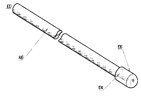
[00039] FIG. la is perspective view of a first embodiment of an optical
fiber including a
substantially conical shaped emitting surface on the tip of the optical fiber,
a substantially
conical shaped reflecting surface axially spaced relative to and facing the
emitting surface, and a
cap enclosing the emitting and reflective surfaces for achieving efficient
3600 radial emission of
the laser energy.
[00040] FIG. lb is a partial, side elevational view of the optical fiber
of FIG. la and an
enlarged detail of the distal portion thereof.
12
CA 02720272 2010-10-01
WO 2009/108956
PCT/US2009/035781
[00041] FIG. 2a is a partial, perspective view of another embodiment of an
optical fiber
received within a blood vessel.
[00042] FIG. 2b is a partial, side elevational view of the optical fiber
of FIG. 2a.
[00043] FIG. 2c is an end elevational view of the optical fiber of FIG. 2a
with part of the
blood vessel removed for clarity.
[00044] FIG. 3 is a somewhat schematic illustration of the optical fiber
of FIGS. 1 or 2
placed within a vein to be treated.
[00045] FIG. 4 is a schematic diagram of a preferred embodiment of a
device including a
laser radiation source, an optical fiber, a temperature sensor, a power
control module, and a
pullback actuator that is controlled by a pullback speed controller.
[00046] FIG. 5 is a partial, perspective view of another embodiment of an
optical fiber
including a protective quartz cap, an optical fiber distal end core with
superficial grooves, a
reflective surface, and a guide wire attached to the distal end of the fiber
and extending distally
therefrom, and an enlarged detail of the attachment of the guide wire to the
cap.
[00047] FIG. 6 is a partial, perspective view of another embodiment of an
optical fiber
comprising an optical fiber set with a guide wire attached to the distal end
of a quartz protective
cap.
[00048] FIG. 7a is a partial, perspective view of another embodiment of an
optical fiber
wherein the optical fiber tip defines a reflective cone.
[00049] FIG. 7b is a partial, cross-sectional view of the optical fiber
tip of FIG. 7a.
[00050] FIG. 8a is a partial, perspective, cross-sectional view of another
embodiment of
an optical fiber including an optical fiber tip with a reflective gap.
[00051] FIG. 8b is a cross-sectional view of the optical fiber tip of FIG.
8a and an
enlarged detail thereof.
13
CA 02720272 2010-10-01
WO 2009/108956 PCT/US2009/035781
[00052] FIG. 9 is a partial, cross-sectional view of another embodiment of
an optical fiber
including an external sleeve slidably mounted over the fiber and/or cap that
defines an internal
reflective surface for preventing the transmission of laser radiation
therethrough and for
controlling the length of the emitting section of the fiber.
[00053] FIG. 10 is a partial, cross-sectional view of another embodiment
of an optical
fiber including a substantially flat emitting face sealed within a protective,
radiation transparent
cap.
[00054] FIG. 11 is a partial, cross-sectional view of another embodiment
of an optical
fiber including a substantially flat emitting face sealed within a protective,
radiation transparent
sleeve.
Detailed Description of Preferred Embodiments
[00055] The currently preferred embodiments are hereinafter described with
reference to
the accompanying drawings wherein like reference numerals are used to indicate
like elements
throughout the various figures. As described further below, the currently
preferred embodiments
provide an improved method and device for safe and efficient low power density
endoluminal
treatment of venous insufficiency. Some currently preferred embodiments also
provide radially
emitting pulsed or continuous energy from an optical fiber. For circular
irradiation, a conical or
near conical fiber distal end is used with an opposing conical shaped
reflective surface anchored
in a cap distal region. In extended radial irradiation, multiple regularly or
otherwise spaced
emission grooves longitudinally positioned at the fiber's end can be used.
[00056] Another feature of some currently preferred embodiments is the
possibility of
achieving an extended emission zone. This can be done by appropriately
arranging the sets of
opposed conical shapes, through a combination of different variables, i.e.,
angle cut of conical
surfaces, spacing between cones, refractive index of cap material, and gas
composition left in the
spacing. In addition, a series of graded lenses, such as a plurality of graded
lenses axially spaced
relative to each other, may be employed. Furthermore, slightly truncated
conical tips also can be
employed with proper allowances for the ray patterns formed in the spacing
area. These
variables can be adjusted to vary the width of the circular cross-section
treated, as well as the
14
CA 02720272 2010-10-01
WO 2009/108956 PCT/US2009/035781
distribution of power density across the spacing length. For example, if
desired, a substantially
uniform power density can be achieved across an entire irradiated cross-
section.
[00057] As shown FIGS. la and lb, a first embodiment of an optical fiber
set is indicated
generally by the reference numeral 100. The optical fiber 100 comprises a
cladding 146, a core
140, and a quartz cap 106. An optical fiber tip preferably defines a
substantially conical-shaped
emitting surface 110 for achieving 360 radial emission. A preferably
substantially conical-
shaped reflective surface 112 is axially spaced relative to and faces the
emitting surface 110 for
enhancing efficiency and designed distribution within a zone of radial
emission. As can be seen,
the emitting and reflecting surfaces assembly is hermetically sealed within a
quartz cap 106 that
is fixedly secured to the end of the fiber and defines an air or other gas
interface at the emitting
surface to achieve radial/annular emission. Accordingly, due to the angle of
the emitting surface
110 and the differences of the indices of refraction of the emitting face 110
and air or other gas
interface provided within the sealed cap 106, the laser radiation is emitted
radially (i.e.,
transverse to or laterally with respect to the elongated axis of the fiber)
and annularly from the
fiber directly onto the surrounding vessel wall. Preferably, the emitting
surface 110 is oriented at
an acute angle with respect to the elongated axis of the fiber that is set for
substantially total
refraction of the emitting radiation laterally with respect to the elongated
axis of the fiber. In
some embodiments, the radiation is emitted laterally and annularly onto the
surrounding vessel
wall, and the annular beam of radiation extends throughout an arc (i.e., the
spread of the beam)
defined by the numerical aperture of the fiber. In some embodiments, the
spread of the annular
beam is defined by an angle within the range of about 30 to about 40 . In
addition, the
approximate center of the beam is preferably oriented at an angle within the
range of about 70
to about 90 relative to the elongated axis of the fiber.
[00058] One advantage of this novel configuration is that substantially
all radiation is
radially emitted, and therefore it significantly enhances radial emission
efficiency in comparison
to the above-described prior art. A laterally or radially emitted annular beam
can define
substantially less volume than an axially or forwardly directed conical-shaped
beam as emitted,
for example, by a flat, bare tipped fiber, and therefore the laterally emitted
beam can more
directly and efficiently transmit radiation into a vessel wall. In addition,
the emitting
characteristics can be adjusted to vary the length of the annular area of the
blood vessel or other
CA 02720272 2010-10-01
WO 2009/108956 PCT/US2009/035781
hollow anatomical structure treated, as well as the distribution of power
density along the length
of such annular section. For example, in another embodiment, a multi-grooved
distal fiber end,
defining a linear distribution of axially spaced grooves, may be used to
irradiate an extended
linear arc sector of the vein wall to, in turn, allow for effective relatively
low power density
treatment. In a preferred embodiment, the fiber, with a linearly distributed
multi-grooved distal
end, is rocked back and forth or rotated (e.g., about one revolution) during
irradiation to achieve
3600 radial stimulation of the vein wall. Alternatively, the grooves can be
offset about the fiber
to provide a roughly circular pattern with either pullback or revolving
motions.
[00059] Turning to FIGS. 2a, 2b and 2c, another embodiment of an optical
fiber is
indicated generally by the reference numeral 200. The optical fiber 200
comprises a normal
section 202 extending along the majority of its length from the proximal end,
which is optically
connected to a laser source, to a laser radiation emitting distal end section
204. The emitting
section 204 comprises several regularly-spaced grooves, preferably spaced
about 1 mm to
several mm apart, for achieving radial laser emission 218 along an emission
zone. Each groove
208 causes some radiation to be partially emitted radially outwardly of the
fiber 218 and the
remaining radiation 216 partially transmitted to a subsequent groove 208.
[00060] The optical fiber tip 210 may define a substantially conical shape
for achieving
360 radial emission and placed opposite to it, there is a preferably conical
reflective surface 212
which, as explained previously, enhances efficiency and distribution of 360
radial emission by
reflecting out any remnant or designed forwardly transmitted energy in 360
radial directions.
[00061] The emission section 204 of the fiber 200 is covered by a
protective cap 206. In
one preferred embodiment, when the wavelength used is highly absorbed in the
target tissue 214,
the protective cap 206 is made of quartz or other radiation transparent or
substantially radiation
transparent material (i.e., a material that permits transmission of the
radiation or a substantial
portion thereof therethrough), such as polymeric Teflon AF, or Teflon PFA, in
order to achieve a
relatively long, flexible emission zone. In another preferred embodiment, when
the wavelength
used is poorly absorbed in the target tissue 214, the protective cap 206 is
made of a radiation
opaque material (i.e., a material that absorbs the emitted radiation) in order
to transform
16
CA 02720272 2010-10-01
WO 2009/108956 PCT/US2009/035781
substantially all or part of the radially emitted radiation into heat in order
to thermally damage
the vein wall. This achieves vein collapse by thermal means instead of direct
laser radiation.
[00062] Turning to FIG. 3, another embodiment of an optical fiber is
indicated generally
by the reference numeral 320 and is shown placed at a pre-determined position
within a vein
314. It can be appreciated from this figure that due to the relatively long
emission zone of the
optical fiber 320, a large portion of the vein can be treated at each position
(e.g., the vein may be
segmentally ablated). The fiber emission section length can be any desired
length, including
without limitation, a length within the range of about 1 cm to about 100 cm,
within the range of
about 1 cm to about 75 cm, or within the range of about 1 cm to about 50 cm.
In a particular
case in which the emission section length coincides with the total length of
the vein section to be
treated, a shorter and simpler treatment may result, as controlled pullback
may be no longer
necessary. In one such embodiment, the entire diseased length may be treated
at once with the
fiber pulled back as the vein wall collapses. In other embodiments, the
grooves are sufficiently
spaced (e.g., within the range of about 1/2 cm to about 2 cm apart, and in one
embodiment, about
1 cm apart), and extend along a sufficient length of the fiber, to treat the
entire blood vessel, or a
desired portion thereof, with the fiber substantially maintained in place and
without pullback
thereof. In other embodiments, the blood vessel is segmentally ablated by
treating extended
sections of the blood vessel in sequence. In one such embodiment, the fiber is
held in place
within a first section of the blood vessel and the laser is fired to treat the
first section, the laser is
then turned off and the fiber is pulled back and placed in a second section of
the vessel, the fiber
is then held in place in the second section of the vessel while the laser is
fired to treat the second
section, and these steps are repeated to treat any additional sections of the
blood vessel as
required. In other embodiments, the laser is not turned off during fiber
pullback or movement
from one vein segment to another. In other embodiments, the fiber is held
stationary while
lasing in some sections of the blood vessel, and is pulled back while lasing
in other sections of
the blood vessel.
[00063] As shown in FIG. 4, another embodiment of an ELA system comprises
a laser
radiation source 424, an optical fiber 420, a temperature sensor 426, a power
control module
428, and a pullback actuator 430 driven by a pullback speed controller 432.
While lasing, the
power control module 428 receives temperature values from the temperature
sensor 426,
17
CA 02720272 2014-03-03
preferably a thermocouple, positioned near the target tissue. In one
embodiment, the temperature
sensor is mounted on the fiber or cap proximate to the emitting/reflecting
surfaces thereof. The
power control module 428 processes information received from the temperature
sensor 426 and
provides feedback to both the laser power source 424 and the pullback speed
controller 432. In
one embodiment, the power control module 428 calculates the ideal or otherwise
desired power
density and pullback speed, and sends this information respectively to the
laser power controller
428 and pull back speed controller 432. The pull back speed controller 432
controls the pullback
actuator 430 to withdraw the fiber through the blood vessel, and the laser
radiation source 424
sets the laser power in accordance with the control signals received from the
control module 428.
One advantage of these embodiments is that the power density and/or pull back
speed of the
optical fiber can be adjusted throughout the endoluminal treatment procedure
to, for example,
ensure vein closure while substantially preventing localized hot spots that
otherwise might give
rise to vein wall perforations, or substantially preventing overheating of the
vein and/or
surrounding tissues that would otherwise unnecessarily cause pain or
discomfort for the patient.
In another embodiment, with manual pullback, the power control module 428
suggests to the
physician the ideal or desired power density and pullback speed values by
showing them on a
display, allowing more efficient and effective manual pullback. The system
and/or components
thereof for monitoring temperature and controlling pullback speed and other
system variables
may be manufactured and used in accordance with the teachings of commonly
assigned U.S.
patent no. 8,257,347, entitled "Vein Treatment Device And Method", and U.S.
patent
publication US 2006/0217692, filed 30 May 2006, entitled "Power Regulated
Medical
Underskin Irradiation".
[00064] In some currently preferred embodiments, a low power density is
applied, for
example about 10W/cm2 or lower, while sufficiently high total energies can be
applied to the
vein in a reasonably short time to assure collagen denaturation, shrinkage and
elimination of the
vein. This can be enhanced by the extended emitting zone (or section) and the
360' radial
irradiation such that during pullback, areas first irradiated by the proximal
side of the emitting
zone continue to receive irradiation from the center and distal side of the
emitting zone.
18
CA 02720272 2010-10-01
WO 2009/108956 PCT/US2009/035781
[00065] Turning to FIG. 5, another embodiment of an optical fiber is
indicated generally
by the reference numeral 500. The optical fiber 500 comprises a normal section
502 for the
majority of its length extending from the proximal end, which is optically
connected to the laser
source, to a laser radiation emitting distal section 504. The emitting section
504 comprises
several regularly or otherwise spaced grooves for achieving radial laser
emission along the
emission zone. The optical fiber tip 510 defines a standard critical angle
distal end, but
preferably defines the illustrated conical shape for achieving 360 radial
emission, and includes a
conical reflective surface 512 axially spaced relative to and oppositely
facing the emitting
surface for enhancing efficiency and effectiveness of radial emission by
reflecting out any
designed or remnant forwardly transmitted energy in radial directions.
[00066] A guide wire 534 is attached to the quartz cap 506 by a mechanical
guide wire
attachment/detachment system 536. While inserting the treatment set into a
blood vessel 514,
the guide wire 534 remains attached to the optical fiber, due to its
illustrated configuration. At
the attachment site, the guide wire 534 is appropriately shaped at 538, so
that the attachment
system 536 prevents disengagement while pushing inwardly but allows detachment
while pulling
backwardly, thus allowing its extraction prior to or at the start of the
treatment. In another
embodiment, the guide wire is attached by means of a medically safe adhesive,
e.g., a wax
orcyanoacrylate. As may be recognized by those of ordinary skill in the
pertinent art based on
the teachings herein, the guide wire may be attached in any of numerous
different ways,
including with any of numerous different adhesives or other attachment
mechanisms, that are
currently known, or that later become known. The guide wire can be detached,
post proper
positioning of the treatment set inside the blood vessel, by means of the
laser radiation, which
softens the adhesive or degrades the adhesive bonding. Once detached, the
guide wire 534 is
removed, leaving the capped optical fiber 500 in the proper position and
prepared for lasing.
While lasing, the optical fiber is withdrawn in the direction toward the
insertion site, shrinking
the blood vessel 514 and preferably occluding the vessel.
[00067] In another preferred embodiment, depicted in FIG. 6, the optical
fiber set 600
comprises an optical fiber, a quartz cap 606 and a guide wire 634. Radial
laser emission is
accomplished through a plurality of superficial grooves 608 with reflective
surfaces 610 formed
at the distal end section of the fiber optical core. In this case, the guide
wire 634 is preferably
19
CA 02720272 2010-10-01
WO 2009/108956 PCT/US2009/035781
attached to the distal end of the cap 606. Thus, the optical fiber set 600 can
be easily introduced
and guided through a blood vessel 614 to the desired position in a single
step, without the need to
remove the guide wire 634. Once in appropriate position, the physician starts
lasing while
withdrawing the optical fiber set 600 toward the insertion site, thereby
shrinking the blood vessel
614 preferably to closure.
[00068] In FIGS. 7a and 7b, another embodiment of an optical fiber is
indicated generally
by the reference numeral 700. The optical fiber 700 achieves radial emission
by means of a
reflective cone 742 placed at the optical fiber tip 700. In this embodiment,
the reflective cone
742 is defined by a concave, substantially conical shaped surface.
Accordingly, radiation
transmitted through the fiber core 740 is radially emitted over 360 when it
reaches the fiber tip.
Preferably, the concave, substantially conical shaped surface of the cone 742
defines an acute
angle with respect to the elongated axis of the fiber that is within the range
of about 30 to about
50 . As with the other embodiments described above, one advantage of this
novel concave,
conical shape, is that it achieves efficient 360 radial emission onto a
surrounding vessel wall.
[00069] In FIGS. 8a and 8b, another embodiment of an optical fiber is
indicated generally
by the reference numeral 800. The optical fiber 800 achieves radial emission
by means of a
conically-shaped reflective gap formed at the optical fiber tip. As can be
seen, the gap 844 is
defined by convex, substantially conical-shaped emitting surface formed at the
distal end of the
fiber core 840, and a concave, substantially conical-shaped surface that is
substantially
transparent to the emitted radiation and is axially spaced relative to the
emitting surface to form
the gap 844 therebetween. In this embodiment, radiation transmitted through
the fiber core 840
is radially emitted when it reaches the fiber tip due to the difference in
refraction properties
between the air or other gas within the gap 844 and fiber core 840.
Accordingly, the radiation is
radially emitted (i.e., in a lateral direction with respect to the elongated
axis of the fiber) in an
annular or circumferential pattern onto an adjacent surrounding vessel wall.
This diffuser tip
configuration leads to efficient 360 radial emission. As can be seen, a
relatively thin wall is
formed between the outer periphery of the gap 844 and the exterior of the
fiber 800 to seal the
gap within the fiber tip and thus maintain the requisite core-gas interface at
the gap for annular
radial laser emission. As with the other embodiments described herein, this
novel configuration
leads to efficient radial emission onto the surrounding vessel wall. As can be
seen, the distal tip
CA 02720272 2010-10-01
WO 2009/108956 PCT/US2009/035781
of the fiber 800 defines an expanded diameter or bulbous portion, which in the
illustrated
embodiment is substantially hemispherical shaped, to facilitate movement of
the tip through a
blood vessel. As may be recognized by those of ordinary skill in the pertinent
art based on the
teachings herein, although the bulbous portion is hemispherical shaped, it may
take any of
numerous different bulbous or like shapes and/or configurations that are
currently known, or that
later become known.
[00070] In another embodiment illustrated in FIG. 9, the cap 906 of the
fiber 900 is
partially covered by a sleeve 946 of radiation reflective material. As
indicated by the arrows in
FIG. 9, the sleeve 946 can be shifted axially relative to the cap 906 and
fiber 900 to control the
axial length of the emitting portion of the fiber. As can be seen, the sleeve
946 can be set to
completely cover a desired number of radially emitting grooves 908, or some
portion or all of the
distal emitting section. Accordingly, one advantage of the embodiment of FIG.
9 is that is
permits a physician to regulate the length of the emitting section or portion
of the fiber. In one
embodiment, the length of the emitting portion is set according to the length
of the vessel 914 or
section thereof to be treated to segmentally ablate such section(s). In
another embodiment, the
extended emitting section is pulled back through the vein while lasing to
progressively lase one
or more treated portions of the vein with substantially the entire extended
emitting section.
When the vein portion is shorter than the emitting fiber length, the sleeve
may be used to cover
the emission portion that is located outside of the vein while lasing. The
sleeve is preferably
made of a reflective material of a type known to those of ordinary skill in
the pertinent art for
performing this function. Even with perfect mirrored surfaces, the reflected
light will pass back
through the fiber such that some portion of the radiation will be captured,
some scattered and
some absorbed. Accordingly, a certain amount of the energy emitted at the
grooves covered by
the sleeve is lost as heat. Nevertheless, as the power density involved is
low, any such heat build
up can be maintained within an acceptable minimum value during an ELA
treatment.
[00071] Turning to FIG. 10, another embodiment of an optical fiber is
indicated generally
by the reference number 1100. The optical fiber 1100 is substantially similar
to the optical fiber
100 described above with reference to FIGS. la and 1 b, and therefore like
reference numerals
preceded by the numeral "11" instead of the numeral "1" are used to indicate
like elements. The
primary difference of the optical fiber 1100 in comparison to the optical
fiber 100 is that the
21
CA 02720272 2010-10-01
WO 2009/108956 PCT/US2009/035781
optical fiber tip defines a substantially flat emitting face 1110 that is
sealed within the protective
a cap 1106. The cap 1106 is made of a material that is substantially
transparent to the emitted
radiation to allow the radiation to pass through it and into the vessel wall.
In one embodiment,
the cap 1106 is made of quartz and is adhesively bonded to the fiber core as
described above;
however, if desired the cap may be made of any of numerous different
materials, and may be
fixedly secured to the distal end of the fiber in any of numerous different
ways, that are currently
known, or that later become known. As can be seen, the protective cap 1106
extends distally
relative to the flat emitting face 1110 of the fiber, and defines a distal end
1107 that is rounded to
facilitate movement of the capped fiber through a tortuous blood vessel. The
distal end 1107 of
the cap 1106 extends distally relative to the flat emitting face 1110 of the
fiber an axial distance
that is preferably within the range of about 2 to about 6 times the diameter
of the fiber core, and
more preferably within the range of about 3 to about 5 times the diameter of
the fiber core. In
the illustrated embodiment, the distal end 1107 of the cap 1106 extends
distally relative to the
flat emitting face 1110 of the fiber an axial distance that is about 4 times
the diameter of the fiber
core. As can be seen, the protective cap 1106 defines an enclosed space 1109
extending between
the flat emitting face 1110 and the distal end 1107 of the cap that allows the
transmitted radiation
to pass through the space and the wall of the cap, but prevents any contact
between the flat
emitting face and the blood vessel wall and otherwise protects the emitting
face of the fiber. In
contrast to the optical fiber 100 described above, the optical fiber 1100 does
not define a
substantially conical-shaped emitting surface or a substantially conical-
shaped reflective surface.
Thus, the optical fiber 1100 emits a substantially conical-shaped beam
forwardly or in the axial
direction of the fiber.
[00072] Turning to FIG. 11, another embodiment of an optical fiber is
indicated generally
by the reference number 1200. The optical fiber 1200 is substantially similar
to the optical fiber
1100 described above in connection with FIG. 10, and therefore like reference
numerals
preceded by the numeral "12" instead of the numeral "11" are used to indicate
like elements.
The primary difference of the optical fiber 1200 in comparison to the optical
fiber 1100 is that
the fiber 1200 includes an open protective sleeve 1206 rather than a closed
protective cap. The
protective sleeve 1206 is made of a material that is substantially transparent
to the emitted
radiation to allow the radiation to pass through it and into the vessel wall.
In one embodiment,
the protective sleeve 1206 is made of quartz and is adhesively bonded to the
fiber core in
22
CA 02720272 2010-10-01
WO 2009/108956 PCT/US2009/035781
substantially the same manner as is the protective cap described above;
however, if desired, the
protective sleeve may be made of any of numerous different materials, and may
be fixedly
secured to the distal end of the fiber in any of numerous different ways, that
are currently known,
or that later become known. As can be seen, the protective sleeve 1206 extends
distally relative
to the flat emitting face 1210 of the fiber, and defines a distal end 1207
that is rounded or curved
inwardly toward a central aperture 1209. The distal end 1207 is curved
inwardly in order to
facilitate movement of the fiber tip through a blood vessel. The protective
sleeve 1207 extends
distally relative to the flat emitting face 1210 of the fiber an axial
distance that is preferably
within the range of about 2 to about 6 times the diameter of the fiber core,
and more preferably
within the range of about 3 to about 5 times the diameter of the fiber core.
In the illustrated
embodiment, the protective sleeve 1207 extends distally relative to the flat
emitting face 1210 of
the fiber an axial distance that is about 4 times the diameter of the fiber
core. In contrast to the
optical fiber 100 described above, the optical fiber 1200 does not define a
substantially conical-
shaped emitting surface or a substantially conical-shaped reflective surface.
Thus, the optical
fiber 1200 emits a substantially conical-shaped beam forwardly or in the axial
direction of the
fiber.
[00073] In the operation of some currently preferred embodiments, the
optical fiber or
other waveguide is first introduced into the vein to be treated. A local
infiltration anesthetic,
such as 0.5% dilute Lidocaine (preferably without Epinephrine) may be
introduced at the access
site, if needed. In one embodiment, about 1/2 ml of such local anesthetic is
used at the access
site. An introducer needle is inserted through the access site and into the
vein to gain access to
the vein. A guide wire then may be introduced through the introducer needle
and into the vein.
Then, an introducer sheath may be introduced over the guide wire into the
vein. The introducer
sheath may take the form of any of numerous different introducer sheaths that
are currently
known, or that later become known, including a short introducer sheath that
provides access to a
relatively short portion of the vein adjacent to the access site (e.g.,
defining a length of less than
about 11 cm, or within the range of about 6 cm to about 11 cm) or a longer
introducer sheath that
can extend up the length of the vein to be treated. The guide wire is then
removed through the
sheath. Then, the optical fiber is introduced through the introducer sheath
until the emitting tip
of the fiber is positioned about 1-1/2 cm or other desired distance below the
sapheno-femoral
junction ("SFJ"). The fiber tip is positioned at the appropriate start point
below the SFJ under
23
CA 02720272 2010-10-01
WO 2009/108956 PCT/US2009/035781
ultrasound guidance and/or by transmitting a red or other noticeable aiming
beam through the
fiber to visually monitor the start position of the fiber tip through the
skin.
[00074] One advantage of the currently preferred embodiments is that the
cap or other
distal portion of the fiber tip is rounded, thus facilitating ease of
insertion through a tortuous vein
and eliminating the need, in many, if not all instances, for an introducer
sheath and guide wire.
In the currently preferred embodiments, the fibers define an outer diameter
within the range of
about 1235 gm to about 1365 gm, the caps define an outer diameter within the
range of about
1800 gm to about 2000 gm, and the rounded distal portion of the cap is defined
by a radius
within the range of about 900 gm to about 1000 gm. Accordingly, although the
use of an
introducer sheath and guide wire is described above, such steps may be
eliminated.
Alternatively, if an introducer sheath is used, it may be removed from the
vein prior to lasing and
pullback of the fiber. For example, if a long introducer sheath is used, the
introducer sheath may
be pulled back and out of the vein prior to lasing and pullback of the fiber.
Similarly, if a tear-
away introducer sheath is used, the sheath may be torn away and removed from
the vein prior to
lasing and pullback of the fiber. If a relatively short introducer sheath is
used, the sheath may be
removed from the vein, or held in place at the access site during lasing and
pullback.
[00075] With the fiber tip at the start position immediately below the SFJ
or other desired
start position, the laser is actuated to emit laser energy into the blood
vessel. With the radial-
emitting fibers, the laser energy is directed preferably radially and
annularly onto the
surrounding wall of the blood vessel. With the flat-tipped fibers, on the
other hand, the laser
energy is emitting in a substantially conical, axially directed beam. As the
radiation is emitted,
the fiber is pulled back at a substantially predetermined rate based on the
wavelength and power
used to damage or kill a sufficient portion of the intravascular endothelium
to achieve vessel
closure. Preferably, the energy per unit length delivered to the blood vessel
is sufficiently high
to close the vein, but sufficiently low to substantially avoid the need for
anesthetic along the
treated length of the vessel. In the currently preferred embodiments, the
energy per unit length
delivered to a treatment area of a blood vessel is on average less than 80
J/cm, preferably less
than about 50 J/cm, more preferably less than about 40 J/cm, more preferably
less than about 30
J/cm, more preferably less than about 20 J/cm, and even more preferably less
than about 10 J/cm.
In some embodiments, the energy per unit length delivered to a treatment area
of a blood vessel
24
CA 02720272 2010-10-01
WO 2009/108956 PCT/US2009/035781
is on average within the range of about 3 J/cm to about 15 J/cm, and
preferably is within the
range of about 5 J/cm to about 10 J/cm. In these embodiments, and as described
further below,
the wavelength of the radiation is preferably relatively strongly absorbed in
water and relatively
weakly absorbed in hemoglobin or oxyhemoglobin (e.g., > to about 1064 nm). One
advantage of
such predetermined energy levels and/or wavelengths is that (i) the energy may
be substantially
entirely absorbed within the wall of the blood vessel, (ii) the intravascular
endothelium is
sufficiently damaged to achieve vessel closure, and (iii) the transmission of
any significant
radiation into the tissues surrounding the blood vessel is substantially
prevented to thereby
substantially avoid the need for an anesthetic along the treated portion of
the vessel.
[000761 Also in the currently preferred embodiments, the energy, such as
laser radiation,
may be applied in a continuous mode, or in a pulsed mode. It has been
discovered that the
delivery of energy in a pulsed mode may allow for the delivery on average of
higher levels of
energy per unit length to a treatment area of a blood vessel substantially
without the application
of an anesthetic to such treatment area, in comparison to the delivery of
laser energy in a
continuous mode (i.e., higher amounts of pulsed energy may be absorbed within
the vessel in
comparison to continuous mode energy, while substantially preventing
transmission of any
significant energy through the vessel wall that otherwise would thermally
damage surrounding
tissue). In addition, as a general matter, and all other factors being equal,
in a pulsed mode, the
greater the percentage that the duty cycle is "off' as opposed to "on", the
higher may be the
energy per unit length delivered on average to a treatment area of a blood
vessel, substantially
without requiring administration of an anesthetic along such treatment area.
In some such
embodiments, more than about 1/2 of the duty cycle is "off', and preferably
about 1/2 to about
2/3 of the duty cycle is off. Pulsing can significantly increase the rate of
decay of the radiation
within the vessel wall tissue in comparison to continuous mode delivery,
thereby resulting in a
lower depth of penetration per given energy delivery rate (e.g., the J/cm
delivered on average by
the intravascular energy delivery device) than without pulsing (e.g.,
continuous mode).
Accordingly, one advantage of delivering energy in a pulsed mode is that it
allows for a higher
energy delivery rate, and thus may allow for a higher amount of energy to be
delivered to the
intravascular endothelium, without the use of an anesthetic along the treated
portion of the
vessel. The term "pulsed mode" is used herein to mean any of numerous
different ways that are
currently known, or that later become known, for subjecting the energy
delivered to the blood
CA 02720272 2010-10-01
WO 2009/108956 PCT/US2009/035781
vessel to a duty cycle (i.e., a recurring period, a fraction of which the
energy delivery is active,
and another fraction of which the energy delivery is inactive), such as a
laser radiation duty
cycle, including without limitation pulsing, repeatedly turning the energy
source on and off, and
interrupting an energy beam, such as with a shutter.
[00077] In some currently preferred embodiments, the wavelength of the
radiation is about
1470 nm, about 30 nm. In other preferred embodiments, the wavelength of the
radiation is
about 1950 nm, about 30 nm. Other embodiments employ radiation at about 810
nm, about
940 nm, about 1064 nm, about 1320 nm, about 2100 nm, about 3000 nm, and about
10,000 nm,
each about 30 nm. One advantage of wavelengths that are significantly more
highly absorbed
in water than in hemoglobin or oxyhemoglobin, is that such wavelengths are not
strongly
absorbed in blood but are strongly absorbed in blood vessel tissue.
Accordingly, such
wavelengths tend to substantially pass through intervening blood between the
emitting surface(s)
of the fiber and the vessel wall and, in turn, are strongly absorbed in the
vessel wall. Such
wavelengths delivered below a predetermined energy delivery rate are
substantially entirely
absorbed within the blood vessel wall tissue to, in turn, damage or kill a
sufficient depth of
intravascular endothelium to facilitate blood vessel closure. Preferably, such
damage to the
intravascular endothelium is at a level on average of at least about 1/3 the
thickness of the
intravascular endothelium, or is on average within the range of about 1/3 to
about 2/3 the
thickness of the intravascular endothelium. As a result, such wavelengths can
be more readily
absorbed at relatively low predetermined energy delivery rates (e.g., less
than about 50 J/cm
delivered on average to the treatment section of the blood vessel, preferably
less than about 40
J/cm, more preferably less than about 30 J/cm, more preferably less than about
20 J/cm, and even
more preferably less than about 10 J/cm) that nevertheless are sufficient to
damage or kill a
sufficient depth of intravascular endothelium to facilitate blood vessel
closure. In addition,
because such radiation is substantially entirely absorbed within the blood
vessel wall, any
heating of tissues that are near or adjacent to the vessel wall is
substantially prevented, and thus
the procedure can be performed substantially without anesthetic about the
treated portion of the
blood vessel (e.g., a local non-tumescent anesthetic may be applied at the
access site only, or
otherwise only at one or a few discrete locations within the physician's
discretion or as requested
by patients on an individual basis). Such wavelengths are preferably greater
than or equal to
26
CA 02720272 2010-10-01
WO 2009/108956 PCT/US2009/035781
about 1064 nm, and including without limitation about 1320 nm, about 1470 nm,
about 1950 nm,
about 2100 nm, about 3000 nm, and about 10,000 nm, each about 50 nm.
[00078] In some embodiments, the wavelength of the radiation is about 1470
nm, about
30 nm, the power is less than about 10 W, preferably less then about 8W, more
preferably less
than about 5W, and most preferably within the range of about 1 W to about 3 W.
In one
embodiment, the laser is fired in a continuous mode (although a pulsed mode
may be employed,
if desired), and the laser is pulled back at a rate within the range of about
1 sec/cm to about 20
sec/cm, more preferably within the range of about 3 sec/cm to about 15 sec/cm,
and most
preferably within the range of about 5 sec/cm to about 10 sec/cm. In one
exemplary
embodiment, an approximately 10 cm length of a GSV was closed by substantially
radially
applying approximately 1470 nm radiation, at a power level of about 2 W, at a
pullback rate of
about 5 sec/cm. In this particular example, a local infiltrate anesthetic was
applied only at the
access site, and was not applied nor otherwise needed throughout the remainder
of the procedure.
[00079] In other exemplary embodiments, a plurality of different veins
(GSV) were closed
by employing a flat-tipped fiber sealed within a quartz cap (see FIG. 10). The
radiation was
about 1470 nm, and the energy per unit length delivered to the blood vessel
was on average
about 10 J/cm (i.e., about 1W at a pullback rate of about 10 sec/cm). In each
of these cases, no
local tumescent or general anesthetic was employed. Rather, a local infiltrate
anesthetic (1/2%
Lidocaine without Epinephrine) was applied only at the patient's request or at
the physician's
discretion. In some cases, the patients had no anesthetic. In other cases, a
small amount was
applied at the access site. In other cases, a small amount was applied at the
access site and
adjacent to the SFJ. One reason for applying a small amount of such local
anesthetic in the areas
adjacent to the SFJ is because the diameter of the vein typically is largest
in this area, and
therefore the pullback rate, and thus the average energy per unit length
delivered on average to
the blood vessel in this region, may be higher than in the distally located
treatment areas.
[00080] In other exemplary embodiments, a plurality of different varicose
veins (GSV)
were closed by employing a flat-tipped fiber sealed within a quartz cap (see
FIG. 10). The
wavelength of radiation applied was about 1470 nm. The primary protocol was to
deliver the
radiation at a rate within the range of about 20 J/cm to about 30 J/cm;
however, some patients
27
CA 02720272 2010-10-01
WO 2009/108956
PCT/US2009/035781
received lower energy delivery rates (within the range of about 10 J/cm to
about 20 J/cm), and
therefore the energy per unit length delivered was on average within the range
of about 10 J/cm
to about 30 J/cm (the average was about 22 J/cm). The primary protocol also
was to deliver
radiation at a power level of about 3W in continuous mode; however, some
patients received
about 3W pulsed at a 50% duty cycle (about 1/2 second on and about 1/2 second
off). The vein
diameters were within the range of about 3 mm to about 22 mm (the average vein
diameter was
about 8.2 mm). All procedures were performed without any tumescent anesthesia
or general
anesthesia, or any pre-shaping or other compression of the veins. Several
patients did not
receive any anesthetic at all, and others received a relatively small volume
of local infiltrate
anesthetic (1/2% Lidocaine without Epinephrine). Of the 31 patients treated,
the average volume
of local anesthetic used throughout the entire procedure was about 28 ml, and
7 patients received
less than 10 ml. As a general matter, it is believed that the lower the energy
delivery rate, the
lesser is the volume of anesthetic required or otherwise desired. In addition,
as a general matter,
pulsed delivery of the laser radiation involved lesser volumes of anesthetic
than continuous mode
delivery. In all cases, the anesthetic was applied locally as deemed necessary
by the physician,
or as requested by the patient. The 24 hours post-op results demonstrated that
over 90% of the
treated veins were occluded with excellent vein wall thickening. In addition,
post-op eccymosis
and reported pain were almost nil; some bruising was reported in only about 5
to 10% of the
patients, primarily at the vein access site; and reported post-op discomfort
was minimal with a
small number of patients reporting use any of OTC pain relief (e.g., aspirin,
acetaminophen,
etc.).
[00081]
Accordingly, a significant advantage of the currently preferred embodiments is
that neither local tumescent anesthesia nor general anesthesia are required.
As indicated above,
in many cases, only a small amount of local infiltrate anesthesia may be
applied at the access site
to the vein, if at all needed. If during the procedure the patient feels any
disgomfort, the
physician may apply a small amount of local infiltrate anesthetic (e.g.,
Lidocaine preferably
without Epinephrine) at the location or area of discomfort. In any event, no
more than about 1
vial (about 50 ml) of local infiltrate anesthesia (e.g., 0.5% Lidocaine
without Epinephrine) is
required on hand during the procedure, and only a small portion of such vial,
if any, may be
needed depending on the length of the vein to be treated and/or the
sensitivity of the patient to
any discomfort perceived or otherwise encountered.
28
CA 02720272 2010-10-01
WO 2009/108956 PCT/US2009/035781
[00082] Some embodiments of the present disclosure comprise administering
sufficient
anesthetic adjacent to the femoral nerve to achieve a sensorial block but not
a femoral motor
block to anesthetize the treatment area. One such procedure comprises the
following steps:
Locating with ultrasound guidance the femoral nerve branch between the SFJ and
femoral artery.
Injecting under ultrasound guidance a predetermined amount of local anesthetic
(e.g., about
1/2% lidocaine) above the nerve at a spot adjacent to the nerve but not
touching the nerve (which
is outside the blood vessel or any sheath surrounding the blood vessel being
treated). The
predetermined amount of local anesthetic is enough to cause a sensorial block,
but not enough to
cause a motor block. In the currently preferred embodiments, the predetermined
amount is
within the range of about 10 to about 30 cc of about 1/2% lidocaine, and most
preferably is within
the range of about 15 to about 25 cc of about 1/2% lidocaine. The volume of
anesthetic may vary
depending upon the dilution ratio (e.g., the concentration of lidocaine in the
saline or other
solution). Typically, if the concentration of lidocaine is higher than the
volume injected is lower,
and vice versa. Typically, it is not necessary to apply any further anesthetic
during the
procedure; however, if desired, a small amount of local anesthetic may be
applied at the access
site, such as with a topical anesthetic, or a few cc's of dilute lidocaine.
The procedure is then
performed as outlined above, e.g., by introducing a needle into the vein;
introducing a short
introducer sheath through the needle and into the vein; introduce a capped
fiber through the
introducer sheath up to the SFJ; firing the laser and pulling back the fiber
at the rate of about 20
J/cm to about 30 J/cm, or otherwise as described herein.
1000831 Other embodiments of the present disclosure comprise the use of an
intravenous
or "IV" drip into the interior of the blood vessel being treated to locally
anesthetize the treatment
area. One such procedure comprises the steps of introducing a small amount of
lidocaine at the
access site to anesthetize the skin at the access site if desired (e.g., a few
cc's of dilute lidocaine);
introducing a needle into the blood vessel to be treated through access site;
introducing a short
introducer sheath through the needle into the blood vessel; introducing a
sheathed fiber through
the short introducer sheath and locating the tip of the sheathed fiber at a
starting point below the
SFJ; the sheathed fiber may be a typical "liquid cooled" fiber that allows the
introduction of a
liquid between the sheath and the fiber, and that includes one or more outlet
ports proximal to the
fiber tip that allow dripping or otherwise allow dispensing of the liquid (in
this case, a dilute
anesthetic solution) into the blood vessel proximal to the fiber tip; dripping
a dilute anesthetic
29
CA 02720272 2010-10-01
WO 2009/108956 PCT/US2009/035781
solution (e.g., dilute lidocaine) into the blood vessel starting at the SFJ or
at a starting point
below the SFJ; after the lidocaine takes effect, firing the laser and pulling
back the fiber at the
desired rate (e.g., at a rate of about of about 20 J/cm to about 30 J/cm, or
otherwise as disclosed
herein); the outlet port(s) for the dilute anesthetic is located proximal of
the fiber tip and
therefore the anesthetic is applied to the portions of the blood vessel just
prior to lasing so that
the portion of the blood vessel that is lased is anesthetized prior to lasing.
[00084] Other embodiments of the present disclosure involve locally
anesthetizing the
treatment area by applying a local, non-tumescent anesthetic prior to
introduction of the fiber
into the blood vessel. In some such embodiments, a small amount of dilute
anesthetic (e.g.,
about 1% dilute lidocaine) is injected at the access site, at a mid-point of
the blood vessel (such
as at or adjacent to "Hunters Crossing"), and at or adjacent to the SFJ,
wherein the amount of
local anesthetic injected at each location is no more than about 3 to about 5
ml, and the total
amount administered is not more than about 9 to about 15 ml.
[00085] In other embodiments, any of numerous other methods or treatments
that are
currently known, or that later become known, may be employed to relax the
patient and/or to
produce analgesia, anesthesia, and/or a decreased sensitivity to painful
stimuli. Such methods or
treatments include without limitation electroanalgesia, electroanesthesia,
neurostimulation,
neuromodulation, and other physical or verbal methods of producing analgesia,
anesthesia,
and/or decreased sensitivity to painful stimuli. Other such methods include
analgesia by
electrical current based on, for example, transcutaneous or percutaneous nerve
stimulation, deep
stimulation, posterior spinal cord stimulation, and transcutaneous cranial
electrical stimulation.
The foregoing description of anesthetics and analgesics is not intended to
imply that any
anesthetic or analgesic is required in connection with the disclosed
endoluminal treatment
devices and methods. Rather, many preferred embodiments do not employ any
anesthetic or
analgesic at all, or at most employ a small amount of local anesthetic or
analgesic at an access
site or other discrete location to address any localized pain that is
perceived or otherwise
encountered by the patient.
[00086] Accordingly, a significant advantage of the devices and procedures
disclosed
herein is that the above-described drawbacks associated with the tumescent
technique may be
CA 02720272 2010-10-01
WO 2009/108956 PCT/US2009/035781
avoided, including the potential toxicity and/or adverse patient reactions
associated with such
anesthetics, the higher incidences of thermal damage to surrounding tissues,
and the post
operative pain and bruising encountered with the relatively high-energy levels
employed with the
tumescent technique procedures. Another advantage of the currently preferred
embodiments
over prior art tumescent technique procedures is that the blood vessel is
maintained at
approximately the same size prior to and after introduction of the energy
application device into
the blood vessel, and the energy is applied into the surrounding wall of the
blood vessel
substantially without pre-shaping, flattening, compressing or moving the wall
of the blood vessel
toward the energy application device.
[00087] As described above, the cap or other structure at the emitting end
of the fiber
imparts a rounded, relatively large diameter distal region to the fiber tip,
thus facilitating ease of
insertion into and pullback through a vein. Another advantage of such expanded
fiber tip
structure in comparison to prior art bare tip fibers is that it displaces a
greater volume or portion
of the vein lumen. Yet another advantage of some currently preferred
embodiments is that the
laser radiation is emitted radially and annularly from the fiber into a
surrounding annular region
of the vein wall, thus transmitting the radiation more directly and
efficiently into the vein wall in
comparison to prior art ELA methods and devices. Yet another advantage of some
currently
preferred embodiments is that the optical fiber tip may define a significantly
greater emitting
surface area in comparison to prior art bare tip or other flat emitting end
face fibers, and further,
the radiation is emitted laterally/radially. As a result, the laser radiation
is transmitted directly
into a significantly larger area of surrounding vein wall tissue, and thus may
be transmitted at
significantly lower power densities in comparison to prior art ELA procedures,
to thereby
facilitate treatment substantially without localized hot spots that otherwise
might cause vein wall
perforations, overheating of surrounding tissues, and associated pain and/or
discomfort to the
patient. Accordingly, a further advantage of the currently preferred
embodiments is that they
may use significantly lesser power levels in comparison to prior art ELA
procedures.
[00088] A further advantage of some currently preferred embodiments is
that the laser
wavelengths employed are highly absorbed in water, and thus highly absorbed in
the blood
vessel wall tissue. As a result, the laser radiation is directly transmitted
into and absorbed by the
surrounding annular portion of the vessel wall or otherwise by a sufficient
depth of intravascular
31
CA 02720272 2010-10-01
WO 2009/108956 PCT/US2009/035781
endothelium to kill or damage the absorbing endothelium and, in turn, achieve
blood vessel
closure. The terms blood vessel closure, close the blood vessel, occlude the
blood vessel, or like
terms, are used herein to mean closure or shrinkage of the blood vessel that
is sufficient to
substantially prevent the flow of blood through the blood vessel following
treatment of the blood
vessel. Yet another advantage of some currently preferred embodiments is that
because the laser
radiation is directly and efficiently transmitted into and absorbed by the
vessel wall, any
significant amount of radiation absorption by the surrounding tissues, and
resulting thermal
damage, is substantially avoided. As a result, the currently preferred
embodiments not only
require less power input than do prior art ELA procedures, but require less
anesthetic, if any, and
allow for the elimination of local tumescent anesthesia and its various
drawbacks and
disadvantages.
[00089] If desired, a saline flush, such as a cold saline flush, may be
employed to cool
and/or numb the vein prior to lasing and fiber pullback. In some such
embodiments, the saline
flush is ice cold (e.g., about 30 F to about 40 F, and more preferably about
32 F to about 35
F) to facilitate numbing the vein prior to treatment. In one embodiment, the
cold saline flush is
introduced into the vein through an introducer sheath and prior to insertion
of the fiber. In
another embodiment, a cold saline flush is introduced through an introducer
sheath after insertion
of the fiber and/or during withdrawal of the introducer sheath prior to
lasing. In another
embodiment, the cold saline flush is introduced through a sheath surrounding
the fiber during
lasing and pullback of the fiber. In the latter embodiment, the cold saline is
introduced through
one or more outlet ports located proximate to the emitting tip of the fiber
(e.g., at the base of the
quartz cap). One such embodiment employs a conventional liquid cooled fiber
sheath
construction.
[00090] In some embodiments ultrasound energy is applied to the fiber or
other waveguide
to facilitate smooth pullback through the vein and/or pullback at a
substantially constant or other
desired rate. In one embodiment, an ultrasound transducer or vibrator is
connected to the
proximal end of the fiber to impart ultrasound vibrations to the emitting tip
or region of the fiber
during lasing and pullback. In another embodiment, the ultrasound transducer
or vibrator is
attached to the cap or otherwise adjacent to the emitting tip or region of the
fiber to impart
ultrasound vibrations thereto during lasing and pullback through the vein.
32
CA 02720272 2014-03-03
[00091] In some embodiments of the present disclosure the fiber is a
fluoropolymer
capped medical fiber, or other fiber based medical laser or light energy
delivery device with a
fluoropolymeric emission surface. One advantage of the fluoropolymeric
emission surface is
that it does not tend to stick against the blood vessel wall or any coagulated
blood within the
vessel, and therefore may be easier to pull back through a blood vessel than
other devices.
[00092] In another preferred embodiment, the optical fiber set adds three
or more shape-
memory expandable arms. While inserting the treatment set, the expandable arms
are in
complete contact with a protective coating. Once in appropriate position, the
expandable arms
are activated by means of an internal/external energy source, expanding their
distal ends, until
contacting the inner surface of the blood vessel. As a consequence, the
optical fiber set is
substantially centered inside the target tissue to further facilitate
substantially evenly heating the
inner surface and further preventing vein wall contact or perforation. The
substantially evenly-
heated surface should in turn more uniformly contract, and efficiently shrink
the blood vessel to
closure where desired.
[00093] In the currently preferred embodiments, the wavelengths are
selected to offer a
reasonably high absorption in the target tissue, such about 1470 nm, about
30 nm, and/or about
1950 nm, about 30 nm. As may be recognized by those of ordinary skill in the
pertinent art,
these wavelengths are only exemplary, however, and any of numerous other
wavelengths that are
currently known, or that later become known, equally may be used, including
without limitation
about 810 nm, 940 nm, 980 nm, 1064 nm, 1320 nm, 2100 nm, 3000 nm, and 10,000
nm, each
about 30 nm. One advantage of the 1470 nm and 1950 nm wavelengths is that they
are highly
absorbed in water, and thus are highly absorbed in the target tissue of the
blood vessel wall.
Absorption of 1470 nm and 1950 nm in the tissue of a blood vessel wall are
about 1-3 orders
higher than for 980 nm, and significantly higher than that order for most
other commercially
available wavelengths.
[00094] The protective radiation transparent caps of the currently
preferred embodiments
may be manufactured and assembled to the fiber in accordance with the
teachings of commonly
assigned U.S. patent application 2007/0106286, entitled "Side Fire Optical
Fiber For High
Power Application".
33
CA 02720272 2014-03-03
[00095] As
indicated above, in certain preferred embodiments, blood vessel wall closure
is
achieved by thermally damaging or killing on average at least about 1/3 the
thickness of the
intravascular endothelium, or thermally damaging or killing a depth of
intravascular endothelium
on average that is within the range of about 1/3 to about 2/3 its thickness.
As also indicated
above, wavelengths that are strongly absorbed in water and applied at
predetermined energy
delivery rates are substantially entirely absorbed at a depth of at least
about 1/3, or within the
range of about 1/3 to about 2/3, the thickness of the intravascular
endothelium to, in turn, prevent
transmission of any significant level of radiation into surrounding tissues,
and thereby avoiding
the need for anesthetic along the treated vessel. Intravascular endothelium
may be damaged to
facilitate blood vessel closure with mechanisms other than radiation. For
example, U.S. Patent
No. 6,402,745 ("the '745 patent") shows an intravenous whip electrode for vein
ablation.
Some
embodiments of the '745 patent do not deliver electrical energy to the
intravascular endothelium,
whereas other embodiments do. In accordance with one embodiment of the present
disclosure,
the intravenous device includes a rotating whip or other device for scraping
or abrading the
intravascular endothelium as disclosed, for example, in the '745 patent, and
an integral
intravascular energy application device that delivers sufficient energy to the
intravascular
endothelium that, combined with the scraping or abrading action of the whip or
other device,
sufficiently damages at least about 1/3 to about 2/3 the depth of the
endothelium to achieve blood
vessel closure. In some such embodiments, the energy application device is an
optical
waveguide that delivers radiation wavelengths strongly absorbed in water
(i.e., about 1064 nm or
greater). In some such embodiments, the radiation is pulsed to allow
relatively high energy
delivery rates substantially without any anesthetic along the treated
segment(s) of the blood
vessel. The abrading or scraping action of the whip or like device may allow
for even lower
34
CA 02720272 2010-10-01
WO 2009/108956 PCT/US2009/035781
energy delivery rates to the blood vessel wall to sufficiently damage the
vessel to closure without
the use of an anesthetic along the treated segment(s) of the vessel.
[00096] Having described various preferred embodiments with reference to
the
accompanying drawings, it is to be understood that the invention is not
limited to these precise
embodiments, and that various changes and modifications may be effected
therein by those
skilled in the art without departing from the scope or spirit of the invention
as defined in the
appended claims. For example, the radiation can be emitted in a pulsed or
continuous mode and
can contain one or more laser wavelengths. In addition, the radiation can be
supplied by means
other than lasers, including without limitation, by LEDs and super luminescent
LEDs. In
addition, the optical fibers may take the form of any of numerous different
optical fibers or
waveguides that are currently known or that later become known, that may
define any of
numerous different cores, claddings, jackets, end caps, protective sleeves,
emitting surfaces,
reflective surfaces, and/or gradient lenses, that are currently known, or that
later become known.
For example, although many of the fibers disclosed herein are capped, fibers
without caps,
including bare tipped fibers, may be employed. Further, the emitting surfaces
may take any of
numerous different shapes or configurations that are currently known, or that
later become
known. For example, although certain embodiments employ emitting surfaces that
are
substantially conical shaped, emitting surfaces defining other arcuate surface
contours (i.e.,
surface contours that are curved), or defining non-arcuate surface contours,
such as one or more
flat, and/or angled emitting surfaces, equally may be employed. In addition,
the methods of
venous treatment may employ any of numerous different devices with or without
anesthetics,
including without limitation, without sheaths or catheters, or with any of
numerous different
types of sheaths or catheters, including without limitation short, long and/or
tear away introducer
sheaths, without guide wires, or with guide wires, including without
limitation, guide wires
attached to, detachable from, or not at all attached to the fiber or
waveguide. In addition, any of
numerous different forms of energy and energy application devices that are
currently known, or
that later become known, equally may be employed to treat blood vessels in
accordance with
various aspects of the inventions disclosed herein. For example, the energy
application device
may take the form of (i) a waveguide or optical fiber that emits laser energy
as described above;
(ii) a microwave catheter or device that emits microwave energy; (iii) an RF
catheter or device
that emits RF energy; (iv) an electrical catheter or device that emits
electrical energy; and (v) an
CA 02720272 2010-10-01
WO 2009/108956 PCT/US2009/035781
ultrasound catheter or device that emits ultrasound energy. Accordingly, this
detailed description
of currently preferred embodiments is to be taken in an illustrative as
opposed to a limiting
sense.
36