Note: Descriptions are shown in the official language in which they were submitted.
CA 02720392 2010-10-01
WO 2009/124203 PCT/US2009/039341
1
SYSTEM AND METHOD FOR IDENTIFYING A POSITION TO INSERT A
SCLERAL PROSTHESIS INTO AN EYE
CROSS-REFERENCE TO RELATED PATENT DOCUMENTS
[0001] This application claims priority under 35 U.S.C.
119(e) to U.S. Provisional Patent Application No. 61/072,757
filed on April 2, 2008, which is hereby incorporated by
reference.
[0002] This application is related to the following U.S.
patent applications and issued patents:
(1) U.S. Patent No. 6,007,578 entitled "Scleral Prosthesis
for Treatment of Presbyopia and Other Eye Disorders"
issued on December 28, 1999;
(2) U.S. Patent No. 6,280,468 entitled "Scleral Prosthesis
for Treatment of Presbyopia and Other Eye Disorders"
issued on August 28, 2001;
(3) U.S. Patent No. 6,299,640 entitled "Scleral Prosthesis
for Treatment of Presbyopia and Other Eye Disorders"
issued on October 9, 2001;
(4) U.S. Patent No. 5,354,331 entitled "Treatment of
Presbyopia and Other Eye Disorders" issued on October
11, 1994;
(5) U.S. Patent No. 5,465,737 entitled "Treatment of
Presbyopia and Other Eye Disorders" issued on November
14, 1995;
(6) U.S. Patent No. 5,489,299 entitled "Treatment of
Presbyopia and Other Eye Disorders" issued on February
6, 1996;
(7) U.S. Patent No. 5,503,165 entitled "Treatment of
Presbyopia and Other Eye Disorders" issued on April 2,
1996;
(8) U.S. Patent No. 5,529,076 entitled "Treatment of
Presbyopia and Other Eye Disorders" issued on June 25,
CA 02720392 2010-10-01
WO 2009/124203 PCT/US2009/039341
2
1996;
(9) U.S. Patent No. 5,722,952 entitled "Treatment of
Presbyopia and Other Eye Disorders" issued on March 3,
1998;
(10) U.S. Patent No. 6,197,056 entitled "Segmented Scleral
Band for Treatment of Presbyopia and Other Eye
Disorders" issued on March 6, 2001;
(11) U.S. Patent No. 6,579,316 entitled "Segmented Scleral
Band for Treatment of Presbyopia and Other Eye
Disorders" issued on June 17, 2003;
(12) U.S. Patent No. 6,926,727 entitled "Surgical Blade for
Use with a Surgical Tool for Making Incisions for
Scleral Eye Implants" issued on August 9, 2005;
(13) U.S. Patent No. 6,991,650 entitled "Scleral Expansion
Device Having Duck Bill" issued on January 31, 2006;
(14) U.S. Patent Application Serial No. 10/080,877 entitled
"System and Method for Making Incisions for Scleral
Eye Implants" filed on February 22, 2002;
(15) U.S. Patent Application Serial No. 10/443,122 entitled
"System and Method for Determining a Position for a
Scleral Pocket for a Scleral Prosthesis" filed on May
20, 2003;
(16) U.S. Patent Application Serial No. 11/137,085 entitled
"Scleral Prosthesis for Treatment of Presbyopia and
Other Eye Disorders" filed on May 24, 2005;
(17) U.S. Patent Application Serial No. 11/199,591 entitled
"Surgical Blade for Use with a Surgical Tool for
Making Incisions for Scleral Eye Implants" filed on
August 8, 2005;
(18) U.S. Patent Application Serial No. 11/252,369 entitled
"Scleral Expansion Device Having Duck Bill" filed on
October 17, 2005;
(19) U.S. Patent Application Serial No. 11/323,283 entitled
CA 02720392 2010-10-01
WO 2009/124203 PCT/US2009/039341
3
"Surgical Blade for Use with a Surgical Tool for
Making Incisions for Scleral Eye Implants" filed on
December 30, 2005;
(20) U.S. Patent Application Serial No. 11/323,284 entitled
"System and Method for Making Incisions for Scleral
Eye Implants" filed on December 30, 2005;
(21) U.S. Patent Application Serial No. 11/322,728 entitled
"Segmented Scleral Band for Treatment of Presbyopia
and Other Eye Disorders" filed on December 30, 2005;
(22) U.S. Patent Application Serial No. 11/323,752 entitled
"Segmented Scleral Band for Treatment of Presbyopia
and Other Eye Disorders" filed on December 30, 2005;
(23) U.S. Provisional Patent Application No. 60/819,995
entitled "Apparatuses, Systems, and Methods Related to
Treating Presbyopia and Other Eye Disorders" filed on
July 11, 2006;
(24) U.S. Patent Application Serial No. 11/827,444 entitled
"Apparatus and Method for Securing Ocular Tissue"
filed on July 11, 2007;
(25) U.S. Patent Application Serial No. 11/827,382 entitled
"Scleral Prosthesis for Treating Presbyopia and Other
Eye Disorders and Related Devices and Methods" filed
on July 11, 2007;
(26) U.S. Provisional Patent Application No. 61/001,593
entitled "Apparatuses and Methods for Forming
Incisions in Ocular Tissue" filed on November 2, 2007;
(27) U.S. Patent Application No. 12/260,694 entitled
"Apparatuses and Methods for Forming Incisions in
Ocular Tissue" filed on October 29, 2008; and
(28) U.S. Provisional Patent Application No. 61/065,149
entitled "Scleral Prosthesis for Ocular Drug Delivery
to Treat Glaucoma, Macular Degeneration, and Other Eye
Disorders or Diseases and Related Method" filed on
CA 02720392 2010-10-01
WO 2009/124203 PCT/US2009/039341
4
February 8, 2008.
All of these patents and patent applications are hereby
incorporated by reference.
TECHNICAL FIELD
[0003] This disclosure is generally directed to the treatment
of presbyopia, hyperopia, primary open angle glaucoma, ocular
hypertension, and other eye disorders. More specifically, this
disclosure is directed to a system and method for identifying a
position to insert a scleral prosthesis into an eye.
CA 02720392 2010-10-01
WO 2009/124203 PCT/US2009/039341
5 BACKGROUND
[0004] It is possible to treat presbyopia, glaucoma, and other
eye disorders by implanting scleral prostheses within the sclera
of a patient's eye. For each individual scleral prosthesis, an
incision is made in the sclera of the patient's eye. The
incision is then extended under the surface of the sclera to form
a scleral "tunnel," and a scleral prosthesis is placed within the
tunnel. One or multiple scleral prostheses may be implanted in a
patient's eye to (among other things) treat presbyopia, glaucoma,
ocular hypertension, elevated intraocular pressure, macular
degeneration, or other eye disorders. This technique is
described more fully in the patents documents incorporated by
reference above.
CA 02720392 2010-10-01
WO 2009/124203 PCT/US2009/039341
6
SUMMARY
[0005] This disclosure provides a system, apparatus, method,
and computer program for identifying a position to insert a
scleral prosthesis into an eye.
[0006] In a first embodiment, a method includes identifying an
actual location of a ciliary body in a patient's eye. The method
also includes identifying a position for a scleral prosthesis to
be inserted into scleral tissue of the patient's eye based on the
identified location of the ciliary body.
[0007] In particular embodiments, the method further includes
forming a scleral tunnel in the scleral tissue of the patient's
eye based on the identified position and inserting the scleral
prosthesis into the scleral tunnel.
[0008] In a second embodiment, a system includes eye
measurement equipment configured to generate information
associated with an actual location of a ciliary body in a
patient's eye. The system also includes a controller configured
to use the information from the eye measurement equipment to
identify a position for a scleral prosthesis to be inserted into
scleral tissue of the patient's eye based on the identified
location of the ciliary body.
[0009] In a third embodiment, an apparatus includes a
controller configured to identify a position for a scleral
prosthesis to be inserted into scleral tissue of a patient's eye.
The controller is configured to identify the position for the
scleral prosthesis using an actual location of a ciliary body in
the patient's eye.
[0010] In a fourth embodiment, a computer readable medium
embodies a computer program. The computer program includes
computer readable program code for receiving information
identifying an actual location of a ciliary body in a patient's
eye. The computer program also includes computer readable
program code for identifying a position for a scleral prosthesis
CA 02720392 2010-10-01
WO 2009/124203 PCT/US2009/039341
7
to be inserted into scleral tissue of the patient's eye based on
the actual location of the ciliary body.
[0011] Other technical features may be readily apparent to one
skilled in the art from the following figures, descriptions, and
claims.
CA 02720392 2010-10-01
WO 2009/124203 PCT/US2009/039341
8
BRIEF DESCRIPTION OF THE DRAWINGS
[0012] For a more complete understanding of this disclosure,
reference is now made to the following description, taken in
conjunction with the accompanying drawing, in which:
[0013] FIGURE 1 illustrates an example system for identifying
a position to insert a scleral prosthesis into an eye in
accordance with this disclosure;
[0014] FIGURE 2 illustrates an example implantation of a
scleral prosthesis into an eye in accordance with this
disclosure;
[0015] FIGURE 3 illustrates an example transillumination of an
eye in accordance with this disclosure;
[0016] FIGURE 4 illustrates an example method for identifying
a position to insert a scleral prosthesis into an eye in
accordance with this disclosure; and
[0017] FIGURE 5 illustrates an example method for using
transillumination or retroillumination to identify a position of
a ciliary body in an eye in accordance with this disclosure.
CA 02720392 2010-10-01
WO 2009/124203 PCT/US2009/039341
9
DETAILED DESCRIPTION
[0018] FIGURES 1 through 5, discussed below, and the various
embodiments used to describe the principles of the present
invention in this patent document are by way of illustration only
and should not be construed in any way to limit the scope of the
invention. Those skilled in the art will understand that the
principles of the invention may be implemented in any type of
suitably arranged device or system.
[0019] FIGURE 1 illustrates an example system 100 for
identifying a position to insert a scleral prosthesis into an eye
in accordance with this disclosure. The embodiment of the system
100 shown in FIGURE 1 is for illustration only. Other
embodiments of the system 100 could be used without departing
from the scope of this disclosure.
[0020] As shown in FIGURE 1, the system 100 includes a
controller 110, which could be coupled to eye measurement
equipment 120. The controller 110 could receive eye measurements
from the eye measurement equipment 120. The eye measurements
could, for example, include an identification of the position of
a ciliary body in each quadrant of a patient's eye. The
controller 110 could then use this information in any suitable
manner.
[0021] The eye measurement equipment 120 includes any suitable
component(s) for taking measurements related to, or otherwise
assisting in the identification of, the location of a ciliary
body in one or more areas of a patient's eye. For example, the
eye measurement equipment 120 could include transillumination or
retroillumination equipment used to illuminate the patient's eye.
Transillumination generally involves directly illuminating one
portion of the patient's eye through the sclera and beneath the
cornea of the eye, or through any other portion of the patient's
eye (and possibly through one of the patient's eyelids).
Retroillumination generally involves indirectly illuminating one
CA 02720392 2010-10-01
WO 2009/124203 PCT/US2009/039341
5 portion of the patient's eye through the sclera and beneath the
cornea of the eye by reflecting light off the back of the eye to
illuminate an image on the opposing quadrant of the eye opposite
the light source. As a particular example, the eye measurement
equipment 120 could include a very bright light source (such as a
10 filtered or unfiltered visible spectrum fiber optic normal or
cold light source or any other suitable light source) and optics
or other components for delivering the light to different areas
of the patient's eye (such as an ACMI/STORZ surgical fiber optic
endoscope). The eye measurement equipment 120 could also include
components supporting other imaging techniques for locating the
ciliary body in one or more areas of a patient's eye, such as
components that support optical coherence tomography (OCT),
ultrasound biomicroscopy (UBM), magnetic resonance imaging (MRI),
or any other suitable imaging techniques.
[0022] The controller 110 could also be coupled to an input
unit 130. The controller 110 could receive any suitable input
data through the input unit 130. The input data could, for
example, include an identification of the position of a ciliary
body in each quadrant of a patient's eye. The input data could
also include other surgical parameters or any other suitable
data. The input data could be provided by a surgeon or other
personnel using the system 100. The input unit 130 includes any
suitable component(s) for providing input to the controller 110.
The input unit 130 could, for instance, include a keypad,
keyboard, mouse, or other or additional input device(s).
[0023] In this example, the controller 110 includes a
processor 140 configured to perform various functions to
facilitate the implantation of one or more scleral prostheses in
a patient's eye. The processor 140 is capable of executing
instructions stored in a memory 145 within the controller 110.
For example, the processor 140 could execute an operating system
150 and a location identification application 160 (which could be
CA 02720392 2010-10-01
WO 2009/124203 PCT/US2009/039341
11
stored in the memory 145). The location identification
application 160 could represent a software application or other
computer instructions that perform various calculations to
identify one or more locations for inserting one or more scleral
prostheses into a patient's eye.
[0024] In some embodiments, the location identification
application 160 can receive measured parameters of an eye that
are provided by the eye measurement equipment 120 and/or input by
a user through the input unit 130. The location identification
application 160 can use this data to identify the one or more
locations for inserting one or more scleral prostheses into the
patient's eye. For example, the location identification
application 160 could identify locations for inserting scleral
prostheses into the patient's eye based on the identified
locations of the ciliary body in four different quadrants of the
patient's eye (obtained during transillumination,
retroillumination, or other technique). The identified locations
could represent the locations where scleral pockets or tunnels
are to be formed in the scleral tissue of the patient's eye.
Additional details regarding the identification of locations for
inserting scleral prosthesis into a patient's eye are provided
below.
[0025] The processor 140 includes any suitable component(s)
for identifying one or more locations for inserting one or more
scleral prostheses into a patient's eye. The processor 140
could, for example, represent a microprocessor, microcontroller,
digital signal processor, application specific integrated circuit
(ASIC), or other processing or computing device. The memory 145
includes any suitable volatile or non-volatile storage and
retrieval device or devices.
[0026] The identified location(s) for inserting one or more
scleral prostheses into a patient's eye could then be used in any
suitable manner. For example, the identified location(s) could
CA 02720392 2010-10-01
WO 2009/124203 PCT/US2009/039341
12
be presented to a surgeon or other personnel on a display 170 for
review. The display 170 includes any suitable component for
displaying information, such as a liquid crystal display (LCD) or
cathode ray tube (CRT) display.
[0027] The identified location(s) for inserting one or more
scleral prostheses into a patient's eye could also be provided to
a surgical tool controller 180. The surgical tool controller 180
controls the operation of a surgical tool, such as a tool for
forming scleral pockets or tunnels in a patient's eye. The
information provided by the controller 110 to the surgical tool
controller 180 could enable the surgical tool controller 180 to
form scleral pockets or tunnels in the identified location(s) of
the patient's eye. As a particular example, the surgical tool
controller 180 may use the information from the location
identification application 160 to automatically determine an
incision point for an incision (where the incision creates a
scleral pocket or tunnel). Any suitable surgical tool could be
controlled by the surgical tool controller 180. Examples of
surgical tools for forming scleral pockets or tunnels are
disclosed in various ones of the patent documents incorporated by
reference above. The surgical tool controller 180 includes any
suitable component(s) for controlling one or more surgical tools.
[0028] In addition, the identified location(s) for inserting
one or more scleral prostheses into a patient's eye could be
provided to a marking plate assembly 190. The marking plate
assembly 190 can form marks on a patient's eye. For example, the
marking plate assembly 190 could make marks on a patient's eye to
identify the locations of scleral pockets or tunnels to be formed
in the patient's eye. The marks could be used by a surgeon or
other personnel, such as when a surgeon manually forms the
scleral pockets or tunnels or when the surgeon uses the marks to
place a surgical tool on the patient's eye. The marking plate
assembly 190 could also act as a template to guide a surgical
CA 02720392 2010-10-01
WO 2009/124203 PCT/US2009/039341
13
cutting device to a proper location, similar to or including
certain fixation tools disclosed in the patent documents
incorporated by reference above. The marking plate assembly 190
could further be customized for a particular patient based on
measurements of that patient's eye(s). The marking plate assembly
190 includes any suitable component(s) for making marks on a
patient's eye and/or for guiding a surgical cutting device.
[0029] Although FIGURE 1 illustrates one example of a system
100 for identifying a position to insert a scleral prosthesis
into an eye, various changes may be made to FIGURE 1. For
example, the functional division shown in FIGURE 1 is for
illustration only. Various components in FIGURE 1 could be
combined, further subdivided, or omitted and additional
components could be added according to particular needs. As a
particular example, the eye measurement equipment 120 could be
omitted if personnel manually measure the locations of the
ciliary body and enter the locations through the input unit 130.
As another particular example, the surgical tool controller 180
could be omitted if the identified locations of scleral pockets
or tunnels are only used to mark the patient's eye using the
marking plate assembly 190. Also, FIGURE 1 illustrates one
example system 100 where transillumination, retroillumination, or
other technique could be used to identify a position of the
ciliary body in the eye so that a location of a scleral
prosthesis can be determined. These techniques could be used
with any other suitable system, or they could be performed
manually.
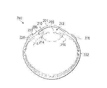
[0030] FIGURE 2 illustrates an example implantation of a
scleral prosthesis into an eye in accordance with this
disclosure. The example implantation shown in FIGURE 2 is for
illustration only. Other implantations of scleral prostheses
could be performed without departing from the scope of this
disclosure.
CA 02720392 2010-10-01
WO 2009/124203 PCT/US2009/039341
14
[0031] As shown in FIGURE 2, an eye 200 includes the white,
tough sclera 202 and the cornea 204. The generally circular
junction of the cornea 204 and the sclera 202 is the limbus 206.
The outline of the limbus 206 is easily identified because this
is where the dark part of the eye (caused by the pigment in the
iris 210 reflecting through the cornea 204) becomes white as the
cornea 204 transitions into the white of the sclera 202. Within
the globe of the eye, a crystalline lens 208 is enclosed in a
thin membranous capsule and is located immediately posterior to
the iris 210, suspended centrally posterior to the pupil 212 on
the optical axis of the eye. The lens 208 is suspended by
zonules 214, which extend between the capsule of the lens 208 and
the ciliary body 216. The ciliary body 216 lies just under the
sclera 202 (just inwardly of the sclera 202) and is attached to
the inner surface of the sclera 202.
[0032] Changes in the shape of the ciliary body 216 (and in
particular the ciliary muscle within the ciliary body) cause
corresponding changes in the shape of the lens 208 when the
effective working distance between the ciliary body 216 and the
lens 208 is satisfactory (such as in a young eye) . In these
circumstances (i.e. a young eye), changes in the shape of the
ciliary body 216 allow the lens 208 to become more convex,
allowing the eye to accommodate or see "at near" (close up). As
the eye ages, the effective working distance between the lens 208
and the ciliary body 216 diminishes, and the eye loses its
ability to accommodate (this condition is called presbyopia).
[0033] As described in more detail in the patent documents
incorporated by reference above, the effective working distance
of the ciliary muscle of the eye can be increased by inserting
one or more scleral prostheses 220 into one or more scleral
pockets or tunnels 222 formed in the sclera 202. This can be
done to help treat presbyopia, glaucoma, ocular hypertension,
elevated intraocular pressure, macular degeneration, or other eye
CA 02720392 2010-10-01
WO 2009/124203 PCT/US2009/039341
5 disorders. Any suitable scleral prosthesis 220 can be implanted
into the sclera 202 of the eye 200. For example, different
scleral prostheses are disclosed in various ones of the patent
documents incorporated by reference above.
[0034] The placement of a scleral prosthesis 220 with respect
10 to the ciliary body 216 can have a large influence on the
effectiveness of the scleral prosthesis 220 in restoring the
effective working distance between the lens 208 and the ciliary
body 216. For example, the beginning or anterior edge of the
ciliary body 216 can generally be found 1.0-5.0 millimeters
15 posterior to the limbus 206 in any give quadrant, with the
balance of the ciliary body 216 moving posteriorly along the
intraocular surface of the sclera 202 for approximately 5.0-7.0
millimeters. However, patients often show some degree of
variability in the distance between the limbus 206 and the
beginning of the ciliary body 216 in their eyes 200. This
variability can exist between patients and actually within the
same patient, such as when this distance varies between quadrants
of the same patient's eye(s) 200. In other words, the ciliary
muscle is not necessarily symmetrical and can vary patient by
patient, eye by eye, and quadrant by quadrant. This variation
can sometimes be significant, such as when the distance to the
forward edge of the ciliary body 216 is 2.0mm posterior to the
limbus 206 in one quadrant and 5.0mm to the forward edge of the
ciliary body 216 in another quadrant (possibly within the same
eye).
[0035] Because of this variation, the corrective effect
provided by a scleral prosthesis 220 could vary depending on the
placement of the scleral prosthesis 220 within the eye 200. For
example, if the scleral prosthesis 220 is implanted at a location
that is quite far from the ciliary body 216 (as defined by its
forward edge) in a particular quadrant of the eye 200, the
corrective effect provided by the scleral prosthesis 220 can be
CA 02720392 2010-10-01
WO 2009/124203 PCT/US2009/039341
16
reduced. In contrast, if the scleral prosthesis 220 is implanted
at a location that is relatively close to the ciliary body 216 in
a particular quadrant of the eye 200, the corrective effect
provided by the scleral prosthesis 220 can be greater.
[0036] In accordance with this disclosure, the actual location
of at least part of the ciliary body 216 in the eye 200 (such as
its forward or anterior edge) can be determined or estimated with
relatively high accuracy. For example, the actual location of
the ciliary body 216 in each of the four quadrants of the eye 200
can be determined or estimated. With the actual location of the
ciliary body 216 known or estimated with higher accuracy, more
optimal locations of the scleral prostheses 220 can be
determined. For instance, the scleral prostheses 220 could be
implanted so that they are no more than 1.0mm-1.25mm away from
the forward edge of the ciliary body 216 of the eye 200, with the
scleral prostheses 220 being posterior to the forward edge of the
ciliary body 216. Of course, other distances can be used, such
as between 2.0mm and 4.0mm. The desired distance between the
ciliary body 216 and a scleral prosthesis 220 could be based on a
number of factors, including a size and a thickness of the
scleral prosthesis 220. In particular embodiments, thicker
scleral prostheses 220 could be used, such as those having a
thickness of approximately 2.5mm, or longer scleral prosthesis,
such as those having a length of approximately 7.0mm. In other
particular embodiments, it may be appropriate to locate the
scleral prostheses 220 on or anterior to the forward edge of the
ciliary body 216, such as at fixed locations.
[0037] Whatever the desired distance from the ciliary body 216
to the scleral prosthesis 220 is, this distance can be more
accurately obtained by identifying the location of the ciliary
body 216 (such as its forward or anterior edge) in the eye 200
prior to implantation of the scleral prosthesis 220. Any
suitable technique could be used to identify the location of the
CA 02720392 2010-10-01
WO 2009/124203 PCT/US2009/039341
17
ciliary body 216 in a patient's eye 200, such as OCT, UBM, MRI,
transillumination, or retroillumination.
[0038] FIGURE 3 illustrates an example transillumination of an
eye in accordance with this disclosure. During transillumination
or retroillumination, one quadrant or other portion of the
patient's eye can be illuminated through the sclera and beneath
the patient's cornea or from another portion of the eye. As
shown in FIGURE 3, the superior nasal quadrant of the patient's
eye (the upper inner portion of the eye) is being illuminated
with light directed into the inferior temporal quadrant of the
patient's eye (the lower outer portion of the eye) through the
patient's upper eyelid.
[0039] During transillumination or retroillumination, the
light creates an elliptical shadow around the limbus 206 of the
patient's eye 200, which as shown in FIGURE 3 is several
millimeters outside of the limbus 206. The elliptical shadow is
caused by the ciliary body 216 of the eye 200. As a result, the
shadow formed in a particular quadrant of the patient's eye 200
can be used to identify the location of the ciliary body 216 in
that quadrant. A mark can be placed on the patient's eye 200 in
the location where the shadow begins on the sclera 202. After
that, the distance between the marked location of the forward
edge of the ciliary body 216 and the limbus 206 can be measured,
such as by using calipers under normal illumination. The marked
location of the forward edge of the ciliary body 216 could also
be used to identify a desired location for a scleral prosthesis
220, such as by identifying a location that is 1.0mm-4.0mm
posterior to the marked location of the ciliary body 216.
[0040] In this way, the actual location of the forward edged
of the ciliary body 216 can be identified in a non-invasive
manner. This can allow more accurate placement of scleral
prostheses 220 in a patient's eye 200, which could result in
improved corrective effect and better resulting eyesight in the
CA 02720392 2010-10-01
WO 2009/124203 PCT/US2009/039341
18
patient.
[0041] Although FIGURE 2 illustrates one example of an
implantation of a scleral prosthesis into an eye, various changes
may be made to FIGURE 2. For example, any number of scleral
prostheses could be inserted into a patient's eye, and each of
the scleral prostheses could have any suitable size and shape.
Although FIGURE 3 illustrates one example of the
transillumination of an eye, various changes may be made to
FIGURE 3. For instance, as noted above, FIGURE 3 illustrates the
transillumination of one quadrant or other portion of the
patient's eye. This transillumination could be repeated for all
quadrants or other portions of the patient's eye. Also,
retroillumination or any other suitable imaging technique could
be used to illuminate the position of the ciliary body in
individual quadrants of the eye.
[0042] FIGURE 4 illustrates an example method 400 for
identifying a position to insert a scleral prosthesis into an eye
in accordance with this disclosure. The embodiment of the method
400 shown in FIGURE 4 is for illustration only. Other
embodiments of the method 400 could be used without departing
from the scope of this disclosure.
[0043] As shown in FIGURE 4, the method 400 includes placing a
reference mark on a patient's eye at step 402. This could
include, for example, a surgeon or other personnel using a
surgical pen or other device to place a mark in a desired
location on the patient's eye. As a particular example, this
could include the surgeon or other personnel placing a reference
mark on the sclera of the patient's eye at either the "12
o'clock" position or the "6 o'clock" position.
[0044] Quadrant marks are placed on the patient's eye at step
404. The quadrant marks could represent marks formed in the four
quadrants of the patient's eye, such as at 450, 135 , 225 , and
315 (as measured from the reference mark) . In particular
CA 02720392 2010-10-01
WO 2009/124203 PCT/US2009/039341
19
embodiments, a marking device with a number of possible marking
lines could be used, and ink can be placed on the four marking
lines to be used to mark the four quadrants of the patient's eye.
The marking device could also include a slot and a reticle
allowing the surgeon or other personnel to generally align the
marking device with the center of the patient's eye and with the
reference mark previously placed on the patient's eye. Any other
or additional marks could also be placed on the patient's eye,
such as clockwise or counterclockwise marks indicating the
direction in which the scleral tunnels or pockets are to be
formed by the surgeon.
[0045] The locations of the ciliary body in the quadrants of
the patient's eye are identified at step 406. This could
include, for example, performing a transillumination or
retroillumination of the patient's eye 200 to identify the
location of the ciliary body 216 in each quadrant of the
patient's eye 200. The identified locations could be measured or
recorded in any suitable manner, such as by measuring the
distance of the ciliary body 216 from the limbus 206 in each
quadrant or measuring the distance of the ciliary body 216 from
the end of the quadrant mark in each quadrant. The distance from
the ciliary body 216 can also be measured to any other suitable
marker of a marking system, such as one representing a consistent
distance from the center of the eye or the limbus.
[0046] The patient is prepared for surgery and the surgical
procedure begins at step 408. This could include, for example,
taking steps necessary or required to perform ocular surgery,
such as placement of the patient and the administration of one or
more medications. This could also include performing a peritomy
or other conjunctival dissection in order to gain access to the
sclera 202 of the patient's eye 200.
[0047] The desired locations of scleral pockets or tunnels are
identified at step 410, and the scleral pockets or tunnels are
CA 02720392 2010-10-01
WO 2009/124203 PCT/US2009/039341
5 formed at step 412. This could include, for example, using the
identified location of the ciliary body 216 in each quadrant of
the patient's eye 200 to identify the desired locations of
scleral pockets or tunnels 222. The desired locations of the
scleral pockets or tunnels 222 could be determined in any
10 suitable manner.
[0048] If the scleral pockets or tunnels 222 are going to be
formed manually, the desired locations of the scleral pockets or
tunnels 222 could be determined as follows. The distance between
the limbus 206 and the identified location of the ciliary body
15 216 in each quadrant is measured, such as by using calipers. A
specified distance (such as lmm) is then added to the measured
distance to produce a desired distance. The desired distance,
when measured from the limbus 206 (or any other suitable marker)
along the quadrant mark for that quadrant (such as by using
20 calipers), defines a point that represents the middle of the
anterior edge of the scleral pocket or tunnel 222. A table top
marker or other indicator could then be placed across and
posterior to that point in order to define the location of the
scleral pocket or tunnel 222. At this point, the scleral pocket
or tunnel 222 could be formed manually, such as by using a curved
or straight cutting blade. This process could then be repeated
for each additional scleral pocket or tunnel 222 to be formed.
[0049] If the scleral pockets or tunnels 222 are going to be
formed using an automated tool, the automated tool typically
includes a footplate used to secure the tool on the patient's
eye. The footplate could include twist picks, locking dual
fixation forceps, or other locking mechanisms for securing the
footplate in place on the patient's eye. Details of example
footplates can be found in various ones of the patent documents
incorporated by reference above. In these situations, the
desired locations of the scleral pockets or tunnels 222 could be
determined as follows. The distance from the anterior edge of
CA 02720392 2010-10-01
WO 2009/124203 PCT/US2009/039341
21
the footplate to the anterior edge of the scleral pocket or
tunnel 222 is calculated (based on the structure of the
footplate). A specified distance (such as lmm) is subtracted
from this calculated distance, and the resulting value is added
to the measured distance between the limbus 206 (or any other
suitable marker) and the identified location of the ciliary body
216 in a particular quadrant. The sum determined here identifies
a point along a quadrant mark that represents the position of the
middle of the anterior edge of the scleral pocket or tunnel 222.
The desired locations of the twist picks, locking dual fixation
forceps, or other locking mechanisms are then determined (such as
by extending the identified point to form a line that is tangent
to the limbus 206 and identifying the desired locations along the
tangent line). Note that this depends on the structure of the
surgical tool's footplate and the placement of the locking
mechanisms on the footplate. Once the desired locations of the
locking mechanisms are identified, the surgical tool can be
placed in the appropriate position so that the locking mechanisms
are located at the desired locations. The locking mechanisms can
then be engaged to lock the surgical tool in place, and the
surgical tool can be used to form a scleral pocket or tunnel 222.
This process could then be repeated for each additional scleral
pocket or tunnel 222 to be formed.
[0050] The scleral pockets or tunnels that have been formed
are then verified at step 414. This could include, for example,
measuring the length of the scleral pockets or tunnels 222 to
verify that they have a desired length. This could also include
measuring the position of each scleral pocket or tunnel 222 from
the identified location of the ciliary body 216 in each quadrant
of the patient's eye 200. This can be done to help ensure that
the scleral pockets or tunnels 222 are formed at desired
distances from the identified locations of the ciliary body 216.
[0051] If the scleral pockets or tunnels are acceptable,
CA 02720392 2010-10-01
WO 2009/124203 PCT/US2009/039341
22
scleral prostheses are inserted into the scleral pockets or
tunnels at step 416. This could include, for example, using a
feeder tube or other instrument to place a scleral prosthesis 220
into a scleral pocket or tunnel 222. The surgical procedure is
then completed at step 418. This could include, for example,
placing locking inserts in the scleral prostheses or placing
sutures in the patient's eye or otherwise completing the
procedure and preparing the patient for recovery.
[0052] Although FIGURE 4 illustrates one example of a method
400 for identifying a position to insert a scleral prosthesis
into an eye, various changes may be made to FIGURE 4. For
example, while shown as a series of steps, various steps in
FIGURE 4 could overlap, occur in parallel, occur in a different
order, or occur multiple times. As a particular example, the
steps shown in FIGURE 4 could be repeated twice, once for each
eye 200 of the patient. As another particular example, a
surgical tool could form a scleral pocket or tunnel 222 and then
insert a scleral prosthesis 220 into the scleral pocket or tunnel
222 during a single operation. In addition, the method 400 could
be used to identify a single location for a single scleral
prosthesis 220 or multiple locations for multiple scleral
prostheses 220.
[0053] FIGURE 5 illustrates an example method 500 for using
transillumination or retroillumination to identify a position of
a ciliary body in an eye in accordance with this disclosure. The
method 500 could, for example, be used during step 406 in the
method 400 of FIGURE 4 (although other techniques could be used
during step 406). The embodiment of the method 500 shown in
FIGURE 5 is for illustration only. Other embodiments of the
method 500 could be used without departing from the scope of this
disclosure.
[0054] As shown in FIGURE 5, a protective cover is placed over
the tip of an endoscopic cable at step 502. This could include,
CA 02720392 2010-10-01
WO 2009/124203 PCT/US2009/039341
23
for example, placing tape, a silicone sheathe, or a hollow steel
tube over the tip of the endoscopic cable. The protective cover
generally protects a patient's eye from heat generated by the
endoscopic cable.
[0055] The patient closes his or her eye at step 504, and the
tip of the endoscopic cable is placed against the patient's eye
at step 506. This could include, for example, placing the tip of
the endoscopic cable against the patient's eyelid in a location
that is opposite the quadrant to be examined. As a particular
example, the tip of the endoscopic cable could be placed against
the patient's eyelid at the inferior nasal quadrant of the
patient's eye when the superior temporal quadrant of the
patient's eye is being examined. At this point, the patient opens
his or her eye at step 508. This could include asking the
patient to look straight forward or in any other direction which
gives optimal illumination of the ciliary body in the quadrant
opposing the light source.
[0056] The light from the endoscopic cable forms a shadow
within the patient's eye, which is caused by the ciliary body of
the patient's eye. The beginning of the shadow represents the
forward or anterior edge of the ciliary body 216, with the
balance of the ciliary body 216 moving posteriorly along the
intraocular surface of the sclera 202 (such as for approximately
5.0-7.0 millimeters). For the purposes of this disclosure, the
forward or anterior edge of the ciliary body 216 is used as the
central point of reference for analysis in determining placement
of the scleral prostheses 220. However, any other suitable
portion of the ciliary body 216 could be used as a reference in
certain applications. The location of the ciliary body in the
target quadrant of the patient's eye, as determined by the
forward edge, is then marked on the sclera at step 510. This
could include, for example, marking the location where the shadow
becomes abruptly dark on the sclera 202 in the target quadrant of
CA 02720392 2010-10-01
WO 2009/124203 PCT/US2009/039341
24
the patient's eye 200. The mark could be placed along a suitable
`clock hour" on the patient's eye 200, such as along a quadrant
mark previously placed on the patient's eye.
[0057] If another location of the ciliary body of the
patient's eye is to be determined (such as in another quadrant)
at step 512, the method 500 returns to step 504. Otherwise, the
distance between the limbus and each marked location of the
ciliary body is measured at step 514. This could include, for
example, measuring the distance between the limbus 206 and each
marked location of the ciliary body 216 using a caliper. This
may also include measuring the distance(s) using normal
illumination and a surgical microscope. At this point, the
location of the ciliary body 216 in each quadrant or other
portion of the patient's eye is known with relatively high
accuracy.
[0058] Although FIGURE 5 illustrates one example of a method
500 for using transillumination or retroillumination to identify
a position of a ciliary body in an eye, various changes may be
made to FIGURE S. For example, while shown as a series of steps,
various steps in FIGURE 5 could overlap, occur in parallel, occur
in a different order, or occur multiple times. As a particular
example, the steps shown in FIGURE 5 could be repeated twice,
once for each eye of the patient. Also, the method 500 could be
used to identify a single location of the ciliary body 216 or
multiple locations of the ciliary body 216.
[0059] In some embodiments, various functions described above
are implemented or supported by a computer program that is formed
from computer readable program code and that is embodied in a
computer readable medium. The phrase "computer readable program
code" includes any type of computer code, including source code,
object code, and executable code. The phrase "computer readable
medium" includes any type of medium capable of being accessed by
a computer, such as read only memory ("ROM"), random access
CA 02720392 2010-10-01
WO 2009/124203 PCT/US2009/039341
5 memory ("RAM"), a hard disk drive, a compact disc ("CD"), a
digital video disc ("DVD"), or any other type of memory.
[0060] It may be advantageous to set forth definitions of
certain words and phrases used throughout this patent document.
The terms "include" and "comprise," as well as derivatives
10 thereof, mean inclusion without limitation. The term "or" is
inclusive, meaning and/or.
[0061] While this disclosure has described certain embodiments
and generally associated methods, alterations and permutations of
these embodiments and methods will be apparent to those skilled
15 in the art. Accordingly, the above description of example
embodiments does not define or constrain this disclosure. Other
changes, substitutions, and alterations are also possible without
departing from the spirit and scope of this disclosure, as
defined by the following claims.