Note: Descriptions are shown in the official language in which they were submitted.
CA 02720478 2010-10-04
WO 2009/121551 PCT/EP2009/002319
PEGylated insulin-like-growth-factor assay
The current invention is in the field of immunoassays, more precisely it is
reported
an immunoassay for the detection and quantification of PEGylated insulin-like-
growth-factor by the formation and determination of a complex of PEGylated
insulin-like-growth-factor and insulin-like-growth-factor-binding-protein.
Background of the Invention
Insulin-like-growth-factors I and II (IGF I and IGF II) are members of the
insulin
superfamily of hormones, growth factors and neuropeptides whose biological
actions are achieved through binding to cell surface receptors, e.g. the
insulin-like-
growth-factor I receptor or the insulin-like-growth-factor II receptor. The
insulin-
like-growth-factor and growth hormone (GH) axis plays a large part in
regulating
fetal and childhood somatic growth. Several decades of basic and clinical
research
have demonstrated that it also is critical in maintaining neoplastic growth
(Khandwala, H.M., et al., Endocr. Rev. 21 (2000) 215-244). Insulin-like-growth-
factor actions are regulated by insulin-like-growth-factor-binding-proteins
(IGFBPs) that act as transporters of insulin-like-growth-factors, protect them
from
degradation, limit or inhibit their binding to receptors, and maintain a
"reservoir" of
biologically inactive insulin-like-growth-factor (Martin, J.L., and Baxter,
R.C., IGF
binding proteins as modulators of IGF actions, in Rosenfeld, R.G., and
Roberts,
C.T. (eds.), The IGF system, Molecular Biology, Physiology, and Clinical
Applications (1999), Humana Press, Totowa, 227-255; Jones, J.L., and Clemmons,
D.R., Endocr. Rev. 12 (1995) 10-21; Khandwala, H.M., et al., Endocr. Rev. 21
(2000) 215-244; Hwa, V., et al., The IGF binding protein superfamily, in
Rosenfeld, R.G., and Roberts, C.T. (eds.), The IGF system, Molecular Biology,
Physiology, and Clinical Applications (1999), Humana Press, Totowa, pp.
315-327). Virtually every level of the insulin-like-growth-factor system
mediated
response on the tumor tissues (IGFs, IGFBPs, IGF receptors) can be targeted
for
therapeutic approaches (Khandwala, H.M., et al., Endocr. Rev. 21 (2000) 215-
244;
Fanayan, S., et al., J. Biol. Chem. 275 (2000) 39146-39151; Imai, Y., et al.,
J. Biol.
Chem. 275 (2000) 18188-18194). It should also be mentioned here that insulin-
like-growth-factor-binding-protein-3 has insulin-like-growth-factor-
independent
anti-proliferative and proapoptotic effects (Wetterau, L.A., et al., Mol. Gen.
Metab.
68 (1999) 161-181; Butt, A.J., et al., J. Biol. Chem. 275 (2000) 39174-39181).
CA 02720478 2010-10-04
WO 2009/121551 PCT/EP2009/002319
-2-
Human insulin-like-growth-factor I is a circulating hormone structurally
related to
insulin. Insulin-like-growth-factor I is traditionally considered the major
mediator
of the actions of growth hormone on peripheral tissues. Insulin-like-growth-
factor I
consists of 70 amino acids and is also named Somatomedin C and defined by
SwissProt No. P01343. Use, activity and production are mentioned in, e.g., le
Bouc, Y., et al., FEBS Lett. 196 (1986) 108-112; de Pagter-Holthuizen, P., et
al.,
FEBS Lett. 195 (1986) 179-184; Sandberg Nordqvist, A.C., et al., Brain Res.
Mol.
Brain Res. 12 (1992) 275-277; Steenbergh, P.H., et al., Biochem. Biophys. Res.
Commun. 175 (1991) 507-514; Tanner, J.M., et al., Acta Endocrinol.
(Copenhagen) 84 (1977) 681-696; Uthne, K., et al., J. Clin. Endocrinol. Metab.
39
(1974) 548-554; EP 0 123 228; EP 0 128 733; US 5,861,373; US 5,714,460;
EP 0 597 033; WO 02/32449; WO 93/02695.
The regulation of insulin-like-growth-factor I function is quite complex. In
the
circulation, only a marginal level of 0.2 % to 1.0 % of insulin-like-growth-
factor I
exist in the free form whereas the majority is bound to insulin-like-growth-
factor-
binding-proteins, which have very high affinities to insulin-like-growth-
factors and
modulate insulin-like-growth-factor I function. The factor can be locally
liberated
by mechanisms releasing insulin-like-growth-factor I such as proteolysis of
insulin-
like-growth-factor-binding-proteins by proteases.
Insulin-like-growth-factor I plays a paracrine role in the developing and
mature
brain (Werther, G.A., et al., Mol. Endocrinol. 4 (1990) 773-778). In vitro
studies
indicate that insulin-like-growth-factor I is a potent non-selective tropic
agent for
several types of neurons in the CNS (Knusel, B., et al., J. Neurosci. 10(1990)
558-
570; Svrzic, D., and Schubert, D., Biochem. Biophys. Res. Commun. 172 (1990)
54-60), including dopaminergic neurons (Knusel, B., et al., J. Neurosci. 10
(1990)
558-570) and oligodendrocytes (McMorris, F.A., and Dubois-Dalcq, M., J.
Neurosci. Res. 21 (1988) 199-209; McMorris, F.A., et al., Proc. Natl. Acad.
Sci.
USA 83 (1986) 822-826; Mozell, R.L., and McMorris, F.A., J. Neurosci. Res. 30
(1991) 382-390)). US 5,093,317 mentions that the survival of cholinergic
neuronal
cells is enhanced by administration of insulin-like-growth-factor II. It is
further
known that insulin-like-growth-factor I stimulates peripheral nerve
regeneration
(Kanje, M., et al., Brain Res. 486 (1989) 396-398) and enhance ornithine
decarboxylase activity (US 5,093,317). US 5,861,373 and WO 93/02695 mention a
method of treating injuries to or diseases of the central nervous system that
predominantly affects glia and/or non-cholinergic neuronal cells by increasing
the
active concentration(s) of insulin-like-growth-factor I and/or analogues
thereof in
CA 02720478 2010-10-04
WO 2009/121551 PCT/EP2009/002319
-3-
the central nervous system of the patient. WO 02/32449 is directed to methods
for
reducing or preventing ischemic damage in the central nervous system of a
mammal by administering to the nasal cavity of the mammal a pharmaceutical
composition comprising a therapeutically effective amount of insulin-like-
growth-
factor I or biologically active variants thereof. Insulin-like-growth-factor I
is
absorbed through the nasal cavity and transported into the central nervous
system
of the mammal in an amount effective to reduce or prevent ischemic damage
associated with an ischemic event. In EP 0 874 641 the use of an insulin-like-
growth-factor I or an insulin-like-growth-factor II for the manufacture of a
medicament for treating or preventing neuronal damage in the central nervous
system is reported.
Reduction of brain and serum levels of free insulin-like-growth-factor I have
been
related to the pathogenesis of sporadic and familial forms of Alzheimer's
disease.
Furthermore, insulin-like-growth-factor I protects neurons against A(3-induced
neurotoxicity (Niikura, T., et al., J. Neurosci. 21 (2001) 1902-1910; Dore,
S., et al.,
Proc. Natl. Acad. Sci. USA 94 (1997) 4772-4777; Dore, S., et al., Ann. NY
Acad.
Sci. 890 (1999) 356-364). Recently, it was shown that peripherally
administered
insulin-like-growth-factor II is capable of reducing brain AR levels in rats
and mice
(Carro, E., et al., Nat. Med. 8 (2002) 1390-1397). Furthermore, the study
demonstrated that in a transgenic AD mouse model prolonged insulin-like-growth-
factor I treatment significantly reduced brain amyloid plaque load. These data
strongly support the idea that insulin-like-growth-factor I is able to reduce
brain A(3
levels and plaque-associated brain dementia by clearing A(3 from the brain.
Insulin-like-growth-factor I and insulin-like-growth-factor II are 67 %
identical
single chain polypeptides of 70 and 67 amino acids, respectively, sharing with
insulin about 40 % sequence identity and presumed structural homology. The
first
29 residues of insulin-like-growth-factors are homologous to the B-chain of
insulin
(B region, 1-29), followed by 12 residues that are analogous to the
C-peptide of proinsulin (C region, 30-41), and a 21-residue region that is
homologous to the A-chain of insulin (A region, 42-62). The carboxy-terminal
octapeptide (D region, 63-70) has no counterpart in insulin and proinsulin
(Murray-
Rust, J., et al., BioEssays 14 (1992) 325-331; Baxter, R.C., et al., J. Biol.
Chem.
267 (1992) 60-65). The insulin-like-growth-factors are the only members of the
insulin superfamily in which the C region is not removed proteolytically after
translation.
CA 02720478 2010-10-04
WO 2009/121551 PCT/EP2009/002319
-4-
Insulin-like-growth-factor-binding-proteins (insulin-like-growth-factor-
binding-
proteins-I to -6) are proteins of 216 to 289 residues, with e.g. mature
insulin-like-
growth-factor-binding-protein-5 consisting of 252 residues (Wetterau, L.A., et
al.,
Mol. Gen. Metab. 68 (1999) 161-181; for review see e.g. Rajaram, S., et al.,
Endocr. Rev. 18 (1997) 801-831). All insulin-like-growth-factor-binding-
proteins
share a common domain organization. The highest conservation is found in the N-
terminal (residues 1 to ca. 100) and C-terminal (from residue 170) cysteine
rich
domains. Twelve conserved cysteines are found in the N-terminal domain and six
in the C-terminal domain. The central, weakly conserved part (L-domain)
contains
most of the cleavage sites for specific proteases (Chemausek, S.D., et al., J.
Biol.
Chem. 270 (1995) 11377-11382). Several different fragments of insulin-like-
growth-factor-binding-proteins have been described and biochemically
characterized so far (Mazerbourg, S., et al., Endocrinology 140 (1999) 4175-
4184).
Mutagenesis studies suggest that the high affinity insulin-like-growth-factor
binding site is located in the N-terminal domain (Wetterau, L.A., et al., Mol.
Gen.
Metab. 68 (1999) 161-181; Chernausek, S.D., et al., J. Biol. Chem. 270 (1995)
11377-11382) and that at least insulin-like-growth-factor-binding-protein-3
and
insulin-like-growth-factor-binding-protein-2 contain two binding determinants,
one
in the N- and one at the C-terminal domains (Wetterau, L.A., et al., Mol. Gen.
Metab. 68 (1999) 161-181). Recently, a group of insulin-like-growth-factor-
binding-protein-related-proteins (IGFBP-rPs) which bind insulin-like-growth-
factors with lower affinity than insulin-like-growth-factor-binding-proteins
have
been described (Hwa, V., et al., The IGF binding protein superfamily in
Rosenfeld,
R.G., and Roberts, C.T. (eds.), The IGF system, Molecular Biology, Physiology,
and Clinical Applications (1999), Humana Press, Totowa, pp. 315-327). Insulin-
like-growth-factor-binding-proteins and IGFBP-rPs share the highly conserved
and
cysteine-rich N-terminal domain which appears to be crucial for several
biological
actions, including their binding to insulin-like-growth-factors and high
affinity
binding to insulin (Hwa et al., 1999). N-terminal fragments of insulin-like-
growth-
factor-binding-protein-3, generated for example by plasma digestion, also bind
insulin and physiologically are thus likely relevant for insulin action.
Beyond the
N-terminal domain, there is a lack of sequence similarity between the insulin-
like-
growth-factor-binding-proteins and IGFBP-rPs.
Summary of the Invention
The first aspect of the current invention is an immunoassay for the detection
of
PEGylated insulin-like-growth-factor comprising a capture antibody and a
tracer
CA 02720478 2010-10-04
WO 2009/121551 PCT/EP2009/002319
-5-
antibody, wherein said capture antibody is a monoclonal anti-(polyethylene
glycol)
antibody, said tracer antibody is a monoclonal anti-digoxygenin antibody, and
said
PEGylated insulin-like-growth-factor is detected as a complex with a
digoxygenylated insulin-like-growth-factor-binding-protein, whereby the
incubation step of said PEGylated insulin-like-growth-factor and said
digoxygenylated insulin-like-growth-factor-binding-protein is for 12 to 24
hours at
room temperature with a concentration of said digoxygenylated insulin-like-
growth-factor-binding-protein of 5.0 gg/ml or less.
In one embodiment said anti-(polyethylene glycol) antibody is conjugated to a
solid
phase and said anti-digoxygenin antibody is conjugated to a detectable label.
In
another embodiment said conjugation is a chemical conjugation. In a further
embodiment said detectable label is selected from enzymes, antigens,
fluorescent
groups, chemoluminescent groups and metal chelate complexes. In still a
further
embodiment said PEGylated insulin-like-growth-factor is an insulin-like-growth-
factor I of SEQ ID NO: 1 or a PEGylated variant thereof. In a further
embodiment
said PEGylated insulin-like-growth-factor is mono-PEGylated. In still another
embodiment said insulin-like-growth-factor-binding-protein is insulin-like-
growth-
factor-binding-protein-3, insulin-like-growth-factor-binding-protein-4, or
insulin-
like-growth-factor-binding-protein-5. In a further embodiment the immunoassay
according to the invention is characterized in that the incubation step of
said
PEGylated insulin-like-growth-factor and said digoxygenylated insulin-like-
growth-factor-binding-protein is of from 18 to 22 hours, preferably 20 hours.
In
still a further embodiment the immunoassay according to the invention is
characterized in that the incubation step of said PEGylated insulin-like-
growth-
factor and said digoxygenylated insulin-like-growth-factor-binding-protein is
with
a concentration of said digoxygenylated insulin-like-growth-factor-binding-
protein
of from 0.1 to 5.0 gg/ml, or of from 0.1 g/ml to 1.0 g/ml.
The second aspect of the current invention is a method for the determination
of
PEGylated insulin-like-growth-factor in a sample comprising the following
steps:
a) providing a sample to be analyzed,
b) incubating an anti-(polyethylene glycol) antibody conjugated to a solid
phase with said sample to form an anti-(polyethylene glycol)
antibody/PEGylated insulin-like-growth-factor-complex,
CA 02720478 2010-10-04
WO 2009/121551 PCT/EP2009/002319
-6-
c) incubating said complex formed in b) with digoxygenylated insulin-like-
growth-factor-binding-protein-4 to form a second complex comprising the
complex formed in b) at room temperature for 12 to 24 hours with a
concentration of said digoxygenylated insulin-like-growth-factor-binding-
protein-4 of 5.0 g/ml or less,
d) incubating said complex formed in c) with a horseradish peroxidase
conjugated anti-digoxygenin antibody to form a third complex comprising
the complex formed in c),
e) determining PEGylated insulin-like-growth-factor by incubating the
complex formed in d) with ABTS and by detection of the formation of a
colored product.
In one embodiment of said method a washing step is performed after steps b),
and/or c), and/or d). In one embodiment said PEGylated insulin-like-growth-
factor
is PEGylated insulin-like-growth-factor-I or a PEGylated variant thereof. In
another embodiment the incubation step of said PEGylated insulin-like-growth-
factor and said digoxygenylated insulin-like-growth-factor-binding-protein-4
is for
18 to 22 hours. In a further embodiment the incubation step of said PEGylated
insulin-like-growth-factor and said digoxygenylated insulin-like-growth-factor-
binding-protein-4 is with a concentration of said digoxygenylated insulin-like-
growth-factor-binding-protein-4 of from 0.1 g/ml to 5.0 g/ml.
A third aspect of the current invention is a method for the quantitative
determination of the amount of PEGylated insulin-like-growth-factor I or a
PEGylated variant thereof in a sample comprising the following steps:
a) providing a sample to be analyzed,
b) providing at least two reference samples each containing a defined but
different amount of PEGylated insulin-like-growth-factor I,
c) incubating separately an anti-(polyethylene glycol) antibody conjugated to
a solid phase with said sample and with said at least two reference samples
containing different amounts of PEGylated insulin-like-growth-factor I to
form an anti-(polyethylene glycol) antibody/PEGylated insulin-like-growth-
CA 02720478 2010-10-04
WO 2009/121551 PCT/EP2009/002319
-7-
factor-complex,
d) incubating separately said complex formed in c) in each of the sample and
reference samples with digoxygenylated insulin-like-growth-factor-binding-
protein-4 to form a second complex comprising the complex formed in c),
whereby the incubating with digoxygenylated insulin-like-growth-factor-
binding-protein-4 is for 12 to 24 hours at room temperature with a
concentration of said digoxygenylated insulin-like-growth-factor-binding-
protein-4 of 5.0 gg/ml or less,
e) incubating separately said complex formed in d) in each of the sample and
reference samples with a horseradish peroxidase conjugated anti-digoxygenin
antibody to form a third complex comprising the complex formed in d),
f) incubating separately the complex formed in e) in each of the sample and
reference samples with ABTS for 5 to 15 minutes and determining the
amount of the formed colored product,
g) quantitatively determining the amount of PEGylated insulin-like-growth-
factor I or of a PEGylated variant thereof in said sample with a calibration
curve calculated based on the amount of the formed colored product in the
reference samples.
A fourth aspect of the current invention is the use of a method according to
the
invention for the follow-up of a patient to whom PEGylated insulin-like-growth-
factor or a PEGylated variant thereof has been administered.
One embodiment of the aspects of the current invention is that said capture
antibody is a mixture of said anti-(polyethylene glycol) antibody comprising
at
least two of said anti-(polyethylene glycol) antibodies that differ in the
antibody
site at which they are conjugated to the solid phase, and said tracer antibody
is a
mixture of said anti-digoxygenin antibody comprising at least two of said anti-
digoxygenin antibodies that differ in the antibody site at which they are
conjugated
to the detectable label. In a further embodiment the conjugation of the
antibody to
its conjugation partner is performed by chemically binding via N-terminal
and/or c-
amino groups (lysine), E-amino groups of different lysines, carboxy-,
sulfhydryl-,
hydroxyl- and/or phenolic functional groups of the amino acid backbone of the
CA 02720478 2010-10-04
WO 2009/121551 PCT/EP2009/002319
-8-
antibody and/or sugar alcohol groups of the carbohydrate structure of the
antibody.
In one embodiment of the invention's aspects the capture antibody mixture or
the
tracer antibody mixture comprises the respective antibody conjugated via an
amino
group and via a carbohydrate structure to their conjugation partner. In a
further
embodiment the conjugation of the capture antibody to the solid phase is
performed
by passive adsorption, or via a specific binding pair. In one embodiment of
the
invention the specific binding pair (first component/second component) is
selected
from Streptavidin or Avidin/biotin, or antibody/antigen, or
lectin/polysaccharide, or
steroid/steroid binding protein, or hormone/hormone receptor, or
enzyme/substrate,
or IgG/Protein A and/or G. In another embodiment the capture antibody is
conjugated to biotin and conjugation to the solid phase is performed via
immobilized Avidin or Streptavidin. In another embodiment the capture antibody
is
an anti-(polyethylene glycol) antibody of the IgM class. In still another
embodiment of the aspects of the invention is the tracer antibody conjugated
to the
detectable label via a specific binding pair. Another embodiment of the
aspects of
the current invention is that the ratio of capture antibody to tracer antibody
is 1:10
to 50:1 (ratio means ratio of antibody molecules irrespective of the molecular
weight of the conjugates which can be different).
Another aspect of the current invention is a kit for the determination of
PEGylated
insulin-like-growth-factor I or of a PEGylated variant thereof in a sample
comprising:
a) a Streptavidin coated micro titer plate,
b) an anti-(polyethylene glycol) antibody conjugated to biotin,
c) an anti-digoxigenin antibody conjugated to horseradish peroxidase,
d) digoxygenylated insulin-like-growth-factor-binding-protein-4,
e) ABTS.
In one embodiment the antibodies in b) and c) are monoclonal antibodies. In
another embodiment the antibody in b) is an antibody of the IgM class and the
antibody in c) is an antibody of the IgG class.
CA 02720478 2010-10-04
WO 2009/121551 PCT/EP2009/002319
-9-
Detailed Description of the Invention
The current invention is directed to an immunoassay for the determination of
PEGylated insulin-like-growth-factor or a PEGylated variant thereof by using a
capture antibody and a tracer antibody, wherein said capture antibody is an
anti-
(polyethylene glycol) antibody and said tracer antibody is an anti-digoxygenin
antibody, wherein said PEGylated insulin-like-growth-factor is determined as a
complex formed between said PEGylated insulin-like-growth-factor and an
insulin-
like-growth-factor-binding-protein, whereby the incubation step of said
PEGylated
insulin-like-growth-factor and said digoxygenylated insulin-like-growth-factor-
binding-protein is for 12 to 24 hours at room temperature with a concentration
of
said digoxygenylated insulin-like-growth-factor-binding-protein of 5.0 g/ml
or
less.
Immunoassays are well known to the skilled artisan. Methods for carrying out
such
assays as well as practical applications and procedures are summarized in
related
textbooks. Examples of related textbooks are Tijssen, P., Preparation of
enzyme-
antibody or other enzyme-macromolecule conjugates (in: "Practice and theory of
enzyme immunoassays" (1990), 221-278, Eds. R.H. Burdon and
v. P.H. Knippenberg, Elsevier, Amsterdam) and various volumes of "Methods in
Enzymology" (Eds. S.P. Colowick, N.O. Caplan, Academic Press), dealing with
immunological detection methods, especially volumes 70, 73, 74, 84, 92 and
121.
Antibodies contain as proteins a number of reactive moieties, such as, for
example,
amino groups (lysines, alpha-amino groups), thiol groups (cystines, cysteine,
and
methionine), carboxylic acid groups (aspartic acid, glutamic acid) and sugar-
alcoholic groups. These can be employed for coupling to a binding partner like
a
surface, a protein, a polymer (such as e.g. PEG, Cellulose or Polystyrol), an
enzyme, or a member of a binding pair (see e.g. Aslam M., and Dent, A.,
Bioconjugation MacMillan Ref. Ltd. (1999) 50-100).
One of the most common reactive groups of proteins is the aliphatic E-amine of
the
amino acid lysine. In general, nearly all antibodies contain abundant lysine.
Lysine
amines are reasonably good nucleophiles above pH 8.0 (pKa = 9.18) and
therefore
react easily and cleanly with a variety of reagents to form stable bonds.
Another
common reactive group in antibodies is the thiol residue from the sulfur-
containing
amino acid cystine and its reduction product cysteine (or half cystine).
Cysteine
contains a free thiol group, which is more nucleophilic than amines and is
generally
CA 02720478 2010-10-04
WO 2009/121551 PCT/EP2009/002319
-10-
the most reactive functional group in a protein. Thiols are generally reactive
at
neutral pH, and therefore can be coupled to other molecules selectively in the
presence of amines. Since free sulfhydryl groups are relatively reactive,
proteins
with these groups often exist with them in their oxidized form as disulfide
groups
or disulfide bonds. In addition to cystine and cysteine, some proteins also
have the
amino acid methionine, which is containing sulfur in a thioether linkage. The
literature reports the use of several thiolating crosslinking reagents such as
Traut's
reagent (2-iminothiolane), succinimidyl (acetylthio) acetate (SATA), or
sulfosuccinimidyl 6-[3-(2-pyridyldithio) propionamido] hexanoate (Sulfo-LC-
SPDP) to provide efficient ways of introducing multiple sullhydryl groups via
reactive amino groups. Reactive esters, particularly N-hydroxysuccinimide
(NHS)
esters, are among the most commonly employed reagents for modification of
amine
groups. The optimum pH for reaction in an aqueous environment is pH 8.0 to
9Ø
Isothiocyanates are amine-modification reagents and form thiourea bonds with
proteins. They react with protein amines in aqueous solution (optimally at pH
9.0
to 9.5). Aldehydes react under mild aqueous conditions with aliphatic and
aromatic
amines, hydrazines, and hydrazides to form an imine intermediate (Schiffs
base).
A Schiffs base can be selectively reduced with mild or strong reducing agents
(such as sodium borohydride or sodium cyanoborohydride) to derive a stable
alkyl
amine bond. Other reagents that have been used to modify amines are acid
anhydrides. For example, diethylenetriaminepentaacetic anhydride (DTPA) is a
bifunctional chelating agent that contains two amine-reactive anhydride
groups. It
can react with N-terminal and E-amine groups of proteins to form amide
linkages.
The anhydride ring opens to create multivalent, metal-chelating arms able to
bind
tightly to metals in a coordination complex.
Another common reactive group in antibodies are carboxylic acids (aspartic
acid,
glutamic acid). Proteins contain carboxylic acid groups at the C-terminal
position
and within the side chains of aspartic acid and glutamic acid. For conjugation
is the
carboxylic acid group usually converted to a reactive ester by the use of a
water-
soluble carbodiimide and reacted with a nucleophilic reagent such as an amine,
hydrazide, or hydrazine. The amine-containing reagent should be weakly basic
in
order to react selectively with the activated carboxylic acid in the presence
of other
amines on the protein. Protein crosslinking can occur when the pH is raised
above
8Ø
Sodium periodate can be used to oxidize the alcohol part of a sugar within a
carbohydrate moiety to an aldehyde. Each aldehyde group can be reacted with an
CA 02720478 2010-10-04
WO 2009/121551 PCT/EP2009/002319
-11-
amine, hydrazide, or hydrazine as described for carboxylic acids. Since the
carbohydrate moiety is predominantly found on the crystallizable fragment (Fc)
region of an antibody, conjugation can be achieved through site-directed
modification of the carbohydrate away from the antigen-binding site.
Thiol-reactive reagents are those that will couple to thiol groups on
proteins,
forming thioether-coupled products. These reagents react rapidly at slight
acidic to
neutral pH and therefore can be reacted selectively in the presence of amine
groups.
Haloacetyl derivatives, e.g. iodoacetamides, form thioether bonds and are
reagents
for thiol modification. In antibodies, the reaction takes place at cysteine
groups that
are either intrinsically present or that result from the reduction of
cystine's
disulfides at various positions of the antibody. Further useful reagents are
maleimides. The reaction of maleimides with thiol-reactive reagents is
essentially
the same as with iodoacetamides. Maleimides react rapidly at slight acidic to
neutral pH.
Amines, hydrazides, and hydrazines are aldehyde and carboxylic acid-reactive
reagents (formation of amide, hydrazone, or alkyl amine bonds). Amines,
hydrazides, and hydrazines can be coupled to carboxylic acids of proteins
after the
activation of the carboxyl group by a water-soluble carbodiimide. The amine-
containing reagent must be weakly basic so that it reacts selectively with the
carbodiimide-activated protein in the presence of the more highly basic E-
amines of
lysine to form a stable amide bond. In the reaction with aldehyde groups,
which
can be generated on antibodies by periodate oxidation of the carbohydrate
residues
on the antibody, a Schiffs base intermediate is formed, which can be reduced
to an
alkyl amine through the reduction of the intermediate with sodium
cyanoborohydride (mild and selective) or sodium borohydride (strong) water-
soluble reducing agents.
The term "immunoassay" as used within the current invention denotes an
immunological determination method, i.e. an in vitro method. With an
immunoassay a direct determination of either the presence and/or the amount of
PEGylated insulin-like-growth-factor or of a PEGylated variant thereof in a
sample
is possible (see e.g. The Immunoassay Handbook, edited by David Wild, M
Stockton Press, 1994). In general comprise immunoassays one or more, in one
embodiment two different, binding molecules specifically binding to the
molecule
to be analyzed in the sample. In one embodiment the immunoassay according to
the invention comprises two different antibodies binding to different, non-
CA 02720478 2010-10-04
WO 2009/121551 PCT/EP2009/002319
-12-
overlapping epitopes on PEGylated insulin-like-growth-factor. In another
embodiment the immunoassay according to the invention comprises one antibody
specifically binding to PEGylated insulin-like-growth-factor or its PEGylated
variant and one molecule binding to a non-overlapping epitope of the molecule
to
be detected. For detection purposes at least one of the binding molecules is
labeled
with a detectable label, e.g. including radioisotopes, enzymes, or dyes, which
can
be detected by radioactive disintegrations, enzymatically catalyzed color-
production, fluorescence output or inhibition, or chemoluminescent output.
Immunological determination methods include methods such as radioimmunoassay
(RIA), enzyme-linked immunosorbent assay (ELISA), fluorescent immunoassay
(FIA) and chemoluminescent assays (CLA). The immunoassay according to the
current invention is in one embodiment a heterogeneous immunoassays. In such
an
assay it is possible to remove not bound molecules present in the sample to be
analyzed from the complex comprising the capture antibody and the analyte,
which
is bound to a solid phase. The separation can be performed by centrifugation,
filtration, magnetic separation or aspiration of the sample fluid from the
solid
phase, and is in one embodiment followed by repeated washing of the solid
phase
bound complex with a buffer. In one embodiment the immunoassay is a sandwich
immunoassay (see e.g. Immunochemistry of Solid-Phase Immunoassay, John E.
Butler, CRC Press, 1991). In this immunoassay the PEGylated insulin-like-
growth-
factor is in a first step bound to a solid phase immobilized antibody
specifically
binding to a first epitope of the PEGylated insulin-like-growth-factor. After
the
complex formation the sample is removed and the complex is repeatedly washed
with a buffer. Afterwards a detection molecule binding to an epitope of the
PEGylated insulin-like-growth-factor which is a non-overlapping epitope to the
first epitope is added to the complex. Said detection molecule is generally
conjugated to a detectable label, either directly (to e.g. a fluorescent
group, a
radiolabel, or a metal-chelate) or indirectly (to e.g. a first partner of a
binding pair).
The PEGylated insulin-like-growth-factor is "sandwiched" between the antibody
and the detection molecule. A second wash step may be performed to remove
unbound detection molecule. Finally the detectable label is detected with a
suitable
detection agent. In one embodiment of the immunoassay and method according to
the invention the antibody binds to the (polyethylene glycol)-part and the
detection
molecule binds to the insulin-like-growth-factor-part of the PEGylated insulin-
like-
growth-factor or its PEGylated variant.
CA 02720478 2010-10-04
WO 2009/121551 PCT/EP2009/002319
- 13-
The term "sample" as used within this application denotes, but is not limited
to, any
quantity of a substance from a living thing or formerly living thing. Such
living
things include, but are not limited to, humans, mice, monkeys, rats, rabbits,
and
other animals. In one embodiment the sample in the immunoassay or method
according to the invention is obtained from mouse, rat, dog, cynomolgus, or
human. In another embodiment the sample in the immunoassay or method
according to the invention is from cynomolgus or human. Such samples include,
but are not limited to, whole blood, serum or plasma from an individual, which
are
the most widely used sources of sample in clinical routine.
The term "solid phase" as used within this application denotes a non-fluid
substance, and includes particles (including microparticles and beads) made
from
materials such as polymer, metal (paramagnetic, ferromagnetic particles),
glass,
and ceramic; gel substances such as silica, alumina, and polymer gels;
capillaries,
which may be made of polymer, metal, glass, and/or ceramic; zeolites and other
porous substances; electrodes; microtiter plates; solid strips; and cuvettes,
tubes or
other spectrometer sample containers. A solid phase component of an assay is
distinguished from inert solid surfaces with which the assay may be in contact
in
that a "solid phase" contains at least one moiety on its surface, which is
intended to
interact with the capture molecule used in the assay. A solid phase may be a
stationary component, such as a tube, strip, cuvette or microtiter plate, or
may be
non-stationary components, such as beads and microparticles. Microparticles
can
also be used as a solid phase for homogeneous assay formats. A variety of
microparticles that allow both non-covalent or covalent attachment of proteins
and
other substances may be used. Such particles include polymer particles such as
polystyrene and poly (methylmethacrylate); gold particles such as gold
nanoparticles and gold colloids; and ceramic particles such as silica, glass,
and
metal oxide particles. See for example Martin, C.R., et al., Analytical
Chemistry-
News & Features (1998) 322A-327A. Solid supports for the immunoassays
according to the invention are widely described in the state of the art (see,
e.g.,
Butler, J.E., Methods 22 (2000) 4-23).
Chromogens (fluorescent or luminescent groups and dyes), enzymes, NMR-active
groups or metal particles, haptens, e.g. digoxigenin, are examples of
"detectable
labels". The detectable label can also be a photoactivatable crosslinking
group, e.g.
an azido or an azirine group. A metal chelate which can be detected by
electrochemoluminescence is in one embodiment the detectable label, with
particular preference being given to ruthenium chelates, e.g. a ruthenium
CA 02720478 2010-10-04
WO 2009/121551 PCT/EP2009/002319
-14-
(bispyridyl)32+ chelate. Suitable ruthenium labeling groups are described, for
example, in EP 0 580 979, WO 90/005301, WO 90/11511 and WO 92/14138.
For direct detection the labeling group can be selected from any known
detectable
group, such as dyes, luminescent labeling groups (such as chemoluminescent
groups, e.g. acridinium esters or dioxetanes), or fluorescent dyes (e.g.
fluorescein,
coumarin, rhodamine, oxazine, resorufin, cyanine and derivatives thereof).
Other
examples of labeling groups are luminescent metal complexes (such as ruthenium
or europium complexes), enzymes (e.g. as used for ELISA or for CEDIA (Cloned
Enzyme Donor Immunoassay, e.g. EP-A-0 061 888)), and radioisotopes.
Indirect detection systems comprise, for example, that the detection reagent
is
labeled with a first partner of a bioaffine binding pair. Examples of suitable
binding
pairs are hapten or antigen/antibody, biotin or biotin analogues such as
aminobiotin, iminobiotin or desthiobiotin/avidin or Streptavidin,
sugar/lectin,
nucleic acid or nucleic acid analogue/complementary nucleic acid, and
receptor/ligand, e.g., steroid hormone receptor/steroid hormone. Preferred
first
binding pair members comprise hapten, antigen and hormone. Especially
preferred
are haptens like digoxin and biotin and analogues thereof. The second partner
of
such binding pair, e.g. an antibody, Streptavidin, etc., usually is labeled to
allow for
direct detection, e.g. by the labels as mentioned above.
In the immunological detection methods according to the current invention
reagent
conditions are chosen which allow for binding of the reagents employed, e.g.
for
binding of an antibody to PEGylated insulin-like-growth-factor. The skilled
artisan
refers to the result of such binding event by using the term "complex". The
complex formed in an assay method according to the present invention either
can
be used to determine the presence or it can be used to determine the
concentration,
i.e. to quantify the amount.
The term "insulin-like-growth-factor" as used within this application denotes
a
protein of SEQ ID NO: I (insulin-like-growth-factor I) or SEQ ID NO: 2
(insulin-
like-growth-factor II) or a variant thereof. A variant of an -insulin-like-
growth-
factor is in one embodiment an insulin-like-growth-factor of SEQ ID NO: I with
the lysine at position 27 substituted with a polar amino acid, and either the
lysine at
position 65 or the lysine at position 68 substituted with a polar amino acid.
The
term "polar amino acid" denotes arginine, glutamine, and asparagine, i.e. the
lysine
is substituted with arginine, glutamine, or asparagine. In one embodiment said
CA 02720478 2010-10-04
WO 2009/121551 PCT/EP2009/002319
- 15-
polar amino acid is arginine. In another embodiment said PEGylated insulin-
like-
growth-factor is a mono-PEGylated insulin-like-growth-factor with the amino
acid
sequence of SEQ ID NO: I with the lysine at position 27 and 65 substituted
with a
polar amino acid and a PEG residue covalently bound to amino acid position 68.
In
a further embodiment said PEGylated insulin-like-growth-factor is a mono-
PEGylated insulin-like-growth-factor with the amino acid sequence of SEQ ID
NO: 1 with the lysine at position 27 and 68 substituted with a polar amino
acid and
a PEG residue covalently bound to amino acid position 65. In still a further
embodiment said PEGylated insulin-like-growth-factor is a mono-PEGylated
insulin-like-growth-factor with the amino acid sequence of SEQ ID NO: 1 with
the
lysine at position 27, or with the lysine at position 27 and 65 and/or 68
substituted
with a polar amino acid and a PEG residue covalently bound to amino terminus
of
said factor.
The first aspect of the current invention is an immunoassay for the detection
of a
PEGylated insulin-like-growth-factor comprising a capture antibody, an insulin-
like-growth-factor/insulin-like-growth-factor-binding-protein-complex, and a
tracer
antibody, wherein
a) said capture antibody is a monoclonal anti-(polyethylene glycol) antibody,
b) said PEGylated insulin-like-growth-factor is detected as a complex with a
digoxygenylated insulin-like-growth-factor-binding-protein,
c) said tracer antibody is a monoclonal anti-digoxygenin antibody.
The antibody against polyethylene glycol (PEG) is biotinylated and in one
embodiment bound to a solid phase, e.g. a Streptavidin coated microtiter
plate. The
PEGylated insulin-like-growth-factor or its PEGylated variant either as
reference
standard or from the test samples binds to the solid-phase conjugated anti-
(polyethylene glycol) antibody in a first incubation step. The term "PEGylated
insulin-like-growth-factor" as used within this application denotes an
"insulin-like-
growth-factor" to which a (polyethylene glycol) residue is covalently
attached.
Thereafter, the digoxygenylated detection reagent, in one embodiment a
digoxygenylated insulin-like-growth-factor-binding-protein, which is present
in
excess, is binding in a second incubation step to the prior formed complex. In
a
mammal derived sample insulin-like-growth-factor will generally be complexed
with endogenous insulin-like-growth-factor-binding-protein. In order to
determine
CA 02720478 2010-10-04
WO 2009/121551 PCT/EP2009/002319
-16-
PEGylated insulin-like-growth-factor the endogenous insulin-like-growth-factor-
binding-protein has to be replaced by the digoxygenylated insulin-like-growth-
factor-binding-protein of the assay, which is therefore added in excess. Anti-
digoxygenin antibody conjugated to horseradish peroxidase and ABTS-solution
are
used as detection system.
In one embodiment the capture antibody is a complete antibody, i.e. it
comprises a
light and a heavy chain whereby the light chain comprises a variable domain
and a
constant domain, and whereby the heavy chain comprises a variable domain, a
CH1, a CH2, a CH3, and optional a CH4 domain as well as a hinge region. The
capture antibody may in a different embodiment be selected from the light
chain,
the variable region of the heavy chain, a Fab, Fab', F(ab)2, or F(ab')2
fragment of
said anti-(polyethylene glycol) antibody, i.e. it is either the light chain,
the variable
region of the heavy chain, the Fab, or Fab', or F(ab)2, or F(ab')2 fragment of
said
anti-(polyethylene glycol) antibody.
Depending on the amino acid sequence of the constant region of the heavy chain
immunoglobulins are assigned to different classes: IgA, IgD, IgE, IgG, and
IgM.
Some of these classes are further divided into subclasses (isotypes), i.e. IgG
in
IgGI, IgG2, IgG3, and IgG4, or IgA in IgAl and IgA2. In one embodiment the
capture antibody is a multimeric antibody, e.g. an IgM.
The conjugation of a tracer and/or capture antibody to its conjugation partner
can
be performed by different methods, such as passive adsorption, chemical
binding,
or binding via a specific binding pair. The term "conjugation partner" as used
herein denotes e.g. a solid phase, a polypeptide, a detectable label, or a
member of
a specific binding pair. In one embodiment the conjugation of the capture
and/or
tracer antibody to its conjugation partner is independently of each other
performed
by chemically binding via N-terminal and/or E-amino groups (lysine), (-amino
groups of different lysines, carboxy-, sulthydryl-, hydroxyl-, and/or phenolic
functional groups of the amino acid backbone of the antibody, and/or sugar
alcohol
groups of the carbohydrate structure of the antibody. In one embodiment the
capture and/or tracer antibody are/is conjugated to its conjugation partner
via a
specific binding pair. In one embodiment the capture antibody is conjugated to
biotin and immobilization to a solid support is performed via solid phase
immobilized Avidin or Streptavidin. In one embodiment the tracer antibody is
conjugated to horseradish peroxidase and is an antibody against digoxygenin.
The
capture antibody is in another embodiment conjugated to the solid phase by
passive
CA 02720478 2010-10-04
WO 2009/121551 PCT/EP2009/002319
-17-
adsorption. An antibody conjugated to the solid phase by passive adsorption
comprises a mixture of antibodies conjugated to the solid phase via different
antibody sites. Thus, the capture antibody conjugated to the solid phase by
passive
adsorption is a mixture of two or more different conjugates wherein the
conjugates
differ in the antibody site, i.e. the antibody amino acid residue, with which
the
conjugation to the solid phase is effected. Passive adsorption is, e. g.,
described by
Butler, J.E., "Solid Phases in Immunoassay", page 205-225 in Diamandis, E.P.
and
Christopoulos, T.K. (Editors): Immunoassay (1996), Academic Press, San Diego.
In one embodiment of the invention, the capture antibody is immobilized via a
specific binding pair. Such a binding pair (first component/second component)
is,
for example, Streptavidin or Avidin/biotin, antibody/antigen (see, for
example,
Hermanson, G.T., et al., Bioconjugate Techniques, Academic Press, 1996),
lectin/polysaccharide, steroid/steroid binding protein, hormone/hormone
receptor,
enzyme/substrate, IgG/Protein A and/or G and/or L, etc. In one embodiment the
capture antibody is conjugated to biotin and immobilization is performed via
immobilized Avidin or Streptavidin. In another embodiment the tracer antibody
is
conjugated to an electrochemiluminescent label, like a ruthenium bispyridyl
complex.
The immunoassay according to the invention employs the specific interaction of
insulin-like-growth-factor with insulin-like-growth-factor-binding-protein.
Insulin-
like-growth-factor-binding-protein specifically binds to insulin-like-growth-
factor
and the formed insulin-like-growth-factor/insulin-like-growth-factor-binding-
protein-complex is detected. This complex cannot be detected directly and,
thus,
further binding partners are required. Therefore, the immunoassay according to
the
invention comprises as core elements:
a) a capture antibody specifically binding to insulin-like-growth-factor,
b) a tracer antibody specifically binding to insulin-like-growth-factor-
binding-protein.
Thus, the immunoassay according to the invention for the detection of a
PEGylated
insulin-like-growth-factor comprises the following compounds (see also Figure
1):
- a solid phase,
CA 02720478 2010-10-04
WO 2009/121551 PCT/EP2009/002319
- 18-
- a capture antibody which is specifically binding to polyethylene glycol and
which is conjugated to the solid phase,
- an insulin-like-growth-factor-binding-protein, which is conjugated either
directly to a detectable label, or is conjugated to a first partner of a
binding
pair,
- optionally, if the insulin-like-growth-factor-binding-protein is conjugated
to
a first partner of a binding pair, a tracer molecule comprising the second
partner of said binding pair.
The anti-(polyethylene glycol) antibody conjugated to the solid phase
specifically
binds to the polyethylene glycol residue of PEGylated compounds. If a sample
containing PEGylated compounds is brought in contact with the anti-
(polyethylene
glycol) antibody conjugated to a solid phase, the PEGylated compounds will be
bound by the anti-(polyethylene glycol) antibody and, thus, will be conjugated
to
the solid phase via the anti-(polyethylene glycol) antibody. The anti-
(polyethylene
glycol) antibody is in one embodiment a monoclonal antibody, and can be of any
immunoglobulin class. In another embodiment said anti-(polyethylene glycol)
antibody is a monoclonal anti-(polyethylene glycol) antibody of the IgM class.
Exemplary anti-(polyethylene glycol) antibodies are reported in US 7,320,791
or
WO 2002/094853. The conjugation of the anti-(polyethylene glycol) antibody to
the solid phase can either be covalently or via a specific binding pair or via
physical interactions.
The solid phase is in one embodiment a well of a micro titer plate. The
conjugation
of said capture anti-(polyethylene glycol) antibody to the solid phase is in
one
embodiment via a specific binding pair, e.g. via the specific binding pair
Streptavidin/biotin, whereby the anti-(polyethylene glycol) antibody is linked
to
biotin via a covalent bond and the solid phase is linked to Streptavidin via a
covalent bond.
The term "insulin-like-growth-factor-binding-protein" encompasses in the
current
invention the insulin-like-growth-factor-binding-proteins insulin-like-growth-
factor-binding-protein-1, insulin-like-growth-factor-binding-protein-2,
insulin-like-
growth-factor-binding-protein-3, insulin-like-growth-factor-binding-protein-4,
insulin-like-growth-factor-binding-protein-5, and insulin-like-growth-factor-
binding-protein-6. The sequences of human insulin-like-growth-factor-binding-
CA 02720478 2010-10-04
WO 2009/121551 PCT/EP2009/002319
-19-
protein-1 to -6 are described in detail in the SwissProt Database
(http://www.expasy.ch) and identified by the following Accession Nos.:
Name Accession No.
insulin-like-growth-factor-binding-protein-1 P 08833
insulin-like-growth-factor-binding-protein-2 P 18065
insulin-like-growth-factor-binding-protein-3 P 17936
insulin-like-growth-factor-binding-protein-4 P 22692
insulin-like-growth-factor-binding-protein-5 P 24593
insulin-like-growth-factor-binding-protein-6 P 24592
The insulin-like-growth-factor-binding-protein in the immunoassay according to
the invention is in one embodiment insulin-like-growth-factor-binding-protein-
3, or
insulin-like-growth-factor-binding-protein-4, or insulin-like-growth-factor-
binding-
protein-5.
In mammal derived samples, e.g. human samples, the insulin-like-growth-factor
will be complexed with one of the endogenous insulin-like-growth-factor-
binding-
proteins-1 to -6, whereas insulin-like-growth-factor-protein-3 is the most
abundant
(Rajaram, S., et al., Endocr. Rev. 18 (1997) 801-831). In one embodiment the
insulin-like-growth-factor-binding-protein in the immunoassay or method
according to the invention is insulin-like-growth-factor-binding-protein-4 of
SEQ
ID NO: 3. The detectable label which is conjugated to the insulin-like-growth-
factor-binding-protein is conjugated via a covalent bond. In one embodiment
the
detectable label is selected from enzymes, antigens, fluorescent groups,
chemoluminescent groups, metal-chelate complexes, and electrochemiluminescent
groups. In another embodiment the detectable label is selected from
digoxygenin,
and ruthenium bispyridyl complexes.
The next aspect of the current invention is a method for the determination of
PEGylated insulin-like-growth-factor in a sample comprising the following
steps:
a) providing a sample to be analyzed,
b) incubating an anti-(polyethylene glycol) antibody conjugated to a solid
phase with said sample to form an anti-(polyethylene glycol)
antibody/PEGylated insulin-like-growth-factor-complex,
CA 02720478 2010-10-04
WO 2009/121551 PCT/EP2009/002319
-20-
c) incubating said complex formed in b) with digoxygenylated insulin-like-
growth-factor-binding-protein-4 to form a complex comprising the complex
formed in b),
d) incubating said complex formed in c) with a horseradish peroxidase
conjugated anti-digoxygenin antibody to form a complex comprising the
complex formed in c),
e) determining PEGylated insulin-like-growth-factor by incubating the
complex formed in d) with ABTS and by the formation of a colored product.
In the determination of PEGylated insulin-like-growth-factor with the method
according to the invention four steps are carried out. In the first step an
anti-
(polyethylene glycol) antibody, which is conjugated to a solid phase, e.g. via
the
specific binding pair Streptavidin/biotin, is incubated with a sample in
question
suspected to contain PEGylated polypeptides, especially to contain PEGylated
insulin-like-growth-factor. In one embodiment the sample is blood serum from
mouse, rat, dog, cynomolgus, or human. The first incubation step in one
embodiment is of from 0.5 hours to 5 hours, e.g. about one hour. The anti-
(polyethylene glycol) antibody specifically binds to PEGylated polypeptides
contained in the sample and is thereby conjugating the PEGylated polypeptide
also
to the solid phase via the anti-(polyethylene glycol) antibody. After the
first
incubation step the solid phase is optionally washed with a buffered solution.
In the second incubation step the complex consisting of the solid phase
conjugated
anti-(polyethylene glycol) antibody and the PEGylated polypeptide formed in
the
first incubation step is incubated with digoxygenylated insulin-like-growth-
factor-
binding-protein-4. A further second complex is formed only if the PEGylated
polypeptide contained in the complex obtained in the first incubation step is
a
PEGylated insulin-like-growth-factor. For the determination of PEGylated
insulin-
like-growth-factor an excess of insulin-like-growth-factor-binding-protein-4
is
added in order to replace endogenous insulin-like-growth-factor-binding-
protein
complexed with the PEGylated insulin-like-growth-factor of the sample. The
second complex consists of the solid-phase conjugated anti-(polyethylene
glycol)
antibody, the thereto bound PEGylated insulin-like-growth-factor and the
thereto
bound digoxygenylated insulin-like-growth-factor-binding-protein-4. The second
incubation step in one embodiment is of from 12 to 24 hours, in another
CA 02720478 2010-10-04
WO 2009/121551 PCT/EP2009/002319
-21-
embodiment of from 18 to 22 hours. After the second incubation step the solid
phase is optionally washed with a buffered solution.
In the third incubation step the complex consisting of the solid phase
conjugated
anti-(polyethylene glycol) antibody, the PEGylated insulin-like-growth-factor
and
the digoxygenylated insulin-like-growth-factor-binding-protein-4 is incubated
with
an anti-digoxygenin antibody, which is conjugated to horseradish peroxidase,
and a
third complex is formed. The third complex consists of the solid-phase
conjugated
anti-(polyethylene glycol) antibody, the thereto bound PEGylated insulin-like-
growth-factor, the thereto bound digoxygenylated insulin-like-growth-factor-
binding-protein-4, and the thereto bound anti-digoxygenin antibody conjugated
to
horseradish peroxidase. The third incubation step in one embodiment is of from
0.5
hours to 5 hours, e.g. about one hour. After the third incubation step the
solid phase
is optionally washed with a buffered solution.
In the fourth incubation step the complex consisting of the solid phase
conjugated
anti-(polyethylene glycol) antibody, the PEGylated insulin-like-growth-factor,
the
digoxygenylated insulin-like-growth-factor-binding-protein-4 and the anti-
digoxygenin antibody conjugated to horseradish peroxidase is incubated with
2,2'-
azino-bis-3-ethylbenzthiazoline-6-sulphonic acid (ABTS), a substrate for the
enzyme horseradish peroxidase, which is converted by the enzyme to a colored
product with an absorbance maximum at 405 nm. The concentration of the colored
compound is proportional to the amount of the horseradish peroxidase and,
thus, to
the amount of the PEGylated insulin-like-growth-factor in the analyzed sample.
A
quantitative determination is therefore possible, if at least two reference
samples
with known PEGylated insulin-like-growth-factor concentration are analyzed, a
smoothing function/calibration curve is determined and therewith the amount of
PEGylated insulin-like-growth-factor is calculated.
The fourth incubation step is in one embodiment stopped, when the optical
density
(OD) of the solution at 405 nm reduced by the optical density of the solution
at
490 nm (reference wavelength, blank) is of from 1.9 to 2.1. In another
embodiment
the fourth incubation step is of from 5 to 15 minutes, in a further embodiment
of
from 8 to 12 minutes.
It has been found that some parameters in the immunoassay have to be carefully
chosen. One of these parameters is the incubation time of the second
incubation
step of the complex consisting of the solid phase conjugated anti-
(polyethylene
CA 02720478 2010-10-04
WO 2009/121551 PCT/EP2009/002319
-22-
glycol) antibody and the PEGylated insulin-like-growth-factor with the
digoxygenylated insulin-like-growth-factor-binding-protein. It has been found
that
a longer incubation time results in an assay having a reduced susceptibility
e.g. for
other components of human serum or by competing insulin-like-growth-factor-
binding-proteins (Figure 3 and 4). Therefore, in one embodiment is the
incubation
time in the second incubation step of the assay according to the invention 12
to 24
hours. A further parameter is the incubation temperature in the second
incubation
step. It has been found that an incubation temperature in the second
incubation step
of 20 to 25 C, i.e. room temperature (Figure 5), is beneficial when compared
to
low temperature, e.g. 4 C (Figure 6). A further parameter that has to be
considered
is the concentration of the employed digoxygenylated insulin-like-growth-
factor-
binding-protein. It has been found that said concentration has to be 5.0 g/ml
or
lower. In one embodiment the concentration of the digoxygenylated insulin-like-
growth-factor-binding-protein is from 0.1 pg/ml to 5.0 pg/ml, in a further
embodiment from 0.1 gg/ml to 1.0 g/ml.
It has to be pointed out that if the provided sample does not contain
PEGylated
insulin-like-growth-factor no complex is formed in the second incubation step
and,
thus, no colored product is formed in the fourth incubation step.
The third aspect of the current invention is a method for the quantitative
determination of the amount of PEGylated insulin-like-growth-factor in a
sample
comprising the following steps:
a) providing a sample to be analyzed,
b) providing reference samples containing known amounts of PEGylated
insulin-like-growth-factor,
c) incubating separately an anti-(polyethylene glycol) antibody conjugated to
a solid phase with each of said sample and at least two reference samples
containing different amounts of PEGylated insulin-like-growth-factor to form
an anti-(polyethylene glycol) antibody/PEGylated insulin-like-growth-factor-
complex,
d) incubating said complex formed in c) in each of the sample and reference
samples with digoxygenylated insulin-like-growth-factor-binding-protein-4
to form a second complex comprising the complex formed in c), whereby the
CA 02720478 2010-10-04
WO 2009/121551 PCT/EP2009/002319
-23-
incubating with digoxygenylated insulin-like-growth-factor-binding-protein-
4 is of from 12 to 20 hours,
e) incubating said complex formed in d) in each of the sample and reference
samples with a horseradish peroxidase conjugated anti-digoxygenin antibody
to form a third complex comprising the complex formed in d),
f) incubating the complex formed in e) in each of the sample and reference
samples with ABTS for 5 to 15 minutes and determining the amount of the
formed colored product,
g) quantitatively determining the amount of PEGylated insulin-like-growth-
factor in said sample based on a calibration curve or smoothing function
calculated based on the amount of the formed colored product in the
reference samples.
The fourth aspect of the current invention is the use of the method according
to the
invention for the follow-up of a patient to whom PEGylated insulin-like-growth-
factor has been administered.
Another aspect of the current invention is a kit for the determination of
PEGylated
insulin-like-growth-factor in a sample comprising:
a) a Streptavidin coated micro titer plate,
b) an anti-(polyethylene glycol) antibody conjugated to biotin,
c) an anti-digoxigenin antibody conjugated to horseradish peroxidase,
d) digoxygenylated insulin-like-growth-factor-binding-protein-4,
e) ABTS.
In one embodiment the antibodies b) and c) are monoclonal antibodies. In
another
embodiment the anti-(polyethylene glycol) antibody is an antibody of the IgM
class
and the anti-digoxygenin antibody is an antibody of the IgG class.
CA 02720478 2010-10-04
WO 2009/121551 PCT/EP2009/002319
-24-
The following examples, sequence listing and figures are provided to aid the
understanding of the present invention, the true scope of which is set forth
in the
appended claims. It is understood that modifications can be made in the
procedures
set forth without departing from the spirit of the invention.
Description of the Figures
Figure 1 Scheme of the immunoassay according to the invention
exemplified with insulin-like-growth-factor I and insulin-like
growth-factor-binding-protein-4; 1: Streptavidin coated microtiter
plate, 2: monoclonal biotinylated anti-(polyethylene glycol)
antibody, 3: PEGylated insulin-like-growth-factor I, 4:
digoxygenylated insulin-like-growth-factor-binding-protein-4, 5:
monoclonal horseradish-peroxidase conjugated anti-digoxygenin
antibody.
Figure 2 Standard curve obtained with reference samples (Example 2); X-
axis: concentration PEGylated insulin-like-growth-factor in
ng/ml, Y-axis: mean absorption signal.
Figure 3 Standard curves obtained with samples i) spiked with reference
amounts of PEGylated insulin-like-growth-factor (diamond), ii)
spiked with 5 % (v/v) human serum and reference amounts of
PEGylated insulin-like-growth-factor (triangle), and iii) spiked
with 10 ng/ml insulin-like-growth-factor-binding-protein-4, 5 %
(v/v) human serum and reference amounts of PEGylated insulin-
like-growth-factor (square); X-axis: concentration of PEGylated
insulin-like-growth-factor in ng/ml, Y-axis: mean absorption
signal; assay conditions: anti-(polyethylene glycol) antibody of
IgM class, digoxygenylated insulin-like-growth-factor-binding-
protein-4 at a concentration of 0.1 g/ml, anti-digoxygenin
antibody-horseradish peroxidase-conjugate at 50 mU/ml, all
incubation times: 1 hour, room temperature, PEGylated insulin-
like-growth-factor is a mixture of N-terminally PEGylated and at
position 68 PEGylated protein.
Figure 4 Standard curves obtained with samples i) spiked with reference
amounts of PEGylated insulin-like-growth-factor (diamond), ii)
CA 02720478 2010-10-04
WO 2009/121551 PCT/EP2009/002319
-25-
spiked with 5 % (v/v) human serum and reference amounts of
PEGylated insulin-like-growth-factor (triangle), and iii) spiked
with 10 ng/ml insulin-like-growth-factor-binding-protein-4, 5 %
(v/v) human serum and reference amounts of PEGylated insulin-
like-growth-factor (square); X-axis: concentration of PEGylated
insulin-like-growth-factor in ng/ml, Y-axis: mean absorption
signal; assay conditions: anti-(polyethylene glycol) antibody of
IgM class, digoxygenylated insulin-like-growth-factor-binding-
protein-4 at a concentration of 0.1 gg/ml, anti-digoxygenin
antibody-horseradish peroxidase-conjugate at 50 mU/ml, all
incubation times: 1 hour except for the incubation with
digoxygenylated insulin-like-growth-factor-binding-protein-4,
which is 20 hours, room temperature, PEGylated insulin-like-
growth-factor is a mixture of N-terminally PEGylated and at
position 68 PEGylated protein.
Figure 5 Standard curves obtained with samples i) spiked with reference
amounts of PEGylated insulin-like-growth-factor and a
concentration of digoxygenylated insulin-like-growth-factor-
binding-protein-4 of 0.1 gg/ml (diamond), ii) spiked with
reference amounts of PEGylated insulin-like-growth-factor,
20 ng/ml insulin-like-growth-factor-binding-protein-4 and a
concentration of digoxygenylated insulin-like-growth-factor-
binding-protein-4 of 0.1 gg/ml (triangle); iii) spiked with
reference amounts of PEGylated insulin-like-growth-factor,
20 ng/ml insulin-like-growth-factor-binding-protein-4 and a
concentration of digoxygenylated insulin-like-growth-factor-
binding-protein-4 of 0.5 g/ml (square); iv) spiked with reference
amounts of PEGylated insulin-like-growth-factor, 20 ng/ml
insulin-like-growth-factor-binding-protein-4 and a concentration
of digoxygenylated insulin-like-growth-factor-binding-protein-4
of 1.0 gg/ml (small square); v) spiked with reference amounts of
PEGylated insulin-like-growth-factor, 20 ng/ml insulin-like-
growth-factor-binding-protein-4 and a concentration of
digoxygenylated insulin-like-growth-factor-binding-protein-4 of
5.0 gg/ml (dashes); vi) spiked with reference amounts of
PEGylated insulin-like-growth-factor, 20 ng/ml insulin-like-
CA 02720478 2010-10-04
WO 2009/121551 PCT/EP2009/002319
-26-
growth-factor-binding-protein-4 and a concentration of
digoxygenylated insulin-like-growth-factor-binding-protein-4 of
10.0 gg/ml (small triangle); X-axis: concentration of PEGylated
insulin-like-growth-factor in ng/ml, Y-axis: mean absorption
signal; assay conditions: anti-(polyethylene glycol) antibody of
IgM class, anti-digoxygenin antibody-horseradish peroxidase-
conjugate at 50 mU/ml, all incubation times: 1 hour except for the
incubation with digoxygenylated insulin-like-growth-factor-
binding-protein-4, which is 20 hours, room temperature,
PEGylated insulin-like-growth-factor is a mixture of N-terminally
PEGylated and at position 68 PEGylated protein.
Figure 6 Standard curves obtained with samples i) spiked with reference
amounts of PEGylated insulin-like-growth-factor and a
concentration of digoxygenylated insulin-like-growth-factor-
binding-protein-4 of 0.1 gg/ml (diamond), ii) spiked with
reference amounts of PEGylated insulin-like-growth-factor,
ng/ml insulin-like-growth-factor-binding-protein-4 and a
concentration of digoxygenylated insulin-like-growth-factor-
20 binding-protein-4 of 0.1 gg/ml (triangle); iii) spiked with
reference amounts of PEGylated insulin-like-growth-factor,
20 ng/ml insulin-like-growth-factor-binding-protein-4 and a
concentration of digoxygenylated insulin-like-growth-factor-
binding-protein-4 of 0.5 gg/ml (square); iv) spiked with reference
amounts of PEGylated insulin-like-growth-factor, 20 ng/ml
insulin-like-growth-factor-binding-protein-4 and a concentration
of digoxygenylated insulin-like-growth-factor-binding-protein-4
of 1.0 g/ml (small square); v) spiked with reference amounts of
PEGylated insulin-like-growth-factor, 20 ng/ml insulin-like-
growth-factor-binding-protein-4 and a concentration of
digoxygenylated insulin-like-growth-factor-binding-protein-4 of
5.0 gg/ml (dashes); vi) spiked with reference amounts of
PEGylated insulin-like-growth-factor, 20 ng/ml insulin-like-
growth-factor-binding-protein-4 and a concentration of
digoxygenylated insulin-like-growth-factor-binding-protein-4 of
10.0 gg/ml (small triangle); X-axis: concentration of PEGylated
insulin-like-growth-factor in ng/ml, Y-axis: mean absorption
CA 02720478 2010-10-04
WO 2009/121551 PCT/EP2009/002319
-27-
signal; assay conditions: anti-(polyethylene glycol) antibody of
IgM class, anti-digoxygenin antibody-horseradish peroxidase-
conjugate at 50 mU/ml, all incubation times: 1 hour except for the
incubation with digoxygenylated insulin-like-growth-factor-
binding-protein-4, which is 20 hours, 4 C, PEGylated insulin-
like-growth-factor is a mixture of N-terminally PEGylated and at
position 68 PEGylated protein.
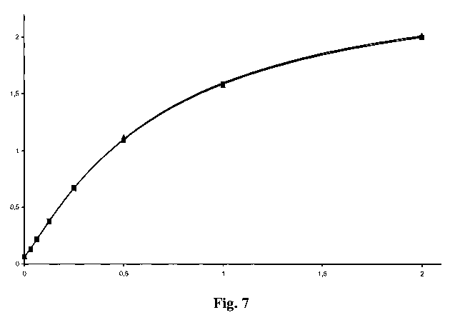
Figure 7 Comparison of standard curves obtained with samples i) spiked
with reference amounts of PEGylated insulin-like-growth-factor
and 5 % (v/v) human serum (triangle), ii) spiked with reference
amounts of PEGylated insulin-like-growth-factor and 5 % (v/v)
mouse serum (triangle); X-axis: concentration of PEGylated
insulin-like-growth-factor in ng/ml, Y-axis: mean absorption
signal; assay conditions: anti-(polyethylene glycol) antibody of
IgM class, anti-digoxygenin antibody-horseradish peroxidase-
conjugate at 25 mU/ml, all incubation times: 1 hour except for the
incubation with digoxygenylated insulin-like-growth-factor-
binding-protein-4, which is 20 hours, room temperature,
PEGylated insulin-like-growth-factor is a mixture of N-terminally
PEGylated and at position 68 PEGylated protein.
Figure 8 Standard curve obtained with reference samples spiked with
reference amounts of PEGylated insulin-like-growth-factor and
5 % (v/v) human plasma; X-axis: concentration PEGylated
insulin-like growth-factor in ng/ml, Y-axis: mean absorption
signal.
Description of the Sequences
SEQ ID NO: 1 Amino acid sequence of human insulin-like-growth-factor I
(amino acids 49 to 118 of Swiss-Prot ID P01343).
SEQ ID NO: 2 Amino acid sequence of human insulin-like-growth-factor II
(amino acids 25 to 91 of Swiss-Prot ID P01344).
CA 02720478 2010-10-04
WO 2009/121551 PCT/EP2009/002319
-28-
SEQ ID NO: 3 Amino acid sequence of human insulin-like-growth-factor-
binding-protein-4 (amino acids 22 to 258 of Swiss-Prot ID
P22692).
Example 1:
Preparation of anti-(polyethylene glycol) antibody conjugated to a microliter
plate
A solution of a biotinylated anti-(polyethylene glycol) antibody with a final
antibody concentration of 2 gg/ml was added to the wells of a 96-well
Streptavidin-
coated microtiter plate (MicroCoat) with 100 l to each well. Afterwards the
solution is incubated at room temperature at 500 rpm for one hour. Thereafter
the
solution is discarded and the wells are washed three times each with 300 Al
washing buffer (lx PBS (phosphate buffered saline) supplemented with 0.05 %
(w/v) n-octylglycosid).
Example 2:
Preparation of samples
a) Standard sample
A stock solution of PEGylated insulin-like-growth-factor I (for preparation of
PEGylated insulin-like-growth-factor I see e.g. WO 2006/066891) with a
concentration of 2 ng/ml in PBS buffer (phosphate buffered saline) supplement
with 0.5 % (w/v) bovine plasma albumin I was prepared. The stock solution was
diluted to the following concentration:
2.00 ng/ml
1.00 ng/ml
0.50 ng/ml
0.25 ng/ml
0.13 ng/ml
0.06 ng/ml
0.03 ng/ml
0.00 ng/ml
CA 02720478 2010-10-04
WO 2009/121551 PCT/EP2009/002319
-29-
b) Reference sample with serum or plasma
A stock solution of PEGylated insulin-like-growth-factor I (for preparation of
PEGylated insulin-like-growth-factor I see e.g. WO 2006/066891) with a
concentration of 2 ng/ml in 5 % pooled blank mouse serum or 5 % pooled blank
human serum or 5 % pooled blank human plasma in PBS buffer (phosphate
buffered saline) supplement with 0.5 % (w/v) bovine plasma albumin 1 was
prepared. The stock solution was diluted to the following concentration:
2.00 ng/ml
1.00 ng/ml
0.50 ng/ml
0.25 ng/ml
0.13 ng/ml
0.06 ng/ml
0.03 ng/ml
0.00 ng/ml
c) Test sample
The unknown test serum sample is diluted 1:20 with 5 % pooled blank mouse
serum in phosphate buffered saline supplemented with 0.5 % (w/v) bovine plasma
albumin 1.
Example 3:
Immunoassay
To the wells of a microtiter plate obtained according to Example 1 were added
100 l of each reference and test sample in duplicate. The wells were
incubated for
one hour with shaking at 500 rpm. Afterwards the solution is discarded and
each
well is washed three times each with 300 l phosphate buffered saline
supplemented with 0.05 % (w/v) n-octylglycosid. Thereafter 100 l of a
solution of
digoxygenylated insulin-like-growth-factor-binding-protein-4 at 100 ng/ml was
added to each well and incubated for 12-24 hours, preferably 20 hours, with
shaking at 500 rpm. Afterwards the solution was discarded and each well was
washed three times each with 300 l phosphate buffered saline supplemented
with
0.05 % (w/v) n-octylglycosid. Thereafter 100 l of a solution of an anti-
CA 02720478 2010-10-04
WO 2009/121551 PCT/EP2009/002319
-30-
digoxygenin antibody conjugated to horseradish peroxidase with a final
concentration of 50 mU/ml was added to each well and incubated for one hour
with
shaking at 500 rpm. Afterwards the solution in the wells was discarded and
each
well was washed three times each with 300 1 phosphate buffered saline
supplemented with 0.05 % (w/v) n-octylglycosid. Thereafter 100 l of an ABTS
solution was added to each well. The reaction was stopped when the highest
standard solution of 2 ng/ml has reached an OD value of 1.9 - 2Ø This
normally
requires between 5 to 15 minutes. The OD of the standard and test samples was
measured at 405 nm and 490 nm. A standard curve of the reference standards was
obtained using a 4-Parameter fit program. With the standard curve the amount
of
PEGylated insulin-like-growth-factor I in the test samples was calculated. The
lower limit of detection and lower limit of quantification have been
calculated to be
at 20 pg/ml and 31 pg/ml, respectively. All steps were carried out at room
temperature.
Table 1: Typical results of the positive control.
Sample Sample Sample Sample
mean STDEV CV [%]
No.1 No. 2 No. 3 concentration
2.0350 2.0550 2.0020 2.00 ng/ml 2.0307 0.0268 1.3%
1.6600 1.6640 1.7330 1.00 ng/ml 1.6857 0.0410 2.4%
1.2090 1.2520 1.2240 0.50 ng/ml 1.2283 0.0218 1.8%
0.7570 0.7340 0.7660 0.25 ng/ml 0.7523 0.0165 2.2%
0.4390 0.4280 0.4360 0.13 ng/ml 0.4343 0.0057 1.3%
0.2500 0.2520 0.2430 0.06 ng/ml 0.2483 0.0047 1.9%
0.1410 0.1590 0.1490 0.03 ng/ml 0.1497 0.0090 6.0%
0.0590 0.0620 0.0660 0.00 ng/ml 0.0623 0.0035 5.6%
CA 02720478 2010-10-04
WO 2009/121551 PCT/EP2009/002319
-31-
Example 4
Biotinylation of anti-(polyethylene glycol) antibody
An antibody against polyethylene glycol was dialyzed against buffer (100 mM
potassium phosphate buffer, pH 8.5). Afterwards the solution was adjusted to a
protein concentration of 10 mg/ml. D-biotinoyl-aminocaproic acid-N-
hydroxysuccinimide ester was dissolved in DMSO and added to the antibody
solution in a molar ratio of 1:5. After 60 minutes the reaction was stopped by
adding L-lysine. The surplus of the labeling reagent was removed by dialysis
against 25 mM potassium phosphate buffer supplemented with 150 mM NaCl, pH
7.5.
Example 5
Digoxigenylation of insulin-like-growth-factor-binding-protein
Insulin-like-growth-factor-binding-protein was dialyzed against
digoxygenylation
buffer (100 mM potassium phosphate buffer, pH 8.5). Afterwards the solution
was
adjusted to a protein concentration of 10 mg/ml. Digoxigenin 3-0-
methylcarbonyl-
c-aminocaproic acid-N-hydroxysuccinimide ester was dissolved in DMSO and
added to the antibody solution in a molar ratio of 1:5. After 60 minutes the
reaction
was stopped by adding L-lysine. The surplus of labeling reagent was removed by
dialysis against 25 mM potassium phosphate buffer supplemented with 150 mM
NaCl, pH 7.5.