Note: Descriptions are shown in the official language in which they were submitted.
CA 02721188 2013-01-04
WO 00176390
PCT/US00/16393
=
APPARATUS FOR REMOVING EMBOLI WITH
INFLATABLE MEMBER AND PUNCTURE RISK REDUCING MEANS
Field_ Of The Invention
This invention relates to apparatus and
methods for protecting against embolization during
vascular interventions, such as carotid artery
angioplasty, endarterectomy, and stenting. More
particularly, the apparatus and methods of the present
invention induce controlled retrograde flow through the
internal carotid artery during an interventional
procedure, without significant blood loss.
packaround of the Invention
Carotid artery stenoses typically manifest in
the common carotid artery, internal carotid artery or
external carotid artery as a pathologic narrowing of
the vascular wall, for example, caused by the
deposition of plaque, that inhibits normal blood flow.
= Endarterectomy, an open surgical procedure,
traditionally has been used to treat such stenosis of
the carotid artery.
An important problem encountered in carotid
artery surgery is that emboli may be formed during the
course of the procedure, and these emboli can rapidly
pass into the cerebral vasculature and cause ischemic
stroke.
CA 02721188 2010-11-15
"r
I 4
'
('
WO 00/76390 PCUUS00/163Y3
- 2 -
In view of the trauma and long recuperation
times generally associated with open surgical
procedures, considerable interest has arisen in the
endovascular treatment of carotid artery stenosis. In
particular, widespread interest has arisen in
transforming interventional techniques developed for
treating coronary artery disease, such as angioplasty
and stenting, for use in the carotid arteries. Such
endovascular treatments, however, are especially prone
to the formation of emboli.
Such emboli may be created, for example, when
an interventional instrument, such as a guide wire or
angioplasty balloon, is forcefully passed into or
through the stenosis, as well as after dilatation and
deflation of the angioplasty balloon or _stent
deployment. Because such instruments are advanced into
the carotid artery in the same direction as blood flow,
emboli generated by operation of the instruments are
carried directly into the brain by antegrade blood
flow.
Stroke rates after carotid artery stenting
have widely varied in different clinical series, from
as low as 4.4% to as high as 30%. One review of
carotid artery stenting including data from twenty-four
major interventional centers in Europe, North America,
South America, and Asia had a combined initial failure
and combined mortality/stroke rate of more than 7%.
Cognitive studies and reports of intellectual changes
after carotid artery stenting indicate that
embolization is a common event causing subclinical
cerebral damage.
Several previously known apparatus and
methods attempt to remove emboli formed during
endovascular procedures by occluding blood flow and
CD, 02721188 2010¨ 11 ¨15
,
e /
ViCIM0631M
PCT/US00/16:343
- 3 -
trapping or suctioning the emboli out of the vessel of
interest. These previously known systems, however,
provide less than optimal solutions to the problems of
effectively removing emboli. Additionally, when used
in stenting, the elements used to occlude blood flow
may dangerously interact with the stent.
Solano et al. U.S. Patent No. 4,921,478
describes cerebral angioplasty methods and devices
wherein two concentric shafts are coupled at a distal
end to a distally-facing funnel-shaped balloon. A
lumen of the innermost shaft communicates with an
opening in the funnel-shaped balloon at the distal end,
and is open to atmospheric pressure at the proximal
end. In use, the funnel-shaped balloon is deployed
proximally (in the direction of flow) of a stenosis,
occluding antegrade flow. An angioplasty balloon
catheter is passed through the innermost lumen and into
the stenosis, and then inflated to dilate the stenosis.
The patent states that when the angioplasty balloon is
deflated, a pressure differential between atmospheric
pressure and the blood distal to the angioplasty
balloon causes a reversal of flow in the vessel that
flushes any emboli created by the angioplasty balloon
through the lumen of the innermost catheter.
While a seemingly elegant solution to the
problem of emboli removal, several drawbacks of the
device and methods described in the Solano et al.
patent seem to have lead to abandonment of that
approach. Chief among these problems is the inability
of that system to generate flow reversal during
placement of the guide wire and the angioplasty balloon
across the stenosis. Because flow reversal does not
occur until after deflation of the angioplasty balloon,
there is a substantial risk that any emboli created
CA 02721188 2010-11-15
\=\ t
I
WO 00/76390 PCUUS00/16393
- 4 -
during placement of the angioplasty balloon will travel
too far downstream to be captured by the subsequent
flow reversal. It is expected that this problem is
further compounded because only a relatively small
volume of blood is removed by the pressure differential
induced after deflation of the angioplasty balloon.
Applicant has determined another drawback of
the method described in the Solano patent: deployment
of the funnel-shaped balloon in the common carotid
artery ("CCA") causes reversal of flow from the
external carotid artery ("ECA") into the internal
carotid artery ("ICA"), due to the lower flow impedance
of the ICA. Consequently, when a guide wire or
interventional instrument is passed across a lesion in
either the ECA or ICA, emboli dislodged from the
stenosis are introduced into the blood flow and carried
into the cerebral vasculature via the ICA.
The insufficient flow drawback identified for
the system of the Solano patent is believed to have
prevented development of a commercial embodiment of the
similar system described in EP Publication No. 0 427
429. EP Publication No. 0 427 429 describes use of a
separate balloon to occlude the ECA prior to crossing
the lesion in the ICA. However, like Solano, that
publication discloses that flow reversal occurs only
when the dilatation balloon in the ICA is deflated.
Chapter 46 of Interventional Neuroradioloav:
Strategies and Practical Techniques (J.J. Connors & J.
Wojak, 1999), published by Saunders of Philadelphia,
PA, describes use of a coaxial balloon angioplasty
system for patients having proximal ICA stenoses. In
particular, a small, deflated occlusion balloon on a
wire is introduced into the origin of the ECA, and a
guide catheter with a deflated occlusion balloon is
CD, 02721188 2010- 11 -15
0
(-1
V030104090
PCT/US00/16:393
- 5 -
positioned in the CCA just proximal to the origin of
the ECA. A dilation catheter is advanced through a
lumen of the guide catheter and dilated to disrupt the
stenosis. Before deflation of the dilation catheter,
the occlusion balloons on the guide catheter and in the
ECA are inflated to block antegrade blood flow to the
= brain. The dilation balloon then is deflated, the
dilation catheter is removed, and blood is aspirated
from the ICA to remove emboli.
Applicant has determined that cerebral damage
still may result from the foregoing previously known
procedure, which is similar to that described in EP
Publication No. 0 427 429, except that the ICA is
occluded prior to the ECA. Consequently, both of these
previously known systems and methods suffer from the
same drawback -- the inability to generate flow
reversal at sufficiently high volumes during placement
of the guide wire and dilation catheter across the
stenosis. Both methods entail a substantial risk that
any emboli created during placement of the balloon will
travel too far downstream to be captured by the flow
reversal.
Applicants note, irrespective of the method
of aspiration employed with the method described in the
foregoing Interventional Neuroradioloav article,
substantial drawbacks are attendant. If, for example,
natural aspiration is used (i.e., induced by the
pressure gradient between the atmosphere and the
artery), then only a relatively small volume of blood
is expected to be removed by the pressure differential
induced after deflation of the angioplasty balloon.
If, on the other hand, an external pump is utilized,
retrieval of these downstream emboli may requii a flow
CA 02721188 2010-11-15
tt ,
(,
V0300/76390 PCT/US00/16393
- 6 -
rate that cannot be sustained for more than a few
seconds, resulting in insufficient removal of emboli.
Furthermore, with the dilation balloon in
position, the occlusion balloons are not inflated until
after inflation of the dilation balloon. Microemboli
generated during advancement of the dilation catheter
into the stenosed segment may therefore be carried by
retrograde blood flow into the brain before dilation,
occlusion, and aspiration are even attempted.
A still further drawback of both the device
in EP Publication No. 0 427 429 and the Interventional
Neuroradioloav device is that, if either is used to
place a stent in the ICA instead of for ICA
angioplasty, the stent often extends beyond the
bifurcation between the ECA and the ICA. The occlusion
balloon placed by guide wire in the ECA may then snag
the stent during retrieval, causing the balloon to
puncture or get caught within the artery, and requiring
emergency surgery to remove the balloon.
Imran U.S. Patent No. 5,833,650 describes a
system for treating stenoses that comprises three
concentric shafts. The outermost shaft includes a
proximal balloon at its distal end that is deployed
proximal of a stenosis to occlude antegrade blood flow.
A suction pump then draws suction through a lumen in
the outermost shaft to cause a reversal of flow in the
vessel while the innermost shaft is passed across the
stenosis. Once located distal to the stenosis, a
distal balloon on the innermost shaft is deployed to
occlude flow distal to the stenosis. Autologous blood
taken from a femoral artery using an extracorporeal
blood pump is infused through a central lumen of the
innermost catheter to provide continued antegrade blood
flow distal to the distal balloon. The third
CA 02721188 2010-11-15
(
WO 00/76390
PCT/US00/16393
- 7 -
, concentric shaft, which includes an angioplasty
balloon, then is advanced through the annulus between
the innermost and outermost catheters to dilate the
stenosis.
Like the device of the Solano patent, the
device of the Imran patent appears to suffer the
drawback of potentially dislodging emboli that are
carried into the cerebral vasculature. In particular,
once the distal balloon of Imranis innermost shaft is
deployed, flow reversal in the vasculature distal to
the distal balloon ceases, and the blood perfused
through the central lumen of the innermost shaft
establishes antegrade flow. Importantly, if emboli are
generated during deployment of the distal balloon,
those emboli will be carried by the perfused blood
directly into the cerebral vasculature, and again pose
a risk of ischemic stroke. Moreover, there is some
evidence that reperfusion of blood under pressure
through a small diameter catheter may contribute to
hemolysis and possible dislodgment of emboli.
In view of these drawbacks of the previously
known emboli removal systems, it would be desirable to
provide methods and apparatus for removing emboli from
within the carotid arteries during interventional
procedures, such as angioplasty or carotid stenting,
that reduce the risk that emboli are carried into the
cerebral vasculature.
It also would be desirable to provide methods
and apparatus for removing emboli from within the
carotid arteries during interventional procedures, such
as angioplasty or carotid stenting, that provide
controlled retrograde blood flow from the treatment
zone, thereby reducing the risk that emboli are carried
into the cerebral vasculature.
CA 02721188 2010-11-15
" WO 0106390
PCT/US00/16393
- 8 -
It further would be desirable to provide
emboli removal methods and apparatus that prevent the
development of reverse flow from the ECA and antegrade
into the ICA once the CCA has been occluded, thereby
enhancing the likelihood that emboli generated by a
surgical or interventional procedure are effectively
removed from the vessel.
It still further would be desirable to
provide an occlusion balloon on a guide wire for
placement in the ECA during stenting of the ICA that
mitigates the risk of snagging the stent during
removal.
It also would be desirable to provide methods
and apparatus for removing emboli during carotid
stenting that enable filtering of emboli and reduced
blood loss, while simultaneously preventing dangerous
interaction between the apparatus and the stent.
Summary of the Invention
In view of the foregoing, it is an object of
this invention to provide methods and apparatus for
removing emboli from within the carotid arteries during
interventional procedures, such as angioplasty or
carotid stenting, that reduce the risk that emboli are
carried into the cerebral vasculature.
It also is an object of the present invention
to provide methods and apparatus for removing emboli
from within the carotid arteries during interventional
procedures, such as angioplasty or carotid stenting,
that provide controlled retrograde blood flow from the
treatment zone, thereby reducing the risk that emboli
are carried into the cerebral vasculature.
It is another r,hject of the present invention
to provide emboli removal methods and apparatus that
CA 02721188 2010-11-15
CT
WO 00f/6390
Per/US00/1 6393
- 9 -
prevent the development of reverse flow between the ECA
and ICA once the common carotid artery has been
occluded, thereby enhancing the likelihood that emboli
generated by a surgical or interventional procedure are
effectively removed from the vessel.
It is a further object of this invention to
provide methods and apparatus for an occlusion balloon
on a guide wire for placement in the ECA during
stenting of the ICA that mitigates the risk of snagging
the stent during removal.
It is yet another object of the present
invention to provide methods and apparatus for removing
emboli during carotid stenting that enable filtering of
emboli and reduced blood loss, while simultaneously
preventing dangerous interaction between the apparatus
and the stent.
The foregoing objects of the present
invention are accomplished by providing interventional
apparatus comprising an arterial catheter, an occlusion
balloon disposed on a guide wire, and a venous return
catheter. Additional, optional, apparatus may also be
provided, including, for example, a blood filter, a
flow control valve, a continuous pump, and/or a flow
sensor, disposed between the arterial and venous return
catheters. The arterial catheter has proximal and
distal ends, an aspiration lumen extending
therebetween, an occlusion element disposed on the
distal end, and a hemostatic port and blood outlet port
disposed on the proximal end that communicate with the
aspiration lumen. The aspiration lumen is sized so
that an interventional instrument, e.g., an angioplasty,
catheter or stent delivery system, may be readily
advanced therethrough¨to the site of a stenosis in
either the ECA (proximal to the balloon) or the ICA.
CA 02721188 2010-11-15
WO 00/76390
PCT/US00/16393
- 10 -
In accordance with the principles of the
present invention, the arterial catheter is
illustratively disposed in the CCA proximal of the
ICA/ECA bifurcation, the occlusion balloon on the guide
wire is disposed in the ECA to occlude flow reversal
from the ECA to the ICA, and the blood outlet port of
the arterial catheter is coupled to the venous return
catheter, with or without the additional, optional
apparatus disposed therebetween. Higher arterial than
venous pressure, especially during diastole, coupled,
for example, with the flow control valve permits
controlled flow reversal in the ICA during an
interventional procedure (other than when a dilatation
balloon is inflated) to flush blood containing emboli
from the vessel. The blood may then be filtered and
reperfused into the body through the venous return
catheter. A higher or more uniform rate of blood flow
between the arterial and venous catheters may be
obtained by use of the continuous pump, and the flow
sensor may alert medical personnel to dangerous blood
flow levels or may provide an automated activation
switch for the external pump.
In stenting applications, the occlusion
balloon on the guide wire is puncture resistant, so as
to prevent dangerous interaction between the balloon
and a stent during retrieval. In a first embodiment,
the apparatus comprises a wedge configured to deflect
the balloon away from contacting a portion of the stent
extending past the ECA/ICA bifurcation during retrieval
of the balloon. In a second embodiment, the apparatus
comprises a balloon that retracts into a capsule prior
to retrieval of the balloon from the ECA.
Methods of using the apparatus of the present
invention are also provided.
CA 02721188 2010-11-15
WO 00/76390
PCT/US00/1 6393
- 11 -
Brief Descrilotion of the Drawinas
Further features of the invention, its nature
and various advantages will be more apparent from the
accompanying drawings and the following detailed
description of the preferred embodiments, in which:
FIGS. lA and 1B are schematic views of prior
art apparatus for protecting against emboli during
carotid intervention;
FIG. 2 is a schematic view of apparatus for
= 10 protecting against emboli during carotid intervention
in accordance with the present invention;
FIGS. 3A-3D are, respectively, a schematic
view, and detailed side and sectional views of the
distal end, of apparatus constructed in accordance with
the present invention;
FIGS. 4A and 4B are views of the distal end
of an alteinative embodiment of the apparatus of FIG.
3;
. FIGS. 5A-5D illustrate a method of using the
apparatus of FIG. 3 in accordance with the principles
of the present invention;
FIGS. EA and 6B are, respectively, a
schematic view and a cross-sectional view of an
alternative embodiment of the apparatus of FIGS. 3;
FIGS: 7A and 7B are side views of a flow
control valve for use with the venous return line of
the present invention shown, respectively, in an open
position and a closed position;
FIGS. BA and 8B are schematic views of an
alternative embodiment of the guide wire balloon
element of the device of FIGS. 3, and a method of using
that device;
FIGS. RA and 9B are schematic views of a
further alternative embodiment of the guide wire
CA 02721188 2010-11-15
(-s
(-
=
W000/76390 PCT/US00/16393
- 12 -
balloon element of the apparatus of FIGS. 3, shown,
respectively, in a deployed configuration and in a
retrieval configuration; and
FIGS. 10A-10B illustrate a method of using
the apparatus of FIGS. 9 in accordance with the
principles of the present invention;
FIGS. 11A-11C are, respectively, detailed
side-sectional views and a cross-sectional view of the
distal end of yet another alternative embodiment of
apparatus of the present invention; and
FIGS. 12A-12C illustrate a method of using
the apparatus of FIG. 3 as adjunct to an emboli removal
filter.
Description of the Preferred Embodiments
Referring to FIGS. LA and 1B, drawbacks of
previously known emboli removal catheters are described
with reference to performing percutaneous angioplasty
of stenosis S in common carotid artery CCA.
With respect to FIG. LA, drawbacks associated
with naturally-aspirated emboli removal systems, such
as described in the above-mentioned patent to Solano
and European Patent Publication, are described. No
flow reversal is induced by those systems until after
balloon 10 of angioplasty catheter 11 first is passed
across the stenosis, inflated, and then deflated.
However, applicant has determined that once member 15
=
of emboli removal catheter 16 is inflated, flow within
the ECA reverses and provides antegrade flow into the
ICA, due to the lower hemodynamic resistance of the
ICA. Consequently, emboli E generated while passing
guide wire 20 or catheter 11 across stenosis S may be
carried irretrievably into the cerebral vasculature
CA 02721188 2010-11-15
(-
(Th
W000/76390
PCT/US00/16393
- 13 -
=
before flow in the vessel is reversed and directed into
the aspiration lumen of emboli removal catheter 16 by
opening the proximal end of the aspiration lumen to
atmospheric pressure. Furthermore, natural-aspiration
may not remove an adequate volume of blood to retrieve
even those emboli that have not yet been carried all
the way into the cerebral vasculature.
In FIG. 1B, system 17 described in the above-
mentioned patent to Imran is shown. As described
hereinabove, deployment of distal balloon 18, and
ejection of blood out of the distal end of the inner
catheter, may dislodge emboli from the vessel wall
distal to balloon 18. The introduction of antegrade
flow through inner catheter 19 is expected only to
exacerbate the problem by pushing the emboli further
into the cerebral vasculature. Thus, while the use of
positive suction in the Imran system may remove emboli
located in the confined treatment field defined by the
proximal and distal balloons, such suction is not
expected to provide any benefit for emboli dislodged
distal of distal balloon 18.
Referring now to FIG. 2, apparatus and
methods of the present invention are described.
Apparatus 30 comprises catheter 31 having an aspiration
lumen and occlusion element 32, and guide wire 35
having inflatable balloon 36 disposed on its distal
end. In accordance with the principles of the present
invention, antegrade blood flow is stopped when both
occlusion element 32 in the CCA and inflatable balloon
36 are deployed. Furthermore, the aspiration lumen of
catheter 31 is connected to a venous return catheter
(described hereinbelow) disposed, for example, in the
patient's femoral vein. In this manner a substantially
continuous flow of blood is induced between the
CA 02721188 2010-11-15
(r' '
(-
WOM06390
PCT/US00/16393
- 14 -
treatment site and the patient's venous vasculature.
Because flow through the artery is towards catheter 31,
any emboli dislodged by advancing a guide wire or
angioplasty catheter 33 across stenosis S causes the
emboli to be aspirated by catheter 31.
Unlike the previously known naturally-
aspirated systems, the present invention provides
substantially continuous retrograde blood flow through
the ICA while preventing blood from flowing retrograde
in the ECA and antegrade into the ICA, thereby
preventing emboli from being carried into the cerebral
vasculature. Because the apparatus and methods of-the
present invention "recycle" emboli-laden blood from the
arterial catheter through the blood filter and to the
venous return catheter, the patient experiences
significantly less blood loss.
Referring now to FIG. api, embolic protection
apparatus 40 constructed in accordance with the
principles of the present invention is described.
Apparatus 40 comprises arterial catheter 41, guide wire
45, venous return line 52, tubing 49 and optional blood
filter 50.
Catheter 41 includes distal occlusion element
42, proximal hemostatic port 43, e.g., a Touhy-Borst
connector, inflation port 44, and blood outlet port 46.
Guide wire 45 includes balloon 46 that is inflated via
inflation port 47. Tubing 49 couples blood outlet port
48 to filter 50 and blood inlet port 51 of venous
return line 52.
Guide wire 45 and balloon 46 are configured
to pass through hemostatic port 43 and the aspiration
lumen of catheter 41 (see FIGS. 3C and 3D), so that the
balloon may be advanced into and occlude the ECA. Port
43 and the aspiration lumen of catheter 41 are sized to
CA 02721188 2010-11-15
õ
WO 00/76390
PCT/US00/16393
- 15 -
,
permit additional interventional devices, such as
angioplasty balloon catheters, atherectomy -devices and
stent delivery systems to be advanced through the
aspiration lumen when guide wire 45 is deployed.
Guide wire 45 preferably comprises a small
diameter flexible shaft having an inflation lumen that
couples inflatable balloon 46 to inflation port 47.
Inflatable balloon 46 preferably comprises a compliant
material, such as described hereinabove with respect to
occlusion element 42 of emboli removal catheter 41.
Venous return line 52 includes hemostatic
port 53, blood inlet port 51 and a lumen that
communicates with ports 53 and 51 and tip 54. Venous
return line 52 may be constructed in a manner per se
known for venous introducer catheters. Tubing 49 may
comprise a suitable length of a biocompatible material,
such as silicone. Alternatively, tubing 49 may be
omitted and blood outlet port 48 of catheter 41 and
blood inlet port 51 of venous return line 52 may be
lengthened to engage either end of filter 50 or each
other.
With respect to FIGS. 38 and 3C, distal
occlusion element 42 comprises expandable bell or pear-
shaped balloon 55. In accordance with manufacturing
techniques which are known in the art, balloon 55
comprises a compliant material, such as polyurethane,
latex or polyisoprene which has variable thickness
along its length to provide a bell-shape when inflated.
Balloon 55 is affixed to distal end 56 of catheter 41,
for example, by gluing or a melt-bond, so that opening
57 in balloon 55 leads into aspiration lumen 58 of
catheter 41. Balloon 55 preferably is wrapped and heat
treated during manufacture so that distal portio¶ 59 of
the balloon extends beyond the distal end of catheter
CA 02721188 2010-11-15
.
W000/76390
PCT/US00/16393
- 16
41 and provides an atraumatic tip or bumper for the
catheter.
As shown in FIG. 3D, catheter 41 preferably
comprises inner layer 60 of low-friction material, such
as polytetrafluoroethylene ("PTFE"), covered with a
layer of flat stainless steel wire braid 61 and polymer
cover 62 (e.g., polyurethane, polyethylene, or PEBAX).
Inflation lumen 63 is disposed within polymer cover 62
and couples inflation port 44 to balloon 55. In a
preferred embodiment of catheter 41, the diameter of
lumen 58 is 7 Fr, and the outer diameter of the
catheter is approximately 9 Fr.
Referring now to FIGS. 4A and 4B, an
alternative embodiment of occlusion element 42 of the
system of FIG. ax is described. In FIGS. 4.A. and 4B,
occlusion element 42 of emboli removal catheter 41
comprises self-expanding wire basket 65 covered with
elastomeric polymer 66, such as latex, polyurethane or
polyisoprene. Alternatively, a tightly knit self-
expanding wire mesh may be used, with or without an
elastomeric covering.
Catheter 41 is surrounded by movable sheath
67. Catheter 41 is inserted transluminally with sheath
67 in a distalmost position, and after basket 65 has
been determined to be in a desired position proximal to
a -stenosis, sheath 67 is retracted proximally to cause
basket 65 to deploy. Upon completion of the procedure,
basket 65 is again collapsed within sheath 67 by moving
the sheath to its distalmost position. Operation of
the system of FIG. aA using the emboli removal catheter
of FIGS. 4A and 4B is similar to that described
hereinbelow for FIGS. 5A-5D, except that the occlusion
element self-expands when sheath 67 is retracted,
.ak 02721188 2010-11-15
(-'
W000/76390
PCT/US00/16393
- 17 -
rather than by infusing an inflation medium to balloon
55.
Referring now to FIGS. ax-sp, use of the
apparatus of FIGS. 3 in accordance with the methods of
the present invention is described. In FIGS. 5,
stenosis S is located in internal carotid artery ICA
above the bifurcation between the internal carotid
artery ICA and the external carotid artery ECA. In a
first step, catheter 41 is inserted, either
percutaneously and transluminally or via a surgical
cut-down, to a position proximal of stenosis S, without
causing guide wire 45 to cross the stenosis. Balloon
55 of distal occlusion element 42 is then inflated,
preferably with a radiopaque contrast solution, via
inflation port 44. As seen in FIG. 5A, this creates
reversal of flow from the external carotid artery ECA
into the internal carotid artery ICA.
Venous return line 52 then is introduced into
the patient's femoral vein, either percutaneously or
via a surgical cut-down. Filter 50 is then coupled
between blood outlet port 48 of catheter 41 and blood
inlet port 51 of venous return line 52 using tubing 49,
and any air is removed from the line. Once this
circuit is closed, negative pressure in the venous
catheter during diastole will establish a low rate
continuous flow of blood through aspiration lumen 58 of
catheter 41, as seen in FIG. 53, to the patient's vein
via venous return line 52.
This low rate continuous flow due to the
difference between venous pressure and arterial
pressure will continue throughout the interventional
procedure. Specifically, blood passes through
aspiration lumen 58 and blood outlet port 48 of
CD, 02721188 2010-11-15
(
I
WO 00/76390 PCMJS00/16393
- 16 -
,
catheter 41, through biocompatible tubing 49 to filter
50, and into blood inlet port 51 of venous return line
52, where it is reperfused into the remote vein.
Filtered emboli collect in filter 50 and may be studied
and characterized upon completion of the procedure.
Continuous blood flow (except during
inflation of any dilatation instruments) with
reperfusion in accordance with the present invention
provides efficient emboli removal with significantly
reduced blood loss. Alternatively, filter 50 may be
omitted, in which case emboli removed from the arterial
side will be introduced into the venous side, and
eventually captured in the lungs. Because of a low
incidence of septal defects, which could permit such
emboli to cross-over to the left ventricle, the use of
filter 50 is preferred.
Referring to FIG. 5C, with balloon 55 of
occlusion element 42 inflated and a retrograde flow
established in the ICA, guide wire 45 and balloon 46
are advanced through aspiration lumen 58. When balloon
46 is disposed within the ECA, as determined, e.g.,
using a fluoroscope and a radiopaque inflation medium
injected into balloon 46, balloon 46 is inflated.
Occlusion of the ECA prevents the development of
reverse flow in the ECA from causing antegrade flow in
the ICA. Another interventional instrument, such as
conventional angioplasty balloon catheter 71 having
balloon 72, is loaded through hemostatic port 43 and
aspiration lumen 58 and positioned within the stenosis.
=
Hemostatic port 43 is closed and instrument 71 is
actuated to disrupt the plaque forming stenosis S.
As seen in FIG. 5D, upon completion of the
angioplasty portion of the procedure using catheter 71,
CA 02721188 2010-11-15
C
W000/76390
PCT/US00/1639.3
- 19 -
balloon 72 is deflated. Throughout the procedure,
except when the dilatation balloon is fully inflated,
the pressure differential between the blood in the ICA
and the venous pressure causes blood in ICA to flow in
a retrograde direction in the ICA into aspiration lumen
58 .of emboli removal catheter 41, thereby flushing any
emboli from the vessel. The blood is filtered and
reperfused into the patient's vein.
Optionally, increased volumetric blood flow
through the extracorporeal circuit may by achieved by
attaching an external pump, such as a roller pump, to
tubing 49. If deemed beneficial, the external pump may
be used in conjunction with device 40 at any point
during the interventional procedure. Likewise, a flow
sensor may be coupled to tubing 49 to generate a signal
corresponding to a flow rate within the tubing. This
sensor may alert medical personnel to dangerous levels
of blood flow through the extracorporeal circuit, or
may provide an automated activation switch for the
external pump. Instrument 71, guide wire 45, emboli
removal catheter 41, and venous return line 52 are then
removed from the patient, completing the procedure.
. As set forth above, the method of the present
invention protects against embolization, first, by
preventing the reversal of blood flow from the ECA to
the ICA when distal occlusion element 42 is inflated,
and second, by providing continuous, low volume blood
flow from the carotid artery to the remote vein in
order to filter and flush any emboli from the vessel
and blood stream. Advantageously, the method of the
present invention permits emboli to be removed with
little blood loss, because the blood is filtered and
Laperfused into the patient. Furthermore, .continuous
CD. 02721188 2010-11-15
'
WO 00/76390 PCIVUS00/16393
- 20 -
removal of blood containing emboli prevents emboli from
migrating too far downstream for aspiration.
Referring now to FIGS. 6, apparatus 140
constructed in accordance with the present invention is
described. Apparatus 140 is an alternative embodiment
of apparatus 40 described hereinabove and comprises
arterial catheter 141 having distal occlusion element
142, proximal hemostatic port 143, inflation port 144
and blood outlet port 148. Guide wire 145 includes
balloon 146 that is inflated via inflation port 147.
Biocompatible tubing 149 couples blood outlet port 148
to filter 150 and to blood inlet port 151 of venous
return line 152. Arterial catheter 141, guide wire
145, venous return line 152 and tubing 149 are
constructed as described hereinabove, except as noted
below.
Guide wire 145 and balloon 146 are configured
to pass through guide wire lumen 164 of catheter 141
(see FIG. 6B), so that the balloon may be advanced into
and occlude the ECA. Additionally, catheter 141
comprises aspiration lumen 158 which is sized to permit
interventional devices, such as angioplasty balloon
catheters, atherectomy devices and stent delivery ,
systems to be advanced through port 143 and the
aspiration lumen. As shown in FIG. 63, one difference
between catheters 41 and 141 is the method of advancing
the guide wire through the catheter: guide wire 45 is
advanced through the aspiration lumen of catheter 41,
whereas guide wire 145 is advanced through separate
guide wire lumen 164 of catheter 141.
Catheter 141 preferably is constructed from
inner layer 160 of low-friction material, such as
polytetrafluoroethylene ("PTFE"), covered with a layer
of flat stainless steel wire braid 161, and polymer
=
CA 02721188 2010- 11 -15
=
- 21 -
cover 162. (e.g., polyurethane, polyethylene, or PEBAX).
Inflation lumen 163 is disposed within polymer cover
=
162 and couples inflation port 144 to occlusion element
142. Guide wire lumen 164 also is disposed within
polymer cover 162, and is sized to permit guide wire
145 and balloon 146 to pass therethrough. In a
preferred embodiment of catheter 141, the diameter of
inflation lumen 163 is .014", the diameter of guide
wire lumen 164 is .020", and the diameter of lumen 158
is 7 Fr. To retain an outer catheter diameter in the
preferred embodiment of approximately 9 Fr., the
thickness of the catheter wall varies around the
circumference from a maximum of 0.026" at the location
of guide wire lumen 164 to a minimum of 0.005" 180
degrees away.
With reference to FIGS. 7A and 7B, a flow
control valve for use with the venous return line is
described. Flow control valve 175 is illustratively
shown connected to a portion of biocompatible tubing
149 of FIG. EA. Valve 175 comprises pinch wheel 177
and member 179. Member 179 comprises channel 181 and
inclined track 183. Pinch wheel 177 includes teeth 178
and is configured to pass through channel 181 along
track 183. Channel 181 is also configured to receive
tubing 149. Pinch wheel 177 may move along track 183
from a first position, shown in FIG. TA, in which it
does not compress tubing 149, to a user-selected second
position, shown in FIG. 7B, at which tubing 149 is
compressed a user-selected degree to reduce or stop
flow through tubing 149.
When used in conjunction with the apparatus
of the present invention, valve 175 allows a selective
degree of constr.ction to be applied to the venous
return line. Thus, valve 175 provides selective
CA 02721188 2010-11-15
- 22 -
control over the amount of reverse flow from the
carotid artery to the femoral vein. Some patients may
tolerate cessation of antegrade flow in the ICA, or a
gentle reversal of flow, but may not tolerate the brain
ischemia associated with full reversal of flow. Valve
175 therefore allows a slow, gentle reversal - or even
cessation - of antegrade flow to be established in the
ICA. As long as flow is stopped or slightly reversed,
emboli will not travel to the brain. Aspiration prior
to deflation of the distal occlusion element ensures
that emboli sitting in the carotid or within the sheath
are removed.
Referring now to FIGS. 8, an alternative
embodiment of the guide wire occlusion apparatus of the
present invention is described. Occlusion apparatus
190 comprises guide wire 191, occlusion balloon 192,
'inflation lumen 193, and wedge 194. Wedge 194 may
comprise a resilient material, such as a polymer or
resilient wire, and reduces the risk that balloon 192
will snag on a stent that extends beyond the
bifurcation of the ICA and ECA.
For the reasons described hereinabove, it is
desirable when performing a stenting procedure in the
ICA to occlude the ECA, to prevent flow reversal from
the ECA and into the ICA. Accordingly, an occlusion
balloon on a guide wire is placed in the ECA and
inflated to block that artery. A stent then may be
placed in the ICA to ensure proper blood flow to the
ICA. It is often desirable, however, for such stents
to extend beyond the bifurcation between the ECA and
the ICA. Consequently, when the occlusion balloon on
the guide wire is deflated and withdrawn from the ECA,
there is a risk that the balloon may snag the stent,
CA 02721188 2010-11-15
c-
WOMVM390
PCT/US00/16393
- 23 -
causing the balloon to puncture or get caught within
the artery, and requiring emergency surgery.
Referring now to FIG. 8B, 'occlusion apparatus
190 is illustratively shown in conjunction with
catheter 41. Stent 195 extends beyond the bifurcation
between the ECA and the ICA and into the CCA. Balloon
192 is deflated and positioned for retrieval. Because
balloon 192 is disposed on guide wire 191 instead of a
traditional, larger diameter balloon catheter, its
cross-sectional diameter is significantly reduced, and
thus the risk that the balloon will snag on stent 195
is reduced. Resilient wedge 194 further reduces this
risk by urging the balloon outward away from the stent
during retrieval of guide wire 191 and balloon 192.
Alternatively, a separate sheath may be advanced over
=guide wire 191 and occlusion balloon 192 to surround
those components, and thereby reduce the risk that the
occlusion balloon or guide wire will snag the stent.
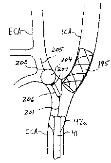
With reference to FIGS. 9A and 9B, an
alternative embodiment of the guide wire occlusion
apparatus of the present invention is described.
Occlusion apparatus 200 comprises guide wire 201 having
inflation lumen 202 and proximally terminating in
inflation port 203, occlusion balloon 204, core wire
205 attached to balloon 204, capsule 206, radiopaque
capsule features 207, and radiopaque balloon feature
208. Core wire 205 is preferably approximately 0.010"
in diameter and is configured to be received within
inflation lumen 202 of guide wire 201. Guide wire 201
is preferably approximately 0.018" in diameter.
Balloon 204 may be inflated via inflation
lumen 202 with a standard or radiopaque inflation
medium. Balloon 204 then extends distally of, but
remains attached to, capsule 206. Upon completion of
CD, 02721188 2010-11-15
(õI.
s.
IVIDOWEMO
PCT/US00/16393
- 24 -
an interventional procedure, such as carotid stenting,
balloon 204 is deflated. 'Proximal retraction of core
wire 205 draws balloon 204 into capsule 206, thereby
preventing snagging during retrieval.
Referring now to FIGS. 10A and 10B, use of
occlusion apparatus 200 in conjunction with arterial
catheter 41 and venous return catheter 52 of FIGS. 3
during carotid stenting is described. With balloon 42a
of occlusion element 42 inflated and a retrograde flow
established in the ICA as described hereinabove,
occlusion apparatus 200 is advanced through aspiration
lumen 58 of catheter 41. Capsule 206 is disposed just
within the ECA, as determined, e.g., using a
fluoroscope and radiopaque capsule features 207, as
=seen in FIG. 10A. Occlusion balloon 204 is then
inflated and its position verified by, for example, a
fluoroscope and radiopaque balloon feature 208 or a
radiopaque inflation medium injected into balloon 204.
Occlusion of the ECA prevents the development of
reverse flow in the ECA from causing antegrade flow in
the ICA. Another interventional instrument, such as
stent 195, is then loaded through hemostatic port 43
and aspiration lumen 58 and positioned across stenosis
S to ensure proper blood flow to the ICA.
Stent 195 may extend beyond the bifurcation
between the ECA and the ICA. Consequently, when the
occlusion-balloon on the guide wire is deflated and
withdrawn from the ECA, there is a risk that the
balloon may snag on the stent, with potentially dire
consequences.
As shown in FIG. 10B, upon completion of the
stenting portion of the procedure, balloon 204 is
deflated, and core wire 205 is proximally retracted to
CA 02721188 2010-11-15
*
W000/76390
PCT/US00/16393
- 25 -
=
draw deflated balloon 204 within capsule 206. Because
balloon 204 is disposed on guide wire 201 instead of a
traditional, larger diameter balloon catheter, its
cross-sectional diameter is significantly reduced, and
thus the risk that the balloon will snag or puncture on
stent 195 is reduced. Capsule 206 further reduces this
risk by protecting the balloon during retrieval of
occlusion apparatus 200. Apparatus 200, emboli removal
catheter 41, and venous return line 52 then are removed
from the patient, completing the procedure.
With reference to FIGS. 11, the distal end of
yet another alternative interventional device suitable
for use in the system of the present invention is
described. Because balloon 55 of FIGS. 3 is attached
to a stepped portion of catheter 41, apparatus 40
requires use of a larger diameter introducer sheath
compared to a standard guide catheter without such a
balloon. At the point of attachment of balloon 55,
however, catheter 41 does not require the torque,
strength, or stiffness of a regular guide catheter.
Balloon 55 has its own inflation means, and, in use,
the tip of catheter 41 does not intubate an artery.
As seen in FIGS. 11A and 11B, distal
occlusion element 210 comprises expandable bell or
pear-shaped balloon 212. Balloon 212 is similar to
balloon 55. In accordance with manufacturing
techniques which are known in the art, balloon 212
comprises a compliant material, such as polyurethane,
latex or polyisoprene which has variable thickness
along its length to provide a bell-shape when inflated.
Balloon 212 is affixed to reduced-thickness distal
region 216 of arterial catheter 214, for example, by
gluing or a melt-bond, so that opening 218 in balloon
212 leads into aspiration lumen 220 of catheter 214.
CD, 02721188 2010-11-15
- 26 -
Balloon 212 preferably is wrapped and heat treated
during manufacture so that distal portion 222 of the
balloon extends beyond the distal end of catheter 214
and provides an atraumatic tip or bumper for the
catheter.
As shown in FIG. 11C, catheter 214 preferably
comprises inner layer 232 of low-friction material,
such as polytetrafluoroethylene ("PTFE"), covered with
a layer of flat stainless steel wire braid 234 and
polymer cover 236 (e.g., polyurethane, polyethylene, or
PEBAX). Inflation lumen 238 is disposed within polymer
cover 236 and couples an inflation port (not shown) to
balloon 212. In a preferred embodiment of catheter
214, the diameter of lumen 220 is 7 Fr, and the outer
diameter of the catheter is approximately 9 Fr.
Arterial catheter 214 is similar to catheter
41 of FIGS. 3, except at reduced-thickness distal
region 216. Distal region 216 preferably achieves its
reduced thickness by omission of wire braid 234 in that
region. Thus, when balloon 212 is deflated, the
composite delivery profile of distal end 216 and
balloon 212 is the substantially same as or smaller
than the delivery profile of the remainder of catheter
214. Preferably, the device has a delivery profile of
approximately 9 Fr (see FIG. 11A). The diameter of
lumen 220 remains the same along the entire length of
catheter 214, preferably around 7 Fr. Catheter 214
therefore requires an introducer sheath of no larger
diameter than those required by a standard guide
catheter.
With reference now to FIGS. 12A-12C, the
apparatus of the present invention also may be used as
an adjunct against embolization when used with a
distally deployed embolic filter. Embolic filter 250
CA 02721188 2013-11-06
WO 00/76390 PM/USN/16393
- 27 -
is illustratively shown in conjunction with catheter
41, and comprises guide wire 252 and expandable mesh
254.
Catheter 41 may provide adjunct protection in
a variety of ways. For example, as depicted in FIG.
12A, reverse flow through catheter 41 may be
= established, as described hereinabove, during
deployment of filter 250 to capture emboli E generated
while passing filter 250 across stenosis S.
Alternatively, if, after deployment, filter 250 becomes
= overly filled with emboli E, reverse flow may .be
established to provide a pressure differential that
aspirates emboli from the filter and into catheter 41,
as depicted in FIG. 12B. Also, in the event filter 250
fails, for example, during retrieval, reverse flow may
be established to prevent emboli E or fragments of the
filter from being carried downstream, as seen in FIG.
12C. In addition to catheter 41, the filter also may
be used in conjunction with balloon 46 so that blood
flow is not unnecessarily reversed in, for example, the
ECA when emboli E are generated in the ICA.
As will of course be understood, the
apparatus of the present invention may be used in
locations other than the carotid arteries. They may,
for example, be used in the coronary aTteries, or in
any other location deemed useful.
While preferred illustrative embodiments of
= the invention are described.above, it will be apparent
to one skilled in the art that various changes and
modifications may be made. The appended claims are
intended to cover all such changes and modifications
that fall within the scope of the invention as described
herein. The scope of the claims should not be limited by
the preferred embodiments set forth in the examples, but
= should receive the broadest interpretation consistent with
the description as a whole.