Note: Descriptions are shown in the official language in which they were submitted.
CA 02721271 2010-10-08
WO 2009/125002 PCT/EP2009/054319
1
A MEDICAL SYSTEM COMPRISING A PERCUTANEOUS PROBE
FIELD OF THE INVENTION
The instant invention relates to medical systems
comprising percutaneous probes, and to uses of such medical
systems.
BACKGROUND OF THE INVENTION
Low intensity ultrasounds are widely used in
medicine for diagnostic procedures, i.e. echography. For 10
years, high intensity ultra-sounds have shown to be an
efficient means to induce tissue necrosis by hyperthermia
for treatment procedures. Various therapeutic probes have
been designed for minimally invasive therapeutic procedures
and can be classified in two groups: external probes and
internal probes.
External probes are designed to mimic the shape of
the surface of the patient's body. Ultrasound transmitters
are displayed in a concentric fashion to optimise the
ultrasound waves focalization.
Internal/interstitial probes are inserted inside
the body of the patient. There are three main categories:
endo-cavity, endovascular or percutaneous probes.
A. Endocavity probes
Endocavity probes are designed to be introduced in
natural body holes such as the rectum, the vagina or the
oesophagus. For example, US 2007/239,011 describes a
medical probe for the delivery of high intensity focused
ultra-sound (HIFU) energy to a patient's organ. Such a
probe comprises a plane-shaped probe body inserted through
a natural cavity of a patient, and a plurality of leaves to
be applied to the surface of the organ, to deliver ultra-
sound energy to the inside of the organ.
B. Endovascular probes
Endovascular flexible probes are in development to
CA 02721271 2010-10-08
WO 2009/125002 PCT/EP2009/054319
2
treat cardiac atrial fibrillation or venous insufficiency.
C. Percutaneous interstitial probes
Percutaneous interstitial probes have initially
received poor interest since they require a tissue
penetration whereas previous probes don't penetrate the
tissue. Nevertheless such percutaneous interstitial probes
have been proposed for treating deep-seated tumours that
cannot be reached with extra-corporeal, endocavity or
endovascular high-intensity focused ultrasound probe. The
ultrasound source is brought as close as possible to the
target in order to minimize the effects of attenuation and
phase aberration along the ultrasound pathway. Most-
described ultrasound percutaneous probes are sideview
emission probes whose active element is water-cooled and
operates at a rather high frequency (above 3 MHz) in order
to promote heating. Most described ultrasound percutaneous
probes are not MRI compatible so that treatment monitoring
is somewhat hazardous.
For clinicians, ultrasounds are a promising
technology. To extend the applicability of ultra-sound
therapy to a broad variety of medical treatments, there is
a need to solve the following inconveniencies:
In particular, external probes, although non
intrusive, have shown consistent inconvenient: ultrasound
attenuation, phase aberration and ultrasound defocalization
by tissue structure (bone, tissue interfaces ...), targeting
limits do to the constant body movement (respiratory,
diaphragm...), long treatment duration, unknown consequences
on crossed normal tissue by the ultrasounds pathway,
complexity of the probes with nowadays hundreds of
ultrasound transducers, complexity to make the system MRI
compatible and MRI adaptable.
In particular, sideview interstitial/internal
probes require clinician manipulation of the probe during
treatment such as a 360 rotation or a longitudinal
CA 02721271 2010-10-08
WO 2009/125002 PCT/EP2009/054319
3
translation to treat the whole lesion leading to a lack of
precision and reproducibility.
In particular for all existing probes, none can
perform histological characterisation or tissue biopsy,
meaning that a biopsy procedure is necessary days before
treatment. For all existing probes, none can perform a
tissue resection after the thermal treatment. Indeed,
hyperthermia treatment of a tumour will gender a serious
tumour volume increase (mass effect) as shown in a previous
clinical trial (Carpentier & al., "Real-time Magnetic
Resonance-Guided Laser Thermal Therapy of Metastatic Brain
Tumors", Neurosurgery, 63 ONS Suppl 1:21-29, 2008). Such
volume increase is most of the time incompatible with
preservation of the normal surrounding tissue and can limit
the development of such minimally invasive ultrasound
therapy systems.
SUMMARY OF THE INVENTION
The instant invention aims to solve at least some
of those cited inconvenients.
To this aim, it is provided a medical system
comprising a percutaneous probe and a computerized system,
the percutaneous probe, made in MRI - compatible materials,
comprising:
- a body having an insertion end, shaped to be
percutaneously inserted into tissue of a patient's body
organ having a region to be analyzed, treated and monitored
during a single medical procedure,
- an optical head of an endo-confocal digital
microscope,
- at least one information collection sensing
device, adapted to collect information about the region of
the organ,
- a plurality of treatment application
transducers, operable as a phased-array, adapted to emit
both focused and not-focused therapeutic ultra-sound waves
CA 02721271 2010-10-08
WO 2009/125002 PCT/EP2009/054319
4
to the region of the organ,
the computerized system comprising a parametrizable
command device and associated equipment adapted to command
a generation of the therapeutic ultra-sound wave.
With these features, the probe can be
percutaneously inserted at a suitable location in any
organ. Further, the probe can be used, during a single
medical procedure, to sense organ information usable for
establishing a diagnostic and characterization, and for
implementing the appropriate therapy.
In some embodiments, one might also use one or more
of the features defined in the dependant claims.
Further, it is provided a method comprising
- providing a body organ having a region to be
analyzed, treated and monitored during a single medical
procedure, said organ being provided with a percutaneous
probe made in MRI compliant materials, and having a body
having an insertion end inserted into tissue of the body
organ,
- collecting information about the region of the
organ with an information collection sensing device of the
probe, and with an optical head of an endo confocal
microscope
- setting parameters of at least one of a focused
and not-focused therapeutic ultra-sound wave to be emitted
to the region of the organ with a plurality of treatment
application transducers (30) of the probe, configured as a
phase-array through a parametrizable command device (50)
and associated equipment (56) of a computerized system.
In some embodiments, one might also use one or more
of the features defined in the method dependant claims.
Advantages of one or more of these embodiments
might include:
- real time monitoring of the therapy,
- tissue histologic characterization and
CA 02721271 2010-10-08
WO 2009/125002 PCT/EP2009/054319
tissue therapeutic treatment during a single procedure,
- ability to extract air bubbles from the
tissue and replace them by liquid to avoid imaging
artifacts,
5 - ability to "real-time" monitor the
therapeutic process and monitor security points for the
treatment,
ability to perform continuous MRI and MR
thermometry monitoring,
- ability to perform immediate post-treatment
MRI imaging sequences for monitoring the therapeutic
process efficiency,
- ability to treat regions either immediately
around the probe and/or delocated areas,
- improved focalization/defocalization of the
therapeutic ultra-sound energy, by several techniques in
order to best fit the lesion geometry,
- ability to perform the therapeutic treatment
even for a moving patient and/or organ,
- ultrasounds emitters organized in a 360
fashion, thereby removing the need to rotate the probe
inside the organ,
- ability to treat tumors of various and
complex shapes,
- diminution of the post-treatment tumoral
volume,
- ability to acquire electro-encephalogram
signals during the treatment.
- ability to erase movement artefacts,
ultrasound attenuations, phase aberrations and/or
ultrasound defocalizations,
- no requirement of a clinician manipulation
during treatment, so that MRI safety and efficacy
monitoring during the treatment becomes reliable,
- ability to allow real time and in vivo
CA 02721271 2010-10-08
WO 2009/125002 PCT/EP2009/054319
6
tissue characterisation, biopsy, post thermal tissue
resection preventing post-treatment mass effect,
- disposable technology prevents inter patient
contamination.
BRIEF DESCRIPTION OF THE DRAWINGS
Other characteristics and advantages of the
invention will readily appear from the following
description of three of its embodiments, provided as a non-
limitative examples, and of the accompanying drawings.
On the drawings :
- Fig. 1 is a schematic view of a medical
apparatus,
- Fig. 2 is a partial sectional view of a probe
inserted into a body organ,
- Figs. 3a, 3b and 3c are perspective views
illustrative of various components of a probe according to
a first embodiment,
- Fig. 4 is a partial sectional view along line
IV-IV On Fig. 3c,
- Fig. 5 is a view similar to Fig. 4 for a variant
embodiment,
- Figs. 6a, 6b and 6c are perspective views
illustrative of various components of a probe according to
a second embodiment,
- Figs. 7a, 7b, and 7c are views similar to Figs.
6a, 6b and 6c, respectively, for a probe according to a
third embodiment,
- Figs. 8a and 8b are perspective views of
components of a probe according to a fourth embodiment,
- Fig. 9 is a sectional view along line IX-IX of
Fig. 8a,
- Fig. 10 is a partial perspective view of a probe
according to a fifth embodiment,
- Figs. 11a and 11b are partial perspective views
of probes according to a sixth and a seventh, respectively,
CA 02721271 2010-10-08
WO 2009/125002 PCT/EP2009/054319
7
embodiment,
- Fig. 12 is a schematic view of a computerized
system operatively associated with the probe,
- Fig. 13 is a diagram showing an example of use of
the medical apparatus, and
- Fig. 14 is a enlarged view of Fig. 2.
On the different figures, the same reference signs
designate like or similar elements.
DETAILED DESCRIPTION
Figure 1 is a schematic view of a medical apparatus
1 comprising a magnetic resonance imaging (MRI) system 2 of
conventional type, suitable to be operated, in particular,
in a thermal imaging mode, in which a patient 3 is
introduced, for example lying on a suitable bed 4.
The medical apparatus 1 further comprises a
computerized system 5 connected to the magnetic resonance
imager so as to receive from the magnetic resonance imager
data enabling the construction of anatomy and/or thermal
magnetic resonance images of the patient 3.
The medical apparatus further comprises a MRI
compatible probe 6 percutaneously inserted into the
patient's body, and operatively associated to the
computerized system 5. The probe 6 is for example
electrically connected to the computerized system 5 through
MRI-compatible wires, for example coaxial wires, which are
known per se and will therefore not be described in more
details here.
As schematically shown on Fig. 2, the probe 6
comprises a somehow cylindrical body 7 of external diameter
D of 4 mm, or less, and preferably of 3 mm or less, and
even more preferably of 2 mm or less, and is shaped to be
introduced into the tissue of an organ 8 of the patient's
body 3. The probe body 7 is interstitial, since it can be
introduced directly into the tissue of the organ 8 without
the necessity to go through the cavity of the organ. The
CA 02721271 2010-10-08
WO 2009/125002 PCT/EP2009/054319
8
insertion tip 9 of the probe body 7 is located within, or
close to a region 10 of the tissue of the organ, which is
to be analyzed and/or treated. The probe 6 could be
applied, for example, to malignant or benign cancers, or
neuro-cognitive brain impairments, as well as for other
tissular pathologies of all other organs that could be
treated by monitored ultrasonic treatment, such as thermal
ablation.
According to a first embodiment, as shown on
figures 3a, 3b and 3c, the probe 6 comprises a plurality of
components : an applicator 11 (figure 3a), a mandrel 12
(figure 3b), and an ultra-sound device 13 (figure 3c).
The applicator 11 comprises a proximally-located
back portion 14 and a somehow cylindrical body 15 defining
an internal cylindrical cavity 16 which extends throughout
the body 15 to a tip in which an end opening 40 is formed.
The medical system can further comprise a
continuous pump 36 to be placed in fluid communication with
the cavity 16 of the applicator 11, or removed therefrom.
The pump can thus be operated to collect tissue fragments
which can later be analyzed, for example by the clinician,
to provide tissue information.
The pump 36 can be operated to extract any material
to be extracted from the patient, such as, for example, air
bubbles that artifact images, and/or inject into the
patient suitable liquids.
The inner mandrel 12 is made of a rigid material
and is shaped so as to be inserted into the cavity 16 so as
to totally obstruct this cavity. The mandrel 12 can for
example have a body 25 of cylindrical shape, of external
diameter equal to the internal diameter of the body 15 of
the applicator 11. Furthermore, the mandrel 12 can have a
back portion 26 of cylindrical shape of outer diameter
equal to the inner diameter of the back portion 14 of the
applicator 11. The mandrel 12 can further comprise a
CA 02721271 2010-10-08
WO 2009/125002 PCT/EP2009/054319
9
pointy tip 57. The mandrel can comprise an internal channel
27 extending from a proximal end 27a in the back portion
26, to a distal end 27b at the insertion end 9 of the probe
and adapted to be placed in fluid communication with a
fluid tank 28, for example through the pump 36. The pump
36 can thus be operated in connection with the mandrel 12
to insert liquid into the patient through the cavity 27 and
extract air bubbles which may form artifacts on the images
therefrom.
In this embodiment, the body 25 comprises, at its
insertion end 9, a confocal endo-microscope head 23 which
is connected through a suitable fiber extending in the
cavity 27 from the confocal microscope head 23 to the back
portion 14, so as to connect the confocal microscope head
23 to the computerized system 5, for example with a not-
shown MRI-compatible wire. The confocal endo-microscope
head can thus be used to collect in vivo and real-time
cellular and tissular information of the organ.
Such miniaturized endo-confocal microscope heads
are known per se and will not be described in more detail
here. Alternatively, two separate channels are provided in
the mandrel 25, one for fluid insertion, and one for the
endoconfocal microscope fiber.
In an alternative embodiment (not shown), the
mandrel back portion 26 will contain an electro mechanical
component able to generate low frequency vibrations (around
10 kHz) within the mandrel body, in order to transmit low
frequency vibrations to the tissue for fragmentation. In
such embodiment, the mandrel might not be MRI compatible.
When the mandrel 12 is inserted through the cavity
16 of the applicator 11, its distal portion will extend
beyond the distal end of the applicator 11, so that the
endo confocal microscope head will be brought in close
proximity to the region to be treated.
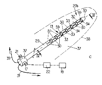
The MRI-compatible ultra-sound device 13 has an
CA 02721271 2010-10-08
WO 2009/125002 PCT/EP2009/054319
external shape which is globally identical to the one of
the mandrel 12, so as to be inserted into the cavity 16 of
the applicator 11. The distal portion 29b of the body 29
of the ultra-sound device comprises a plurality of ultra-
5 sound transducers 30 which are, for example, spaced from
each other both longitudinally and circumferentially,
disposed all around the circumference and the length of the
part of the ultra-sound device which is to be inserted in
the region to be treated. All the transducers 30 are
10 connected as a phased-array device to the computerized
control system 5 through MRI compatible wires 31, which
extend from the back portion 32 of the ultra-sound device
to the computerized control system 5. The transducers 30
can operate as ultra-sound emitters and/or as ultra-sound
receivers. Such micro ultra-sound transducers are known
per se, and will not be described in more details here
(piezoelectric composite technology, capacitive
micromachined ultrasonic transducers technology, etc...).
When they are operated to detect ultra-sounds, the
transducers thus can collect information about the organ.
The back portion 32 comprises an in-flow aperture
17 in fluid communication with a fluid tank 18 preferably
provided outside the patient's body. As shown on Figs 4
and 5, the in-flow aperture 17 is in fluid communication
with a micro circuitry of the body 29 comprising at least
one in-flow channel 19 extending in the thickness of the
body 29, from the back portion 32 to the tip 9 where it is
in fluid communication with at least one out-flow channel
20 which extends from the tip 9 to an out-flow aperture 21
of the back portion 32.
A suitable micro pump such as a pulsed micro-pump
22 is located in the fluid line, so as to generate a flow
of fluid from the fluid tank 18 to the out-flow aperture
21. Such pulsed micro-pumps are known per se and will not
be described in more details here.
CA 02721271 2010-10-08
WO 2009/125002 PCT/EP2009/054319
11
The outer surface of the ultra-sound device 13 can
further comprise other MRI compatible sensors, such as, for
example, electro-physiological signal sensors 33 such as
carbon contact electrodes for detecting electro-
encephalograms, electro-metabolic bio sensors 59,
Other MRI compatible sensors can be provided at the
outer surface of the body 15, such as temperature sensors
34 adapted to locally detect the temperature. The sensors
33, 34 are connected to the computerized system 5 through
suitable MRI compatible wires 35 extending from the back
portion 32 of the ultra-sound device 13, and are thus
operable to collect information about the organ.
Such micro sensors are known per se and will not be
described in more details here.
When the ultra-sound device 13 is inserted through
the cavity 16 of the applicator 11, its distal portion 29b
will extend beyond the distal end of the applicator such
that the ultra-sound transducers 30 are directly coupled to
the tissue of the organ so as to emit and/or receive ultra-
sounds to/from the tissue.
In the embodiment of Fig. 4, the coupling is
performed by way of circulating the cooling fluid in the
micro-circuitry 19, 20. As shown by Fig. 5, other
configurations are possible for the flow of the cooling
fluid such as having the in-flow channel 19 and the out-
flow channel 20 in the centre of the probe.
The transducers 30 can be configured to be operable
in an operating volume 37 around the probe having an
external envelope 38 located at least 20 mm from the probe
(not to scale on Fig. 3c) and preferably at least 30 mm
from the probe. The transducers 30 can be further
configurable to focus ultra-sounds to a focal area in the
operation volume. For example, the transducers 30 are
configurable to focus the ultra-sounds to a focal area
having between 2 (or 3) and 10 mm of diameter. In another
CA 02721271 2010-10-08
WO 2009/125002 PCT/EP2009/054319
12
operative mode, the ultra-sounds are not focused.
The transducers 30 are configurable to operate in
one or a plurality of frequencies, for example chosen in
the range extending from 500 kHz to 10 MHz. Indeed by
modulating the ultrasound emission frequencies, different
physical effects, biological effects and tissue treatment
effects can be produced such as cavitation phenomenons,
tissue fragmentation, sonoporation of the cell membranes
and thermal tissue necrosis. For thermal tissue necrosis,
an embodiment is optimal between 3 MHz to 10 Mhz
frequencies. The transducers 30 are configurable to operate
in a plurality of emission intensities, time durations, and
pulsed or continuous modes. Additionally, they can operate
in phase or out of phase, in a specific setup of
synchronizations.
The transducers 30 are configurable to emit
sufficient energy to raise the temperature by 30 degrees
Celsius, preferably by 60 degrees Celsius, within less than
2 minutes, preferably less than 20 seconds, in the whole
envelope 38 (cylinder of 30 mm around the probe). Possibly,
they can perform this temperature rise within less than 1
second, preferably within less than 100 milliseconds, at a
focal spot of 2 mm of diameter.
Thus, the probe can be set to operate in ultrasound
imaging and/or elastography modes, in an ultrasound
therapeutic mode or a fragmentation mode.
As can be visible from the various embodiments
described in the application, other arrangements and
configurations are possible within the scope of the
invention. For example, a second embodiment is shown on
Figs. 6a to 6c. This second embodiment differs from the
first embodiment described above in relation to Figs. 3a to
5 mainly by the fact that the cooling fluid circuits are
provided within the shell of the applicator 11, as well as
the sensors 33, 34 and 59 and the head 40 of the endo
CA 02721271 2010-10-08
WO 2009/125002 PCT/EP2009/054319
13
confocal microscope. Thus, in this embodiment, the ultra-
sound transducer 30 are coupled to the tissue of the organ
through the body 15 of the applicator 11.
According to a third embodiment, as shown on
figures 7a to 7c, the applicator 11 of the second
embodiment is modified to incorporate the ultra-sound
transducers 30 to form a combined applicator and ultra-
sound device 39. The combined device 39 further comprises
a plurality of openings 40 formed on the lateral face of
the body 15 and in a fluid communication with the internal
cavity 16.
In this third embodiment, the probe can comprise a
rigid mandrel 12 identical to the one of the second
embodiment.
The probe can further comprise a cooling mandrel 41
shaped to be introduced into the internal cavity 16 of the
combined device 39, and comprising a micro circuitry
comprising an in-flow channel 19 in fluid communication
with a fluid tank 18 through an in-flow aperture 17 of the
back portion 42 of the cooling mandrel, and an out-flow
channel 20 in fluid communication with the in-flow channel
19 and exiting from the back portion 42 at an out-flow
aperture 21.
According to a fourth embodiment, as shown on
figures 8a, 8b and 9, the probe comprises only two
components. Indeed, the combined applicator and ultra-
sound device 39 of the third embodiment (figure 7a) is
modified to incorporate the fluid micro circuitry which, in
the third embodiment, is provided through the independent
cooling mandrel 41. Thus, in the fourth embodiment, as
shown on figure 8a, in addition to the features of the
combined device of the third embodiment, an in-flow channel
19 extends from the in-flow aperture 17 provided in the
head 14, whereas the internal cavity 16 itself serves as
the out-flow channel. The in-flow channel 19 is thus in
CA 02721271 2010-10-08
WO 2009/125002 PCT/EP2009/054319
14
fluid communication with the cavity 16 at the insertion tip
9 of the device.
In this fourth embodiment, the probe still
comprises the rigid mandrel 12 of the two previous
embodiments.
A fifth embodiment is partially shown on figure 10.
This fifth embodiment differs from the third or fourth
embodiment in that it has no side openings.
Figure 11a partially shows a sixth embodiment of a
device 39. When compared to the device of the fifth
embodiment, this sixth embodiment differs by a concave
arrangement of the transducers, for example in a central
portion of the device 39. For example, no sensors are
found in this central portion. Thus, instead of being
purely cylindrical, the body 15 has, in this embodiment, a
thinned central portion. According to another embodiment,
as shown on figure llb, the central portion could be
convex.
Such convex or concave shapes could be used for any
of the ultra-sound carrying components of the above
embodiments.
It is contemplated that other geometries could be
used provided the ultra-sound device is still able to
provide therapeutic ultra-sound energy with the required
power.
The ultra-sound waves 51 are schematically
illustrated on these figures.
Figure 12 now describes a schematic representation
of an embodiment of the computerized system 5. This system
could be embodied on one or on a plurality of programmable
machines and comprise both hardware and software
components. The operating system and interfaces of the
computerized system can be of any conventional type, and
will not be described in more details here. The system 5
comprises network corrections, data archiving and storage
CA 02721271 2010-10-08
WO 2009/125002 PCT/EP2009/054319
software and hardware, image manipulation software
including metric and editing functions,
The computerized system 5 can comprise an
echographic and elastographic imaging software 43 of
5 conventional type suitable for obtaining a 2D or a 3D image
based on ultra-sound detection data provided from the probe
6, and resulting from the detection by the probe 6 of
ultra-sound emitted by the probe 6 and reflected by the
organ.
10 The computerized system can further comprise an
EEG-reading software 44 adapted to read data provided from
the probe 6, and detected by the electro-physiological
sensors 33 of the probe.
The computerized system can further comprise a
15 thermal software 47 receiving data from the temperature
sensors 34 of the probe 6 and adapted to determine
temperature data of the tissue based on these received
signals.
The computerized system can further comprise a
confocal microscope image software 48 adapted to receive
data from the confocal microscope head 23 of the probe 6,
and to form an image from this data.
The computerized system 5 can further comprise an
MRI image software 45 receiving data from the MRI system 2
and adapted to reconstruct an MRI anatomy image of the
patient from this data in conventional way.
The computerized system can further comprise a
thermal image software 46 connected to the MRI system 2 and
adapted to treat data provided from the MRI system 2 to
provide the user with a thermal image and with thermal
ablation image of the patient.
The computerized system 5 can further comprise a
planning software 49 adapted to gather information and/or
data about the patient and/or the studied organ and/or
region from the echographic imaging software 43, the EEG
CA 02721271 2010-10-08
WO 2009/125002 PCT/EP2009/054319
16
software 44, the MRI image software 45, the confocal
microscope image software 48, as well as if necessary, any
other patient data, or organ data, obtained by any other
suitable way. The planning software 49 enables the
clinician to evaluate the relevant information, and to
determine the best suit of action for the patient.
The computerized system can further include a
simulation software 60 that can elaborate the optimal
parameter settings for the treatment, based on the various
information available.
For example, the simulation software 60 will use a
model of the probe (including its position and orientation
in the patient's reference frame, for example obtained from
the MRI), a model of the tumor (acoustic impedance and
other relevant parameters), and the geometry of the planned
ablation, to estimate the appropriate settings to use for
each of the transducers.
The computerized system 5 can further comprise a
command software 50 adapted to gather information from the
planning software 49, the MRI thermal image software 46,
the thermal software 47. The command software 50 comprises
a setting device 55 enabling to set the probe 6 in
therapeutic or fragmentation mode (see below), by setting
the parameters of the ultra-sound transducers to emit the
necessary ultra-sound energy, as determined from the
simulation and the planning software 49, toward the
appropriate region (appropriate focal area) of the patient.
The ultra-sound can be set to operate in thermal- and/or
cavity-dominant modes, and the focal area can be
dynamically modified, for example under MRI image guidance,
or ultra-sound imaging.
The command software 50 can also be used to operate
the other mechanical parts of the system such as the pump
22, through a pulsed-pump command 53, the pump 36 through a
continuous pump command 54, and the ultra-sound device in
CA 02721271 2010-10-08
WO 2009/125002 PCT/EP2009/054319
17
imaging mode, through the setting device 55. The command
software 50 is connected to power amplifiers 56 which
deliver the necessary power to the transducers 30. The
switch between the different operation modes of the
transducers could be commanded by a foot switch operated by
the clinician.
In the therapeutic mode, the system can operate in
a step-by-step user-commanded style, or in a computer-
controled automatic style.
Fig. 13 now describes a possible application of the
above described medical system.
At step 101, the applicator 11 comprising the inner
rigid mandrel 12 is percutaneously inserted into the tissue
of the patient's organ, either by hand or under image
monitoring (external echography or MRI). Alternatively, a
stereotaxis frame or a robot arm could be used, for example
for cerebrally inserting the probe.
At step 102, the position of the probe relative to
the organ is monitored. For example, an image of the
patient is obtained from the MRI system, or from an
external CT-scan or echographic system. In alternative or
in addition, the image could be obtained by the probe 6
itself, operating in an "imaging" mode. To do so the
mandrel has to be removed and the ultra-sound device 13
inserted. In this mode, the ultrasound transducers 30 are
commanded, by the command software 50, to operate in an
imaging mode, in which they emit ultra-sounds, for example
within the operation volume 37, and to detect the reflected
ultra-sounds, the obtained detection data being thus sent
to the echographic imaging software 43 of the computerized
system.
At step 103, the inner rigid mandrel 12 is removed
while simultaneously bringing liquid through the channel 27
by the pump 36 to avoid air introduction. The continuous
pump 36 is then directly connected to the internal cavity
CA 02721271 2010-10-08
WO 2009/125002 PCT/EP2009/054319
18
16, so as to perform a biopsy by aspiring through the
opening(s) 40, a part of the organ. Alternatively, a
syringe head or a biopsy needle could be used. This part
can be further analyzed to confirm the characteristics of
the tissue. In addition or in alternative, tissue
characterization could be performed by disconnecting the
pump 36 and inserting the microscope optical fiber through
the cavity 16 (or 27), so as to use the endo-confocal
microscope, the data of which is provided to the confocal
microscope image software 48 of the computerized system.
At step 104, data from the electro-physiologic
sensors 33 is obtained and is forwarded to the electro
encephalogram reading software 44 of the computerized
system 5.
Using the data obtained at steps 102, 103, and/or
104, at step 105, the target volume and the probe position
in reference to the targeted volume are defined, for
example by the clinician using the planning software 49.
The target volume can further be defined manually or
automatically, using external images, such as MRI images or
the like.
Based on the location and size of the target
volume, at step 105', the therapeutic ultra-sound energy,
phases, durations for the various ultrasound transducers
are simulated and calculated. At step 106, the ultra-
sound emission parameters are set, for example using the
command software 50. The size and the location of the
focal area, the ultra-sound power and frequency are set in
the command software 50. Low frequency ultra-sound waves
can be used for applying therapeutic ultra-sound energy to
regions remote from the probe 6, whereas higher frequencies
can be used for close regions. The probe 6 is thus set to
operate in a thermal "therapeutic" treatment mode, either
by non destructive (metabolism stimulation, ...) or
destructive (coagulation, vaporization) mode.
CA 02721271 2010-10-08
WO 2009/125002 PCT/EP2009/054319
19
At step 107, the ultra-sounds are delivered to the
region 10 of the organ 8. As shown on Fig. 14, the probe 6
could be operated in de-focused mode inside the envelope
38. It can also be operated in a focused mode active on
specific focal areas 58. Simultaneously, cooling
physiological fluid is pumped into the micro-circuitry of
the probe by the pump 22, under command of the command
software 50, so as to efficiently cool the probe. Real-
time thermal MRI images are provided to the thermal image
software 46 of the computerized system for monitoring the
temperature rise in the probe and/or in the organ and/or
security points for the treatment. Necrosis prediction
could be performed by the computerized system 5 by summing
in time the obtained thermal data, to obtain thermal
deposited doses within the specific volume. The reflected
ultra-sounds are detected by the transducers 30, and real-
time images are formed at the computerized system 5, which
enables a real-time ultra-sound monitoring.
At step 108, the efficiency of the therapy ultra-
sound application is monitored, for example from the
detected MRI image, or by ultra-sound imaging (echography,
elastography). If the therapeutic treatment is not judged
sufficient by the clinician, the process continues back
from step 105. However, if the clinician finds out that a
sufficient thermal treatment was performed, the process
moves to step 109.
At step 109, the treated region 10 is mechanically
fragmented by ultra-sounds. The probe 6 is set to operate
in a "fragmentation" mode by emission of pulsed ultra-sound
to the treated region of the organ. The "fragmentation"
mode also is a therapeutic mode. This results in a
fragmentation (shearing) of the region, by breaking the
inter-cellular adhesions using the jet-steam / cavitation
technique. Of course, the ultra-sound parameters, in this
mode, can be set from the command software 50. In this
CA 02721271 2010-10-08
WO 2009/125002 PCT/EP2009/054319
mode, the probe is continuously cooled by the pump 22. In
another method, the fragmentation of the tissue can be
performed by low frequency vibration (around 10 kHz [1-50
kHz]) emitted by the electromechanical mandrel back portion
5 26 through the mandrel body (if reintroduced) to transmit
low frequency vibration within the treated tissue, or
alternatively by yet another specific mechanical-stress
inducing mandrel.
At step 110, the efficiency of the fragmentation
10 step 109 is monitored. If this step is not judged
sufficiently efficient by the clinician, the process moves
back to step 109 whereas, if it is judged to be
sufficiently efficient, the process moves to step 111 where
the fragmented tissue is aspirated outside the body.
15 At step 111, the optical fiber of the endo-confocal
microscope is removed, the pump 36 connected to the cavity
16 performs a soft aspiration, at controlled negative
pressure, commanded by the command software 50 of the
computerized system, so as to allow the decrease in volume
20 of the treated region.
At step 112, it is monitored whether the treatment
can be judged satisfactory by the clinician. If this is
not the case, the process moves back to step 111. If it is
decided to end the procedure, the probe 6 is removed from
the organ and the process is ended.
The above description is just an exemplary
description of one possible embodiment of the above
described probe and system and it is within the reach of
the person skilled in the art to repeat, bypass, or change
the order of the above steps, or to add additional steps,
dependent of the pathology to be treated. Thus, the probe
could allow the treatment of the tissue lesions by the
following modes and physical agents: thermal blood
coagulation, thermal reversible ischemia, non-thermal
mechanical jet steam and cavitation means, sonoporation or
CA 02721271 2010-10-08
WO 2009/125002 PCT/EP2009/054319
21
combinations thereof.
Furthermore, the interstitial probe could be used
in a non-interstitial way, by being placed in close
proximity, but outside an organ to be treated. The probe
could be disposable, or be sterilized between subsequent
uses.
Furthermore, in another embodiment, the probe could
be permanently installed in the organ of the patient, and
be remote-controlled by a suitable computer, for example
implanted inside the patient, at periodical examinations.
Although the above embodiments show a probe having
numerous features, it should be noted that not all of these
features necessarily need to be part of the inventive
probe. For example, the information collection device
could be comprised of only one or more of the ultra-sound
transducers 30 in imaging mode, the electro physiological
sensors 33, the temperature sensors 34, and the endo-
confocal microscope head 40.