Note: Descriptions are shown in the official language in which they were submitted.
CA 02722002 2010-10-20
. -
COMPOSITE LACRIMAL INSERT AND RELATED METHODS
TECHNICAL FIELD
This patent document pertains generally to ophthalmic devices, and
particularly to ocular implants. More particularly, but not by way of
limitation, this
patent document pertains to lacrimal implants, methods of making such
implants,
and methods of treating ocular, respiration or other diseases or disorders
using such
implants.
BACKGROUND
A variety of challenges face patients and physicians in the area of ocular and
respiration disease or disorder management, including adequate drug delivery
to the
eyes or nasal passage and treatment of dry eyes. In ocular management, for
example, many current ocular drug delivery systems require repetitive manual
drug
administration and are often ineffective due to a lack of patient compliance
or
inadequate drug concentrations reaching the eye. Many current tear flow
blockage
techniques also have drawbacks, including being irreversible in nature.
In order to eye treat infection, inflammation of the eye, glaucoma and other
ocular diseases or disorders, drugs are often required to be administered to
the eye.
A conventional method of drug delivery is by topical drop application to the
eye's
surface. Topical eye drops, though effective, can be inefficient. As one
example,
when an eye drop is instilled in an eye, it often overfills the conjunctival
sac (i.e.,
the pocket between the eye and the lids) causing a substantial portion of the
drop to
be lost due to overflow of the lid margin and spillage onto the cheek. In
addition, a
large portion of the drop remaining on the ocular surface can be washed away
into
and through a lacrimal canaliculus, thereby diluting the concentration of the
drug
before it can treat the eye. Moreover, topically applied drugs often have a
peak
ocular effect for about two hours post-application, after which additional
applications of the drugs should be, but are often not, administered to
maintain the
desired drug therapeutic benefit.
1
- ' CA 02722002 2010-10-20
To compound ocular management difficulty, patients often do not use their
eye drops as prescribed. This poor compliance can be due to, for example, an
initial
stinging or burning sensation caused by the eye drop and experience by a
patient.
Instilling eye drops in one's own eye can be difficult, in part because of the
normal
reflex to protect the eye. Therefore, one or more drops may miss the eye.
Older
patients may have additional problems instilling drops due to arthritis,
unsteadiness,
and decreased vision. Pediatric and psychiatric populations pose difficulties
as well.
Conditions of dry eye have been treated by blocking the tear flow from the
eye into and through the lacrimal canaliculus. This has involved closing the
canalicular canal by stitching the punctal opening shut or by using electrical
or laser
cauterization to seal the punctal opening. Although such procedures can
provide the
desired result of blocking tear flow to treat a dry eye, they are
unfortunately not
reversible without reconstructive surgery.
In a field different from ocular management, control of respiration-related
(e.g., allergies) diseases or disorders often requires repetitive manual
digestion or
other intake of a medication, and as such, can be ineffective due to a lack of
patient
compliance or non-localized drug delivery.
EXEMPLARY ASPECTS, EXAMPLES, AND EMBODIMENTS OF THE
INVENITON
The present inventors have recognized, among other things, one promising
approach of drug delivery to an eye or nasal passage system, for example, can
be to
place a removable, drug-releasing lacrimal implant into a lacrimal punctum. It
is
believed that by allowing for the sustained release of one or more drugs, the
present
lacrimal implants can overcome some of the drawbacks associated with current
drug
administration (i.e., manual drop instillation or digestion), such as poor
patient
compliance, waste, untimely application, or non-localized delivery. One
promising
approach to successful blocking of tear flow from the eye is to place a
removable,
but retainable, lacrimal implant into the lacrimal punctum. The present
inventors
have further recognized, among other things, the lacrimal implant can benefit
from
one or more of the ability to be easily implanted and removed via controlled
biasing
2
- CA 02722002 2010-10-20
of the lacrimal punctum or canaliculus, the ability to be securely and
comfortably
retainable in the lacrimal punctum upon implantation, and, when made and used
as a
drug delivery system, the ability to allow for the sustained, localized
release of one
or more drugs at a desired therapeutic level for an extended period of time.
Lacrimal implants for treating diseases or disorders are disclosed. Methods
of making such implants, and methods of treating ocular or respiration
diseases or
disorders using such implants are also disclosed.
In Example 1, a lacrimal implant comprises an implant body, including first
and second portions, sized and shaped for at least partial insertion into a
lacrimal
canaliculus, the first portion including a first biocompatible polymer
configured to
swell less than 100 wt% when in contact with an aqueous medium, the second
portion including a second biocompatible polymer configured to swell greater
than
100 wt% when in contact with an aqueous medium; wherein the first and second
biocompatible polymers adhere at a junction between the first portion and the
second portion.
In Example 2, the lacrimal implant of Example 1 is optionally configured
such that the first biocompatible polymer includes a urethane-based material.
In Example 3, the lacrimal implant of at least one of Examples 1 or 2 is
optionally configured such that the first biocompatible polymer includes a
polyurethane polymer or copolymer.
In Example 4, the lacrimal implant of at least one of Examples 1-3 is
optionally configured such that the second biocompatible polymer includes a
urethane-based material.
In Example 5, the lacrimal implant of at least one of Examples 1-4 is
optionally configured such that the second biocompatible polymer includes a
hydrogel-forming polyurethane polymer or copolymer.
In Example 6, the lacrimal implant of at least one of Examples 1-5 is
optionally configured such that the second biocompatible polymer includes a
hydrogel-forming polyurethane polymer or copolymer, and wherein the polymer or
copolymer can swell from about 100 wt% to about 200 wt% when contacted with an
aqueous medium.
3
- ' CA 02722002 2010-10-20
In Example 7, the lacrimal implant of at least one of Examples 1-6 is
optionally configured such that the second biocompatible polymer includes a
polyurethane hydrogel adapted to swell 500-2000 wt% upon exposure to an
aqueous
medium.
In Example 8, the lacrimal implant of at least one of Examples 1-7 is
optionally configured such that the first polymer, the second polymer, or
both,
includes a polyurethane-silicone copolymer, a polyurethane-carbonate
copolymer,
an aliphatic polyurethane, an aromatic polyurethane, or any combination
thereof.
In Example 9, the lacrimal implant of at least one of Examples 1-8
optionally comprises an exterior coating or sheath, the coating or sheath
being
configured to expand when the second polymer comes into contact with the
aqueous
medium and swells thereby.
In Example 10, the lacrimal implant of at least one of Examples 1-9 is
optionally configured such that the second portion is disposed as a coating on
a first
part of an external surface of the first portion, and wherein a second part of
the
external surface of the first portion is disposed adjacent to the proximal end
and is
uncoated.
In Example 11, the lacrimal implant of at least one of Examples 1-10 is
optionally configured such that the junction comprises an intermediate member
including a third biocompatible polymer, the third biocompatible polymer
configured to adhere to both the first biocompatible polymer and the second
biocompatible polymer, and configured to swell upon contact with an aqueous
medium to a greater degree than the first polymer but to a lesser degree than
the
second polymer.
In Example 12, the lacrimal implant of Example 11 is optionally configured
such that the third polymer includes a polyurethane polymer or copolymer, a
polyurethane-silicone copolymer, a polyurethane-carbonate copolymer, an
aliphatic
polyurethane, an aromatic polyurethane, or any combination thereof.
In Example 13, the lacrimal implant of Example 12 is optionally configured
such that the third polymer is configured to absorb about 50% to about 200%
water.
4
. CA 02722002 2010-10-20
In Example 14, the lacrimal implant of at least one of Examples 11-13 is
optionally configured such that the strength of an adhesion between the first
biocompatible polymer and the third biocompatible polymer, or between the
second
biocompatible polymer and the third biocompatible polymer, or both, is
stronger
than a strength of adhesion between the first and second biocompatible
polymers.
In Example 15, the lacrimal implant of at least one of Examples 1-14 is
optionally configured such that the first portion comprises a base member
extending
from a proximal end, configured to sit at or near a lacrimal punctum when
implanted and including a first diameter, to a distal end portion, configured
for
insertion through the lacrimal punctum into the lacrimal canaliculus when
implanted
and having a second diameter less than the first diameter.
In Example 16, the lacrimal implant of Example 15 is optionally configured
such that a shape of the base member is configured to provide sufficient
surface area
for adhesion of the first biocompatible polymer to the second biocompatible
polymer, such that the first portion and the second portion do not separate at
the
junction when the implant body is withdrawn under tension from the lacrimal
canaliculus and the second portion has swelled from contact with the aqueous
medium.
In Example 17, the lacrimal implant of Example 16 is optionally configured
such that a surface of the first portion is chemically modified or is treated
with
ionizing radiation or electron beam radiation to bond with the second portion
to
resist separation under tension.
In Example 18, the lacrimal implant of at least one of Examples 15-17 is
optionally configured such that the base member includes one or more arm
members
protruding from an outer surface thereof.
In Example 19, the lacrimal implant of Examples 18 is optionally configured
such that at least one of the one or more arm members protrude laterally
relative to a
longitudinal axis of the base member.
In Example 20, the lacrimal implant of at least one of Examples 18 or 19 is
optionally configured such that the one or more arm members comprise a cross-
sectional size greater than an adjacent portion of the base member.
- CA 02722002 2010-10-20
-
In Example 21, the lacrimal implant of at least one of Examples 15-20 is
optionally configured such that the base member includes one or more voids
sized
to receive a portion of the second portion upon coupling.
In Example 22, the lacrimal implant of at least one of Examples 15-21 is
optionally configured such that the second portion comprises an expandable
retention member coupled at least partially over the base member, the
expandable
retention member configured to swell via absorption of an aqueous medium after
insertion into the lacrimal canaliculus.
In Example 23, the lacrimal implant of Example 22 is optionally configured
such that the expandable retention member substantially envelops the base
member.
In Example 24, the lacrimal implant of at least one of Examples 22 or 23 is
optionally configured such that the expandable retention member includes a gel
configured to at least partially conform to a size and shape of the lacrimal
canaliculus.
In Example 25, the lacrimal implant of at least one of Examples 22-24 is
optionally configured such that the base member includes a longitudinal axis,
and
wherein the expandable retention member includes at least one longitudinal
swelling
axis extending laterally relative to the longitudinal axis of the base member.
In Example 26, the lacrimal implant of at least one of Examples 22-25 is
optionally configured such that the expandable retention member is configured
to at
least partially extend toward a horizontal section of the lacrimal
canaliculus.
In Example 27, the lacrimal implant of at least one of Examples 22-26 is
optionally configured such that the expandable retention member is configured
to at
least partially extend toward an ampulla of a lacrimal canaliculus.
In Example 28, the lacrimal implant of at least one of Examples 1-27
optionally comprises a first supply of a first active agent included in the
first
portion, the first supply configured to provide a release of the first active
agent to an
eye.
In Example 29, the lacrimal implant of Example 28 is optionally configured
such that the first portion includes a cavity extending inward from a proximal
end of
6
= CA 02722002 2010-10-20
the first portion, the cavity comprising the first supply to provide the
release of the
first active agent to the eye.
In Example 30, the lacrimal implant of at least one of Examples 28 or 29 is
optionally configured such that the first supply includes a solid matrix
comprising a
mixture of silicone and the first active agent.
In Example 31, the lacrimal implant of at least one of Examples 28-30 is
optionally configured such that the first supply is dispersed within the first
polymer.
In Example 32, the lacrimal implant of at least one of Examples 28-31 is
optionally configured such that the first supply includes at least one exposed
surface
near a proximal end of the first portion to provide release of the first
active agent to
the eye.
In Example 33, the lacrimal implant of Example 32 is optionally configured
such that the at least one exposed surface is positioned above the proximal
end of
the first portion such that the first supply at least partially protrudes
outside of the
implant body.
In Example 34, the lacrimal implant of at least one of Examples 28-33
optionally comprises a second supply of a second active agent included in the
second portion, the second supply configured to provide a release of the
second
active agent to one or both of a lacrimal canaliculus wall or a nasolacrimal
system
after contact with an aqueous medium.
In Example 35, the lacrimal implant of at least one of Examples 28-34 is
optionally configured such that the first or second active agent comprises an
anti-.
glaucoma medicament, an antimicrobial agent, a corticosteroid or other anti-
inflammatory, a decongestant, an agent that prevents of modifies an allergic
response, a mast cell stabilizer, a cycloplegic, a mydriatic, or a combination
thereof.
In Example 36, the lacrimal implant of Example 35 is optionally configured
such that the anti-glaucoma medicament comprises an adrenergic agonist, an
adrenergic antagonists, a systemic or topical carbonic anhydrase inhibitor, a
prostaglandin or hypotensive lipid, or a combination thereof.
7
- CA 02722002 2010-10-20
_ -
In Example 37, the lacrimal implant of Example 36 is optionally configured
such that the prostaglandin comprises bimatoprost, travoprost, or latanoprost,
or a
combination thereof.
In Example 38, the lacrimal implant of Example 35 is optionally configured
such that the antimicrobial agent comprises an antibiotic, tetracycline,
chlortetracycline, bacitracin, neomycin, polymyxin, gramicidin, cephalexin,
oxytetracycline, chloramphenicol, rifampicin, ciprofloxacin, tobramycin,
gentamycin, erythromycin, penicillin, sulfonamides, sulfadiazine,
sulfacetamide,
sulfamethizole, sulfisoxazole, nitrofurazone, sodium propionate, an
antifungal,
amphotericin B, miconazole, an antiviral, idoxuridine trifluorothymidine,
acyclovir,
gancyclovir, or interferon, or a combination thereof.
In Example 39, the lacrimal implant of Example 36 is optionally configured
such that the corticosteroid or other anti-inflammatory comprises
cyclosporine,
hydrocortisone, hydrocortisone acetate, dexamethasone, dexamethasone 21-
phosphate, fluocinolone, medrysone, methylprednisolone, prednisolone,
prednisolone 21-phosphate, prednisolone acetate, fluoromethalone,
betamethasone,
triamcinolone, triamcinolone acetonide, cortisone, flumetholone, a non
steroidal
anti-inflammatory, salicylate, indomethacin, ibuprofen, diclofenac,
flurbiprofen,
piroxicam indomethacin, ibuprofen, naxopren, piroxicam, nabumetone, ketorolac,
bromfenac, nepafenac or suprofen, or combinations thereof.
In Example 40, the lacrimal implant of Example 36 is optionally configured
such that the decongestant comprises a vasoconstrictor, phenylephrine,
naphazoline,
or tetrahydrazoline, or a combination thererof.
In Example 41, the lacrimal implant of Example 36 is optionally configured
such that the agent that prevents or modifies an allergic response comprises
an
antihistamine, a cytokine inhibitor, a leucotriene inhibitor, an IgE
inhibitor, an
immunomodulator, cyclosporine, or a combination thereof.
In Example 42, the lacrimal implant of at least one of Examples 1-41 is
optionally configured such that an intermediate section of the first portion
includes a
coupling void and a coupling arm sized and shaped to lock within the coupling
void.
8
CA 02722002 2010-10-20
. -
In Example 43, the lacrimal implant of at least one of Examples 1-42 is
optionally configured such that the implant body is non-biodegradable when
implanted within a human subject.
In Example 44, the lacrimal implant of at least one of Examples 1-42
optionally comprises a lateral projection affixed to a proximal end of the
first
portion, the lateral projection having a size and shape configured to rest on
an
exterior of the lacrimal punctum when a distal end of the first portion is
positioned
within a lacrimal canaliculus.
In Example 45, the lacrimal implant of at least one of Examples 1-44 is
optionally configured such that the implant body in physical shape comprises a
substantially cylindrical or conical region.
In Example 46, a kit comprises the lacrimal implant of at least one of
Examples 1-45, and an instruction for using the lacrimal implant to treat an
eye
disease.
In Example 47, a method of making the lacrimal implant of at least one of
Examples 1-45 comprises use of a melt of the polyurethane polymer or
copolymer.
In Example 48, a method of making the lacrimal implant of at least one of
Examples 1-45 comprises injection molding the first portion, the second
portion, or
both, using respectively a melt of the first polymer, the second polymer, or
melts of
both polymers.
In Example 49, a method of making the lacrimal implant of at least one of
Examples 1-45 comprises injection molding the first portion, the second
portion, or
both, using respectively a melt of the first polymer, the second polymer, or
melts of
both polymers, wherein an intermediate member is disposed as a melt between
the
first portion and the second portion.
In Example 50, a lacrimal implant comprises a unitary implant body sized to
at least partially pass through a lacrimal punctum and be at least partially
positioned
within a lacrimal canaliculus, the implant body extending from a proximal end
portion to a distal end portion, the proximal end portion including a cavity,
the distal
end portion including a base member having a diameter less than a diameter of
the
proximal end portion; a drug supply included in the cavity, the drug supply
9
CA 02722002 2010-10-20
, .
configured to provide a release of a first active agent to an eye; and an
expandable
retention member coupled at least partially over the base member, the
expandable
retention member including a dehydrated material hydratable an aqueous medium
to
swell from a first diameter to a second diameter greater than the first
diameter.
In Example 51, the lacrimal implant of Example 50 is optionally configured
such that the expandable retention member fully envelopes the base member.
In Example 52, the lacrimal implant of at least one of Examples 50 or 51 is
optionally configured such that the implant body includes a urethane-based
material.
In Example 53, the lacrimal implant of at least one of Examples 50-52
optionally comprises a second active agent included in the expandable
retention
member, the expandable retention member configured to provide a release of the
second active agent after contact with the aqueous medium.
In Example 54, the lacrimal implant of at least one of Examples 50-53 is
optionally configured such that the base member includes one or more arm
members
laterally protruding from an outer surface thereof.
In Example 55, the lacrimal implant of at least one of Examples 50-54 is
optionally configured such that the expandable retention member includes a
urethane-based hydrogel material.
In Example 56, the lacrimal implant of at least one of Examples 50-55 is
optionally configured such that the second diameter of the expandable
retention
member is configured to be at least about 5 times greater than the first
diameter of
the expandable retention member.
In Example 57, the lacrimal implant of at least one of Examples 50-56 is
optionally configured such that the length of the base member is at least
about one-
third the total length of the implant body.
In Example 58, the lacrimal implant of at least one of Examples 50-57 is
optionally configured such that the length of the base member is at least
about one-
half the total length of the implant body.
In Example 59, a kit comprises the lacrimal implant of any of claims 50-58,
and an instruction for using the lacrimal implant to treat an eye disease.
- CA 02722002 2010-10-20
In Example 60, a method comprises forming an implant body, including first
and second portions, sized for at least partial insertion into a lacrimal
canaliculus,
including forming the first portion from a first biocompatible polymer
configured to
swell less than 100 wt% when in contact with an aqueous medium, and forming
the
second portion from a second biocompatible polymer configured to swell greater
than 100 wt% when in contact with an aqueous medium; and coupling the first
and
second biocompatible polymers at a junction between the first portion and the
second portion.
In Example 61, the method of Example 60 optionally comprises disposing an
intermediate member including a third biocompatible polymer between the first
and
second portions, the third biocompatible polymer configured to adhere to the
first
biocompatible polymer on a first surface and adhere to the second
biocompatible
polymer on a second surface.
In Example 62, the method of at least one of Examples 60 or 61 is optionally
configured such that forming the first portion includes forming a base member
extending from a proximal end, configured to sit at or near a lacrimal punctum
when
implanted, to a distal end portion, configured for insertion through the
lacrimal
punctum into the lacrimal canaliculus when implanted, including forming one or
more arm members protruding from an outer surface of the base member.
In Example 63, the method of Example 62 is optionally configured such that
adhering the first and second biocompatible polymers includes coupling an
expandable retention member at least partially over the base member such that
an
outer surface of the expandable retention member can absorb the aqueous medium
after insertion into the lacrimal canaliculus.
In Example 64, the method of Example 63 is optionally configured such that
coupling the expandable retention member at least partially over the base
member
includes fully surrounding the base member with the expandable retention
member.
In Example 65, the method of at least one of Examples 60-64 optionally
comprises providing a first supply of a first active agent in the first
portion, the first
supply configured 10 provide a release of the first active agent to an eye.
11
- = CA 02722002 2010-10-20
_ .
In Example 66, the method of at least one of Examples 60-65 is optionally
configured such that forming the first and second portions from a polymer
includes
forming a unitary implant body from a urethane-based material.
In Example 67, the method of at least one of Examples 63-66 optionally
comprises disposing an intermediate member between an outer surface of the
base
member and an inner surface of the expandable retention member, the
intermediate
member configured to absorb a greater amount of fluid than the polymer of the
second portion but less fluid than a swellable polymer of the expandable
retention
member.
In Example 68, the method of at least one of Examples 60-67 is optionally
configured such that forming the implant body includes forming a cavity
extending
inward from a proximal end of the first portion, including configuring the
cavity to
include a drug supply and positioning at least one exposed surface of the drug
supply near the proximal end.
In Example 69, the method of Example 68 is optionally configured such that
positioning the at least one exposed surface of the drug supply near the
proximal
end includes positioning the at least one exposed surface above the proximal
end
such that the first supply at least partially protrudes outside of the implant
body.
In Example 70, the method of at least one of Examples 63-69 is optionally
configured such that forming the second portion including the base member
includes forming one or more arm members protruding laterally from an outer
surface of the base member, to thereby increase the surface area for coupling
of the
expandable retention member.
In Example 71, the method of at least one of Examples 63-70 is optionally
configured such that coupling the expandable retention member includes molding
a
urethane-based hydrogel over the base member.
In Example 72, the method of at least one of Examples 63-72 is optionally
configured such that coupling the expandable retention member includes dip
coating
the base member with a urethane-based hydrogel.
12
CA 02722002 2010-10-20
In Example 73, the method of at least one of Example 63-72 is optionally
configured such that coupling the expandable retention member includes
disposing a
hydro gel sleeve over an outer surface of the base member.
In Example 74, a method of treating a subject having an eye disorder
comprises inserting a lacrimal implant into at least one lacrimal canaliculus,
the
lacrimal implant comprising, an implant body, including first and second
portions,
sized and shaped for at least partial insertion into a lacrimal canaliculus,
the first
portion including a first biocompatible polymer configured to swell less than
100
wt% when in contact with an aqueous medium, the second portion including a
second biocompatible polymer configured to swell greater than 100 wt% when in
contact with an aqueous medium; wherein the first and second biocompatible
polymers adhere at a junction between the first portion and the second
portion.
In Example 75, the method of Example 74 is optionally configured such that
inserting the lacrimal implant includes partially inserting the implant body
through a
lacrimal punctum until a removal projection extending laterally from the
proximal
end of the first portion is positioned outside and adjacent to the lacrimal
punctum.
In Example 76, the method of at least one of Examples 74 or 75 is optionally
configured such that inserting the lacrimal implant includes positioning a
supply of
an active agent, included in the first portion, adjacent an eye of the
subject.
In Example 77, the method of Example 76 is optionally configured such that
the active agent is configured to treat at least one of a glaucoma disease or
a
seasonal allergy.
In Example 78, the method of at least one of Examples 76 or 77 is optionally
configured such that a period of time over which the active agent is released
includes at least one week, at least one month, or at least three months.
In Example 79, the method of at least one of Examples 74-78 optionally
comprises removing the inserted implant body from the lacrimal punctum.
In Example 80, the method of Example 79 optionally comprises replacing
the lacrimal implant that has been removed with a second lacrimal implant
including
a supply of an active agent following an interval of time.
13
CA 02722002 2010-10-20
In Example 81, the method of Example 80 is optionally configured such that
replacing the lacrimal implant is repeated until the subject no longer
requires
treatment.
In Example 82, a method of administering a medicament to the eye, or to
surrounding tissue, or both, of a patient having a malcondition for treatment
of
which administration of the medicament is medically indicated comprises
inserting
the lacrimal implant of any of claims 1 through 45, or a lacrimal implant made
by
the method of any of claims 47 through 49, wherein the implant comprises the
medicament dispersed therein, into a punctal canal of a patient in need
thereof, such
that the implant is placed in contact with an aqueous medium and the implant
undergoes swelling to secure the implant within the punctal canal, and such
that the
medicament is released into the eye or surrounding tissues, or both, from the
implant over a period of time.
In Example 83, a method of administering a medicament to the eye, or to
surrounding tissue, or both, of a patient having a malcondition for treatment
of
which administration of the medicament is medically indicated comprises
inserting
the lacrimal implant of any of claims 1 through 45, or a lacrimal implant made
by
the method of any of claims 47 through 49, wherein the implant comprises a
drug-
releasing insert with the medicament dispersed therein, into a punctal canal
of a
patient in need thereof, such that the implant is placed in contact with an
aqueous
medium and the implant undergoes swelling to secure the implant within the
punctal
canal, and such that the medicament is released into the eye or surrounding
tissues,
or both, from the drug-releasing insert over a period of time.
In Example 84, a method of administering a medicament to the eye, or to
surrounding tissue, or both, of a patient having a malcondition for treatment
of
which administration of the medicament is medically indicated comprises
inserting
the lacrimal implant of any of claims 1 through 45, or a lacrimal implant made
by
the method of any of claims 47 through 49, wherein the first portion comprises
an
active substance dispersed therein, into a punctal canal of a patient in need
thereof,
such that the second polymer is placed in contact with an aqueous medium and
the
second polymer undergoes swelling to secure the implant within the punctal
canal,
14
e. CA 02722002 2010-10-20
-
and such that the active agent is released into the eye or surrounding
tissues, or both,
from the first polymer over a period of time.
In Example 85, a method of administering a medicament to the eye, or to
surrounding tissue, or both, of a patient having a malcondition for treatment
of
which administration of the medicament is medically indicated comprises
inserting
the lacrimal implant of any of claims 1 through 45, or a lacrimal implant made
by
the method of any of claims 47 through 49, wherein the first portion comprises
a
drug-releasing insert with an active substance dispersed therein, into a
punctal canal
of a patient in need thereof, such that the second polymer is placed in
contact with
an aqueous medium and the second polymer undergoes swelling to secure the
implant within the punctal canal, and such that the active agent is released
into the
eye or surrounding tissues, or both, from the drug-releasing insert over a
period of
time.
In Example 86, the method of at least one of Examples 82-85 is optionally
configured such that the period of time is about 1 week to about 6 months.
In Example 87, the method of at least one of Examples 82-86 is optionally
configured such that the medicament comprises an anti-glaucoma medicament, an
antimicrobial agent, a corticosteroid or other anti-inflammatory, a
decongestant, an
agent that prevents of modifies an allergic response, a mast cell stabilizer,
a
cycloplegic, a mydriatic, or a combination thereof.
In Example 88, the method of Example 87 is optionally configured such that
the anti-glaucoma medicament comprises an adrenergic agonist, an adrenergic
antagonists, a systemic or topical carbonic anhydrase inhibitor, a
prostaglandin or
hypotensive lipid, or a combination thereof.
In Example 89, the method of Example 88 is optionally configured such that
the prostaglandin comprises bimatoprost, travoprost, or latanoprost, or a
combination thereof.
In Example 90, the method of at least one of Examples 82-89 is optionally
configured such that the medicament is indicated for treating high eye
pressure/intraocular pressure in people with open-angle glaucoma or ocular
hypertension.
CA 02722002 2010-10-20
In Example 91, the method of Example 87 is optionally configured such that
the antimicrobial agent comprises an antibiotic, tetracycline,
chlortetracycline,
bacitracin, neomycin, polymyxin, gramicidin, cephalexin, oxytetracycline,
chloramphenicol, rifampicin, ciprofloxacin, tobramycin, gentamycin,
erythromycin,
penicillin, sulfonamides, sulfadiazine, sulfacetamide, sulfamethizole,
sulfisoxazole,
nitrofurazone, sodium propionate, an antifungal, amphotericin B, miconazole,
an
antiviral, idoxuridine trifluorothymidine, acyclovir, gancyclovir, or
interferon, or a
combination thereof
In Example 92, the method of Example 87 is optionally configured such that
the corticosteroid or other anti-inflammatory comprises cyclosporine,
hydrocortisone, hydrocortisone acetate, dexamethasone, dexamethasone 21-
phosphate, fluocinolone, medrysone, methylprednisolone, prednisolone,
prednisolone 21-phosphate, prednisolone acetate, fluoromethalone,
betamethasone,
triamcinolone, triamcinolone acetonide, cortisone, flumetholone, a non
steroidal
anti-inflammatory, salicylate, indomethacin, ibuprofen, diclofenac,
flurbiprofen,
piroxicam indomethacin, ibuprofen, naxopren, piroxicam, nabumetone, ketorolac,
bromfenac, nepafenac, or suprofen or combinations thereof
In Example 93, the method of Example 87 is optionally configured such that
the decongestant comprises a vasoconstrictor, phenylephrine, naphazoline, or
tetrahydrazoline, or a combination thererof.
In Example 94, the method of Example 87 is optionally configured such that
the agent that prevents of modifies an allergic response comprises an
antihistamine,
a cytokine inhibitor, a leucotriene inhibitor, an IgE inhibitor, an
immunomodulator,
cyclosporine, or a combination thereof.
These and other examples, advantages, and features of the present lacrimal
implants and methods will be set forth in part in following Detailed
Description.
This Summary is intended to provide an overview of the subject matter of the
present patent document. It is not intended to provide an exclusive or
exhaustive
explanation of the present subject matter. The Detailed Description is
included to
provide further information about the present patent document.
16
CA 02722002 2010-10-20
BRIEF DESCRIPTION OF THE DRAWINGS
In the drawings, like numerals have been used to describe similar
components throughout the several views. Like numerals having different letter
suffixes have been used to represent different instances of similar
components. The
drawings illustrate generally, by way of example, but not by way of
limitation,
various embodiments discussed in the present document.
FIGS. 1-2 illustrate examples of schematic views of anatomical tissue
structures associated with the eye, such tissue structures
providing a suitable environment in which the present
lacrimal implants can be used.
FIG. 3 illustrates an example of a present lacrimal implant showing
a
"half and half' implant design.
FIG. 4A illustrates an example of an isometric view of a present
lacrimal implant, which is configured to be retained within a
lacrimal punctum and canalicular anatomy.
FIG. 4B illustrates an example of a cross-sectional view of a
present
lacrimal implant taken along a line parallel to a longitudinal
axis of the implant, such as along line 3B-3B of FIG. 3A.
FIG. 4C illustrates another example of a cross-section view of a
present lacrimal implant taken along a line parallel to a
longitudinal axis of the implant.
FIG. 5 illustrates an example of an assembly of a present lacrimal
implant, which is configured to be retained within a lacrimal
punctum and canalicular anatomy.
FIG. 6 illustrates an example of a schematic view of a present
lacrimal implant, which is retained within a lacrimal punctum
and canalicular anatomy.
FIGS. 7A-7G illustrate examples of cross-sectional views of other present
lacrimal implants, each of which is configured to be retained
within a lacrimal punctum and canalicular anatomy.
17
CA 02722002 2010-10-20
FIG. 8 illustrates an example of a method of manufacturing a
present
lacrimal implant, which is configured to be retained within a
lacrimal punctum and canalicular anatomy.
FIGS. 9-12 illustrate and chart example experimental results of a
present
lacrimal implant, which is configured to be retained within a
lacrimal punctum and canalicular anatomy.
DETAILED DESCRIPTION
In this patent document, biocompatible lacrimal implants and related
methods providing secure, biasing retention within a lacrimal punctum of an
eye are
described. The lacrimal implants can comprise an implant body configured for
at
least partial insertion through the lacrimal punctum and into an associated
lacrimal
canaliculus. The implant body can include first and second portions, in which
the
first portion is formed from a polymer and includes a first diameter and the
second
portion is also formed from a polymer and includes a base member having a
second
diameter. In various examples, the second diameter of the base member is less
than
the first diameter of the first body portion. An expandable retention member
is
coupled at least partially over the base member and is configured to swell via
absorption of lacrimal fluid after insertion into the lacrimal punctum. In
this way, at
least a portion of the expandable retention member can be biased against at
least a
portion of a lacrimal canaliculus wall to retain an implant position of the
lacrimal
implant. In an example, the lacrimal implant includes a punctual plug. In
various
examples, the lacrimal implant can further comprise a drug or other agent
supply
included in at least one of the first portion or the expandable retention
member, such
as to provide a sustained release of a therapeutic agent to one or both of an
eye or a
nasal passage, for instance.
The present lacrimal implants can be securely retained in or near an eye,
such as for one or more of successfully blocking the flow of tears from the
eye, or
providing sustained delivery of a drug or other therapeutic agent to the eye,
nasal
passage or other portion of the nasolacrimal system. Configuring the lacrimal
implant to include an expandable retention member coupled at least partially
over a
18
CA 02722002 2010-10-20
second, smaller diameter portion of the implant body can inhibit the lacrimal
implant from inadvertently coming out of an implanted lacrimal punctum and
canalicular position, and can be used to at least partially block movement of
a fluid
through the lacrimal canaliculus. In addition, by configuring the expandable
retention member to be coupled at least partially over the second, smaller
diameter
portion of the implant body, adequate adhesion between the expandable
retention
member (or optionally, an intermediate swellable member) and the implant body
is
possible via a relatively large surface coupling area.
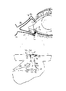
FIGS. 1-2 illustrate examples of schematic views of anatomical tissue
structures associated with an eye 100. The anatomical tissue structures shown
are
suitable for treatment using the lacrimal implants and methods discussed
herein.
The eye 100 is a spherical structure including a wall having three layers: the
outer
sclera 102, the middle choroid layer 104 and the inner retina 106. The sclera
102
includes a tough fibrous coating that protects the inner layers. It is mostly
white
except for the transparent area at the front, the cornea 108, which allows
light to
enter the eye 100.
The choroid layer 104, situated inside the sclera 102, contains many blood
vessels and is modified at the front of the eye 100 as the pigmented iris 110.
The
biconvex lens 112 is situated just behind the pupil. The chamber 114 behind
the
lens 112 is filled with vitreous humour, a gelatinous substance. The anterior
and
posterior chambers 116 are situated between the cornea 108 and iris 110,
respectively and filled with aqueous humour. At the back of the eye 100 is the
light-detecting retina 106.
The cornea 108 is an optically transparent tissue that conveys images to the
back of the eye 100. It includes avascular tissue to which nutrients and
oxygen are
supplied via bathing with lacrimal fluid and aqueous humour as well as from
blood
vessels that line the junction between the cornea 108 and sclera 102. The
cornea
108 includes one pathway for the permeation of drugs into the eye 100.
Other anatomical tissue structures associated with the eye 100 include the
lacrimal drainage system, which includes a secretory system 230, a
distributive
system and an excretory system. The secretory system comprises secretors that
are
19
CA 02722002 2010-10-20
stimulated by blinking and temperature change due to tear evaporation and
reflex
secretors that have an efferent parasympathetic nerve supply and secrete tears
in
response to physical or emotional stimulation. The distributive system
includes the
eyelids 202 and the tear meniscus around the lid edges of an open eye, which
spread
tears over the ocular surface by blinking, thus reducing dry areas from
developing.
The excretory part of the lacrimal drainage system includes, in flow order of
drainage, the lacrimal puncta, the lacrimal canaliculi, the lacrimal sac 204
and the
lacrimal duct 206. From the lacrimal duct 206, tears and other flowable
materials
drain into a passage of the nasal system. The lacrimal canaliculi include an
upper
(superior) lacrimal canaliculus 208 and a lower (inferior) lacrimal
canaliculus 210,
which respectively terminate near the eye 100 in an upper 212 and lower 214
lacrimal punctum. The upper 212 and lower 214 punctum are slightly elevated at
the medial end of a lid margin at the junction 216 of the ciliary and lacrimal
portions
near a conjunctival sac 218. The upper 212 and lower 214 punctum are generally
round or slightly ovoid openings surrounded by a connective ring of tissue.
Each of
the puncta 212, 214 leads into a vertical portion 220, 222 of their respective
canaliculus before turning more horizontal at a canaliculus curvature 250 to
join one
another at the entrance of the lacrimal sac 204. The canaliculi 208, 210 are
generally tubular in shape and lined by stratified squamous epithelium
surrounded
by elastic tissue, which permits them to be dilated or biased. As shown, a
lacrimal
canaliculus ampulla 252 exists near an outer edge of the canaliculus curvature
250.
In accordance with features of the present subject matter, a lacrimal implant
can be
inserted through either punctum and into its associated canaliculus.
FIG. 3 illustrates an example of a lacrimal implant 300 that can be insertable
into a lacrimal punctum 212, 214 (FIG. 2). The insertion of the lacrimal
implant
300 into a lacrimal punctum 212, 214 can allow for one or more of inhibition
or
blockage of tear flow through a lacrimal canaliculus 208, 210 (FIG. 2) (e.g.,
to treat
dry eyes), or the sustained delivery of a therapeutic agent to an eye (e.g.,
to treat an
infection, inflammation, glaucoma or other ocular diseases or disorders) or
nasal
passage (e.g., to treat a sinus or allergy disorder). In some examples, a
period of
time over which the agent is delivered includes at least one week, at least
one
-* CA 02722002 2010-10-20
_
month, or at least three months. In some examples, the lacrimal implant 300
includes a width between about 0.3 millimeters to about 1.5 millimeters. In
some
examples, the lacrimal implant 300 includes a length between about 1.5
millimeters
to about 6 millimeters, such as between about 2 millimeters to about 3
millimeters.
In an example, the lacrimal implant 300 comprises a polyurethane polymer
or copolymer. Typically, lacrimal implants are formed of silicone polymers,
which
can be quite hydrophobic and furthermore are usually prepared by
polymerization of
a silicone precursor in the presence of a catalyst. However, polyurethane
polymers
and copolymers can be thermoplastic, and can therefore be melted and cast into
a
desired form. A medicament can be dispersed within the polyurethane melt,
either
in molten form itself or as a dispersion of a solid material. Polyurethane
polymers
and copolymers can also be dissolved in various organic solvents, such as
dichloromethane or tetrahydrofuran, then cast into a desired form with removal
of
the solvent, such as by evaporation. Again, a medicament can be dispersed or
dissolved in the organic solvent along with the polyurethane, such that upon
removal of the solvent, the polyurethane containing the medicament in a
desired
form is obtained.
In FIG. 3, the lacrimal implant 300 is shown as a "half and half' implant
design. In this example, an expandable retention member 314, such as a
swellable
material that can be bonded or otherwise coupled over a portion of the
lacrimal
implant 300 such that it envelops, at least in part, a portion of the lacrimal
implant
300, forms a junction with a first portion 304 of the body of the lacrimal
implant
300. The first portion 304 of the body of the lacrimal implant 300 can be
formed of
a first polymer that is biocompatible and swells less than 100 wt% when in
contact
with an aqueous medium, such as a biocompatible polyurethane polymer or
copolymer. For example, the first polymer can comprise a polyurethane-silicone
copolymer. An example is PursilTM, a biocompatible, non-biodegradable
copolymer
adapted for medical use. Other similar materials can also be used, provided
they are
both biocompatible and substantially non-swelling or minimally swelling when
put
in contact with an aqueous medium. For example, hydrophilic polyurethanes that
do not substantially swell upon contact with an aqueous medium can be used to
21
- = CA 02722002 2010-10-20
improve surface wetability of the non-hydrogel component of the lacrimal
implant
300. The expandable retention member 314 can be formed of a second polymer
that
is biocompatible and swells greater than 100 wt% to form a hydrogel when in
contact with an aqueous medium, such as a biocompatible hydrogel-forming
polyurethane polymer or copolymer. For example, hydrogel-forming materials TG-
500 or TG-2000, adapted to swell by as much as 500-2000 wt% upon exposure to
an
aqueous medium, can be used.
In the half and half design, the expandable retention member 314 is
uncovered. The junction between the first portion 304 of the implant body and
the
expandable retention member 314 includes an intermediate member 350,
optionally
comprising the third polymer which can have an intermediate degree of swelling
in
an aqueous medium, and is sufficiently strong to hold the implant intact under
a
degree of tension, such as when the implant is removed from the punctal canal.
The
third polymer can be a biocompatible polyurethane polymer or copolymer adapted
to adhere to both the first and second polymers. It can be a moderately
swelling
polymer upon exposure to the aqueous medium.
FIG. 4A illustrates first portion 304 of the body of the lacrimal implant
another example of the lacrimal implant 300 that can be insertable into a
lacrimal
punctum 212, 214 (FIG. 2). In various examples, the lacrimal implant 300
comprises an implant body 402, including first 304 and second 406 portions,
which
is sized and shaped for at least partial insertion into a lacrimal punctum
212, 214.
The first portion 304 is formed from a polymer and includes a first diameter
408.
The second portion 406 is also formed from a polymer and includes a base
member
412 (e.g., mandrel or spine-like member) having a second diameter 410, which
is
less than the first diameter 408. In an example, the first 304 and second 406
portions are integrally coupled and comprise a unitary implant body 402. In an
example, as shown in FIGS. 7E-7G, the first 304 and second 406 portions are
separate elements, which can be coupled to one another via an engagement
between
a coupling void and a coupling arm, for instance.
An expandable retention member 314, such as a swellable material, can be
bonded or otherwise coupled over the base member 412 such that it envelops, at
22
- CA 02722002 2010-10-20
least in part, a portion of the base member 412. In an example, the expandable
retention member substantially envelops the base member 412. As the expandable
retention member 314 absorbs or otherwise retains lacrimal or other fluid,
such as
upon insertion into a lacrimal punctum 212, 214, its size increases and its
shape may
change thereby urging itself against and slightly biasing a wall of the
associated
canaliculus 208, 210. It is believed that the expandable retention member 314
will
provide retention comfort to a subject and may improve lacrimal implant 300
retention via controlled biasing of the canaliculus 208, 210 wall.
The positioning of the expandable retention member 314 over a portion of
the implant body 402 allows the retention member 314 to be freely exposed to
lacrimal fluid in situ, thereby allowing for a wide range of potential
expansion rates.
Further, the base member 412 provides an adequate coupling surface area to
which
the expandable retention member 314, for example, can adhere such that the
material of the expandable retention member 314 does not remain in a lacrimal
punctum 212, 214 after the lacrimal implant 300 is removed from the subject.
As
shown in this example, the expandable retention member 314 can include a non-
expanded, "dry or dehydrated" state, which aids insertion through a lacrimal
punctum 212, 214 and into the associated lacrimal canaliculus 208, 210. Once
placed into a lacrimal canaliculus 208, 210, the expandable retention member
314
can absorb or other retain lacrimal fluid to form an expanded structure.
In some examples, the implant body 402 can include a cylindrical-like
structure comprising a cavity 416 disposed near a proximal end 418 of the
first
portion 304. In this example, the cavity 416 extends inward from the proximal
end
418 and includes a first drug-releasing or other agent-releasing drug supply
420 to
provide a sustained drug or other agent release to an eye 100. The drug or
other
agent release can occur, at least in part, via an exposed surface of the drug
supply
420. In an example, such as is shown in FIG. 4B, the exposed surface of the
drug
supply 420 can be positioned above the proximal end 418 such that the drug
supply
420 at least partially protrudes outside of the implant body 402. In some
examples,
the exposed surface of the drug supply 420 can be flush or slightly below the
23
CA 02722002 2016-09-01
proximal end 418 such that the drug supply 420 does not protrude outside of
the
implant body 402.
In some examples, by controlling geometry or a drug concentration gradient
near the exposed surface, a predetermined drug or agent release rate can be
achieved. For instance, the exposed surface can be constructed with a specific
geometry or other technique appropriate to control the release rate of the
drug or
other agent onto an eye 100, such as on an acute basis, or on a chronic basis
between outpatient doctor visits, for example. Further discussion regarding
effective release rates of one or more drugs or other agents from a drug
supply 420
can be found in commonly-owned DeJuan et al., U.S. Patent Publication No. 2007-
0243230.
The implant body 402 can include an integral feedback or other projection
422, such as projections extending laterally at least partially from or around
the
proximal end 418 of the first implant body portion 304. In an example, the
projection 422 includes a partially trimmed head portion extending 360 degrees
around the proximal end 418 from an outer implant body surface. In an example,
the projection 422 includes a full head portion extending 360 degrees around
the
proximal end 418 from an outer implant body surface. In an example, the
projection
422 includes a cross-sectional shape similar to a flat disk (i.e., relatively
flat top and
bottom surfaces). In various examples, the projection 422 can be configured to
seat
against or near a punctal opening 212, 214 when the second portion 406 of the
implant body 402 is positioned within the associated canalicular lumen 208,
210,
such as for inhibiting or preventing the lacrimal implant 300 from passing
completely within the canalicular lumen, for providing tactile or visual
feedback
information to an implanting user (e.g., as to whether the implant is fully
implanted), or for removing the lacrimal implant 300 from an implant position.
In
an example, the projection 422 includes a portion having a diameter of about
0.5-2.0
mm to prevent the lacrimal implant 300 from passing down into the canaliculus
208,
210.
24
- - CA 02722002 2010-10-20
FIG. 4B illustrates an example of a cross-sectional view of a lacrimal
implant 300 taken along a line parallel to a longitudinal axis of the implant,
such as
along line 4B-4B of FIG. 4A. As shown in FIG. 4B, the lacrimal implant 300
comprises an implant body 402, including first 304 and second 406 portions,
which
is sized and shaped for at least partial insertion into a lacrimal punctum
212, 214
(FIG. 2). The first portion 304 is formed from a polymer and includes a first
diameter 408. The second portion 406 is also formed from a polymer and
includes a
base member 412 (e.g., mandrel or spine) having a second diameter 410, which
is
less than the first diameter 408. In an example, the base member 412 is at
least
about one-third the total length of the implant body 402. In an example, the
base
member 412 is at least about one-half the total length of the implant body
402. In
the example shown, the implant body 402 also includes an integral feedback or
other projection 422, such as a projection extending laterally at least
partially from
or around a proximal end 418 of the first implant body portion 304.
In various examples, the implant body 402 can be molded or otherwise
formed using an elastic material, such as silicone, polyurethane or other
urethane-
based material, or combinations thereof. In an example, one or both of the
first 304
and second 406 portions include a urethane-based material. In an example, one
or
both of the first 304 and second 406 portions include a silicone-based
material, such
as 4840TM or PurSilTM. In an example, one or both of the first 304 and second
406
portions include a copolymer material, such as polyurethane/silicone,
urethane/carbonate. silicone/polyethylene glycol (PEG) or
silicone/2hydroxyethyl
methacrylate (HEMA). In various examples, the implant body 402 is configured
to
be non-absorbable in situ and is sufficiently strong to address issues of
cutting
strength (e.g., during insertion and removal of the lacrimal implant 300) and
dimensional stability.
An expandable retention member 314, such as a swellable material, can be
bonded or otherwise coupled over the base member 412 such that it envelops, at
least in part, a portion of the base member 412. As the expandable retention
member absorbs or otherwise retains lacrimal fluid, such as upon insertion
into a
lacrimal punctum 212, 214, its size increases and its shape may change thereby
CA 02722002 2016-09-01
urging itself against and slightly biasing a wall of the associated
canaliculus 208,
210. In various examples, the expandable retention member 314 can be molded or
otherwise formed using a swellable material. In an example, the expandable
retention member 314 includes a polyurethane hydrogel, such as TG-2000Tm, TG-
500 TM, or other urethane-based hydrogel. In an example, the expandable
retention
member 314 includes a thermoset polymer, which may be configured to swell
anisotropically. In an example, the expandable retention member 314 includes a
gel, which does not maintain its shape upon expansion, but rather conforms to
fit the
shape of a canaliculus lumen wall or other surrounding structure.
In some examples, the lacrimal implant 300 includes a base member 412
including polyurethane or other urethane-based material and an expandable
retention member 314 including a biocompatible polyurethane or other urethane-
based swellable material. In an example, a polyurethane hydrogel is coupled
directly to an outer surface, such as a plasma-treated outer surface, of the
base
member 412. As further discussed in commonly-owned Utkhede et al., U.S. Patent
Publication No. 2009-0104243, urethane-based polymer and copolymer materials
allow for a variety of processing methods and bond well to one another.
In some examples, the lacrimal implant 300 includes an intermediate
member 350 positioned between a portion of the implant body 402, such as the
base
member 412, and a portion of the expandable retention member 314. The
intermediate member 350 can include a material configured to absorb, when
implanted, a greater amount of lacrimal fluid than the polymer of the base
member
412 but less lacrimal fluid than the swellable polymer of the expandable
retention
member 314. The intermediate member 350 can provide the lacrimal implant 300
with integrity, such as between a substantially non-swelling polymer of the
implant
body 402 and a swelling polymer of the expandable retention member 314. For
instance, when the polymer of the expandable retention member 314 swells upon
exposure to moisture, it is possible that the expanding polymer will, in the
absence
of the intermediate member 350, swell away from the underlying, non-swelling
polymer of the base member 412. In an example, the intermediate member 350
includes PurSi1TM and is dip or otherwise coated onto an outer surface of the
base
26
CA 02722002 2016-09-01
member 412. In an example, the intermediate member 350 includes a polyurethane
configured to absorb about 10% to about 500% water, such as Tecophilic TM
urethanes or Tecophilic TM solution grade urethanes.
In certain examples, the implant body 402 can include a cavity 416 disposed
near the proximal end 418 of the first portion 304. In an example, the first
cavity
416 extends inward about 2 millimeters or less from the proximal end 418, and
houses a first drug-releasing or other agent-releasing drug supply 420 to
provide a
sustained drug or other agent release to an eye 100 (FIG. 2). In various
examples,
the drug supply 420 stores and slowly dispenses an agent to the eye 100 as
they are
leached out, for example, by tear film fluid. In an example, the drug supply
420
includes a plurality of therapeutic agent inclusions 452, which can be
distributed in
a matrix 454. In an example, the inclusions 452 comprise a concentrated form
of
the therapeutic agent (e.g., a crystalline agent form). In an example, the
matrix 454
comprises a silicone matrix or the like, and the distribution of inclusions
452 within
the matrix are homogeneous or non-homogeneous. In an example, the agent
inclusions 452 include droplets of oil, such as Latanoprost oil. In still
another
example, the agent inclusions 452 include solid particles, such as Bimatoprost
particles in crystalline form. The inclusions can be of many sizes and shapes.
For
instance, the inclusions can include microparticles having dimensions on the
order
of about 1 micrometer to about 100 micrometers.
In the example shown, the drug supply 420 includes a sheath body 456
disposed over at least a portion thereof such as to define at least one
exposed surface
458 of the drug supply. In an example, the sheath body 456 comprises
polyimide.
The exposed surface 458 can be located at or near the proximal end 418 of the
implant body 402 such as to contact a tear or a tear film fluid and release
the
therapeutic agent at one or more therapeutic levels over a sustained time
period
when the lacrimal implant 300 is inserted into a lacrimal punctum 212, 214.
Further
discussion regarding configuring and manufacturing of the drug supply 420 can
be
found in commonly-owned DeJuan et al., U.S. Patent Publication No. 2007-
0269487.
27
CA 02722002 2016-09-01
In certain examples, the expandable retention member can include a second
drug-releasing or other agent-releasing drug supply 460 to provide a sustained
drug
or other agent release to one or both of a wall of a lacrimal canaliculus 208,
210 or a
nasolacrimal system. The drug supply 460 can be configured to store and slowly
dispense an agent after contact with lacrimal fluid within a lacrimal
canaliculus 208,
210. In an example, the agent included in the expandable retention member can
comprise medicaments, therapeutic agents, or antimicrobials (e.g., silver).
FIG. 4C illustrates an example of a cross-sectional view of a lacrimal
implant 300 taken along a line parallel to a longitudinal axis of the implant.
As
shown in FIG. 4B, some of the present lacrimal implants 300 are configured to
include a feedback or other projection 422 at or near a proximal end 418 for
inhibiting or preventing the lacrimal implant 300 from passing completely
within
the canalicular lumen, and others are configured to be inserted completely
below the
punctal opening 212, 214. As shown in FIG. 4C, the lacrimal implant 300
comprises an implant body 402 without a feedback or other projection 322 (FIG.
3A) at or near a proximal end 418 of a first implant body portion 304.
Accordingly,
the lacrimal implant 300 can, in some examples, be completely inserted within
the
canalicular lumen.
FIG. 5 illustrates an example of an assembly of a present lacrimal implant
300. As discussed, the lacrimal implant 300 can include a unitary implant body
402
sized to at least partially pass through a lacrimal punctum 212, 214 (FIG. 2)
and be
positioned within a lacrimal canaliculus 208, 210 (FIG. 2). The implant body
402
can extend from a proximal end portion 502 to a distal end portion 504. In the
example shown, the proximal end portion 502 includes a cavity 416 and the
distal
end portion 504 includes a base member 412 having a diameter 410 less than a
diameter of the proximal end portion 502. In an example, a drug core 420 can
be
included in the cavity 416 and configured to provide a release of an agent to
an eye.
In various examples, an expandable retention member 314 can be coupled at
least
partially over the base member 412 and includes a "dry or dehydrated" material
28
' CA 02722002 2010-10-20
_
hydratable by lacrimal or other fluid to swell from a first diameter to a
second
diameter greater than the first diameter.
The base member 412 can be coupled with the expandable retention member
314 in a variety of ways. In an example, as shown in FIG. 5, a preformed,
swellable
(e.g., hydrogel) sleeve 506 can be slid over an outer surface 508 of the base
member
412. In an example, the expandable retention member 314 can be dip or
otherwise
coated onto the base member 412. In an example, a first polymer-based (e.g.,
urethane-based) base member 412 and a first polymer-based (e.g., urethane-
based)
expandable retention member 314 can be injection molded concurrently (e.g.,
via bi-
injection molding), thereby allowing the lacrimal implant 300 to be formed in
a
minimum number of steps. In an example, a multi-shot molding process can be
used, which involves sequential injection of separate materials into different
locations in a mold. In an example, an insert over-molding process can used
such
that the base member 412 is first molded then placed into a second mold for
over-
molding with a polymer for the expandable retention member 314. In various
examples, the expandable retention member 314 may extend along any desired
length portion of the implant body 402.
In certain examples, one or both of the distal body portion 504 or a distal
end
510 of the expandable retention member 314 can include a taper 512 to self-
dilate
anatomical tissue, such one or both of a lacrimal punctum 212, 214 (FIG. 2) or
associated lacrimal canaliculus 208, 210, to a sufficient diameter as the
lacrimal
implant 300 is being implanted. In this way, the lacrimal implant 300 can be
implanted in various size ocular anatomies without the need for pre-dilation
via a
separate enlarging tool. The taper 512 can be formed so as to not be traumatic
to an
inner lining of the lacrimal punctum 212, 214 or the lacrimal canaliculus 208,
210.
As shown, the taper 512 can generally narrow from a location near an
intermediate portion of the swellable sleeve 506 to the distal end 510 of the
sleeve
506, such as from a diameter of about 0.5 millimeters to a diameter of about
0.1
millimeters or less. Among other factors, a determination of a desirable taper
512
for a given implant situation can be made by balancing implant properties,
such as a
implant body 402 strength desirable for implant insertion with a desire to
have a
29
CA 02722002 2016-09-01
soft, flexible and conforming implant body (e.g., to conform to a lacrimal
canaliculus anatomy) upon implantation. In some examples, a lubricious coating
disposed on, or impregnated in, one of both of an outer surface of the implant
body
402 or an outer surface of the expandable retention member 314 can be used to
further aid insertion of the lacrimal implant 300 into the anatomical tissue.
In an
example, the lubricious coating can include a silicone lubricant. In an
example, the
outer surface of the implant body 402 or the expandable retention member 314
can
be treated via plasma or radiation in order to securely bond a thin coating of
lubricious material (e.g., hydrogel).
In various examples, the outer surface of one or both of the implant body
402 or the expandable retention member 314 can be formed, or surface treated
to be,
generally smooth to inhibit bacteria from attaching to the lacrimal implant
300 and
incubating. The generally smooth outer surface can also prevent damage to the
inner lining of the receiving anatomical tissue, such as a lacrimal punctum
212, 214
(FIG. 2) or the associated lacrimal canaliculus 208, 210 (FIG. 2), during
implantation. As further discussed in commonly-owned Rapacki, U.S. Patent
Publication No. 2009-0298390, the outer surface of the plug body 402, for
instance,
can be improved via a polishing procedure using dichloride methane or other
suitable media in conjunction with a tumbling process.
In some examples, an antimicrobial coating can be disposed on, or
impregnated in, at least a portion of the outer surface of one or both of the
implant
body 402 or the expandable retention member 314 to further prevent bacteria
growth on the implant body. In an example, the antimicrobial coating can
include
an agent selected from the group consisting of 2-bromo-2-nitropropane-1,3-
diol, 5-
bromo-5-nitro-1,3-dioxane, 7-ethyl bicyclooxazolidine, benzalkonium chloride,
benzethonium chloride, benzoic acid, benzyl alcohol, boric acid, bronopol,
cetylpyridinium chloride, chlorhexidine digluconate, chloroacetamide,
chlorobutanol, chloromethyl isothiazolinone and methyl isothiazoline,
dimethoxane,
dimethyl oxazolidine, dimethyl hydroxymethyl pyrazole, chloroxylenol,
dehydroacetic acid, diazolidinyl urea, dichlorobenzyl alcohol, DMDM hydantoin,
CA 02722002 2010-10-20
-
ethyl alcohol, formaldehyde, glutaraldehyde, hexachlorophene, hexetidine,
hexamethylenetramine, imidazolidinyl urea, iodopropynyl butylcarbamate,
isothiazolinones, methenammonium chloride, methyldibromo glutaronitrile, MDM
hydantoin, minocycline, ortho phenylphenol, p-chloro-m-cresol, parabens
(butylparaben, ethylparaben, methylparaben), phenethyl alcohol,
phenoxyethanol,
piroctane olamine, polyaminopropyl biguanide, polymethoxy bicyclic
oxazolidine,
polyoxymethylene, polyquaternium-42, potassium benzoate, potassium sorbate,
propionic acid, quaternium-15, rifampin, salicylic acid, selenium disulfide,
sodium
borate, sodium iodate, sodium hydroxymethylglycinate, sodium propionate,
sodium
pyrithione, sorbic acid, thimerosal, triclosan, triclocarban, undecylenic
acid, zinc
phenosulfonate, and zinc pyrithione. In an example, the antimicrobial coating
can
include a material selected from the group consisting of silver lactate,
silver
phosphate, silver citrate, silver acetate, silver benzoate, silver chloride,
silver iodide,
silver iodate, silver nitrate, silver sulfadiazine, silver palmitate or one or
more
mixtures thereof. In an example, the antimicrobial coating can include at
least one
of an antibiotic or an antiseptic. For instance, the antimicrobial coating can
include
a temporary anesthetic lasting, on average, between a few hours and a day. In
still
other examples, the antimicrobial coating can include a drug use to treat an
underlying disease, such as a bolus for immediate effect.
FIG. 6 illustrates an example of a schematic view of a lacrimal implant 300
implanted in a lower lacrimal punctum 214 and associated canaliculus 210. In
some
examples, a lacrimal implant 300 can be implanted in an upper lacrimal punctum
212 and associated canaliculus 208. In this example, the lacrimal implant 300
comprises a implant body 402, including first 304 and second 406 portions,
which is
sized and shaped for at least partial insertion into the lacrimal punctum 214.
The
first portion 304 is fbrmed from a polymer and includes a first diameter 408
(FIG.
4B). The second portion 406 is also formed from a polymer and includes a base
member 412 (e.g., mandrel or spine-like member) having a second diameter 410
(FIG. 4B) less than the first diameter 408. An expandable retention member
314,
such as a swellable material, can be bonded or otherwise coupled over the base
member 412 such that it envelops, at least in part, a portion of the base
member 412.
31
- = CA 02722002 2010-10-20
In certain examples, an outer surface of one or both of the implant body 402
or the
expandable retention member 314 can include grooves or a coating of a wicking
material such as to allow fluid flow around the implant body 402.
As shown, the first portion 304 of the implant body 402 can be configured to
rest, at a proximal end 418, against the punctal opening 214 and rest, at a
distal end
602, within the associated lacrimal canaliculus 210. In this example, an
integral
feedback or other projection 422 extending around the proximal end 418
inhibits or
prevents the lacrimal implant 300 from passing completely within the
canalicular
lumen 210.
As further shown, the second portion 406, including the base member 412 at
least partially enveloped by the expandable retention member 314, can be
configured to rest and retain the lacrimal implant 300 within the associated
lacrimal
canaliculus 210. As the expandable retention member absorbs or otherwise
retains
lacrimal or other fluid, such as upon insertion into the lacrimal punctum 214,
its size
increases radially or longitudinally and its shape may change thereby urging
itself
against and slightly biasing a wall of the associated canaliculus 210, while
still
being comfortable to the subject. In an example, a longitudinal swelling
direction of
the expandable retention member 314 extends laterally relative to a
longitudinal axis
606 of the implant body 402. In an example, a portion 608 of the expandable
retention member 314 is configured to at least partially extend toward a
horizontal
section of the lacrimal canaliculus 210. In an example, a portion 610 of the
expandable retention member 314 is configured to at least partially extend
toward
an ampulla 252 of the lacrimal canaliculus 210. In an example, the swellable
material of the expandable retention member 314 can comprise a hydrogel having
a
water content, at a fully hydrated state, of about 500% to about 2000% by
weight
and, at a "dry or dehydrated" state, of less than about 10%, such as about 1%.
In
certain examples, the expandable retention member 314 is configured to allow
for
an expansion capacity of up to about one times its "dry or dehydrated" volume,
up
to about five times its "dry or dehydrated" volume, or up to about ten times
its "dry
or dehydrated" volume.
32
- - CA 02722002 2010-10-20
Forceps or another insertion tool can be used to insert the lacrimal implant
300 in a lacrimal punctum 212, 214 and the associated canaliculus 208, 210. In
various examples, the second portion 406 of the implant body 402 can be
advanced
into the depth of a lacrimal canaliculus 208, 210 by manipulation of the
inserter tool
until a feedback or other projection 422, if present, is seated against the
punctal
opening 212, 214. When it is desired to remove the lacrimal implant 300, the
projection 432 can be readily grasped with the forceps, for example, and
withdrawn
from the punctal opening 212, 214. It is believed the insertion and removal
processes associated with the present lacrimal implants 300 may, in some
instances,
be simply performed in a short amount of time by a general ophthalmologist in
his/her office without the need for special skills or expensive equipment. In
some
instances, after a first lacrimal implant has been removed, a second lacrimal
implant
including a supply of an agent is inserted into the subject until he/she no
longer
requires treatment.
In certain examples, the implant body 402 can include a cavity 416 disposed
near the proximal end 418 of the first portion 304. In this example, the first
cavity
418 extends inward from the proximal end 418 and includes a first drug-
releasing or
other agent-releasing drug supply 420. The lacrimal implant 300 can be
oriented
with the expandable retention member 314 aligned for placement in a lacrimal
canaliculus 208, 210, while the drug supply 420 and the proximal end 418 are
substantially aligned with the exterior of the punctum openings 212, 214 to
provide
a sustained drug or other agent release to an eye 100 (e.g., to treat an
infection,
inflammation, glaucoma or other ocular disease or disorder). The drug or other
agent release can occur, at least in part, via an exposed surface of the drug
supply
420. In this example, the exposed surface of the drug supply 420 can be
positioned
above the proximal end 418 such that the drug supply 420 at least partially
protrudes
outside of the implant body 402.
In certain examples, the expandable retention member can include a second
drug-releasing or other agent-releasing drug supply 460 to provide a sustained
drug
or other agent release to one or both of a wall of a lacrimal canaliculus 208,
210 or a
nasolacrimal system. The drug supply 460 can be configured to store and slowly
33
- CA 02722002 2010-10-20
, =
dispense an agent after contact with lacrimal fluid within a lacrimal
canaliculus 208,
210. In an example, the agent included in the expandable retention member can
comprise medicaments, therapeutic agents, or antimicrobials (e.g., silver).
FIGS. 7A-7G illustrate examples of lacrimal implants 300 that can be
insertable into a lacrimal punctum 212, 214 (FIG. 2). The insertion of the
lacrimal
implants 300 into a lacrimal punctum 212, 214 can allow for one or more of
inhibition or blockage of tear flow through a lacrimal canaliculus 208, 210
(FIG. 2)
(e.g., to treat dry eyes), or the sustained delivery of a therapeutic agent to
an eye
(e.g., to treat an infection, inflammation, glaucoma or other ocular diseases
or
disorders) or nasal passage (e.g., to treat a sinus or allergy disorder).
In various examples, the lacrimal implants 300 comprises an implant body
402, including first 304 and second 406 portions, which is sized and shaped
for at
least partial insertion into a lacrimal punctum 212, 214. The first portion
304 is
formed from a polymer and includes a first diameter 408 (FIG. 4B). The second
portion 406 is also formed from a polymer and includes a base member 412
(e.g.,
mandrel or spine-like member) having a second diameter 410 (FIG. 4B) less than
the first diameter 408. In some examples, such as are shown in FIGS. 7A-7D,
the
first 304 and second 406 portions are integrally coupled and comprise a
unitary
implant body 402. In some examples, such as are shown in FIGS. 7E-7G, the
first
304 and second 406 portions are separate elements, which can be coupled to one
another via an engagement between a coupling void and a coupling arm, for
instance. In various examples, the base member 412 can include one or more arm
members 702 protruding from an outer surface 508 thereof.
An expandable retention member 314, such as a swellable material, can be
bonded or otherwise coupled over the base member 412 such that it envelops, at
least in part, a portion of the base member 412. In an example, the expandable
retention member substantially envelops the base member 412. The one or more
arm members 702 increase the surface area for bonding or other coupling
between
one or more of the expandable retention member 314, an intermediate member 350
(FIG. 4B), and the base member 412. The expandable retention member 314, for
example, would will the spaces between the one or more arm members 702 upon
34
CA 02722002 2010-10-20
application. As the expandable retention member 314 absorbs or otherwise
retains
lacrimal or other fluid, such as upon insertion into a lacrimal punctum 212,
214, its
size increases and its shape may change thereby urging itself against and
slightly
biasing a wall of the associated canaliculus 208, 210.
In the example of FIG. 7A, the lacrimal implant 300 includes a unitary
implant body 402 extending from a first portion 304 to a second portion 406.
The
first portion 304 can include a cavity 416 disposed near a proximal end 418 of
the
first portion 304. The cavity 416 can include a first drug-releasing or other
agent-
releasing drug supply 420 to provide a sustained drug or other agent release
to an
eye. The implant body 402 can further include an integral feedback or other
projection 422 extending laterally at least partially from or around the
proximal end
318. In various examples, the projection 422 can be configured to seat against
or
near a punctal opening 212, 214 when the second portion 406 of the implant
body
402 is positioned within the associated canalicular lumen 208, 210, such as
for
inhibiting or preventing the lacrimal implant 300 from passing completely
within
the canalicular lumen, for providing tactile or visual feedback information to
an
implanting user (e.g., as to whether the implant is fully implanted), or for
removing
the lacrimal implant 300 from an implant position.
As shown, the second portion 406 can include a base member 412 (e.g.,
mandrel or spine-like member) having a diameter 410 (FIG. 4B) less than a
diameter 408 (FIG. 4B) of the first portion 304. In this example, the base
member
412 includes one or more arm members 702 in the form of ribs 704. Each rib 704
can include a curved or non-curved projection extending from an outer surface
of
the base member 412, such as projecting laterally relative to a longitudinal
axis of
the base member 412. In this example, three pairs of ribs 704 extend from the
base
member 412; however, more and fewer than three pairs of ribs 704 may also be
used
without departing from the scope of the present subject matter. The ribs 704
can
provide strength to the lacrimal implant 300 by increasing the surface area to
which
one or both of the expandable retention member 314 or an intermediate member
350
(FIG. 4B) are coupled to the base member 412.
CA 02722002 2010-10-20
eit
S.
In the example of FIG. 7B, the lacrimal implant 300 includes a unitary
implant body 402 extending from a first portion 304 to a second portion 406.
The
first portion 304 can include a cavity 416 disposed near a proximal end 418 of
the
first portion 304. The cavity 416 can include a first drug-releasing or other
agent-
releasing drug supply 420 to provide a sustained drug or other agent release
to an
eye. The implant body 402 can further include an integral feedback or other
projection 422 extending laterally at least partially from or around the
proximal end
418. In various examples, the projection 422 can be configured to sit against
or near
a punctal opening 212, 214 when the second portion 406 of the implant body 402
is
positioned within the associated canalicular lumen 208, 210, such as for
inhibiting
or preventing the lacrimal implant 300 from passing completely within the
canalicular lumen, for providing tactile or visual feedback information to an
implanting user (e.g., as to whether the implant is fully implanted), or for
removing
the lacrimal implant 300 from an implant position.
As shown, the second portion 406 can include a base member 412 (e.g.,
mandrel or spine-like member) having a diameter 410 (FIG. 4B) less than a
diameter 408 (FIG. 4B) of the first portion 304. In this example, the base
member
412 includes one or more arm members 702 in the form of disks 706. Each disk
706
can include a relatively flat top or bottom surface and can include a greater
cross-
sectional size than an adjacent portion of the base member 412. The one or
more
disks 706 can be spaced apart along a longitudinal axis of the base member 412
and
extend from an outer surface of the base member 412. In an example, three
disks
706 extend from base member 412; however, more and fewer than three disks 706,
such as one disk 706, may also be used without departing from the scope of the
present subject matter. The disks 706 can provide strength to the lacrimal
implant
300 by increasing the surface area to which one or both of the expandable
retention
member 314 or an intermediate member 350 (FIG. 4B) are coupled to the base
member 412.
In the example of FIG. 7C, the lacrimal implant 300 includes a unitary
implant body 402 extending from a first portion 304 to a second portion 406.
The
first portion 304 can include a cavity 416 disposed near a proximal end 418 of
the
36
CA 02722002 2010-10-20
-
first portion 304. The cavity 416 can include a first drug-releasing or other
agent-
releasing drug supply 420 to provide a sustained drug or other agent release
to an
eye. The implant body 402 can further include an integral feedback or other
projection 422 extending laterally at least partially from or around the
proximal end
418. In various examples, the projection 422 can be configured to seat against
or
near a punctal opening 212, 214 when the second portion 406 of the implant
body
402 is positioned within the associated canalicular lumen 208, 210, such as
for
inhibiting or preventing the lacrimal implant 300 from passing completely
within
the canalicular lumen, for providing tactile or visual feedback information to
an
implanting user (e.g., as to whether the implant is fully implanted), or for
removing
the lacrimal implant 300 from an implant position.
As shown, the second portion 406 can include a base member 412 (e.g.,
mandrel or spine-like member) having a diameter 410 (FIG. 4B) less than a
diameter 408 (FIG. 4B) of the first portion 304. In this example, the base
member
412 includes one or more arm members 702 in the form of spikes 708. Each spike
708 can protrude radially outward from an outer surface of the base member 412
at
any location around a circumference of the base member 412. In an example, six
or
more spikes 708 extend from base member 412; however, fewer than six spikes
708
may also be used without departing from the scope of the present subject
matter.
The spikes 708 can provide strength to the lacrimal implant 300 by increasing
the
surface area to which one or both of the expandable retention member 314 or an
intermediate member 350 (FIG. 4B) are coupled to the base member 412.
In the example of FIG. 7D, the lacrimal implant 300 includes a unitary
implant body 402 extending from a first portion 304 to a second portion 406.
The
first portion 304 can include a cavity 416 disposed near a proximal end 418 of
the
first portion 304. The cavity 416 can include a first drug-releasing or other
agent-
releasing drug supply 420 to provide a sustained drug or other agent release
to an
eye. The implant body 402 can further include an integral feedback or other
projection 422 extending laterally at least partially from or around the
proximal end
418. In various examples, the projection 422 can be configured to sit against
or near
a punctal opening 212, 214 when the second portion 406 of the implant body 402
is
37
= ` CA 02722002 2010-10-20
positioned within the associated canalicular lumen 208, 210, such as for
inhibiting
or preventing the lacrimal implant 300 from passing completely within the
canalicular lumen, for providing tactile or visual feedback information to an
implanting user (e.g., as to whether the implant is fully implanted), or for
removing
the lacrimal implant 300 from an implant position.
As shown, the second portion 406 can include a base member 412 (e.g.,
mandrel or spine-like member) having a diameter 410 (FIG. 4B) less than a
diameter 408 (FIG. 4B) of the first portion 304. In this example, the base
member
412 includes one or more arm members 702 having a curved shape, such as a fish
hook-like shape 710. Each fish hook-like projection 710 can protrude radially
outward from an outer surface of the base member 412 at any location around a
circumference of the base member 412. In an example, at least one of the fish
hook-
like projections include a barb or other backward pointing ridge. In this
example,
three pairs of fish hook-like projections 710 extend from the base member 412;
however, more and fewer than three pairs of fish hook-like projections 710 may
also
be used without departing from the scope of the present subject matter. The
fish
hook-like projections 710 or other curved shaped projections can provide
strength to
the lacrimal implant 300 by increasing the surface area to which one or both
of the
expandable retention member 314 or an intermediate member 350 are coupled to
the
base member 412.
In the example of FIG. 7E and 7G, the lacrimal implant 300 includes a
implant body 402 comprising first 304 and second 406 separable portions, which
can be coupled to one another via an engagement between a coupling void 712
and
a coupling arm 714, for instance. In an example, a polymer forming the first
portion
304, such as a silicone-based material, is different than a polymer forming
the
second portion 406, such as a polyester, silk, polycarbonate, urethane-based
material
or other material having a melting temperature higher than that at which the
polymer of the first portion 304 is cured or having a greater hardness. The
first
portion 304 can include a cavity 416 disposed near a proximal end 418 of the
first
portion 304. The cavity 416 can include a first drug-releasing or other agent-
releasing drug supply 420 to provide a sustained drug or other agent release
to an
38
CA 02722002 2010-10-20
eye. The implant body 402 can further include an integral feedback or other
projection 422 extending laterally at least partially from or around the
proximal end
418. In various examples, the projection 422 can be configured to sit against
or near
a punctal opening 212, 214 when the second portion 406 of the implant body 402
is
positioned within the associated canalicular lumen 208, 210, such as for
inhibiting
or preventing the lacrimal implant 300 from passing completely within the
canalicular lumen, for providing tactile or visual feedback information to an
implanting user (e.g., as to whether the implant is fully implanted), or for
removing
the lacrimal implant 300 from an implant position.
As shown, the second portion 406 can include a base member 412 (e.g.,
mandrel or spine-like member) comprising a variety of shapes and having a
diameter 410 (FIG. 4B) less than a diameter 408 of the first portion 304. In
these
examples, the base member 412 includes one or more arm members 702 extending
from an outer surface of the base member 412, such as projecting laterally
relative
to a longitudinal axis of the base member 412, which, in the example of FIG.
7E,
coincides with a longitudinal axis of the first portion 304 and, in the
example of
FIG. 7G, is angled 718 relative to the axis of the first portion 304. In an
example,
the angled 718 intersection between the longitudinal axis of the base member
412
and the longitudinal axis of the first portion 304 is formed upon or after
implantation of the implant body 402 into a lacrimal canaliculus 208, 210. The
one
or more arm members 702 can provide strength to the lacrimal implant 300 by
increasing the surface area to which one or both of the expandable retention
member
314 or an intermediate member 350 (FIG. 4B) are coupled to the base member
412.
In the example of FIG. 7F, the lacrimal implant 300 includes an implant
body 402 comprising first 304 and second 406 separable portions, which can be
coupled to one another via an engagement between a coupling void 712 and a
coupling arm 714, for instance. In an example, a polymer forming the first
portion
304, such as a silicone-based material, is different than a polymer forming
the
second portion 406, such as a polyester, silk, polycarbonate, urethane-based
material
or other material having a melting temperature higher than that at which the
polymer of the first portion 304 is cured or having a greater hardness. The
first
39
CA 02722002 2010-10-20
_ .
portion 304 can include a cavity 416 disposed near a proximal end 418 of the
first
portion 304. The cavity 416 can include a first drug-releasing or other agent-
releasing drug supply 420 to provide a sustained drug or other agent release
to an
eye. The implant body 402 can further include an integral feedback or other
projection 422 extending laterally at least partially from or around the
proximal end
418. In various examples, the projection 422 can be configured to sit against
or near
a punctal opening 212, 214 when the second portion 406 of the implant body 402
is
positioned within the associated canalicular lumen 208, 210, such as for
inhibiting
or preventing the lacrimal implant 300 from passing completely within the
canalicular lumen, for providing tactile or visual feedback information to an
implanting user (e.g., as to whether the implant is fully implanted), or for
removing
the lacrimal implant 300 from an implant position.
As shown, the second portion 406 can include a base member 412 (e.g.,
mandrel or spine-like member) having a diameter 410 (FIG. 4B) less than a
diameter 408 (FIG. 4B) of the first portion 304. In this example, the base
member
412 includes one or more arm members 702 extending from an outer surface of
the
base member 412, such as projecting in a balloon-like shape from a juncture
between the coupling void 712 and the coupling arm 714. In an example, the one
or
more arm members 702 include one or more voids 616 sized to receive a portion
of
the expandable retention member 314 or an intermediate member 350. The one or
more arm members 702 can provide strength to the lacrimal implant 300 by
increasing the surface area to which one or both of the expandable retention
member
314 or an intermediate member 350 (FIG. 4B) are coupled to the base member
412.
FIG. 8 is a block diagram illustrating an example of a method 800 of
manufacturing a lacrimal implant configured to be at least partially
insertable into a
lacrimal punctum. At 802, a implant body, including first and second portions,
is
formed. Forming the first portion can include melt, molding or otherwise
processing a polymer into a shape having a first diameter. Forming the second
portion can include melt, molding or otherwise processing a polymer into a
base
member shape having a second diameter less than the first diameter. In an
example,
forming the fist portion, the second portion, or both can include injection
molding
CA 02722002 2010-10-20
using respectively a melt of the first polymer, the second polymer, or both
polymers.
Polyurethane polymers and copolymers are adapted for melt processing, thus
avoiding both the added complexity of solvent casting technology, the cost of
dealing with the necessary solvents, and the possibility of residual solvents
in the
polymeric materials of the implant. In an example, forming the implant body
further includes forming a cavity extending inward form a proximal end of the
first
portion. In an example, forming the implant body further includes forming one
or
more arm members protruding laterally from an outer surface of the base
member.
In some examples, the implant body is formed from a urethane-based material,
such
as polyurethane. In some examples, the implant body is formed from a silicone
material. Optionally, one or more portions of an outer surface of the base
member
are plasma treated to encourage coupling to a covering element.
At 804, an intermediate member is optionally disposed on the outer surface
of the base member. In various examples, the intermediate member includes a
third
polymer configured to absorb a greater amount of fluid (e.g., lacrimal fluid)
than a
polymer of the implant body. In an example, the third polymer can be
incorporated
into the lacrimal implant as a melt. In an example, the intermediate member
can be
applied to the base member using an injection molding process, or it can be
applied
using a solvent-dip coating process. For instance, the dip-coating process can
be
used to apply a thin layer of the intermediate member onto the outer surface
of the
base member.
At 806, an expandable retention member is coupled at least partially over the
base member, and optionally, the intermediate member. In an example, the
expandable retention member is coupled over the base member such that the base
member is entirely or substantially surrounded. In various examples, the
expandable retention member includes a polymer configured to absorb a greater
amount of fluid than both the implant body and the intermediate member. In an
example, the expandable retention member includes a urethane-based material,
such
as urethane-based hydrogel, and is molded over the base member, which can also
include a urethane-based material. In an example, the expandable retention
member
is formed using an injection molding process. For instance, a urethane-based
base
41
- = CA 02722002 2010-10-20
member and a urethane-based expandable retention member could be melted and
injected into a mold through two separate mold ports, such as is performed in
a
multi-shot molding process. In another example, either the base member or the
expandable retention member could be molded individually, then the other
element
could be injected into the remaining portion of the mold, such as is performed
in an
insert over-molding process. In an example, the expandable retention member is
dip
coated onto an outer surface of the base member. In an example, the expandable
retention member includes a hydrogel sleeve (e.g., hydrogel tubing) configured
to
slide onto the outer surface of the base member and be coupled using, for
instance, a
urethane-based adhesive such as Tecoflex TM 1-MP.
At 808, a drug supply is disposed in the cavity of the first body portion. In
various examples, the drug supply stores and slowly dispenses an agent to the
eye as
they are leached out, for example, by tear film fluid. The drug or other agent
release
can occur, at least in part, via an exposed surface of the drug supply. In an
example,
the exposed surface of the drug supply can be positioned above the proximal
end
such that the drug supply at least partially protrudes outside of the implant
body. In
some examples, the exposed surface of the drug supply can be flush or slightly
below the proximal end such that the drug supply does not protrude outside of
the
implant body.
The lacrimal implant can also be made by processes including insert
overmolding where the implant body can be molded out of one material and once
completed can be placed into a second mold where the next material is injected
around the part; multi-component molding where there is simultaneous injection
of
multiple materials into a mold either through the same injection nozzle or
separate
nozzles; multi-shot molding, where there is sequential injection of separate
materials into different locations of the mold; and extrusion of a hydrogel
sleeve
which is then bonded (via adhesive or melt bonded) to a molded implant body.
Sheath Body Examples:
In various ways, the sheath body surrounding and optionally included in the
drug supply can comprise appropriate shapes and materials to control migration
of
one or agents from the supply. In some examples, the sheath body is configured
to
42
- = CA 02722002 2010-10-20
_
be conformable to an implant anatomy, such as an anatomy of a lacrimal punctum
or canaliculus. As discussed, in some examples, the sheath body houses the
drug
supply and can fit snugly against an outer surface of the matrix/agent
mixture. The
sheath body can be made from a material that is substantially impermeable to
the
agents so that the rate of migration of the agents is largely controlled by an
exposed
surface area of the drug supply that is not covered by the sheath body. In
many
examples, migration of the agents through the sheath body can be about one
tenth of
the migration of the agent through the exposed surface of the drug supply, or
less.
Suitable sheath body materials can include, among others, polyimide,
polyethylene
terephthalate (PET). The sheath body can have a thickness, as defined from the
sheath surface adjacent the outer matrix/agent mixture surface to an opposing
sheath
surface away from the outer surface, of about 0.00025 inches to about 0.0015
inches. The total diameter of the sheath that extends across the drug supply
ranges
from about 0.2 millimeters to about 1.2 millimeters. The drug supply can be
formed
by dip coating the matrix in the sheath body. In some examples, the sheath
body
can comprise a tube into which the matrix/agent mixture is introduced. The
sheath
body can also be dip coated around the matrix/agent mixture, for example dip
coated around a pre-formed matrix/agent core.
The sheath body can be provided with one or more additional features such
as to facilitate clinical use of the lacrimal implants discussed herein. For
example,
the sheath can receive a drug supply that is exchangeable in situ, while the
implant
body remains implanted in the patient, or after its removal. In some examples,
the
sheath body can be provided with one or more external protrusions that apply
force
to the sheath body when squeezed, which cause the matrix/agent mixture to be
ejected from the sheath body. A replacement drug supply can then be positioned
in
the sheath body.
Therapeutic Agent Examples:
A therapeutic agent (or simply "agent") can comprise, among other things, a
drug made from one or any combination of the following or their equivalents,
derivatives or analogs, including, anti-glaucoma medications, (e.g. adrenergic
agonists, adrenergic antagonists (beta blockers), carbonic anhydrase
inhibitors
43
-" CA 02722002 2010-10-20
_
(CAIs, systemic and topical), parasympathomimetics, prostaglandins and
hypotensive lipids, and combinations thereof), antimicrobial agent (e.g.,
antibiotic,
antiviral, antiparacytic, antifungal, etc.), a corticosteroid or other anti-
inflammatory
(e.g., an NSAID or other analgesic and pain management compounds), a
decongestant (e.g., vasoconstrictor), an agent that prevents of modifies an
allergic
response (e.g., an antihistamine, cytokine inhibitor, leucotriene inhibitor,
IgE
inhibitor, immunomodulator), a mast cell stabilizer, cycloplegic, mydriatic or
the
like.
Example available agents include, but are not limited to, thrombin inhibitors;
antithrombogenic agents; thrombolytic agents; fibrinolytic agents; vasospasm
inhibitors; vasodilators; antihypertensive agents; antimicrobial agents, such
as
antibiotics (such as tetracycline, chlortetracycline, bacitracin, neomycin,
polymyxin,
gramicidin, cephalexin, oxytetracycline, chloramphenicol, rifampicin,
ciprofloxacin,
tobramycin, gentamycin, erythromycin, penicillin, sulfonamides, sulfadiazine,
sulfacetamide, sulfamethizole, sulfisoxazole, nitrofurazone, sodium
propionate),
antifungals (such as amphotericin B and miconazole), and antivirals (such as
idoxuridine trifluorothymidine, acyclovir, gancyclovir, interferon);
inhibitors of
surface glycoprotein receptors; antiplatelet agents; antimitotics; microtubule
inhibitors; anti-secretory agents; active inhibitors; remodeling inhibitors;
antisense
nucleotides; anti-metabolites; antiproliferatives (including antiangiogenesis
agents);
anticancer chemotherapeutic agents; anti-inflammatories (such as
hydrocortisone,
hydrocortisone acetate, dexamethasone 21-phosphate, fluocino lone, medrysone,
methylprednisolone, prednisolone 21-phosphate, prednisolone acetate,
fluoromethalone, betamethasone, triamcinolone, triamcinolone acetonide); non
steroidal anti-inflammatories (NSAIDs) (such as salicylate, indomethacin,
ibuprofen, diclofenac, flurbiprofen, piroxicam indomethacin, ibuprofen,
naxopren,
piroxicam and nabumetone). Examples of such anti-inflammatory steroids
contemplated for use with the present lacrimal implants, include triamcinolone
acetonide (generic name) and corticosteroids that include, for example,
triamcinolone, dexamethasone, fluocinolone, cortisone, prednisolone,
flumetholone,
and derivatives thereof.); antiallergenics (such as sodium chromoglycate,
antazoline,
44
CA 02722002 2010-10-20
methapyriline, chlorpheniramine, cetrizine, pyrilamine, prophenpyridamine);
anti
proliferative agents (such as 1,3-cis retinoic acid, 5-fluorouracil, taxol,
rapamycin,
mitomycin C and cisplatin); decongestants (such as phenylephrine, naphazoline,
tetrahydrazoline); miotics and anti-cholinesterase (such as pilocarpine,
salicylate,
carbachol, acetylcholine chloride, physostigmine, eserine, diisopropyl
fluorophosphate, phospholine iodine, demecarium bromide); antineoplastics
(such
as carmustine, cisplatin, fluorouracil3; immunological drugs (such as vaccines
and
immune stimulants); hormonal agents (such as estrogens,--estradiol,
progestational,
progesterone, insulin, calcitonin, parathyroid hormone, peptide and
vasopressin
hypothalamus releasing factor); immunosuppressive agents, growth hormone
antagonists, growth factors (such as epidermal growth factor, fibroblast
growth
factor, platelet derived growth factor, transforming growth factor beta,
somatotrapin, fibronectin); inhibitors of angiogenesis (such as angiostatin,
anecortave acetate, thrombospondin, anti-VEGF antibody); dopamine agonists;
radiotherapeutic agents; peptides; proteins; enzymes; extracellular matrix;
components; ACE inhibitors; free radical scavengers; chelators; antioxidants;
anti
polymerases; photodynamic therapy agents; gene therapy agents; and other
therapeutic agents such as prostaglandins, antiprostaglandins, prostaglandin
precursors, including antiglaucoma drugs including beta-blockers such as
Timolol,
betaxolol, levobunolol, atenolol, and prostaglandin analogues such as
bimatoprost,
travoprost, latanoprost etc; carbonic anhydrase inhibitors such as
acetazolamide,
dorzolamide, brinzolamide, methazolamide, dichlorphenamide, diamox; and
neuroprotectants such as lubezole, nimodipine and related compounds; and
parasympathomimetrics such as pilocarpine, carbachol, physostigmine and the
like.
Additional agents that can be used with the present lacrimal implants
include, but are not limited to, drugs that have been approved under Section
505 of
the United States Federal Food, Drug, and Cosmetic Act or under the Public
Health
Service Act, some of which can be found at the U.S. Food and Drug
Administration
(FDA) website http://www.accessdataSda.goviscripts/cder/drugsatfda/index. The
present lacrimal implants can also be used with drugs listed in the Orange
Book,
either in paper or in electronic form, which can be found at the FDA Orange
Book
CA 02722002 2010-10-20
website (http://www.fda.gov/cder/ob/)), that has or records the same date as,
earlier
date than, or later date than, the filing date of this patent document. For
example,
these drugs can include, among others, dorzolamide, olopatadine, travoprost,
bimatoprost, cyclosporin, brimonidine, moxifloxacin, tobramycin, brinzolamide,
aciclovir timolol maleate, ketorolac tromethamine, prednisolone acetate,
sodium
hyaluronate, nepafenac, bromfenac,diclofenac, flurbiprofen, suprofenac,
binoxan,
patanol, dexamethasone/tobramycin combination, moxifloxacin, or acyclovir.
Examples of diseases or disorders that can be treated with above-listed
agents include, but are not limited to, glaucoma, pre- and post-surgical
ocular
treatments, dry eye, anti-eye allergy, anti-infective, post-surgical
inflammation or
pain, or respiration-related disorders, such as allergies In some examples,
the
therapeutic agent can include a lubricant or a surfactant, for example a
lubricant to
treat dry eye. In other examples, the therapeutic agent can include an
absorbent
capable of absorbing tear from an eye.
Drug Supply Examples:
The drug supply can comprise one or more agents, and in some examples,
one or more matrix materials to provide sustained release of the agents. The
one or
more agents can migrate from an exposed surface of the drug supply to the
target
tissue (e.g., ciliary muscles of an eye) based, at least in part, on a
solubility of the
agents in the matrix. The rate of migration of the agents from the exposed
surface
can also be related to the concentration of agents dissolved in the matrix. In
some
examples, the concentration of agents dissolved in the drug supply can be
controlled
to provide the desired release rate of the agents. In addition or in
combination, the
rate of migration of agents from the exposed surface can be related to one or
more
properties of the matrix in which the agents dissolve, such as the properties
of a
silicone matrix formulation. In some examples, the agents included in the drug
supply can include liquid, solid, solid gel, solid crystalline, solid
amorphous, solid
particulate, or dissolved forms. In one such example, liquid Latanoprost
droplets or
solid Bimatoprost particles are dispersed in a silicone matrix.
The drug supply can comprise one or more biocompatible materials capable
of providing a sustained release of the one or more agents. Although the drug
46
CA 02722002 2010-10-20
. =
supply is primarily discussed above with respect to an example comprising a
matrix
including a substantially non-biodegradable silicone matrix with dissolvable
inclusions of the agents located therein, the drug supply can include other
structures
that provide sustained release of the agents, for example a biodegradable
matrix, a
porous drug supply, a liquid drug supply or a solid drug supply. A matrix that
includes the agents can be formed from either biodegradable or non-
biodegradable
polymers. In some examples, a non-biodegradable drug supply can include
silicone,
acrylates, polyethylenes, polyurethane, polyurethane, hydrogel, polyester
(e.g.,
DACRON® from E. I. Du Pont de Nemours and Company, Wilmington, Del.),
polypropylene, polytetrafluoroethylene (PTFE), expanded PTFE (ePTFE),
polyether
ether ketone (PEEK), nylon, extruded collagen, polymer foam, silicone rubber,
polyethylene terephthalate, ultra high molecular weight polyethylene,
polycarbonate
urethane, polyurethane, polyimides, stainless steel, nickel-titanium alloy
(e.g.,
Nitinol), titanium, stainless steel, cobalt-chrome alloy (e.g., ELGILOY®
from
Elgin Specialty Metals, Elgin, Ill.; CONICHROME® from Carpenter Metals
Corp., Wyomissing, Pa.). In some examples, a biodegradable drug supply can
comprise one or more biodegradable polymers, such as protein, hydrogel,
polyglycolic acid (PGA), polylactic acid (PLA), poly(L-lactic acid) (PLLA),
poly(L-glycolic acid) (PLGA), polyglycolide, poly-L-lactide, poly-D-lactide,
poly(amino acids), polydioxanone, polycaprolactone, polygluconate, polylactic
acid-polyethylene oxide copolymers, modified cellulose, collagen,
polyorthoesters,
polyhydroxybutyrate, polyanhydride, polyphosphoester, poly(alpha-hydroxy acid)
and combinations thereof. In some examples, the drug supply can comprise a
hydrogel polymer.
EXPERIMENTAL EXAMPLES
In order that the present lacrimal implants can be more fully understood, the
following examples are given by way of illustration.
Experimental Example 1
47
- = CA 02722002 2010-10-20
FIG. 9 illustrates a lacrimal implant 300 comprising an implant body, which
includes a base member, and an expandable retention member coupled at least
partially over the base member. In this example, the implant body includes a
polyurethane/silicone copolymer and the expandable retention element includes
a
polyurethane hydrogel sleeve.
The lacrimal implant 300 was tested by soaking in a saline solution and
changes to both diameter and length of the expandable retention member were
measured. At 902, the implant body and the expandable retention member are
shown at t=0 minutes. At 904, the implant body and the expandable retention
member are shown at t=5 minutes. At 906, the implant body and the expandable
retention member are shown at t=10 minutes. At 908, the implant body and the
expandable retention member are shown at t=30 minutes. At 910, the implant
body
and the expandable retention member are shown at t=5 minutes. Table 912 shows
that the expandable retention element increased in dimensional size to 1.23
millimeters (a dimensional change of 2.24 times the original dimension) in 60
minutes, and further increased to 1.31 millimeters (a dimensional change of
2.38
times the original dimension) in 24 hours.
Experimental Example 2
FIG. 10 illustrates a lacrimal implant 300 comprising a implant body, which
includes a base member, and an expandable retention member coupled at least
partially over the base member. In this example, the implant body includes a
polyurethane/silicone copolymer and the expandable retention element includes
a
polyurethane hydrogel sleeve.
The lacrimal implant 300 was tested by soaking in a saline solution and
changes to both diameter and length of the expandable retention member were
measured. At 1002, the implant body and the expandable retention member are
shown at t=0 minutes. At 1004, the implant body and the expandable retention
member are shown at t=5 minutes. At 1006, the implant body and the expandable
retention member are shown at t=10 minutes. At 1008, the implant body and the
expandable retention member are shown at t=30 minutes. At 1010, the implant
48
..
CA 02722002 2010-10-20
..
body and the expandable retention member are shown at t=5 minutes. Table 1012
shows that the expandable retention element increased in dimensional size to
1.17
millimeters (a dimensional change of 2.02 times the original dimension) in 60
minutes, and further increased to 1.24 millimeters (a dimensional change of
2.14
times the original dimension) in 24 hours.
49
- = CA 02722002 2010-10-20
=
Experimental Example 3
FIG. 11 illustrates two lacrimal implants 300, each comprising a implant
body having a first portion and a second portion. In this example, the first
portion
of the implant body includes a silicone-urethane copolymer, and the second
portion
of the implant body includes a urethane hydrogel (TG-500Tm).
The lacrimal implant 300 was tested by soaking in a saline solution and
changes to a diameter of the second body portion were measured. At 1102, the
implant body, including the first and second portions, are shown at t=0
minutes. At
1104, the implant body, including the first and second portions, are shown at
t=2
minutes. At 1106, the implant body, including the first and second portions,
are
shown at t=5 minutes. At 1108, the implant body, including the first and
second
portions, are shown at t=10 minutes. Table 1110 shows, among other things that
the
second portion of the implant body increased in dimensional size from 0.44
millimeters to 0.76 millimeters (a 72% dimensional change) in only 2 minutes.
Experimental Example 4
FIG. 12 illustrates two lacrimal implants 300, each comprising a implant
body having a first portion and a second portion. In this example, the first
portion
of the implant body includes a silicone-urethane copolymer, and the second
portion
of the implant body includes a urethane hydrogel (TG-2000Tm).
The lacrimal implant 300 was tested by soaking in a saline solution and
changes to a diameter of the second body portion were measured. At 1202, the
implant body, including the first and second portions, are shown at t=0
minutes. At
1204, the implant body, including the first and second portions, are shown at
t=2
minutes. At 1206, the implant body, including the first and second portions,
are
shown at t=5 minutes. At 1208, the implant body, including the first and
second
portions, are shown at t=10 minutes. At 1210, the implant body, including the
first
and second portions, are shown at t=30 minutes. Table 1212 shows, among other
things, that the second portion of the implant body increased in dimensional
size
from 0.47 millimeters to 0.92 millimeters (a 95% dimensional change) in only 2
minutes.
_ = CA 02722002 2010-10-20
_ =
Closing Notes:
Among other things, lacrimal implants and related methods providing secure
retention within a lacrimal punctum and canaliculus of an eye are discussed
herein.
The implant body can include first and second portions, in which the first
portion is
formed from a polymer and includes a first diameter and the second portion is
also
formed from a polymer and includes a base member having a second diameter. In
various examples, the second diameter of the base member is less than the
first
diameter of the first body portion. An expandable retention member is coupled
at
least partially over the base member and is configured to swell via absorption
of
lacrimal fluid after insertion into the lacrimal punctum. In this way, at
least a
portion of the expandable retention member can be biased against at least a
portion
of a lacrimal canaliculus wall to retain an implant position of the lacrimal
implant.
In various examples, the lacrimal implant can further comprise a drug or other
agent
supply included in at least one of the first portion or the expandable
retention
member, such as to provide a sustained release of a therapeutic agent to one
or both
of an eye or a nasal passage, for instance.
The present lacrimal implants can be securely retained in or near an eye,
such as for one or more of successfully blocking the flow of tears from the
eye, or
providing sustained delivery of a drug or other therapeutic agent to the eye,
nasal
passage or other portion of the nasolacrimal system. Configuring the lacrimal
implant to include an expandable retention member coupled at least partially
over a
second, smaller diameter portion of the implant body can inhibit the lacrimal
implant from inadvertently coming out of an implanted lacrimal punctum and
canalicular position, and can be used to at least partially block movement of
a fluid
through the lacrimal canaliculus. For instance, it may be possible to control
the
amount of swelling of the expandable retention element to set the lacrimal
implant
in place, but to prevent over swelling which may tend to push the implant out
of the
implanted position. In addition, by configuring the expandable retention
member to
be coupled at least partially over the second, smaller diameter portion of the
implant
body, adequate adhesion between the expandable retention member (or
optionally,
51
CA 02722002 2010-10-20
an intermediate swellable member) and the implant body is possible via a
relatively
large surface coupling area.
The above Detailed Description includes references to the accompanying
drawings, which form a part of the Detailed Description. The drawings show, by
way of illustration, specific embodiments in which the invention can be
practiced.
These embodiments are also referred to herein as "examples."
In this document, the terms "a" or "an" are used, as is common in patent
documents, to include one or more than one, independent of any other instances
or
usages of "at least one" or "one or more." In this document, the term "or" is
used to
refer to a nonexclusive or, such that "A or B" includes "A but not B," "B but
not
A," and "A and B," unless otherwise indicated. In this document, the term
"about"
is used to refer to an amount that is approximately, nearly, almost, or in the
vicinity
of being equal to a stated amount.
In this document, the term "proximal" refers to a location relatively closer
to
a hand of a physician implanting a lacrimal implant into a patient, and the
term
"distal" refers to a location relatively further from the hand of the
physician,
particularly during the implanting of the implant into the patient.
In this document, the term "hydrogel" is used to refer to an absorbing or
otherwise retaining material (e.g., adsorbing material), such as super-
absorbent
polymers, hydrocolloids, and water-absorbent hydrophilic polymers, for
example.
In some examples, the term "hydrogel" refers to super-absorbent polymer
particles
in a "dry or dehydrated" state, more specifically, particles containing from
no water
up to an amount of water less than the weight of the particles, such as less
than
about 5%, by weight, water. In some examples, the term "hydrogel" refers to a
super-absorbent polymer in the "dry or dehydrated" state when the hydrogel is
not
expandable and also refers to its hydrated or expanded state, more
specifically,
hydrogels that have absorbed at least their weight in water, such as several
times
their weight in water. As the hydrogel material absorbs fluid, it size can
increase
and its shape can change to bias against at least a portion of a lacrimal
canaliculus
ampulla or lacrimal canaliculus wall, for example.
52
CA 02722002 2010-10-20
In this document, the term "medicament" is used to refer to an active agent
that is suitable for use in medical treatment, such as a medicinal compound or
drug.
In this document, the term "active agent" refers to a molecular entity that
exerts an effect on a living organism.
In this document, the term "polymer" refers to an organic macromolecule
containing one or more repeating units, as is well known in the art. A
"copolymer"
refers to a polymer in which there are at least two types of repeating units
included.
A copolymer can be a block copolymer, in which there are segments containing
multiple repeating units of one type, bonded to segments containing multiple
repeating units of a second type.
In this document, the term "hydrophilic polymer" refers to a polymer that
can be wetted by water, i.e., does not have a water-repellant surface. A
hydrophilic
polymer can absorb water to a small degree, for example about 0-100 wt% of
water,
but does not greatly swell in volume as does a hydrogel-forming polymer.
In this document, the term "polyurethane" refers to a variety of polymer or
copolymer containing repeating units bonded covalently through urethane, i.e.,
carbamate, bonds, -N-C(0)-0- wherein the N and 0 atoms are attached to an
organic radical. The organic radical can be aliphatic, aromatic, or mixed; can
contain other functional groups. Each radical, other than the radicals at the
ends of
the molecular chains, is bonded via two (or more) urethane groups to other
radicals.
A polyurethane polymer contains only urethane-type groups joining the
repeating
units. A polyurethane copolymer, such as a polyurethane-silicone copolymer or
a
polyurethane-carbonate copolymer, contains urethane and other types of groups
joining the repeating units, i.e., silicone and carbonate type groups
respectively.
Examples include ElastEonTM by AorTech, a polyurethane-silicone copolymer,
TecoflexTm by Lubrizol, an aliphatic flexible polyurethane, TecothaneTm by
Lubrizol, a thermoplastic polyurethane, and CarbothaneTM by Lubrizol, a
polyurethane/polycarbonate copolyrner.
A polyurethane-silicone copolymer contains segments of polyurethane
chains and segments of silicone chains, as is well known in the art. An
example of a
polyurethane-silicone copolymer is "PursilTm", a product of Polymer
Technologies
53
CA 02722002 2010-10-20
_ .
Inc., of Berkeley, CA, described by the manufacturer as a family of aliphatic,
thermoplastic silicone polyether urethane copolymers. These polymers are
formed
by the incorporation of silicone in the polymer backbone together with
polyether
soft segments, and the use of Surface-Modifying End GroupsTM (SME) to
terminate
the polymer chain. A polyurethane-carbonate copolymer contains urethane
segments and carbonate (-O-C(0)0-) segments. An example of a polyurethane-
carbonate copolymer is Carbothane TPUTm (Lubrizol).
"TG-500" and "TG-2000" are polyurethane hydrogel-forming polymers
manufactured by the Thermedics Polymer Products division of Lubrizol Advanced
Materials, Inc., of Wilmington, MA. They are described by the manufacturer as
aliphatic, polyether based thermoplastic polyurethanes capable of forming
hydrogels. Such hydrogel-forming polymers can absorb greater than 100 wt%, for
example up to 500-2000 wt% of water, and consequently swell in physical
dimensions.
In the appended claims, the terms "including" and "in which" are used as the
plain-English equivalents of the respective terms "comprising" and "wherein."
Also, in the following claims, the terms "including" and "comprising" are open-
ended, that is, a system, assembly, device, article, or process that includes
elements
in addition to those listed after such a term in a claim are still deemed to
fall within
the scope of that claim. Moreover, in the following claims, the terms "first,"
"second," and "third," etc. are used merely as labels, and are not intended to
impose
numerical requirements on their objects.
The above description is intended to be illustrative, and not restrictive. For
example, the above-described examples (or one or more features thereof) can be
used in combination with each other. Other embodiments can be used, such as by
one of ordinary skill in the art upon reviewing the above description. Also,
in the
above Detailed Description, various features can be grouped together to
streamline
the disclosure. The Abstract is to allow the reader to quickly ascertain the
nature of
the technical disclosure.
54