Note: Descriptions are shown in the official language in which they were submitted.
CA 02722223 2014-04-07
,
NON-INVASIVE POWER ADJUSTABLE INTRAOCULAR LENS
TECHNICAL FIELD OF THE INVENTION
The present invention relates generally to the human eye and more particularly
to
intraocular lenses (IOLs).
1
CA 02722223 2010-10-21
WO 2009/137362
PCT/US2009/042571
BACKGROUND OF THE INVENTION
The human eye in its simplest terms functions to provide vision by
transmitting
light through a clear outer portion called the cornea, and focusing the image
by way of a
lens onto a retina. The quality of the focused image depends on many factors,
including
the size and shape of the eye, and the transparency of the cornea and lens.
Age and/or
disease often cause the lens to become less transparent. Thus, vision
deteriorates because
of the diminished light which can be transmitted to the retina. This
deficiency in the lens
of the eye is medically known as a cataract.
An accepted treatment for this condition is surgical removal of the lens and
replacement of the lens function by an IOL. For many years most IOLs were made
of
poly (methylmethacrylate), a material with good optical characteristics and
compatibility
with the tissues of the eye. A disadvantage of PMMA is, however, that it is a
very rigid
material and the incision must be made large enough for implantation of the
JUL. If the
optical properties are not correctly matched, a need for a second JUL is
required.
All incisions in the eye are accompanied by trauma, and so, although foldable
lenses have been a great improvement, there is still a need for an IOL that
can be adjusted
without an additional incision. Complications of conventional IOL
implantation, namely
decentration and posterior capsular opacification, may also require
adjustment.
Hydrogels are a class of materials that are very interesting for an injectable
lens
zo
because they have the added advantage that their aqueous composition
approximates that
of the natural lens. Hydrogels can be made by crosslinking aqueous polymer or
monomer/crosslinker solutions. Since monomers are often toxic, the use of
polymers is
preferred for applications in the eye. Polymers, to which a reactive group is
attached, for
2
CA 02722223 2010-10-21
WO 2009/137362
PCT/US2009/042571
example, an acrylate group, can be polymerized in the presence of water and
form a
hydrogel. However, the injectable lens may also not have the desired optical
properties.
3
CA 02722223 2015-11-18
SUMMARY OF THE INVENTION
Detailed embodiments of the present invention provide an improved ocular
implant.
This ocular implant includes an intraocular lens (IOL) and a number of
haptics. The IOL
passes optical energy. A microstructure within the IOL places the IOL under
tension. The
microstructure is operable to be broken in a controlled manner to release
tension in the IOL
and reshape the IOL. This may be done at any time post operatively and in
conjunction with
wavefront aberrometry to provide improved results. The haptics mechanically
couple to the
IOL in order to position and secure the IOL within the eye.
Certain exemplary embodiments can provide an ocular implant, comprising: an
intraocular lens (TOL) operable to pass optical energy, the IOL comprising a
controlled
microstructure, the microstructure operable to place the IOL under tension,
wherein the
microstructure is operable to adjust tension in a controlled manner in
response to an external
stimulus, the microstructure comprising: a first ring; a second ring,
substantially concentric
with the first ring; a plurality of members operable to link the first ring
and the second ring;
the members operable to change length in response to an external stimulus to
adjust the
tension within the IOL, each of the members comprising a pocket of heat-
absorbing material
that shrinks when exposed to heat to change the length of the respective
member; a plurality
of haptics coupled to the IOL operable to position the IOL within an eye.
Certain exemplary embodiments can provide an ocular implant, comprising: a
multifocal intraocular lens (IOL) operable to pass optical energy in both
photopic and
mesopic conditions, the IOL comprises: a controlled microstructure, the
microstructure
operable to place the IOL under tension, wherein the microstructure is
operable to adjust
tension in a controlled manner in response to an external stimulus, the
microstructure
comprising: a first ring; a second ring, substantially concentric with the
first ring; a plurality
4
CA 02722223 2015-11-18
of members operable to link the first ring and the second ring; the members
operable to
change length in response to an external stimulus to adjust the tension within
the IOL, each of
the members comprising a pocket of heat-absorbing material that shrinks when
exposed to
heat to change the length of the respective member; a central diffractive
region; and an outer
refractive region; a plurality of haptics coupled to the IOL operable to
position the IOL within
an eye.
Other embodiments can provide a method to correct for visual impairment of
aphakia.
In one embodiment this involves removing a natural lens from an eye when the
lens may be
diseased, or damaged through accident. Next an IOL may be inserted within the
eye and then
secured and positioned with a number of haptics. A need for power adjustment
of the in vivo
IOL is determined. This may be done using wavefront aberrometry or other like
diagnostic
procedures. An implanted IOL having a controlled micro structure that places
the IOL under
tension can then be reshaped by adjusting the tension therein. This may be
done by making
appropriate changes to the micro structure of the IOL. These changes may
involve using an
external stimulus to reshape members, break crosslinlcs or induce cavities
within the IOL.
Another embodiment may have a predefined control structure wherein members of
the
structure may be adjusted in length to change the tension and reshape the IOL
as desired. The
changes to the controlled micro structure of the IOL are implemented with an
external
stimulus. Wavefront aberrometry and other diagnostic procedures may be applied
after the
adjustment to measure the effectiveness and determine if there is a need for
further adjustment
and, if so, whether or not the current micro structure will support that
adjustment.
Other advantages of the present invention will become more apparent to one
skilled in
the art upon reading and understanding the detailed description of the
preferred embodiments
described herein with reference to the following drawings.
5
CA 02722223 2010-10-21
WO 2009/137362
PCT/US2009/042571
BRIEF DESCRIPTION OF THE DRAWINGS
For a more complete understanding of the present invention and the advantages
thereof, reference is now made to the following description taken in
conjunction with the
accompanying drawings in which like reference numerals indicate like features
and
wherein:
FIG. 1 illustrates the anatomy of the eye in which an intraocular lens (IOL)
in
accordance with embodiments of the present invention may be implanted;
FIG. 2 depicts an IOL in accordance with embodiments of the present invention;
FIGs. 3A and 3B provide a cross section of an intraocular lens (IOL) operable
to
be adjusted in vivo in accordance with embodiments of the present invention;
FIG. 4 depicts an IOL in accordance with embodiments of the present invention;
FIGs. 5A and 5B show how a laser may be applied to an internal structure of an
IOL in order to effect changes in accordance with embodiments of the present
invention;
FIG. 6 provides a logic flow diagram of a method to correct for visual
impairments such as aphakia of the eye in accordance with embodiments of the
present
invention; and
FIG. 7 provides a logic flow diagram of a method to power adjust an in vivo
IOL
to correct for visual impairments in accordance with embodiments of the
present
invention.
6
CA 02722223 2010-10-21
WO 2009/137362
PCT/US2009/042571
DETAILED DESCRIPTION OF THE INVENTION
Preferred embodiments of the present invention are illustrated in the FIGs.,
like
numerals being used to refer to like and corresponding parts of the various
drawings.
An improved design for an ocular implant is provided. This ocular implant
includes an intraocular lens (IOL) and a number of haptics. The IOL passes
optical
energy. A microstructure within the IOL places the IOL under tension. The
microstructure is operable to be broken in a controlled manner to release
tension in the
IOL and reshape the IOL. This may be done at any time post operatively and
with
wavefront aberrometry to provide improved results. The haptics mechanically
couple to
the IOL in order to position and secure the IOL within the eye.
FIG. 1 illustrates the anatomy of an eye into which the improved design for an
ocular implant provided by the present invention may be implanted. Eye 100
includes
cornea 102, iris 104, pupil 106, lens 108, lens capsule 110, zonules, ciliary
body, sclera
112, vitreous gel 114, retina 116, macula, and optic nerve 118. Cornea 102 is
a clear,
dome-shaped structure on the surface of the eye that is transparent to visible
light entering
the eye. Iris 104, the colored part of the eye, is a muscle surrounding the
pupil that
relaxes and contracts to control the amount of light entering the eye. Pupil
106 is the
round, central opening of the iris. Lens 108 is the structure inside the eye
that helps to
focus light on the retina. Lens capsule 110 is an elastic bag that envelops
the lens,
helping to control lens shape when the eye focuses on objects at different
distances.
Zonules are slender ligaments that attach the lens capsule to the inside of
the eye, holding
the lens in place. The Ciliary body is the muscular area attached to the lens
that contracts
and relaxes to control the size of the lens for focusing. Sclera 112 is the
tough, outermost
7
CA 02722223 2010-10-21
WO 2009/137362
PCT/US2009/042571
layer of the eye that maintains the shape of the eye. Vitreous gel 114 is
located towards
the back of the eyeball and helps to maintain the curvature of the eye. Retina
116 is a
light-sensitive nerve layer in the back of the eye that receives light and
converts it into
signals to send to the brain. The macula is the area in the back of the eye
that contains
functions for seeing fine detail. Optic nerve 118 connects and transmits
signals from the
eye to the brain.
FIG. 2 depicts an JUL in accordance with embodiments of the present invention.
IOL 200 is an artificial lens implanted in the eye to restore vision after a
natural lens has
been removed. The need for the JUL 200 may be due to cataract, disease or
accidents.
The lens of the IOL 200 may be convex on both sides (biconvex) and made of a
soft
plastic that can be folded prior to insertion, allowing placement through an
incision
smaller than the optic diameter of the lens. After surgical insertion into the
eye, the lens
gently unfolds to restore vision. The supporting arms (haptics) 202 provide
for proper
positioning of the JUL 200 within the eye.
JUL 200 may be positioned in the posterior chamber of the eye, replacing the
natural lens 108. This position allows JUL 200 to correct the visual
impairment of
aphakia (absence of the natural lens 108). JUL 200 may have a biconvex optic
that is
shaped using a process designed to provide increased depth of focus. The JUL
200 may
be used in adult patients with and without presbyopia who desire near,
intermediate and
distance vision with increased independence from glasses following cataract
surgery. JUL
200 can provide good near, intermediate and distance vision with increased
independence
from glasses in patients who have undergone cataract surgery. IOL 200 delivers
quality
vision for various lighting situations. In brightly lit conditions, the
central portion 204
8
CA 02722223 2010-10-21
WO 2009/137362
PCT/US2009/042571
sends light waves simultaneously to both near and distant focal points, while,
in dimly lit
conditions, the surrounding area 206 sends greater energy to distance vision.
FIGs. 3A and 3B provide a cross section of an JUL 300 operable to be adjusted
in
vivo in accordance with embodiments of the present invention. JUL 300 can be
made of a
hydrogel type material or of a non-hydrogel material with controlled
microstructure
through crosslinks 304 or morphology. FIG. 3A shows a femtosecond laser
directed
towards the center of the IOL 300. Following laser treatment, FIG. 3A shows
that the
radius of curvature on both sides of the biconvex JUL has increased. FIG. 3A
shows that
JUL 300 flattens in response to the external stimulus. Similarly FIG. 3B shows
a laser
treatment applied to the periphery of IOL 300. Following laser treatment, FIG.
3B shows
that the radius of curvature on both sides of the biconvex IOL 300 has
decreased. FIG.
3B shows that IOL 300 gaining increased optical power in response to the
external laser
stimulus
Currently there is no approved procedure or device for adjusting JUL power in
vivo. The power adjustment provided in accordance with embodiments of the
present
invention may be accomplished using a femtosecond laser. A major advantage of
JUL
300 and the power adjustment procedure provided in accordance with embodiments
of
the present invention is that the procedure is non-invasive and can be carried
out to
achieve a target refraction post-operatively. The femtosecond laser is used to
create
micro cavities 302 within the JUL, or to break up the cross-links 304, in
order release
the tension within the lens.
Creating micro cavities or micro perforations 302 at the center of the JUL 300
is
expected to increase lens power and doing the same at the periphery of the JUL
300 is
9
CA 02722223 2010-10-21
WO 2009/137362
PCT/US2009/042571
expected to flatten the curvature of the lens. The laser can also be applied
to a specific
region of the JUL 300 for cylinder correction, including correction for
surgically induced
astigmatism. As will be apparent to one skilled in the art, such regional
correction could
be used to make adjustments for a number of other aberrations in addition to
cylindrical
aberrations. The femtosecond laser is based on near-1R microsurgical lasers
and has
capability to create micron size implosions with low pulse energies of 2-4
micro joules,
minimal acoustical shock wave, and without undesirable cavitations and
bubbles.
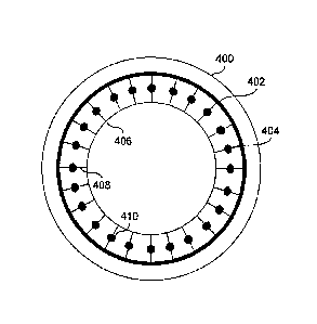
FIG. 4 depicts an JUL 400 in accordance with embodiments of the present
invention. JUL 400 may be loaded with an Internal Structure 402 which may be
[0
deformed using a femtosecond laser to cause the power of IOL 400 to be
adjusted. As
shown in FIG. 4, IOL 400 is preloaded with a structure 402 which includes two
concentric rings 404 and 406. Concentric Rings 404 and 406 have members 408
which
may have localized regions or pockets of heat-absorbing material or dye 410.
FIGs. 5A and 5B show how a laser may be applied to Structure 402 in order to
effect changes. In FIG. 5A, heat-absorbing pockets 410, which may shrink when
exposed
to heat, increasing the stressor tension on inner ring 406 and outer ring 404,
cause the IOL
to be deformed. Similarly, as shown in FIG. 5B, links or members 408 may be
broken
using the femtosecond laser in order to release tension between concentric
rings 406 and
408.
zo The
embodiments of the JUL and procedure of this invention have many
advantages over the prior art. For example, the non-invasive nature of the JUL
power
(sphere and cylinder) adjustment is a very desirable feature. Further, the IOL
may be a
fully cured lens without any significant unreacted monomer, oligomer or
initiator.
CA 02722223 2010-10-21
WO 2009/137362
PCT/US2009/042571
Further still, this power adjustment need not be performed within a limited
postoperative
period. Rather, the adjustment can be performed when a need arises for IOL
power
adjustment.
Another embodiment of the present invention relates to the use of ultrafast
laser
technology to perform ultrafine cutting to modify the shape of an JUL. The
ultra short
pulses in the range of pico to femto seconds at wavelengths in the vicinity of
1 micron can
be used to ablate features into or materials away from an JUL to induce the
shape of the JUL
to change. Embodiments of the present invention use the ultrafast laser in a
real-time
application where a pseudophakic patient has their vision refined by using the
ultra fast
laser which is tracked to the moving eye. The laser performs intraocular
surgical
modification of a pre installed JUL designed to have its shape adapted based
on the action
of the femtosecond laser. The ultrafast pulses can be used to ablate or cut
material from a
specially preloaded controlled micro structure within the JUL, causing the JUL
to deform
in a desired manner. Such a microstructure was presented in FIGs. 3A, 3B, 4,
5A and 5B.
A laser pulse duration of interest is from 100 fs to 10 picoseconds and the
wavelength can
range from 500 nm to 1.1 microns. Dyes can be used as part of the lens
material to
preferentially absorb the ultrafast laser pulses.
Embodiments of the present invention may employ the simultaneous, or at least
contemporaneous, use of wavefi-ont aberrometry to assess the aberration
structure at all
phases of the treatment from pre-implant through to the point in the procedure
where the
post-op aberrations are minimized by adjustment of the structure of the JUL.
This
approach does not require any alteration of the material properties of the IOL
itself or
require the optical portion of the IOL to be modified with the laser. Rather,
the cutting or
11
CA 02722223 2010-10-21
WO 2009/137362
PCT/US2009/042571
ablating action of the femtosecond laser can either be used to break load
bearing members
of the IOL that will allow the lens to flatten in one or more directions and
take a more
desirable shape OR it can be used to heat and shrink a mechanical element that
will be
able to induce a desired stress to the IOL, causing the lens to take a new
steeper shape.
Although the use of femtosecond lasers is specifically described, it will be
apparent to one
skilled in the art that other lasers or external stimuli may be suitable for
performing the
adjustments to the IOL described herein.
The possibility of making intraoperative adjustment to the shape of an IOL,
and
hence its optical power, when combined with the diagnostic power of wavefront
0 aberrometry provides an opportunity to combine two successful
technologies with a
powerful new laser that has recently been introduced into the surgical
armamentarium,
namely the femtosecond laser. This type of laser is capable of penetrating the
cornea
without causing any significant heating or mechanical disruption, but can be
focused to
pinpoint accuracy into the anterior chamber to allow precision cutting
(ablation) and or
5 heating of materials within the anterior chamber. The width of the cut
or ablation and the
amount of associated debris can be very small, about 1-3 microns. Lower peak
laser
powers can be used to generate heat when the laser is interacting with a
section of
material that has been properly doped to permit preferential and very
localized heating. In
particular, an IOL can be pre-stressed to induce a specific spherical
aberration in the lens
:0 that can be released as appropriate to shift the sign of the spherical
aberration or induce
compensating astigmatism as needed. This can be done to compensate for any
residual
corneal spherical aberration or astigmatism that was either pre-existing or
induced by the
surgery. The use of the femtosecond laser can be delayed in order to let the
cornea
12
CA 02722223 2010-10-21
WO 2009/137362
PCT/US2009/042571
completely heal from the JUL insertion procedure. This allows the lens to be
adjusted in
the presence of quiet and stable ocular optics.
FIG. 6 provides a logic flow diagram of an embodiment of a method to correct
for
visual impairments such as aphakia of the eye. Operation 600 begins with the
removal of
a natural lens from an eye in Step 602. The IOL, which may be a multi-focal
IOL, may
then be inserted within the eye. The lenses of the JUL may be convex on both
sides (bi-
convex) and made of a soft plastic that can be folded prior to insertion. This
folding
allows placement through a reduced-size incision wherein the incision is
smaller than the
optic diameter of the JUL. After surgical insertion into the eye in step 604,
the JUL may
gently unfold to restore vision. In Step 606, the JUL is positioned and
secured within the
eye. This may be done with the use of supporting arms (haptics) to provide for
proper
positioning of the JUL within the eye. Embodiments of the present invention
may place
or position the IOL in posterior chamber of the eye to replace the natural
lens as shown in
FIG. 1. This position allows the JUL to correct visual impairments such as the
absence of
a natural lens caused by disease or accident. The lens itself may be a multi-
focal IOL as
discussed previously. This allows patients with and without presbyopia who
desire near
intermediate and distant vision to experience independence from glasses
following
surgery, such as cataract surgery.
FIG. 7 provides a logic flow diagram of an embodiment of a method to power
adjust an in vivo JUL to correct for visual impairments in accordance with
embodiments
of the present invention. This method allows power adjustments to be made to
the JUL.
Operation 700 begins at Step 702, wherein a need for power adjustment of an in
vivo JUL
is determined. This may be done using wavefront aberrometry or other like
diagnostic
13
CA 02722223 2010-10-21
WO 2009/137362
PCT/US2009/042571
procedures. If the JUL in place has a controlled micro structure that places
the JUL under
tension, it may be possible then to reshape the JUL by adjusting the tension
therein. This
may be done by making appropriate changes to the micro structure of the JUL.
These
changes are identified in Step 704. These changes may involve using an
external
stimulus, such as a femtolaser, to reshape members, break crosslinks or induce
cavities
within the JUL. Another embodiment may involve an JUL having a predefined
control
structure wherein members of the structure may be adjusted in length to change
the
tension and reshape the JUL as desired. In Step 706, the changes to the
controlled micro
structure of the JUL are implemented with an external stimulus. Wavefront
aberrometry
and other diagnostic procedures may be applied after the adjustment, either
immediately
or following a period of post surgical adaptation, to measure the
effectiveness and
determine if there is a need for further adjustment and, if so, whether or not
the current
micro structure will support that adjustment.
In summary, embodiments of the present invention provide an improved design
for
an ocular implant. This ocular implant includes an intraocular lens (JUL) and
a number
of haptics. The IOL passes optical energy. A microstructure within the JUL
places the
IOL under tension. The microstructure is operable to be broken in a controlled
manner to
release tension in the IOL and reshape the JUL. This may be done at any time
post
operatively and in conjunction with wavefront aberrometry to provide improved
results.
The haptics mechanically couple to the JUL in order to position and secure the
IOL within
the eye.
The JUL embodiments of this invention can be a multifocal JUL that passes
optical energy in both photopic and mesopic conditions. The multifocal JUL may
include
14
CA 02722223 2010-10-21
WO 2009/137362
PCT/US2009/042571
both a diffractive region and a refractive region. The diffractive region may
be a central
region or optic zone of the lens that includes concentric steps of gradually
varying step
heights in order to allocate energy based on lighting conditions and activity
in order to
create a full range of quality vision, (i.e. near to distant). This allows
conditions where
the natural lens of the eye must be replaced to be corrected.
Embodiments of the present invention allow patients having visual impairment
to
have clear distance vision at smaller pupil conditions, i.e. photopic
conditions, and have
improved vision at larger pupil, i.e. mesopic conditions.
As one of average skill in the art will appreciate, the term "substantially"
or
to "approximately", as may be used herein, provides an industry-accepted
tolerance to its
corresponding term. Such an industry-accepted tolerance ranges from less than
one
percent to twenty percent and corresponds to, but is not limited to, component
values,
integrated circuit process variations, temperature variations, rise and fall
times, and/or
thermal noise. As one of average skill in the art will further appreciate, the
term
"operably coupled", as may be used herein, includes direct coupling and
indirect coupling
via another component, element, circuit, or module where, for indirect
coupling, the
intervening component, element, circuit, or module does not modify the
information of a
signal but may adjust its current level, voltage level, and/or power level. As
one of
average skill in the art will also appreciate, inferred coupling (i.e., where
one element is
coupled to another element by inference) includes direct and indirect coupling
between
two elements in the same manner as "operably coupled". As one of average skill
in the
art will further appreciate, the term "compares favorably", as may be used
herein,
indicates that a comparison between two or more elements, items, signals,
etc., provides a
CA 02722223 2014-04-07
,
desired relationship. For example, when the desired relationship is that
signal 1 has a
greater magnitude than signal 2, a favorable comparison may be achieved when
the
magnitude of signal 1 is greater than that of signal 2 or when the magnitude
of signal 2 is
less than that of signal 1.
Although the present invention is described in detail, it should be understood
that
various changes, substitutions and alterations can be made hereto.
16