Note: Descriptions are shown in the official language in which they were submitted.
CA 02722749 2010-10-27 PCT/US 2009/041 610 - 01-03-2010
DEVICE PLATFORM FOR MEDICAL PROCEDURES
PRIORITY CLAIM
[00011 This invention claims the benefit of priority of U.S. Provisional
Application Serial
No. 61/048,647, entitled "Device Platform for Medical Procedures," filed April
29, 2008, the
disclosure of which is hereby incorporated by reference in its entirety.
TECHNICAL FIELD
[00021 The present embodiments relate generally to the field of medical
devices, and more
particularly, to apparatus and methods for facilitating positioning of one or
more medical
components.
BACKGROUND INFORMATION
[00031 Medical components may be advanced into the human or animal body for a
wide
range of diagnostic and therapeutic purposes. For example, catheters, needles,
extraction
baskets, stents, snares and numerous other components may be inserted into a
patient's
vasculature, gastric system, or other ducts or passageways in order to
facilitate imaging
and/or treatment of various conditions.
[00041 The medical components may be inserted and advanced through an outer
cover,
sheath, endoscope or similar introducer device. In the case of an endoscopic
procedure, an
endoscope may be advanced towards a target site using an imaging system, and
one or more
medical components may be advanced through a working lumen of the endoscope
towards
the target site.
[0005] There are instances in which multiple medical components may be used in
a series of
smaller procedures during a single, larger operation. For example, in a
"rendezvous
procedure," a combination of one or more smaller procedures may be performed
such as the
injection of a radiopaque dye, cannulation of a passageway, drainage of a
cyst, delivery of a
therapeutic agent to a target site, the use of fine needle aspiration to
obtain tissue or fluid
samples, dilation of a target site, insertion of a stent, and so forth.
1
AMENDED SHEET
CA 02722749 2010-10-27 PCT/US 2009/041 610 - 01-03-2010
[0006] It may become difficult to precisely position a medical component
within a patient's
vessel or duct, particularly when multiple components are used at the same
time. Moreover,
incremental longitudinal advancement or retraction of the multiple medical
components with
respect to each other may become difficult to achieve. Furthermore, it may
become difficult
to exchange one medical component for another. JP Patent Publication 2001-
170063-A
discloses a flat rail plate that may be coupled to an endoscope, the flat rail
plate having a
groove formed therein, whereby connectors coupled to medical devices are
movable along
the groove. The flat rail plate is not movable relative to the endoscope, and
further, it is not
possible to move the rail plate and any of the connectors in tandem relative
to the introducer
device.
[0007] Accordingly, there is a need for apparatus and methods that facilitate
insertion and/or
positioning of one or more medical components, such that improved positioning
of the
medical components may be achieved in an easy-to-use manner.
SUMMARY
[00081 The present embodiments provide apparatus and methods suitable for
facilitating
positioning one or more medical components. In one embodiment, the apparatus
comprises a
rail member having proximal and distal ends and a generally longitudinal axis.
A first
connector is adapted to be selectively coupled to the rail member, and a first
medical
component is adapted to be coupled to the first connector. The first connector
is disposed for
selective longitudinal movement along the rail member, thereby advancing or
retracting the
first medical component upon corresponding movement of the first connector.
[0009] The apparatus may further comprise a second connector adapted to be
coupled to the
rail member and a second component. The second connector is disposed for
selective
longitudinal movement along the rail member, thereby advancing and retracting
the second
medical component upon corresponding movement of the second connector. The
second
connector is configured for independent longitudinal movement relative to the
first connector.
[0010] An adapter may be coupled to the distal end of the rail member, and
further may be
coupled to a port of an introducer device, such as an endoscope, in order to
align the first
connector with the port of the introducer device to facilitate insertion of
the first medical
component into the introducer device. The rail member may comprise measurement
indicia
2
AMENDED SHEET
CA 02722749 2010-10-27 PCT/US 2009/041 610 - 01-03-2010
for identifying a longitudinal distance in which the first connector and the
first medical
component have been advanced or retracted with respect to the rail member. In
this manner,
one or more components, such as a catheter and a needle, may be inserted into
a human or
animal body in a controlled fashion with respect to one another.
[00111 The first and second connectors each may comprise a rail engaging
portion and a
component engaging portion. The rail engaging portion may comprise a bore
having a
greater diameter than an outer diameter of the rail member to permit movement
of the
connectors over the rail member. The rail engaging portion of the connectors
also may
comprises a guide member adapted for movement within a slot formed in the rail
member,
thereby substantially inhibiting rotational movement of the connectors with
respect to the rail
member. An actuator, such as a thumb screw, may be provided for selectively
inhibiting
longitudinal movement of the connectors with respect to the rail member,
thereby securing
the longitudinal positioning of the first and second medical components.
10012] The component engaging portion of the first and second connectors each
may
comprise a bore adapted to receive a proximal end of the first medical
component. The first
and second medical components are adapted to be securely connected to the
component
engaging portion, for example, using an actuator such as a thumb screw.
[0013] In one exemplary method of operation, the introducer device may
comprise an
endoscope having an accessory port that enables insertion of first and second
medical
components into a working lumen of the endoscope. In one embodiment, the first
medical
component may comprise a catheter having a lumen, and the second medical
component may
comprise a needle adapted to be advanced through the lumen of the catheter.
The catheter
and the needle may be advanced through the working lumen of the endoscope to a
target site.
Advantageously, using the rail member in conjunction with first and second
connectors, the
first and second medical components may be independently advanced a fixed
amount with
respect to one another, and with respect to the endoscope, to perform the
objectives of a
particular procedure.
[0014] Other systems, methods, features and advantages of the invention will
be, or will
become, apparent to one with skill in the art upon examination of the
following figures and
detailed description. It is intended that all such additional systems,
methods, features and
advantages be within the scope of the invention, and be encompassed by the
following
claims.
3
AMENDED SHEET
CA 02722749 2010-10-27 PCT/US 2009/041 610 - 01-03-2010
BRIEF DESCRIPTION OF THE DRAWINGS
[0015] The invention can be better understood with reference to the following
drawings and
description. The components in the figures are not necessarily to scale,
emphasis instead
being placed upon illustrating the principles of the invention. Moreover, in
the figures, like
referenced numerals designate corresponding parts throughout the different
views.
[0016] FIG. I is a perspective view of a first embodiment of apparatus for
facilitating
positioning of one or more medical components.
[0017] FIG. 2 is a perspective view illustrating features of a connector of
FIG. 1.
[0018] FIG. 3 is a perspective view showing the apparatus of FIG. I coupled to
an accessory
port of an endoscope.
[0019] FIG. 4 is a perspective view showing exemplary first and second medical
components
being used in conjunction with the apparatus of FIGS. 1-3.
[0020] FIG. 5 is a cross-sectional view along line A--A of FIG. 4.
[0021] FIG. 6 is a perspective view illustrating the second medical component
being
advanced with respect to the first medical component.
[0022] FIG. 7 is a perspective view illustrating the first medical component
being advanced
with respect to the second medical component.
[0023] FIG. 8 is a perspective view illustrating the second medical components
being
removed from the rail member.
[0024] FIG. 9 is a perspective view showing an alternative first medical
component, along
with a second medical component, being used in conjunction with the apparatus
of FIGS. 1-3.
[0025] FIG. 10 is a cross-sectional view along line B--B of FIG. 9.
[0026] FIG. I 1 is a perspective view of an alternative embodiment of
apparatus for
facilitating positioning of one or more medical components.
DETAILED DESCRIPTION OF THE PREFERRED EMBODIMENTS
[0027] In the present application, the term "proximal" refers to a direction
that is generally
towards a physician during a medical procedure, while the term "distal" refers
to a direction
that is generally towards a target site within a patient's anatomy during a
medical procedure.
[0028] Referring now to FIG. 1, a first embodiment of apparatus suitable for
facilitating
insertion of one or more medical components is described. The apparatus 20
generally
4
AMENDED SHEET
CA 02722749 2010-10-27 PCT/US 2009/041 610 - 01-03-2010
comprises a rail member 30 having proximal and distal ends 32 and 34,
respectively, and a
generally longitudinal axis L. The apparatus 20 further comprises at least a
first connector
50, which is adapted for longitudinal movement with respect to the rail member
30, as set
forth in greater detail below.
[0029) The first connector 50 may comprise a rail engaging portion 52 and a
component
engaging portion 62, as shown in FIG. 2. The rail engaging portion 52 may
comprise a
substantially hollow member having a bore 54 formed therethrough. The bore 54
may
comprise a diameter that may be slightly greater than an outer diameter of the
rail member
30. Accordingly, the rail engaging portion 52 of the first connector 50 may be
disposed over,
and slide along, the rail member 30, as generally depicted in FIG. 1.
[0030] The rail member 30 may comprise a generally cylindrical cross-sectional
shape, as
depicted in FIG. 1. If the rail member 30 comprises a generally cylindrical
shape, then the
bore 54 of the rail engaging portion 52 also may comprise a generally
cylindrical shape to
permit selective longitudinal movement of the first connector 50 with respect
to the rail
member 30. In alternative embodiments, the rail member 30 may comprise other
suitable
exterior shapes, such as an oval or rectangular cross-sectional shape, in
which case the bore
54 of the rail engaging portion 52 may comprise a corresponding interior
shape.
[0031] In one embodiment, the rail engaging portion 52 may comprise a guide
member 58,
which may be longitudinally oriented and disposed within the bore 54, as shown
in FIG. 2.
The guide member 58 may be disposed for longitudinal movement within a
longitudinally
oriented slot 37 formed in a lateral surface of the rail member 30, as
depicted in FIG. 1.
Accordingly, the guide member 58 may permit longitudinal movement of the first
connector
50 with respect to the rail member 30, while substantially inhibiting
rotational movement of
the first connector 50 with respect to the rail member 30.
[0032] The first connector 50 further may comprise a mechanism for securing
its longitudinal
movement with respect to the rail member 30. In one embodiment, the mechanism
comprises
a thumb screw 55 having a threaded portion 56, as shown in FIGS. 1-2. The
threaded portion
56 may be coupled to a bore 59 having internal threading, thereby allowing
incremental
adjustment of the positioning of the thumb screw 55 within the bore 54 of the
rail engaging
portion 52. The thumb screw 55 may be actuated such that the threaded portion
56 is
disposed within the bore 54, thereby engaging an outer surface of the rail
member 30 and
inhibiting longitudinal movement of the first connector 50 with respect to the
rail member 30.
5
AMENDED SHEET
CA 02722749 2010-10-27 PCT/US 2009/041 610 - 01-03-2010
[00331 The first connector 50 further comprises a component engaging portion
62, which
may comprise features that are similar to the rail engaging portion 52. More
specifically, the
component engaging portion 62 may comprise a substantially longitudinally
oriented bore 64
formed therein, as shown in FIGS. 1-2. The bore 64 may be configured for
receiving a
proximal portion of one or more medical components, as explained in further
detail below.
The first connector 50 also may comprise a mechanism for securing a medical
component
within the bore 64 of the component engaging portion 62. In one embodiment, a
thumb
screw 65 having a threaded portion 66 may be coupled to a bore 69 having
internal threading,
as shown in FIG. 2. The thumb screw 65 may be incrementally positioned within
the bore 64
of the component engaging portion 62 to engage and effectively secure a
medical component
positioned therein, as explained further below.
[00341 In the embodiments shown herein, the apparatus 20 comprises the first
connector 50
and also a second connector 70. Unless otherwise noted, the first and second
connectors 50
and 70 are substantially identical. The first connector 50 may be coupled to
the proximal end
of a first medical component, while the second connector 70 may be coupled to
the proximal
end of a second medical component. As will be explained further below, the
first and second
connectors 50 and 70 may be longitudinally positioned with respect to each
other, via the rail
member 30, to thereby incrementally longitudinally position the first medical
component with
respect to the second medical component as well as with respect to the
endoscope 110.
While two connectors 50 and 70 are depicted, greater or fewer connectors may
be used at any
time in conjunction with the rail member 30 to facilitate positioning of one
or more medical
components.
[00351 The apparatus 20 further may comprise an adapter 90, which may couple
the distal
end 34 of the rail member 30 to an introducer device, such as an endoscope
110, as shown in
FIG. 3. The adapter 90 may comprise any suitable shape configured to couple
the rail
member 30 to the introducer device. By way of example, the adapter 90 may
comprise a
male luer lock adapter having a distal region 92, which may be removably
coupled to an
accessory port 125 of the endoscope 110, as shown in FIG. 3.
[0036] Further, the apparatus 20 may comprise a fitting 35, which is adapted
to be coupled
between the adapter 90 and the rail member 30, as shown in FIG. 3. The fitting
35 may be
removably or permanently coupled to the distal end 34 of the rail member 30.
The fitting 35
may comprise a C-shaped clip 36, shown substantially from the side in FIG. 3,
that is adapted
6
AMENDED SHEET
CA 02722749 2010-10-27 PCT/US 2009/041 610 - 01-03-2010
to be coupled to a notch 94 formed in the adapter 90. When the C-shaped clip
36 is secured
within the notch 94, e.g., using a snap-fit connection, the rail member 30 may
be securely
coupled to the adapter 90 and also the endoscope 110. While a C-shaped clip 36
is depicted
in FIG. 1 and FIG. 3, any suitable mechanism may be used for removably
coupling the rail
member 30 to the adapter 90.
[00371 The rail member 30 may be coupled to the accessory port 125 of the
endoscope 110
such that the bores 64 and 84 of the first and second connectors 50 and 70,
respectively, are
substantially aligned with the adapter 90, as shown in FIG. 3. Therefore, when
medical
components are secured within the bores 64 and 84 of the first and second
connectors 50 and
70, respectively, the first and second connectors 50 and 70 may be
longitudinally advanced
over the rail member 30 to longitudinally advance the medical components
through the
adapter 90 and into a working lumen 128 of the endoscope 110, as explained
further below.
[00381 The rail member 30 may be formed from any suitable material, such as
plastic or
stainless steel. The overall longitudinal length of the rail member 30, i.e.,
the length between
the proximal end 32 and the distal end 34, may comprise any suitable length
for permitting
movement of one or more connectors along the rail member 30. In one
embodiment, the
longitudinal length of the rail member 30 may be about 20 to about 30
centimeters. Further,
the rail member 30 may comprise measurement indicia 38, which may allow a
physician to
know exactly how far the first and second connectors 50 and 70 are moved with
respect to the
endoscope 110 and further with respect to one another.
[00391 The apparatus 20 may be used in conjunction with any suitable
introducer device,
such as the endoscope 110 shown in FIG. 3. The exemplary endoscope shown
comprises a
side-viewing endoscope 110 having proximal and distal ends 132 and 134,
respectively. The
endoscope 110 may comprise fiber optic components for illuminating and
capturing an image
distal to the endoscope 110. A physician may view the images distal to the
endoscope using
an eyepiece 120. A fiber optic cable 122 may be coupled between the endoscope
110 and a
suitable light source. A control section 123 may be provided to facilitate
actuation of various
components associated with the endoscope 110.
[00401 Further, the endoscope 110 may comprise a working lumen 128, as shown
in FIG. 5.
The working lumen 128 is sized to accommodate at least one medical component
therein, as
generally described with respect to FIGS. 4-10. An adjustable guiding element
may be
disposed near the distal end 134 of the endoscope 110 to cause components
advanced through
7
AMENDED SHEET
CA 02722749 2010-10-27 PCT/US 2009/041 610 - 01-03-2010
the working lumen 128 to exit at a predetermined angle with respect to the
endoscope 110. It
should be noted that, for illustrative purposes, the optical components
associated with the
endoscope 110 are not shown in the cross-sectional view of FIG. 5.
[0041] Optionally, an endoscopic ultrasound procedure may be performed, in
which case the
endoscope 110 may be retrofitted with an ultrasound transducer. The ultrasound
transducer
may produce accurate and detailed images of the digestive tract and
surrounding organs,
particularly due to the proximity of the ultrasound transducer to the organ or
tract being
imaged.
[0042] Referring now to FIGS. 4-8, a first exemplary method of using the
apparatus 20 is
described. In a first step, the rail member 30 may be coupled to the accessory
port 125 of the
endoscope 110, as generally set forth above. In one embodiment, the rail
member 30 may be
coupled to the adapter 90 by coupling the C-fitting 36 to the notch 94 of the
adapter 90.
Other techniques or mechanisms may be employed to couple the rail member 30 to
the
endoscope 110.
[0043] In a next step, proximal ends of one or more medical components, such
as a dilation
catheter 150 and a needle 170, may be coupled to the first and second
connectors 50 and 70,
respectively. It should be noted that the dilation catheter 150 may be coupled
to the first
connector 50 before the first connector 50 is secured to the rail member 30,
and similarly, the
needle 170 may be coupled to the second connector 70 before the second
connector 70 is
secured to the rail member 30, which may facilitate the loading process.
[0044] In one exemplary step, a proximal end 152 of the dilation catheter 150
may be
coupled to the first connector 50, for example, using a luer fitting 160. The
luer fitting 160
may be positioned within the bore 64 (see FIGS. 1-3) of the component engaging
portion 62
of the first connector 50. At least a portion of the luer fitting 160
comprises an outer
diameter that is smaller than an inner diameter of the bore 64, thereby
allowing the luer
fitting 160 to be at least partially disposed within the bore 64, as shown in
FIG. 4. In a next
step, the thumb screw 65 may be actuated to cause the threaded portion 66 (see
FIG. 2) to be
advanced radially inward, thereby engaging an outer surface of the luer
fitting 160. This
action secures the proximal end 152 of the dilation catheter 150 to the first
connector 50.
[00451 Similarly, a proximal end 172 of the needle 170 may be coupled to the
second
connector 70, for example, using a luer fitting 180, which may be positioned
within the bore
84 of the component engaging portion 82 of the second connector 70. The thumb
screw 85
8
AMENDED SHEET
CA 02722749 2010-10-27 PCT/US 2009/041 610 - 01-03-2010
may be actuated to engage an outer surface of the luer fitting 180, thereby
securing the
proximal end 172 of the needle 170 to the second connector 70.
[0046] In a next step, a portion of the dilation catheter 150 may be inserted
into the working
lumen 128 of the endoscope 110 and the first connector 50 may then be coupled
to the rail
member 30. More specifically, the distal end 154 of the catheter 150 (see FIG.
7) may be
advanced distally through the adapter 90 and into the working lumen 128 of the
endoscope
110, as depicted in FIGS. 4-5. The working lumen 128 of the endoscope 110 may
have an
inner diameter of about 4.0 - 5.5 mm, while the overall diameter of the
endoscope 110 may
be about 10-14 mm. An outer diameter of the catheter 150 may be about 3.0 mm
and may
comprise a lumen 155 having a diameter of about 1.5 mm. Such dimensions are
provided for
exemplary reference purposes and are not intended to be limiting.
[0047] The distal end 154 of the catheter 150 may be advanced distally within
the working
lumen 128, for example, until it is disposed just proximal to the distal end
134 of the
endoscope 110. During this time, the guide member 58 of the first connector 50
(see FIG. 2)
may be aligned and advanced distally within the longitudinally oriented slot
37 of the rail
member 30. When positioned at a desired longitudinal location, the thumb screw
55 may be
actuated to secure the longitudinal position of the first connector 50.
Accordingly, the distal
end 154 of the catheter 150 may be securely disposed at a predetermined
longitudinal
location within the working lumen 128 of the endoscope 110.
[0048] Referring still to FIG. 4, the needle 170 then may be disposed through
the lumen 155
of the catheter 150 and coupled to the rail member 30. In particular, a distal
end 174 of the
needle 170 may be loaded in a distal direction through the luer fitting 160
that is coupled to
the proximal end 152 of the catheter 150. Then the needle 170 may be advanced
distally
through the lumen 155 of the catheter 150 until the needle 170 is positioned
at a desired
location. For example, the distal end 174 of the needle 170 may be advanced
distally within
the lumen 155 until it is aligned with the distal end of the catheter 155,
which may be
disposed just proximal to the distal end 134 of the endoscope 110. During this
time, the
second connector 70 may be aligned with, and advanced distally over, the rail
member 30.
When positioned at a desired longitudinal location, the thumb screw 75 may be
actuated to
temporarily secure the positioning of the second connector 70 and the needle
170, as shown
in FIG. 4.
9
AMENDED SHEET
CA 02722749 2010-10-27 PCT/US 2009/041 610 - 01-03-2010
[0049] The endoscope 110 may be advanced towards a target site within a
patient's anatomy
using suitable imaging techniques. If an endoscopic ultrasound procedure is
performed, the
endoscope 110 may be retrofitted with an ultrasound transducer, as noted
above. The
ultrasound transducer may produce accurate and detailed images, particularly
when placed
within a certain distance of a desired target site. For example, during a
translumenal
procedure, the ultrasound transducer may facilitate imaging of a target site
in an adjacent
vessel or duct when disposed within an original vessel or duct in close
proximity to the
selected target site. If the distance is too great for use of an ultrasound
transducer, then
fluoroscopic techniques may be employed, optionally in conjunction with
radiopaque bands
disposed on the endoscope 110.
(0050] During positioning of the endoscope 110, the rail member 30 may be
coupled to the
auxiliary channel 125 of the endoscope 110, further with the dilation catheter
150 and the
needle 170 disposed within the working lumen 128 of the endoscope 110 via the
first and
second connectors 50 and 70, respectively. In this instance, the dilation
catheter 150 and the
needle 170 may be pre-positioned within the working lumen 128 of the endoscope
110 during
insertion, and therefore, substantially ready for use upon final positioning
of the endoscope
110.
[0051] Alternatively, the endoscope 110 may be advanced into a patient's body
without the
dilation catheter 150 and the needle 170 loaded into the working lumen 128
and/or without
the rail member 30 coupled to the auxiliary channel 125. For example, the
endoscope 110
may be advanced to a desired location within the patient's anatomy.
Optionally, a wire guide
then may be loaded through the working lumen 128 of the endoscope 110 and
advanced
towards a target site. The catheter 150 may be advanced distally over the wire
guide, through
the working lumen 128 of the endoscope 110, and positioned at a desired
location. The rail
member 30 then may be secured to the endoscope 110 and the longitudinal
positioning of the
catheter 150 may be secured by coupling the proximal end 152 of the catheter
150 to the rail
member 30 using the first connector 50. Subsequently, the wire guide may be
proximally
retracted and removed from the lumen 155 of the catheter 150. Then, the needle
170 may be
distally advanced through the lumen 155 of the catheter 150 and secured in the
position
shown in FIG. 4 using the second connector 70.
[0052] The above-described techniques are exemplary techniques for preparing
the dilation
catheter 150 and the needle 170 for subsequent use with the endoscope 110 and
the rail
AMENDED SHEET
CA 02722749 2010-10-27 PCT/US 2009/041 610 - 01-03-2010
member 30. Other loading and insertion techniques may be employed to couple
the dilation
catheter 150 and the needle 170 to the endoscope 110 and the rail member 30.
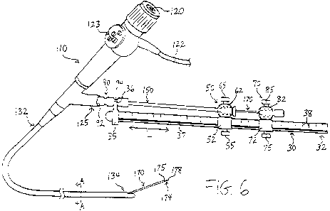
[0053] Referring now to FIG. 6, in a next step, it may become desirable to
move the needle
170 a fixed distance with respect to the dilation catheter 150. The thumb
screw 75 may be
actuated in a direction away from the rail member 30 to permit longitudinal
movement of the
second connector 70 with respect to the rail member 30. Using the measurement
indicia 38, a
physician may know exactly how far the second connector 70 is being moved
distally with
respect to the fixed first connector 50, and therefore, how much the needle
170 is being
advanced distally with respect to the dilation catheter 150 and the endoscope
110. As shown
in FIG. 6, such distal movement of the second connector 70 may cause the
distal end 174 of
the needle 170 to extend distally beyond the distal end 134 of the endoscope
110. Using the
eyepiece 120, a physician may view the positioning of the distal end 174 of
the needle 170.
[0054] The distal end 174 of the needle 170 may comprise a sharpened tip 178.
Further, the
needle 170 may comprise a bore 175 extending between the proximal and distal
ends 172 and
174, as depicted in FIGS. 5-6. The needle 170 may comprise an endoscopic
ultrasound
(EUS) needle, or any other suitable needle. Moreover, the needle 170 may be
used for
puncturing tissue, obtaining translumenal access, draining cysts or
pseudocysts, obtaining
tissue samples, providing aspiration, injecting fluids, delivering sclerosing
agents or other
therapeutic agents, and other purposes.
[0055] In one exemplary procedure, the needle 170 may be used for obtaining
translumenal
access from the gastrointestinal tract to a target site, for example, the
celiac plexus. In this
case, the distal end of the endoscope 110 may be placed at the appropriate
location within the
stomach and the needle 170 may be advanced, as shown in FIG. 6, to
translumenally pierce
through the stomach wall, through the peritoneal cavity, and into the celiac
plexus. As will
be apparent, many other applications, both translumenal and otherwise, are
possible.
[0056] Referring now to FIG. 7, in a next optional step, it may become
desirable to move the
dilation catheter 150 a fixed distance with respect to the needle 170 and the
endoscope 110.
The thumb screw 55 may be actuated in a direction away from the rail member 30
to permit
longitudinal movement of the first connector 50 with respect to the rail
member 30. Using
the measurement indicia 38, a physician may know exactly how far the first
connector 50 is
being moved distally with respect to the second connector 70 and the endoscope
110. As
shown in FIG. 7, such distal movement of the first connector 50 may cause the
distal end 154
11
AMENDED SHEET
CA 02722749 2010-10-27 PCT/US 2009/041 610 - 01-03-2010
of the dilation catheter 150 to extend distally beyond the distal end 134 of
the endoscope 110.
It should be noted that in this configuration, the distal end 154 of the
catheter 150 may extend
distally beyond the sharpened tip 178 of the needle 170, thereby covering the
distal end 174
of the needle 170 and reducing the likelihood of the needle inadvertently
cutting tissue.
[00571 The distal end 154 of the dilation catheter 150 may comprise a widened
section 158,
which may be used, for example, to cannulate a body passageway or dilate a
stenosis. As
will be explained below with respect to FIGS. 9-10, a balloon inflation
catheter may be
employed in lieu of the dilation catheter 150 shown in the above-described
embodiments.
[0058) Referring now to FIG. 8, once a physician has performed one or more
desired
procedures using the dilation catheter 150 and/or the needle 170, the needle
170 may be
removed from the ensdoscope 110 and the rail member 30. Specifically, the
thumb screw 75
may be actuated to permit longitudinal movement of the second connector 70.
The rail
engaging portion 72 of the second connector 70 then may be retracted
proximally in a linear
direction until the second connector 70 is beyond the proximal end 32 of the
rail member 30,
' as shown in FIG. 8. At this time, the second connector 70 further may be
retracted
proximally to withdraw the needle 170 from the lumen 155 of the dilation
catheter 150.
[0059) With the needle 170 removed from the lumen 155 of the dilation catheter
150, another
component may be introduced through the lumen 155 of the dilation catheter
150. A
proximal end of the new component may be coupled to the component engaging
portion 82
of the second connector 70, while a distal end of the new component may be
inserted through
the lumen 155 of the dilation catheter 150.
[0060] Further, a suction source may be coupled to the catheter 150 and used
to aspirate a
cyst or other condition through the lumen 155 of the catheter 150. With the
needle 170
having been removed from the lumen 155, a greater diameter of flow is provided
to yield
enhanced suction capability, which may advantageously drain a cyst in a faster
manner.
[0061) Alternatively, the dilation catheter 150 also may be removed from the
ensdoscope 110
and the rail member 30. In either case, one or more new medical components may
be
advanced over the rail member 30 and into the endoscope 110.
[0062) In this manner, any number of medical components may be exchanged and
used in
conjunction with the rail member 30. By coupling proximal ends of the medical
components
to connectors, such as the first and second connectors 50 and 70, the
components may be
advanced or retracted a desired distance using the measurement indicia 38.
Further, multiple
12
AMENDED SHEET
CA 02722749 2010-10-27 PCT/US 2009/041 610 - 01-03-2010
components may be advanced or retracted in a simple linear fashion with
respect to one
another, and with respect to the endoscope 110.
[0063] It should be noted that the second component, which is disposed
proximal to the first
component on the rail member 30, need not be inserted through a lumen of the
first
component. For example, in the example of FIGS. 4-8, when the dilation
catheter 150 is
inserted through the working lumen 128 of the endoscope 110, the needle 170 or
other
medical device may be advanced adjacent to an outer surface of the dilation
catheter 150
within the working lumen 128, as opposed to through the lumen 155 of the
dilation catheter
150.
[0064] Referring now to FIGS. 9-10, in an alternative embodiment, a balloon
inflation
catheter 150' may be used in lieu of a non-inflatable dilation catheter 150.
The balloon
catheter 150' preferably comprises a first lumen 155 for receiving a second
component, such
as the needle 170, and further comprises a second, inflation lumen 188, as
shown in FIG. 10.
The inflation lumen 188 is in fluid communication with a balloon coupled to a
distal region
of the catheter 150'. In use, a Y-shaped connector 185 may be coupled to the
proximal end
of the balloon catheter 150', as shown in FIG. 9. The second component, such
as the needle
170, may be advanced through the Y-shaped connector 185 and into the lumen 155
of the
catheter 150'. The other inlet port 186 of the Y-shaped connector 185 may be
coupled to an
inflation source (not shown) via tubing 187. The inflation source and the
tubing 187 are
placed in fluid communication with the inflation lumen 188 to permit selective
inflation and
deflation of the balloon of the catheter 150'. Like the embodiments described
above, the first
and second connectors 50 and 70 may be advanced or retracted with respect to
one another a
fixed distance to achieve the objects of a particular medical procedure.
[0065] Referring now to FIG. 11, an alternative apparatus 20' generally
comprises the rail
member 30, the first connector 50, and the second connector 70, each of which
preferably are
provided as described in detail above. The apparatus 20' further comprises an
adapter
connector 250 in lieu of the fitting 35 above.
[0066] The adapter connector 250 preferably is provided in accordance with the
first
connector 50 and comprises a rail engaging portion 252, an adapter engaging
portion 262,
and thumb screws 255 and 265. The thumb screw 255, which may be provided in
accordance
with the thumb screw 55 described above, may be actuated to selectively engage
an outer
13
AMENDED SHEET
CA 02722749 2010-10-27 PCT/US 2009/041 610 - 01-03-2010
surface of the rail member 30 to selectively pen-nit and inhibit longitudinal
movement of the
rail member 30 with respect to the adapter 90.
[0067] Advantageously, the rail member 30 may be advanced relative to the
endoscope 110
when the thumb screw 255 is unscrewed, to thereby simultaneously advance the
first and
second connectors 50 and 70, along with any associated medical components. By
advancing
the rail member 30 to simultaneously advance the first and second connectors
50 and 70, the
relative positioning of multiple medical components may be maintained. For
example, if the
dilation catheter 150 and the needle 170 described above are coupled to the
first and second
connectors 50 and 70, respectively, then advancement or retraction of the rail
member 30 via
the adapter connector 250 may simultaneously advance or retract the dilation
catheter 150
and the needle 170 in tandem. Therefore, in the embodiment of FIG. 11, a
physician need not
advance two different medical components separately when it is desired to
maintain their
relative positioning. Notably, the thumb screw 265 may be independently
actuated to unlock
the adapter connector 250 from the adapter 90.
[0068] While two medical components have been shown herein, greater or fewer
components
and connectors may be employed. For example, only one medical component, and
therefore
one first connector 50, may be used with the endoscope 110 and the rail member
30.
Alternatively, three or more medical components may be used at any given time,
in which
case three or more associated connectors may be advanced with respect to one
another along
the rail member 30, or the rail member 30 may be advanced relative to the
endoscope 110 in
tandem with the three or more associated connectors, as described in FIG. 11.
[0069) It will be appreciated that a wide variety of medical components may be
used in
conjunction with the apparatuses 20 and 20'. For example, catheters, needles,
extraction
baskets and snares may be delivered using the rail member 30, either alone or
in conjunction
with one another. Further, self-expanding or balloon-expandable stents may be
delivered
using the apparatuses 20 and 20'. Still further medical components and
applications are
intended to be within the scope of the present embodiments. Moreover, while
the introducer
device has been shown as an exemplary endoscope 110, the apparatuses 20 and
20' may be
used in conjunction with various other introducer devices. Still further, it
should be noted
that Touhy-Borst adapters may be used in conjunction with the apparatuses 20
and 20' to
facilitate closure and prevent air from entering into the systems.
14
AMENDED SHEET
= CA 02722749 2010-10-27 PCT/US 2009/041 610 - 01-03-2010
[00701 While various embodiments of the invention have been described, it will
be apparent
to those of ordinary skill in the art that many more embodiments and
implementations are
possible within the scope of the invention. Accordingly, the invention is not
to be restricted
except in light of the attached claims and their equivalents.
AMENDED SHEET _