Note: Descriptions are shown in the official language in which they were submitted.
CA 02722765 2010-10-27
WO 2009/134769 PCT/US2009/041932
NANOPARTICLES FOR USE IN PHARMACEUTICAL COMPOSITIONS
BACKGROUND
Particulate carriers are commonly used in the pharmaceutical arts. For
example,
particulate carriers have been used with adsorbed or entrapped antigens in
attempts to elicit
adequate immune responses. Such carriers present multiple copies of a selected
antigen to the
immune system and are believed to promote trapping and retention of antigens
in local lymph
nodes. The particles can be phagocytosed by macrophages and can enhance
antigen presentation
through cytokine release.
For example, commonly owned International Publication No. WO 98/33487 and co-
pending Pub. No. US 2003/0049298 describe the use of antigen-adsorbed and
antigen-
encapsulated microparticles to stimulate immunological responses, including
cell-mediated
immunological responses, as well as methods of making the microparticles.
Polymers used to
form the microparticles include poly(lactide) and poly(lactide-co-glycolide)(
PLG).
Commonly owned International Publication No. WO 00/06123 and WO 01/36599 and
U.S.
Patent No. 6,884,435 disclose methods of making microparticles having adsorbed
macromolecules, including polynucleotides and polypeptide antigens. The
microparticles
comprise, for example, a biodegradable polymer and are formed using, for
example, cationic,
anionic or nonionic detergents. Microparticles containing anionic detergents
can be used with
positively charged macromolecules, such as polypeptides. Microparticles
containing cationic
detergents can be used with negatively charged macromolecules, such as DNA.
The use of such
microparticles to stimulate immunological responses, including cell-mediated
immunological
responses, is also disclosed.
Commonly owned International Patent Appin. No. PCT/US06/46212 describes
sterile-
filtered lyophilized nanoparticle compositions which contain at least one
biodegradable polymer,
at least one surfactant, at least one cryoprotective agent, and at least one
antigen. Also disclosed
are methods of making and using such compositions and kits supplying such
compositions.
Nanoparticles are created using the nanoprecipitation method.
1
CA 02722765 2010-10-27
WO 2009/134769 PCT/US2009/041932
SUMMARY OF THE INVENTION
In various aspects of the present invention, nanoparticle compositions are
provided which
comprise (a) nanoparticles comprising one or more biodegradable polymers and
(b) one or more
pharmaceuticals associated with the nanoparticles.
In certain embodiments, the nanoparticle compositions are sterile filtered
nanoparticle
compositions, which may or may not be lyophilized.
In certain embodiments, the nanoparticles within the compositions of the
present invention
typically have a size distribution in which the Z average and/or the D(v,0.5)
value is less than
200 nm, and more typically less than 150 nm and in which the D(v,0.9) is less
than 250 nm, and
more typically less than 200 nm.
In certain embodiments, the biodegradable polymers within the nanoparticles
are
synthetic biodegradable polymers, for example, selected from polyesters
including poly(a-
hydroxy acids) and polycaprolactones, polyorthoesters, polyanhydrides,
polycyanoacrylates, and
combinations thereof, among others.
In some aspects of the invention, the pharmaceuticals may be immunogenic
species.
In certain embodiments, the immunogenic species are species that stimulate an
adaptive
immune response. For example, the immunogenic species in these embodiments may
comprise
one or more antigens. Examples of antigens include polypeptide-containing
antigens,
polysaccharide-containing antigens, and polynucleotide-containing antigens,
among others.
Antigens can be derived, for example, from tumor cells and from pathogenic
organisms such as
viruses, bacteria, fungi and parasites, among other sources.
In certain embodiments, the immunogenic species are species that stimulate an
innate
immune response. For example, the immunogenic species may be an activator of
one or more of
the following receptors, among others: Toll-like receptors (TLRs), nucleotide-
binding
oligomerization domain (NOD) proteins, and receptors that induce phagocytosis,
such as
scavenger receptors, mannose receptors and P-glucan receptors.
In certain embodiments, the immunogenic species may be selected, for example,
from
one or more of the following immunological adjuvants: bacterial
lipopolysaccharides,
peptidoglycan, bacterial lipoproteins, bacterial flagellins, imidazoquinoline
compounds,
2
CA 02722765 2010-10-27
WO 2009/134769 PCT/US2009/041932
immunostimulatory oligonucleotides, single-stranded RNA, saponins, lipotechoic
acid, ADP-
ribosylating toxins and detoxified derivatives thereof, polyphosphazene,
muramyl peptides,
thiosemicarbazone compounds, tryptanthrin compounds, and lipid A derivatives,
among others.
In certain embodiments, the immunogenic species may be selected, for example,
from
one or more small molecule immunopotentiators. For example, the immunogenic
species may
be selected from imidazoquinoline compounds such as resimiquod, imiquimod and
imidazoquinoline 090, among others.
In certain embodiments, the compositions of the invention optionally comprise
at least
one surfactant. In some embodiments, the compositions of the invention
optionally comprise at
least one cryoprotective agent. In some embodiments, the compositions of the
invention
optionally comprise at least one surfactant and at least one cryoprotective
agent. Examples of
cryoprotective agents include polyols, carbohydrates and combinations thereof,
among others.
Examples of surfactants include non-ionic surfactants, cationic surfactants,
anionic surfactants,
and zwitterionic surfactants, among others. Surfactants and/or cryoprotective
agents may be
added, for example, to ensure that lyophilized nanoparticles can be
resuspended without an
unacceptable increase in size (e.g., without significant aggregation).
Other aspects of the invention are directed to methods of producing
nanoparticle
compositions that comprise at least one biodegradable polymer.
For example, in some embodiments of the invention, nanoparticle compositions
are
produced from a method that comprises contacting a first liquid that comprises
one or more
biodegradable polymers dissolved in a first solvent with a second liquid that
comprises a second
solvent which is miscible with the first solvent while being a non-solvent for
the one or more
biodegradable polymers, such that nanoparticles are formed. The first and
second liquids are
contacted under conditions of gentle shaking, preferably with little or no
stirring. For example,
gentle shaking is implemented using a gyrotory shaker, among other
possibilities.
In some embodiments of the invention, nanoparticle compositions are produced
from a
method that comprises contacting a first liquid that comprises one or more
biodegradable
polymers dissolved in a first solvent with a second liquid that comprises a
buffer and a second
solvent which is miscible with the first solvent while being a non-solvent for
the one or more
biodegradable polymers, such that nanoparticles are formed.
3
CA 02722765 2010-10-27
WO 2009/134769 PCT/US2009/041932
The first solvent may comprise, for example, one or more hydrophilic organic
solvent
species, which may be selected, for example, from acetone and ethanol, among
others. The
second solvent may comprise, for example, water, among other possibilities.
In certain of the above embodiments, the first liquid is added to the second
liquid in a
dropwise fashion, among other possibilities.
In certain embodiments, the nanoparticles are optionally recovered after
formation.
In certain embodiments, the nanoparticles are optionally lyophilized after
formation.
In certain embodiments, the first solvent is more volatile than the second
solvent and is
allowed to evaporate.
In certain embodiments, the biodegradable polymer concentration in the first
liquid
ranges from 0.25% w/v to 5% w/v (e.g., ranging from 0.25% w/v to 0.5% w/v to
1% w/v to 2%
w/v to 3% w/v to 5% w/v), more typically from 0.5% w/v to 3% w/v.
Methods such as the foregoing are advantageous, for example, in that the yield
for the
nanoparticles, based on the amount of biodegradable polymer in the solution
that is recovered in
the form of nanoparticles, can be high, for example, ranging from 90% to 95%
or more.
In some embodiments, one or more pharmaceuticals are added either during or
after
nanoparticle formation. For example, one or more immunogenic species (e.g.,
species that
stimulate an immune response, for instance, species that stimulate an innate
immune response, an
adaptive immune response, or a combination of innate and adaptive responses)
may be added
either during or after nanoparticle formation.
For instance, in certain embodiments, one or more pharmaceuticals may be added
to the
nanoparticles after their formation. For example, one or more pharmaceuticals
may be attached
to the nanoparticles (e.g., adsorbed or conjugated to the surface of the
nanoparticles) and/or
admixed with the nanoparticles in a liquid or solid composition (e.g., in
solution, as an aqueous
suspension, colyophilized with the nanoparticles, etc.), or otherwise
associated with the
nanoparticles.
In certain embodiments, one or more pharmaceuticals may be added to the
nanoparticles
during their formation. For example, in the method described above, the first
liquid may further
comprise (in addition to one or more biodegradable polymers dissolved in a
first solvent) one or
more pharmaceuticals, which may be, for example, dissolved or suspended in the
first liquid.
4
CA 02722765 2010-10-27
WO 2009/134769 PCT/US2009/041932
Consequently, the one or more pharmaceuticals become entrapped in the
nanoparticles
concurrent with their formation.
In embodiments where the first liquid comprises a pharmaceutical and the
second liquid
comprises a buffer, the buffer may be selected to maintain the pH of the
second liquid at a point
where the pharmaceutical is predominantly uncharged.
In embodiments where the first liquid comprises a pharmaceutical, where the
second
liquid comprises a buffer, and where the pharmaceutical is a proton-accepting
pharmaceutical,
the buffer may be selected to maintain a pH that is greater than the pKa of
the pharmaceutical.
In embodiments where first liquid comprises a pharmaceutical, where the second
liquid
comprises a buffer, and where the pharmaceutical is a proton donating
pharmaceutical, the buffer
may be selected to maintain a pH that is less than the pKa of the
pharmaceutical.
Where two pharmaceuticals are employed, they can be, for example, attached to
or
entrapped within the same population of nanoparticles, or attached to or
entrapped within
separate populations of nanoparticles, among other possibilities.
In certain embodiments, the amount of pharmaceutical provided (relative to the
amount
of biodegradable polymer) ranges from 0.25% w/w to 5% w/w (e.g., ranging from
0.25% w/w to
0.5% w/w to 1% w/w to 2% w/w to 3% w/w to 5% w/w).
Methods such as the foregoing are advantageous, for example, in that the
encapsulation
efficiency for the pharmaceutical, can be quite high, for example, ranging
from 50% to 60% to
70% to 80% to 90% or more.
Still other aspects of the invention are directed to methods of delivering the
nanoparticle
compositions of the invention to a host animal (e.g., for therapeutic,
prophylactic, or diagnostic
purposes). The above described nanoparticles compositions may be used, for
example, to
stimulate an innate immune response, an adaptive immune response, or both, in
a host animal.
The host animal is preferably a vertebrate animal. Delivery of the
nanoparticle compositions of
the invention can be performed by any known method.
These and other aspects, embodiments, and advantages of the present invention
will
become more readily apparent to those of ordinary skill in the art in view of
the disclosure
herein.
CA 02722765 2015-08-26
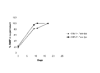
BRIEF DESCRIPTION OF THE DRAWINGS
Fig. 1 is a plot of the in vitro release profile of a non-lyophilized
suspension at a SMIP
concentration 1% and 2 % w/w relative to the polymer.
Fig. 2 is a plot of the in vitro release profile of a lyophilized suspension
at a SMIP
concentration 1% and 2% w/w relative to the polymer.
DETAILED DESCRIPTION OF THE INVENTION
The practice of the present invention will employ, unless otherwise indicated,
conventional methods of chemistry, polymer chemistry, biochemistry, molecular
biology,
immunology and pharmacology, within the skill of the art. Such techniques are
explained fully in
the literature. See, e.g., Remington's Pharmaceutical Sciences, 18th ed.
(Easton, Pennsylvania:
Mack Publishing Company, 1990); Methods In Enzymology (S. Colowick and N.
Kaplan, eds.,
Academic Press, Inc.); Weir, D.M., Handbook of Experimental Immunology,Vols. I-
IV, 5th ed.
(Blackwell Publishers, 1996); Sambrook, J. et al, Molecular Cloning: A
Laboratory Manual, 3rd
ed. (Cold Spring Harbor Laboratory Press, 2001); Ausubel, F.M. et al., Short
Protocols In
Molecular Biology, 5th ed. (Current Protocols, 2002); Handbook of Surface and
Colloidal
Chemistry (Birdi, K.S., ed, CRC Press, 2003) and Seymour/Carraher's Polymer
Chemistry, 7th
ed. (CRC Press, 2007).
As used in this specification and any appended claims, the singular forms "a,"
"an" and
"the" include plural references unless the content clearly dictates otherwise.
Thus, for example,
the term "nanoparticle" refers to one or more nanoparticles, and the like.
Unless stated otherwise or unless the context clearly dictates otherwise, all
percentages
and ratios herein are given on a weight basis.
A. Definitions
In describing the present invention, the following terms are intended to be
defined as
indicated below.
6
CA 02722765 2010-10-27
WO 2009/134769 PCT/US2009/041932
The term "nanoparticle" as used herein, refers to a particle of less than
1,000 nm in
diameter.
Particle size can be determined (measured) using methods available in the art.
For
example, particle size can be determined using photon correlation
spectroscopy, dynamic light
scattering or quasi-elastic light scattering. These methods are based on the
correlation of particle
size with diffusion properties of particles obtained from Brownian motion
measurements.
Brownian motion is the random movement of the particles due to bombardment by
the solvent
molecules that surround the particles. The larger the particle, the more
slowly the Brownian
motion will be. Velocity is defined by the translational diffusion
coefficient. The value
measured refers to how a particle moves within a liquid (hydrodynamic
diameter). The diameter
that is obtained is the diameter of a sphere that has the same translational
diffusion coefficient as
the particle.
Particle size can also be determined using static light scattering, which
measures the
intensity of light scattered by particles in a solution at a single time.
Static light scattering
measures light intensity as a function of scattering angle and solute
concentration. Particles
passing though a light source, for example, a laser beam, scatter light at an
angle that is inversely
proportional to their size. Large particles generate a diffraction pattern at
low scattering angles
with high intensity, whereas small particles give rise to wide angle low
intensity signals. Particle
size distributions can be calculated if the intensity of light scattered from
a sample are measured
as a function of angle. The angular information is compared with a scattering
model (e.g., Mie
theory) in order to calculate the size distribution.
Generally, particle size is determined at room temperature and involves
multiple analyses
of the sample in question (e.g., at least 3 repeat measurements on the same
sample) to yield an
average value for the particle diameter.
For photon correlation spectroscopy, Z average (also called the cumulant mean
or
hydrodynamic diameter) is typically calculated from cumulants (monomodal)
analysis.
For static light scattering measurements (and also for photon correlation
spectroscopy in
some embodiments), volume-based size parameters may be measured. For instance,
the D(v,0.5)
(where v means volume) is a size parameter whose value is defined as the point
where 50% of
the particles (volume basis) in the composition, as measured, have a size that
is less than the
7
CA 02722765 2010-10-27
WO 2009/134769 PCT/US2009/041932
D(v,0.5) value, and 50% of the particles in the composition have a size that
is greater than the
D(v,0.5) value. Similarly, the D(v,0.9) is a size parameter whose value is
defined as the point
where 90% (volume basis) of the particles in the composition have a size that
is less than the
D(v,0.9) value, and 10% of the particles in the composition have a size that
is greater than the
D(v,0.9) value.
The nanoparticles within the compositions of the present invention typically
have a size
distribution in which the Z average and/or the D(v,0.5) value is less than 200
nm, and more
typically less than 150 nm and in which the D(v,0.9) is less than 250 nm, and
more typically less
than 200 nm.
As defined herein, an "organic solvent species" is a solvent species that
comprises at least
one carbon atom.
As defined herein, an "aqueous" liquid is a water-containing liquid, typically
a liquid
containing more than 50 wt% water, for example, from 50 to 75 to 90 to 95 wt%
or more water.
As defined herein, an "aqueous" solvent is a water-containing solvent,
typically a solvent
containing more than 50 wt% water, for example, from 50 to 75 to 90 to 95 wt%
or more water.
As defined herein, a "nanoparticle suspension" is a liquid phase that contains
nanoparticles.
An "aqueous nanoparticle suspension" is a water-containing liquid that further
contains
nanoparticles. Aqueous nanoparticle suspensions in accordance with the
invention typically
contain more than 50 wt% water, for example from 50 to 75 to 90 to 95 wt% or
more water.
The nanoparticles of the invention are typically formed from polymers that are
substantially non-toxic and biodegradable. Such materials include polyesters
such as poly(a-
hydroxy acids) and polylactones (e.g., polycaprolactone), polyorthoesters,
polyanhydrides, and
polycyanoacrylates (e.g., polyalkylcyanoacrylate or "PACA"), among others.
More typically,
nanoparticles for use with the present invention are polymer nanoparticles
derived from poly(a-
hydroxy acids), for example, from a poly(lactide) ("PLA") such as poly(D,L-
lactide), a
copolymer of lactide and glycolide, such as a poly(D,L-lactide-co-glycolide)
or poly(L-lactide-
co-glycolide) (both referred to as "PLG"), or a copolymer of D,L-lactide and
caprolactone. The
polymer nanoparticles may be formed from polymers which have a variety of
molecular weights
and, in the case of the copolymers, such as PLG, a variety of monomer (e.g.,
lactide:glycolide)
8
CA 02722765 2010-10-27
WO 2009/134769 PCT/US2009/041932
ratios. Polymers are also available in a variety of end groups. These
parameters are discussed
further below.
The term "surfactant" comes from the phrase "surface active agent".
Surfactants
accumulate at interfaces (e.g., at liquid-liquid, liquid-solid and/or liquid-
gas interfaces) and
change the properties of that interface. As used herein, surfactants include
detergents, dispersing
agents, suspending agents, emulsion stabilizers, and the like.
As defined herein, "carbohydrates" include monosaccharides, oligosaccharides
and
polysaccharides, as well as substances derived from monosaccharides,
oligosaccharides and
polysaccharides, for example, by reduction (e.g., alditols), by oxidation of
one or more terminal
groups to carboxylic acids (e.g., glucuronic acid), or by replacement of one
or more hydroxy
group(s) by a hydrogen atom or an amino group (e.g., beta-D-glucosamine and
beta-D-
galactosamine).
As defined herein, a "monosaccharide" is a polyhydric alcohol, i.e., an
alcohol that further
comprises either an aldehyde group (in which case the monosaccharide is an
aldose) or a keto
group (in which case the monosaccharide is a ketose). Monosaccharides
typically contain from
3-10 carbons. Moreover, monosaccharides commonly have the empirical formula
(CH20)n
where n is an integer of three or greater, typically 3-10. Examples of 3-6
carbon aldoses include
glyceraldehyde, erythrose, threose, ribose, 2-deoxyribose, arabinose, xylose,
lyxose, allose,
altrose, glucose, mannose, gulose, idose, galactose, and talose. Examples of 3-
6 carbon ketoses
include dihydroxyacetone, erythrulose, ribulose, xylulose, psicose, fructose,
sorbose, and
tagatose. Naturally occurring monosaccharides are normally found in the D-
isomer form, as
opposed to the L-form.
As defined herein "oligosaccharide" refers to a relatively short
monosaccharide polymer,
i.e., one containing from 2 to 30 monosaccharide units. As defined herein, a
"polysaccharide" is
a monosaccharide polymer that is beyond oligosaccharide length (i.e., one
containing more than
30 monosaccharide units). Moreover, as used herein, the term "polysaccharide"
also refers to a
monosaccharide polymer that contains two or more linked monosaccharides. To
avoid
ambiguity, the second definition is to be applied at all times, unless there
are explicit indications
to the contrary. The term "polysaccharide" also includes polysaccharide
derivatives, such as
amino-functionalized and carboxyl-functionalized polysaccharide derivatives,
among many
9
CA 02722765 2010-10-27
WO 2009/134769 PCT/US2009/041932
others. Monosaccharides are typically linked by glycosidic linkages. Specific
examples include
disaccharides (such as sucrose, lactose, trehalose, maltose, gentiobiose and
cellobiose),
trisaccharides (such as raffinose), tetrasaccharides (such as stachyose), and
pentasaccharides
(such as verbascose).
As used herein the term "saccharide" encompasses monosaccharides,
oligosaccharides and
polysaccharides. A "saccharide-containing species" is a molecule, at least a
portion of which is a
saccharide. Examples include saccharide cryoprotective agents, saccharide
antigens, antigens
comprising saccharides conjugated to carrier peptides, and so forth.
As used herein, a "cryoprotective agent" is an agent that protects a
composition from
experiencing adverse effects upon freezing and thawing. For example, in the
present invention,
cryoprotective agents may be added to prevent substantial nanoparticle
agglomeration from
occurring when the lyophilized compositions of the invention are resuspended.
A "polynucleotide" is a nucleic acid polymer. As used herein, a
"polynucleotide" can
include as few as 5, 6, 7 or 8 nucleotides. Furthermore, a "polynucleotide"
can include both
double- and single-stranded sequences and refers to, but is not limited to,
cDNA from viral,
procaryotic or eucaryotic mRNA, genomic RNA and DNA sequences from viral (e.g.
RNA and
DNA viruses and retroviruses) or procaryotic DNA, and synthetic DNA sequences.
The term
also captures sequences that include any of the known base analogs of DNA and
RNA. The term
further includes modifications, such as deletions, additions and substitutions
(generally
conservative in nature), to a native sequence, for example, where the nucleic
acid molecule
encodes an antigenic protein. These modifications may be deliberate, as
through site-directed
mutagenesis, or may be accidental, such as through mutations of hosts that
produce antigens.
As defined herein an "oligonucleotide" is a polynucleotide having in the range
of 5 to 100
nucleotides, more typically, 5 to 30 nucleotides in size.
As defined herein, a "polynucleotide-containing species" is a molecule, at
least a portion of
which is a polynucleotide.
The term "polypeptide" refers to a polymer of amino acid residues and is not
limited to a
minimum length of the product. Thus, proteins, peptides, oligopeptides,
dimers, multimers, and
the like, are included within the definition. Proteins for use herein include
full length proteins
CA 02722765 2010-10-27
WO 2009/134769 PCT/US2009/041932
and protein fragments. In certain embodiments, modifications to the native
sequence, such as
deletions, additions and substitutions (generally conservative in nature), are
employed.
A "polypeptide-containing species" is a molecule, at least a portion of which
is a
polypeptide. Examples include polypeptides, glycoproteins, metalloproteins,
lipoproteins,
saccharide antigens conjugated to carrier proteins, and so forth.
The term "pharmaceutical" refers to biologically active compounds such as
antibiotics,
antiviral agents, growth factors, hormones, antigens, immunological adjuvants,
and the like.
The term "adjuvant" refers to any substance that assists or modifies the
action of a
pharmaceutical, including but not limited to immunological adjuvants, which
increase and/or
diversify the immune response to an antigen. Hence, immunological adjuvants
are compounds
that are capable of potentiating an immune response to antigens. Immunological
adjuvants can
potentiate humoral and/or cellular immunity. In some embodiments,
immunological adjuvants
stimulate an innate immune response. Immunological adjuvants may also be
referred to herein
as "immunopotentiators."
As used herein, an "antigen" refers to a molecule containing one or more
epitopes (e.g.,
linear, conformational or both) that elicit an immunological response. The
term may be used
interchangeably with the term "immunogen."
As used herein, an "epitope" is that portion of given species (e.g., an
antigenic molecule or
antigenic complex) that determines its immunological specificity. An epitope
is within the
scope of the present definition of antigen. Commonly, an epitope is a
polypeptide or
polysaccharide in a naturally occurring antigen. In artificial antigens, it
can be a low molecular
weight substance such as an arsanilic acid derivative. Normally, a B-cell
epitope will include at
least about 5 amino acids but can be as small as 3-4 amino acids. A T-cell
epitope, such as a CTL
epitope, will typically include at least about 7-9 amino acids, and a helper T-
cell epitope will
typically include at least about 12-20 amino acids.
The term "antigen" as used herein denotes both subunit antigens, i.e.,
antigens which are
separate and discrete from a whole organism with which the antigen is
associated in nature, as
well as killed, attenuated or inactivated bacteria, viruses, parasites or
other pathogens or tumor
cells. Antibodies such as anti-idiotype antibodies, or fragments thereof, and
synthetic peptide
mimotopes, which can mimic an antigen or antigenic determinant, are also
captured under the
11
CA 02722765 2010-10-27
WO 2009/134769 PCT/US2009/041932
definition of antigen as used herein. Similarly, an oligonucleotide or
polynucleotide that
expresses an immunogenic protein, or antigenic determinant in vivo, such as in
nucleic acid
immunization applications, is also included in the definition of antigen
herein.
An "immunological response" or "immune response" to an antigen or composition
is the
development in a subject of a humoral and/or a cellular immune response to
molecules present in
the composition of interest.
Immune responses include innate and adaptive immune responses. Innate immune
responses are fast-acting responses that provide a first line of defense for
the immune system. In
contrast, adaptive immunity uses selection and clonal expansion of immune
cells having
somatically rearranged receptor genes (e.g., T- and B-cell receptors) that
recognize antigens from
a given pathogen or disorder (e.g., a tumor), thereby providing specificity
and immunological
memory. Innate immune responses, among their many effects, lead to a rapid
burst of
inflammatory cytokines and activation of antigen-presenting cells (APCs) such
as macrophages
and dendritic cells. To distinguish pathogens from self-components, the innate
immune system
uses a variety of relatively invariable receptors that detect signatures from
pathogens, known as
pathogen-associated molecular patterns, or PAMPs. The addition of microbial
components to
experimental vaccines is known to lead to the development of robust and
durable adaptive
immune responses. The mechanism behind this potentiation of the immune
responses has been
reported to involve pattern-recognition receptors (PRRs), which are
differentially expressed on a
variety of immune cells, including neutrophils, macrophages, dendritic cells,
natural killer cells,
B cells and some nonimmune cells such as epithelial and endothelial cells.
Engagement of PRRs
leads to the activation of some of these cells and their secretion of
cytokines and chemokines, as
well as maturation and migration of other cells. In tandem, this creates an
inflammatory
environment that leads to the establishment of the adaptive immune response.
PRRs include
nonphagocytic receptors, such as Toll-like receptors (TLRs) and nucleotide-
binding
oligomerization domain (NOD) proteins, and receptors that induce phagocytosis,
such as
scavenger receptors, mannose receptors and P-glucan receptors. Reported TLRs
(along with
examples of some reported ligands, which may be used as immunogenic species in
various
embodiments of the invention) include the following: TLR1 (bacterial
lipoproteins from
Mycobacteria, Neisseria), TLR2 (zymosan yeast particles, peptidoglycan,
lipoproteins,
12
CA 02722765 2010-10-27
WO 2009/134769 PCT/US2009/041932
glycolipids, lipopolysaccharide), TLR3 (viral double-stranded RNA, poly:IC),
TLR4 (bacterial
lipopolysaccharides, plant product taxol), TLR5 (bacterial flagellins), TLR6
(yeast zymosan
particles, lipotechoic acid, lipopeptides from mycoplasma), TLR7 (single-
stranded RNA,
imiquimod, resimiquimod, and other synthetic compounds such as loxoribine and
bropirimine),
TLR8 (single-stranded RNA, resimiquimod) and TLR9 (CpG oligonucleotides),
among others.
Dendritic cells are recognized as some of the most important cell types for
initiating the priming
of naive CD4 helper T (TH) cells and for inducing CD8 T cell differentiation
into killer cells.
TLR signaling has been reported to play an important role in determining the
quality of these
helper T cell responses, for instance, with the nature of the TLR signal
determining the specific
type of TH response that is observed (e.g., TH1 versus TH2 response). A
combination of
antibody (humoral) and cellular immunity are produced as part of a TH1-type
response, whereas
a TH2-type response is predominantly an antibody response. Various TLR ligands
such as CpG
DNA (TLR9) and imidazoquinolines (TLR7, TLR8) have been documented to
stimulate
cytokine production from immune cells in vitro. The imidazoquinolines are the
first small, drug-
like compounds shown to be TLR agonists. For further information, see, e.g.,
A. Pashine, N. M.
Valiante and J. B. Ulmer, Nature Medicine 11, S63-S68 (2005), K. S. Rosenthal
and D. H.
Zimmerman, Clinical and Vaccine Immunology, 13(8), 821-829 (2006), and the
references cited
therein.
For purposes of the present invention, a "humoral immune response" refers to
an immune
response mediated by antibody molecules, while a "cellular immune response" is
one mediated
by T-lymphocytes and/or other white blood cells. One important aspect of
cellular immunity
involves an antigen-specific response by cytolytic T-cells ("CTLs"). CTLs have
specificity for
peptide antigens that are presented in association with proteins encoded by
the major
histocompatibility complex (MHC) and expressed on the surfaces of cells. CTLs
help induce
and promote the intracellular destruction of intracellular microbes, or the
lysis of cells infected
with such microbes. Another aspect of cellular immunity involves an antigen-
specific response
by helper T-cells. Helper T-cells act to help stimulate the function, and
focus the activity of,
nonspecific effector cells against cells displaying peptide antigens in
association with MHC
molecules on their surface. A "cellular immune response" also refers to the
production of
13
CA 02722765 2010-10-27
WO 2009/134769 PCT/US2009/041932
cytokines, chemokines and other such molecules produced by activated T-cells
and/or other
white blood cells, including those derived from CD4 and CD8 T-cells.
A composition such as an immunogenic composition or a vaccine that elicits a
cellular
immune response may thus serve to sensitize a vertebrate subject by the
presentation of antigen
in association with MHC molecules at the cell surface. The cell-mediated
immune response is
directed at, or near, cells presenting antigen at their surface. In addition,
antigen-specific T-
lymphocytes can be generated to allow for the future protection of an
immunized host. The
ability of a particular antigen or composition to stimulate a cell-mediated
immunological
response may be determined by a number of assays known in the art, such as by
lymphoproliferation (lymphocyte activation) assays, CTL cytotoxic cell assays,
by assaying for
T-lymphocytes specific for the antigen in a sensitized subject, or by
measurement of cytokine
production by T cells in response to restimulation with antigen. Such assays
are well known in
the art. See, e.g., Erickson et al. (1993) J. Immunol. 151:4189-4199; Doe et
al. (1994) Eur. J.
Immunol. 24:2369-2376. Thus, an immunological response as used herein may be
one which
stimulates the production of CTLs and/or the production or activation of
helper T-cells. The
antigen of interest may also elicit an antibody-mediated immune response.
Hence, an
immunological response may include, for example, one or more of the following
effects among
others: the production of antibodies by, for example, B-cells; and/or the
activation of suppressor
T-cells and/or y6 T-cells directed specifically to an antigen or antigens
present in the
composition or vaccine of interest. These responses may serve to neutralize
infectivity, and/or
mediate antibody-complement, or antibody dependent cell cytotoxicity (ADCC) to
provide
protection to an immunized host. Such responses can be determined using
standard
immunoassays and neutralization assays, well known in the art.
Immunogenic compositions in accordance with the present invention display
"enhanced
immunogenicity" for a given antigen when they possess a greater capacity to
elicit an immune
response than the immune response elicited by an equivalent amount of the
antigen in a differing
composition (e.g., wherein the antigen is administered as a soluble protein).
Thus, a composition
may display "enhanced immunogenicity," for example, because the composition
generates a
stronger immune response, or because a lower dose or fewer doses of antigen is
necessary to
achieve an immune response in the subject to which it is administered. Such
enhanced
14
CA 02722765 2010-10-27
WO 2009/134769 PCT/US2009/041932
immunogenicity can be determined, for example, by administering the
compositions of the
invention, and antigen controls, to animals and comparing assay results of the
two.
As used herein, "treatment (including variations thereof, for example, "treat"
or "treated")
refers to any of (i) the prevention of a pathogen or disorder in question
(e.g. cancer or a
pathogenic infection, as in a traditional vaccine), (ii) the reduction or
elimination of symptoms
associated with a pathogen or disorder in question, and (iii) the substantial
or complete
elimination of a pathogen or disorder in question. Treatment may thus be
effected
prophylactically (prior to arrival of the pathogen or disorder in question) or
therapeutically
(following arrival of the same).
The terms "effective amount" or "pharmaceutically effective amount" of a
pharmaceutical
composition of the present invention refer herein to a sufficient amount of
the immunogenic
composition to treat or diagnose a condition of interest. The exact amount
required will vary
from subject to subject, depending, for example, on the species, age, and
general condition of the
subject; the severity of the condition being treated; in the case of an
immunological response, the
capacity of the subject's immune system to synthesize antibodies, for example,
and the degree of
protection desired; and the mode of administration; among other factors. An
appropriate
"effective" amount in any individual case may be determined by one of ordinary
skill in the art.
Thus, a "therapeutically effective amount" will typically fall in a relatively
broad range that can
be determined through routine trials.
By "vertebrate subject" or "vertebrate animal" is meant any member of the
subphylum
cordata, including, without limitation, mammals such as cattle, sheep, pigs,
goats, horses, and
humans; domestic animals such as dogs and cats; and birds, including domestic,
wild and game
birds such as cocks and hens including chickens, turkeys and other
gallinaceous birds. The term
does not denote a particular age. Thus, both adult and newborn animals are
covered.
By "pharmaceutically acceptable" or "pharmacologically acceptable" is meant a
material
which is not biologically or otherwise undesirable, e.g., the material may be
administered to an
individual without causing any excessively undesirable biological effects in
the individual or
interacting in an excessively deleterious manner with any of the components of
the composition
in which it is contained.
CA 02722765 2010-10-27
WO 2009/134769 PCT/US2009/041932
The term "excipient" refers to any essentially accessory substance that may be
present in
the finished dosage form. For example, the term "excipient" includes vehicles,
binders,
disintegrants, fillers (diluents), lubricants, suspending/dispersing agents,
and so forth.
By "physiological pH" or a "pH in the physiological range" is meant a pH in
the range of
approximately 7.2 to 8.0 inclusive, more typically in the range of
approximately 7.2 to 7.6
inclusive.
As used herein, the phrase "vector construct" generally refers to any assembly
that is
capable of directing the expression of a nucleic acid sequence(s) or gene(s)
of interest. A "DNA
vector construct" refers to a DNA molecule that is capable of directing the
expression of a
nucleic acid sequence(s) or gene(s) of interest. One specific type of DNA
vector construct is a
plasmid, which is a circular episomal DNA molecule capable of autonomous
replication within a
host cell. Typically, a plasmid is a circular double stranded DNA loop into
which additional
DNA segments can be ligated. pCMV is one specific plasmid that is well known
in the art.
Other DNA vector constructs are known, which are based on RNA viruses. These
DNA vector
constructs typically comprise a promoter that functions in a eukaryotic cell,
5' of a cDNA
sequence for which the transcription product is an RNA vector construct (e.g.,
an alphavirus
RNA vector replicon), and a 3' termination region. Other examples of vector
constructs include
RNA vector constructs (e.g., alphavirus vector constructs) and the like. As
used herein, "RNA
vector construct", "RNA vector replicon" and "replicon" refer to an RNA
molecule that is
capable of directing its own amplification or self-replication in vivo,
typically within a target
cell. The RNA vector construct is used directly, without the requirement for
introduction of
DNA into a cell and transport to the nucleus where transcription would occur.
By using the
RNA vector for direct delivery into the cytoplasm of the host cell, autonomous
replication and
translation of the heterologous nucleic acid sequence occurs efficiently.
B. General Methods
As indicated above, in various aspects of the invention, nanoparticle
compositions
are provided which comprise (a) nanoparticles comprising at least one
biodegradable polymer
and (b) at least one pharmaceutical associated with the nanoparticles. Much of
the following
16
CA 02722765 2010-10-27
WO 2009/134769 PCT/US2009/041932
discussion is directed to immunogenic species as exemplary pharmaceuticals.
However, the
invention is not so-limited.
Other aspects of the invention are directed to methods of producing
nanoparticle
compositions that comprise at least one biodegradable polymer.
1. Nanop article Compositions
Useful polymers for forming nanoparticles compositions in accordance with the
present
invention include homopolymers, copolymers and polymer blends, both natural
and synthetic.
Such polymers may be derived, for example, from homopolymers and copolymers of
the
following: poly(alpha-hydroxy acids) including polyglycolic acid (PGA) (also
known as
polyglycolide), polylactic acid (PLA) (also known as polylactide) and
polyhydroxybutyric acid
(also known as polyhydroxybutyrate); polydioxanone; polycaprolactone;
polyorthoesters;
polycyanoacrylates, polyanhydrides; and combinations thereof. More typical are
poly(a-hydroxy
acids) such as poly(L-lactide), poly(D,L-lactide) (both referred to as "PLA"
herein), copolymers
of lactide and glycolide, such as poly(L-lactide-co-glycolide) and poly(D,L-
lactide-co-glycolide)
(both designated as "PLG" herein).
The above polymers are available in a variety of molecular weights, and the
appropriate
molecular weight for a given use is readily determined by one of skill in the
art. Thus, for
example, a suitable molecular weight for PLA may be on the order of about 2000
to 5000. A
suitable molecular weight for PLG may range from about 5,000 to about 200,000.
Where copolymers are employed, copolymers with a variety of monomer ratios may
be
available. For example, where PLG is used to form the nanoparticles, a variety
of
lactide:glycolide molar ratios will find use herein, and the ratio is largely
a matter of choice,
depending in part on any coadministered species (e.g., adsorbed, entrapped, or
otherwise
associated with the nanoparticles) and the rate of degradation desired. For
example, a 50:50
PLG polymer, containing 50% lactide and 50% glycolide, will provide a faster
resorbing
copolymer, while 75:25 PLG degrades more slowly, and 85:15 and 90:10, even
more slowly, due
to the increased lactide component. Mixtures of nanoparticles with varying
lactide:glycolide
ratios may also find use herein in order to achieve the desired release
kinetics. Degradation rate
17
CA 02722765 2010-10-27
WO 2009/134769 PCT/US2009/041932
of the nanoparticles of the present invention can also be controlled by such
factors as polymer
molecular weight and polymer crystallinity.
Where used PLG copolymers are typically those having a lactide/glycolide molar
ratio
ranging, for example, from 10:90 to 20:80 to 25:75 to 40:60 to 45:55 to 55:45
to 60:40 to 75:25
to 80:20 to 90:10, and having a molecular weight ranging, for example, from
5,000 to 10,000 to
20,000 to 40,000 to 50,000 to 70,000 to 100,000 to 200,00 Daltons, among
others.
PLG copolymers are also available with a variety of end groups, including acid
end groups
and ester end groups.
PLG copolymers with varying lactide:glycolide ratios, molecular weights and
end groups
are readily available commercially from a number of sources including from
Boehringer
Ingelheim, Germany, Birmingham Polymers, Inc., Birmingham, AL, USA and
Lakeshore
Biomaterials, Birmingham, AL, USA. Some exemplary PLG copolymers, available
from
Boehringer Ingelheim, include: (a) RG 502, a PLG having predominantly alkyl
ester end groups
on one of the chain ends, a 50:50 lactide/glycolide molar ratio and a
molecular weight of 12,000
Da, (b) RG 503, a PLG having predominantly alkyl ester end groups on one of
the chain ends, a
50:50 lactide/glycolide molar ratio and a molecular weight of 34,000 Da, (c)
RG 504, a PLG
having predominantly alkyl ester end groups on one of the chain ends, a 50:50
lactide/glycolide
molar ratio and a molecular weight of 48,000 Da, (d) RG 752, a PLG having
predominantly alkyl
ester end groups on one of the chain ends, a 75:25 lactide/glycolide molar
ratio and a molecular
weight of 22,000 Da, (e) RG 755, a PLG having predominantly alkyl ester end
groups on one of
the chain ends, a 75:25 lactide/glycolide molar ratio and a molecular weight
of 68,000 Da, (f)
RG 502H, a PLG having a 50:50 lactide/glycolide molar ratio, and having
predominantly free
carboxyl end groups on one of the chain ends, and (g) RG 503H, a PLG having a
50:50
lactide/glycolide molar ratio, and having predominantly free carboxyl end
groups on one of the
chain ends.
Nanoparticles in accordance with the invention can be prepared using various
suitable
methods.
In certain embodiments of the invention, a first liquid that comprises one or
more
biodegradable polymers dissolved in a first solvent is contacted with a second
liquid that
18
CA 02722765 2010-10-27
WO 2009/134769 PCT/US2009/041932
comprises a second solvent which is miscible with the first solvent while
being a non-solvent for
the one or more biodegradable polymers, such that nanoparticles are formed.
In certain of these embodiments, the second solvent is an aqueous solvent.
In certain embodiments, the second liquid comprises a buffer.
In certain embodiments, the first liquid may be contacted with the second
liquid by a
variety of suitable techniques, with the general idea being that the two
liquids are combined with
minimal intermixing. For example, the first liquid may be carefully poured
onto the second
liquid, or the first liquid may be gently injected into or onto the second
liquid, among other
possibilities. In one embodiment the first liquid is added in a drop-wise
fashion to the surface of
the second liquid.
In certain embodiments, during or after bringing the first and second liquids
into contact,
the liquids are allowed to interact with one another under conditions of
gentle agitation (e.g.,
gentle shaking, preferably with little or no stirring), or under conditions of
no agitation
whatsoever, to yield nanoparticles. Gentle shaking may be implemented, for
example, using a
gyrotory shaker, among other possibilities.
It has been found that combining the liquids with gentle shaking can result in
particles with
uniform size distribution at higher yields than are obtained with stirring.
For example, the yield,
based on the amount of biodegradable polymer that is recovered in the form of
nanoparticles , is
higher with the method in which the liquids are combined with shaking (e.g.,
ranging from 90%
or more) than the yield with the method in which the liquids are combined with
stirring (e.g., 60
% or less).
The first and second liquids may be combined in any suitable relative volume.
For
example, the first and second liquids may be combined relative volumes
selected from 1:10 to
1:5 to 1:2 to 1:1 to 2:1 to 5:1 to 10:1, more typically from 1:2 to 2:1, even
more typically about
1:1.
The biodegradable polymer concentration in the first liquid may be set at any
suitable
level, but typically ranges from 0.25% w/v to 5% w/v (e.g., ranging from 0.25%
w/v to 0.5% w/v
to 1% w/v to 2% w/v to 3% w/v to 4% w/v to 5% w/v), more typically 0.5% w/v to
3% w/v. In
general, the polymer concentration will affect the particle size, with lower
concentrations
19
CA 02722765 2010-10-27
WO 2009/134769 PCT/US2009/041932
yielding lower particle sizes. The polymer concentration may also affect the
encapsulation
efficiency of any pharmaceuticals that are introduced during the nanoparticle
formation process.
The first solvent may comprise, for instance, one or more organic solvent
species, for
example, one or more hydrophilic organic solvent species which may be selected
from acetone
and ethanol, among many others.
The second solvent may comprise, for example, water and/or one or more
hydrophilic
organic solvent species, among other possibilities. For instance, the second
liquid may be
selected from deionized water, normal saline, and buffered solutions. The
latter solutions can (a)
provide a tonicity, i.e., osmolality, that is essentially the same as normal
physiological fluids and
(b) maintain a pH compatible with normal physiological conditions. In other
embodiments, the
tonicity and/or pH characteristics of the compositions of the present
invention may be adjusted
after nanoparticle formation.
The second liquid may also comprise a buffer, for instance, to enhance
encapsulation
efficiency. Buffers are available which can maintain the pH of the second
liquid within a desired
pH range when the first and second liquids are brought into contact with one
another. For
example, buffers are commercially available which are designed to maintain pH
values ranging
from 2 or less to 3 to 4 to 5 to 6 to 7 to 8 to 9 to 10 to 11 to 12 or more.
The following are a few
examples of commercially available buffers (available from Sigma-Aldrich
and/or Polysciences
Inc.): citrate buffer solution (pH-4.8), sodium acetate buffer (pH-5.2, pH-
7.0), phosphate
citrate buffer (pH-5.0), SSC buffer (pH-7.0), PBS (pH-7.2 to 7.5), SSPE buffer
(pH-7.4), Tris
buffered saline (pH-7.4), Tris-phosphate-EDTA (pH ¨ 8.0), triethylammonium
bicarbonate
buffer (pH-8.0), Tris-EDTA (pH-8.0), Tris-borate-EDTA (pH-8.3), Tris-glycine
(pH-8.3), Tris
acetate-EDTA (pH-8.3), triethylammonium bicarbonate buffer (pH-8.5), Tris-
glycine-SDS
Buffer (pH-8.6), glycine buffer solution (pH-9.2), and sodium bicarbonate-
sodium carbonate
buffer (pH-9.6), among others.
In certain embodiments, the first solvent is more volatile that the second
solvent. In these
embodiments, the first solvent may be removed, for example, by evaporation
under ambient
conditions or by evaporation under reduced pressure and/or elevated
temperature.
In some embodiments, one or more additional species are added during or after
nanoparticles formation. Such additional species can include, for instance,
pharmaceuticals such
CA 02722765 2010-10-27
WO 2009/134769 PCT/US2009/041932
as immunogenic species (e.g., species that stimulate an innate immune
response, species that
stimulate an adaptive immune response, or both, including immunological
adjuvants,
immunopotentiators, and antigens), surfactants, cryoprotective agents, and
other supplemental
components such as tonicity adjusting agents, pH adjusting agents, and so
forth.
In these embodiments, the one or more additional species may be entrapped
within the
nanoparticles, associated with the surfaces of the nanoparticles (e.g.,
adsorbed or conjugated to
the surfaces of the nanoparticles), admixed with the nanoparticles in a liquid
or solid
composition (e.g., provided in solution, as an aqueous suspension,
colyophilized with the
nanoparticles, etc.), and/or otherwise associated with the nanoparticles.
In some embodiments of the invention, one or more additional species are added
during the
above-described method of nanoparticles formation. For instance, the first
liquid, the second
liquid, or both, may contain additional species as desired.
As a specific example, in addition to one or more biodegradable polymers
dissolved in an
organic solvent, the first liquid may further comprise one or more
pharmaceuticals (which may
be, for example, dissolved or suspended in the first liquid). Consequently,
the one or more
pharmaceuticals become entrapped in the nanoparticles.
In certain of these embodiments, the second liquid may comprise a buffer. The
buffer
may be selected, for example, to maintain the pH of the second liquid at a
point where the
pharmaceutical is predominantly uncharged. In embodiments where the
pharmaceutical is a
proton-accepting pharmaceutical, the buffer may be selected, for example, to
maintain a pH that
is greater than the pKa of the pharmaceutical. In embodiments where the
pharmaceutical is a
proton donating pharmaceutical, the buffer may be selected to maintain a pH
that is less than the
pKa of the pharmaceutical.
In certain embodiments, the amount of pharmaceutical provided (e.g.,
immunogenic
species, etc.) ranges from 0.25% w/w to 5% w/w relative to the amount of
biodegradable
polymer used in the process (e.g., ranging from 0.25% w/w to 0.5% w/w to 1%
w/w to 2% w/w
to 3% w/w to 4% w/w to 5% w/w).
Where the pharmaceutical is added to the first liquid, for polymer
concentrations ranging
from 0.5% w/v to 3% w/v (5g/m1 to 30 g/m1) in the first liquid, a range of
0.5% w/w to 3% w/w
for the pharmaceutical relative the amount of polymer corresponds to overall
concentrations in
21
CA 02722765 2010-10-27
WO 2009/134769 PCT/US2009/041932
the first liquid ranging from 0.0025% w/v to 0.09% w/v (i.e., 25 to 900
micrograms per mL,
more typically 100 to 600 micrograms per mL).
Methods such as the foregoing are advantageous, for example, in that the
encapsulation
efficiency for the pharmaceutical can be quite high, for example, ranging from
50% to 60% to
70% to 80% to 90% or more.
Vigorous agitation upon combining the first and second liquids (e.g., stirring
with a
magnetic stir bar), in the absence of a buffer in the second liquid, result in
much lower
encapsulation efficiencies.
In some embodiments of the invention, one or more additional species are added
subsequent to nanoparticle formation (and typically subsequent to organic
solvent removal, as
well as subsequent to washing steps, if any). For example, pharmaceuticals
such as
immunogenic species (e.g., antigens, immunological adjuvants,
immunopotentiators, etc.), agents
for adjusting tonicity and/or pH, surfactants, cryoprotective agents, and so
forth, may be added
subsequent to nanoparticle formation. Frequently, these additional species are
added to the
nanoparticles as an aqueous solution or dispersion. The resulting admixture
may be lyophilized
in some embodiments.
As noted above, the additional species may be associated with the surfaces of
the
nanoparticles (e.g., adsorbed or conjugated to the surfaces of the
nanoparticles) and/or otherwise
associated or non-associated with the nanoparticles to varying degrees (e.g.,
admixed with the
nanoparticles in a liquid dispersion, in a solid composition, etc.), among
other possibilities.
Where two pharmaceuticals (e.g., immunogenic species, etc.) are employed, they
can be,
for example, attached to (e.g., adsorbed or conjugated to) or entrapped within
the same
population of nanoparticles, or attached to or entrapped within separate
populations of
nanoparticles, among other possibilities.
Compositions in accordance with some embodiments of the invention can be
sterile filtered
(e.g., using a 200 micron filter) at any time after nanoparticle formation,
for example, after
nanoparticle formation but before the addition of any additional species,
after nanoparticle
formation and after the addition of any additional species, and so forth.
The nanoparticles within the compositions of the present invention (including
lyophilized
compositions that have been resuspended) typically have a size distribution in
which the Z
22
CA 02722765 2010-10-27
WO 2009/134769 PCT/US2009/041932
average and/or the D(v,0.5) value is less than 200 nm, and more typically less
than 150 nm and
in which the D(v,0.9) is less than 250 nm, and more typically less than 200
nm.
Taking as an example nanoparticles formed using PLG, there are several
advantages of the
techniques of the present invention, as compared with microparticle forming
techniques based on
oil-in-water and water-in-oil-in-water emulsification. A first benefit is the
ease of preparation.
The nanoparticle method is a single step technique and does not need high-
shear homogenization
as does the microparticle method, only gentle shaking. In addition, the entire
emulsion-based
microparticle particle preparation process is typically aseptic, whereas, due
to their small size,
nanoparticles may be sterile filtered post particle preparation, leading to
less strict production
requirements.
Furthermore, the type of organic solvent used with the two methods is
different. The
nanoparticle method can be performed using acetone whereas the microparticle
method typically
involves the use of dichloromethane (DCM ) as a solvent. The U.S. Food and
Drug
Administration (FDA) classifies DCM as a Class 2 solvent and has established
limits on the
amounts of allowable residual solvent which may be present in pharmaceutical
products,
whereas acetone is a Class 3 solvent for which the FDA has established higher
limits on the
allowable amounts.
23
CA 02722765 2010-10-27
WO 2009/134769 PCT/US2009/041932
2. Immunological Adjuvants
As previously indicated, one or more immunological adjuvants may optionally be
provided in the compositions of the invention. They may be, for example,
entrapped within the
nanoparticles, associated with the surfaces of the nanoparticles (e.g.,
adsorbed or conjugated to
the surfaces of the nanoparticles) and/or otherwise associated with the
nanoparticles to varying
degrees (e.g., admixed with the nanoparticles in a liquid suspension, admixed
with the
nanoparticles in a solid composition, for instance, colyophilized with the
nanoparticles, etc.),
among other possibilities
Immunological adjuvants for use with the invention include, but are not
limited to, one or
more of the following:
a. Mineral Containing Compositions
Mineral containing compositions suitable for use as adjuvants include mineral
salts, such
as aluminum salts and calcium salts. The invention includes mineral salts such
as hydroxides
(e.g. oxyhydroxides), phosphates (e.g. hydroxyphosphates, orthophosphates),
sulfates, etc. (see,
e.g., Vaccine Design: The Subunit and Adjuvant Approach (Powell, M.F. and
Newman, M.J.
eds.) (New York: Plenum Press) 1995, Chapters 8 and 9), or mixtures of
different mineral
compounds (e.g. a mixture of a phosphate and a hydroxide adjuvant, optionally
with an excess of
the phosphate), with the compounds taking any suitable form (e.g. gel,
crystalline, amorphous,
etc.), and with adsorption to the salt(s) being preferred. The mineral
containing compositions
may also be formulated as a particle of metal salt (WO 00/23105).
Aluminum salts may be included in vaccines of the invention such that the dose
of A13 is
between 0.2 and 1.0 mg per dose.
In one embodiment, the aluminum based adjuvant for use in the present
invention is alum
(aluminum potassium sulfate (A1K(SO4)2)), or an alum derivative, such as that
formed in-situ by
mixing an antigen in phosphate buffer with alum, followed by titration and
precipitation with a
base such as ammonium hydroxide or sodium hydroxide.
Another aluminum-based adjuvant for use in vaccine formulations of the present
invention is aluminum hydroxide adjuvant (Al(OH)3) or crystalline aluminum
oxyhydroxide
(A100H), which is an excellent adsorbant, having a surface area of
approximately 500m2/g. In
24
CA 02722765 2010-10-27
WO 2009/134769 PCT/US2009/041932
another embodiment, the aluminum based adjuvant is aluminum phosphate adjuvant
(A1PO4) or
aluminum hydroxyphosphate, which contains phosphate groups in place of some or
all of the
hydroxyl groups of aluminum hydroxide adjuvant. Preferred aluminum phosphate
adjuvants
provided herein are amorphous and soluble in acidic, basic and neutral media.
In another embodiment, the adjuvant comprises both aluminum phosphate and
aluminum
hydroxide. In a more particular embodiment thereof, the adjuvant has a greater
amount of
aluminum phosphate than aluminum hydroxide, such as a ratio of 2:1, 3:1, 4:1,
5:1, 6:1, 7:1, 8:1,
9:1 or greater than 9:1, by weight aluminum phosphate to aluminum hydroxide.
In another
embodiment, aluminum salts in the vaccine are present at 0.4 to 1.0 mg per
vaccine dose, or 0.4
to 0.8 mg per vaccine dose, or 0.5 to 0.7 mg per vaccine dose, or about 0.6 mg
per vaccine dose.
Generally, the preferred aluminum-based adjuvant(s), or ratio of multiple
aluminum-
based adjuvants, such as aluminum phosphate to aluminum hydroxide is selected
by optimization
of electrostatic attraction between molecules such that the antigen carries an
opposite charge as
the adjuvant at the desired pH. For example, aluminum phosphate adjuvant (iep
= 4) adsorbs
lysozyme, but not albumin at pH 7.4. Should albumin be the target, aluminum
hydroxide
adjuvant would be selected (iep 11.4). Alternatively, pretreatment of aluminum
hydroxide with
phosphate lowers its isoelectric point, making it a preferred adjuvant for
more basic antigens.
b. Oil-Emulsions
Oil-emulsion compositions and formulations suitable for use as adjuvants (with
or
without other specific immunostimulating agents such as muramyl peptides or
bacterial cell wall
components) include squalene-water emulsions, such as MF59 (5% Squalene, 0.5%
Tween 80,
and 0.5% Span 85, formulated into submicron particles using a microfluidizer).
See WO
90/14837. See also, Podda (2001) Vaccine 19: 2673-2680; Frey et al. (2003)
Vaccine 21:4234-
4237. MF59 is used as the adjuvant in the FLUADTM influenza virus trivalent
subunit vaccine.
Particularly preferred oil-emulsion adjuvants for use in the compositions are
submicron
oil-in-water emulsions. Preferred submicron oil-in-water emulsions for use
herein are
squalene/water emulsions optionally containing varying amounts of MTP-PE, such
as a
submicron oil-in-water emulsion containing 4-5% w/v squalene, 0.25-1.0% w/v
Tween 80TM
(polyoxyethylenesorbitan monooleate), and/or 0.25-1.0% Span 85TM (sorbitan
trioleate), and,
optionally, N-acetylmuramyl-L-alanyl-D-isogluatminyl-L-alanine-2-(1'-2'-
dipalmitoyl-sn-
CA 02722765 2010-10-27
WO 2009/134769 PCT/US2009/041932
glycero-3-huydroxyphosphophoryloxy)-ethylamine (MTP-PE), for example, the
submicron oil-
in-water emulsion known as "MF59" (WO 90/14837; U.S. Patent No. 6,299,884;
U.S. Patent No.
6,451,325; and Ott et al., "MF59 -- Design and Evaluation of a Safe and Potent
Adjuvant for
Human Vaccines" in Vaccine Design: The Subunit and Adjuvant Approach (Powell,
M.F. and
Newman, M.J. eds.) (New York: Plenum Press) 1995, pp. 277-296). MF59 contains
4-5% w/v
Squalene (e.g. 4.3%), 0.25-0.5% w/v Tween 8OTM, and 0.5% w/v Span 85 TM and
optionally
contains various amounts of MTP-PE, formulated into submicron particles using
a microfluidizer
such as Model 110Y microfluidizer (Microfluidics, Newton, MA). For example,
MTP-PE may
be present in an amount of about 0-500 iag/dose, more preferably 0-250
iag/dose and most
preferably, 0-100 yig/dose. As used herein, the term "MF59-0" refers to the
above submicron oil-
in-water emulsion lacking MTP-PE, while the term MF59-MTP denotes a
formulation that
contains MTP-PE. For instance, "MF59-100" contains 100 tg MTP-PE per dose, and
so on.
MF69, another submicron oil-in-water emulsion for use herein, contains 4.3%
w/v squalene,
0.25% w/v Tween 8OTM, and 0.75% w/v Span 85TM and optionally MTP-PE. Yet
another
submicron oil-in-water emulsion is MF75, also known as SAF, containing 10%
squalene, 0.4%
Tween 8OTM, 5% pluronic-blocked polymer L121, and thr-MDP, also microfluidized
into a
submicron emulsion. MF75-MTP denotes an MF75 formulation that includes MTP,
such as from
100-400 tg MTP-PE per dose.
Submicron oil-in-water emulsions, methods of making the same and
immunostimulating
agents, such as muramyl peptides, for use in the compositions, are described
in detail in WO
90/14837; U.S. Patent No. 6,299,884; and U.S. Patent No. 6,451,325.
Complete Freund's adjuvant (CFA) and incomplete Freund's adjuvant (IFA) may
also be
used as adjuvants in the invention.
c. Saponin Formulations
Saponin formulations are also suitable for use as adjuvants in the invention.
Saponins are
a heterologous group of sterol glycosides and triterpenoid glycosides that are
found in the bark,
leaves, stems, roots and even flowers of a wide range of plant species.
Saponins isolated from
the bark of the Quillaia saponaria Molina tree have been widely studied as
adjuvants. Saponins
can also be commercially obtained from Smilax ornata (sarsaprilla),
Gypsophilla paniculata
26
CA 02722765 2010-10-27
WO 2009/134769 PCT/US2009/041932
(brides veil), and Saponaria officianalis (soap root). Saponin adjuvant
formulations include
purified formulations, such as QS21, as well as lipid formulations, such as
ISCOMs. Saponin
adjuvant formulations include STIMULON adjuvant (Antigenics, Inc., Lexington,
MA).
Saponin compositions have been purified using High Performance Thin Layer
Chromatography
(HP-TLC) and Reversed Phase High Performance Liquid Chromatography (RP-HPLC).
Specific purified fractions using these techniques have been identified,
including QS7, QS17,
QS18, QS21, QH-A, QH-B and QH-C. Preferably, the saponin is QS21. A method of
production
of QS21 is disclosed in U.S. Patent No. 5,057,540. Saponin formulations may
also comprise a
sterol, such as cholesterol (see WO 96/33739).
Combinations of saponins and cholesterols can be used to form unique particles
called
Immunostimulating Complexes (ISCOMs). ISCOMs typically also include a
phospholipid such
as phosphatidylethanolamine or phosphatidylcholine. Any known saponin can be
used in
ISCOMs. Preferably, the ISCOM includes one or more of Quil A, QHA and QHC.
ISCOMs are
further described in EP 0 109 942, WO 96/11711 and WO 96/33739. Optionally,
the ISCOMS
may be devoid of (an) additional detergent(s). See WO 00/07621.
A review of the development of saponin based adjuvants can be found in Barr et
al.
(1998) Adv. Drug Del. Rev. 32:247-271. See also Sjolander et al. (1998) Adv.
Drug Del. Rev.
32:321-338.
d. Virosomes and Virus Like Particles (VLPs)
Virosomes and Virus Like Particles (VLPs) are also suitable as adjuvants.
These
structures generally contain one or more proteins from a virus optionally
combined or formulated
with a phospholipid. They are generally non-pathogenic, non-replicating and
generally do not
contain any of the native viral genome. The viral proteins may be
recombinantly produced or
isolated from whole viruses. These viral proteins suitable for use in
virosomes or VLPs include
proteins derived from influenza virus (such as HA or NA), Hepatitis B virus
(such as core or
capsid proteins), Hepatitis E virus, measles virus, Sindbis virus, Rotavirus,
Foot-and-Mouth
Disease virus, Retrovirus, Norwalk virus, human Papilloma virus, HIV, RNA-
phages, Q13-phage
(such as coat proteins), GA-phage, fr-phage, AP205 phage, and Ty (such as
retrotransposon Ty
protein pl). VLPs are discussed further in WO 03/024480; WO 03/024481; Niikura
et al. (2002)
Virology 293:273-280; Lenz et al. (2001) J. Inununol. 166(9):5346-5355; Pinto
et al. (2003) J.
27
CA 02722765 2010-10-27
WO 2009/134769 PCT/US2009/041932
Infect. Dis. 188:327-338; and Gerber et al. (2001) J. Virol. 75(10):4752-4760.
Virosomes are
discussed further in, for example, Gluck et al. (2002) Vaccine 20:B10-B16.
Immunopotentiating
reconstituted influenza virosomes (IRIV) are used as the subunit antigen
delivery system in the
intranasal trivalent INFLEXALTM product (Mischler and Metcalfe (2002) Vaccine
20 Suppl
5:B17-B23) and the INFLUVAC PLUSTM product.
e. Bacterial or Microbial Derivatives
Adjuvants suitable for use in the invention include bacterial or microbial
derivatives such
as:
(1) Non-toxic derivatives of enterobacterial lipopolysaccharide (LPS): Such
derivatives
include Monophosphoryl lipid A (MPL) and 3-0-deacylated MPL (3dMPL). 3dMPL is
a
mixture of 3 De-O-acylated monophosphoryl lipid A with 4, 5 or 6 acylated
chains. A preferred
"small particle" form of 3 De-O-acylated monophosphoryl lipid A is disclosed
in EP 0 689 454.
Such "small particles" of 3dMPL are small enough to be sterile filtered
through a 0.22 micron
membrane (see EP 0 689 454). Other non-toxic LPS derivatives include
monophosphoryl lipid
A mimics, such as aminoalkyl glucosaminide phosphate derivatives, e.g., RC-
529. See Johnson
et at. (1999) Bioorg. Med. Chem. Lett. 9:2273-2278.
(2) Lipid A Derivatives: Lipid A derivatives include derivatives of lipid A
from
Escherichia coli such as 0M-174. 0M-174 is described for example in Meraldi et
al. (2003)
Vaccine 21:2485-2491; and Pajak et al. (2003) Vaccine 21:836-842.
(3) Immunostimulatory oligonucleotides: Immunostimulatory oligonucleotides or
polymeric molecules suitable for use as adjuvants in the invention include
nucleotide sequences
containing a CpG motif (a sequence containing an unmethylated cytosine
followed by guanosine
and linked by a phosphate bond). Bacterial double stranded RNA or
oligonucleotides containing
palindromic or poly(dG) sequences have also been shown to be
immunostimulatory. The CpG's
can include nucleotide modifications/analogs such as phosphorothioate
modifications and can be
double-stranded or single-stranded. Optionally, the guanosine may be replaced
with an analog
such as 2'-deoxy-7-deazaguanosine. See Kandirnalla et al. (2003) Nucl. Acids
Res. 31(9): 2393-
2400; WO 02/26757; and WO 99/62923 for examples of possible analog
substitutions. The
adjuvant effect of CpG oligonucleotides is further discussed in Krieg (2003)
Nat. Med. 9(7):831-
28
CA 02722765 2010-10-27
WO 2009/134769 PCT/US2009/041932
835; McCluskie et al. (2002) FEMS Immunol. Med. Microbiol. 32:179-185; WO
98/40100; U.S.
Patent No. 6,207,646; U.S. Patent No. 6,239,116; and U.S. Patent No.
6,429,199.
The CpG sequence may be directed to TLR9, such as the motif GTCGTT or TTCGTT.
See
Kandimalla et al. (2003) Biochem. Soc. Trans. 31 (part 3):654-658. The CpG
sequence may be
specific for inducing a Thl immune response, such as a CpG-A ODN, or it may be
more specific
for inducing a B cell response, such a CpG-B ODN. CpG-A and CpG-B ODNs are
discussed in
Blackwell et al. (2003) J. Immunol. 170(8):4061-4068; Krieg (2002) TRENDS
Immunol. 23(2):
64-65; and WO 01/95935. Preferably, the CpG is a CpG-A ODN.
Preferably, the CpG oligonucleotide is constructed so that the 5' end is
accessible for
receptor recognition. Optionally, two CpG oligonucleotide sequences may be
attached at their 3'
ends to form "immunomers". See, for example, Kandimalla et al. (2003) BBRC
306:948-953;
Kandimalla et al. (2003) Biochem. Soc. Trans. 31(part 3):664-658; Bhagat etal.
(2003) BBRC
300:853-861; and W003/035836.
Immunostimulatory oligonucleotides and polymeric molecules also include
alternative
polymer backbone structures such as, but not limited to, polyvinyl backbones
(Pitha et al. (1970)
Biochem. Biophys. Acta 204(1):39-48; Pitha et al. (1970) Biopolymers 9(8):965-
977), and
morpholino backbones (U.S. Patent No. 5,142,047; U.S. Patent No. 5,185,444). A
variety of
other charged and uncharged polynucleotide analogs are known in the art.
Numerous backbone
modifications are known in the art, including, but not limited to, uncharged
linkages (e.g., methyl
phosphonates, phosphotriesters, phosphoamidates, and carbamates) and charged
linkages (e.g.,
phosphorothioates and phosphorodithioates).
(4) ADP-ribosylating toxins and detoxified derivatives thereof: Bacterial ADP-
ribosylating toxins and detoxified derivatives thereof may be used as
adjuvants in the invention.
Preferably, the protein is derived from E. coli (i.e., E. coli heat labile
enterotoxin "LT"), cholera
("CT"), or pertussis ("PT"). The use of detoxified ADP-ribosylating toxins as
mucosal adjuvants
is described in WO 95/17211 and as parenteral adjuvants in WO 98/42375.
Preferably, the
adjuvant is a detoxified LT mutant such as LT-K63, LT-R72, and LTR192G. The
use of ADP-
ribosylating toxins and detoxified derivatives thereof, particularly LT-K63
and LT-R72, as
adjuvants can be found in the following references: Beignon et al. (2002)
Infect. Immun.
70(6):3012-3019; Pizza et al. (2001) Vaccine 19:2534-2541; Pizza et al. (2000)
Int. J. Med.
29
CA 02722765 2010-10-27
WO 2009/134769 PCT/US2009/041932
Microbiol. 290(4-5):455-461; Scharton-Kersten et al. (2000) Infect. Immun.
68(9):5306-5313;
Ryan et al. (1999) Infect. Immun. 67(12):6270-6280; Partidos et al. (1999)
Immunol. Lett.
67(3):209-216; Peppoloni et al. (2003) Vaccines 2(2):285-293; and Pine et al.
(2002) J. Control
Release 85(1-3):263-270. Numerical reference for amino acid substitutions is
preferably based
on the alignments of the A and B subunits of ADP-ribosylating toxins set forth
in Domenighini
et al. (1995) Mol. Microbiol. 15(6):1165-1167.
f. Bioadhesives and Mucoadhesives
Bioadhesives and mucoadhesives may also be used as adjuvants. Suitable
bioadhesives
include esterified hyaluronic acid microspheres (Singh etal. (2001) J. Cont.
Release 70:267-276)
or mucoadhesives such as cross-linked derivatives of polyacrylic acid,
polyvinyl alcohol,
polyvinyl pyrollidone, polysaccharides and carboxymethylcellulose. Chitosan
and derivatives
thereof may also be used as adjuvants in the invention (see WO 99/27960).
g. Liposomes
Examples of liposome formulations suitable for use as adjuvants are described
in U.S.
Patent No. 6,090,406; U.S. Patent No. 5,916,588; and EP Patent Publication No.
EP 0 626 169.
h. Polyoxyethylene ether and Polyoxyethylene Ester Formulations
Adjuvants suitable for use in the invention include polyoxyethylene ethers and
polyoxyethylene esters (see, e.g., WO 99/52549). Such formulations further
include
polyoxyethylene sorbitan ester surfactants in combination with an octoxynol
(WO 01/21207) as
well as polyoxyethylene alkyl ethers or ester surfactants in combination with
at least one
additional non-ionic surfactant such as an octoxynol (WO 01/21152).
Preferred polyoxyethylene ethers are selected from the following group:
polyoxyethylene-9-lauryl ether (laureth 9), polyoxyethylene-9-steoryl ether,
polyoxytheylene-8-
steoryl ether, polyoxyethylene-4-lauryl ether, polyoxyethylene-35-lauryl
ether, and
polyoxyethylene-23-lauryl ether.
i. Polyphosphazene (PCPP)
PCPP formulations suitable for use as adjuvants are described, for example, in
Andrianov
et al. (1998) Biomaterials 19(1-3):109-115; and Payne et al. (1998) Adv. Drug
Del. Rev.
31(3):185-196.
CA 02722765 2010-10-27
WO 2009/134769 PCT/US2009/041932
j. Muramyl peptides
Examples of muramyl peptides suitable for use as adjuvants include N-acetyl-
muramyl-
L-threonyl-D-isoglutamine (thr-MDP), N-acetyl-normuramy1-1-alanyl-d-
isoglutamine (nor-
MDP), and N-acetylmuramy1-1-alanyl-d-isoglutaminy1-1-alanine-2-(1'-2'-
dipalmitoyl-sn-glycero-
3-hydroxyphosphoryloxy)-ethylamine MTP-PE).
k. Imidazoquinoline Compounds
Examples of imidazoquinoline compounds suitable for use as adjuvants include
Imiquimod and its analogues, which are described further in Stanley (2002)
Clin. Exp. Dermatol.
27(7):571-577; Jones (2003) Curr. Opin. Investig. Drugs 4(2):214-218; and U.S.
Patent Nos.
4,689,338; 5,389,640; 5,268,376; 4,929,624; 5,266,575; 5,352,784; 5,494,916;
5,482,936;
5,346,905; 5,395,937; 5,238,944; and 5,525,612.
NII,
I )----R2
Ri
Preferred imidazoquinolines are those of the formula, ,
where R1
and R2 are independently selected from the group consisting of hydrogen, alkyl
of one to ten
carbon atoms, hydroxyalkyl of one to ten carbon atoms, alkoxyalkyl of one to
ten carbon atoms,
acyloxyalkyl wherein the acyloxy moiety is alkanoyloxy of one to five carbon
atoms or
RA
-N.
benzoyloxy and wherein the alkyl moiety contains one to six carbon atoms,
R3 wherein
R3 and R4 are independently selected from the group consisting of hydrogen and
alkyl of one to
ten carbon atoms, benzyl, (phenyl)ethyl and phenyl, where the benzyl,
(phenyl)ethyl or phenyl
substituent are optionally substituted on the benzene ring by one or two
moieties independently
selected from the group consisting of alkyl of one to four carbon atoms,
alkoxy of one to four
carbon atoms and halogen. The preceding alkyl groups may be linear, branched
and/or cyclic.
Particularly preferred imidazoquinolines for the practice of the present
invention include
31
CA 02722765 2010-10-27
WO 2009/134769 PCT/US2009/041932
NH2
N N
N
imiquimod, resiquimod, and , the latter of which is also referred
to herein
as "imidazoquinoline 090". See, e.g., Int. Pub. Nos. WO 2006/031878 to
Valiante et al. and WO
2007/109810 to Sutton et al.
1. Thiosemicarbazone Compounds
Examples of thiosemicarbazone compounds suitable for use as adjuvants, as well
as
methods of formulating, manufacturing, and screening for such compounds,
include those
described in WO 04/60308. The thiosemicarbazones are particularly effective in
the stimulation
of human peripheral blood mononuclear cells for the production of cytokines,
such as TNF- a.
m. Trvptanthrin Compounds
Examples of tryptanthrin compounds suitable for use as adjuvants, as well as
methods of
formulating, manufacturing, and screening for such compounds, include those
described in WO
04/64759. The tryptanthrin compounds are particularly effective in the
stimulation of human
peripheral blood mononuclear cells for the production of cytokines, such as
TNF- a.
n. Human Immunomodulators
Human immunomodulators suitable for use as adjuvants include cytokines, such
as
interleukins (e.g. IL-1, IL-2, IL-4, IL-5, IL-6, IL-7, IL-12, etc.),
interferons (e.g. interferon-y),
macrophage colony stimulating factor (M-CSF), and tumor necrosis factor (TNF).
The invention may also comprise combinations of aspects of one or more of the
adjuvants
identified above. For example, the following adjuvant compositions may be used
in the
invention:
(1) a saponin and an oil-in-water emulsion (WO 99/11241);
(2) a saponin (e.g., Q521) + a non-toxic LPS derivative (e.g. 3dMPL) (see WO
94/00153);
(3) a saponin (e.g., Q521) + a non-toxic LPS derivative (e.g. 3dMPL) + a
cholesterol;
(4) a saponin (e.g., Q521) + 3dMPL + IL-12 (optionally + a sterol) (WO
98/57659);
(5) combinations of 3dMPL with, for example, Q521 and/or oil-in-water
emulsions (see EP 0
835 318; EP 0 735 898; and EP 0 761 231);
32
CA 02722765 2010-10-27
WO 2009/134769 PCT/US2009/041932
(6) SAF, containing 10% Squalane, 0.4% Tween 80, 5% pluronic-block polymer
L121, and thr-
MDP, either microfluidized into a submicron emulsion or vortexed to generate a
larger particle
size emulsion;
(7) RibiTM adjuvant system (RAS), (Ribi Immunochem, Hamilton, MT) containing
2% Squalene,
0.2% Tween 80, and one or more bacterial cell wall components from the group
consisting of
monophosphorylipid A (MPL), trehalose dimycolate (TDM), and cell wall skeleton
(CWS),
preferably MPL + CWS (DetoxTm);
(8) one or more mineral salts (such as an aluminum salt) + a non-toxic
derivative of LPS (such as
3dPML);
(9) one or more mineral salts (such as an aluminum salt) + an
immunostimulatory
oligonucleotide (such as a nucleotide sequence including a CpG motif).
3. Antigens
As previously indicated, one or more antigens may optionally be provided in
the
compositions of the invention. Antigens may be entrapped within the
nanoparticles, associated
with the surfaces of the nanoparticles (e.g., adsorbed or conjugated to the
surfaces of the
nanoparticles) and/or otherwise associated with the nanoparticles to varying
degrees (e.g.,
admixed with the nanoparticles in a liquid suspension, admixed with the
nanoparticles in a solid
composition, for instance, colyophilized with the nanoparticles), among other
possibilities.
Each antigen may be provided in an effective amount (e.g., an amount effective
for use in
therapeutic, prophylactic, or diagnostic methods in accordance with the
invention). For example,
the compositions of the present invention may be used to treat or prevent
infections caused by
any of the below-listed pathogens.
Antigens for use with the invention include, but are not limited to, one or
more of the
following antigens set forth below, or antigens derived from one or more of
the pathogens set
forth below:
a. Bacterial Antigens
Bacterial antigens suitable for use in the invention include proteins,
polysaccharides,
lipopolysaccharides, and outer membrane vesicles which may be isolated,
purified or derived
from a bacterium. In addition, bacterial antigens include bacterial lysates
and inactivated
33
CA 02722765 2010-10-27
WO 2009/134769 PCT/US2009/041932
bacteria formulations. Bacteria antigens can be produced by recombinant
expression. Bacterial
antigens preferably include epitopes which are exposed on the surface of the
bacteria during at
least one stage of its life cycle. Bacterial antigens are preferably conserved
across multiple
serotypes. Bacterial antigens include antigens derived from one or more of the
bacteria set forth
below as well as the specific antigens examples identified below.
Neisseria meningitides: Meningitides antigens include proteins (such as those
identified in
W099/24578; W099/36544; W099/57280; W000/22430; Tettelin et al. (2000) Science
287:1809-1815; W096/29412; and Pizza et al. (2000) Science 287:1816-1820),
saccharides
(including a polysaccharide, oligosaccharide or lipopolysaccharide), or outer-
membrane vesicles
(WO 01/52885; Bjune et al. (1991) Lancet 338(8775):1093-1096; Fuskasawa et al.
(1999)
Vaccine 17:2951-2958; and Rosenqist et al. (1998) Dev. Biol. Strand 92:323-
333) purified or
derived from N. meningitides serogroup such as A, C, W135, Y, and/or B.
Meningitides protein
antigens can be selected from adhesions, autotransporters, toxins, Fe
acquisition proteins, and
membrane associated proteins (preferably integral outer membrane protein).
Streptococcus pneumoniae: Streptococcus pneumoniae antigens include a
saccharide
(including a polysaccharide or an oligosaccharide) and/or protein from
Streptococcus
pneumoniae. Saccharide antigens can be selected from serotypes 1, 2, 3, 4, 5,
6B, 7F, 8, 9N, 9V,
10A, 11A, 12F, 14, 15B, 17F, 18C, 19A, 19F, 20, 22F, 23F, and 33F. Protein
antigens can be
selected from a protein identified in WO 98/18931; WO 98/18930; U.S. Patent
No. 6,699,703;
U.S. Patent No. 6,800,744; WO 97/43303; and WO 97/37026. Streptococcus
pneumoniae
proteins can be selected from the Poly Histidine Triad family (PhtX), the
Choline Binding
Protein family (CbpX), CbpX truncates, LytX family, LytX truncates, CbpX
truncate-LytX
truncate chimeric proteins, pneumolysin (Ply), PspA, PsaA, Sp128, Sp101,
Sp130, Sp125 or
Sp133.
Streptococcus pyo genes (Group A Streptococcus): Group A Streptococcus
antigens
include proteins identified in WO 02/34771 and WO 2005/032582 (including GAS
40), fusions
of fragments of GAS M proteins (including those described in WO 02/094851; and
Dale (1999)
Vaccine 17:193-200, and Dale (1996) Vaccine 14(10): 944-948), fibronectin
binding protein
(Sfbl), Streptococcal heme-associated protein (Shp), and Streptolysin S
(SagA).
34
CA 02722765 2010-10-27
WO 2009/134769 PCT/US2009/041932
Moraxella catarrhalis: Moraxella antigens include antigens identified in WO
02/18595;
and WO 99/58562, outer membrane protein antigens (HMW-OMP), C-antigen, and/or
LPS.
Bordetella pertussis: Pertussis antigens include petussis holotoxin (PT) and
filamentous
haemagglutinin (FHA) from B. pertussis, optionally also combination with
pertactin and/or
agglutinogens 2 and 3 antigen.
Staphylococcus aureus: Staph aureus antigens include S. aureus type 5 and 8
capsular
polysaccharides optionally conjugated to nontoxic recombinant Pseudomonas
aeruginosa
exotoxin A, such as StaphVAXTM, and antigens derived from surface proteins,
invasins
(leukocidin, kinases, hyaluronidase), surface factors that inhibit phagocytic
engulfment (capsule,
Protein A), carotenoids, catalase production, Protein A, coagulase, clotting
factor, and
membrane-damaging toxins (optionally detoxified) that lyse eukaryotic cell
membranes
(hemolysins, leukotoxin, leukocidin).
Staphylococcus epidermis: S. epidermidis antigens include slime-associated
antigen
(SAA).
Clostridium tetani (Tetanus): Tetanus antigens include tetanus toxoid (TT),
preferably
used as a carrier protein in conjunction/conjugated with the compositions of
the present
invention.
Cornynebacterium diphtheriae (Diphtheria): Diphtheria antigens include
diphtheria
toxin, preferably detoxified, such as CRM197. Additionally, antigens capable
of modulating,
inhibiting or associated with ADP ribosylation are contemplated for
combination/co-
administration/conjugation with the compositions of the present invention. The
diphtheria
toxoids may be used as carrier proteins.
Haemophilus influenzae B (Hib): Hib antigens include a Hib saccharide antigen.
Pseudomonas aeruginosa: Pseudomonas antigens include endotoxin A, Wzz protein,
P.
aeruginosa LPS, more particularly LPS isolated from PA01 (05 serotype), and
Outer Membrane
Proteins, including Outer Membrane Proteins F (OprF) (Price et al. (2001)
Infect Immun.
69(5):3510-3515).
Legionella pneumophila. Bacterial antigens can be derived from Legionella
pneumophila.
CA 02722765 2010-10-27
WO 2009/134769 PCT/US2009/041932
Streptococcus agalactiae (Group B Streptococcus): Group B Streptococcus
antigens
include protein and saccharide antigens, such as those identified in WO
02/34771; WO
03/093306; WO 04/041157; and WO 2005/002619 (including proteins GBS 80, GBS
104, GBS
276 and GBS 322, and including saccharide antigens derived from serotypes Ia,
Ib, Ia/c, II, III,
IV, V, VI, VII and VIII).
Neiserria gonorrhoeae: Gonorrhoeae antigens include Por (or porin) protein,
such as
PorB (see, e.g., Zhu et at. (2004) Vaccine 22:660-669), a transferring binding
protein, such as
TbpA and TbpB (see, e.g., Price et al. (2004) Infect. Immun. 71(1):277-283),
an opacity protein
(such as Opa), a reduction-modifiable protein (Rmp), and outer membrane
vesicle (OMV)
preparations (see, e.g., Plante et al. (2000) J. Infect. Dis. 182:848-855); WO
99/24578; WO
99/36544; WO 99/57280; and W002/079243).
Chlamydia trachomatis: Chlamydia trachomatis antigens include antigens derived
from
serotypes A, B, Ba and C (agents of trachoma, a cause of blindness), serotypes
L1, L2 & L3
(associated with Lymphogranuloma venereum), and serotypes, D-K. Chlamydia
trachomas
antigens also include antigens identified in WO 00/37494; WO 03/049762; WO
03/068811; and
WO 05/002619, including PepA (CT045), LcrE (CT089), ArtJ (CT381), DnaK
(CT396), CT398,
OmpH-like (CT242), L7/L12 (CT316), OmcA (CT444), AtosS (CT467), CT547, Eno
(CT587),
HrtA (CT823), and MurG (CT761).
Treponema pallidum (Syphilis): Syphilis antigens include TmpA antigen.
Haemophilus ducreyi (causing chancroid): Ducreyi antigens include outer
membrane
protein (DsrA).
Enterococcus faecalis or Enterococcus faecium: Antigens include a
trisaccharide repeat
and other Enterococcus derived antigens provided in U.S. Patent No. 6,756,361.
Helicobacter pylori: H pylori antigens include Cag, Vac, Nap, HopX, HopY and
urease
antigen.
Staphylococcus saprophyticus: Antigens include the 160 kDa hemagglutinin of S.
saprophyticus antigen.
Yersinia enterocolitica Antigens include LPS (Xu et al. (2002) Infect. Immun.
70(8):
4414-4423).
36
CA 02722765 2010-10-27
WO 2009/134769 PCT/US2009/041932
E. coli: E. coli antigens can be derived from enterotoxigenic E. coli (ETEC),
enteroaggregative E. coli (EAggEC), diffusely adhering E. coli (DAEC),
enteropathogenic E.
coli (EPEC), or enterohemorrhagic E. coli (EHEC).
Bacillus anthracis (anthrax): B. anthracis antigens are optionally detoxified
and can be
selected from A-components (lethal factor (LF) and edema factor (EF)), both of
which can share
a common B-component known as protective antigen (PA). In certain embodiments,
the
compositions of the present invention do not include an anthrax antigen.
Yersinia pestis (plague): Plague antigens include FI capsular antigen (Gosfeld
et al.
(2003) Infect. Immun. 71(1)): 374-383), LPS (Fields et al. (1999) Infect.
Immun. 67(10): 5395-
5408), Yersinia pestis V antigen (Hill et al. (1997) Infect. Immun. 65(11):
4476-4482).
Mycobacterium tuberculosis: Tuberculosis antigens include lipoproteins, LPS,
BCG
antigens, a fusion protein of antigen 85B (Ag85B) and ESAT-6 optionally
formulated in cationic
lipid vesicles (Olsen et al. (2004) Infect. Immun. 72(10): 6148-6150),
Mycobacterium
tuberculosis (Mtb) isocitrate dehydrogenase associated antigens (Banerjee et
al. (2004) Proc.
Natl. Acad. Sci. USA 101(34):12652-12657), and MPT51 antigens (Suzuki et al.
(2004) Infect.
Immun. 72(7):3829-3837).
Rickettsia: Antigens include outer membrane proteins, including the outer
membrane
protein A and/or B (OmpB) (Chao et al. (2004) Biochim. Biophys. Acta.
1702(2):145-152), LPS,
and surface protein antigen (SPA) (Carl et al. (1989) J. Autoimmun. 2 Supp1:81-
91).
Listeria monocytogenes . Bacterial antigens can be derived from Listeria
monocytogenes.
Chlamydia pneumoniae: Antigens include those identified in WO 02/02606.
Vibrio cholerae: Antigens include proteinase antigens, LPS, particularly
lipopolysaccharides of Vibrio cholerae II, 01 Inaba 0-specific
polysaccharides, V. cholera
0139, antigens of IEM108 vaccine (Liang et al. (2003) Infect. Immun.
71(10):5498-5504), and
Zonula occludens toxin (Zot).
Salmonella typhi (typhoid fever): Antigens include capsular polysaccharides
preferably
conjugates (Vi, i.e. vax-TyVi).
Borrelia burgdorferi (Lyme disease): Antigens include lipoproteins (such as
OspA,
OspB, Osp C and Osp D), other surface proteins such as OspE-related proteins
(Erps), decorin-
37
CA 02722765 2010-10-27
WO 2009/134769 PCT/US2009/041932
binding proteins (such as DbpA), and antigenically variable VI proteins, such
as antigens
associated with P39 and P13 (an integral membrane protein, Noppa et al. (2001)
Infect. Immun.
69(5):3323-3334), VlsE Antigenic Variation Protein (Lawrenz et al. (1999) J.
Clin. Hicrobiol.
37(12): 3997-4004).
Porphyromonas gingivalis: Antigens include P. gin givalis outer membrane
protein
(OMP).
Klebsiella: Antigens include OMPs, including OMP A, and polysaccharides
optionally
conjugated to tetanus toxoid.
Other bacterial antigens include capsular antigens, polysaccharide antigens or
protein
antigens of any of the above. Further bacterial antigens also include outer
membrane vesicle
(OMV) preparations. Additionally, antigens include live, attenuated, and/or
purified versions of
any of the aforementioned bacteria. Antigens can be derived from gram-negative
or gram-
positive bacteria. Antigens can be derived from aerobic or anaerobic bacteria.
Additionally, any of the above bacterial-derived saccharides (polysaccharides,
LPS, LOS
or oligosaccharides) can be conjugated to another agent or antigen, such as a
carrier protein (for
example CRM197). Such conjugation can be direct conjugation effected by
reductive amination
of carbonyl moieties on the saccharide to amino groups on the protein, as
provided in U.S. Patent
No. 5,360,897; and Roy et al. (1984) Can. J. Biochem. Cell Biol. 62(5):270-
275. In another
embodiment, the saccharides can be conjugated through a linker, such as, with
succinamide or
other linkages provided in Hermanson, G.T., Bioconjugate Techniques, 1st ed.,
Academic Press
(1996) and Wong, S.S., CRC, Chemistry of Protein Conjugation and Cross-
Linking, 1st ed.,
CRC-Press (1991).
b. Viral Antigens
Viral antigens suitable for use in the invention include inactivated (or
killed) virus,
attenuated virus, split virus formulations, purified subunit formulations,
viral proteins which may
be isolated, purified or derived from a virus, and Virus Like Particles
(VLPs). Viral antigens can
be derived from viruses propagated on cell culture or other substrate or
expressed recombinantly.
Viral antigens preferably include epitopes which are exposed on the surface of
the virus during at
least one stage of its life cycle. Viral antigens are preferably conserved
across multiple serotypes
38
CA 02722765 2010-10-27
WO 2009/134769 PCT/US2009/041932
or isolates. Viral antigens include antigens derived from one or more of the
viruses set forth
below as well as the specific antigens examples identified below.
Orthomyxovirus: Viral antigens may be derived from an Orthomyxovirus, such as
Influenza A, B and C. Orthomyxovirus antigens may be selected from one or more
of the viral
proteins, including hemagglutinin (HA), neuraminidase (NA), nucleoprotein
(NP), matrix protein
(M1), membrane protein (M2), one or more of the transcriptase components (PB1,
PB2 and PA).
Preferred antigens include HA and NA.
Influenza antigens may be derived from interpandemic (annual) flu strains.
Influenza
antigens may be derived from strains with the potential to cause pandemic a
pandemic outbreak
(i.e., influenza strains with new haemagglutinin compared to the
haemagglutinin in currently
circulating strains, or influenza strains which are pathogenic in avian
subjects and have the
potential to be transmitted horizontally in the human population, or influenza
strains which are
pathogenic to humans). Influenza antigens may be derived from viruses grown in
eggs or cell
culture.
Paramyxoviridae viruses: Viral antigens may be derived from Paramyxoviridae
viruses,
such as Pneumoviruses (RSV), Paramyxoviruses (PIV) and Morbilliviruses
(Measles).
Pneumovirus: Viral antigens may be derived from a Pneumovirus, such as
Respiratory
syncytial virus (RSV), Bovine respiratory syncytial virus, Pneumonia virus of
mice, and Turkey
rhinotracheitis virus. Preferably, the Pneumovirus is RSV. Pneumovirus
antigens may be
selected from one or more of the following proteins, including surface
proteins Fusion (F),
Glycoprotein (G) and Small Hydrophobic protein (SH), matrix proteins M and M2,
nucleocapsid
proteins N, P and L and nonstructural proteins NS1 and NS2. Preferred
Pneumovirus antigens
include F, G and M. See e.g., Johnstone et al. (2004)J. Gen. Virol. 85(Pt
11):3229-3238).
Pneumovirus antigens may also be formulated in or derived from chimeric
viruses. For example,
chimeric RSV/PIV viruses may comprise components of both RSV and PIV.
Paramyxovirus: Viral antigens may be derived from a Paramyxovirus, such as
Parainfluenza virus types 1-4 (PIV), Mumps, Sendai viruses, Simian virus 5,
Bovine
parainfluenza virus and Newcastle disease virus. Preferably, the Paramyxovirus
is PIV or
Mumps. Paramyxovirus antigens may be selected from one or more of the
following proteins:
Hemagglutinin Neuraminidase (1-1N), Fusion proteins Fl and F2, Nucleoprotein
(NP),
39
CA 02722765 2010-10-27
WO 2009/134769 PCT/US2009/041932
Phosphoprotein (P), Large protein (L), and Matrix protein (M). Preferred
Paramyxovirus
proteins include HN, Fl and F2. Paramyxovirus antigens may also be formulated
in or derived
from chimeric viruses. For example, chimeric RSV/PIV viruses may comprise
components of
both RSV and PIV. Commercially available mumps vaccines include live
attenuated mumps
virus, in either a monovalent form or in combination with measles and rubella
vaccines (MMR).
Morbillivirus: Viral antigens may be derived from a Morbillivirus, such as
Measles.
Morbillivirus antigens may be selected from one or more of the following
proteins:
hemagglutinin (H), Glycoprotein (G), Fusion factor (F), Large protein (L),
Nucleoprotein (NP),
Polymerase phosphoprotein (P), and Matrix (M). Commercially available measles
vaccines
include live attenuated measles virus, typically in combination with mumps and
rubella (MMR).
Picornavirus: Viral antigens may be derived from Picornaviruses, such as
Enteroviruses,
Rhinoviruses, Heparnavirus, Cardioviruses and Aphthoviruses. Antigens derived
from
Enteroviruses, such as Poliovirus are preferred.
Enterovirus: Viral antigens may be derived from an Enterovirus, such as
Poliovirus types
1, 2 or 3, Coxsackie A virus types 1 to 22 and 24, Coxsackie B virus types 1
to 6, Echovirus
(ECHO) virus) types 1 to 9, 11 to 27 and 29 to 34 and Enterovirus 68 to 71.
Preferably, the
Enterovirus is poliovirus. Enterovirus antigens are preferably selected from
one or more of the
following Capsid proteins VP1, VP2, VP3 and VP4. Commercially available polio
vaccines
include Inactivated Polio Vaccine (IPV) and Oral poliovirus vaccine (OPV).
Heparnavirus: Viral antigens may be derived from a Heparnavirus, such as
Hepatitis A
virus (HAV). Commercially available HAV vaccines include inactivated HAV
vaccine.
Togavirus: Viral antigens may be derived from a Togavirus, such as a
Rubivirus, an
Alphavirus, or an Arterivirus. Antigens derived from Rubivirus, such as
Rubella virus, are
preferred. Togavirus antigens may be selected from El, E2, E3, C, NSP-1, NSPO-
2, NSP-3 and
NSP-4. Togavirus antigens are preferably selected from El, E2 and E3.
Commercially available
Rubella vaccines include a live cold-adapted virus, typically in combination
with mumps and
measles vaccines (MMR).
Flavivirus: Viral antigens may be derived from a Flavivirus, such as Tick-
borne
encephalitis (TBE), Dengue (types 1, 2, 3 or 4), Yellow Fever, Japanese
encephalitis, West Nile
encephalitis, St. Louis encephalitis, Russian spring-summer encephalitis,
Powassan encephalitis.
CA 02722765 2010-10-27
WO 2009/134769 PCT/US2009/041932
Flavivirus antigens may be selected from PrM, M, C, E, NS-1, NS-2a, NS2b, NS3,
NS4a, NS4b,
and NS5. Flavivirus antigens are preferably selected from PrM, M and E.
Commercially
available TBE vaccine include inactivated virus vaccines.
Pestivirus: Viral antigens may be derived from a Pestivirus, such as Bovine
viral diarrhea
(BVDV), Classical swine fever (CSFV) or Border disease (BDV).
Hepadnavirus: Viral antigens may be derived from a Hepadnavirus, such as
Hepatitis B
virus. Hepadnavirus antigens may be selected from surface antigens (L, M and
S), core antigens
(HBc, HBe). Commercially available HBV vaccines include subunit vaccines
comprising the
surface antigen S protein.
Hepatitis C virus: Viral antigens may be derived from a Hepatitis C virus
(HCV). HCV
antigens may be selected from one or more of El, E2, E1/E2, NS345 polyprotein,
NS 345-core
polyprotein, core, and/or peptides from the nonstructural regions (Houghton et
al. (1991)
Hepatology 14:381-388).
Rhabdovirus: Viral antigens may be derived from a Rhabdovirus, such as a
Lyssavirus
(Rabies virus) and Vesiculovirus (VSV). Rhabdovirus antigens may be selected
from
glycoprotein (G), nucleoprotein (N), large protein (L) and nonstructural
proteins (NS).
Commercially available Rabies virus vaccine comprise killed virus grown on
human diploid cells
or fetal rhesus lung cells.
Caliciviridae: Viral antigens may be derived from Calciviridae, such as
Norwalk virus,
and Norwalk-like Viruses, such as Hawaii Virus and Snow Mountain Virus.
Coronavirus: Viral antigens may be derived from a Coronavirus, SARS, Human
respiratory coronavirus, Avian infectious bronchitis (IBV), Mouse hepatitis
virus (MHV), and
Porcine transmissible gastroenteritis virus (TGEV). Coronavirus antigens may
be selected from
spike (S), envelope (E), matrix (M), nucleocapsid (N), and Hemagglutinin-
esterase glycoprotein
(HE). Preferably, the Coronavirus antigen is derived from a SARS virus. SARS
viral antigens
are described in WO 04/92360;
Retrovirus: Viral antigens may be derived from a Retrovirus, such as an
Oncovirus, a
Lentivirus or a Spumavirus. Oncovirus antigens may be derived from HTLV-1,
HTLV-2 or
HTLV-5. Lentivirus antigens may be derived from HIV-1 or HIV-2. Retrovirus
antigens may
be selected from gag, poi, env, tax, tat, rex, rev, nef, vif, vpu, and vpr.
HIV antigens may be
41
CA 02722765 2010-10-27
WO 2009/134769 PCT/US2009/041932
selected from gag (p24gag and p55gag), env (gp160 and gp41), pol, tat, nef,
rev vpu,
miniproteins, (preferably p55 gag and gp140v delete). HIV antigens may be
derived from one or
more of the following strains: HIVinb, HIV
sF2, HIVLAv, HIVLAI, HIV, HIV-1CM235, HIV-lus4.
Reovirus: Viral antigens may be derived from a Reovirus, such as an
Orthoreovirus, a
Rotavirus, an Orbivirus, or a Coltivirus. Reovirus antigens may be selected
from structural
proteins kl, k2, k3, jtl, u2, ul, c2, or c3, or nonstructural proteins aNS,
uNS, or als. Preferred
Reovirus antigens may be derived from a Rotavirus. Rotavirus antigens may be
selected from
VP1, VP2, VP3, VP4 (or the cleaved product VP5 and VP8), NSP 1, VP6, NSP3,
NSP2, VP7,
NSP4, or NSP5. Preferred Rotavirus antigens include VP4 (or the cleaved
product VP5 and
VP8), and VP7.
Parvovirus: Viral antigens may be derived from a Parvovirus, such as
Parvovirus B19.
Parvovirus antigens may be selected from VP-1, VP-2, VP-3, NS-1 and NS-2.
Preferably, the
Parvovirus antigen is capsid protein VP-2.
Delta hepatitis virus (HDV): Viral antigens may be derived HDV, particularly 6-
antigen
from HDV (see, e.g., U.S. Patent No. 5,378,814).
Hepatitis E virus (HEV): Viral antigens may be derived from HEV.
Hepatitis G virus (HGV): Viral antigens may be derived from HGV.
Human Herpesvirus: Viral antigens may be derived from a Human Herpesvirus,
such as
Herpes Simplex Viruses (HSV), Varicella-zoster virus (VZV), Epstein-Barr virus
(EBV),
Cytomegalovirus (CMV), Human Herpesvirus 6 (HHV6), Human Herpesvirus 7 (HHV7),
and
Human Herpesvirus 8 (HHV8). Human Herpesvirus antigens may be selected from
immediate
early proteins (a), early proteins (p), and late proteins (y). HSV antigens
may be derived from
HSV-1 or HSV-2 strains. HSV antigens may be selected from glycoproteins gB,
gC, gD and gH,
fusion protein (gB), or immune escape proteins (gC, gE, or gI). VZV antigens
may be selected
from core, nucleocapsid, tegument, or envelope proteins. A live attenuated VZV
vaccine is
commercially available. EBV antigens may be selected from early antigen (EA)
proteins, viral
capsid antigen (VCA), and glycoproteins of the membrane antigen (MA). CMV
antigens may be
selected from capsid proteins, envelope glycoproteins (such as gB and gH), and
tegument
proteins
42
CA 02722765 2010-10-27
WO 2009/134769 PCT/US2009/041932
Papovaviruses: Antigens may be derived from Papovaviruses, such as
Papillomaviruses
and Polyomaviruses. Papillomaviruses include HPV serotypes 1, 2, 4, 5, 6, 8,
11, 13, 16, 18, 31,
33, 35, 39, 41, 42, 47, 51, 57, 58, 63 and 65. Preferably, HPV antigens are
derived from
serotypes 6, 11, 16 or 18. HPV antigens may be selected from capsid proteins
(L1) and (L2), or
El ¨ E7, or fusions thereof HPV antigens are preferably formulated into virus-
like particles
(VLPs). Polyomyavirus viruses include BK virus and JK virus. Polyomavirus
antigens may be
selected from VP1, VP2 or VP3.
Other antigens, compositions, methods, and microbes for use in the invention
are
described in Plotkin, S.A. et al., Vaccines, 4111 ed., W.B. Saunders Co.
(2004); Murray, P.R. et al.,
Medical Microbiology 5th ed., Mosby Elsevier (2005); Joklik, W.K. (ed.),
Virology, 3rd ed.,
Appleton & Lange (1988); Howley, P.M. et al. (eds.), Fundamental Virology, 4th
ed., Lippincott
Williams & Wilkins (1991); and Fields, B.N. et al. (eds.), Fields Virology,
4th ed., Lippincott
Williams & Wilkins (2001).
c. Fungal Antigens
Fungal antigens for use in the invention can be derived from one or more of
the fungi set
forth below.
Fungal antigens may be derived from Dermatophytres, including: Epidermophyton
floccusum, Microsporum audouini, Microsporum canis, Microsporum distortum,
Microsporum
equinum, Microsporum gypsum, Microsporum nanum, Trichophyton concentricum,
Trichophyton equinum, Trichophyton gallinae, Trichophyton gypseum,
Trichophyton megnini,
Trichophyton mentagrophytes, Trichophyton quinckeanum, Trichophyton rubrum,
Trichophyton
schoenleini, Trichophyton tonsurans, Trichophyton verrucosum, T. verrucosum
var. album, var.
discoides, var. ochraceum, Trichophyton violaceum, and/or Trichophyton
faviforme.
Fungal pathogens may be derived from Aspergillus fumigatus, Aspergillus
flavus,
Aspergillus niger, Aspergillus nidulans, Aspergillus terreus, Aspergillus
sydowi, Aspergillus
flavatus, Aspergillus glaucus, Blastoschizomyces capitatus, Candida albicans,
Candida enolase,
Candida tropicalis, Candida glabrata, Candida krusei, Candida parapsilosis,
Candida
stellatoidea, Candida kusei, Candida parakwsei, Candida lusitaniae, Candida
pseudotropicalis,
Candida guilliermondi, Cladosporium carrionii, Coccidioides immitis,
Blastomyces dermatidis,
Cryptococcus neofornzans, Geotrichutn clavatunz, Histoplasma capsulatum,
Klebsiella
43
CA 02722765 2010-10-27
WO 2009/134769 PCT/US2009/041932
pneumoniae, Paracoccidioides brasiliensis, Pneumocystis carinii, Pythiumn
insidiosum,
Pityrosporum ovate, Saccharomyces cerevisae, Saccharomyces boulardii,
Saccharomyces
pombe, Scedosporium apiosperum, Sporothrix schenckii, Trichosporon beigelii,
Toxoplasma
gondii, Penicillium marneffei, Malassezia spp., Fonsecaea spp., Wangiella
spp., Sporothrix spp.,
Basidiobolus spp., Conidiobolus spp., Rhizopus spp, Mucor spp, Absidia spp,
Mortierella spp,
Cunninghamella spp, Saksenaea spp., Alternaria spp, Curvularia spp,
Helminthosporium spp,
Fusarium spp, Aspergillus spp, Penicillium spp, Monolinia spp, Rhizoctonia
spp, Paecilomyces
spp, Pithomyces spp, and Cladosporium spp.
Processes for producing a fungal antigens are well known in the art (see U.S.
Patent No.
6,333,164). In a preferred method, a solubilized fraction extracted and
separated from an
insoluble fraction obtainable from fungal cells of which cell wall has been
substantially removed
or at least partially removed, characterized in that the process comprises the
steps of: obtaining
living fungal cells; obtaining fungal cells of which cell wall has been
substantially removed or at
least partially removed; bursting the fungal cells of which cell wall has been
substantially
removed or at least partially removed; obtaining an insoluble fraction; and
extracting and
separating a solubilized fraction from the insoluble fraction.
d. STD Antigens
The compositions of the invention can include one or more antigens derived
from a
sexually transmitted disease (STD). Such antigens can provide for prophylactis
or therapy for
STDs such as chlamydia, genital herpes, hepatits (such as HCV), genital warts,
gonorrhoea,
syphilis and/or chancroid (see WO 00/15255). Antigens may be derived from one
or more viral
or bacterial STDs. Viral STD antigens for use in the invention may be derived
from, for
example, HIV, herpes simplex virus (HSV-1 and HSV-2), human papillomavirus
(HPV), and
hepatitis (HCV). Bacterial STD antigens for use in the invention may be
derived from, for
example, Neiserria gonorrhoeae, Chlamydia trachomatis, Treponema pallidum,
Haemophilus
ducreyi, E. coli, and Streptococcus agalactiae. Examples of specific antigens
derived from these
pathogens are described above.
e. Respiratory Antigens
The compositions of the invention can include one or more antigens derived
from a
pathogen which causes respiratory disease. For example, respiratory antigens
may be derived
44
CA 02722765 2010-10-27
WO 2009/134769 PCT/US2009/041932
from a respiratory virus such as Orthomyxoviruses (influenza), Pneumovirus
(RSV),
Paramyxovirus (Ply), Morbillivirus (measles), Togavirus (Rubella), VZV, and
Coronavirus
(SARS). Respiratory antigens may be derived from a bacteria which causes
respiratory disease,
such as Streptococcus pneumoniae, Pseudomonas aeruginosa, Bordetella
pertussis,
Mycobacterium tuberculosis, Mycoplasma pneumoniae, Chlamydia pneumoniae,
Bacillus
anthracis, and Moraxella catarrhalis . Examples of specific antigens derived
from these
pathogens are described above.
f. Pediatric Vaccine Antigens
The compositions of the invention may include one or more antigens suitable
for use in
pediatric subjects. Pediatric subjects are typically less than about 3 years
old, or less than about
2 years old, or less than about 1 years old. Pediatric antigens can be
administered multiple times
over the course of 6 months, 1, 2 or 3 years. Pediatric antigens may be
derived from a virus
which may target pediatric populations and/or a virus from which pediatric
populations are
susceptible to infection. Pediatric viral antigens include antigens derived
from one or more of
Orthomyxovirus (influenza), Pneumovirus (RSV), Paramyxovirus (PIV and Mumps),
Morbillivirus (measles), Togavirus (Rubella), Enterovirus (polio), HBV,
Coronavirus (SARS),
and Varicella-zoster virus (VZV), Epstein Barr virus (EBV). Pediatric
bacterial antigens include
antigens derived from one or more of Streptococcus pneumoniae, Neisseria
meningitides,
Streptococcus pyo genes (Group A Streptococcus), Moraxella catarrhalis,
Bordetella pertussis,
Staphylococcus aureus, Clostridium tetani (Tetanus), Cornynebacterium
diphtheriae
(Diphtheria), Haemophilus influenzae B (Hib), Pseudomonas aeruginosa,
Streptococcus
agalactiae (Group B Streptococcus), and E. coll. Examples of specific antigens
derived from
these pathogens are described above.
g. Antigens suitable for use in Elderly or Immunocompromised Individuals
The compositions of the invention can include one or more antigens suitable
for use in
elderly or immunocompromised individuals. Such individuals may need to be
vaccinated more
frequently, with higher doses or with adjuvanted formulations to improve their
immune response
to the targeted antigens. Antigens which may be targeted for use in elderly or
immunocompromised individuals include antigens derived from one or more of the
following
pathogens: Neisseria nzeningitides, Streptococcus pneunzoniae, Streptococcus
pyogenes (Group
CA 02722765 2010-10-27
WO 2009/134769 PCT/US2009/041932
A Streptococcus), Moraxella catarrhalis, Bordetella pertussis, Staphylococcus
aureus,
Staphylococcus epidermis, Clostridium tetani (Tetanus), Cornynebacterium
diphtheriae
(Diphtheria), Haemophilus influenzae B (Rib), Pseudomonas aeruginosa,
Legionella
pneumophila, Streptococcus agalactiae (Group B Streptococcus), Enterococcus
faecalis,
Helicobacter pylori, Clamydia pneumoniae, Orthomyxovirus (influenza),
Pneumovirus (RSV),
Paramyxovirus (PIV and Mumps), Morbillivirus (measles), Togavirus (Rubella),
Enterovirus
(polio), HBV, Coronavirus (SARS), Varicella-zoster virus (VZV), Epstein Barr
virus (EBV),
Cytomegalovirus (CMV). Examples of specific antigens derived from these
pathogens are
described above.
h. Antigens suitable for use in Adolescent Vaccines
The compositions of the invention can include one or more antigens suitable
for use in
adolescent subjects. Adolescents may be in need of a boost of a previously
administered
pediatric antigen. Pediatric antigens which may be suitable for use in
adolescents are described
above. In addition, adolescents may be targeted to receive antigens derived
from an STD
pathogen in order to ensure protective or therapeutic immunity before the
beginning of sexual
activity. STD antigens which may be suitable for use in adolescents are
described above.
i. Tumor Antigens
The compositions of the invention can include one or more tumor or cancer
antigens.
Tumor antigens can be, for example, peptide-containing tumor antigens, such as
a polypeptide
tumor antigen or glycoprotein tumor antigens. A tumor antigen can also be, for
example, a
saccharide-containing tumor antigen, such as a glycolipid tumor antigen or a
ganglioside tumor
antigen. A tumor antigen can further be, for example, a polynucleotide-
containing tumor antigen
that expresses a polypeptide-containing tumor antigen, for instance, an RNA
vector construct or
a DNA vector construct, such as plasmid DNA.
Tumor antigens include (a) polypeptide-containing tumor antigens, including
polypeptides (which can range, for example, from 8-20 amino acids in length,
although lengths
outside this range are also common), lipopolypeptides and glycoproteins, (b)
saccharide-
containing tumor antigens, including poly-saccharides, mucins, gangliosides,
glycolipids and
glycoproteins, and (c) polynucleotides that express antigenic polypeptides.
46
CA 02722765 2010-10-27
WO 2009/134769 PCT/US2009/041932
Tumor antigens can be, for example, (a) full length molecules associated with
cancer
cells, (b) homologs and modified forms of the same, including molecules with
deleted, added
and/or substituted portions, and (c) fragments of the same. Tumor antigens can
be provided in
recombinant form. Tumor antigens include, for example, class I-restricted
antigens recognized
by CD8+ lymphocytes or class II-restricted antigens recognized by CD4+
lymphocytes.
Numerous tumor antigens are known in the art, including: (a) cancer-testis
antigens such as NY-ESO-1, SSX2, SCP1 as well as RAGE, BAGE, GAGE and MAGE
family
polypeptides, for example, GAGE-1, GAGE-2, MAGE-1, MAGE-2, MAGE-3, MAGE-4,
MAGE-5, MAGE-6, and MAGE-12 (which can be used, for example, to address
melanoma,
lung, head and neck, NSCLC, breast, gastrointestinal, and bladder tumors), (b)
mutated antigens,
for example, p53 (associated with various solid tumors, e.g., colorectal,
lung, head and neck
cancer), p21/Ras (associated with, e.g., melanoma, pancreatic cancer and
colorectal cancer),
CDK4 (associated with, e.g., melanoma), MUM1 (associated with, e.g.,
melanoma), caspase-8
(associated with, e.g., head and neck cancer), CIA 0205 (associated with,
e.g., bladder cancer),
HLA-A2-R1701, beta catenin (associated with, e.g., melanoma), TCR (associated
with, e.g., T-
cell non-Hodgkins lymphoma), BCR-abl (associated with, e.g., chronic
myelogenous leukemia),
triosephosphate isomerase, KIA 0205, CDC-27, and LDLR-FUT, (c) over-expressed
antigens,
for example, Galectin 4 (associated with, e.g., colorectal cancer), Galectin 9
(associated with,
e.g., Hodgkin's disease), proteinase 3 (associated with, e.g., chronic
myelogenous leukemia),
WT 1 (associated with, e.g., various leukemias), carbonic anhydrase
(associated with, e.g., renal
cancer), aldolase A (associated with, e.g., lung cancer), PRAME (associated
with, e.g.,
melanoma), HER-2/neu (associated with, e.g., breast, colon, lung and ovarian
cancer), alpha-
fetoprotein (associated with, e.g., hepatoma), KSA (associated with, e.g.,
colorectal cancer),
gastrin (associated with, e.g., pancreatic and gastric cancer), telomerase
catalytic protein, MUC-1
(associated with, e.g., breast and ovarian cancer), G-250 (associated with,
e.g., renal cell
carcinoma), p53 (associated with, e.g., breast, colon cancer), and
carcinoembryonic antigen
(associated with, e.g., breast cancer, lung cancer, and cancers of the
gastrointestinal tract such as
colorectal cancer), (d) shared antigens, for example, melanoma-melanocyte
differentiation
antigens such as MART-1/Melan A, gp100, MC1R, melanocyte-stimulating hormone
receptor,
tyrosinase, tyrosinase related protein-1/TRP1 and tyrosinase related protein-
2/TRP2 (associated
47
CA 02722765 2010-10-27
WO 2009/134769 PCT/US2009/041932
with, e.g., melanoma), (e) prostate associated antigens such as PAP, PSA,
PSMA, PSH-P1,
PSM-P1, PSM-P2, associated with e.g., prostate cancer, (0 immunoglobulin
idiotypes
(associated with myeloma and B cell lymphomas, for example), and (g) other
tumor antigens,
such as polypeptide- and saccharide-containing antigens including (i)
glycoproteins such as
sialyl Tn and sialyl Lex (associated with, e.g., breast and colorectal cancer)
as well as various
mucins; glycoproteins may be coupled to a carrier protein (e.g., MUC-1 may be
coupled to
KLH); (ii) lipopolypeptides (e.g., MUC-1 linked to a lipid moiety); (iii)
polysaccharides (e.g.,
Globo H synthetic hexasaccharide), which may be coupled to a carrier proteins
(e.g., to KLH),
(iv) gangliosides such as GM2, GM12, GD2, GD3 (associated with, e.g., brain,
lung cancer,
melanoma), which also may be coupled to carrier proteins (e.g., KLH).
Other tumor antigens include p15, Hom/Me1-40, H-Ras, E2A-PRL, H4-RET, IGH-IGK,
MYL-RAR, Epstein Barr virus antigens, EBNA, human papillomavirus (HPV)
antigens,
including E6 and E7, hepatitis B and C virus antigens, human T-cell
lymphotropic virus
antigens, TSP-180, p185erbB2, p180erbB-3, c-met, mn-23H1, TAG-72-4, CA 19-9,
CA 72-4,
CAM 17.1, NuMa, K-ras, p16, TAGE, PSCA, CT7, 43-9F, 5T4, 791 Tgp72, beta-HCG,
BCA225, BTAA, CA 125, CA 15-3 (CA 27.29\BCAA), CA 195, CA 242, CA-50, CAM43,
CD68\KP1, CO-029, FGF-5, Ga733 (EpCAM), HTgp-175, M344, MA-50, MG7-Ag, MOV18,
NB/70K, NY-CO-1, RCAS1, SDCCAG16, TA-90 (Mac-2 binding protein\cyclophilin C-
associated protein), TAAL6, TAG72, TLP, TPS, and the like. These as well as
other cellular
components are described for example in United States Patent Publication No.
2002/0007173
and references cited therein.
Polynucleotide-containing antigens in accordance with the present invention
typically
comprise polynucleotides that encode polypeptide cancer antigens such as those
listed above.
Preferred polynucleotide-containing antigens include DNA or RNA vector
constructs, such as
plasmid vectors (e.g., pCMV), which are capable of expressing polypeptide
cancer antigens in
vivo.
Tumor antigens may be derived, for example, from mutated or altered cellular
components. After alteration, the cellular components no longer perform their
regulatory
functions, and hence the cell may experience uncontrolled growth.
Representative examples of
altered cellular components include ras, p53, Rb, altered protein encoded by
the Wilms' tumor
48
CA 02722765 2010-10-27
WO 2009/134769 PCT/US2009/041932
gene, ubiquitin, mucin, protein encoded by the DCC, APC, and MCC genes, as
well as receptors
or receptor-like structures such as neu, thyroid hormone receptor, platelet
derived growth factor
(PDGF) receptor, insulin receptor, epidermal growth factor (EGF) receptor, and
the colony
stimulating factor (C SF) receptor. These as well as other cellular components
are described for
example in U.S. Patent No. 5,693,522 and references cited therein.
Bacterial and viral antigens, may be used in conjunction with the compositions
of the
present invention for the treatment of cancer. In particular, carrier
proteins, such as CR1V1197,
tetanus toxoid, or Salmonella typhimurium antigen may be used in
conjunction/conjugation with
compounds of the present invention for treatment of cancer. The cancer antigen
combination
therapies will show increased efficacy and bioavailability as compared with
existing therapies.
Additional information on cancer or tumor antigens can be found, for example,
in
Moingeon (2001) Vaccine 19:1305-1326; Rosenberg (2001) Nature 411:380-384;
Dermine et al.
(2002) Brit. Med. Bull. 62:149-162; Espinoza-Delgado (2002) The Oncologist
7(suppl 3):20-33;
Davis et al. (2003)J. Leukocyte Biol. 23:3-29; Van den Eynde et al. (1995)
Curr. Opin.
Immunol. 7:674-681; Rosenberg (1997) Immunol. Today 18:175-182; Offringa et
al. (2000)
Curr. Opin. Immunol. 2:576-582; Rosenberg (1999) Immunity 10:281-287; Sahin et
al. (1997)
Curr. Opin. Immunol. 9:709-716; Old et al. (1998) J. Exp. Med. 187:1163-1167;
Chaux et al.
(1999)J. Exp. Med. 189:767-778; Gold et al. (1965)J. Exp. Med. 122:467-468;
Livingston et al.
(1997) Cancer Immunol. Immunother. 45:1-6; Livingston et al. (1997) Cancer
Immunol.
Immunother. 45:10-19; Taylor-Papadimitriou (1997) Immunol. Today 18:105-107;
Zhao et al.
(1995)J. Exp. Med. 182:67-74; Theobald et al. (1995) Proc. NatL Acad. Sci. USA
92:11993-
11997; Gaudernack (1996) Immunotechnology 2:3-9; WO 91/02062; U.S. Patent No.
6,015,567;
WO 01/08636; WO 96/30514; U.S. Patent No. 5,846,538; and U.S. Patent No.
5,869,445.
Further antigens may also include an outer membrane vesicle (OMV) preparation.
Additional formulation methods and antigens (especially tumor antigens) are
provided in
U.S. Patent Publication No. 2004/0202680. See also U.S. Patent No. 6,884,435.
j. Antigen References
The compositions of the invention can include antigens described in any of the
following
references:
49
CA 02722765 2010-10-27
WO 2009/134769
PCT/US2009/041932
1 International Publication No. W099/24578.
2 International Publication No. W099/36544.
3 International Publication No. W099/57280.
4 International Publication No. W000/22430.
Tettelin et al. (2000) Science 287:1809-1815.
6 International Publication No. W096/29412.
7 Pizza et al. (2000) Science 287:1816-1820.
8 International Publication No. WO 01/52885.
9 Bjune et al. (1991) Lancet 338(8775):1093-1096.
Fuskasawa et al. (1999) Vaccine 17:2951-2958.
11 Rosenqist et al. (1998) Dev. Biol. Strand 92:323-333.
12 Constantino et al. (1992) Vaccine 10:691-698.
13 Constantino et al. (1999) Vaccine 17:1251-1263.
14 Watson (2000) Pediatr. Infect. Dis. J. 19:331-332.
Rubin (2000) Pediatr. Clin. North Am. 47:269-285.
16 Jedrzejas (2001) Microbiol. Mol. Biol. Rev. 65:187-207.
17 International Publication No. WO 02/02606.
18 Kalman et al. (1999) Nature Genetics 21:385-389.
19 Read et al. (2000) Nucleic Acids Res. 28:1397-1406.
Shirai et al. (2000) J. Infect. Dis. 181(Suppl 3):S524-S527.
21 International Publication No. W099/27105.
22 International Publication No. W000/27994.
23 International Publication No. W000/37494.
24 International Publication No. W099/28475.
Bell (2000) Pediatr. Infect. Dis. J. 19:1187-1188.
26 Iwarson (1995) APMIS 103:321-326.
27 Gerlich et al. (1990) Vaccine 8 Suppl:S63-S68, S79-S80.
28 Hsu et al. (1999) Clin. Liver Dis. 3:901-915.
29 Gastofsson et al. (1996) N. EngL J. Med. 334:349-355.
CA 02722765 2010-10-27
WO 2009/134769
PCT/US2009/041932
30 Rappuoli et al. (1991) TIBTECH 9:232-238.
31 Plotkin, S.A. et al., Vaccines, 4th ed., W.B. Saunders Co. (2004)
32 Del Guidice et al. (1998) Mol. Aspects Med. 19:1-70.
33 International Publication No. W093/018150.
34 International Publication No. W099/53310.
35 International Publication No. W098/04702.
36 Ross et al. (2001) Vaccine 19:135-142.
37 Sutter et al. (2000) Pediatr. Clin. North Am. 47:287-308.
38 Zimmerman & Spann (1999) Am. Fam. Physician 59:113-118, 125-126.
39 Dreensen (1997) Vaccine 15 Suppl:52-56.
40 MMWR Morb. Mortal Wkly Rep. (1998) 16:47(1):12, 19.
41 McMichael (2000) Vaccine 19 Suppl 1:S101-S107.
42 Schuchat (1999) Lancet 353(9146):51-56.
43 GB patent applications 0026333.5, 0028727.6 & 0105640.7.
44 Dale (1999) Infect. Disclin. North Am. 13:227-243.
45 Ferretti et al. (2001) Proc. Natl. Acad. Sci. USA 98: 4658-4663.
46 Kuroda et al. (2001) Lancet 357(9264):1225-1240.
47 Ala'Aldeen et al. (2001) Lancet 357(9264):1218-1219.
48 Ramsay et al. (2001) Lancet 357(9251):195-196.
49 Lindberg (1999) Vaccine 17 Suppl 2:S28-S36.
50 Buttery & Moxon (2000) J. R. Coil Physicians Long 34:163-168.
51 Ahmad & Chapnick (1999) Infect. Dis. Clin. North Am. 13:113-133.
52 Goldblatt (1998) J. Med. Micro biol. 47:663-667.
53 European Patent No. EP 0 477 508B1.
54 U.S. Patent No. 5,306,492.
55 International Publication No. W098/42721.
56 Cruse et al. (eds.) Conjugate Vaccines, particularly vol. 10:48-114.
57 Hermanson, G.T., Bioconjugate Techniques, 1st ed., Academic Press (1996).
58 European Patent Publication No. 0 372 501.
59 European Patent Publication No. 0 378 881.
51
CA 02722765 2015-08-26
60 European Patent Publication No. 0 427 347.
61 International Publication No. WO 93/17712.
62 International Publication No. WO 98/58668.
63 European Patent Publication No. 0 471 177.
64 International Publication No. W000/56360.
65 International Publication No. WO 00/67161.
4. Further Pharmaceuticals
In addition to immunogenic species such as those above (e.g., immunological
adjuvants
and antigens), a near limitless variety of further pharmaceuticals may be
used. Examples of such
pharmaceuticals include the following, among many others: antibiotics and
antiviral agents,
nonsteroidal antiinflammatory drugs, analgesics, vasodilators, cardiovascular
drugs,
psychotropics, neuroleptics, antidepressants, antiparkinson drugs, beta
blockers, calcium channel
blockers, bradykinin inhibitors, ACE-inhibitors, vasodilators, prolactin
inhibitors, steroids,
hormone antagonists, antihistamines, serotonin antagonists, heparin,
chemotherapeutic agents,
antineoplastics and growth factors, including but not limited to PDGF, EGF,
KGF, IGF-1 and
IGF-2, FGF, hormones including peptide hormones such as insulin, proinsulin,
growth hormone,
GHRH, LHRH, EGF, somatostatin, SNX-111, BNP, insulinotropin, ANP, FSH, LH, PSH
and
hCG, gonadal steroid hormones (androgens, estrogens and progesterone), thyroid-
stimulating
hormone, inhibin, cholecystokinin, ACTH, CRF, dynorphins, endorphins,
endothelin, fibronectin
fragments, galanin, gastrin, insulinotropin, glucagon, GTP-binding protein
fragments, guanylin,
the leukokinins, magainin, mastoparans, dennaseptin, systemin, neuromedins,
neurotensin,
pancreastatin, pancreatic polypeptide, substance P, secretin, thymosin, and
the like, enzymes,
transcription or translation mediators, intermediates in metabolic pathways,
and
immunomodulators, such as any of the various cytokines including interleukin-
1, interleukin-2,
interleukin-3, interleukin-4, and gamma-interferon.
52
CA 02722765 2010-10-27
WO 2009/134769 PCT/US2009/041932
5. Surfactants and/or Cryoprotective Agents
As noted above, one or more surfactants and/or one or more cryoprotective
agents may be
optionally added to the compositions of the invention, for example, to ensure
that lyophilized
nanoparticles can be resuspended without an unacceptable increase in size
(e.g., without
significant aggregation).
Surfactants include cationic, anionic, zwitterionic, and nonionic surfactants.
Cationic
surfactants include, for example, cetyltrimethylammonium bromide or "CTAB"
(e.g., cetrimide),
benzalkonium chloride, DDA (dimethyl dioctodecyl ammonium bromide), and DOTAP
(dioleoy1-3-trimethylammonium-propane), among others. Anionic surfactants
include, for
example, SDS (sodium dodecyl sulfate), SLS (sodium lauryl sulfate), DSS
(disulfosuccinate),
and sulphated fatty alcohols, among others. Nonionic surfactants include, for
example, PVA
(polyvinyl alcohol), povidone (also known as polyvinylpyrrolidone or PVP),
sorbitan esters,
polysorbates, polyoxyethylated glycol monoethers, polyoxyethylated alkyl
phenols, and
poloxamers, among others.
In some embodiments, one or more surfactants is/are added to the compositions
of the
invention in an amount effective to promote nanoparticle suspension (and
resuspension after
lyophilization). The weight ratio of the surfactant to the biodegradable
polymer may range, for
example, from less than 0.001:1 to 0.5:1 or more, for example, ranging from
0.005:1 to 0.1:1,
among other ratios. In general ionic surfactants are used in lower ratios than
nonionic
surfactants.
Common cryoprotective agents include (a) amino acids such as glutamic acid and
arginine, among others; (b) polyols, including diols such as ethylene glycol,
propanediols such as
1,2-propylene glycol and 1,3-propylene glycol, and butane diols such as 2,3-
butylene glycol,
among others, triols such as glycerol, among others, as well as other higher
polyols; and (c)
carbohydrates including, for example, (i) monosaccharides (e.g., glucose,
galactose, and fructose,
among others), (ii) polysaccharides including disaccharides (e.g., sucrose,
lactose, trehalose,
maltose, gentiobiose and cellobiose, among others), trisaccharides (e.g.,
raffinose, among
others), tetrasaccharides (e.g., stachyose among others), pentasaccharides
(e.g., verbascose
among others), as well as numerous other higher polysaccharides, and (iii)
alditols such as
53
CA 02722765 2010-10-27
WO 2009/134769 PCT/US2009/041932
xylitol, sorbitol, and mannitol, among others (in this regard, is noted that
alditols are higher
polyols, as well as being carbohydrates).
In some embodiments, one or more cryoprotective agents is/are added to the
compositions of the invention in an amount effective to promote nanoparticle
suspension (and
resuspension after lyophilization). The weight ratio of the cryoprotecitve
agent to the
biodegradable polymer may range, for example, from less than 0.01:1 to 0.5:1
or more, for
example, ranging from 0.05:1 to 0.1:1, among other ratios.
6. Supplemental Components
The pharmaceutical compositions of the present invention may optionally
include one or
more of a wide variety of supplemental components including one or more
pharmaceutically
acceptable excipients. For example, vehicles such as water, saline, glycerol,
polyethylene glycol,
ethanol, etc. may be used. Other excipients, such as wetting or emulsifying
agents, tonicity
adjusting agents, biological buffering substances, and the like, may be
present. A biological
buffer can be virtually any species which is/are pharmacologically acceptable
and which provide
the formulation with the desired pH, i.e., a pH in the physiological range.
Examples of buffered
systems include phosphate buffered saline, Tris buffered saline, Hank's
buffered saline, and the
like. Other buffer systems include those set forth above for use in the
nanoparticle formation
process.
Depending on the final dosage form, other excipients known in the art can also
be
introduced, including binders, disintegrants, fillers (diluents), lubricants,
glidants (flow
enhancers), compression aids, sweeteners, flavors, preservatives,
suspensing/dispersing agents,
film formers/coatings, and so forth.
7. Administration
Nanoparticle compositions in accordance with the invention can be administered
parenterally, e.g., by injection (which may be needleless). The compositions
can be injected
subcutaneously, intradermally, intramuscularly, intravenously,
intraarterially, or
intraperitoneally, for example. Other modes of administration include nasal,
mucosal,
54
CA 02722765 2010-10-27
WO 2009/134769 PCT/US2009/041932
intraoccular, rectal, vaginal, oral and pulmonary administration, and
transdermal or
transcutaneous applications.
In some embodiments, the compositions of the present invention can be used for
site-
specific targeted delivery. For example, intravenous administration of the
compositions can be
used for targeting the lung, liver, spleen, blood circulation, or bone marrow.
Treatment may be conducted according to a single dose schedule or a multiple
dose
schedule. A multiple dose schedule is one in which a primary course of
administration may be
given, for example, with 1-10 separate doses, followed by other doses given at
subsequent time
intervals, chosen to maintain and/or reinforce the therapeutic response, for
example at 1-4
months for a second dose, and if needed, a subsequent dose(s) after several
months. The dosage
regimen will also be, at least in part, determined by the need of the subject
and be dependent on
the judgment of the practitioner.
Furthermore, if prevention of disease is desired, the compositions are
generally
administered prior to the arrival of the primary occurrence of the infection
or disorder of interest.
If other forms of treatment are desired, e.g., the reduction or elimination of
symptoms or
recurrences, the compositions are generally administered subsequent to the
arrival of the primary
occurrence of the infection or disorder of interest.
C. Experimental
Below are examples of specific embodiments for carrying out the present
invention. The
examples are offered for illustrative purposes only, and are not intended to
limit the scope of the
present invention in any way.
Efforts have been made to ensure accuracy with respect to numbers used (e.g.,
amounts,
temperatures, etc.), but some experimental error and deviation should, of
course, be allowed for.
Materials.
Polylactide-co-glycolide(PLG), RG502H with a copolymer ratio of 50:50 was
obtained
from Boehringer Ingleheim (Ingelheim, Germany). Sucrose and mannitol for
lyophilization
were obtained from Sigma Chemicals (St Louis, MO). Polyvinyl alcohol (PVA)
(MW=15000)
was obtained from ICN Biomedicals (now MP Biomedicals, Irvine, CA). Acetone
was obtained
CA 02722765 2010-10-27
WO 2009/134769 PCT/US2009/041932
from EMD Chemicals (Gibbstown, NJ). Escherichia Coli-derived recombinant
meningococcal
vaccine candidate MB1 from Novartis Vaccines was isolated and purified as
described here. M.
Comanducci, et al., "NadA, a Novel Vaccine Candidate of Neisseria
meningitides," J. Exp. Med.
195:1445-1454 (2002). Tris-EDTA buffer was obtained from Novartis Vaccines.
SMIP
(Imidazoquinoline 090) synthesis is described in Int. Pub. Nos. WO 2006/031878
to Valiante et
al. and WO 2007/109810 to Sutton etal.
Example 1. Preparation of Nanoparticles.
Poly(lactide-co-glycolide) nanoparticles were prepared based on the solvent
displacement
method. An organic phase with PLG dissolved in acetone was added dropwise to
Tris EDTA
buffer placed on a gyrotory shaker-model G2 (New Brunswick Scientific Co,
Inc., NJ, USA) at
120 rpm and the acetone was allowed to evaporate overnight. The SMIP was
encapsulated by
co-dissolving it with PLG in the organic phase. Sizes of the particles
prepared depended on the
PLG concentration as seen below. There was no surfactant present in either the
aqueous phase
or the organic phase.
The size distribution of the particles [D(v,0.5)] was determined with a
Zetasizer 3000HsA
(Malvern, Worcestershire, UK), at a scattering angle of 90 at 25 C. Each
nanoparticle
preparation was analyzed with 10 readings per sample after dilution of
nanoparticles in water.
The measurements were run in triplicates. This instrument measures the size of
the particles
based on dynamic light scattering. The particles were considered as
nanoparticles if there were
no aggregates and a single monodisperse peak was obtained. The zeta potential
was measured
with the Zetasizer with a typical diluted concentration of 0.2mg/m1PLG in
water.
The different sized nanoparticles (see Table 1 below) were formulated by
adjusting the
initial PLG concentration in the organic phase. Small particles (-130nm) were
prepared with
10m1 of 0.05% (w/v) PLG in acetone added drop-wise to 10m1 of water.
Intermediate sized
particles (-180nm) were made with 10m1 of 2% (w/v) PLG in acetone added to
10m1 of water.
Large particles (-240nm) were prepared with 10m1 of 3.0% PLG (w/v) in acetone
added to 10m1
of water. The zeta potential for the particles was ¨45mV.
56
CA 02722765 2010-10-27
WO 2009/134769 PCT/US2009/041932
PLG Concentration Particle size
RG 502H 0.5% w/v (5 mg/ml) 130nm
RG 502H 1% w/v (10 mg/ml) 160nm
RG 502H 2% w/v (20 mg/ml) 180nm
RG 502H 2.5% w/v (25 mg/ml) 225nm
RG 502H 3% w/v (30 mg/ml) 240nm
Table 1. Particle Size vs. PLG concentration.
Since the nanoparticle formation process does not use any surfactant, a known
volume of
the nanoparticle suspension was freeze-dried and the PLG content was
determined. More
specifically, the PLG content of the suspension was determined by aliquoting a
lml volume into
four pre-weighed vials, which were lyophilized and weighed again with the
average net weight
used as the PLG content. The PLG recovery was found to be consistently greater
than 90% for
polymer concentrations ranging from 10mg/m1 to 25mg/ml. With concentrations
higher than
25mg/ml, the particle size was greater than 225 nm and these concentrations
were not
investigated further.
Initially, the nanoparticle suspension was agitated with a magnetic stirrer.
However, this
caused polymer to aggregate forming clumps, resulting in lower recovery for
the nanoparticles.
This issue was successfully addressed by placing the nanoparticle suspension
on a gyrotory
shaker, which prevented aggregation of particles.
Example 2. SMIP Encapsulation
All the particles that were used for the encapsulation of SMIP were formed
using RG
502H 2% w/v (20 mg/mi) with SMIP concentrations of 1% and 2% w/w relative to
the polymer.
Nanoparticles with a theoretical SMIP loading level of 2% w/w of PLG were
prepared for
the SMIP encapsulation study. Both water (pH 5.5-6) and Tris EDTA buffer (pH 7-
8) were used
for the aqueous phases. The aqueous phase pH influences the ionization of a
given adjuvant and
hence its solubility. Since, a basic SMIP is being employed here, Tris EDTA
buffer was used for
the aqueous phase, which maintained the pH of the suspension between 7 and 8.
57
CA 02722765 2010-10-27
WO 2009/134769 PCT/US2009/041932
The encapsulation efficiency was calculated as a ratio of mass of SMIP in the
nanoparticles to the mass of SMIP used in the formulation. More particularly,
the nanoparticle
suspension was aliquoted (1m1) into 2 vials and centrifuged at 16000RPM for 30
minutes. The
amount of SMIP in the supernatant was determined using an ultra performance
liquid
chromatography (Waters Acquity UPLC, Milford, MA, USA) assay. The pellet was
washed
three times and the amount of SMIP in the pellet was determined after
hydrolyzing the pellet
with sodium hydroxide, followed by neutralization with hydrochloric acid.
Encapsulation
efficiency was calculated as ratio of the SMIP in the pellet to the total SMIP
content of the
suspension. Mass balance was achieved by hydrolyzing lml of the suspension and
determining
the total SMIP content in lml of the suspension.
The encapsulation efficiency ranged from 40-50% when water was used in the
aqueous
phase whereas with Tris EDTA buffer as the aqueous phase, the encapsulation
efficiency was
greater than 90%. The increased encapsulation efficiency is most likely due to
a change in the
degree of ionization. Without wishing to be bound by theory, it is
hypothesized that, because a
SMIP in base form was employed, increasing the pH may have reduced its
migration into the
aqueous phase, enhancing SMIP encapsulation in the nanoparticle.
Example 3. Protein Adsorption.
MenB protein was adsorbed to the SMIP-encapsulated particles of Example 2 by
incubating them in a Tris EDTA buffer (and no other excipients) overnight on a
lab rocker at
4 C. After protein adsorption, additional surfactant and sugar excipients (10%
PVA w/w of
polymer as well as 4% Sucrose and 3% Mannitol w/v of reconstitution) were
added before
lyophilization. An advantage to nanoparticles in comparison with
microparticles is the greater
available surface area that facilitates increased protein loading levels. The
increased protein
loading level allows for delivery of the same amount of protein antigen with
less PLG and less
amount of surfactant used in lyophilization.
Excipients were added to the nanoparticle suspensions immediately prior to
lyophilization.
The suspensions were placed in glass vials and frozen at -80 C for 30 minutes.
Lyophilization
was carried out in a Labconce Freeze Dry System, Freezone 4.5 (Kansas City,
MO) operating at
58
CA 02722765 2010-10-27
WO 2009/134769 PCT/US2009/041932
-49 C and vacuum less than 133x10-3 mBar for approximately 24 h. All samples
were
reconstituted with lml sterile water for injection.
The reconstituted samples were centrifuged, and the supernatant was separated
from the
pellet. The amount of protein in the supernatant was measured by high-
performance liquid
chromatography (HPLC) with a gel-permeation column with ultraviolet (UV)
detection at
214nm, and was semi-quantitatively confirmed by the SDS-PAGE gels. The
detection limit of
the UV detector is This leads to a typical error of less than 5% for the
amount of protein
adsorbed. The results from the size exclusion chromatography assay indicated
that there was no
protein-antigen present in the supernatant, which implies that the protein was
at least 95%
associated with the particles (allowing for 5% error).
The formulation was also characterized for size distribution, pH, osmolarity
and endotoxin
content post-lyophilization after reconstituting in lml of sterile water. The
size distribution was
determined with Zetasizer 3000HsA (Malvern, Worcestershire, UK), and the
results are
presented in Table 2.
RG 502H nanoparticles/2% 177nm 196.5nm
SMIP (w/w)/protein-antigen
adsorbed on surface
RG 502H nanoparticles/1% 182nm 202.5nm
SMIP (w/w)/protein antigen
adsorbed on surface
Table 2.
The pH was measured using pH-indicator strips (colorpHast, EMD Chemicals,
Gibbstown,
NJ). The pH of the nanoparticle suspension post-lyophilization was found to be
around 5.5.
The osmolarity of the formulation was determined using a vapor pressure
osmometer
(Wescor Inc., Logan, UT). The osmolarity of the formulations were in the range
of 260-320
mOsm/L.
The endotoxin content of the formulation was determined by placing the
appropriately
diluted PLG nanoparticle formulation in an Endosafe PTS system (Charles River
Laboratories,
Wilmington, MA, USA). This is an FDA-licensed endotoxin detection system that
utilizes an
LAL test cartridge to determine the endotoxin levels in the sample and has a
sensitivity of 0.01-
1.0 EU/ml. The endotoxin levels as determined by the Endosafe PTS system were
less than 1.43
EU/ml.
59
CA 02722765 2015-08-26
Example 4. In vitro release of SMIP
In vitro release studies for lyophilized and non-lyophilized formulations with
adsorbed
protein were compared using nanosuspensions (ml) having SMIP concentrations of
1% and 2%
w/w relative to the polymer. The polymer concentrations used to form the
nanoparticles were
maintained at 2% w/v. In vitro release profiles were measured by determining
the SMIP content
in the supernatant of the nanosuspensions (volume Im1). The formulations were
reconstituted in
sterile water to give a total SMIP concentration of 12Oug/ml. The suspensions
were kept stirring
at 37 C. The suspensions were periodically centrifuged and the supernatant was
analyzed using
an ultra performance liquid chromatography assay to determine the amount of
SMIP in the
supernatant.
As seen from Fig. 1, the non-lyophilized suspensions released the SMIP content
completely in two weeks. As seen from Fig. 2, on the other hand, the
lyophilized formulations
released only about 40% of the total SMIP content in 2 weeks. Without wishing
to be bound by
theory, it was hypothesized that this could be due to the interaction of the
PVA with the PLGA,
such that the PVA forms a protective layer around PLGA.
Although preferred embodiments of the subject invention have been described in
some
detail, it is understood that obvious variations can be made without departing
from the scope of
the invention.