Note: Descriptions are shown in the official language in which they were submitted.
CA 02723801 2010-11-08
WO 2009/137793 PCT/US2009/043340
TISSUE ENGINEERED CONSTRUCTS
CROSS-REFERENCE TO RELATED APPLICATIONS/PATENTS &
INCORPORATION BY REFERENCE
This application claims the benefit of U.S. provisional patent application
Ser. No.
61/052,160, filed May 9, 2008, the entire disclosure of which is incorporated
herein by
reference. Any and all references cited in the text of this patent
application, including
any U.S. or foreign patents or published patent applications, International
patent
applications, as well as, any non-patent literature references, including any
manufacturer's instructions, are hereby expressly incorporated herein by
reference.
GOVERNMENT SUPPORT
Research supporting this application was supported by the DOD Medical Free
Electron Laser Program. The government has certain rights in the invention.
FIELD OF THE INVENTION
The present invention relates to a field of biocompatible membranes, tubes and
conduits which comprising a photosensitizer which is capable of being
crosslinked to
form a three dimensional structure which can be implanted into a subject to
assist in
tissue bonding and nerve maintenance and development.
BACKGROUND OF THE INVENTION
Surgical management of the nerve gap remains a significant challenge for the
reconstructive surgeon. The current standard of care requires the harvest of
nerve grafts
for interposition between the nerve ends, resulting in an inevitable
neurological deficit at
the donor site. Recent research has focused on the development of alternative
methods
of bridging the nerve gap. Biocompatible nerve guidance conduits have been
developed
using a number of biological and engineered materials in an attempt to avoid
the need
for autologous tissue.
Photochemical tissue bonding (PTB) is a promising new tissue repair technique.
Visible laser light is combined with a photoreactive dye to create chemical
bonds
between the tissue surfaces. This technique has been successfully applied in a
number of
experimental tissue repair models. It has been previously demonstrated that
PTB can be
-1-
CA 02723801 2010-11-08
WO 2009/137793 PCT/US2009/043340
effectively used for peripheral nerve repair (Johnson et al 2006, in press).
This work
indicated that circumferential bonding at the repair site resulted in
excellent preservation
of neural architecture. It has also been shown that photochemical sealing of
the repair
site can enhance the histological and functional outcome of peripheral
neurorrhaphy.
To permit neural regeneration, guidance tubes must have sufficient mechanical
strength to resist collapse in-vivo. Conventional cross-linking techniques
include
chemical cross-linking using glutaraldehyde, formaldehyde or polyepoxy
compounds
and physical cross-linking using gamma irradiation, ultraviolet irradiation or
heat
treatments. A major disadvantage of these techniques is the time required to
achieve
sufficient cross-linking, which may be hours or even days.
Accordingly, there remains a need for a rapidly cross-linked nerve conduit and
methods for making such conduits which can optimize the local environment for
regeneration across the nerve gap with minimal toxicity and which are easier
to fabricate
and implant.
SUMMARY OF THE INVENTION
In one aspect, the invention provides a tissue sealing device comprising a
shaped
biocompatible material, said material comprising at least a first section of
cross-linked
moieties and at least a second section of uncross-linked moieties, wherein
said first and
second sections are configured so that said second section is contactable with
a tissue to
be sealed and wherein said uncross-linked moieties can be cross-linked with
proteins of
said tissue to be sealed upon contact of said second section and said tissue
with a
photosensitizer agent and irradiation with electromagnetic energy.
In certain aspects, the photosensitizer agent of a tissue sealing device of
the
invention is selected from the group consisting of xanthene (including, but
not limited to
Rose Bengal), flavin, phenothiazine, triphenylmethyl, cyanine, Mono azo dye,
Azine
mono azo dye, Phenothia-zine dye, rhodamine dye, Benzyphen-oxazine dye,
oxazine,
anthroqui-none dye, and porphyrin.
In other aspects, the cross-linked moieties of a tissue sealing device of the
invention are proteins.
In still other aspects, the biocompatible material of a tissue sealing device
of the
invention is a biocompatible membrane, including, but not limited to amniotic
-2-
CA 02723801 2010-11-08
WO 2009/137793 PCT/US2009/043340
membrane (including, but not limited to human amniotic membrane), SIS, fascia,
dura
matter, peritoneum, and pericardium.
In some aspects of a tissue sealing device of the invention, the biocompatible
material is in the shape of a tube.
In certain aspects, the second section of a tissue sealing device of the
invention is
a border region. In certain aspects, particularly when the biocompatible
material of the
tissue sealing device of the invention is in the shape of a tube, the border
region can be
at one or both ends of said material.
In yet other aspects, a tissue sealing device of the invention is cross-linked
with
electromagnetic energy applied at an irradiance less than 1.5 W/cm2, in some
cases of
about 0.50 W/cm2.
In another aspect, the invention provides a tissue sealing device preform
comprising a biocompatible material having at least a first section and a
second section,
wherein said first section includes a photosensitizer agent and said second
section is free
of said photosensitizer agent, such that when said preform is irradiated with
electromagnetic energy, moieties in said first section are crosslinked to
other moieties of
said material and moieties in said second section remain uncrosslinked.
In some aspects, the cross-linked moieties of a tissue sealing preform of the
invention are proteins.
In certain aspects, the photosensitizer agent of a tissue sealing preform of
the
invention is selected from the group consisting of xanthene (including, but
not limited to
Rose Bengal), flavin, phenothiazine, triphenylmethyl, cyanine, Mono azo dye,
Azine
mono azo dye, Phenothia-zine dye, rhodamine dye, Benzyphen-oxazine dye,
oxazine,
anthroqui-none dye, and porphyrin.
In still other aspects, the biocompatible material of a tissue sealing preform
of
the invention is a biocompatible membrane, including, but not limited to
amniotic
membrane (including, but not limited to human amniotic membrane), SIS, fascia,
dura
matter, peritoneum, and pericardium.
In some aspects of a tissue sealing preform of the invention , the
biocompatible
material is in the shape of a tube.
In certain aspects, the second section of a tissue sealing preform of the
invention
is a border region. In certain aspects, particularly when the biocompatible
material of
-3-
CA 02723801 2010-11-08
WO 2009/137793 PCT/US2009/043340
the tissue sealing device of the invention is in the shape of a tube, the
border region can
be at one or both ends of said material.
In yet other aspects, a tissue sealing preform of the invention is cross-
linked with
electromagnetic energy applied at an irradiance less than 1.5 W/cm2, in some
cases of
about 0.50 W/cm2.
In another aspect, the invention provides a three-dimensional biocompatible
structure comprising a biocompatible material in the shape of said structure,
said
structure comprising least a first section of cross-linked moieties and at
least a second
section of uncross-linked moieties, wherein said first and second sections are
configured
so that said second section is contactable with a tissue and wherein said
uncross-linked
moieties can be cross-linked with proteins of said tissue upon contact of said
second
region and said tissue with a photosensitizer agent and irradiation with
electromagnetic
energy.
In certain aspects, the biocompatible material of a three-dimensional
biocompatible structure of the invention is a biocompatible membrane,
including, but
not limited to amniotic membrane (including, but not limited to human amniotic
membrane), SIS, fascia, dura matter, peritoneum, and pericardium.
In other aspects, the photosensitizer agent of a three-dimensional
biocompatible
structure of the invention is selected from the group consisting of xanthene
(including,
but not limited to Rose Bengal), flavin, phenothiazine, triphenylmethyl,
cyanine, Mono
azo dye, Azine mono azo dye, Phenothia-zine dye, rhodamine dye, Benzyphen-
oxazine
dye, oxazine, anthroqui-none dye, and porphyrin.
In still other aspects of a three-dimensional biocompatible structure of the
invention, the biocompatible material is in the shape of a tube.
In certain aspects, the second section of a three-dimensional biocompatible
structure of the invention is a border region. In certain aspects,
particularly when the
biocompatible material of the tissue sealing device of the invention is in the
shape of a
tube, the border region can be at one or both ends of said material.
In yet other aspects, a three-dimensional biocompatible structure of the
invention
is cross-linked with electromagnetic energy applied at an irradiance less than
1.5
W/cm2, in some cases of about 0.50 W/cm2.
In another aspect, the invention provides a biocompatible conduit comprising a
biocompatible material, said material comprising at least a first section of
cross-linked
-4-
CA 02723801 2010-11-08
WO 2009/137793 PCT/US2009/043340
moieties and at least a second section of uncross-linked moieties, wherein
said first and
second sections are configured so that said second section is contactable with
a tissue
and wherein said uncross-linked moieties can be cross-linked with proteins of
said tissue
upon contact of said second region and said tissue with a photosensitizer
agent and
irradiation with electromagnetic energy.
In some aspects, the cross-linked moieties of a biocompatible conduit of the
invention are proteins.
In certain aspects, the biocompatible material of a biocompatible conduit of
the
invention is a biocompatible membrane, including, but not limited to amniotic
membrane (including, but not limited to human amniotic membrane), SIS, fascia,
dura
matter, peritoneum, and pericardium.
In other aspects, the photosensitizer agent of a biocompatible conduit of the
invention is selected from the group consisting of xanthene (including, but
not limited to
Rose Bengal), flavin, phenothiazine, triphenylmethyl, cyanine, Mono azo dye,
Azine
mono azo dye, Phenothia-zine dye, rhodamine dye, Benzyphen-oxazine dye,
oxazine,
anthroqui-none dye, and porphyrin.
In still other aspects of a biocompatible conduit of the invention, the
biocompatible material or conduit is in the shape of a tube.
In certain aspects, the second section of a biocompatible conduit of the
invention
is a border region. In certain aspects, particularly when the biocompatible
material of
the tissue sealing device of the invention is in the shape of a tube, the
border region can
be at one or both ends of said material.
In yet other aspects, a biocompatible conduit of the invention is cross-linked
with
electromagnetic energy applied at an irradiance less than 1.5 W/cm2, in some
cases of
about 0.50 W/cm2.
In another aspect, the invention provides a biocompatible conduit comprising
an
amniotic membrane comprising at least a first section of cross-linked proteins
and at
least a second section of uncross-linked proteins, wherein said first and
second sections
are configured so that said second section is contactable with a tissue and
wherein said
uncross-linked proteins can be cross-linked with proteins of said tissue upon
contact of
said second region and said tissue with a photosensitizer agent and
irradiation with
electromagnetic energy.
-5-
CA 02723801 2010-11-08
WO 2009/137793 PCT/US2009/043340
In some aspects, the photosensitizer agent of a biocompatible conduit of the
invention is selected from the group consisting of xanthene (including, but
not limited to
Rose Bengal), flavin, phenothiazine, triphenylmethyl, cyanine, Mono azo dye,
Azine
mono azo dye, Phenothia-zine dye, rhodamine dye, Benzyphen-oxazine dye,
oxazine,
anthroqui-none dye, and porphyrin.
In still other aspects of a biocompatible conduit of the invention , the
biocompatible material is in the shape of a tube.
In certain aspects, the second section of a biocompatible conduit of the
invention
is a border region. In certain aspects, particularly when the biocompatible
material of
the tissue sealing device of the invention is in the shape of a tube, the
border region can
be at one or both ends of said material.
In yet other aspects, a biocompatible conduit of the invention is cross-linked
with
electromagnetic energy applied at an irradiance less than 1.5 W/cm2, in some
cases of
about 0.50 W/cm2.
In another aspect, the invention provides, a method of forming a shaped tissue
sealing device, said method comprising: contacting at least a first section of
a
biocompatible material with a photosensitizer agent, wherein at least a second
section of
said biocompatible membrane is not contacted with said photosensitizer agent;
forming
said biocompatible material into a desired shape; applying electromagnetic
energy to
said biocompatible material in an amount and duration sufficient to form cross-
links
between moieties of said first section, whereby a shaped tissue sealing device
is formed.
In certain aspects, the cross-linked moieties of method of forming a shaped
tissue
sealing device of the invention are proteins.
In certain aspects, the biocompatible material of the method of forming a
shaped
tissue sealing device of the invention is a biocompatible membrane, including,
but not
limited to amniotic membrane (including, but not limited to human amniotic
membrane),
SIS, fascia, dura matter, peritoneum, and pericardium.
In some aspects, the second section of a method of forming a shaped tissue
sealing device of the invention is a border region.
In other aspects of the method of forming a shaped tissue sealing device, said
shaped tissue sealing device has a three-dimensional shape, which may be a
tube.
In still other aspects of the method of forming a shaped tissue sealing
device, the
photosensitizer agent is selected from the group consisting of xanthene
(including, but
-6-
CA 02723801 2010-11-08
WO 2009/137793 PCT/US2009/043340
not limited to Rose Bengal), flavin, phenothiazine, triphenylmethyl, cyanine,
Mono azo
dye, Azine mono azo dye, Phenothia-zine dye, rhodamine dye, Benzyphen-oxazine
dye,
oxazine, anthroqui-none dye, and porphyrin.
In yet other aspects of the method of forming a shaped tissue sealing device,
the
electromagnetic energy is applied at an irradiance less than 1.5 W/cm2, in
some cases of
about 0.50 W/cm2. In certain aspects of the method of forming a shaped tissue
sealing
device said electromagnetic energy is not applied to said second section.
In still yet another aspect, the method of forming a shaped tissue sealing
device
further comprises the step of obtaining said cross-linkable material.
In another aspect, the invention provides a method for making a biocompatible
conduit, said method comprising: contacting at least a first section of a
biocompatible
material with a photosensitizer agent, wherein at least a second section of
said
biocompatible membrane is not contacted with said photosensitizer agent;
forming said
biocompatible material into a conduit; applying electromagnetic energy to said
biocompatible material in an amount and duration sufficient to form cross-
links between
moieties of said first section, whereby a biocompatible conduit is formed.
In certain aspects, the cross-linked moieties of the method for making a
biocompatible conduit of the invention are proteins.
In certain aspects, the biocompatible material of the method for making a
biocompatible conduit of the invention is a biocompatible membrane, including,
but not
limited to amniotic membrane (including, but not limited to human amniotic
membrane),
SIS, fascia, dura matter, peritoneum, and pericardium.
In some aspects, the second section of the method for making a biocompatible
conduit of the invention is a border region.
In still other aspects of the method for making a biocompatible conduit, the
photosensitizer agent is selected from the group consisting of xanthene
(including, but
not limited to Rose Bengal), flavin, phenothiazine, triphenylmethyl, cyanine,
Mono azo
dye, Azine mono azo dye, Phenothia-zine dye, rhodamine dye, Benzyphen-oxazine
dye,
oxazine, anthroqui-none dye, and porphyrin.
In yet other aspects of the method for making a biocompatible conduit, the
electromagnetic energy is applied at an irradiance less than 1.5 W/cm2, in
some cases of
about 0.50 W/cm2. In certain aspects of the method of forming a shaped tissue
sealing
device said electromagnetic energy is not applied to said second section.
-7-
CA 02723801 2010-11-08
WO 2009/137793 PCT/US2009/043340
In still yet another aspect, the method for making a biocompatible conduit
further
comprises the step of obtaining said cross-linkable material.
In another aspect, the invention provides a method for adhering neural tissue,
comprising: contacting a neural tissue with a conduit, said conduit comprising
a
biocompatible material, said material comprising at least a first section of
cross-linked
moieties and at least a second section of uncross-linked moieties, wherein
said neural
tissue is contacted with the second section of the material; treating the
neural tissue
and/or the second section of the biocompatible material with a
photosensitizing agent;
and applying electromagnetic energy to the neural tissue and the second
section of the
biocompatible material in an amount and duration sufficient to form cross-
links between
proteins in the neural tissue and moieties the second section of the
biocompatible
material, thereby creating a tissue seal between the neural tissue and the
conduit.
In some aspects of the method for adhering neural tissue, the photosensitizer
agent is selected from the group consisting of xanthene (including, but not
limited to
Rose Bengal), flavin, phenothiazine, triphenylmethyl, cyanine, Mono azo dye,
Azine
mono azo dye, Phenothia-zine dye, rhodamine dye, Benzyphen-oxazine dye,
oxazine,
anthroqui-none dye, and porphyrin.
In other aspects of the method for adhering neural tissue, a circumferential,
watertight seal is created between the neural tissues and the conduit.
In still other aspects of the method for adhering neural tissue, the
intraneural
neurotrophic environment is maintained within the conduit.
In certain aspects, the biocompatible material of the method for adhering
neural
tissue is selected from the group consisting of a blood vessel, acellular
muscle and
nerve. In other aspects, the biocompatible material of the method for adhering
neural
tissue is a synthetic absorbable polymer (including, but not limited to PGA).
In still
other aspects, the biocompatible material of the method for adhering neural
tissue is
human amniotic membrane.
In certain aspects, the cross-linked moieties of the method for adhering
neural
tissue of the invention are proteins.
In yet other aspects of the method for adhering neural tissue , the
electromagnetic energy is applied at an irradiance less than 1.5 W/cm2, in
some cases of
2
about 0.50 W/cm.
-8-
CA 02723801 2010-11-08
WO 2009/137793 PCT/US2009/043340
In another aspect, the method for adhering neural tissue, further comprises
the
step of forming said conduit. In still another aspect, in the method for
adhering neural
tissue, said step of contacting comprises placing said neural tissue inside
said conduit.
In another aspect, the invention provides a method for adhering neural tissue,
comprising: contacting a neural tissue with a conduit, said conduit comprising
amniotic
membrane, said amniotic membrane comprising at least a first section of cross-
linked
protein and at least a second section of uncross-linked protein, wherein said
neural tissue
is contacted with the second section of the amniotic membrane; treating the
neural tissue
and the second section of the amniotic membrane with a photosensitizing agent;
and
applying electromagnetic energy to the neural tissue and the second section of
the
amniotic membrane in an amount and duration sufficient to form cross-links
between
proteins in the neural tissue and moieties the second section of the amniotic
membrane,
thereby creating a tissue seal between the neural tissue and the conduit.
In some aspects of the method for adhering neural tissue, the photosensitizer
agent is selected from the group consisting of xanthene (including, but not
limited to
Rose Bengal), flavin, phenothiazine, triphenylmethyl, cyanine, Mono azo dye,
Azine
mono azo dye, Phenothia-zine dye, rhodamine dye, Benzyphen-oxazine dye,
oxazine,
anthroqui-none dye, and porphyrin.
In other aspects of the method for adhering neural tissue, a circumferential,
watertight seal is created between the neural tissues and the conduit.
In still other aspects of the method for adhering neural tissue, the
intraneural
neurotrophic environment is maintained within the conduit.
In yet other aspects of the method for adhering neural tissue , the
electromagnetic energy is applied at an irradiance less than 1.5 W/cm2, in
some cases of
about 0.50 W/cm2.
In another aspect, the method for adhering neural tissue, further comprises
the
step of forming said conduit. In still another aspect, in the method for
adhering neural
tissue, said step of contacting comprises placing said neural tissue inside
said conduit.
In another aspect, the invention provides a tissue sealing device comprising a
shaped biocompatible material, said material comprising at least a first
section of cross-
linked moieties and at least a second section of uncross-linked moieties,
wherein said
first and second sections are configured so that said second section is
contactable with a
tissue to be sealed and wherein said uncross-linked moieties can be cross-
linked with
-9-
CA 02723801 2010-11-08
WO 2009/137793 PCT/US2009/043340
proteins of said tissue to be sealed upon contact of said second region and
said tissue
with a photosensitizer agent and irradiation with electromagnetic energy, said
tissue
sealing device produced by contacting said first section of said biocompatible
material
with a photosensitizer agent, wherein said second section of said
biocompatible material
is not contacted with said photosensitizer agent; forming said biocompatible
material
into a desired shape; applying electromagnetic energy to said biocompatible
material
wherein cross-links are formed between moieties of said first section, whereby
a shaped
tissue sealing device is formed.
In some aspects, the cross-linked moieties of a tissue sealing device of the
invention are proteins.
In certain aspects, the photosensitizer agent of a tissue sealing device of
the
invention is selected from the group consisting of xanthene (including, but
not limited to
Rose Bengal), flavin, phenothiazine, triphenylmethyl, cyanine, Mono azo dye,
Azine
mono azo dye, Phenothia-zine dye, rhodamine dye, Benzyphen-oxazine dye,
oxazine,
anthroqui-none dye, and porphyrin.
In still other aspects, the biocompatible material of a tissue sealing device
of the
invention is a biocompatible membrane, including, but not limited to amniotic
membrane (including, but not limited to human amniotic membrane), SIS, fascia,
dura
matter, peritoneum, and pericardium.
In some aspects of a tissue sealing device of the invention , the
biocompatible
material is in the shape of a tube.
In certain aspects, the second section of a tissue sealing device of the
invention is
a border region. In certain aspects, particularly when the biocompatible
material of the
tissue sealing device of the invention is in the shape of a tube, the border
region can be
at one or both ends of said material.
In yet other aspects, a tissue sealing device of the invention is cross-linked
with
electromagnetic energy applied at an irradiance less than 1.5 W/cm2, in some
cases of
about 0.50 W/cm2.
In another aspect, the invention provides a conduit comprising amniotic
membrane, said membrane comprising at least a first section of cross-linked
proteins and
at least a second section of uncross-linked proteins, wherein said first and
second
sections are configured so that said second section is contactable with a
tissue to be
sealed and wherein said uncross-linked proteins can be cross-linked with
proteins of said
-10-
CA 02723801 2010-11-08
WO 2009/137793 PCT/US2009/043340
tissue to be sealed upon contact of said second region and said tissue with a
photosensitizer agent and irradiation with electromagnetic energy, said
conduit produced
by contacting said first section of said amniotic membrane with a
photosensitizer agent,
wherein said second section of said amniotic membrane is not contacted with
said
photosensitizer agent; forming said amniotic membrane into a conduit; applying
electromagnetic energy to said amniotic membrane wherein cross-links are
formed
between moieties of said first section, whereby a conduit is formed.
In certain aspects, the photosensitizer agent of a conduit of the invention is
selected from the group consisting of xanthene (including, but not limited to
Rose
Bengal), flavin, phenothiazine, triphenylmethyl, cyanine, Mono azo dye, Azine
mono
azo dye, Phenothia-zine dye, rhodamine dye, Benzyphen-oxazine dye, oxazine,
anthroqui-none dye, and porphyrin.
In certain aspects, the second section of a conduit of the invention is a
border
region. In certain aspects,, the border region can be at one or both ends of
said material.
In yet other aspects, a conduit of the invention is cross-linked with
electromagnetic energy applied at an irradiance less than 1.5 W/cm2, in some
cases of
about 0.50 W/cm2.
In another aspect, the invention provides a kit comprising the tissue sealing
device of the invention, and packaging materials therefor.
In certain aspects, the photosensitizer agent of the kit is selected from the
group
consisting of xanthene (including, but not limited to Rose Bengal), flavin,
phenothiazine,
triphenylmethyl, cyanine, Mono azo dye, Azine mono azo dye, Phenothia-zine
dye,
rhodamine dye, Benzyphen-oxazine dye, oxazine, anthroqui-none dye, and
porphyrin.
In other aspects, the cross-linked moieties of the kit are proteins.
In still other aspects, the biocompatible material of the kit is a
biocompatible
membrane, including, but not limited to amniotic membrane (including, but not
limited
to human amniotic membrane), SIS, fascia, dura matter, peritoneum, and
pericardium.
In some aspects of the kit, the biocompatible material is in the shape of a
tube.
In certain aspects, the second section of the kit of the invention is a border
region. In certain aspects, particularly when the biocompatible material of
the kit is in
the shape of a tube, the border region can be at one or both ends of said
material.
-11-
CA 02723801 2010-11-08
WO 2009/137793 PCT/US2009/043340
In yet other aspects, the kit also includes instructions for use of said
tissue
sealing device for the repair of a human tissue (including but not limited to
human
neural tissue).
In still another aspect, the invention encompasses a kit comprising an
amniotic
membrane conduit comprising a border region, and packaging materials therefor.
In certain aspects, particularly when the conduit of the kit of the invention
is in
the shape of a tube, the border region can be at one or both ends of said
conduit.
In yet other aspects, the kit also includes instructions for use of said
tissue
sealing device for use of said conduit for peripheral nerve repair.
In yet another aspect, the invention provides a kit comprising a biocompatible
membrane, a photosensitizer agent, and instructions for forming said
biocompatible
membrane into a tissue sealing device of the invention. In certain aspects,
the kit also
includes instructions for use of said tissue sealing device for the repair of
a human tissue
(including but not limited to human neural tissue).
In certain aspects, the photosensitizer agent of the kit is selected from the
group
consisting of xanthene (including, but not limited to Rose Bengal), flavin,
phenothiazine,
triphenylmethyl, cyanine, Mono azo dye, Azine mono azo dye, Phenothia-zine
dye,
rhodamine dye, Benzyphen-oxazine dye, oxazine, anthroqui-none dye, and
porphyrin.
In still other aspects, the biocompatible material of the kit is a
biocompatible
membrane, including, but not limited to amniotic membrane (including, but not
limited
to human amniotic membrane), SIS, fascia, dura matter, peritoneum, and
pericardium.
In some aspects of the kit, the biocompatible material is in the shape of a
tube.
In certain aspects, the second section of the kit of the invention is a border
region. In certain aspects, particularly when the biocompatible material of
the kit is in
the shape of a tube, the border region can be at one or both ends of said
material.
Other aspects of the invention are described in the following disclosure, and
are
within the ambit of the invention.
BRIEF DESCRIPTION OF THE DRAWINGS
The following Detailed Description, given by way of example, but not intended
to limit the invention to specific embodiments aspects described, may be
understood in
conjunction with the accompanying drawings, which incorporated herein by
reference.
-12-
CA 02723801 2010-11-08
WO 2009/137793 PCT/US2009/043340
Various features and aspects of the present invention will now be described by
way of
non-limiting examples and with reference to the accompanying drawings, in
which:
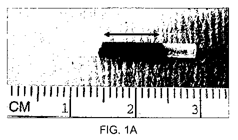
Figure 1 shows (A) a human amniotic membrane conduit with the pink central
area having been treated with 0.1% Rose Bengal and illuminated with a nd:YAG
laser at
532nm. The border region is shown as the not treated (i.e. not pink) terminal
ends. (B) a
collagen conduit with a free edge of the rolled collagen which has been sealed
using
PTB.
Figure 2 shows conduits in situ. (A) Amnion conduit secured with sutures.
Arrow shows the crosslinked central area which has maintained its tubular
structure
following rehydration. (B) Collagen conduit secured with sutures. Pink area
indicates
where the free edge has been treated with PTB. (C) Amnion conduit integrated
with
PTB. Arrow indicates where the proximal nerve end has been enveloped in the
conduit.
The conduit has been sealed to the nerve and itself using PTB. (D) Collagen
conduit
sealed with PTB.
Figure 3a shows appearance of amnion conduits at twelve weeks post-
operatively. (A) shows the nerve regeneration within an amnion conduit secured
with
sutures. (B) shows a PTB sealed conduit. The conduit is still present in both
cases
(arrows).
Figure 3b shows gross appearance of conduits following harvest at 12 weeks
post operatively. (A); amnion conduit secured with sutures. (B); amnion
conduit sealed
with PTB. The Rose Bengal stained conduit is still evident in both cases. (C);
a thin
band of neural tissue bridges the gap in the collagen conduit suture group.
The conduit
has been completely resorbed. (D); there was no neural regeneration in the
collagen
conduit PTB group. (E); autologous nerve graft.
Figure 4 shows a chart showing (A) Gastrocnemius muscle mass preservation
compared to the contralateral control muscle; and (B) Myocyte diameter
preservation
compared to contralateral control muscle. (NS= non significant. ** p<0.01)
Figure 5 shows axonal regeneration within the conduits. (A) Autologous nerve
graft showing organized regeneration with axons forming distinct fascicles.
(B) Amnion
nerve graft sealed with PTB. The area occupied by regenerating axons is large
and there
is minimal fibrous ingrowth. (C) Amnion nerve graft secure with sutures. The
central
area is occupied by axons but there is more fibrous tissue within the conduit.
(Toluidine
Blue 40x).
-13-
CA 02723801 2010-11-08
WO 2009/137793 PCT/US2009/043340
Figure 6 shows 1 m sections from the midpoint of the nerve conduits which
show regenerated axons in the (A) Autologous nerve graft, (B) Amnion conduit
secured
with sutures, (C) Amnion conduit secured with PTB and (D) Collagen conduit
secured
with sutures.
Figure 7 shows 1 m sections from 5mm distal to the nerve conduits show
regenerated axons in the (A) Autologous nerve graft, (B) Amnion conduit
secured with
sutures, (C) Amnion conduit secured with PTB and (D) Collagen conduit secured
with
sutures. No regeneration is evident in the distal stump of nerves treated with
collagen
conduits sealed with PTB (E). (Toluidine Blue, original magnification 200x).
Figure 8 shows a chart showing the total fiber counts measured within the
conduit at the midpoint. NS= non significant. ** p<0.01
DETAILED DESCRIPTION OF THE INVENTION
The present invention relates to a biocompatible membranes, tubes and conduits
which comprising a photosensitizer which is capable of being crosslinked to
form a three
dimensional structure which can be implanted into a subject to assist in
tissue bonding
and nerve maintenance and development. Significantly, the membranes and other
structures may be partially cross linked using a partial treatment with a
photosensitizer
thereby leaving one or more border regions which allows for further bonding of
the
structure to tissue or other biomaterial. This allows a generally rigid
structure (formed
by photo crosslinking) to be incorporated directly into tissues and act as
conduits or
other structures for healing and/or cell growth. This is particularly useful
when a
biological material or conduit is used to bridge between nerve ends.
Definitions
Unless defined otherwise, all technical and scientific terms used herein have
the
meaning commonly understood by a person skilled in the art to which this
invention
belongs. The following references, the entire disclosures of which are
incorporated
herein by reference, provide one of skill with a general definition of many of
the terms
used in this invention: Singleton et al., Dictionary of Microbiology and
Molecular
Biology (2nd ed. 1994); The Cambridge Dictionary of Science and Technology
(Walker
ed., 1988); The Glossary of Genetics, 5th Ed., R. Rieger et al. (eds.),
Springer Verlag
-14-
CA 02723801 2010-11-08
WO 2009/137793 PCT/US2009/043340
(1991); and Hale & Marham, The Harper Collins Dictionary of Biology (1991). As
used
herein, the following terms may have the meanings ascribed to them below,
unless
specified otherwise. However, it should be understood that other meanings that
are
know or understood by those having ordinary skill in the art are also
possible, and within
the scope of the present invention.
As used herein, the term "biocompatible structure" refers to a structure
having
three-dimensions wherein the structure is compatible with living tissue or a
living
system. In that regard, a biocompatible structure is nontoxic and/or non-
injurious to the
living tissue or living system over the period of contact/exposure. Moreover,
a
biocompatible structure does not cause a substantial immunological reaction or
rejection
over the period of contact/exposure.
As used herein, the term "biocompatible material" refers to a material that
includes molecules, such as protein molecules, that, when contacted with a
photosensitizer agent and electromagnetic energy, will form cross-links
between the
proteins, and the photosensitizer agent. Biocompatible materials according to
the
invention include biological membrane and also biocompatible membranes
composed of
synthetic polymers such as, but not limited to, polylactic acid (PLA), poly-L-
lactic acid
(PLLA), poly-D-lactic acid (PDLA), polyglycolide, polyglycolic acid (PGA),
polylactide-co-glycolide (PLGA), polydioxanone, polygluconate, polylactic acid-
polyethylene oxide copolymers, modified cellulose, collagen,
polyhydroxybutyrate,
polyhydroxpriopionic acid, polyphosphoester, poly(alpha-hydroxy acid),
polycaprolactone, polycarbonates, polyamides, polyanhydrides, polyamino acids,
polyorthoesters, polyacetals, polycyanoacrylates, degradable urethanes,
aliphatic
polyesterspolyacrylates, polymethacrylate, acyl substituted cellulose
acetates, non-
degradable polyurethanes, polystyrenes, polyvinyl chloride, polyvinyl
flouride,
polyvinyl imidazole, chlorosulphonated polyolifins, polyethylene oxide,
polyvinyl
alcohol, teflon RTM, nylon silicon, and shape memory materials, such as
poly(styrene-
block-butadiene), polynorbomene, hydrogels, metallic alloys, and oligo(c-
caprolacto-
ne)diol as switching segment/oligo(p-dioxyanone)diol as physical crosslink.
Other
suitable polymers can be obtained by reference to The Polymer Handbook, 3rd
edition
(Wiley, N.Y., 1989).
By "biological membrane" or "biocompatible membrane" can mean, but in no
way is limited to an organized layer or cells taken from any animal. In
preferred
-15-
CA 02723801 2010-11-08
WO 2009/137793 PCT/US2009/043340
embodiments, the biological membrane is an amniotic membrane. In other
exemplary
embodiments, the biological membrane can be taken from the amnion of a mammal,
for
example a cow, pig, sheep, or the like. In another preferred embodiment, the
biological
membrane may be taken from, for example, a human pregnancy, post partum. A
biological membrane or biocompatible membrane can also include endothelium,
fascia,
pericardium, pleural lining, acellular muscle, blood vessel, dura matter,
peritoneum, and
mucosal membrane (such as small intestine submucosa, SIS). A biocompatible
membrane can include synthetic membrane such as, but not limited to membranes
made
from an absorbable synthetic polymer, PGA, silicone, or other polymers such as
polylactic acid (PLA), poly-L-lactic acid (PLLA), poly-D-lactic acid (PDLA),
polyglycolide, polylactide-co-glycolide (PLGA), polydioxanone, polygluconate,
polylactic acid-polyethylene oxide copolymers, modified cellulose, collagen,
polyhydroxybutyrate, polyhydroxpriopionic acid, polyphosphoester, poly(alpha-
hydroxy
acid), polycaprolactone, polycarbonates, polyamides, polyanhydrides, polyamino
acids,
polyorthoesters, polyacetals, polycyanoacrylates, degradable urethanes,
aliphatic
polyesterspolyacrylates, polymethacrylate, acyl substituted cellulose
acetates, non-
degradable polyurethanes, polystyrenes, polyvinyl chloride, polyvinyl
flouride,
polyvinyl imidazole, chlorosulphonated polyolifins, polyethylene oxide,
polyvinyl
alcohol, teflon RTM, nylon silicon, and shape memory materials, such as
poly(styrene-
block-butadiene), polynorbomene, hydrogels, metallic alloys, and
oligo(.epsilon.-
caprolacto- ne)diol as switching segment/oligo(p-dioxyanone)diol as physical
crosslink.
It will be understood by those of skill in the art that one or more of the
foregoing
polymer constituents may be modified to include appropriate side chains (e.g.,
groups
containing amino substituents) that permit cross-linking of the polymers.
As used herein, the term "shaped" with respect to, for example, a "shaped
biocompatible material" refers to a predetermined physical or spatial form of
a
biocompatible material, biocompatible membrane, amniotic membrane, and the
like.
Shaped can refer to a material or membrane that is manipulated into a
particular physical
or spatial form such as a flat or substantially planar sheet, tube, conduit,
sphere, or
geometric solid (whether or not the shape has a hollow or solid interior).
Shaped can
also refer to a material having an intended three-dimensional physical or
spatial form.
Shaped can also refer to any of the foregoing physical and/or spatial
configurations
-16-
CA 02723801 2010-11-08
WO 2009/137793 PCT/US2009/043340
wherein the shaped structure is at least partially cross-linked so as to
substantially retain
the shape.
As used herein the term "preform" refers to a precursor to a shaped
biocompatible material. A preform can refer to a biocompatible material that
has not yet
been set into a given shape. Alternatively, a preform can refer to a
biocompatible
material that has been set into a given shape, but which is not able to
substantially retain
that shape.
As used herein, the term "border region" refers to the portion of a
biocompatible
structure that forms a contact point with tissue of an individual into which
the
biocompatible structure has been implanted and to which the biocompatible
structure is
intended to be adhered; that is, the region of a biocompatible structure that
will be cross-
linked to the tissue of the individual into which it is implanted. For
example, when the
biocompatible structure is a tube or conduit, the border region is a region,
present at one
or both terminal ends of the tube or conduit, having at least 5% of the total
length of the
tube. Where the biocompatible structure has a three-dimensional shape other
than a tube
or conduit, the border region is at least a portion of the edge of the
structure (such as, for
example, the peripheral 1mm or more of the biocompatible structure) that is
intended to
be adhered to the tissue of an individual into which it is implanted. The
border region in
such a structure can also be a portion of the biocompatible structure not at
the edge, but
which is nonetheless intended to be adhered to a tissue of the individual into
which it is
implanted. A border region also includes a region of a planar biocompatible
membrane
that, when the biocompatible membrane is shaped into a biocompatible
structure, will
form a border region of such biocompatible structure.
By "electromagnetic energy" can mean, but in no way limited to electromagnetic
radiation, or the like. For example, electromagnetic radiation can include
light having a
wavelength in the visible range or portion of the electromagnetic spectrum, or
in the
ultra violet and infrared regions of the spectrum.
By "luminal anatomical structure" can mean, but in no way limited to a
structure
that is found on the luminal surface of, for example, a blood vessel or
another
anatomical conduit.
By "luminal surface" can mean, but in no way limited to the inner surface. A
lumen is an interior space or cavity, for example, the interior of a blood
vessel. The
luminal surface of a blood vessel is the side facing the blood. For example,
the luminal
-17-
CA 02723801 2010-11-08
WO 2009/137793 PCT/US2009/043340
(or apical) side of an epithelial cell is the side that communicates with the
lumen of the
tube the epithelium lines.
The term "photo sensitizer agent" can mean, but in no way limited to a
chemical
compound that produces a biological effect upon photoactivation or a
biological
precursor of a compound that produces a biological effect upon
photoactivation, or the
like. Exemplary photo sensitizers can be those that absorb electromagnetic
energy. The
photo sensitizers of the invention can include photosensitizer fragments
and/or
derivatives of known photo sensitizers, which have the same or substantially
the same
function as the known photo sensitizers, which means that function which is at
least
about 50% of the function of an original photo sensitizer, more preferably
about 60% or
70%, or still more preferably about 80% or 90%, or even more preferably about
95% or
99% the function of the known photosensitizer compound. A photosensitizer
agent can
be, but is not limited to a xanthenes, e.g., Rose Bengal and erythrosin;
flavins, e.g.,
riboflavin; thiazines, e.g., methylene blue; porphyrins and expanded
porphyrins, e.g.,
protoporphyrin I through protoporphyrin IX, coproporphyrins, uroporphyrins,
mesoporphyrins, hematoporphyrins and sapphyrins; chlorophylis, e.g.,
bacteriochlorophyll A, phenothiazine, cyanine, Mono azo dye (e.g., Methyl
Red), Azine
mono azo dye (e.g., Janus Green B), Phenothia-zine dye (e.g., Toluidine Blue),
rhodamine dye (e.g., Rhodamine B base), Benzyphen-oxazine dye (e.g., Nile Blue
A,
Nile Red), oxazine (e.g., Celestine Blue), and anthroqui-none dye (e.g.,
Remazol
Brilliant Blue R). Exemplary photosensitizer agents may include, but are not
limited to,
Rose Bengal, riboflavin-5-phosphate, and methylene blue.
The photo sensitizers of the invention can include "photoactive dyes," which,
as
used herein, refers to those photo sensitizers that produce a fluorescent
signal when
activated. The photoactive dyes of the invention may also be fragments and/or
derivatives of a known photoactive dyes which have the same or substantially
the same
function as a known photoactive dye, which means a function that is at least
about 50%
of the function of a known photoactive dye, more preferably about 60% or 70%,
or still
more preferably about 80% or 90%, or even more preferably about 95% or 99% the
function of a known photoactive dye.
Depending on the wavelength and power of light administered, a photosensitizer
can be activated to fluoresce and, therefore, act as a photoactive dye, but
not produce a
-18-
CA 02723801 2010-11-08
WO 2009/137793 PCT/US2009/043340
phototoxic species. The wavelength and power of light can be adapted by
methods
known to those skilled in the art to bring about a phototoxic effect where
desired.
By "photoactivatable membrane device" can mean, but in no way limited to a
membrane that is capable of photoactivation, or the like. Photoactivation can
be used to
describe the process by which energy is absorbed by a compound, e.g., a photo
sensitizer,
thus "exciting" the compound, which then becomes capable of converting the
energy to
another form of energy, preferably chemical energy.
The term "photo sensitizer composition," as used herein, refers to chemical
constructs having one or more photo sensitizers (or fragments and/or
derivatives thereof),
as well as other materials, such as linkers, backbones, targeting moieties and
binders,
that may be couple thereto.
As used herein, the term "fluorescent dye" refers to dyes that are fluorescent
when illuminated with light but do not produce reactive species that are
phototoxic.
Any compound or moiety of the invention that is fluorescent in one or more
states can contain one or more "fluorophores," which refers to a compound or
portion
thereof which exhibits fluorescence. The term "fluorogenic" refers to a
compound or
composition that becomes fluorescent or demonstrates a change in its
fluorescence (such
as an increase or decrease in fluorescence intensity or a change in its
fluorescence
spectrum) upon interacting with another substance, for example, upon binding
to a
biological compound or metal ion, upon reaction with another molecule or upon
metabolism by an enzyme. Fluorophores may be substituted to alter their
solubility,
spectral properties and/or physical properties. Numerous fluorophores and
fluorogenic
compounds and compositions are known to those skilled in the art and include,
but are
not limited to, benzofurans, quinolines, quinazolines, quinazolinones,
indoles,
benzazoles, indodicarbocyanines, borapolyazaindacenes and xanthenes, with the
latter
including fluoresceins, rhodamines and rhodols as well as other fluorophores
described
in Haugland, Molecular Probes, Inc. Handbook of Fluorescent Probes and
Research
Chemicals, (9th ed., including the CD-ROM, September 2002), and include the
photo sensitizers, photoactive dyes, and fluorescent compounds and moieties of
the
invention.
As used herein, the term "detectable" or "directly detectable," or the like,
refers
to the presence of a detectable signal generated from a compound of the
invention, e.g.,
-19-
CA 02723801 2010-11-08
WO 2009/137793 PCT/US2009/043340
a photo sensitizer, that is detectable by observation, instrumentation, or
film without
requiring chemical modifications or additional substances.
The term "subject" is used herein to refer to a living animal, including a
human.
As used herein, the term "substantially retains" as it relates to a three-
dimensional shape of a biocompatible structure refers to the retention of a
three-
dimensional shape to the extent that the biocompatible structure can be used
for its
intended purpose. "Substantially retains" refers to no greater than a 5% or
more change
in a given dimension of a biocompatible structure, for example no greater than
a 5%
change, 10%, 15%, 20%, 25%, 30%, 35%, 40%, 45%, 50%, 55%, 60%, 70%, 75% or
80% change in a given dimension, provided that the biocompatible structure can
still be
used for its intended purpose. For example, a linear human amniotic membrane
tube
intended for use as a conduit to permit nerve regeneration can undergo a 5% or
more
change in its linear shape (i.e., it can be curved), but only to the extent
that it can
function as a nerve conduit.
As used herein, the term "neural tissue" refers to neural tissue of the
central or
peripheral nervous system. Neural tissue can refer to peripheral nervous
tissue, such as
a peripheral nerve, a dorsal or ventral ramus, spinal nerve, or ganglion, and
can also
refer to central nervous tissue such as the spinal cord.
In this disclosure, "comprises," "comprising," "containing" and "having" and
the
like can have the meaning ascribed to them in U.S. Patent law and can mean "
includes,"
"including," and the like; "consisting essentially of" or "consists
essentially" likewise
has the meaning ascribed in U.S. Patent law and the term is open-ended,
allowing for the
presence of more than that which is recited so long as basic or novel
characteristics of
that which is recited is not changed by the presence of more than that which
is recited,
but excludes prior art embodiments.
Other definitions appear in context throughout this disclosure.
Biocompatible materials
The present invention provides shaped biocompatible structures and tissue
sealing devices that can be used for a wide array of applications such as
nerve repair,
surgical wound closure, stents, and the like. The structures described herein
can be
formed by contacting a biocompatible material with a photosensitizer agent,
where upon
application of electromagnetic energy, molecules in the material are able to
form cross-
links with the photosensitizer agent. The result is an increase in the
rigidity of the
-20-
CA 02723801 2010-11-08
WO 2009/137793 PCT/US2009/043340
biocompatible material such that the three-dimensional structure is formed and
the
structure substantially retains its desired shape. Biocompatible materials are
materials
that comprise molecules, such as protein molecules, that, when contacted with
a
photosensitizer agent and electromagnetic energy, will form cross-links
between the
cross-linkable molecules, and the photosensitizer agent. Biocompatible
materials
according to the invention can include biocompatible membranes, either natural
or
synthetic. Biocompatible membranes useful according to the invention can be
biological
membranes which are an organized layer or cells taken from an animal or
produced
synthetically. In one embodiment, the biological membrane is an amniotic
membrane.
In other exemplary embodiments, the biological membrane can be taken from the
amnion of a mammal, for example a cow, pig, sheep, or the like. In another
embodiment, the biological membrane may be taken from, for example, a human
pregnancy, postpartum. Biological membranes also include endothelium, fascia,
pericardium, pleural lining, acellular muscle, blood vessel, dura matter,
peritoneum, and
mucosal membrane (such as small intestine submucosa, SIS). Biocompatible
materials
include biocompatible membranes composed of synthetic polymers such as, but
not
limited to, polylactic acid (PLA), poly-L-lactic acid (PLLA), poly-D-lactic
acid (PDLA),
polyglycolide, polyglycolic acid (PGA), polylactide-co-glycolide (PLGA),
polydioxanone, polygluconate, polylactic acid-polyethylene oxide copolymers,
modified
cellulose, collagen, polyhydroxybutyrate, polyhydroxpriopionic acid,
polyphosphoester,
poly(alpha-hydroxy acid), polycaprolactone, polycarbonates, polyamides,
polyanhydrides, polyamino acids, polyorthoesters, polyacetals,
polycyanoacrylates,
degradable urethanes, aliphatic polyesterspolyacrylates, polymethacrylate,
acyl
substituted cellulose acetates, non-degradable polyurethanes, polystyrenes,
polyvinyl
chloride, polyvinyl flouride, polyvinyl imidazole, chlorosulphonated
polyolifins,
polyethylene oxide, polyvinyl alcohol, teflon RTM, nylon silicon, and shape
memory
materials, such as poly(styrene-block-butadiene), polynorbomene, hydrogels,
metallic
alloys, and oligo(.epsilon.-caprolacto- ne)diol as switching segment/oligo(p-
dioxyanone)diol as physical crosslink. Other suitable polymers can be obtained
by
reference to The Polymer Handbook, 3rd edition (Wiley, N.Y., 1989). One of
skill in the
art will readily appreciate that the foregoing polymers can be uses in
biocompatible
materials as described herein provided that they are adapted to be amenable to
cross-
-21-
CA 02723801 2010-11-08
WO 2009/137793 PCT/US2009/043340
linking by the methods of the invention (e.g., provided that the polymers
contain suitable
amion containing side chains or moieties).
Amnionic membranes for forming three-dimensional structures
The amniotic membrane is the translucent innermost layer of the three layers
forming the fetal membranes, and is derived from the fetal ectoderm. The
amniotic
membrane contributes to homeostasis of the amniotic fluid. At maturity, the
amniotic
membrane is composed of epithelial cells on a basement membrane, which in turn
is
connected to a thin connective tissue membrane or mesenchymal layer by
filamentous
strands. In one embodiment of the invention, amniotic membrane is obtained
from a
human, although amniotic membrane may also be obtained from other mammals such
as
sheep, pig, cow.
Human amniotic membrane (HAM) is a substrate that can be photochemically
modified to make shaped biocompatible structures. Native HAM is a transparent,
20 m
thick tissue that is flimsy in nature although somewhat tear resistant.
Crosslinking of
HAM provides enhanced rigidity and mechanical strength to the material
HAM in its native form can be used for photochemical tissue bonding to seal
tissues by crosslinking at the interface between the HMA and the body tissue,
e.g.
peripheral nerve cornea, sclera and conjunctiva. In this process a
photosensitizer agent is
applied superficially to the HAM, which is then placed in intimate contact
with the
target tissue and illuminated in situ to form a tight seal or coverage of the
native tissue,
such as in sealing HAM nerve wraps.
The isolated amniotic membranes that can be used in the exemplary embodiment
of the present invention may be obtained from a commercial source, for example
from
suppliers such as AmbioDry and AmbioDry2 from OKTO Ophtho and AMNIOGRAFT
from Bio-Tissue. Alternatively, the amniotic membrane may be recombinant, or
naturally occurring and sterilized. The amniotic tissue may be obtained
postpartum and
then preserved by any number of methods known to one of skill in the art (e.g.
glycerol,
lyophilization, gluteraldehyde, etc). Additionally, amniotic membranes that
are derived
from non-humans may be used. Methods for obtaining and preparing amniotic
membrane are known in the art and are described, for example, in
US20070031471, the
contents of which are incorporated herein in their entirety.
The membranes of the exemplary embodiment of the present invention can be,
for example, between 10 m, 15 m, 20 m, 25 m, 30 m, 35 m or more m in
-22-
CA 02723801 2010-11-08
WO 2009/137793 PCT/US2009/043340
thickness. In certain exemplary embodiments, the membrane is 20 m in
thickness, and
is a human amniotic membrane.
Photoactivation and Photosensitizer Agents
Photoactivation, as referred to herein, e.g., can be used to describe the
process
by which energy in the form of electromagnetic radiation is absorbed by a
compound,
e.g., a photosensitizer agent, thus "exciting" the compound, which then
becomes capable
of converting the energy to another form of energy, preferably chemical
energy. The
electromagnetic radiation can include energy, e.g., light, having a wavelength
in the
visible range or portion of the electromagnetic spectrum, or the ultra violet
and infrared
regions of the spectrum. The chemical energy can be in the form of a reactive
species,
e.g., a reactive oxygen species, e.g., a singlet oxygen, superoxide anion,
hydroxyl
radical, the excited state of the photo sensitizer, photosensitizer free
radical or substrate
free radical species. The photoactivation process can involve an insubstantial
transfer of
the absorbed energy into heat energy. Preferably, photoactivation occurs with
a rise in
temperature of less than 3 degrees Celsius (C), more preferably a rise of less
than 2
degrees C and even more preferably, a rise in temperature of less than 1
degree C as
measured, e.g., by an imaging thermal camera that looks at the tissue during
irradiation.
The camera can be focused in the area of original dye deposit, e.g., the wound
area, or
on an area immediately adjacent the wound area, to which dye will diffuse. As
used
herein, a photosensitizer agent is a chemical compound that produces a
biological effect
upon photoactivation or a biological precursor of a compound that produces a
biological
effect upon photoactivation. Exemplary photo sensitizers can be those that
absorb
electromagnetic energy, such as light. While not wishing to be bound by
theory, the
photosensitizer agent may act by producing an excited photosensitizer or
derived species
that interacts with tissue, e.g., amniotic membrane, to form a bond, e.g., a
covalent bond
or crosslink. Certain exemplary photo sensitizers typically have chemical
structures that
include multiple conjugated rings that allow for light absorption and
photoactivation. A
number of photo sensitizers are known to one of skill in the art, and
generally include a
variety of light-sensitive dyes and biological molecules. Examples of
photosensitizer
agent include, but are not limited to, xanthenes, e.g., Rose Bengal and
erythrosin;
flavins, e.g., riboflavin; thiazines, e.g., methylene blue; porphyrins and
expanded
-23-
CA 02723801 2010-11-08
WO 2009/137793 PCT/US2009/043340
porphyrins, e.g., protoporphyrin I through protoporphyrin IX, coproporphyrins,
uroporphyrins, mesoporphyrins, hematoporphyrins and sapphyrins; chlorophylis,
e.g.,
bacteriochlorophyll A, phenothiazine, cyanine, Mono azo dye (e.g., Methyl
Red), Azine
mono azo dye (e.g., Janus Green B), Phenothia-zine dye (e.g., Toluidine Blue),
rhodamine dye (e.g., Rhodamine B base), Benzyphen-oxazine dye (e.g., Nile Blue
A,
Nile Red), oxazine (e.g., Celestine Blue), anthroqui-none dye (e.g., Remazol
Brilliant
Blue R), and photosensitive derivatives thereof. Exemplary photosensitizer
agents
according to the methods of the invention as described herein are compounds
capable of
causing a photochemical reaction capable of producing a reactive intermediate
when
exposed to light, and which do not release a substantial amount of heat
energy. Some
exemplary photo sensitizers include Rose Bengal (RB); riboflavin-5-phosphate
(R-5-P);
methylene blue (MB); and N-hydroxypyridine-2-(1H)-thione (N-HTP).
In certain exemplary embodiments, a photosensitizer agent, e.g., RB, R-5-P,
MB,
or N-HTP, can be dissolved in a biocompatible buffer or solution, e.g., saline
solution,
and used at a concentration of from about 0.1 mM to 10 mM, preferably from
about 0.5
mM to 5 mM, more preferably from about 1 mM to 3 mM.
A photosensitizer agent can be administered to a biocompatible material as
described herein. Photosensitizer agents can be brushed or sprayed onto one or
both
surfaces of a biocompatible membrane prior to the application of
electromagnetic
energy. Other methods for applying photosensitizer agent (e.g., such as
submerging the
membrane in photosensitizer agent) can be envisioned by one of skill in the
art. In one
embodiment, photosensitizer agent is not applied to the entirety of the
biocompatible
membrane pior to forming a three-dimensional structure, and a portion of the
biocompatible membrane is left free of photosensitizer agent. As described in
further
detail below, upon exposure to electromagnetic energy, the portion of the
biological
membrane that contains photosensitizer agent will form cross-links, while the
portion
that is free of photosensitizer agent will not form cross-links.
The electromagnetic radiation, e.g., light, can be applied to the tissue at an
appropriate wavelength, energy, and duration, to cause the photosensitizer to
undergo a
reaction to affect the structure of the amino acids in the tissue, e.g., to
cross-link a tissue
protein, thereby creating a tissue seal. The wavelength of light can be chosen
so that it
corresponds to or encompasses the absorption of the photo sensitizer, and
reaches the
area of the tissue that has been contacted with the photo sensitizer, e.g.,
penetrates into
-24-
CA 02723801 2010-11-08
WO 2009/137793 PCT/US2009/043340
the region where the photosensitizer is injected. The electromagnetic
radiation, e.g.,
light, necessary to achieve photoactivation of the photosensitizer agent can
have a
wavelength from about 350 nm to about 800 nm, preferably from about 400 to 700
nm
and can be within the visible, infra red or near ultra violet spectra. The
energy can be
delivered at an irradiance of about between 0.5 and 5 W/cm2, preferably
between about
1 and 3 W/cm2. The duration of irradiation can be sufficient to allow cross-
linking of
one or more proteins of the tissue, e.g., of a tissue collagen. For example,
in corneal
tissue, the duration of irradiation can be from about 30 seconds to 30
minutes, preferably
from about 1 to 5 minutes. The duration of irradiation can be substantially
longer in a
tissue where the light has to penetrate a scattering layer to reach the wound,
e.g., skin or
tendon. For example, the duration of irradiation to deliver the required dose
to a skin or
tendon wound can be at least between one minute and two hours, preferably
between 30
minutes to one hour.
Suitable sources of electromagnetic energy can include but not limited to
commercially available lasers, lamps, light emitting diodes, or other sources
of
electromagnetic radiation. Light radiation can be supplied in the form of a
monochromatic laser beam, e.g., an argon laser beam or diode-pumped solid-
state laser
beam. Light can also be supplied to a non-external surface tissue through an
optical fiber
device, e.g., the light can be delivered by optical fibers threaded through a
small gauge
hypodermic needle or an arthroscope. Light can also be transmitted by
percutaneous
instrumentation using optical fibers or cannulated waveguides.
The choice of energy source can generally be made in conjunction with the
choice of photosensitizer employed in the method. For example, an argon laser
can be an
energy source suitable for use with RB or R-5-P because these dyes are
optimally
excited at wavelengths corresponding to the wavelength of the radiation
emitted by the
argon laser. Other suitable combinations of lasers and photo sensitizers are
known to
those of skill in the art. Tunable dye lasers can also be used with the
methods described
herein.
The photosensitizer agents of the current invention afford several beneficial
aspects for cross-linking biocompatible membranes such as amnion. For example,
the
electromagnetic energy used to photoactivate the photosensitizer agent can
typically
penetrate further into tissues than other cross-linking energy sources, such
as UV rays.
Additionally, the current methods provide an alternative to using ionizing
radiation to
-25-
CA 02723801 2010-11-08
WO 2009/137793 PCT/US2009/043340
cross link the biocompatible membrane, which is well known to be detrimental
to
surrounding tissues. Furthermore, the photosensitizer agents useful in the
invention can
be non-toxic and the light initiation described herein provides a greater
degree of control
over the extent of cross-linking in the biocompatible membrane.
Shaped biocompatible structures
The invention relates to shaped biocompatible structures (such as a tissue
sealing
device) that can be formed by placing a biocompatible material comprising a
photosensitizer agent into a desired shape and exposing the membrane to
electromagnetic energy, whereby cross-links are formed in the membrane,
whereby the
rigidity of the membrane is increased such that the membrane is able to
substantially
retain the desired shape. In one embodiment, the shaped biocompatible
structure (i.e.,
tissue sealing device) comprises a first section of cross-linked moieties and
a second
section of noncross-linked moieties. The first section of cross-linked
moieties confers
rigidity to the structure. The second section of noncross-linked moieties is
configured so
that it is contactable with a tissue (e.g., nerve tissue) wherein the non-
cross-linked
moieties can be cross-linked with protein molecules of the tissue by
contacting one or
both of the structure and tissue with a photosensitizer agent and exposing the
structure
and tissue to electromagnetic energy. In one embodiment the noncross-linked
section of
a shaped biocompatible structure is a border region, meaning that it is a
section that is
intended to be used to bond the biocompatible structure to a host tissue. A
border region
can be located at any position on a biocompatible structure that is intended
to be cross-
linked to a host tissue.
Examples of biocompatible structures that can be formed using biocompatible
membranes described herein include, but are not limited to, conduits, shunts,
stents,
patches, wound closure devices, and hernia repair patches. Biocompatible
structures
(i.e., shaped biocompatible structures) can also include scaffolding or
framework
structures on which additional tissues are grown or which can be implanted in
the body
to give three dimensional shape to tissue. Such framework structures include
structures
that mimic cartilagenous portions of the human body such as the ear or nose,
or
structures that are used in plastic surgical applications such as implants for
the lips,
cheeks, and the like. Thee-dimensional biocompatible structures according to
the
invention can also be used to fill space in a body cavity or other body space
to maintain
the proper anatomical relationship of surrounding structures, such as, for
example,
-26-
CA 02723801 2010-11-08
WO 2009/137793 PCT/US2009/043340
inserting a shaped biocompatible structure into the body to fill the space
previously
occupied by an organ or other tissue.
Previous research has shown that the physical properties of a membrane, can be
altered by photocrosslinking the constitutive proteins. For example, in one
example, a
tube was prepared by applying rose bengal to a strip of biological membrane,
wrapping
3-4 layers around a rod, irradiating and then removing the rod [Irish
Association of
Plastic Surgeons, Galway, Ireland, May 10-12, 2007. Preparation and
Integration of
Nerve Conduits using a Photochemical Technique. O'Neill et al.]. Previous
studies have
also shown that flat layers of human amniotic membrane can be photocrosslinked
together [unpublished].
Further, the amniotic membranes of the exemplary embodiment of the present
invention may be modified to change their consistency. For example, amniotic
membranes with enhanced rigidity as biocompatible devices are described in
WO06002128.
A shaped biocompatible structure may be formed prior to deployment, during, or
after deployment, in order to conform and/or alter the topology of the
structure to which
it is to be applied. In one embodiment, the shaped biocompatible structure is
a tube that
can be used as a conduit.
In one embodiment the shaped biocompatible structure is a conduit, such as a
pre-formed conduit, made of partially cross-linked amniotic membrane. A piece
of
amniotic membrane is obtained (for example, as described hereinabove) and
photosensitizer dye is partially applied to the central section of the
membrane, leaving a
portion of the membrane free of said photosensitizer agent (i.e., a border
region). The
membrane is then wrapped around a cylindrical support having an appropriate
diameter
and illuminated with electromagnetic energy, such as green light. Subsequent
removal
of the support results in a partially cross-linked amniotic membrane conduit
for
implantation. To implant the conduit, such as for peripheral nerve repair,
photosensitizer agent is subsequently applied to the luminal surface of the
border region,
and the nerve stumps are inserted into the conduit and sealed by forming cross-
links
between the conduit and the peripheral nerve, for example, by applying
electromagnetic
energy in the form of green light.
In certain embodiments, the shaped biocompatible structure is designed to
alter
the topology of a luminal anatomic structure.
-27-
CA 02723801 2010-11-08
WO 2009/137793 PCT/US2009/043340
In one such example, the shaped biocompatible structure may be formed as a
sheet of membrane, for example a sheet of amniotic membrane. This
configuration may
be preferable for use in imparting stability to one portion of the luminal
anatomical
structure.
In certain other examples, the intraluminal covering device that attaches to a
luminal anatomic structure can at least partially cover the anatomical
structure in a
manner that either at least partially maintains the patency of said luminal
anatomic
structure.
In other examples, the membrane, preferably the exemplary biological
membrane, attaches to a luminal anatomic structure that does not move within
said
structure following deployment. In other preferred examples, the biological
membrane
can attach to a luminal anatomic structure that at least partially covers the
anatomical
structure in a manner that either at least partially stabilizes of said
luminal anatomic
structure.
It may be preferred that the membrane attaches to a luminal anatomic structure
does not damage said structure.
In another example, this topology may be used to repair a defect in an
anatomical
structure. In certain cases, it may be preferable to use the membrane of the
invention to
treat, repair, or cover only one portion of an anatomical structure, and leave
the other
portion of the anatomical structure intact. For example, to cover only a
portion of the
luminal anatomic structure that may utilize an alteration while leaving the
remainder of
the luminal anatomic structure intact. One example of this can be a covering
or a stent,
such as an intraluminal stent. Such a stent or covering can, for example,
impart
mechanical stability, act as a cover, or maintain at least partial patency of
the structure it
is covering (e.g. a luminal anatomic structure). The stent or covering may in
certain
examples be a resizable stent or covering that at least imparts mechanical
stability,
covers, or maintains at least partial patency of the anatomic structure. In
this exemplary
way, the stent or covering does not need to be fitted in diameter to be of a
predetermined
size, and overlapping areas of the shaped biocompatible structure take up the
slack upon
deployment of the device.
In another exemplary embodiments of the present invention, a number of
different device patterns are described that enhance or enable different
biological
functions or capabilities.
-28-
CA 02723801 2010-11-08
WO 2009/137793 PCT/US2009/043340
The shaped biocompatible structure may be conformed to be in a certain
exemplary geometry. For example, the shaped biocompatible structure may be
conformed in a cylinder, a plane, a sphere, a geometry preformed to the
contour of the
tissue of interest, or preformed to a desired contour to effect the best
clinical treatment.
In certain preferred examples, the cylinder or tube is used as a conduit,
stent or a
covering.
In other examples, the edges of the shaped biocompatible structure are
tapered,
and in certain preferred embodiments, may contain projections. The projections
can
comprise amniotic membrane, metal struts, nitinol struts, plastic struts, or
composites,
such as Polytetrafluoroethylene (PTFE), teflon, plastic, rubber, nitinol, or
biodegradable
composites or the like. .
The shaped biocompatible structure may be configured with holes. The shaped
biocompatible structure may have be configured to have 1, 2, 3, 5, 10, 20, 50,
100, 150,
200, 300, 500, or more holes, or different number of holes. The holes in the
membrane
can be of any geometry and may be configured to allow for passage of
intraluminal
tissues such as, but not limited to, blood, bile, or lymph to pass through.
The exemplary
minimum diameter of the holes may be between 10, 20, 30 40, 50, 75, 100, 200,
400,
500, 600, 750 pm in order to allow the passage of red and white blood cells,
but other
diameters are conceivable, and are within the scope of the present invention.
The
exemplary pattern of holes may be configured to allow endothelial or
epithelial cells or
other cells to migrate through the shaped biocompatible structure.
The holes and intervening spaces may be configured to impart further
mechanical stability to the shaped biocompatible structure. For example, the
edges of the
shaped biocompatible structure may be tapered to further significantly improve
endothelial or epithelial cell migration.
Accordingly, it is one object of the present invention that the exemplary
shaped
biocompatible structure that attaches to a luminal anatomic structure promotes
re-
endothelialization or re-epithelialization of said anatomic structure. Such
exemplary
membrane device thereby can be configured to allow the endothelial or
epithelial cells of
the luminal anatomic structure to migrate and cover the biological membrane
following
deployment of the device. Promotion of this healing process can be facilitated
by
adjusting an exemplary biological membrane thickness, number and size of holes
or
-29-
CA 02723801 2010-11-08
WO 2009/137793 PCT/US2009/043340
openings, and by applying other pharmacological agents to the biological
membrane
device that facilitate said re-endo or re-epithelialization
The exemplary shaped biocompatible structure may be, in certain embodiments,
comprised of layers of membrane, for example, amniotic membrane, configured to
impart substantially more thickness and/or mechanical stability to the
membrane device.
The membranes of the exemplary embodiments of the present invention, may be
modified to change shape or configuration. For example, the shaped
biocompatible
structures can be comprised of layers of one or more, for example, 2, 3, 5,
10, 20, 30, 50
or more membrane sheets. These sheets can be affixed to each other, in certain
examples, by electromagnetic radiation.
In one exemplary embodiment, the layers may be affixed to one another by
means of applying electromagnetic radiation to layers of amniotic membrane
comprised
of photoactivatable dye.
In the foregoing embodiments, a first section or portion of the biocompatible
membrane is contacted with photosensitizer agent and a second section or
portion is kept
free of photosentisizing agent so as to create a noncross-linked border region
in the final
shaped structure. Shaped structures formed in this way will, therefore, only
be partially
cross-linked following application of electromagnetic energy. This partial
cross-linking
permits the shaped biocompatible structure to be deployed in a subject such
that
photosensitizer is applied to the non-cross linked border region of the
structure (and/or is
applied to the tissue to which the structure is to be adhered), wherein
subsequent
application of electromagnetic energy will function to create cross-links
between the
biocompatible membrane of the device at the border region and the target
tissue to
which the device is to be adhered. A border region may be formed at any
location of the
shaped structure that is intended for contact and bonding to the tissue of a
subject. For
example, the border region of a conduit may be located at either or both ends
of the
conduit, and/or may be located at some site in the conduit internal to the
ends. In the
context of a tube or conduit, a border region can occupy 5-40% of the total
length of the
tube or conduit. In one embodiment, as measured along the long axis of the
tube or
conduit, the border region can be 1mm or more in length. For example the
border region
can be 2, 5, 10, 15, 20, 25, 30, 35, 40, 45, 50, 55, 60, 65, 70, 75, 80, 85,
90, 95, 100, 200,
300, 400, 500, 600, 700, 800, 900, or 1000 mm or more in length as measured
along the
long axis of the tube or catheter.
-30-
CA 02723801 2010-11-08
WO 2009/137793 PCT/US2009/043340
A border region need not be continuous with respect to the biocompatible
membrane, but instead, may be discontinuous or located in discrete areas of
the
biocompatible membrane. For example, if the ultimate shape of a shaped
biocompatible
structure will have one or more specific points of contact with a host tissue,
those points
of contact can be created as border areas (by not applying a photosensitizer
agent to the
corresponding regions of the biocompatible membrane), regardless of whether
the
border region is at the edge of the biocompatible membrane, and regardless of
whether
the border region represents a continuous area of the biocompatible membrane.
A shaped biocompatible structure may be insertable or may be implantable. In
one embodiment, the shaped structure may be pre-formed or partially pre-formed
prior
to implantation. The application of photosensitizer agent and/or
electromagnetic energy
may occur in situ in a subject or may be performed ex vivo prior to
implantation of a
device in a subject.
Methods using shaped biocompatible structures
The shaped biocompatible structures (such as a tissue sealing device)
described
herein can be suitable for use in a variety of applications, including in
vitro laboratory
applications, ex vivo tissue treatments, but especially in in vivo procedures
on living
subjects, e.g., humans, and especially in nerve repair and repair of luminal
anatomical
structures.
In one embodiment, the shaped biocompatible structures described herein can be
used as a tissue sealing device in nerve repair. A pre-formed conduit made
from
biocompatible membrane such as human amniotic membrane can be used to bridge a
defect in neural tissue (such as a transection, nerve crush, partial
transection, or other
lesion), whereby an intraneural neurotrophic environment can be maintained
within the
conduit. In one embodiment, a biocompatible conduit as described herein can be
used to
bridge a gap between the cut ends of a peripheral nerve. It will be
understood, however,
that the phrase "bridge a gap" does not require a physical separation of the
two ends of a
nerve, but also includes a situation where the ends of a nerve are in contact
with each
other, but some or all of the nerve fibers have been severed or otherwise
damaged. For
example, a partially cross-linked conduit can be formed as described above.
The site of
nerve transection in a subject is then exposed under surgical conditions.
Photosensitizer
agent is then applied to the luminal surface of the conduit at least covering
the border
region, although photosensitizer may be applied a portion of the already cross-
linked
-31-
CA 02723801 2010-11-08
WO 2009/137793 PCT/US2009/043340
conduit. Photosensitizer agent may also or alternatively applied to the nerve
that will be
inserted into the conduit. Each cut end of the nerve is placed in the conduit
and
electromagnetic energy is applied to cross-link the border region of the
conduit to the
nerve stumps. In addition, one or more sutures may also be used to secure the
ends of
the transected nerve within the conduit. Sealing the nerve in the pre-formed
conduit in
this way preferably results in a watertight seal being formed between the
neural tissue
and the conduit. In addition to the foregoing, the conduit can be reinforced
by placing
one or more sutures through the conduit and tissue to be repaired. In one
embodiment,
the photosensitizer agent is only applied to one end of the conduit, while the
other end of
the conduit is secured with one or more sutures.
In a further embodiment a shaped biocompatible structure can be used in tissue
repair applications such as hernia repair. For example, a piece of
biocompatible
membrane may be treated with photosensitizer agent, whereby a border region at
the
perimeter of the biocompatible membrane is left untreated:
Treated with photosensitizer agent
Not treated with photosensitizer
age
The membrane can then be exposed to electromagnetic energy whereby the
treated portion of the membrane is cross-linked and has increased rigidity
relative to the
untreated border region. This partially cross-linked membrane patch can then
be
adhered to a facial, muscle, or other tissue layer in an individual having a
hernia or other
anatomical defect, wherein the border region is first treated with a
photosensitizer agent,
whereby subsequent exposure to electromagnetic energy bonds the membrane patch
to
the tissue of the individual by cross-linking the membrane at the border
region with the
tissue of the individual. In addition, the patch can be reinforced by placing
one or more
sutures through the border region and the tissue of the individual.
The invention also provides methods for stabilizing luminal anatomical
structures and for treating or preventing atherosclerotic plaques.
-32-
CA 02723801 2010-11-08
WO 2009/137793 PCT/US2009/043340
The exemplary methods described herein can be used, for example, for tissue
bonding. Tissue bonding can be used to seal anatomical sites, for instance,
after injury,
or after a surgical procedure, or as part of a prophylactic measure to prevent
against a
disease or pathological event. In one example, for instance, an exemplary
biological
membrane tissue bonding technique/procedure has been previously used to seal
neurorraphy sites [Photochemical Sealing Improves Outcome Following Peripheral
Neurorrhaphy. A.C. O'Neill, M.A. Randolph, K. E. Bujold, I.E. Kochevar, R.W.
Redmond, J. M. Winograd submitted to Experimental Neurology ], incorporated by
reference in its entirety herein. In this example, Rose Bengal-stained
biological
membrane was wrapped around the repair site (rat sciatic nerve) and exposed to
30
J/cm2 (on each side) 532 nm (irradiance = 0.5 W/cm2) using a frequency doubled
Nd/
YAG laser. For example, the biological membrane can additionally rapidly bond
to
vocal fold (epithelial, lamina propria and muscle layers) [unpublished],
incorporated by
reference in its entirety herein. In this example, bonding of a biological
membrane to
cornea (without epithelial layer) an energy density of 100 J/cm2 is typically
used.
Biological membrane has also been bonded to dermis, epidermis and tracheal
submucosa.
Methods for stabilizing luminal structures can include the steps of contacting
a
biological membrane with a photosensitizer agent and deploying the biological
membrane photosensitizer complex to the luminal anatomical structure of
interest, and
then applying electromagnetic energy, thereby adhering the biological membrane
to the
luminal anatomical structure. In one embodiment, the biological membrane is
pre-
formed into a shaped biocompatible structure such as a stent.
Another exemplary embodiment of the method according to the present
invention can be provided for stabilizing a luminal anatomical structure. The
exemplary
method can comprise contacting a biological membrane with a photosensitizer
agent and
then deploying the biological membrane to the luminal anatomical structure in
need of
stabilization, applying electromagnetic energy to the biological membrane-
photosensitizer complex in a manner effective to bond the tissue, and thereby
stabilizing
a luminal anatomical structure.
The invention also includes methods for treating or preventing an
atherosclerotic
plaque. The method comprises identifying an atherosclerotic plaque, contacting
a
biological membrane with a photosensitizer agent wherein a portion of the
membrane is
-33-
CA 02723801 2010-11-08
WO 2009/137793 PCT/US2009/043340
not contacted with the photosensitizer agent so as to form a border region,
deploying the
biological membrane to the atherosclerotic plaque, and applying
electromagnetic energy
to the biological membrane photosensitizer complex in a manner effective to
bond the
tissue, and thus treating or preventing an atherosclerotic plaque. In one
embodiment,
prior to deployment of the membrane, the membrane is exposed to
electromagnetic
energy to partially cross-link the membrane.
According to yet another embodiment of the present invention, methods for
promoting one or more of cell growth and migration in a luminal anatomical
structure of
interest are provided. The exemplary method can comprise contacting a
biological
membrane with a photosensitizer agent, deploying the biological membrane
photosensitizer complex to the luminal anatomical structure of interest, and
applying
electromagnetic energy, and thereby promoting cell growth and migration in a
luminal
anatomical structure of interest.
According to another embodiment of the invention, a shaped biocompatible
structure can be formed and used to give structural shape to overlying tissues
such as
skin. For example, a shaped biocompatible structure can be used as an implant
in
cosmetic surgical applications, such as, for example, facial reconstruction
(e.g, lip,
cheek, brow or neck augmentation or reconstruction), scar repair, or repair of
damage
from traumatic injury that decreased the supporting structures underlying the
skin or
other tissue.
Kits
In one embodiment, the invention provides kits comprising a shaped
biocompatible structure as described herein and packaging materials therefor.
In one
embodiment, the kit includes a pre-formed shaped biocompatible structure
(e.g., a tissue
sealing device), while in another embodiment, the kit includes a Biocompatible
material
(e.g., a tissue sealing device pre-form) and a photo sensitizer agent with
instructions for
forming a shaped biocompatible structure. In either of the foregoing
embodiments, the
kit can also include written instructions that describe how to use the shaped
biocompatible structure for a given purpose. For example, the instructions can
describe
how to use a tubular shaped biocompatible structure as a conduit for nerve
repair. The
instructions can include a description of methods for adhering a shaped
biocompatible
structure to anatomical structures such as nerve or other tissues, for
stabilizing a luminal
anatomical structure, for treating or preventing an atherosclerotic plaque, or
for
-34-
CA 02723801 2010-11-08
WO 2009/137793 PCT/US2009/043340
promoting one or more of cell growth and migration in or on a shaped
biocompatible
structure or tissue of interest as described herein. The exemplary kits can
include
packaging materials such as a container for storage, e.g., a light-protected
and/or
refrigerated container for storage of the shaped biocompatible structure
and/or
photosensitizer agent. A photosensitizer agent included in the kits can be
provided in
various forms, e.g., in powdered, lyophilized, crystal, or liquid form.
EXAMPLES
This example is designed to show the difference between a human amniotic
membrane of the invention as implanted by further photo cross-linking to
indigenous
nerve tissue as compared to the implantation of an amniotic membrane of the
invention
implanted by sutures and a collagen based membrane which is entirely cross-
linked
before implantation by sutures.
Methods
Preparation of Amnion Conduits
Human placenta was obtained with the approval of the institutional ethics
committee. The placenta was washed with Earle's Balanced Salt Solution (Gibco,
Grand
Island, NY) several times to remove any residual blood clots from the
membrane. The
amniotic membrane was peeled away from the chorion and placed on
nitrocellulose
paper (epithelial side down) which was cut into segments for storage. Segments
were
placed in storage medium which consisted of a 1:1 solution of 100% glycerol
and
Dulbeccos Modified Eagle's Medium (Gibco, Grand Island, NY) with lml of
Penicillin-Streptomycin solution (Gibco, Grand Island, NY) added to each 100m1
of the
media. Segments were then frozen at -20 C overnight and -80 C for long-term
storage. Segments were defrosted at room temperature immediately prior to
conduit
preparation.
2x 3cm segments of amnion were prepared and thoroughly rinsed in PBS for a
period of 2 hours to remove all glycerol. Segments were laid out on a flat
surface and
blotted to remove excess fluid. 0.1% (w/v) Rose Bengal dye (Aldrich,
Milwaukee,
WI) in phosphate buffered saline was applied to the central lcm of the amnion
segment
on the epithelial surface and allowed to absorb for one minute. Excess dye was
removed and
the amnion was then wrapped around a 1 6G angiocatheter to create the conduit
tube.
-35-
CA 02723801 2010-11-08
WO 2009/137793 PCT/US2009/043340
The dye treated area was exposed to green laser light at 532 nm from a Compass
415
continuous wave Nd/YAG laser (Coherent Inc., Santa Clara, CA), at an
irradiance of
0.5 W/cm2 for a period of 2 minutes. The angiocatheter was rotated during this
time to
ensure all areas were exposed to the laser. The amnion conduit was then dried
on the
angiocatheter at 60 C overnight (Figure 1A).
Preparation of the Collagen Conduit
A 1 x 2cm segment of collagen sheeting (Collagen Matrix Film, Collagen Matrix
Inc, NJ), was prepared and soaked in PBS. The collagen segment was then
wrapped
around a 16G angiocatheter and allowed to dry for 30 minutes. Next, 0.1% (w/v)
Rose
Bengal solution was applied at the overlap and allowed to absorb for 1 minute
before
excess dye was removed The dye treated area was irradiated using the nd:YAG
laser at
an irradiance of 0.5W/cm2 for a period of 1 minute. Conduits were not further
treated,
as the material is partially cross-linked during manufacture. The collagen
conduit was
dried at room temperature overnight (Figure 1B).
Both the amnion and collagen conduits were trimmed to 1.5cm prior to use to
permit a 2.5mm overlap at each end and a 1cm gap between the nerve ends.
Surgical Procedure
The institutional Subcommittee on Research Animal Care at Massachusetts
General Hospital approved all procedures in this study. Forty male Sprague
Dawley rats
(Charles River Laboratories, Wilmington, MA), weighing 250-350g were
anesthetized
with an intraperitoneal injection of pentobarbital sodium (50mg/kg, Abbott
Laboratories
Chicago, Il). The right sciatic nerve was then exposed through a dorso-lateral
muscle
splitting incision. Using an operating microscope (Codman, Randolph, MA), the
nerve was dissected from the surrounding tissues and a 1cm segment was sharply
excised using a scalpel blade Animals were then randomized to one of six
experimental
groups:
Group 1: Autologous Nerve Graft (n=8)
The excised segment of nerve was reversed and replaced into the nerve gap.
This served as an autologous nerve graft which is the current gold standard in
the
clinical management of nerve gaps. The reversed nerve graft was secured to the
-36-
CA 02723801 2010-11-08
WO 2009/137793 PCT/US2009/043340
proximal and distal nerve stumps using 10/0 epineurial sutures (approximately
6 sutures
at each end)
Group 2: Amnion Conduit (n=8)
The proximal and distal segments of the severed nerve were inserted into the
amnion conduit and secured with a single 10/0 nylon epineurial suture at
either end
(Figure 2). The PTB treated area of the conduit maintained its tubular
structure
following rehydration and in-vivo placement.
Group 3: Amnion Conduit + PTB (n=8)
The proximal and distal segments of the severed nerve were inserted into the
amnion conduit. The conduit / nerve overlap area was treated with 0.1% (w/v)
Rose
Bengal solution. The dye treated areas were irradiated using the nd:YAG laser
at an
irradiance of 0.5W/cm2 for a period of 1 minute at either end (Figure 2).
Group 4: Collagen Conduit (n=8)
The proximal and distal segments of the severed nerve were inserted into the
collagen conduit and secured with a single 10/0 nylon epineurial suture at
either end
(Figure 2).
The proximal and distal segments of the severed nerve were inserted into the
collagen conduit. The conduit / nerve overlap area was treated with dye and
irradiated as
described above (Group 3).
Following the above procedures the muscle and skin were closed using
absorbable
4/0 polyglactin sutures (Ethicon, Somerville, NJ). Animals were permitted to
mobilize
freely. They were housed in the animal facility of the Massachusetts General
Hospital,
where they had free access to water and rat chow.
Evaluation
At 12 weeks post-operatively animals were re-anesthetized and the right
sciatic
nerve was exposed. The nerves were examined grossly for continuity, neuroma
formation
and evidence of nerve regeneration across the gap.
The nerve segment distal to the conduit was pinched with fine forceps and
determined to have a positive pinch-reflex test if there was contraction of
the leg
muscles.
Nerve harvest and histology.
-37-
CA 02723801 2010-11-08
WO 2009/137793 PCT/US2009/043340
Conduits were harvested en bloc, including 5mm of nerve proximally and
distally
and fixed in a 2% glutaraldehyde (Polysciences, Warrington PA) / 2%
paraformaldehyde
(USB, Cleveland, OH) solution. Nerves were then post-fixed in 1% Osmium
tetroxide,
dehydrated in alcohol and embedded in araldite resin. 1 pm sections were made
at the
mid point of the conduit and immediately distal to the conduit using a
microtome (Leica,
Germany). Sections were stained with 0.5% (w/v) Toluidine blue for light
microscopy.
The total number of fibers present at the midpoint of the conduit and the 5mm
distal to
the conduit were calculated from 200x images using Metamorph Imaging Software
v4.6
(Universal Imaging Corporation TM).
The mean fiber diameter and myelin thickness in the distal nerve were
calculated for axons in one 200x field for each nerve.
Gastrocnemius Muscle Preservation
The right gastrocnemius muscle and the contralateral normal gastrocnemius
muscle
were harvested from each animal and the wet weights recorded. The percentage
of gastrocnemius muscle mass preserved was calculated (right gastrocnemius
muscle
mass/ left gastrocnemius muscle mass x 100) for each animal.
Muscles were then fixed in 4% paraformaldehyde for 24 hours prior to
embedding in JB4 (Polysciences, Warrington MA). 2 m sections were made and
stained
with Masons Trichrome for light microscopy. Myocyte diameters were measured
using
Metamorph Imaging Software v4.6 (Universal Imaging Corporation TM).
Statistics
Analysis of the data was performed using Sigmastat TM for Windows v2.3.
Statistical significance was set at p-value <0.05. Analysis of Variance
(ANOVA) and
Tukeys pairwise comparison tests were used to evaluate the differences between
the
study groups.
Results
Gross findings
There was good regeneration across the autologous nerve grafts in all animals.
The amnion conduits were still visible upon harvest at 12 weeks post-
operatively.
The Rose Bengal staining was apparent on the central section (Figure 3b). The
conduits
could be seen to contain nerve tissue, crossing the entire length of the
conduit (Figure
3a). The collagen conduits had completely resorbed at 12 weeks. When collagen
conduits were secured with sutures (group 4) a band of neural tissue connected
the
-38-
CA 02723801 2010-11-08
WO 2009/137793 PCT/US2009/043340
proximal and distal stumps in all cases (Figure 3b). However, when collagen
conduits
were integrated using PTB (group 5) there was no neural regeneration across
the gap
(Figure 3b). No further quantitative analysis was performed on nerve or
muscles
from this group. The pinch reflex was positive in all animals in all groups
except the
collagen conduit / PTB group.
Muscle mass
Gastrocnemius muscle mass preservation was greatest in the autologous nerve
graft group (5 1.83+/-7.92) but this did not differ significantly from the
amnion conduit /
PTB group (46.07 +/- 7.56 p>0.05). When amnion conduits were secured with
suture the
muscle mass preservation was significantly lower than that seen in amnion
conduit/ PTB
group (35.15+/-8.12 p< 0.01). Lowest muscle mass preservation was observed in
animals
treated with collagen conduits (Figure 4a).
Muscle Histomorph
Gastrocnemius myocyte diameters were greatest in the autologous nerve graft
group (76.25+/- 6.36). The amnion conduit / PTB group showed significantly
greater
muscle fiber diameters than the animal treated with amnion conduits secured
with sutures
(69.85+/- 4.69 vs 60.3 +/-6.85 p<0.01).
Nerve Histology
Myelinated fibers were present within the conduits in all cases in groups 1-4.
Amnion conduits sealed with PTB contained significantly more myelinated fibers
than
amnion conduits secured with sutures (Figure 5). In the amnion/ PTB group
nerve fibers
filled the entire conduit while in the amnion / suture conduits fibers were
concentrated in
the center of the conduit with increased fibrous tissue peripherally (Figure
5).
Regeneration was best in the autologous nerve group but this was not
significantly better
than the amnion conduits sealed with PTB (Figure 5 and 6). Regeneration was
also
observed in the collagen conduits secured with sutures but the area of
regeneration was
reduced (Figure 5).
Myelinated fibers were also present distal to the conduits in all cases in
groups 1-4 (figure 7). The total fiber counts followed the same pattern
observed within
the conduits, with the greatest number of fibers being present in the
autologous nerve
graft but this was not significantly superior to the amnion / PTB group. The
amnion
conduit sealed with PTB contained significantly more myelinated fibers in the
distal nerve
than the amnion/ suture group. The lowest number of distal fibers was observed
in the
-39-
CA 02723801 2010-11-08
WO 2009/137793 PCT/US2009/043340
collagen suture group. Histology also confirmed the absence of regenerated
fibers
distally in the collagen/PTB group.
Fiber diameter and myelin thickness in the distal end of the amnion / PTB
treated
nerves were comparable to those observed in the autologous nerve group and
significantly better than the amnion / suture group (Table 1).
Table 1. Sciatic Function Indices
Group 4 Weeks 8 Weeks 12 Weeks
Nerve Graft -92.4 3.8 -69.2 2.4* -60.3 3.2**
Amnion / Suture -90.5 5.4 -76.8 2.8 -71.8 2.9
Amnion / PTB -92.1 4.1 -72.9 3.2 -62.0 3.17**
Sciatic function indices in each of the experimental groups at 4 week
intervals post-
operatively (* indicates statistical significance compared to all groups apart
from the
amnion / PTB group.** indicates statistical significance compared to other
groups, p <
0.01.).
Table 2. Nerve Histomorphometry
Group Fiber Count Fiber Diameter. (pm) Myelin Thick. (pm)
Nerve Graft 5633.7 389.3** 4.62 1.41** 1.98 0.32**
Amnion / Suture 3578.5 386.7 2.05 1.54 0.98 0.36
Amnion / PTB 5186.3 286.4** 4.11 1.67** 1.55 0.54**
Histomorphometric parameters five millimeters distal to the conduits at 12
weeks
postoperatively (** indicates statistical significance, p < 0.01). No
regeneration
occurred in the collagen/PTB group.
Equivalents
Those skilled in the art will recognize, or be able to ascertain using no more
than
routine experimentation, many equivalents of the specific embodiments of the
invention
described herein. Such equivalents are intended to be encompassed by the
following
claims.
-40-