Note: Descriptions are shown in the official language in which they were submitted.
CA 02724081 2010-11-10
WO 2009/142904 PCT/US2009/042875
CONICAL DIFFUSER TIP
BACKGROUND OF THE INVENTION
[0001] The present disclosure relates generally to vascular access devices and
methods, including catheter assemblies and devices used with catheter
assemblies.
Generally, vascular access devices are used for communicating fluid with the
vascular
system of patients. For example, catheters are used for infusing fluid, such
as saline
solution, various medicaments, and/or total parenteral nutrition, into a
patient,
withdrawing blood from a patient, and/or monitoring various parameters of the
patient's vascular system.
[0002] A variety of clinical circumstances, including massive trauma, major
surgical procedures, massive burns, and certain disease states, such as
pancreatitis and
diabetic ketoacidosis, can produce profound circulatory volume depletion. This
depletion can be caused either from actual blood loss or from internal fluid
imbalance.
In these clinical settings, it is frequently necessary to infuse blood and/or
other fluid
rapidly into a patient to avert serious consequences.
[0003] Additionally, the ability to inject large quantities of fluid in a
rapid
manner may be desirable for certain other medical and diagnostic procedures.
For
example, a power injection of a contrast agent may be desirable for conducting
a
scanning procedure, such as a computed tomography (CT) scan. For this
procedure,
an injection rate of about 1 to 10 ml/second is needed to ensure sufficient
distribution
of the contrast agent during the scanning procedure. A power injection of a
highly
viscous liquid may also be desirable. For example, a medical or diagnostic
procedure
may require a rapid injection of a fluid with a high viscosity at an injection
rate of
about 1 to 10 ml/second. Power injections at this injection rate produce
significant
back pressure within the infusion system that may result in a failure of the
infusion
system components.
[0004] In the past, power injection of highly viscous fluids, as well as rapid
infusions to replace large amounts of fluids has been a major problem to the
medical
and surgical teams attending patients with these acute needs. A common method
of
rapid infusion involves the simultaneous use of a plurality of infusion sites.
Frequently, a plurality of medical personnel is required to establish and
oversee the
various infusion sites and to ensure the flow of fluids from their respective
fluid
-1-
CA 02724081 2010-11-10
WO 2009/142904 PCT/US2009/042875
sources. This method may be limited by the number of peripheral or central
sites that
can be physically accessed in a given patient, the number of people attending
the
fluids being infused, as well as the efficiency of infusing the fluids during
a dire,
hypovolemic event. It is not uncommon for four to five anesthesiologists or
technicians to stand in attendance during transplant operations lasting more
than
twenty-four hours attempting to infuse massive quantities of blood through
five or six
venous catheters.
[0005] Patients who have undergone massive trauma or surgery such as liver
transplantations or other elective procedures may require voluminous
quantities of
fluids to maintain a viable circulatory state. Although it is not uncommon for
an
anesthesiologist or surgeon in a major trauma center to encounter massive
exsanguinations of ten liters or more, it is unusual to successfully
resuscitate a patient
with such massive blood volume loss using traditional methods.
[0006] Traditionally, rapid infusion therapy entails the use of a venous
catheter attached to a peristaltic pump and a fluid source. A patient is
infused as a tip
portion of the catheter is inserted into the vasculature of a patient and the
pump forces
a fluid through the catheter and into the patient's vein. Intravenous infusion
rates may
be defined as either routine, generally up to 999 cubic centimeters per hour
(cc/hr), or
rapid, generally between about 999 cc/hr and 90,000 cc/hr (1.5 liters per
minute) or
higher. Current rapid infusion therapies utilize a catheter and catheter tip
with
geometries identical to those used with traditional, routine infusion rates.
These
geometries include a tapering catheter tip such that the velocity of a fluid
is
accelerated as the fluid moves through the catheter tip and exits into a
patient's
vasculature. This acceleration of the infused fluid is undesirable for several
reasons.
[0007] For example, the tapered catheter results in a greater backpressure
and/or recoil force for the remainder of the catheter assembly. This effect is
undesirable due to the limitations of the pumping capacity of the infusion
pump as
well as the limited structural integrity of the components and subcomponents
of the
infusion system. For example, if the backpressure becomes too great, the
pump's
efficiency may decrease and certain seals or connections within the infusion
system
may fail. Additionally, a greater recoil force may cause the catheter tip to
shift within
the patient's vein thereby displacing the catheter and/or damaging the
patient's vein
and/or injection site.
-2-
CA 02724081 2010-11-10
WO 2009/142904 PCT/US2009/042875
[0008] Additionally, the accelerated infusant may infiltrate the patient's
vein
wall thereby damaging the patients vein and leading to extravasation. Not only
is this
uncomfortable and/or painful to the patient, but infiltration may also
decrease the
infusion rate and prevent the patient from receiving the needed infusant.
Accordingly,
the problem of backpressures and/or recoil forces during rapid infusion
procedures
remains to be solved. The present disclosure presents systems and methods to
significantly limit and/or prevent such undesirable recoil forces during rapid
infusion
procedures.
BRIEF SUMMARY OF THE INVENTION
[0009] The systems and methods of the present disclosure have been
developed in response to problems and needs in the art that have not yet been
fully
resolved by currently available infusion systems and methods. Thus, these
systems
and methods are developed to provide for safer and more efficient infusion
procedures.
[0010] One aspect of the present disclosure provides a catheter for use in
high
pressure infusion therapies. The catheter includes a catheter tip, a catheter
adapter and
a length of catheter tubing. The catheter tubing is generally comprises a
uniform bore.
The bore of the catheter tubing is selected based on the needs of the infusion
therapy.
The catheter tip comprises a proximal end and a distal end.
[0011] The catheter tip is generally conical with the proximal end having an
inner and outer diameter equal to the inner and outer diameter of the catheter
tubing.
The second end of the catheter tip comprises an inner and outer diameter that
is larger
than the inner and outer diameter of the first end of the catheter tip,
respectively.
Thus, the inner surface of the catheter tip splays outwardly from the first
end to the
second end in a conical manner. The resulting catheter tip is conically shaped
and
serves a diffusing function for an infusant with the catheter tip.
[0012] The geometry of the conical catheter tip reduces the recoil force of
the
catheter thereby allowing the use of higher flow rates for infusion therapies.
Unlike
the prior art catheters, the velocity of an infusant within the conically
shaped catheter
tip actually decreases from the first end to the second end of the catheter
tip. By
diffusing the infusant, the exit velocity of the infusant is reduced, thereby
reducing the
likelihood of venous infiltration. Additionally, the decreased exit velocity
decreases
-3-
CA 02724081 2010-11-10
WO 2009/142904 PCT/US2009/042875
the recoil force of the catheter. This reduces the likelihood of displacing
the catheter
during high pressure infusion therapies.
[0013] Several geometric factors must be considered when implementing the
current invention. For example, a divergence angle for the catheter tip, as
well as an
area ratio and length must be selected for a targeted vasculature, infusant
and infusion
therapy. Each of these geometric factors is constrained by the targeted
vasculature.
For example, the divergence angle and length of the catheter tip must provide
a
maximum outlet diameter of the catheter tip that is approximately less than,
or equal
to 50% of inner diameter of the targeted vasculature. Therefore, in one
embodiment
the divergence angle is within a range of about 5-20 and the area ratio is
within a
range of about 2-30%.
[0014] The tip described above may also be incorporated into an infusion
system. The infusion system may include a variety of components and
subcomponents for a given infusion therapy. For example, an infusion system
may
include an infusion pump, a filtering device, access ports, an intravenous
fluid source
and/or an introducer needle.
[0015] Unlike conventional over-the-needle catheter systems, the flared,
conical configuration requires the use of a splittable introducer needle. The
catheter
tip is compressed within a shaft of the splittable introducer needle such that
the outer
diameter of the compressed catheter tip is equal to, or less than the inner
diameter of
the needle shaft. As such, the catheter tubing and compressed catheter tip may
be
slidably housed within the needle shaft of the splittable introducer needle.
[0016] The splittable introducer needle is used to introduce the catheter into
a
vascular system of a patient. Following insertion of the needle, the catheter
tip and
catheter tubing are advanced into the patient's vein. The splittable
introducer needle
is then withdrawn from the patient and divided into at least two halves
without
disrupting the placement of the catheter tip. The catheter adapter may then be
secured
to the patient by any suitable technique.
[0017] Following advancement of the catheter tip into the vascular system of
the patient, the compressed catheter tip relaxes and/or decompresses into the
conical
diffuser shape. Additionally or alternatively, the catheter tip may include a
shrunken,
dehydrated polymer material that is rehydrated and restored to the conical
diffuser
-4-
CA 02724081 2010-11-10
WO 2009/142904 PCT/US2009/042875
shape upon introduction into the aqueous environment of the patient's vascular
system.
BRIEF DESCRIPTION OF THE SEVERAL VIEWS OF THE DRAWINGS
[0018] In order that the manner in which the above-recited and other features
and advantages of the invention are obtained will be readily understood, a
more
particular description of the invention briefly described above will be
rendered by
reference to specific embodiments thereof which are illustrated in the
appended
drawings. These drawings depict only typical embodiments of the invention and
are
not therefore to be considered to limit the scope of the invention.
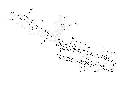
[0019] Figure 1 is a perspective view of a catheter with a conical diffuser
tip.
[0020] Figure 2 is a perspective view of a catheter with a conical diffuser
tip
as incorporated into an infusion system.
[0021] Figure 3 is a perspective view of a catheter with a conical diffuser
tip
as housed within a splittable introducer needle.
[0022] Figure 3a is a detailed perspective view of a conical diffuser tip as
compressed within the tip of a splittable introducer needle.
[0023] Figure 4 is a perspective view of a catheter with a conical diffuser
tip
as inserted into a cross-sectioned patient via a splittable introducer needle.
[0024] Figure 5 is a perspective view of a catheter with a conical diffuser
tip
following a division of the splittable introducer needle.
[0025] Figure 6 is a perspective view of a catheter with a conical diffuser
tip
as inserted into a cross-sectioned patient, following removal of the
splittable
introducer needle.
[0026] Figure 7 is a chart demonstrating the relationship between catheter
gauge, injection rate and recoil force.
DETAILED DESCRIPTION OF THE INVENTION
[0027] The presently preferred embodiments of the present invention will be
best understood by reference to the drawings, wherein like reference numbers
indicate
identical or functionally similar elements. It will be readily understood that
the
components of the present invention, as generally described and illustrated in
the
figures herein, could be arranged and designed in a wide variety of different
configurations. Thus, the following more detailed description, as represented
in the
-5-
CA 02724081 2010-11-10
WO 2009/142904 PCT/US2009/042875
figures, is not intended to limit the scope of the invention as claimed, but
is merely
representative of presently preferred embodiments of the invention.
[0028] Referring now to Figure 1, a section of a catheter 10 is illustrated.
The
catheter 10 comprises a catheter tube 12 and a catheter tip 18. The catheter
tube 12
may comprise any length where the length is selected based on the intended
application of the catheter 10. For example, the catheter 10 length may vary
from a
few centimeters for peripheral access to many centimeters for central access
procedures.
[0029] The catheter tube 12 is generally tubular having an inner diameter 14.
The tube wall 22 of the catheter tube 12 is generally uniform in thickness
thereby
providing a uniform bore along the entire length of the catheter tube 12. The
catheter
tube 12 adjoins the catheter tip 18 at the terminal end 16 of the catheter
tube 12. The
catheter tube 12 and the catheter tip 18 are generally comprised of the same
material,
but may be comprised of different materials as discussed in detail below.
[0030] The tube wall 22 thickness of the catheter tube 12 is selected so as to
achieve a desired flexibility or rigidity for the catheter 10. The bore of the
catheter
tube 12 is selected based on the intended application of the catheter 10. For
example,
where an application calls for administration of a thick or viscous liquid, a
large bore
catheter tube may be desirable due to flow and volume restrictions of smaller
bore
catheter tubes. Additionally, where an application calls for administration of
large
volumes of a liquid, a large bore catheter tube may be desirable due to the
flow and
volume restrictions of small bore catheter tubes.
[0031] The catheter tip 18 comprises a first end 24 and a second end 26. The
first end 24 adjoins the terminal end 16 of the catheter tube 12, as
illustrated.
Additionally, the second end 26 comprises a first opening 28 of the catheter
10. The
catheter tip 18 further comprises a first inner diameter 30 and a second inner
diameter
32. The first inner diameter 30 is equal to the inner diameter 14 of the
terminal end
16 of the catheter tube 12. The second inner diameter 32 of the catheter tip
18 is
greater than the first inner diameter 30 of the catheter tip 18.
[0032] The catheter tip 18 is generally conical. As such, the inner surface 34
of the catheter tip 18 gradually flares outward from the first inner diameter
30 to the
second inner diameter 32 thereby forming a conical diffuser tip 18.
Additionally, the
inner surface 34 of the tube wall 38 is tapered such that the thickness of the
tube wall
-6-
CA 02724081 2010-11-10
WO 2009/142904 PCT/US2009/042875
38 decreases or thins from the first end 24 to the second end 26. Thus, the
tube wall
38 thickness terminates at the first opening 28 of the catheter 10.
Conversely, the
outer surface 36 of the tube wall 38 is generally planer and comprises a
generally
conical shape as previously described.
[0033] Referring now to Figure 2, the catheter 10 is illustrated as
incorporated
into an infusion system 100. The infusion system 100 may include a variety of
components and/or subcomponents for various infusion therapies. For example,
the
infusion system 100 may include an infusion pump, such as a peristaltic pump,
as well
as a filtering device. The infusion system 100 may also include a power
injector for
injecting CT scan contrast agents, as well as fluids of high viscosity as
required for
various medical and diagnostic procedures. Additionally, the catheter tubing
12 may
include a plurality of access ports for accessing the infusion system 100. The
infusion
system 100 may also include an introducer needle as well as an adapter to
house the
introducer needle. Configurations of connectors, splicers and/or adapters may
also be
incorporated into the infusion system 100 within the scope of the current
invention.
[0034] As illustrated, the infusion system 100 includes a section of catheter
tubing 12, a catheter 10 and an intravenous (IV) fluid source 102. The
catheter tubing
12 is connected to the IV fluid source 102 and the catheter 10. A fluid from
the IV
fluid source 102 is infused into a patient 90 following insertion of the
catheter tip 18
through an insertion site 94 and into the vascular system 92. As such, a fluid
communication is established between the IV fluid source and the vascular
system 92
of the patient 90.
[0035] Referring now to Figures 3-5, unlike conventional over-the-needle
catheter systems, the current catheter 10 is introduced into the vascular
system 92 of a
patient 90 via a splittable introducer needle 80. The splittable introducer
needle 80
comprises a needle shaft 82 and a needle tip 84. The needle tip 84 is beveled
so as to
provide a cutting surface for piercing a patient's skin 90. The needle shaft
82
comprises an inner diameter selected to slidably house the catheter 10,
catheter tubing
12 and the catheter tip 18. The catheter tip 18 is compressed into a fluted
configuration such that the outer diameter of the compressed catheter tip 18
is less
than, or equal to the inner diameter of the splittable needle shaft 82 (see
Figure 3a for
detail). As such, the catheter tip 18 is slidably housed within the needle
shaft 82, as
illustrated.
-7-
CA 02724081 2010-11-10
WO 2009/142904 PCT/US2009/042875
[0036] The needle shaft 82 further comprises at least two score marks 86. The
score marks 86 are located opposite one another and comprise a groove running
the
length of the needle shaft 82. The score marks are located on the inner
surface of the
needle shaft 82 thereby providing a smooth outer surface for the needle shaft
82. The
score marks 86 provide at least two thinned portions of the needle shaft 82
where the
needle shaft 82 may be easily separated into at least two pieces.
[0037] The splittable introducer needle 80 further comprises a first and
second
gripping handles 96, 98. The gripping handles 96, 98 are attached to a distal
half 110
and a proximal half 112 of the splittable introducer needle 80, respectively.
The
gripping handles 96, 98 may be used for holding and maneuvering the needle
shaft 82
during insertion. Additionally, the first and second gripping handles 96, 98
provide a
gripping surface for separating the splittable introducer needle 80 following
insertion
of the catheter 10. Finally, the splittable introducer needle 80 comprises a
channel
114 through which the catheter tubing 12 may slidably extend.
[0038] Referring now to Figure 4, the catheter 10 is illustrated during
insertion into a patient 90 via a splittable introducer needle 80. As
illustrated, the
needle tip 84 is used to penetrate the patient 90 at an insertion site 94.
Following
penetration, the needle tip 84 is advanced into the vascular system 92 of the
patient
90. A user then advances the catheter tip 18 beyond the needle tip 82 and into
the
vascular system 92. A user may advance the catheter tip 18 by pushing the
catheter
adapter 88 towards the first and second gripping handles 96, 98 of the
splittable
introducer needle 80. Once the catheter tip 18 is inserted into the vascular
system 92,
a user may occlude the vascular system at an external point 104 adjacent the
catheter
tip 18. By so doing, the user may immobilize the catheter tip 18 within the
vascular
system 92. After immobilizing the catheter tip 18, the user may remove the
needle tip
84 and needle shaft 82 from the patient 90 while leaving the catheter tip 18
within the
vascular system 92.
[0039] Following advancement of the catheter tip 18 into the vascular system
92, the compressed catheter tip 18 may relax and uncompress. The decompression
of
the catheter tip 18 may be accomplished by any means. For example, in one
embodiment the catheter tip 18 is mechanically compressed due to the
restricted inner
diameter of the needle shaft 82. As such, when the compressed catheter tip 18
is no
-8-
CA 02724081 2010-11-10
WO 2009/142904 PCT/US2009/042875
longer housed within the needle shaft 82, the catheter tip 18 is no longer
mechanically
compressed and may therefore relax and uncompress.
[0040] Conversely, in another embodiment the catheter tip 18 is constructed of
a dehydrated material, such as a dehydrated polymer. As such, the compressed
state
of the catheter tip 18 is a result of the shrunken state of the dehydrated
material. Thus,
when the catheter tip 18 is exposed to the aqueous environment of the vascular
system
92 the dehydrated material is hydrated and the catheter tip 18 expands. In
this
embodiment, the dehydrated catheter tip 18 may be fused to the hydrated
catheter 10
by any method of plastic joining. For example, the plastic joining method may
include induction, electrofusion, laser welding, mechanical bonding and/or
chemical
bonding.
[0041] Referring now to Figure 5, the splittable introducer needle 80 is
illustrated as divided into a first half 110 and a second half 112. The
channel 114 of
the needle shaft 82 is shown housing a portion of the catheter tubing 12.
Additionally,
the score marks 86 are illustrated running the length of each half 110, 112 of
the
needle shaft 82. The splittable introducer needle 80, as illustrated, is one
form of a
splittable introducer needle compatible with the present invention. However,
it is
anticipated that any splittable introducer needle may used and/or adapted for
use with
the conical diffuser tip 18. Additionally, other methods of inserting a
catheter 10
comprising the conical diffuser tip 18 may be used, such as surgical
implantation.
[0042] Referring now to Figure 6, a catheter 10 is illustrated following
removal of the splittable introducer needle 80. The catheter 10 and catheter
tip 18 are
positioned within the vascular system 92 of the patient 90 in accordance with
the
previous discussion. As illustrated, the catheter tip 18 is fully decompressed
and
conically shaped. The catheter adapter 88 is positioned on the patient 90
adjacent to
the insertion site 94. The catheter adapter 88 may be secured to the patient
90 by
means of a fastener, such as a steri-strip. Finally, the catheter adapter 88
is positioned
and/or secured so as to permit a section of catheter tubing 12 to remain
uninserted.
The uninserted portion of the catheter tubing 12 provides a gentle transition
from the
catheter adapter to the vascular system 92 thereby preventing restricted flow
through
the catheter 10.
-9-
CA 02724081 2010-11-10
WO 2009/142904 PCT/US2009/042875
[0043] Technical Discussion
[0044] Additional objects, advantages, and novel features of this invention
will become apparent to those skilled in the art upon examination of the
following
technical discussion. It should be appreciated that this technical discussion
is not to
be considered as limiting the scope of the invention, but merely as being
illustrative
and representative thereof.
[0045] The geometry of the conical diffuser tip reduces a nozzle recoil force
of
the catheter 10 thereby allowing the use of higher flow rates for infusion
therapies. As
previously discussed, higher flow rates are desirable for infusion therapies
requiring
rapid infusion of large volumes of infusate. Unlike the conical diffuser tip,
conventional catheter tips taper inward thereby decreasing the inner diameter
of the
catheter through which an infusant exits the catheter. This tapered
configuration
accelerates an infusant through the tapered portion of the catheter tip.
Additionally,
the decreased inner diameter results in an increased back pressure or recoil
force
within the catheter. As previously discussed, an increased recoil force is
undesirable
due to the possibility of displacing the inserted catheter and/or injuring the
vasculature
of a patient.
[0046] Nozzle recoil force is calculated based on the principle of
conservation
of linear momentum. As such, the recoil force of a catheter tip may be
calculated
from Equation 1.
[0047] 1 1 1
rTI
u .a~m E uat n 1
1Y 2
2 DI
[0048] The recoil force Fr may be calculated for the catheter tip where Q is
the volumetric flowrate, p is the density of the infusant, and the diameters
Di and D2
are first diameter and second diameter, respectively. A conventional catheter
tip will
comprise a second diameter D2 that is larger than the tapered first diameter
Di.
Therefore, according to Equation 1, a conventional catheter comprises a
positive
recoil force Fr dependent upon the density of the infusant. For example, for a
constant volumetric flowrate Q, the recoil force Fr of the conventional
catheter tip
increases as the density p of the contrast media increases. Likewise, for a
constant
density p of an infusant, the recoil force Fr increases as the volumetric
flowrate Q
-10-
CA 02724081 2010-11-10
WO 2009/142904 PCT/US2009/042875
increases. An application of Equation 1 to a conventional, tapered catheter
tip is
explained below.
[0049] A recoil force Fr was calculated for a variety of conventional catheter
tips using Equation 1. Four catheters were selected including an 18, 20, 22,
and 24
gauge catheters. Each catheter comprised a conventional, tapered catheter tip.
A first
inner diameter and a second inner diameter of the catheter tip were measured
and
recorded for each catheter gauge. A standard contrast media was selected with
a
density p of 1406 kg/m3. A range of volumetric flowrates Q was selected
between 1
ml/second and 10 ml/second. The results are shown in Figure 7.
[0050] As shown, the recoil force Fr for a variety of conventional, tapered
catheter tips was calculated using Equation 1. According to the results, for a
given
injection rate (x-axis) an increase in the gauge of the catheter results in an
increased
recoil force Fr. Generally, a higher catheter gauge is desirable to reduce
patient
trauma during the placement and removal of the catheter. However, according to
the
results of Example 1, a higher gauge catheter, at a higher injection rate
results in
increased recoil force. Thus, for infusion therapies requiring high infusion
rates, use
of a less invasive, higher gauged conventional catheter may not be beneficial.
[0051] As demonstrated above, the recoil force of a catheter is directly
proportional to the fluid velocity at the catheter tip opening. Therefore, a
decrease in
the fluid velocity at the catheter tip opening will result in a decreased
recoil force Fr.
The geometry of the conical diffuser tip provides for a decreased fluid
velocity at the
catheter tip. As such, the recoil force of the conical diffuser tip is
decreased or
eliminated. Therefore, the conical diffuser tip permits the safe use of higher
gauge
catheters at higher infusion rates.
[0052] Flow dynamics through a catheter is largely dependent upon the
geometry of the catheter tip. Conventional catheter tips are tapered inwardly
and
therefore cause an infusant to accelerate while passing through the catheter
tip. The
flared or outwardly tapered conical diffuser tip 18, as shown in the figures,
decreases
the velocity of an infusant 72 while passing through the diffuser tip 18. The
decreased
velocity of the infusant 72 reduces or eliminates recoil force within the
catheter 10.
Therefore, a higher gauged catheter comprising a conical diffuser tip may be
used for
high infusion rate procedures without displacing the inserted catheter or
damaging the
patient's vasculature.
-11-
CA 02724081 2010-11-10
WO 2009/142904 PCT/US2009/042875
[0053] Referring again to Figure 1, several additional geometric factors may
be considered when optimizing an infusion system. An infusion system is
optimized
by allowing an infusant to flow, without flow separation, into an unoccluded
vein.
Flow separation is a disturbance whereby the flow of the infusant 72 becomes
detached from the diffuser tip 18. Once detached, the flow of the infusant 72
takes the
forms of eddies and vortices. The catheter tip may be configured to avoid flow
separation and optimize flow of the infusant. Important geometric factors
include the
maximum outlet diameter 42, and the degree of divergence 44.
[0054] The maximum outlet diameter 42 is the widest point of the conical
diffuser tip 18. At all infusion rates, maximal in vivo blood flow rates are
significantly reduced when the maximum outlet diameter 42 of the catheter tip
18
exceeds approximately 50% of the inner diameter of the targeted vasculature.
For
example, where a targeted vasculature has an inner diameter of 6.0mm, a
maximum
outlet diameter 42 of greater than 3.0mm will reduce the flow efficiency of
the
infusion system. For this example, if the maximum outlet diameter 42 exceeds
3.0mm, the vasculature of the patient may become occluded thereby decreasing
the
flow efficiency of an infusant into the patient.
[0055] Therefore, regardless of the gauge of the catheter tube 12, the
maximum outlet diameter 42 of the catheter 42 must be less than, or equal to
50% of
the inner diameter of a targeted vasculature. Generally, a targeted
vasculature may
comprise an inner diameter from about 1mm to 1.5cm. Commonly used veins in the
forearm have an inner diameter of about 6.4mm. Therefore, in one embodiment, a
maximal outer diameter 42 will range from 0.5mm to 0.75cm and in another
embodiment the maximum outlet diameter 42 is 3.2mm.
[0056] The degree of divergence 44 is the angle at which the conical diffuser
tip 18 is splayed. A divergence angle 44 must be chosen to provide maximum
decrease of an infusant's 72 velocity through the diffuser tip 18 while
minimizing
flow separation. An optimal divergence angle 44 is selected from a range of
about 5-
20 . In one embodiment, a divergence angle 44 of less than about 14 should be
selected with a preferred range of about 5-14 and an optimal divergence angle
44 of
about 8 .
[0057] The geometry of the conical diffuser tip 18 is selected to minimize the
infusant's velocity at the second end 26 of the catheter 10. In a preferred
embodiment,
-12-
CA 02724081 2010-11-10
WO 2009/142904 PCT/US2009/042875
the geometry of the diffuser tip 18 is selected such that the fluid velocity
of an
infusant 72 is slowed through the diffuser tip 18. For example, in one
embodiment an
infusant 72 is infused at a desired rate whereupon the diffuser tip 18
diffuses the
infusant 72 such that the recoil force of the catheter 10 is eliminated. In
another
embodiment, the diffuser tip 18 decreased an infusant's velocity such that the
inserted
catheter 10 is not displaced and the vasculature of the patient is not damaged
by the
infusant 72. Therefore, in this embodiment the decreased velocity of the
infusant 72
prevents an undesirable recoil force for the catheter 10. As such, the
diffuser tip 18
prevents the infusant 72 from being accelerated into a patient's vein at an
unsafe
velocity during high rates of infusion.
[0058] The conical diffuser tip 18 utilizes a round cross-sectional shape.
Although the present embodiment demonstrates a round cross-sectional shape,
other
shapes including oval, square and/or rectangular shapes may be used as needed.
Generally, the geometry and shape of the catheter tubing 12 and the conical
diffuser
tip 18 is selected to optimize the flow of an infusant 72 through the catheter
10.
Additionally, the geometry and shape of the catheter 10 and diffuser tip 18
are
selected to reduce the velocity of an infusant 72 as the infusant travels
through the
conical diffuser tip 18.
[0059] In a preferred embodiment, the catheter 10, including the catheter
tubing 12 and the diffuser tip 18, is fabricated from a polymeric material
such as
nylon, PVC, PVP, silicone, polyurethane and/or polyethylene. Additionally, a
preferred embodiment may include a radiopaque filler, such as a chemical salt
of
bismuth or barium and/or an element such as platinum or tungsten.
[0060] The present invention may be embodied in other specific forms without
departing from its structures, methods, or other essential characteristics as
broadly
described herein and claimed hereinafter. The described embodiments are to be
considered in all respects only as illustrative, and not restrictive. The
scope of the
invention is, therefore, indicated by the appended claims, rather than by the
foregoing
description. All changes that come within the meaning and range of equivalency
of
the claims are to be embraced within their scope.
-13-