Note: Descriptions are shown in the official language in which they were submitted.
CA 02724197 2015-04-20
FLUID FLOW CONTROL DEVICE WITH
RETRACTABLE CANNULA
1. Field of the Invention
[00011 This invention relates to a fluid flow control device and, in a
preferred
embodiment, to a medical device having a cannula, often a needle, that is
insertable
into a patient for use in infusing, collecting or extracting fluids. One
aspect of the
invention relates to a medical device having an actuator that is manipulated
to modify a
fluid flow path between the cannula and an external fluid source or receptacle
following
the infusion or extraction. Another aspect of the invention relates to a
mechanism that
functions as a clamp when attached to a fluid flow line. Another aspect of the
invention
relates to a mechanism that retracts the cannula inside the device to prevent
accidental
needlesticks following use and to prevent reuse of the contaminated cannula.
Although
the subject invention is particularly preferred for use in intravascular
("IV") applications,
it can also be used beneficially, for example, in epidural, intraosseous and
intraocular
applications, and with any body fluid.
2. Description of Related Art
[0002] Intravascular ("IV") infusion sets are well-known in the art for
delivering
fluids and/or medications to a patient by means of a cannula connected to
tubing. IV
infusion devices frequently have attached wings that facilitate handling
during insertion
of the cannula, help stabilize the device, and can be secured to limit
movement of the
device during use. Blood collection devices operate on the same principle, but
in
reverse. Blood is collected from a vein or artery through a cannula that is
connected
1
CA 02724197 2010-11-10
WO 2009/151704
PCT/US2009/037742
through the body of the device to a blood collection receptacle. Following use
of a
conventional infusion or fluid collection system, the cannula is contaminated
with blood
and/or other bodily fluid, and care must be taken to avoid reusing the device
and to
avoid accidentally sticking either healthcare workers or patients, and thereby
spreading
blood-borne pathogens. The use of caps or covers that must be replaced over
the
cannula after withdrawal from a patient are not a satisfactory solution
because they
increase the risk for an accidental stick, or can become loose and fall off,
thereby again
exposing the contaminated cannula.
[0003] U.S. 5,779,679 to Shaw, entitled "Winged IV Set With Retractable
Cannula," and U.S. 6,210,371 to Shaw, entitled "Winged IV Set," both disclose
an IV
infusion set with a retractable cannula. In both of these patents, the
retractable cannula
is held by a retraction member having a tubing connector on its back end
portion that
establishes fluid communication between the cannula and an IV tube. The
retraction
member is held in its non-retracted position against the force of a compressed
spring by
a pair of releasable latches disposed on opposite sides of the housing. Once
the
latches are released, the spring forces the retraction member, and
consequently the
cannula, back into the housing. However, because an IV tube is connected
directly to
the retraction member, retraction of the cannula causes the IV tube to move
rearwardly ,
as well. If the tube is not free to move rearwardly during retraction, the
retraction
member and the cannula may not be fully retracted.
[0004] An infusion and fluid collection device are needed in which the cannula
can be retracted without causing or relying upon rearward movement of the
connected
tubing, and in such manner that the device is rendered non-reusable and that
the fluid
flow path is interrupted, relocated and sealed off in conjunction with
retraction of the
cannula.
2
DALLAS: 057532900286: 1692982v1
CA 02724197 2015-04-20
SUMMARY OF THE INVENTION
[0004a] Certain exemplary embodiments can provide a device comprising: a
housing; a cannula; a retraction mechanism biasing the cannula rearwardly; a
retraction cavity having a front portion, wherein the retraction cavity is
pivotally
connected to the housing such that the front portion can move arcuately from a
first position to a second position; wherein, when the front portion of the
retraction
cavity is in the first position, the cannula projects forwardly from the
housing and
the retraction cavity is not coaxial to or aligned with the cannula; wherein,
when
the front portion of the retraction cavity is in the second position, the
front portion is
aligned with the cannula and the retraction mechanism retracts the cannula
into
the retraction cavity such that the cannula no longer projects forwardly from
the
housing.
[0004b] Certain exemplary embodiments can provide a device comprising: a
housing; a cannula; a retraction mechanism biasing the cannula rearwardly; an
actuator, pivotably mounted to the housing; the actuator having a front
portion and
further comprising a retraction cavity and a separate fluid flow path disposed
in a
non-coaxial, spaced apart relation to each other; wherein the actuator can be
pivoted relative to the housing so as to arcuately move the front portion from
a first
position into a second position; wherein, in the first position, the cannula
projects
forwardly from the housing, the separate fluid flow path provides fluid
communication from the cannula through the device, and the retraction cavity
is
not aligned with the cannula; wherein, in the second position, the separate
fluid
flow path is not in fluid communication with the cannula, thereby preventing
fluid
flow through the device; the retraction cavity is sufficiently aligned with
the cannula
to allow the retraction mechanism to retract the cannula into the retraction
cavity;
and the retraction mechanism retracts the cannula into the retraction cavity
so that
the cannula no longer projects forwardly from the housing.
3
CA 02724197 2015-04-20
[0005] The described embodiments provide a device that is particularly well
suited for use in the medical field, but is not necessarily limited to medical
use.
According to one preferred embodiment of the invention, a medical device is
disclosed that has a cannula and can be configured and used for fluid
injection,
infusion or extraction. Depending upon its configuration, the subject device
can be
used, for example: As part of an infusion set or as a collection device for
venous or
arterial blood; for other body fluids such as spinal fluid, cerebral fluid,
amniotic
fluid, and the like that are well known to healthcare workers; or for solid
matter
contained in suspensions or slurries such as, for example, medications, lipids
or
bone marrow. When the device is used for infusing fluids or medication, the
fluid
source can be, for example, an IV drip bag or a syringe. When the device is
used
for collecting blood, the fluid receptacle can be, for example, a blood
collection
bag, an evacuated tube or a syringe. When the device is attached to a fluid
flow
line, it can also be used as a clamp. Prior to retraction of the cannula, the
device
prevents fluid leakage into or out of the fluid flow path. The cannula is
typically a
needle having a front end that is beveled to facilitate insertion into tissue
or into
another medical device such as a port.
[0006] According to another embodiment of the invention, the device
comprises an offset fluid flow path and a retraction chamber fixed in a
position that
is in-line with the cannula. Retraction is initiated by an actuator that can
be
repositioned axially in relation to the cannula, thereby also blocking the
fluid flow
path.
[0007] According to another embodiment of the invention, the device
comprises an in-line fluid flow path and an offset retraction cavity.
Retraction is
initiated by an actuator that can be repositioned laterally in relation to the
cannula,
thereby blocking and sealing off the fluid flow path.
3a
CA 02724197 2015-04-20
[0008] According to another embodiment of the invention, the device
comprises an in-line fluid flow path and an offset retraction chamber.
Retraction is
initiated by an actuator that can be repositioned arcuately in relation to the
cannula, thereby blocking and sealing off the fluid flow path.
[0009] According to a preferred embodiment of the invention, a device is
disclosed that preferably comprises a housing; a cannula projecting forwardly
from the
3b
CA 02724197 2010-11-10
WO 2009/151704
PCT/US2009/037742
housing; a connector useful for attaching the device to a fluid source or
receptacle; a
fluid flow path establishing fluid communication between the cannula and the
connector;
a retraction mechanism biasing the cannula away from its projecting position;
and an
actuator supported by the housing and configured to modify the fluid flow path
so as to
terminate fluid flow through the device, seal off the fluid flow path, and
release the
retraction mechanism to retract the cannula into the housing. Laterally
extending finger
grips and/or stabilization wings with finger pads are desirably provided to
facilitate
manipulation of the device by a user, to resist rolling of the device on an
underlying
surface, and to provide surfaces that can be secured to a patient during use.
[0010] According to another preferred embodiment of the invention, the
actuator
portion of the device comprises two elongate, most preferably cylindrical,
cavities,
including one cavity that defines a portion of the fluid flow path and another
that is a
retraction cavity configured to receive a portion of the retraction mechanism
and
cannula following retraction. The retraction mechanism preferably comprises a
holder
for the needle or cannula, and a biasing member such as a compression spring.
The
actuator is preferably movable by the user from a first position to a second
position to
modify the fluid flow path by interrupting, displacing, redirecting or
reconfiguring at least
part of the path, thereby cutting off fluid flow along the original flow path
through the
device. At least part of the actuator is either slidably mounted or rotatably,
most
preferably pivotally, connected inside the housing. Movement of the actuator
from the
first position to the second position also desirably releases the retraction
mechanism,
allowing the biasing member to force the cannula holder and at least part of
the cannula
back inside the retraction cavity, and to force all of the cannula back inside
the housing
to prevent accidental needle sticks and to prevent reuse of the device. The
use of
devices having retractable needles, the avoidance of accidental needlesticks
and
disabling the device to prevent reuse are important to significantly reducing
the spread
of disease by blood-borne pathogens to healthcare workers, other patients, and
those
who may handle such devices following use.
4
DALLAS: 057532900286: 1692982v1
CA 02724197 2010-11-10
WO 2009/151704
PCT/US2009/037742
BRIEF DESCRIPTION OF THE DRAWINGS
[0011] The apparatus of the invention is further described and explained in
relation to the following figures of the drawings wherein:
FIG. 1 is a front elevation view of an embodiment of the invention having an
interruptable fluid flow path, a retractable cannula projecting forwardly out
of the
housing, a rearwardly slidable actuator, and stabilization wings;
FIG. 2 is a cross-sectional side elevation view taken along line 2-2 of FIG.
1;
FIG. 3 is an enlarged view of the device of FIG. 2 following interruption of
the
fluid flow path and retraction of the cannula;
FIG. 4 is a cross-sectional front elevation view of another embodiment of the
invention having an interruptible fluid flow path, a retractable cannula
projecting
forwardly out of the housing, and a laterally slidable actuator;
FIG. 5 is a cross-sectional front elevation view of the embodiment of FIG. 4
following interruption of the fluid flow path and retraction of the cannula
into the
retraction cavity;
FIG. 6 is a perspective view of another embodiment of the invention having an
interruptable fluid flow path, a retractable cannula projecting forwardly out
of the
housing, and a pivotable actuator;
FIG. 7 is an exploded perspective view of the embodiment of FIG. 6;
FIG. 8 is an enlarged top plan view (oppositely directed) of the embodiment of
FIG. 6;
FIG. 9 is an enlarged cross-sectional plan view, partially broken away, of the
embodiment of FIG. 8, with the cannula projecting forwardly and a tubing
segment
(shown in phantom outline) disposed in fluid communication with the fluid flow
path
through the actuator, cannula holder and cannula;
FIG. 10 is an enlarged cross-sectional plan view of the embodiment of FIG. 9
following interruption of the fluid flow path and retraction of the cannula;
FIG. 11 is a perspective view of another embodiment of the invention having an
interruptable fluid flow path, a retractable cannula projecting forwardly out
of the
DALLAS. 0575329.00286: 1692982v1
CA 02724197 2010-11-10
WO 2009/151704
PCT/US2009/037742
housing (shown covered by a protective guard), and an actuator pivotably
connected to
the housing;
FIG. 12 is an exploded perspective view of the embodiment of FIG. 11;
FIG. 13 is an enlarged top plan view of the embodiment of FIG. 11;
FIG. 14 is a front elevation view of the embodiment of FIG. 13, with the
protective
cover shown in phantom outline;
FIG. 15 is an enlarged cross-sectional plan view, partially broken away, of
the
embodiment of FIG. 13, with the cannula projecting forwardly and the
protective cover
shown in phantom, showing the fluid flow path through the cannula, cannula
holder and
actuator;
FIG. 16 is a cross-sectional plan view of the embodiment of FIG. 15 following
interruption of the fluid flow path and retraction of the cannula into the
retraction cavity;
FIG. 17 is a perspective view of another embodiment of the invention having an
interruptable fluid flow path that is particularly useful with liquid fluids,
a retractable
cannula projecting forwardly out of the housing (hidden from view by a
removable
protective cover), and an actuator pivotably connected to the housing;
FIG. 18 is an exploded perspective view of the embodiment of FIG. 17;
FIG. 19 is an enlarged cross-sectional plan view, partially broken away, of
the
embodiment of FIG. 17, with the cannula projecting forwardly and the
protective cover
shown in phantom outline, and showing the fluid flow path through the cannula,
cannula
holder, actuator and Luer connector set; and
FIG. 20 is a cross-sectional plan view of the embodiment of FIG. 19 following
interruption of the fluid flow path and retraction of the cannula into the
retraction cavity.
6
DALLAS: 0575329.00286: 1692982v1
CA 02724197 2010-11-10
WO 2009/151704
PCT/US2009/037742
DESCRIPTION OF THE PREFERRED EMBODIMENTS
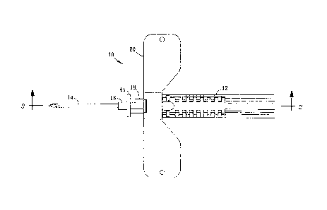
[0012] Referring to FIG. 1, device 10 can be used, for example, as part of a
medical apparatus for collecting blood, blood gases or other bodily fluids
from a patient,
or for infusing a patient with fluids of the type typically administered
intravenously or
otherwise. As shown, device 10 comprises housing 12 and forwardly projecting
cannula
14 attached to cannula holder 16, which is more visible in FIGS. 2 and 3.
Prior to use of
device 10, the beveled point of cannula 14 is desirably shielded by a
protective cover.
Actuator 18 is slidably supported by housing 12 and comprises a plurality of
opposed
flexible latches that secure the end opposite handle 44 inside housing 12.
Handle 44 at
the front of actuator 18 facilitates manual manipulation of actuator 18
relative to housing
12 to terminate fluid flow through device 10 and initiate retraction of
cannula 14 inside
housing 12 as described in greater detail below.
[0013] Optional stabilization wings 20 extend laterally from housing 12 and
facilitate handling of device 10 by the user. When used, wings 20 also provide
a
surface that will restrict rotation of housing 12 when device 10 is secured to
a patient
using tape, sutures, or other similarly effective means. After a desired
volume of fluid
has been administered to or withdrawn from a patient as therapeutically
prescribed, the
fluid flow through cannula 14 can be terminated and cannula 14 can be
retracted inside
housing 12 by applying rearwardly directed manual pressure to the front side
of handle
44 of actuator 18 while simultaneously grasping housing 12. If desired,
cannula 14 can
be retracted into housing 12 without first withdrawing it from the patient.
Usage of
device 10 and retraction of cannula 14 are discussed more fully below in
relation to
FIGS. 2 and 3.
[0014] Referring to FIG. 2, device 10 is shown without the optional
stabilization
wings attached. Housing 12 of device 10 comprises two elongated portions
including
lower section 26 and upper section 28. Lower section 26 further comprises
smaller
diameter front end 32 and a larger diameter back end 34 with transition zone
defined by
shoulder 30 disposed between front end 32 and back end 34. Upper section 28
extends rearwardly from a point above shoulder 30 to an open back end above
back
end 34 of lower section 26, and comprises a longitudinal bore that serves as a
connector into which tubing segment 24 is insertable. The end of tubing
segment 24 is
7
DALLAS: 0575329.00286: 1692982v1
CA 02724197 2010-11-10
WO 2009/151704
PCT/US2009/037742
desirably maintained inside upper section 28 of housing 12 by any suitable
means such
as, for example, frictional engagement or by using a clamping device,
adhesive, or the
like. Depending upon the desired use of device 10, tubing segment 24 can be of
any
desired length that is suitable for connecting device 10 to a fluid source,
such as an IV
bag, or to a fluid receptacle, such as a blood collection system. Lower
section 26
preferably further comprises a retraction cavity 36 disposed between back end
34 and
transition zone shoulder 30. As shown, back end 34 of retraction cavity 36
comprises
an opening that is closed by end cap 42. Lower section 26 further comprises
aperture
38 disposed in the top wall, slightly forward of transition zone shoulder 30.
Hole 38
aligns with hole 40 in the bottom of upper section 28, and the alignment of
holes 38, 40
helps establish a fluid flow path between cannula 14 and tubing segment 24.
[0015] The opening at front end 32 of lower section 26 is closed by slidably
engaged tubular actuator 18, which comprises handle 44 extending upwardly from
the
front edge. Actuator 18 is movable from a first position, where handle 44 is
spaced
apart from front end 32 of lower section 26, to a second position, where
handle 44 abusts
and is adjacent to front end 32 of lower section 26. Actuator 18 is desirably
sized
lengthwise to extend into lower section 26 of housing 12 a sufficient distance
to cover
and close hole 38 in lower section 26 when actuator 18 is moved to the second
position
abutting against front end 32. Actuator 18 also desirably comprises a pair of
diametrically opposed latches extending outwardly from the rear end of
actuator 18. As
sliding end cap 18 is moved towards its second position, the latches slide
over and
engage a projection shoulder on the inside wall of lower section 26, thereby
locking
actuator 18 in its second position and preventing its removal from housing 12.
[0016] Retraction mechanism 13 supports cannula 14 and comprises cannula
holder 16, cannula holder plug 46, and spring 22. Following installation of
actuator 18 in
the front of lower section 26, cannula holder 16, spring 22 and cannula holder
plug 46
are desirably preassembled and inserted into lower section 26 through the
opening in
back end 34 and through retraction cavity 36. Prior to insertion, resilient
cannula holder
plug 46 is desirably inserted into frictional engagement with a recess inside
the larger
diameter section 58 of cannula holder 16. Spring 22 is desirably placed over
the
smaller diameter front section of cannula holder 16, where it slides
rearwardly into
8
DALLAS 0575329.00286: 1692982v1
CA 02724197 2010-11-10
WO 2009/151704
PCT/US2009/037742
abutting engagement with annular shoulder 52. The assembled unit is then
oriented so
that fluid passageway 60 is aligned with aperture 38 in the top wall of lower
section 26,
and advanced past transition zone shoulder 30. As the forwardly extending tip
of
cannula holder 16 projects forwardly of actuator 18, spring 22 seats against
the annular
shoulder inside the front opening of actuator 18 and is compressed to the
position
shown in FIG. 2. Actuator 18 resists the force of the compressed spring and is
prevented from moving forwardly away from housing 12 under the spring force by
the
resilient latches securing actuator 18 to housing 12 as previously described.
This
configuration of elements allows cannula 14 to be in fluid communication with
tubing 24,
as fluid flows through hollow cannula 14 and cannula holder 16 into cannula
holder plug
46, through hole 60 in the top of cannula holder plug 46, through hole 38 in
top of
retraction cavity housing 26, through hole 40 in the bottom of tubing assembly
housing
28 and into tubing 24. Similarly, fluid can flow in the opposite direction and
pass from
tubing 24, through aligned holes 40, 38 and 60 and out cannula 14. Rear end
cap 42 is
installed in the open end of lower section 26 following installation of
cannula 14 and
cannula holder plug 46, and is frictionally held inside rear end 34 of lower
section 26.
[0017] Although cannula 14 can be secured in fixed relation to cannula holder
16 prior to insertion of retraction mechanism 13 into housing 12, cannula 14
is desirably
inserted into the bore of projecting tip of cannula holder 16 after cannula
holder 16 is
installed inside housing 12. As seen in FIG. 2, the opening at the forwardly
extending
end of cannula holder 16 is tapered to facilitate the insertion and attachment
of cannula
14. Cannula 14 can be frictionally held inside the bore of cannula holder 16
but is
desirably attached in fixed relation to cannula holder 16 using glue or any
other similarly
effective means known to those of ordinary skill in the art. The open portion
of the
beveled point of cannula 14 desirably faces upwardly to facilitate insertion
into a patient.
As shown in FIG. 2, open back end 62 of hollow cannula 14 extends through back
end
58 of cannula holder 16 into open front section 54 of cannula holder plug 46.
It should
be appreciated, however, that cannula 14 needs only extend into cannula holder
16 a
sufficient distance to facilitate reliable engagement between them.
[0018] When device 10 is assembled as described above, compressed spring
22, or any other similarly effective biasing means, biases cannula 14 and
cannula
9
DALLAS: 0575329.00286: 1692982v1
CA 02724197 2010-11-10
WO 2009/151704
PCT/US2009/037742
holder 16 rearwardly. The frictional holding force exerted against inside
surface 50 of
the smaller diameter front portion of lower section 26 by cannula holder plug
46 should
be great enough to resist the biasing force exerted against annular shoulder
52 by
spring 22 in combination with the force exerted back against cannula holder
plug 46
through cannula 14 and cannula holder 16 during insertion of cannula 14 into a
patient.
Otherwise, cannula 14 could retract prematurely without movement of actuator
18
relative to housing 12.
[0019] When the fluid infusion or extraction procedure is complete and
retraction
of cannula 14, the user can initiate retraction by applying rearwardly
directed pressure
to handle 44 while maintaining housing 12 in a stationary position, either by
gripping its
textured outside surface portion (visible in FIG. 1) or by pressing down on
the optional
wings (likely already secured to the patient). The manual pressure applied to
handle 44
causes actuator 18 to move backwards relative to housing 12. Desirably,
retraction is
initiated while the cannula, typically a needle, is still inserted in the
patient. As actuator
18 moves backwards, cannula holder 16 and cannula holder plug 46 are also
forced
backwards due to the contact between back end 64 of actuator 18 and annular
shoulder
52 of cannula holder 16. It can be observed in FIG. 2 that back end 64 of
actuator 18
abuts the adjacent portion of forwardly facing annular shoulder 52 of cannula
holder 16,
while the rear end of that part of actuator 18 as depicted beneath spring 22
is slightly
separated from annular shoulder 52. This slight separation causes the
rearwardly
directed force exerted by the user on handle 44 to be concentrated against one
side of
annular shoulder 52 rather than being evenly distributed around the
circumference, and
is believed to reduce the manual force required to initiate retraction.
[0020] Referring to FIG. 3, in response to the rearward movement of actuator
18, cannula holder plug 46 passes through the transition zone (past shoulder
30) and
into larger diameter retraction cavity 36 of lower section 26. As cannula
holder plug 46
moves, the friction force between outside surface 48 of cannula holder plug 46
and
inside wall 50 of lower section 26 is reduced, and as cannula holder plug 46
passes the
transition zone and enters retraction cavity 36, the frictional holding force
is completely
eliminated. At the point where the frictional holding force is sufficiently
reduced by the
combined finger force of the user as applied through handle 44 of actuator 18
and the
DALLAS: 0575329.00286: 1692982v1
CA 02724197 2010-11-10
WO 2009/151704
PCT/US2009/037742
biasing force of compressed spring 22, spring 22 forces cannula holder 16 and
cannula
holder plug 46 backwards into retraction cavity 36, thereby simultaneously
causing
cannula holder 16 to draw the beveled tip of cannula 14 inside housing 12. It
will be
apparent to those of skill in the art upon reading this disclosure that
actuator 18 should
be long enough that its range of travel relative to housing 12 is sufficient
to force
cannula holder plug 46 past shoulder 30.
[0021] As shown in FIG. 3, following retraction, cannula holder plug 46
desirably
abuts, or nearly abuts, rear end cap 42 of lower section 26. Lower section 26
is
desirably sized such that the entirety of cannula 14 is contained within lower
section 26
and does not protrude from front end 32. After retraction, the top edge of
actuator 18
blocks the fluid flow path between cannula 14 and hole 38 in the top of lower
section 26.
This prevents fluid from escaping tubing 24 that is still connected to upper
section 28 of
housing 12.
[0022] Another embodiment of the invention is disclosed and described in
relation to FIGS. 4 and 5. Referring to FIG. 4, device 70 is preferred for use
as part of a
blood collection apparatus or an IV infusion set. Device 70 comprises a
substantially
rectangular housing having front wall 72 with forwardly projecting conical
nose 74; rear
wall 92 with open slot 88; side wall 98; and cooperating, substantially flat
bottom wall
104 and a corresponding top wall (not visible in the cross-sectional view)
that
interconnect walls 72, 92 and 98. The edges of bottom wall 104 and the
corresponding
top wall (not visible) that are opposite side wall 98 are not visible in FIG.
4, but extend
between front wall 72 and back wall 92 at a point slightly beyond the side of
slot 88 that
is farthest removed from wall 98. Sliding track 94, seen behind front wall 72,
is
preferably unitarily formed as part of front wall 72.
[0023] As shown and described, the housing of device 70 defines a structure
into which retraction mechanism 76 and actuator 96 are installable. Retraction
mechanism 76 preferably further comprises cannula holder 78 having a larger
diameter
head 80, and a biasing member exerting a rearwardly directed force against
cannula
holder 78. A preferred biasing member is compression spring 86. Retraction
mechanism 76 is installable into front wall 72, nose 74 and sliding track 94
of the
housing from the rear, preferably prior to the installation of cannula 84 and
actuator 96.
11
=
DALLAS: 0575329.00286: 1692982v1
CA 02724197 2010-11-10
WO 2009/151704
PCT/US2009/037742
Coil spring 86 is placed over the forwardly extending end of cannula holder 78
and
cannula holder 78 is then inserted into nose 74 until the forwardly facing end
of spring
86 seats against the annular shoulder inside the front opening of nose 74 that
is
disposed around cannula holder 78. As spring 86 is compressed, a portion of
annular
shoulder 82 on the forwardly facing surface of head 80 abuts against the
rearwardly
facing surface of front wall 72 that is adjacent to the opening through nose
74. While
retraction mechanism 76 is held in place (as by temporarily clamping the
portion of
cannula holder 78 extending forwardly out of nose 74), actuator 96 is
desirably inserted
into sliding track 94 from the side of the housing opposite wall 98, and is
moved laterally
to a position as shown in FIG. 4 where sealing member 95 provides a fluid-
tight seal
permitting fluid flow between head 80 of cannula holder 78 into fluid flow
path 100 of
actuator 96.
[0024] Actuator 96 is preferably an elongate, substantially rectangular body
made to slidably engage at least one guide or sliding track 94 on the inside
of the
housing to facilitate lateral movement of actuator 96 within the housing. The
interior of
actuator 96 preferably comprises an in-line fluid flow path 100 defined by
wall sections
106, 108, and a retraction chamber 102 that is offset from cannula 84 while
actuator 96
is in the use position. Resilient sealing member 95, preferably an elastomeric
0-ring or
another similarly effective sealing member, is disposed in a recess at the
forward end of
fluid flow path 100 through actuator 96, where it can provide sealing
engagement with
the rearwardly facing surface of enlarged head 80 of cannula holder 78. It
will be
observed that resilient sealing member 95 seals against fluid leakage either
into or out
of fluid flow path 100.
[0025] When actuator 96 is positioned as shown in FIG. 4, spring 86 is
maintained in its compressed state and continuously biases cannula holder 78
in a
rearward direction until such time as actuator 96 is selectively repositioned
following use
of device 70. Once retraction mechanism 76 and actuator 96 are installed
inside the
housing, the rear end of cannula 84 can be inserted into the axial bore of
cannula 78
and glued or otherwise secured in place. Although not shown, a frictionally
engageable,
removable protective cover is desirably provided for cannula 84 following its
installation
in cannula holder 78.
12
DALLAS: 0575329 00286: 1692982v1
CA 02724197 2010-11-10
WO 2009/151704
PCT/US2009/037742
[0026] Prior to use, device 70 is preferably connected to a fluid source or
fluid
receptacle by means of a flexible tubing segment 90 that is insertable into or
otherwise
attachable to tubing connector 103 through slot 88 by conventional means. When
actuator 96 is positioned as shown in FIG. 4, a substantially linear fluid
flow path is
established between cannula 84 and tubing segment 90. Tubing connector 103 can
be
a section of the bore inside actuator 96 that is tapered slightly to receive
and frictionally
engage a free end of tubing segment 90, or can be configured for attachment of
a
tubing segment by other known means such as, for example, luer connectors,
threaded
connectors, clamps, adhesive, and the like. Tubing segment 90 is preferably
flexible
polymeric tubing of any length and material that are suitable for the intended
use.
When configured as shown in FIG. 4, device 70 can be used to transfer fluids
from an
external source to be discharged through the cannula, or can be extracted or
withdrawn
from an external source through the cannula and subsequently discharged from
the end
of tubing segment 90 that is opposite to tubing connector 103.
[0027] Following use, retraction is initiated by moving actuator 96 from its
use
position to its retraction position by applying manual force to actuator 96 in
a direction
that is substantially perpendicular to the longitudinal axis through cannula
84 and
cannula holder 78. Referring to FIG. 5, as actuator 96 is moved laterally
toward wall 98,
fluid flow path 100 through actuator 96 is shifted laterally into a position
where it is no
longer opposed to head 80 of cannula holder 78. Simultaneously, head 80 is
acted
upon by the biasing force of compressed spring 86 to propel cannula holder 78
into
retraction cavity 102 of actuator 96, thereby withdrawing cannula 84 inside
the housing.
To produce this result, it will be apparent that the distance between back
wall 92 and
the front tip of nose 74 must be sufficiently great to receive the pointed end
of cannula
84 at least into nose 74. Also, the length of uncompressed spring 86 is
desirably such
that head 80 will be maintained a sufficient distance from the front opening
of nose 74
that the tip of cannula 84 does not again protrude from the front of device 70
following
retraction, particularly if device 70 is rotated to a vertical position with
the cannula
pointing down.
[0028] Another embodiment of the invention, in which the actuator is
repositioned arcuately relative to the housing to initiate retraction, is
described in
13
DALLAS 0575329.00286: 1692982v1
CA 02724197 2010-11-10
WO 2009/151704
PCT/US2009/037742
relation to FIGS. 6-10. Referring first to FIGS. 6-8, medical device 110 is
disclosed that
comprises housing 112, actuator 114, a retraction mechanism 118, and a
forwardly
projecting cannula, preferably needle 122. Housing 112 further comprises a
hollow
body having substantially flat top and bottom surfaces, an inclined finger pad
134, a
forwardly extending, open neck 136, open side and back sections including
actuator
stop rail 140, recessed wall section 168, and aligned, oppositely disposed
apertures 126
for pivotably attaching actuator 114 to housing 112.
[0029] Actuator 114 preferably comprises actuator contact surface 132, contact
surface 166, actuator positioning rail 138, outwardly projecting mounting
bosses 128
insertable into mating engagement with apertures 126 of housing 112, and
tubing
aperture 130. Retraction mechanism 118 preferably comprises a needle holder
having
a forwardly extending, small diameter portion 106 and a larger-diameter head
108
disposed rearwardly of small diameter portion 106. Compression spring 116 is
configured to slide over small diameter portion 106 and to abut against the
forwardly
facing annular surface of head 108. A sealing member, preferably 0-ring 120,
is further
described below.
[0030] Referring to FIG. 9, the retraction mechanism is inserted into neck 136
of
housing 112 from the rear, with small diameter portion 106 of the needle
holder
projecting forwardly through the opening in the front. Spring 116 slidably
engages small
diameter portion 106 and the forward end of spring 116 is seated against an
annular
shoulder adjacent to the front opening inside neck 136. The other end of
spring 116
abuts against an annular shoulder of head 108. Spring 116 is compressed, and
is
maintained in the pre-retraction position by an opposing force exerted against
head 108
by actuator 114. Actuator 114 is disposed in its use position relative to
housing 112,
with mounting bosses 128 pivotably inserted into apertures 126 and with
contact
surface 166 abutting against the inside surface of housing 112 that is
adjacent to
recessed wall section 168. Actuator 114 comprises fluid flow path 154 bounded
by
walls 150, 152, and retraction cavity 164. Space 124 in housing 112 is
provided to
receive a portion of actuator 114 when it is repositioned to terminate fluid
flow and
initiate retraction.
14
DALLAS: 0575329.00286: 1692982v1
CA 02724197 2010-11-10
WO 2009/151704
PCT/US2009/037742
[0031] With actuator 114 in this position, fluid flow path 154 through
actuator
114 is disposed in fluid communication with axial passageway 144 through the
needle
holder and with the inside of needle 122. Sealing member 120, seated in recess
148,
desirably provides fluid-tight sealing engagement between the forward end of
walls 150,
152 and head 108 of the needle holder. This sealing engagement is facilitated
by an
annular shoulder 146 of head 108. Tubing connector 156 desirably comprises
outwardly
tapering walls 142 at the rear end of fluid flow path 154 and is adapted to
receive and
engage an end of tubing segment 162 (shown in phantom outline). Projecting
boss 158
of housing 112, located adjacent to the forwardly facing end of retraction
cavity 164 is
provided to prevent actuator 114 from inadvertently being moved from the pre-
retraction
position to the retraction position. In practice, the spacing between boss 158
and facing
surface 160 of actuator 114 is desirably less than that shown for illustrative
purposes in
FIG. 9.
[0032] After the transfer of fluids through device 110 in either direction has
been
completed to the extent desired, the fluid flow is easily terminated by
repositioning
actuator 114 relative to housing 112 by applying pressure against actuator
contact
surface 132, which causes actuator 114 to pivot in the direction shown by
arrow 160.
Although some manual pressure is required to overcome the resistance of
pushing
surface 160 over boss 158 and to move sealing member 120 past head 108, the
required force is desirably such that it can easily be applied by an adult
user. It will
again be observed that resilient sealing member 120 seals against fluid
leakage either
into or out of fluid flow path 154.
[0033] Referring to FIG. 10, after actuator 114 is repositioned relative to
housing
112 as shown, fluid flow between needle 122 and tubing segment 162 is blocked,
fluid
flow path 154 is offset from the opening through neck 136 and from passageway
144
through head 108. Furthermore, as soon as wall 150 clears head 108, retraction
cavity
164 is pivoted into coaxial alignment with the opening through neck 136, and
the biasing
force of compressed spring 122 projects the needle holder into the retraction
cavity,
simultaneously withdrawing the tip of needle 122 from the patient and into
housing 112
to avoid accidental needle sticks and prevent reuse of device 110.
DALLAS: 0575329.00286: 1692982v1
CA 02724197 2010-11-10
WO 2009/151704
PCT/US2009/037742
[0034] Another embodiment of the invention is described in relation to FIGS.
11-
16. This embodiment is particularly preferred for use in collecting fluids
comprising
gases, such as arterial blood gases, intended for subsequent analysis, and
also
comprises an actuator that is repositioned arcuately relative to the housing
to initiate
retraction. Referring first to FIGS. 11-14, medical device 200 is disclosed
that
comprises housing 226, actuator 204, needle holder 220, spring 222, hermetic
sealing
element 218 and forwardly projecting needle 224. Housing 226 further comprises
a
hollow body having substantially flat top and bottom surfaces 202, oppositely
disposed
and integrally formed finger grips with textured gripping surfaces 206, a
forwardly
extending, tapered neck 238 with opening 228, an open side and back, and
aligned,
oppositely disposed apertures 230 for pivotably attaching actuator 204 to
housing 226.
Protective cover 232 is desirably provided to protect needle 224 prior to use,
and should
be removed from needle 224 prior to use.
[0035] Actuator 204 preferably comprises actuator contact surface 234,
outwardly projecting mounting bosses 212 insertable into mating engagement
with
apertures 230 of housing 204, retraction cavity 216, recess 214 around the
opening of
the fluid flow path, and a tubing connector 208 that extends rearwardly from
housing
226. Referring to FIGS. 13 and 14, tubing connector 208 further comprises half
of a
Luer connector 236 to facilitate attachment of device 200 to another fluid
source or
receptacle, depending upon the intended use.
[0036] Referring to FIGS. 15-16, the retraction mechanism is installed by
inserting it into axial passageway 250 through neck 238 of the housing from
the rear,
with small diameter portion 221 of needle holder 220 projecting forwardly
through
opening 228 in the front. Spring 222 slidably engages small diameter portion
221 and
the forward end of spring 222 is seated against an annular shoulder 252
adjacent to
front opening 228 of neck 238. The other end of spring 222 abuts against an
annular
shoulder of larger diameter head 223 of needle holder 220. Spring 222 is
compressed,
and is maintained in the pre-retraction position by an opposing force exerted
against
head 223 by actuator 204. Actuator 204 is disposed in its use position
relative to
housing 226 with mounting bosses 212 pivotably inserted into apertures 230
(FIG. 12)
and with actuator contact surface 234 abutting against the inside surface of
housing 226
16
DALLAS: 057532900286: 1692982v1
CA 02724197 2010-11-10
WO 2009/151704
PCT/US2009/037742
that is underneath the nearest adjacent textured gripping surface 206.
Actuator 204
comprises fluid flow path 242 and retraction cavity 216. Space 244 in housing
226 is
provided to receive a portion of actuator 204 when it is repositioned to
terminate fluid
flow and initiate retraction.
[0037] With actuator 204 in the position shown in FIG. 15, fluid flow path 242
through actuator 204 is disposed in fluid communication with axial passageway
225
through the needle holder and with axial passageway 258 inside needle 224.
Hermetic
sealing member 246, seated in recess 214, desirably provides fluid-tight
sealing
engagement between the forward end of fluid flow path 242 and head 223 of the
needle
holder. Tubing connector 208 desirably comprises stepped bore 240 providing
fluid
communication with fluid flow path 242. Projecting boss 235 of housing 226,
located
adjacent to the forwardly facing end of retraction cavity 216 is provided to
prevent the
actuator from being moved inadvertently from the pre-retraction position to
the retraction
position shown in FIG. 16.
[0038] Referring to FIG. 16, after the transfer of fluids through device 200
in
either direction has been completed to the extent desired, the fluid flow is
easily
terminated by repositioning the actuator relative to housing 226 by applying
pressure
against actuator contact surface 234, which causes the actuator to pivot in
the direction
shown by arrow 260. Although some manual pressure is required to overcome the
resistance of pushing surface 237 (FIG. 15) over boss 235 and to move sealing
member
246 past head 223, the required force is desirably within the range that can
be applied
smoothly by an adult user. After the actuator is repositioned relative to
housing 112 as
shown in FIG. 16, fluid flow between needle 224 and tubing connector 208 is
blocked,
fluid flow path 242 is offset from the opening through passageways 248, 250.
Furthermore, as soon as retraction cavity 164 is pivoted into coaxial
alignment with the
passageways 248, 250, the biasing force of compressed spring 224 projects
needle
holder 221, 223 into the retraction cavity, simultaneously withdrawing the tip
of needle
224 from the patient and into housing 226 to avoid accidental needle sticks
and prevent
reuse.
[0039] Another embodiment of the invention is described in relation to FIGS.
17-
20. This embodiment, which is particularly preferred for use in extracting,
collecting or
17
DALLAS: 0575329.00286: 1692982v1
CA 02724197 2010-11-10
WO 2009/151704
PCT/US2009/037742
infusing fluids, also comprises an actuator that is repositioned rotationally,
most
preferably arcuately, relative to the housing to initiate retraction.
Referring first to FIGS.
17-18, medical device 300 is disclosed that comprises housing 304, actuator
316,
needle holder 312, spring 310, sealing element 314 and forwardly projecting
needle
308. Housing 304 further comprises a hollow body having substantially flat top
and
bottom surfaces, oppositely disposed and integrally formed finger grips with
textured
gripping surfaces 340, a forwardly extending, tapered neck 306 with a
forwardly
extending opening, an open side and back, and aligned, oppositely disposed
apertures
324 for pivotably attaching actuator 316 to housing 304. Protective cover 302
is
desirably provided to protect needle 308 prior to use, and should be removed
from
needle 308 prior to use.
[0040] Referring to FIGS. 18 and 19, in this embodiment of the invention, an
external connector body 322 is provided that is attachable to fluid flow
passage 328
through actuator 316 by means of tubing segment 318 having a length that is
appropriate for attachment of another device (not shown) that is either a
source of, or
receptacle for, fluids to be infused into or extracted from, a patient.
Referring to FIG.
19, tubing segment 318 (which can range in length, for example, from one to
four feet or
more) is preferably inserted into the rear of actuator 316 and glued, welded,
clamped or
otherwise secured in place to establish fluid communication with fluid flow
path 328, and
is likewise attachable to connector body 322 through an opening in nose 320,
thereby
establishing fluid communication with stepped axial bore 332 through connector
body
322. Connector 334 at the rear of connector body 322 is desirably provided,
most
preferably with half of a standard Luer connector, to facilitate attachment to
another
device, preferably a fluid source or receptacle. The end of tubing segment
inserted into
nose 320 of connector body 322 is also preferably attached using an adhesive
or by any
other suitable conventional means. Connector body 322 can also optionally be
provided with oppositely directed stabilization wings 336 if desired for use
in securing
connector body 322 to another surface or article.
[0041] Prior to the installation of actuator 316 inside housing 304, the
retraction
mechanism comprising needle holder 312 and spring 310 is preferably installed
by
inserting it into neck 306 of housing 304 from the rear as previously
described in relation
18
DALLAS. 0575329.00286: 1692982v1
CA 02724197 2010-11-10
WO 2009/151704
PCT/US2009/037742
to the embodiment of FIGS. 11-17, with the smaller diameter portion of needle
holder
312 projecting forwardly through the opening in the front of neck 306. Spring
310 is
compressed during installation, and is maintained in the pre-retraction
position by an
opposing force exerted against the head of needle holder 312 by actuator 316.
In FIG.
19, actuator 316 is disposed in its use position inside housing 304 as
previously
described for actuator 204 in relation to housing 226 of FIG. 15. Actuator 316
comprises fluid flow path 328 and retraction cavity 330. Space 326 in housing
304 is
provided to receive a portion of actuator 316 when it is repositioned to
terminate fluid
flow and initiate retraction.
[0042] With actuator 316 in the position shown in FIG. 19, fluid flow path 328
through actuator 316 is disposed in fluid communication with axial passageway
through
needle holder 312 and with the axial passageway 258 inside needle 308.
Elastomeric
sealing member 314 desirably provides fluid-tight sealing engagement between
the
forward end of fluid flow path 328 and the head of needle holder 312. The rear
portion
of fluid flow path 328 desirably comprises tapering walls to receive and
engage an end
of tubing segment 318 as previously described. A projecting boss as previously
described in relation to boss 235 of housing 226 of FIG. 15 is desirably
located adjacent
to the forwardly facing end of retraction cavity 330 to prevent actuator 316
from being
moved inadvertently from the pre-retraction position of FIG. 19 to the
retraction position
shown in FIG. 20.
[0043] Referring to FIG. 20, after the transfer of fluids through device 300
in
either direction has been completed to the extent desired, the fluid flow is
easily
terminated by repositioning actuator 316 relative to housing 304 by applying
pressure
against the actuator contact surface as indicated by arrow 338, which causes
actuator
316 to pivot in the direction shown by arrow 338. After the actuator is
repositioned
relative to housing 304 as shown in FIG. 20, fluid flow between needle 308 and
tubing
connector body 322 is blocked, and fluid flow path 328 is offset from the
opening
through nose 306. Furthermore, as soon as retraction cavity 330 is pivoted
into coaxial
alignment with the opening through neck 306 of housing 304, the biasing force
of
compressed spring 308 forces needle holder 312 into retraction cavity 330,
simultaneously withdrawing the tip of needle 308 from the patient and into
housing 304
19
DALLAS: 0575329.00286: 1692982v1
CA 02724197 2010-11-10
WO 2009/151704
PCT/US2009/037742
to avoid accidental needlesticks and prevent reuse, thereby reducing the
related
potential for spreading fluid-borne pathogens to another person.
[0044] As disclosed herein, all housings, actuators, cannula holders,
protective
covers, end caps and tubing connectors can be made of any suitable material
such as,
for example, plastic, metal, ceramic, glass, or the like. For medical
application such as
IV infusion and blood collection, the use of molded polypropylene is
preferred.
Similarly, depending upon the intended use or application, cannulas suitable
for use in
the invention can be made of metal, plastic or ceramic materials, with metal
being
preferred. Resilient parts used as fluid sealing members or cannula holder
plugs are
desirably made of rubber, other elastomeric polymers, or rubber-modified
plastic.
[0045] When using the devices disclosed in relation to FIGS. 1-5, and assuming
that the housing is maintained in a stationary position during retraction, a
tubing
segment connected to the rear of the device is not moved axially, laterally or
directionally when the actuator is repositioned to terminate the fluid flow
and withdraw
the cannula into the housing. When using the devices of FIGS. 6-20, wherein
flow is
terminated and the cannula is retracted by arcuate repositioning of the
actuator as it
pivots relative to the housing, the axial, lateral and directional movements
of an
attached tubing segment are slight compared to the travel distance previously
associated with the use of conventional devices.
[0046] Other alterations and modifications of the invention will likewise
become
apparent to those of ordinary skill in the art upon reading the present
disclosure, and it
is intended that the scope of the invention disclosed herein be limited only
by the
broadest interpretation of the appended claims to which the inventors are
legally
entitled.
DALLAS: 0575329.00286: 1692982v1