Note: Descriptions are shown in the official language in which they were submitted.
CA 02724817 2010-11-18
WO 2009/141622 PCT/GB2009/001288
-1-
Underwater Hyperspectral Imaging
This invention relates to hyperspectral imaging of aquatic specimens and
scenes.
When viewing a scene using a traditional digital imaging sensor or by eye, the
intensity of light from each point or pixel of the imaged scene can be
determined for
each of three wavelength bands (centred around red, green and blue for a
digital
camera, and yellowish-green, green and bluish-violet for the human eye).
Information about the full spectral emissions (i.e. a continuous graph of
intensity
over wavelength) of the scene can, at best, be represented in only a three-
dimension
colour space, necessitating a loss of information.
Multispectral sensors have been used in research into aquatic (freshwater,
brackish
water and salt water) environments for about 30 years. Multispectral sensors
are
divided into more than three discrete colour bands and so give more detailed
spectral
information. They typically have a minimum wavelength resolution of 10 nm.
They have typically been carried in satellites, aeroplanes, buoys and boats to
analyse
upwelling radiance remotely, and in underwater vehicles to measure both
upwelling
and downwelling radiance in situ. In both cases the light measured by the
sensor
comes from natural illumination that is incident on the water.
Hyperspectral sensors are also known. These have a much better wavelength
resolution than multispectral sensors - at least 10 nm or better and can
operate over
a broad range of wavelengths including visible light and typically also into
ultraviolet and infrared frequencies. It is also known to use hyperspectral
sensors
for imaging purposes in passive remote sensing. A hyperspectral imager (also
known as an imaging spectrometer, imaging spectroscope, imaging
spectroradiometer, superspectral or ultraspectral imager), is capable of
determining
CA 02724817 2010-11-18
WO 2009/141622 PCT/GB2009/001288
-2-
the light intensity from each point or pixel of a scene for each of a large
number
(typically hundreds) of wavelength bands, each no more than 10 nm wide. This
results in far more spectral information about the scene being preserved than
is the
case when just three bands are available, as for conventional imaging.
Because hyperspectral imagers give such detailed spectral information for each
pixel
in the image, independently of each other, it is possible to identify regions
containing particular types of matter, such as chemical substances and
organisms, by
using their known unique spectra.
Applications for hyperspectral imagers include mineral exploration,
agriculture,
astronomy and environmental monitoring. They are typically used in aeroplanes
(so-called "remote" viewing).
An overview of the use of hyperspectral sensors in oceanography is given is
"The
New Age of Hyperspectral Oceanography" by Chang et al. in Oceanography, June
2004, pp.23-29. WO 2005/054799 discloses the use of a hyperspectral imager
from
airborne platforrns to observe coastal marine environments remotely. The use
of an
airborne hyperspectral imager for mapping kelp forest distribution close to
the shore
is described in "Kelp forest mapping by use of airborne hyperspectral imager"
by
Volent et al. in Journal of Applied Remote Sensing, Vol. 1, 011503 (2007).
However, the applicant has realised that taking hyperspectral images remotely
from
the air or from space has several limitations. For example, even for very
optically
clear water, such as can be found in the Arctic, it is not possible to
distinguish
features of the sea bottom or of suspended matter beyond a depth of a few
metres.
In more typical marine waters, even this limited visibility is drastically
reduced and
is normally less than a metre or so - in murkier waters maybe only a few
centimetres
might be penetrable by light. This limits the usefulness of this technique.
Additional problems occur due to interference from the air between the water
surface and the remote imager; for example, due to clouds and to Rayleigh
scattering. It is also necessary to take into account the angle of the sun in
the sky.
CA 02724817 2010-11-18
WO 2009/141622 PCT/GB2009/001288
-3-
Furthermore, the spatial resolution of conventional remote sensing systems,
such as
a hyperspectral imager mounted in an aeroplane, is typically relatively low.
When viewed from a first aspect the invention provides an apparatus for
placement
on or in a body of water for hyperspectral imaging of material in the water
comprising an artificial light source and a hyperspectral imager which are
arranged
so that in use light exits the apparatus beneath the surface of the water and
is
reflected by said material before re-entering the apparatus beneath the
surface of the
water and entering said hyperspectral imager, wherein said hyperspectral
imager is
adapted to produce hyperspectral image data having at least two spatial
dimensions.
In accordance with the invention there is provided a new method and apparatus
for
aquatic hyperspectral imaging (optical measurements by using artificial light
sources) which open up the possibility for wider and more accurate uses of
hyperspectral imagers in underwater environments. Two-dimensional
hyperspectral
images of underwater material can be obtained from in situ apparatus; i.e.
apparatus
that is at least partially submerged. By having control of the light source,
more
accurate measurements of reflectance and transmission characteristics can be
made,
since there is no need to calibrate for solar angle above the horizon, and
there are no
atmospheric distortions to worry about. Moreover hyperspectral imaging can be
carried out at any depth, rather than just at the surface as with remote
sensing
approaches.
Moreover by carrying its own artificial light source, the apparatus can be
used to
image material at much greater depths; either because it can be made bright
enough
to penetrate further, or because the apparatus itself can be submerged to the
required
depth. A further advantage given by the on-board light source is that the
emission
spectrum of the light source can be chosen or tailored to the reflectance
spectrum of
the material being looked for or expected and the optical properties of the
water.
These optical properties are affected by coloured dissolved organic matter,
suspended matter, phytoplankton etc. Thus if a particular material is being
searched
CA 02724817 2010-11-18
WO 2009/141622 PCT/GB2009/001288
-4-
for, the light source can be chosen to ensure that it is illuminated by all
the desired
wavelengths corresponding to peaks in its reflection spectrum. Equally the
appropriate light source can be chosen absorption and scattering properties of
the
water in which the unit is operating can be
For example an apparatus in accordance with the invention, such as an
autonomous
underwater vehicle (AUV), remotely operated vehicle (ROV), could be provided
with a plurality of light sources. Each light source could be used in
different
conditions or when looking for different materials; or indeed they could be
blended
together in varying proportions to give further lighting options.
Indeed in a set of preferred embodiments the apparatus comprises means for
adjusting the spectrum of light emitted by the light source or by a plurality
of light
source. This allows the possibility of "tuning" the overall spectral output of
the
light source(s) as needed. This could be an adjustment made for each mission
or
could be adjusted dynamically - either manually or under programmed or
feedback
control. For example a calibration surface having known reflectance
characteristics
could be deployed and feedback control used to alter the output spectrum
depending
on the spectrum of the light reflected from the calibration surface until a
desired
spectrum is achieved. A non-limiting example of such a calibration surface is
a
white Teflon disc deployed in front of the hyperspectral imager at a given
distance.
The hyperspectral imager could though be calibrated using other instruments
such as
a (non-imaging) spectroradiometer, spectrophotometer or a spectrofluorometer.
The
apparatus may comprise further instruments such as a spectrophotometer, a
spectrofluorometer, an acoustic Doppler current profiler (ADCP), a chlorophyll
fluorescence sensor (passive Chl a fluorometer (no artificial excitation light
source),
blue excitation light stimulated Chla fluorometer or LED laser Chla
fluorometer), a
coloured dissolved organic matter (cDOM) sensor, a backscattering meter, a
turbidity meter, a temperature sensor or a salinity meter. Determinations from
these
other instruments may be used to adjust the output of the hyperspectral imager
and/or the light source.
CA 02724817 2010-11-18
WO 2009/141622 PCT/GB2009/001288
-5-
In one example the spectrum adjusting means could comprise one or more optical
filters selectably placeable in the path of the emitted light. Preferably more
than one
filter is available, each filter having a unique spectral-filtering
characteristic.
Alternatively or additionally the light source may comprise a plurality of
light-
emitting elements each with differing emission spectra, the spectrum adjusting
means comprising means for altering the power supplied to respective elements
in
order to give a required overall output spectrum. The light emitting elements
could
comprise light emitting diodes (LEDs). The LEDs could emit light substantially
at a
single-frequency - e.g. red, green or blue light - or could contain phosphors
that emit
light across a range of frequencies - e.g. white light. A mixture of coloured
and
white LEDs could be employed.
It is important to note that the present invention is not concerned with
simple
hyperspectrometers (e.g. spectroradiometers) providing a spectral analysis of
effectively a single light beam travelling along a single path.. A
hyperspectral
imager on the other hand can produce a two-dimensional representation of a
scene
containing hyperspectral information for each of many points across the scene.
The addition of spatial dimension information over simple hyperspectral sensor
output data, allows hyperspectral imagers to be used in a wide variety of
applications. In general it allows the identification of underwater material
of interest
in situ in an aquatic environment (bio-geo-chemistry). This can have many
useful
applications such as enhanced environmental monitoring; developing theme-maps
of
materials of interest that are geolocalized and have a time tag; creating a
time-series
of hyperspectral images of a region including a given material of interest;
monitoring and surveillance of materials of interests in a given region;
identification
of unusual activities (e.g. mass occurrence of a given organism, planktonic or
benthic; oil leakage; leakage of other minerals/chemicals; metal
disintegration) .
The hyperspectral imager could, for example, use dispersive spectrography
(DS),
Fourier transform spectrography (FTS) or Hadamard transform spectrography
CA 02724817 2010-11-18
WO 2009/141622 PCT/GB2009/001288
-6-
(HTS). Dispersive spectrography generates a spectrum by optically dispersing
incoming radiation according to its spectral components while FTS and HTS use
the
Michelson interferometer principle to generate a spectrum by modulating
incoming
radiation in the time domain through interference by use of moving mirrors or
a
Hadamard array respectively; the modulated radiation in the time domain is
then
Fourier transformed into spectral components. Preferably the imager uses
dispersive
spectrography; this reduces the need for moving parts and permits a compact,
robust
and low-cost construction with relatively low power consumption, and good
resistance to the low temperatures that may be experienced underwater.
Preferably
the imager operates using the push-broom technique. Preferably movement of the
whole apparatus (e.g. forward motion of an underwater vehicle) enables an area
of
interest to be continuously imaged; this contrasts with FTS and HTS approaches
in
which separate, discrete images would need to be formed and then assembled to
image a large area. Preferably it has no independently moving parts; this
contrast
with FTS which requires a moving mirror and HTS which requires a moving
grating
or mask.
The apparatus could be tethered to a ship or other vessel. Such a tether could
comprise an umbilical power supply. Alternatively and preferably the apparatus
could move independently; e.g. it might comprise a portable power supply such
as
batteries or means for generating its own power. Whether tethered or
untethered,
control of the apparatus could be exercised from a support vessel, or even
from land,
or the apparatus could be completely autonomous. In some preferred embodiments
the apparatus is not physically connected to any above-surface apparatus, and
comprises a battery power supply, which may be lead-acid or nickel-based, but
is
preferably lithium-based so as to be relatively compact and light-weight.
Alternatively or additionally, the apparatus may comprise any other suitable
power
supply such as a combustion engine, a nuclear reactor, or a capacitor (e.g. a
super
capacitors).
The apparatus preferably comprises image capture means, such as a digital
video
camera, for capturing frames from the hyperspectral imager for subsequent
analysis;
CA 02724817 2010-11-18
WO 2009/141622 PCT/GB2009/001288
-7-
it may additionally or alternatively comprise image processing means arranged
to
process captured images from the hyperspectral imager; it may, for example, be
arranged to compile time-sequential frames into a representation of a scene.
The apparatus could be a floating vessel. In a set of preferred embodiments
however it is adapted to be fully submersible. Embodiments of the invention
comprise a housing made substantially of metal, e.g. aluminium or marine
steel.
In a preferred set of embodiments part of the housing or hull is transparent
to permit
the exit and entrance of light from/to the light source and imager. For
example it
could comprise one or more transparent panels, e.g. made of soda glass,
quartz,
acrylic glass or other suitable material. In some embodiments, the entire
housing
could be constructed of transparent material.
Alternatively the light source and/or hyperspectral imager (or at least an
optical part
thereof) could be provided in a separate pod attached to the rest of the
vessel.
The housing is advantageously designed to withstand external pressures of at
least 2
bars; preferably at least 10 bars; and possibly at least 100 bars. In some
embodiments where a vessel in accordance with the invention is required to be
used
in the very deepest parts of the ocean it may be necessary for the housing to
withstand pressures of the order of 1000 bars.
The invention also extends to a method of generating hyperspectral images.
When
viewed from a further aspect, the invention provides a method of imaging
material
beneath the surface of a body of water comprising:
illuminating said material with an artificial light source from beneath the
surface of the water;
receiving from beneath the surface of the water light reflected from said
material into a hyperspectral imager; and
said imager generating hyperspectral image data from said material, said
image data having at least two spatial dimensions.
CA 02724817 2010-11-18
WO 2009/141622 PCT/GB2009/001288
-S-
Preferably the apparatus is as described in accordance with the first aspect
of the
invention. Preferably the artificial light source is provided in the same
unit, such as
a vessel or underwater platform, as the imager. It is envisaged however that
it could
be provided on an attached unit, or even a separate, unconnected unit.
In a set of embodiments the method comprises the further step of adjusting the
output spectrum of the artificial light source. In some embodiments the
hyperspectral imager is used to determine whether a desired spectrum for the
artificial light is achieved. The method may comprise the further step of
locating a
spectral filter in the path of the artificial light; it may also or instead
comprise the
step of selectively illuminating elements from among a set of spectrally-
distinct
light-emitting elements.
In a set of preferred embodiments the apparatus is used to locate or map the
extent
of one or more organisms or other material by the characteristic spectral
fingerprint(s) thereof. However this relies on these spectral fingerprints
being
known. The spectral fingerprints might be obtainable from an existing library,
database or other source. However in a preferred set of embodiments a library
is
built up or extended by using a hyperspectral imager to obtain a spectral
profile of a
specimen (object of interest). That specimen can be identified by other means -
e.g.
visually by an expert or by independent analysis - and the profile associated
with the
identity of the material. In some preferred embodiments, a combination of
analysis
methods are used to build up the database; especially preferred is to use a
hyperspectral imager in combination with high-precision liquid chromatography
(HPLC) and/or liquid-chromatography mass spectrometry (LC-MS). These latter
techniques are preferably used to isolate and characterise a substance (e.g.
molecules) that contributes to an optical signature for a specimen. For
example,
HPLC may be used to characterise optically different types of chlorophylls
and/or
carotenoids.
CA 02724817 2010-11-18
WO 2009/141622 PCT/GB2009/001288
-9-
This is considered to be novel and inventive in its own right and thus when
viewed
from a further aspect, the invention also provides a method of identifying an
underwater material comprising:
analysing a specimen of a material extracted from a body of water using a
hyperspectral imager to determine a hyperspectral profile of said material and
storing said hyperspectral profile;
taking an image of an underwater scene in a body of water using said
hyperspectral imager or a furtherhyperspectral imager;
generating an observed hyperspectral profile from said scene; and
comparing said observed hyperspectral profile with said stored hyperspectral
profile to identify said material and recording a positive identification if
the
comparison is sufficiently close.
Thus it will be seen by the person skilled in the art that underwater material
can be
identified based on a prior analysis of a sample of that material. The
specimen may,
for example, be a mineral; a protein; a pigment; oil; a metal (e.g. copper,
iron);
disintegrating metal (e.g. rust); a bacterium; a eukaryote; a marine
invertebrate; a
marine vertebrate; microphytobenthos; macrobenthos; a benthic filter feeder; a
phytoplankton; a zooplankton; a larva; a fish; kelp; an alga; sediment; a
biological
mat (bacteria and microscopic eukaryotes covering sediments); a hydrocarbon;
vegetation; wood; an artefact (e.g. a ship-wreck or a lost item); a
hydrothermal vent;
a cold seep; or a plurality, or any combination, of the above.
Imaging may be conducted near the water surface, within the water column or on
the
bottom, both for marine and fresh water.
Once reflectance, R(lamda), and/or transmission, T(lamda) (where lainda is the
wavelength of light) characteristics are obtained for an object of interest,
preferably
embedded in water to mimic natural conditions, this information can further be
used
to calibrate and compensate for the effects of optical path length in water
masses of
different types (e.g. case I and II waters where the content of phytoplankton,
coloured dissolved organic matter and suspended matter needs top be adjusted
for
CA 02724817 2010-11-18
WO 2009/141622 PCT/GB2009/001288
-10-
since they will alter the spectral characteristics of the emitted light to the
hyperspectral imager due to different spectral attenuation coefficients,
K(lambda), in
the water).
Measurements of R(lamda) from a given object of interest made under controlled
conditions may be used to adjust for the optical path length (distance from
the light
source to the object and back to the hyperspectral imager) and / or to
determine
optical characteristics of the intervening water.
Preferably the apparatus comprises an optical sensor and means for estimating
a
spectral attenuation coefficient of the ambient water using an output from
said
optical sensor. Preferably such estimations are made continually or
continuously.
Preferably these estimations are used to adjust the output of the artificial
light
source; e.g. to tune the spectral output of one or more lamps (LED, halogen,
HID,
etc.) so that a predetermined light spectrum will be received at a target
object and/or
to compensate for the attenuation of reflected or emitted light returning to
the
apparatus. The predetermined light spectrum may be a substantially uniform
energy
across the visible spectrum e.g. 400 - 700 nm (i.e. white light), or it may be
of any
other appropriate shape.
Preferably the method comprises the step of storing said hyperspectral profile
in a
database of hyperspectral profiles. Preferably the method then also comprises
the
step of retrieving the hyperspectral profile from the database. This allows,
for
example, entirely new chemical species and/or biological entities, previously
unknown to man, to be highlighted as they will not be found to be in the
database of
known substances. Such discoveries may have applications to the food, energy
and
pharmaceutical industries (e.g. bio-prospecting), among others.
Preferably the same hyperspectral imager, or one with the same optical
characteristics is used. In this way, no correction for optical artefacts
unique to a
particular imager is required.
CA 02724817 2010-11-18
WO 2009/141622 PCT/GB2009/001288
-11-
Preferably the step of taking an image of an underwater scene comprises use of
apparatus according to the first aspect of the invention.
Data are preferably stored on a hard disk. Analysis of the data may be
performed;
e.g. discriminant analysis, principal component analysis, standard error of
replicate
measurements, or mean coefficient of variation. The step of recording a
positive
identification could comprise displaying on a display or storing in a volatile
or non-
volatile memory or other digital data storage medium.
Preferably the step of analysing comprises using the hyperspectral imager in
an
apparatus comprising an objective lens, e.g. by coupling the hyperspectral
imager to
a microscope. Preferably the specimen is submerged in liquid, preferably
water,
preferably seawater. Many materials and objects, including aquatic specimens
such
as algae, have different spectral characteristics when they are in water
compared
with in air. There are therefore significant advantages in analysing them in a
liquid.
It will be appreciated that, in addition to having advantageous optical
effects (e.g. no
reflected light from light source, imitating the spectral characteristics of
the object of
interest in situ under controlled conditions in the laboratory), the apparatus
of this
aspect of the invention allows controlled measurements in the laboratory of
marine
organisms of different taxa to be taken in vivo (i.e. with the specimen alive
and in
good shape). Nonetheless, it may be desirable on occasions to generate
hyperspectral images of specimens that are dead or decaying.
The apparatus may further comprise additional means for determining in vivo
spectral absorption or fluorescence excitation spectra; or for performing high
precision liquid chromatography (HPLC), liquid chromatography mass
spectrometry
(LC-MS), or nuclear magnetic resonance spectroscopy (NMR). These additional
means may facilitate the isolation, identification, characterisation and
quantification
of entities such as pigments or other bio-molecules or bio-active molecules;
this
information may subsequently be used for in situ underwater bio-prospecting of
substances of interests (e.g. bioactive substances). It may thereby be
possible in situ
CA 02724817 2010-11-18
WO 2009/141622 PCT/GB2009/001288
-12-
to identify an object of interest and also to determine its optically-active
chemical
composition.
For example, a mat of cyanobacteria on a seafloor may give an hyperspectral
image
reflectance drop at 440, 490, 545 and 680 nm. From previous HPLC analysis it
is
known that the 440 and 680 mn peaks are related to the absorption peaks of Chl
a;
the 490 nm peak corresponds to zeaxanthin; and the 545 nm peak corresponds to
phycoerythrin. If some of the pigments were unknown, subsequent analysis could
be performed using LC-MS to find the molecular weight of the given compound;
this would allow it to be characterised and added to the database.
This is considered to be novel and inventive in its own right and thus when
viewed
from a further aspect, the invention also provides a method of identifying an
underwater material comprising:
taking an image of an underwater scene in a body of water using a
hyperspectral imager;
generating an observed hyperspectral profile from said scene; and
using a database to compare said observed hyperspectral profile with a stored
optical profile to identify a molecule and recording a positive identification
of that
molecule in the scene if the coinparison-is sufficiently close.
The molecule may be a pigment such as a chlorophyll, carotenoid,
phycobiliprotein
or axylene. Preferably a plurality of different molecules are identified in
the scene
and preferably the method further comprises the step of identifying said
material
from said identification of the molecule(s).
In any of the foregoing aspects, the hyperspectral imaging component is
preferably
arranged to distinguish between wavelengths to a resolution finer than 10 nm;
more
preferably between 0.5 and 2 nm; and most preferably finer than 1 nm; e.g. 0.5
mn.
Advantageously, the spectral resolution of the imaging component is
adjustable;
preferably while the apparatus is deployed. Thus the spectral resolution can
be set
to match the prevailing conditions, noting that the signal-to-noise ratio may
be
CA 02724817 2010-11-18
WO 2009/141622 PCT/GB2009/001288
-13-
improved if the spectral resolution is made coarser. For example in murky
waters or
when imaging far-away objects, the spectral resolution maybe made coarser to,
say,
between 5 and 10 nm, so as to enhance the signal-to-noise ratio (at the
expense of
spectral resolution). The hyperspectral imaging component is preferably
arranged to
image over the whole spectrum of visible light; e.g. 400 - 700 nm. It may
alternatively or additionally be arranged to image outside the visible
spectrum; e.g.
at wavelengths below 400 nm and/or above 700 nm.
The hyperspectral imaging component preferably has a maximum dimension less
than 1 metre and more preferably less than 50 cm; e.g. between 20 and 30 cm.
Preferably it has a second-largest dimension less than 50 cm; more preferably
less
than 10 cm; e.g. approximately 5 cm. The person skilled in the art will
appreciate
that this is considerably smaller than many previous hyperspectral imagers;
this
allows the present imaging component to fit into commercially-available UUVs,
AUVs, underwater gliders and ROVs.
The hyperspectral imaging component of the present invention is preferably
also
under 5 kg in weight; more preferably under 1 kg; e.g. between 500 and 1000 g.
It
preferably has a power consumption of less than 10 W; more preferably less
than 5
W; most preferably less than 2 W.
When viewed from another aspect the invention provides an apparatus for
imaging a
specimen comprising an objective lens, a hyperspectral imager in optical
communication with said lens, a vessel suitable for holding a specimen in
liquid
such that at least a part of said specimen is situated in the focal plane of
said lens.
The invention extends to a method of imaging a specimen immersed in liquid in
a
container using a hyperspectral imager.
Thus it will be seen that an apparatus is provided which may be used in a
laboratory
situation to analyse samples in a fluid using a hyperspectral imager. As above
preferably the liquid is water such as seawater.
CA 02724817 2010-11-18
WO 2009/141622 PCT/GB2009/001288
-14-
The specimen could be static during the analysis. Preferably however the
apparatus
comprises means operable to move said vessel relative to said lens in a
direction
parallel to said focal plane. This allows a hyperspectral image with two
spatial
dimensions to be built up. This might be useful for example in establishing an
area
of an object comprising a certain material and obtaining an averaged
hyperspectral
profile across that area. Thus a preferred method comprises moving the
specimen
relative to an objective lens of said imager in a direction parallel to the
focal plane of
the lens and forming a two-dimensional image of the specimen.
Preferably the apparatus comprises an artificial light source e.g. a halogen,
xenon,
metal halide (HID, light arc) lamp. The advantages of an artificial light
source are
discussed above in relation to the first aspect of the invention. Light from
the light
source may be directed onto or through the specimen by optical diffusers,
optical
fibres and/or mirrors. The apparatus may be arranged to generate images using
light
reflected from the specimen, or light transmitted through the specimen, or
both.
There may be an air gap between the front of the objective lens and the
surface of
the fluid, but preferably the objective lens is at least partially immersed in
the fluid.
Thus optical interference due to the light passing through air between the
fluid
surface and the objective lens is avoided.
Preferably this method uses apparatus as set out in the preceding aspect of
the
invention.
Various aspects and features of the invention have been set out above.
Features
described with reference to one aspect should not be understood as being
limited to
that aspect only, but rather as also being applicable to any of the other
aspects where
appropriate.
Certain preferred embodiments of the invention will now be described, by way
of
example only, with reference to the accompanying drawings, in which:
CA 02724817 2010-11-18
WO 2009/141622 PCT/GB2009/001288
-15-
Figure 1 is a schematic, perspective drawing of the principle components of
a hyperspectral imager as used in embodiments of the invention;
Figure 2 is figurative diagram showing a vertical cross-section through an
underwater vehicle embodying the invention;
Figure 3 is a perspective drawing of the exterior of an underwater
hyperspectral imager embodying the invention;
Figure 4 is a perspective drawing of a light source for use with embodiments
of the invention;
Figure 5 is a perspective view of a hyperspectral microscopic imager in
accordance with the invention; and
Figure 6 shows the analysis of a specimen of a red alga using a magnifying
hyperspectral imager in accordance with the invention.
First an example of the use of a hyperspectral imager to form an image having
two
spatial dimensions will be described with reference to Figure 1. Figure 1
shows how
light passes from a scene 2 through the optics of a push-broom hyperspectral
imager
during the capture of a single frame. Only a thin strip 4 of the scene is
imaged
during each time frame, extending in the direction of the Y axis and having
width
AX. Light from the scene first passes through an objective lens 8 which
focuses it
through an entrance slit 10. The slit excludes light other than that emanating
from
the strip 4. Its width is set to relate desired width AX to the width of a
single row of
pixels of a CCD image sensor 18. A collector lens 12 then directs light
through a
grism 14, which is a combination of a grating and a prism arranged to create a
dispersed spectrum. The spectral dispersion occurs over the X axis, orthogonal
to
the spatial dimension Y of the strip 4. A camera lens 16 then focuses the
spectrally
dispersed light onto a CCD image sensor 18.
In order to build up an image of a two-dimensional scene, the objective lens 8
and
other optics are, over time, moved laterally relative to the scene 2 in the
direction of
the X axis. The speed of motion is determined such that each sequential frame
captures a strip 4 of the scene along the Y axis immediately adjacent the
preceding
strip. The sequential frames can be processed and composed to generate a
CA 02724817 2010-11-18
WO 2009/141622 PCT/GB2009/001288
-16-
hypercube. If desired, this hypercube can be used to generate two-dimensional
flat
greyscale images indicating light intensity at each pixel for a given single
optical
wavelength range. The wavelength resolution of the apparatus is determined by
the
number of pixels on the CCD sensor 18 in the direction of the X axis.
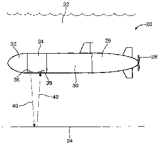
Figure 2 shows an autonomous underwater vehicle (AUV) 20 according to an
embodiment of the invention in a body of water 22 above a seabed 24. A
suitable
AUV is the REMUS developed by the Woods Hole Oceanographic Institution. The
AUV 20 comprises a tail section 26 containing the propulsion motor and
controller
circuitry fora propeller 28. A mid-body section 30 houses various operational
components of the vehicle. Between the mid-body section 30 and a nose cone 32
is
an optics section 34. The optics section 34 comprises a watertight chamber
carrying
a hyperspectral imager and a light source (not shown). A transparent outlet
window
36 allows light 40 from the light source to emerge towards a scene of
interest, such
as the seabed 24. Light 42 returning from the scene enters through a
transparent
inlet window 38 behind which is located the objective lens 8 of a
hyperspectral
imager.
Figure 3 shows another embodiment of an underwater apparatus 44 embodying the
invention. This apparatus 44 is not self-propelling but rather can be lowered
into the
water attached to a floater and so be immersed in the water for towing by a
boat for
example, or carried by a human diver. It comprises a watertight housing 46
made of
aluminium or marine steel having a transparent window 48 made of soda glass or
quartz to allow the passage of light into, and optionally out of, the imager
44. It also
has a display panel 50 for turning the system on and off, tuning the frame,
gain, iris
and gamma controls. Inside the housing 46, there is a hyperspectral imager,
batteries and video recorder and there may be one or more lamps. The apparatus
44
may also carry external underwater lamps (not shown) such as an Underwater
Kinetics Light Cannon 100, which can be used to obtain 6000 degrees Kelvin
colour
temperature. The imager can be used in any orientation; i.e. it can be pointed
horizontally, up or down.
CA 02724817 2010-11-18
WO 2009/141622 PCT/GB2009/001288
- 17-
In both cases above the apparatus could carry several lamps which can be used
individually or in combination to provide a customised illumination. This can
be
used to minimise the effects of absorption and scattering in the water between
the
light source, imaged material and the imager, and can also ensure that the
correct
wavelengths in the imaged material are excited.
The lamp 52 shown in Figure 4 is also suitable for use with imagers embodying
the
invention, such as those of Figures 2 and 3 and takes the idea of blending
light
sources one step further. The lamp 52 comprises a plurality of light emitting
diode
(LED) lamps 54 which can be selectively illuminated. Some of the LEDs are
white,
emitting light in the range 350-800 nm. Others are blue (emitting light in 400-
500
nm range), green (500-600 nm), and red (600-700 mn).
The spectrum of light emanating from the lamp 52 can be tuned by selecting
which
LEDs to activate, depending on the optical properties of the water (which vary
with
distance to the target object due to the spectral attenuation coefficient of
water, and
which can vary due to optically-active components such as phytoplankton,
coloured
dissolved organic matter and total suspended matter).
Either of the two underwater apparatus described above can be used to capture
and
record two-dimensional hyperspectral images beneath the water. By carrying its
own artificial light source, the imaging apparatus can measure much more
accurate
hyperspectral information than is possible using airborne remote sensing. For
example the effects of solar horizontal, and of atmospheric scattering and
distortion
are removed. Moreover the path length of the emitted and reflected light
through the
water can be relatively short, whatever depth the imaged material is at.
One application of the principles of the invention is in mapping or
prospecting for
materials by using a database of spectral profiles that correspond to known
materials
such as particular compounds, substances or organisms to compare against the
spectral profiles measured from the captured images. The spectral profiles on
the
CA 02724817 2010-11-18
WO 2009/141622 PCT/GB2009/001288
-18-
database might be commercially or publicly available. However below a method
of
building up or adding to such a database will be described.
Figure 5 shows a hyperspectral microscopic imager 56 for use in the method
mentioned above forming an embodiment of another part of the invention. The
imager 56 comprises a microscope component 60, adapted from a conventional
optical microscope, such as a Leitz Leca MS5 microscope (1-80x), and a
hyperspectral imaging component 58, such as an Astrovid StellaCam II Video
Camera [AV-STCA2] with a pixel array of 640 x 480, containing optics as
described
with reference to Figure 1. The objective lens of the hyperspectral imaging
component 58 may, by way of example, have a focal length of 25 mm and f: 1.6.
The hyperspectral imaging component 58 has an image capture means; for example
an ARCOS pocket video recorder AV400 capturing AVI video at 25 frames/sec. In
one example, each video frame recorded (spectral profile), consists of the
light
spectrum from 363 to 685 rim dispersed over 640 pixels, giving a resolution of
0.5
nm/pixel. The spatial resolution perpendicular to the moving direction in this
example is 193 pixels.
The imager 56 further comprises a moveable platform 62, which can be moved in
the direction indicated by the arrow by a stepper motor located underneath the
platform. By way of example, the stepper motor may have a gear exchange of
1:500
giving a speed of 2.59 mm/sec. The platform 62 carries a watertight sample
container 64, such as a Petri dish, which can hold a specimen in a volume of
liquid.
The container 64 is also arranged to direct light through a specimen from
beneath,
for example by means of a mirror and a diffuser, when determining optical
transmission characteristics of a specimen; or with a light source above for
determining optical reflectance. The imager 56 also comprises one or more
light
sources directable onto the upper surface of a specimen, preferably from an
off axis
angle such as at 45 degree to the vertical. The same light source maybe used
for
either transmissive or reflective analysis and may consist of a halogen or
other light
source directed appropriately through two fibre optic bundles. This light
source can
CA 02724817 2010-11-18
WO 2009/141622 PCT/GB2009/001288
-19-
be used when determining the reflectance characteristics of a specimen. The
objective lens of the microscope component 60 may be lowered into the fluid
carried
in the sample container 64, to mitigate any optical interference that might be
caused
due to the fluid-air and air-lens boundaries when the objective lens is
located out of
the fluid.
In use, a sample is placed in fluid; such as sea water, in the sample
container 64.
The stepper motor moves the platform 62 in the direction of the arrow while
the
hyperspectral imaging component 58 captures sequential spectral image strips
across
the specimen orthogonal to the direction of motion. These strips can be
combined as
explained above with reference to Figure 1. In particular, processing may be
performed using YaPlaySpecX software (Fred Sigernes, UNIS, cf. Sigernes et al.
2000 Applied Optics) to compose monochromatic images from an AVI video,
forming an spectral image cube. Depending on the light source selected, two-
dimensional images of either spectral transmittance or spectral reflectance of
the
specimen in the liquid can be generated at high magnification through use of
the
imager 56.
If desired, average spectral characteristics (with statistical information on
e.g. error
estimates) for an area of interest captured with the hyperspectral microscopic
imager 56, can be found by averaging information from an image hypercube in
the
spectral direction. The average spectral characteristics measured for
reflection,
Er(lamda) (mW/nm), or transmission, Et(lamda) (mW/nm), may be adjusted for the
halogen lamp (or other light source) radiant intensity spectrum for
reflection,
Ehr(lamda) (mW/nm), and for transmission, Eht(lamda) (mW/nm), to give a
comparable reflectance or transmittance spectrum with optical density. The
dimensionless reflectance spectra is then R(X) = Er(lamda) / Ehr(lamda) and
the
dimensionless transmittance spectra is T(lamda) = Et(lamda) / Eht(lamda).
Figure 6 shows an image A of a specimen of a red alga to be analysed using a
magnifying hyperspectral imager in accordance with the invention. It also
shows a
magnified monochromatic image B of the specimen in water (at 600 nm
CA 02724817 2010-11-18
WO 2009/141622 PCT/GB2009/001288
-20-
wavelength) captured using the hyperspectral imager. Three distinct regions 1,
2, 3
are indicated, for which the average reflectance, R(lamda), over the region is
to be
determined. Fig 6-C shows the R(lamda) spectra 1, 2, 3 obtained. It also shows
the
corresponding spectral absorbance spectrum OD, measured with a
spectrophotometer, which validates the reflectance measurements (they should
be
inversely related). The reflectance measurements have been adjusted to
compensate
for the halogen lamp radiant intensity spectrum, Eh(),).
Once an averaged spectrum for a region of interest has been obtained, this can
be
used to identify other instances of the same material in other situations; in
particular,
it can be used with the apparatus described earlier to identify the same
material
underwater using in situ hyperspectral imaging apparatus.