Note: Descriptions are shown in the official language in which they were submitted.
CA 02725498 2013-05-03
MULTI-FIELD CHARGED PARTICLE CANCER THERAPY METHOD AND
APPARATUS
BACKGROUND OF THE INVENTION
FIELD OF THE INVENTION
This invention relates generally to treatment of solid cancers. More
particularly, the
invention relates to charged particle irradiation beam control in cancer
therapy.
DISCUSSION OF THE PRIOR ART
Cancer Treatment
Several distinct forms of radiation therapy exist for cancer treatment
including:
brachytherapy, traditional electromagnetic X-ray therapy, and proton therapy.
Proton
therapy systems typically include: a beam generator, an accelerator, and a
beam
transport system to move the resulting accelerated protons to a plurality of
treatment
rooms where the protons are delivered to a tumor in a patient's body.
Proton therapy works by aiming energetic ionizing particles, such as protons
accelerated with a particle accelerator, onto a target tumor. These particles
damage the
DNA of cells, ultimately causing their death. Cancerous cells, because of
their high rate
of division and their reduced ability to repair damaged DNA, are particularly
vulnerable
to attack on their DNA.
Charged Particle Cancer Therapy
Patents related to the current invention are summarized here.
Proton Beam Therapy System
F. Cole, et. al. of Loma Linda University Medical Center "Multi-Station Proton
Beam
Therapy System", U.S. patent no. 4,870,287 (September 26, 1989) describe a
proton
beam therapy system for selectively generating and transporting proton beams
from a
1
CA 02725498 2013-05-03
single proton source and accelerator to a selected treatment room of a
plurality of
patient treatment rooms.
Gantry
T. Yamashita, et. al. "Rotating Irradiation Apparatus", U.S. patent no.
7,381,979 (June 3,
2008) describe a rotating gantry having a front ring and a rear ring, each
ring having
radial support devices, where the radial support devices have linear guides.
The
system has thrust support devices for limiting movement of the rotatable body
in the
direction of the rotational axis of the rotatable body.
T. Yamashita, et. al. "Rotating Gantry of Particle Beam Therapy System" U.S.
patent no.
7,372,053 (May 13, 2008) describe a rotating gantry supported by an air
braking system
allowing quick movement, braking, and stopping of the gantry during
irradiation
treatment.
M. Yanagisawa, et. aL "Medical Charged Particle Irradiation Apparatus", U.S.
patent no.
6,992,312 (January 31, 2006); M. Yanagisawa, et. al. "Medical Charged Particle
Irradiation Apparatus", U.S. patent no. 6,979,832 (December 27, 2005); and M.
Yanagisawa, et. al. "Medical Charged Particle Irradiation Apparatus", U.S.
patent no.
6,953,943 (October 11, 2005) all describe an apparatus capable of irradiation
from
upward and horizontal directions. The gantry is rotatable about an axis of
rotation
where the irradiation field forming device is eccentrically arranged, such
that an axis of
irradiation passes through a different position than the axis of rotation.
H. Kaercher, et. al. "Isokinetic Gantry Arrangement for the lsocentric
Guidance of a
Particle Beam And a Method for Constructing Same", U.S. patent no. 6,897,451
(May
24, 2005) describe an isokinetic gantry arrangement for isocentric guidance of
a particle
beam that can be rotated around a horizontal longitudinal axis.
G. Kraft, et. aL "Ion Beam System for Irradiating Tumor Tissues", U.S. patent
no.
6,730,921 (May 4, 2004) describe an ion beam system for irradiating tumor
tissues at
2
CA 02725498 2013-05-03
various irradiation angles in relation to a horizontally arranged patient
couch, where the
patient couch is rotatable about a center axis and has a lifting mechanism.
The system
has a central ion beam deflection of up to 15 degrees with respect to a
horizontal
direction.
M. Pavlovic, et. al. "Gantry System and Method for Operating Same", U.S.
patent no.
6,635,882 (October 21, 2003) describe a gantry system for adjusting and
aligning an ion
beam onto a target from a freely determinable effective treatment angle. The
ion beam
is aligned on a target at adjustable angles of from 0 to 360 degrees around
the gantry
rotation axis and at an angle of 45 to 90 degrees off of the gantry rotation
axis yielding a
cone of irradiation when rotated a full revolution about the gantry rotation
axis.
Movable Patient
N. Rigney, et. al. "Patient Alignment System with External Measurement and
Object
Coordination for Radiation Therapy System", U.S. patent no. 7,199,382 (April
3, 2007)
describe a patient alignment system for a radiation therapy system that
includes
multiple external measurement devices that obtain position measurements of
movable
components of the radiation therapy system. The alignment system uses the
external
measurements to provide corrective positioning feedback to more precisely
register the
patient to the radiation beam.
Y. Muramatsu, et. al. "Medical Particle Irradiation Apparatus", U.S. patent
no. 7,030,396
(April 18, 2006); Y. Muramatsu, et. al. "Medical Particle Irradiation
Apparatus", U.S.
patent no. 6,903,356 (June 7, 2005); and Y. Muramatsu, et. al. "Medical
Particle
Irradiation Apparatus", U.S. patent no. 6,803,591 (October 12, 2004) all
describe a
medical particle irradiation apparatus having a rotating gantry, an annular
frame located
within the gantry such that it can rotate relative to the rotating gantry, an
anti-correlation
mechanism to keep the frame from rotating with the gantry, and a flexible
moving floor
engaged with the frame in such a manner to move freely with a substantially
level
bottom while the gantry rotates.
3
CA 02725498 2013-05-03
H. Nonaka, et. al. "Rotating Radiation Chamber for Radiation Therapy", U.S.
patent no.
5,993,373 (November 30, 1999) describe a horizontal movable floor composed of
a
series of multiple plates that are connected in a free and flexible manner,
where the
movable floor is moved in synchrony with rotation of a radiation beam
irradiation
section.
Patient Positioning
Y. Nagannine, et. al. "Patient Positioning Device and Patient Positioning
Method", U.S.
patent no. 7,212,609 (May 1, 2007) and Y. Nagamine, et. al. "Patient
Positioning Device
and Patient Positioning Method", U.S. patent no. 7,212,608 (May 1, 2007)
describe a
patient positioning system that compares a comparison area of a reference X-
ray image
and a current X-ray image of a current patient location using pattern
matching.
D. Miller, et. al. "Modular Patient Support System", U.S. patent no. 7,173,265
(February
6, 2007) describe a radiation treatment system having a patient support system
that
includes a modularly expandable patient pod and at least one immobilization
device,
such as a moldable foam cradle.
K. Kato, et. al. "Multi-Leaf Collimator and Medical System Including
Accelerator", U.S.
patent no. 6,931,100 (August 16, 2005); K. Kato, et. al. "Multi-Leaf
Collimator and
Medical System Including Accelerator", U.S. patent no. 6,823,045 (November 23,
2004);
K. Kato, et. al. "Multi-Leaf Collimator and Medical System Including
Accelerator", U.S.
patent no. 6,819,743 (November 16, 2004); and K. Kato, et. al. "Multi-Leaf
Collimator
and Medical System Including Accelerator", U.S. patent no. 6,792,078
(September 14,
2004) all describe a system of leaf plates used to shorten positioning time of
a patient
for irradiation therapy. Motor driving force is transmitted to a plurality of
leaf plates at
the same time through a pinion gear. The system also uses upper and lower air
cylinders and upper and lower guides to position a patient.
4
CA 02725498 2013-05-03
Problem
There exists in the art of particle beam therapy of cancerous tumors a need
for charged
particle irradiation beam control. More particularly, there exists in the art
a need for
efficient delivery of charged particles to the tumor, where efficiency is the
fraction of
energy deposited in the tumor relative to the fraction of energy deposited in
healthy
tissue.
SUMMARY OF THE INVENTION
The invention comprises a multi-field charged particle irradiation beam method
and
apparatus used in radiation therapy of cancerous tumors.
DESCRIPTION OF THE FIGURES
Figure 1 illustrates component connections of a particle beam therapy system;
Figure 2 illustrates a charged particle therapy system;
Figure 3 illustrates an ion beam generation system;
Figure 4 illustrates straight and turning sections of a synchrotron;
Figure 5 illustrates bending magnets of a synchrotron;
Figure 6 provides a perspective view of a bending magnet;
Figure 7 illustrates a cross-sectional view of a bending magnet;
Figure 8 illustrates a cross-sectional view of a bending magnet;
Figure 9 illustrates a magnetic tuming section of a synchrotron;
Figures 10A and B illustrate an RF accelerator and an RF accelerator
subsystem,
respectively;
Figure 11 illustrates a magnetic field control system;
5
CA 02725498 2013-05-03
Figure 12 illustrates a charged particle extraction and intensity control
system;
Figure 13 illustrates a proton beam position verification system;
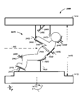
Figure 14 illustrates a patient positioning system from: (A) a front view and
(B) a top
view;
Figure 15 provides X-ray and proton beam dose distributions;
Figures 16 A-E illustrate controlled depth of focus irradiation;
Figures 17 A-E illustrate multi-field irradiation;
Figure 18 illustrates dose efficiency enhancement via use of multi-field
irradiation;
Figures 19A-C and Figure 19E illustrate distal irradiation of a tumor from
varying
rotational directions and Figure 19D illustrates integrated radiation
resulting from distal
radiation;
Figure 20 provides two methods of multi-field irradiation implementation;
Figure 21 illustrates multi-dimensional scanning of a charged particle beam
spot
scanning system operating on: (A) a 2-D slice or (B) a 3-D volume of a tumor;
Figure 22 illustrates an electron gun source used in generating X-rays coupled
with a
particle beam therapy system;
Figure 23 illustrates an X-ray source proximate a particle beam path;
Figure 24 illustrates a semi-vertical patient positioning system;
Figure 25 illustrates respiration monitoring;
Figure 26 illustrates a patient positioning, immobilization, and repositioning
system;
Figure 27 shows particle field acceleration timed to a patient's respiration
cycle; and
Figure 28 illustrates adjustable particle field acceleration timing.
6
CA 02725498 2013-05-03
DETAILED DESCRIPTION OF THE INVENTION
The invention comprises a multi-field charged particle irradiation beam method
and
apparatus used in radiation therapy of cancerous tumors.
In one embodiment, a method and apparatus for efficient radiation dose
delivery to a
tumor is described. Preferably, radiation is delivered through an entry point
into the
tumor and Bragg peak energy is targeted to a distal or far side of the tumor
from an
ingress point. Delivering Bragg peak energy to the distal side of the tumor
from the
ingress point is repeated from multiple rotational directions.
Beam intensity is
proportional to radiation dose delivery efficiency. The multi-field
irradiation process with
energy levels targeting the far side of the tumor from each irradiation
direction provides
even and efficient charged particle radiation dose delivery to the tumor.
Preferably, the
charged particle therapy is timed to patient respiration via control of
charged particle
beam injection, acceleration, extraction, and/or targeting methods and
apparatus.
For example, radiation is delivered through an entry point into the tumor and
Bragg
peak energy is targeted to a distal or far side of the tumor from an ingress
point.
Delivering Bragg peak energy to the distal side of the tumor from the ingress
point is
repeated from multiple rotational directions. Preferably, beam intensity is
proportional to
radiation dose delivery efficiency. Preferably, the charged particle therapy
is timed to
patient respiration via control of charged particle beam injection,
acceleration,
extraction, and/or targeting methods and apparatus. Optionally, multi-axis
control of the
charged particle beam is used simultaneously with the multi-field irradiation.
Combined,
the system allows multi-field and multi-axis charged particle irradiation of
tumors
yielding precise and accurate irradiation dosages to a tumor with distribution
of harmful
ingress energy about the tumor.
In another embodiment, the system relates to a combined rotation / raster
method and
apparatus, referred to as multi-field charged particle cancer therapy. The
system uses
a fixed orientation charged particle source, such as a proton source, relative
to a
rotating patient to yield tumor irradiation from multiple directions.
Preferably, the system
7
CA 02725498 2013-05-03
combines layer-wise tumor irradiation from many directions with controlled
energy
proton irradiation to deliver peak proton beam energy within a selected tumor
volume or
irradiated slice. Optionally, the selected tumor volume for irradiation from a
given angle
is a distal portion of the tumor. In this manner ingress Bragg peak energy is
circumferentially spread about the tumor minimizing damage to healthy tissue
and peak
proton energy is efficiently, accurately, and precisely delivered to the
tumor.
In yet another embodiment, a multi-field imaging and a multi-field charged
particle
cancer therapy method and apparatus is used that is coordinated with patient
respiration via use of feedback sensors used to monitor and/or control patient
respiration. Optionally, the respiration monitoring system uses thermal and/or
force
sensors to determine where a patient is in a respiration cycle in combination
with a
feedback signal control delivered to the patient to inform the patient when
breath control
is required. Preferably, the multi-field imaging, such as X-ray imaging, and
the charged
particle therapy are performed on a patient in a partially immobilized and
repositionable
position. X-ray and/or proton delivery is timed to patient respiration via
control of
charged particle beam injection, acceleration, extraction, and/or targeting
methods and
apparatus.
In still yet another embodiment, a multi-axis charged particle irradiation
method and
apparatus is described, optionally used in combination with multi-field
irradiation. The
multi-axis controls includes separate control of one or more of horizontal or
x-axis
position, vertical or y-axis position, energy control, and intensity control
of the charged
particle irradiation beam. Optionally, the separate control is independent
control.
Optionally, the charged particle beam is additionally controlled in terms of
timing.
Timing is coordinated with patient respiration and/or patient rotational
positioning.
Combined, the system allows multi-axis and multi-field charged particle
irradiation of
tumors yielding precise and accurate irradiation dosages to a tumor with
distribution of
harmful healthy tissue volume ingress energy about the tumor.
8
CA 02725498 2013-05-03
In another embodiment, the system uses a radio-frequency (RF) cavity system to
induce betatron oscillation of a charged particle stream. Sufficient amplitude
modulation
of the charged particle stream causes the charged particle stream to hit a
material, such
as a foil. The foil decreases the energy of the charged particle stream, which
decreases
a radius of curvature of the charged particle stream in the synchrotron
sufficiently to
allow a physical separation of the reduced energy charged particle stream from
the
original charged particle stream. The physically separated charged particle
stream is
then removed from the system by use of an applied field and deflector.
In still another embodiment, the system comprises intensity control of a
charged particle
beam acceleration, extraction, and/or targeting method and apparatus used in
conjunction with charged particle beam radiation therapy of cancerous tumors.
Particularly, intensity of a charged particle stream of a synchrotron is
described in
combination with turning magnets, edge focusing magnets, concentrating
magnetic field
magnets, winding and control coils, and extraction elements of the
synchrotron. The
system reduces the overall size of the synchrotron, provides a tightly
controlled proton
beam, directly reduces the size of required magnetic fields, directly reduces
required
operating power, and allows continual acceleration of protons in a synchrotron
even
during a process of extracting protons from the synchrotron.
Used in combination with the invention, novel design features of a charged
particle
beam cancer therapy system are described. Particularly, a negative ion beam
source
with novel features in the negative ion source, ion source vacuum system, ion
beam
focusing lens, and tandem accelerator is described. Additionally, tuming
magnets, edge
focusing magnets, magnetic field concentration magnets, winding and correction
coils,
flat magnetic field incident surfaces, and extraction elements are described
that
minimize the overall size of the synchrotron, provide a tightly controlled
proton beam,
directly reduce the size of required magnetic fields, directly reduce required
operating
power, and allow continual acceleration of protons in a synchrotron even
during a
process of extracting protons from the synchrotron. The ion beam source system
and
synchrotron are preferably computer integrated with a patient imaging system
and a
9
CA 02725498 2013-05-03
patient interface including respiration monitoring sensors and patient
positioning
elements. Further, intensity control of a charged particle beam acceleration,
extraction,
and/or targeting method and apparatus used in conjunction with charged
particle beam
radiation therapy of cancerous tumors is described. More particularly,
intensity, energy,
and timing control of a charged particle stream of a synchrotron is described.
The
synchrotron control elements allow tight control of the charged particle beam,
which
compliments the tight control of patient positioning to yield efficient
treatment of a solid
tumor with reduced tissue damage to surrounding healthy tissue. In addition,
the
system reduces the overall size of the synchrotron, provides a tightly
controlled proton
beam, directly reduces the size of required magnetic fields, directly reduces
required
operating power, and allows continual acceleration of protons in a synchrotron
even
during a process of extracting protons from the synchrotron. All of these
systems are
preferably used in conjunction with an X-ray system capable of collecting X-
rays of a
patient in (1) a positioning system for proton treatment and (2) at a
specified moment of
the patient's respiration cycle. Combined, the systems provide for efficient,
accurate,
and precise noninvasive tumor treatment with minimal damage to surrounding
healthy
tissue.
Cyclotron / Synchrotron
A cyclotron uses a constant magnetic field and a constant-frequency applied
electric
field. One of the two fields is varied in a synchrocyclotron. Both of these
fields are
varied in a synchrotron. Thus, a synchrotron is a particular type of cyclic
particle
accelerator in which a magnetic field is used to turn the particles so they
circulate and
an electric field is used to accelerate the particles. The synchroton
carefully
synchronizes the applied fields with the travelling particle beam.
By increasing the fields appropriately as the particles gain energy, the
charged particles
path can be held constant as they are accelerated. This allows the vacuum
container
for the particles to be a large thin torus. In reality it is easier to use
some straight
sections between the bending magnets and some turning sections giving the
torus the
shape of a round-cornered polygon. A path of large effective radius is thus
constructed
CA 02725498 2013-05-03
using simple straight and curved pipe segments, unlike the disc-shaped chamber
of the
cyclotron type devices. The shape also allows and requires the use of multiple
magnets
to bend the particle beam.
The maximum energy that a cyclic accelerator can impart is typically limited
by the
strength of the magnetic fields and the minimum radius / maximum curvature, of
the
particle path. In a cyclotron the maximum radius is quite limited as the
particles start at
the center and spiral outward, thus this entire path must be a self-supporting
disc-
shaped evacuated chamber. Since the radius is limited, the power of the
machine
becomes limited by the strength of the magnetic field. In the case of an
ordinary
electromagnet, the field strength is limited by the saturation of the core
because when
all magnetic domains are aligned the field may not be further increased to any
practical
extent. The arrangement of the single pair of magnets also limits the economic
size of
the device.
Synchrotrons overcome these limitations, using a narrow beam pipe surrounded
by
much smaller and more tightly focusing magnets. The ability of this device to
accelerate
particles is limited by the fact that the particles must be charged to be
accelerated at all,
but charged particles under acceleration emit photons, thereby losing energy.
The
limiting beam energy is reached when the energy lost to the lateral
acceleration
required to maintain the beam path in a circle equals the energy added each
cycle.
More powerful accelerators are built by using large radius paths and by using
more
numerous and more powerful microwave cavities to accelerate the particle beam
between corners. Lighter particles, such as electrons, lose a larger fraction
of their
energy when tuming. Practically speaking, the energy of electron/positron
accelerators
is limited by this radiation loss, while it does not play a significant role
in the dynamics of
proton or ion accelerators. The energy of those is limited strictly by the
strength of
magnets and by the cost,
11
CA 02725498 2013-05-03
CHARGED PARTICLE BEAM THERAPY
Throughout this document, a charged particle beam therapy system, such as a
proton
beam, hydrogen ion beam, or carbon ion beam, is described. Herein, the charged
particle beam therapy system is described using a proton beam. However, the
aspects
taught and described in terms of a proton beam are not intended to be limiting
to that of
a proton beam and are illustrative of a charged particle beam system. Any
charged
particle beam system is equay applicable to the techniques described herein.
Referring now to Figure 1, a charged particle beam system 100 is illustrated.
The
charged particle beam preferably comprises a number of subsystems including
any of:
a main controller or irradiation control module 110; an injection system 120;
a
synchrotron 130 that typically includes: (1) an accelerator system 132 and (2)
an
extraction system 134; a scanning / targeting / delivery system 140; a patient
interface
module 150; a display system 160; and/or an imaging system 170.
An exemplary method of use of the charged particle beam system 100 is
provided. The
main controller 110 controls one or more of the subsystems to accurately and
precisely
deliver protons to a tumor of a patient. For example, the main controller 110
obtains an
image, such as a portion of a body and/or of a tumor, from the imaging system
170.
The main controller 110 also obtains position and/or timing information from
the patient
interface module 150. The main controller 110 then optionally controls the
injection
system 120 to inject a proton into a synchrotron 130. The synchrotron
typically contains
at least an accelerator system 132 and an extraction system 134. The main
controller
preferably controls the proton beam within the accelerator system, such as by
controlling speed, trajectory, and timing of the proton beam. The main
controller then
controls extraction of a proton beam from the accelerator through the
extraction system
134. For example, the controller controls timing, energy, and/or intensity of
the
extracted beam. The controller 110 also preferably controls targeting of the
proton
beam through the scanning / targeting / delivery system 140 to the patient
interface
module 150. One or more components of the patient interface module 150 are
preferably controlled by the main controller 110. Further, display elements of
the
12
CA 02725498 2013-05-03
display system 160 are preferably controlled via the main controller 110.
Displays, such
as display screens, are typically provided to one or more operators and/or to
one or
more patients. In one embodiment, the main controller 110 times the delivery
of the
proton beam from all systems, such that protons are delivered in an optimal
therapeutic
manner to the patient.
Herein, the main controller 110 refers to a single system controlling the
charged particle
beam system 100, to a single controller controlling a plurality of subsystems
controlling
the charged particle beam system 100, or to a plurality of individual
controllers
controlling one or more sub-systems of the charged particle beam system 100.
Synch rotron
Herein, the term synchrotron is used to refer to a system maintaining the
charged
particle beam in a circulating path; however, cyclotrons are alternatively
used, albeit
with their inherent limitations of energy, intensity, and extraction control.
Further, the
charged particle beam is referred to herein as circulating along a circulating
path about
a central point of the synchrotron. The circulating path is alternatively
referred to as an
orbiting path; however, the orbiting path does not refer a perfect circle or
ellipse, rather
it refers to cycling of the protons around a central point or region.
Referring now to Figure 2, an illustrative exemplary embodiment of one version
of the
charged particle beam system 100 is provided. The number, position, and
described
type of components is illustrative and non-limiting in nature.
In the illustrated
embodiment, an injector system 210 or ion source or charged particle beam
source
generates protons. The protons are delivered into a vacuum tube that runs
into,
through, and out of the synchrotron. The generated protons are delivered along
an
initial path 262. Focusing magnets 230, such as quadrupole magnets or
injection
quadrupole magnets, are used to focus the proton beam path. A quadrupole
magnet is
a focusing magnet. An injector bending magnet 232 bends the proton beam toward
the
plane of the synchrotron 130. The focused protons having an initial energy are
introduced into an injector magnet 240, which is preferably an injection
Lamberson
13
CA 02725498 2013-05-03
magnet. Typically, the initial beam path 262 is along an axis off of, such as
above, a
circulating plane of the synchrotron 130. The injector bending magnet 232 and
injector
magnet 240 combine to move the protons into the synchrotron 130. Main bending
magnets 250 or dipole magnets or circulating magnets are used to turn the
protons
along a circulating beam path 264. A dipole magnet is a bending magnet. The
main
bending magnets 250 bend the initial beam path 262 into a circulating beam
path 264.
In this example, the main bending magnets 250 or circulating magnets are
represented
as four sets of four magnets to maintain the circulating beam path 264 into a
stable
circulating beam path. However, any number of magnets or sets of magnets are
optionally used to move the protons around a single orbit in the circulation
process. The
protons pass through an accelerator 270. The accelerator accelerates the
protons in
the circulating beam path 264. As the protons are accelerated, the fields
applied by the
magnets are increased. Particularly, the speed of the protons achieved by the
accelerator 270 are synchronized with magnetic fields of the main bending
magnets 250
or circulating magnets to maintain stable circulation of the protons about a
central point
or region 280 of the synchrotron. At separate points in time the accelerator
270 / main
bending magnet 250 combination is used to accelerate and/or decelerate the
circulating
protons while maintaining the protons in the circulating path or orbit. An
extraction
element of the inflector/deflector system 290 is used in combination with a
Lamberson
extraction magnet 292 to remove protons from their circulating beam path 264
within the
synchrotron 130. One example of a deflector component is a Lamberson magnet.
Typically the deflector moves the protons from the circulating plane to an
axis off of the
circulating plane, such as above the circulating plane. Extracted protons are
preferably
directed and/or focused using an extraction bending magnet 237 and extraction
focusing magnets 235, such as quadrupole magnets along a transport path 268
into the
scanning / targeting / delivery system 140. Two components of a scanning
system 140
or targeting system typically include a first axis control 142, such as a
vertical control,
and a second axis control 144, such as a horizontal control. In one
embodiment, the
first axis control 142 allows for about 100 mm of vertical scanning of the
proton beam
268 and the second axis control 144 allows for about 700 mm of horizontal
scanning of
the proton beam 268. A nozzle system 146 is used for imaging the proton beam
and/or
14
CA 02725498 2013-05-03
as a vacuum barrier between the low pressure beam path of the synchrotron and
the
atmosphere. Protons are delivered with control to the patient interface module
150 and
to a tumor of a patient. All of the above listed elements are optional and may
be used in
various permutations and combinations.
Ion Beam Generation System
An ion beam generation system generates a negative ion beam, such as a
hydrogen
anion or Fl- beam; preferably focuses the negative ion beam; converts the
negative ion
beam to a positive ion beam, such as a proton or H+ beam; and injects the
positive ion
beam into the synchrotron 130. Portions of the ion beam path are preferably
under
partial vacuum. Each of these systems are further described, infra.
Referring now to Figure 3, an exemplary ion beam generation system 300 is
illustrated.
As illustrated, the ion beam generation system 300 has four major elements: a
negative
ion source 310, a first partial vacuum system 330, an optional ion beam
focusing
system 350, and a tandem accelerator 390.
Still referring to Figure 3, the negative ion source 310 preferably includes
an inlet port
312 for injection of hydrogen gas into a high temperature plasma chamber 314.
In one
embodiment, the plasma chamber includes a magnetic material 316, which
provides a
magnetic field barrier 317 between the high temperature plasma chamber 314 and
a
low temperature plasma region on the opposite side of the magnetic field
barrier. An
extraction pulse is applied to a negative ion extraction electrode 318 to pull
the negative
ion beam into a negative ion beam path 319, which proceeds through the first
partial
vacuum system 330, through the ion beam focusing system 350, and into the
tandem
accelerator 390.
Still referring to Figure 3, the first partial vacuum system 330 is an
enclosed system
running from the hydrogen gas inlet port 312 to the tandem accelerator 390
foil 395.
The foil 395 is sealed directly or indirectly to the edges of the vacuum tube
320
providing for a higher pressure, such as about 10 torr, to be maintained on
the first
CA 02725498 2013-05-03
partial vacuum system 330 side of the foil 395 and a lower pressure, such as
about 10-7
torr, to be maintained on the synchrotron side of the foil 390. By only
pumping first
partial vacuum system 330 and by only semi-continuously operating the ion beam
source vacuum based on sensor readings, the lifetime of the semi-continuously
operating pump is extended. The sensor readings are further described, infra.
Still referring to Figure 3, the first partial vacuum system 330 preferably
includes: a first
pump 332, such as a continuously operating pump and/or a turbo molecular pump;
a
large holding volume 334; and a semi-continuously operating pump 336.
Preferably, a
pump controller 340 receives a signal from a pressure sensor 342 monitoring
pressure
in the large holding volume 334. Upon a signal representative of a sufficient
pressure in
the large holding volume 334, the pump controller 340 instructs an actuator
345 to open
a valve 346 between the large holding volume and the semi-continuously
operating
pump 336 and instructs the semi-continuously operating pump to turn on and
pump to
atmosphere residual gases out of the vacuum line 320 about the charged
particle
stream. In this fashion, the lifetime of the semi-continuously operating pump
is
extended by only operating semi-continuously and as needed. In one example,
the
semi-continuously operating pump 336 operates for a few minutes every few
hours,
such as 5 minutes every 4 hours, thereby extending a pump with a lifetime of
about
2,000 hours to about 96,000 hours.
Further, by isolating the inlet gas from the synchrotron vacuum system, the
synchrotron
vacuum pumps, such as turbo molecular pumps can operate over a longer lifetime
as
the synchrotron vacuum pumps have fewer gas molecules to deal with. For
example,
the inlet gas is primarily hydrogen gas but may contain impurities, such as
nitrogen and
carbon dioxide. By isolating the inlet gases in the negative ion source system
310, first
partial vacuum system 330, ion beam focusing system 350 and negative ion beam
side
of the tandem accelerator 390, the synchrotron vacuum pumps can operate at
lower
pressures with longer lifetimes, which increases the efficiency of the
synchrotron 130.
16
CA 02725498 2013-05-03
Still referring to Figure 3, the ion beam focusing system 350 includes two or
more
electrodes where one electrode of each electrode pair partially obstructs the
ion beam
path with conductive paths 372, such as a conductive mesh. In the illustrated
example,
three ion beam focusing system sections are illustrated, a two electrode ion
focusing
section 360, a first three electrode ion focusing section 370, and a second
three
electrode ion focusing section 380. In a given electrode pair, electric field
lines, running
between the conductive mesh of a first electrode and a second electrode,
provide
inward forces focusing the negative ion beam. Multiple such electrode pairs
provide
multiple negative ion beam focusing regions. Preferably the two electrode ion
focusing
section 360, first three electrode ion focusing section 370, and second three
electrode
ion focusing section 380 are placed after the negative ion source and before
the tandem
accelerator and/or cover a space of about 0.5, 1, or 2 meters along the ion
beam path.
Ion beam focusing systems are further described, infra.
Still referring to Figure 3, the tandem accelerator 390 preferably includes a
foil 395,
such as a carbon foil. The negative ions in the negative ion beam path 319 are
converted to positive ions, such as protons, and the initial ion beam path 262
results.
The foil 395 is preferably sealed directly or indirectly to the edges of the
vacuum tube
320 providing for a higher pressure, such as about 10-5 torr, to be maintained
on the
side of the foil 395 having the negative ion beam path 319 and a lower
pressure, such
as about 10-7 torr, to be maintained on the side of the foil 390 having the
proton ion
beam path 262. Having the foil 395 physically separating the vacuum chamber
320 into
two pressure regions allows for a system having fewer and/or smaller pumps to
maintain the lower pressure system in the synchrotron 130 as the inlet
hydrogen and its
residuals are extracted in a separate contained and isolated space by the
first partial
vacuum system 330.
Referring again to Figure 1, another exemplary method of use of the charged
particle
beam system 100 is provided. The main controller 110, or one or more sub-
controllers,
controls one or more of the subsystems to accurately and precisely deliver
protons to a
tumor of a patient. For example, the main controller sends a message to the
patient
17
CA 02725498 2013-05-03
indicating when or how to breath. The main controller 110 obtains a sensor
reading
from the patient interface module, such as a temperature breath sensor or a
force
reading indicative of where in a breath cycle the subject is. The main
controller collects
an image, such as a portion of a body and/or of a tumor, from the imaging
system 170.
The main controller 110 also obtains position and/or timing information from
the patient
interface module 150. The main controller 110 then optionally controls the
injection
system 120 to inject hydrogen gas into a negative ion beam source 310 and
controls
timing of extraction of the negative ion from the negative ion beam source
310.
Optionally, the main controller controls ion beam focusing using the ion beam
focusing
io lens system 350; acceleration of the proton beam with the tandem
accelerator 390;
and/or injection of the proton into the synchrotron 130. The synchrotron
typically
contains at least an accelerator system 132 and an extraction system 134. The
synchrotron preferably contains one or more of: turning magnets, edge focusing
magnets, magnetic field concentration magnets, winding and correction coils,
and flat
magnetic field incident surfaces, some of which contain elements under control
by the
main controller 110. The main controller preferably controls the proton beam
within the
accelerator system, such as by controlling speed, trajectory, and/or timing of
the proton
beam. The main controller then controls extraction of a proton beam from the
accelerator through the extraction system 134. For example, the controller
controls
timing, energy, and/or intensity of the extracted beam. The controller 110
also
preferably controls targeting of the proton beam through the targeting /
delivery system
140 to the patient interface module 150. One or more components of the patient
interface module 150 are preferably controlled by the main controller 110,
such as
vertical position of the patient, rotational position of the patient, and
patient chair
positioning / stabilization / control elements. Further, display elements of
the display
system 160 are preferably controlled via the main controller 110. Displays,
such as
display screens, are typically provided to one or more operators and/or to one
or more
patients. In one embodiment, the main controller 110 times the delivery of the
proton
beam from all systems, such that protons are delivered in an optimal
therapeutic
manner to the patient.
18
CA 02725498 2013-05-03
Circulating System
A synchrotron 130 preferably comprises a combination of straight sections 410
and ion
beam turning sections 420. Hence, the circulating path of the protons is not
circular in a
synchrotron, but is rather a polygon with rounded corners.
In one illustrative embodiment, the synchrotron 130, which as also referred to
as an
accelerator system, has four straight elements and four turning sections.
Examples of
straight sections 410 include the: inflector 240, accelerator 270, extraction
system 290,
and deflector 292. Along with the four straight sections are four ion beam
turning
sections 420, which are also referred to as magnet sections or turning
sections.
Turning sections are further described, infra.
Referring now to Figure 4, an exemplary synchrotron is illustrated. In this
example,
protons delivered along the initial proton beam path 262 are inflected into
the circulating
beam path with the inflector 240 and after acceleration are extracted via a
deflector 292
to a beam transport path 268. In this example, the synchrotron 130 comprises
four
straight sections 410 and four bending or turning sections 420 where each of
the four
tuming sections use one or more magnets to turn the proton beam about ninety
degrees. As is further described, infra, the ability to closely space the
tuming sections
and efficiently tum the proton beam results in shorter straight sections.
Shorter straight
sections allows for a synchrotron design without the use of focusing
quadrupoles in the
circulating beam path of the synchrotron. The removal of the focusing
quadrupoles
from the circulating proton beam path results in a more compact design. In
this
example, the illustrated synchrotron has about a five meter diameter versus
eight meter
and larger cross-sectional diameters for systems using a quadrupole focusing
magnet in
the circulating proton beam path.
Referring now to Figure 5, additional description of the first bending or
turning section
420 is provided. Each of the turning sections preferably comprises multiple
magnets,
such as about 2, 4, 6, 8, 10, or 12 magnets. In this example, four turning
magnets 510,
520, 530, 540 in the first turning section 420 are used to illustrate key
principles, which
19
CA 02725498 2013-05-03
are the same regardless of the number of magnets in a turning section 420. A
turning
magnet 510 is a particular type of main bending or circulating magnet 250.
In physics, the Lorentz force is the force on a point charge due to
electromagnetic fields.
The Lorentz force is given by equation 1 in terms of magnetic fields with the
election
field terms not included.
F = q(v X B)
eq. 1
In equation 1, F is the force in newtons; B is the magnetic field in Teslas;
and v is the
instantaneous velocity of the particles in meters per second.
Referring now to Figure 6, an example of a single magnet bending or turning
section
510 is expanded. The turning section includes a gap 610 through which protons
circulate. The gap 610 is preferably a flat gap, allowing for a magnetic field
across the
gap 610 that is more uniform, even, and intense. A magnetic field enters the
gap 610
through a magnetic field incident suiface and exits the gap 610 through a
magnetic field
exiting surface. The gap 610 runs in a vacuum tube between two magnet halves.
The
gap 610 is controlled by at least two parameters: (1) the gap 610 is kept as
large as
possible to minimize loss of protons and (2) the gap 610 is kept as small as
possible to
minimize magnet sizes and the associated size and power requirements of the
magnet
power supplies. The flat nature of the gap 610 allows for a compressed and
more
uniform magnetic field across the gap 610. One example of a gap dimension is
to
accommodate a vertical proton beam size of about 2 cm with a horizontal beam
size of
about 5 to 6 cm.
As described, supra, a larger gap size requires a larger power supply. For
instance, if
the gap 610 size doubles in vertical size, then the power supply requirements
increase
by about a factor of 4. The flatness of the gap 610 is also important. For
example, the
flat nature of the gap 610 allows for an increase in energy of the extracted
protons from
about 250 to about 330 MeV. More particularly, if the gap 610 has an extremely
flat
CA 02725498 2013-05-03
surface, then the limits of a magnetic field of an iron magnet are reachable.
An
exemplary precision of the flat surface of the gap 610 is a polish of less
than about 5
microns and preferably with a polish of about 1 to 3 microns. Unevenness in
the
surface results in imperfections in the applied magnetic field. The polished
flat surface
spreads unevenness of the applied magnetic field.
Still referring to Figure 6, the charged particle beam moves through the gap
610 with an
instantaneous velocity, v. A first magnetic coil 620 and a second magnetic
coil 630 run
above and below the gap 610, respectively. Current running through the coils
620, 630
results in a magnetic field, B, running through the single magnet turning
section 510. In
this example, the magnetic field, B, runs upward, which results in a force, F,
pushing the
charged particle beam inward toward a central point of the synchrotron, which
turns the
charged particle beam in an arc.
Still referring to Figure 6, a portion of an optional second magnet bending or
turning
section 520 is illustrated. The coils 620, 630 typically have retum elements
640, 650 or
turns at the end of one magnet, such as at the end of the first magnet tuming
section
510. The tums 640, 650 take space. The space reduces the percentage of the
path
about one orbit of the synchrotron that is covered by the turning magnets.
This leads to
portions of the circulating path where the protons are not tumed and/or
focused and
allows for portions of the circulating path where the proton path defocuses.
Thus, the
space results in a larger synchrotron. Therefore, the space between magnet
tuming
sections 660 is preferably minimized. The second tuming magnet is used to
illustrate
that the coils 620, 630 optionally run along a plurality of magnets, such as
2, 3, 4, 5, 6,
or more magnets. Coils 620, 630 running across multiple tuming section magnets
allows for two turning section magnets to be spatially positioned closer to
each other
due to the removal of the steric constraint of the tums, which reduces and/or
minimizes
the space 660 between two tuming section magnets.
Referring now to Figures 7 and 8, two illustrative 90 degree rotated cross-
sections of
single magnet bending or tuming sections 510 are presented. Referring now to
Figure
21
CA 02725498 2013-05-03
8, the magnet assembly has a first magnet 810 and a second magnet 820. A
magnetic
field induced by coils, described infra, runs between the first magnet 810 to
the second
magnet 820 across the gap 610. Retum magnetic fields run through a first yoke
812
and second yoke 822. The combined cross-section area of the return yokes
roughly
approximates the cross-sectional area of the first magnet 810 or second magnet
820.
The charged particles run through the vacuum tube in the gap 610. As
illustrated,
protons run into Figure 8 through the gap 610 and the magnetic field,
illustrated as
vector B, applies a force F to the protons pushing the protons towards the
center of the
synchrotron, which is off page to the right in Figure 8. The magnetic field is
created
using windings. A first coil makes up a first winding coil 850 and a second
coil of wire
makes up a second winding coil 860. Isolating or concentrating gaps 830, 840,
such as
air gaps, isolate the iron based yokes from the gap 610. The gap 610 is
approximately
flat to yield a uniform magnetic field across the gap 610, as described supra.
Still again to Figure 7, the ends of a single bending or tuming magnet are
preferably
beveled. Nearly perpendicular or right angle edges of a tuming magnet 510 are
represented by dashed lines 774, 784. The dashed lines 774, 784 intersect at a
point
790 beyond the center of the synchrotron 280. Preferably, the edge of the
turning
magnet is beveled at angles alpha, a, and beta, 13, which are angles formed by
a first
line 772, 782 going from an edge of the turning magnet 510 and the center 280
and a
second line 774, 784 going from the same edge of the tuming magnet and the
intersecting point 790. The angle alpha is used to describe the effect and the
description of angle alpha applies to angle beta, but angle alpha is
optionally different
from angle beta. The angle alpha provides an edge focusing effect. Beveling
the edge
of the tuming magnet 510 at angle alpha focuses the proton beam.
Multiple turning magnets provide multiple magnet edges that each have edge
focusing
effects in the synchrotron 130. If only one turning magnet is used, then the
beam is
only focused once for angle alpha or twice for angle alpha and angle beta.
However, by
using smaller turning magnets, more turning magnets fit into the tuming
sections 420 of
the synchrotron 130. For example, if four magnets are used in a tuming section
420 of
22
CA 02725498 2013-05-03
the synchrotron, then for a single turning section there are eight possible
edge focusing
effect surfaces, two edges per magnet. The eight focusing surfaces yield a
smaller
cross-sectional beam size. This allows the use of a smaller gap 610.
The use of multiple edge focusing effects in the turning magnets results in
not only a
smaller gap 610, but also the use of smaller magnets and smaller power
supplies. For
a synchrotron 130 having four turning sections 420 where each turning sections
has
four turning magnets and each turning magnet has two focusing edges, a total
of thirty-
two focusing edges exist for each orbit of the protons in the circulating path
of the
synchrotron 130. Similarly, if 2, 6, or 8 magnets are used in a given tuming
section, or if
2, 3, 5, or 6 tuming sections are used, then the number of edge focusing
surfaces
expands or contracts according to equation 2.
M FE
TFE = 1VTS *¨*¨
eq. 2
NTS M
where TFE is the number of total focusing edges, NTS is the number of turning
sections, M is the number of magnets, and FE is the number of focusing edges.
Naturally, not all magnets are necessarily beveled and some magnets are
optionally
beveled on only one edge.
The inventors have determined that multiple smaller magnets have benefits over
fewer
larger magnets. For example, the use of 16 small magnets yields 32 focusing
edges
whereas the use of 4 larger magnets yields only 8 focusing edges. The use of a
synchrotron having more focusing edges results in a circulating path of the
synchrotron
built without the use of focusing quadrupoles magnets. All prior art
synchrotrons use
quadrupoles in the circulating path of the synchrotron. Further, the use of
quadrupoles
in the circulating path necessitates additional straight sections in the
circulating path of
the synchrotron. Thus, the use of quadrupoles in the circulating path of a
synchrotron
results in synchrotrons having larger diameters, circulating beam pathlengths,
and/or
larger circumferences.
23
CA 02725498 2013-05-03
In various embodiments of the system described herein, the synchrotron has any
combination of:
= at least 4 and preferably 6, 8, 10, or more edge focusing edges per 90
degrees
of turn of the charged particle beam in a synchrotron having four tuming
sections;
= at least about 16 and preferably about 24, 32, or more edge focusing
edges per
orbit of the charged particle beam in the synchrotron;
= only 4 tuming sections where each of the turning sections includes at
least 4 and
io preferably 8 edge focusing edges;
= an equal number of straight sections and turning sections;
= exactly 4 turning sections;
= at least 4 edge focusing edges per turning section;
= no quadrupoles in the circulating path of the synchrotron;
= a rounded comer rectangular polygon configuration;
= a circumference of less than 60 meters;
= a circumference of less than 60 meters and 32 edge focusing surfaces;
and/or
= any of about 8, 16, 24, or 32 non-quadrupole magnets per circulating path
of the
synchrotron, where the non-quadrupole magnets include edge focusing edges.
Referring again to Figure 8, the incident magnetic field surface 870 of the
first magnet
810 is further described. Figure 8 is not to scale and is illustrative in
nature. Local
imperfections or unevenness in quality of the finish of the incident surface
870 results in
inhomogeneities or imperfections in the magnetic field applied to the gap 610.
Preferably, the incident surface 870 is flat, such as to within about a zero
to three
micron finish polish, or less preferably to about a ten micron finish polish.
Referring still to Figure 8, additional magnet elements are described. The
first magnet
810 preferably contains an initial cross sectional distance 890 of the iron
based core.
The contours of the magnetic field are shaped by the magnets 810, 820 and the
yokes
24
CA 02725498 2013-05-03
812, 822. The iron based core tapers to a second cross sectional distance 892.
The
magnetic field in the magnet preferentially stays in the iron based core as
opposed to
the gaps 830, 840. As the cross-sectional distance decreases from the initial
cross
sectional distance 890 to the final cross-sectional distance 892, the magnetic
field
concentrates. The change in shape of the magnet from the longer distance 890
to the
smaller distance 892 acts as an amplifier. The concentration of the magnetic
field is
illustrated by representing an initial density of magnetic field vectors 894
in the initial
cross section 890 to a concentrated density of magnetic field vectors 896 in
the final
cross section 892. The concentration of the magnetic field due to the geometry
of the
turning magnets results in fewer winding coils 850, 860 being required and
also a
smaller power supply to the coils being required.
In one example, the initial cross-section distance 890 is about fifteen
centimeters and
the final cross-section distance 892 is about ten centimeters. Using the
provided
numbers, the concentration of the magnetic field is about 15/10 or 1.5 times
at the
incident surface 870 of the gap 610, though the relationship is not linear.
The taper 842
has a slope, such as about 20, 40, or 60 degrees. The concentration of the
magnetic
field, such as by 1.5 times, leads to a corresponding decrease in power
consumption
requirements to the magnets.
Referring still to Figure 8, the first magnet 810 preferably contains an
initial cross
sectional distance 890 of the iron based core. The contours of the magnetic
field are
shaped by the magnets 810, 820 and the yokes 812, 822. In this example, the
core
tapers to a second cross sectional distance 892 with a smaller angle theta, 0.
As
described, supra, the magnetic field in the magnet preferentially stays in the
iron based
core as opposed to the gaps 830, 840. As the cross-sectional distance
decreases from
the initial cross sectional distance 890 to the final cross-sectional distance
892, the
magnetic field concentrates. The smaller angle, theta, results in a greater
amplification
of the magnetic field in going from the longer distance 890 to the smaller
distance 892.
The concentration of the magnetic field is illustrated by representing an
initial density of
magnetic field vectors 894 in the initial cross section 890 to a concentrated
density of
CA 02725498 2013-05-03
. magnetic field vectors 896 in the final cross section 892. The concentration
of the
= magnetic field due to the geometry of the tuming magnets results in fewer
winding coils
850, 860 being required and also a smaller power supply to the winding coils
850, 860
being required.
Still referring to Figure 8, optional correction coils 852, 862 are
illustrated that are used
to correct the strength of one or more tuming magnets. The correction coils
852, 862
supplement the winding coils 850, 860. The correction coils 852, 862 have
correction
coil power supplies that are separate from winding coil power supplies used
with the
io winding coils 850, 860. The correction coil power supplies typically
operate at a fraction
of the power required compared to the winding coil power supplies, such as
about 1, 2,
3, 5, 7, or 10 percent of the power and more preferably about 1 or 2 percent
of the
power used with the winding coils 850, 860. The smaller operating power
applied to the
correction coils 852, 862 allows for more accurate and/or precise control of
the
correction coils. Correction coils are used to adjust for imperfection in the
turning
magnets 510, 520, 530, 540. Optionally, separate correction coils are used for
each
turning magnet allowing individual tuning of the magnetic field for each
turning magnet,
which eases quality requirements in the manufacture of each turning magnet.
Referring now to Figure 9, an example of winding coils and correction coils
about a
plurality of turning magnets 510, 520, 530, 540 in an ion beam turning section
420 is
illustrated. One or more high precision magnetic field sensors are placed into
the
synchrotron and are used to measure the magnetic field at or near the proton
beam
path. For example, the magnetic sensors 950 are optionally placed between
tuming
magnets and/or within a turning magnet, such as at or near the gap 610 or at
or near
the magnet core or yoke. The sensors are part of a feedback system to the
correction
coils. Thus, the system preferably stabilizes the magnetic field in the
synchrotron
elements rather that stabilizing the current applied to the magnets.
Stabilization of the
magnetic field allows the synchrotron to come to a new energy level quickly.
This
allows the system to be controlled to an operator or algorithm selected energy
level with
each pulse of the synchrotron and/or with each breath of the patient.
26
CA 02725498 2013-05-03
The winding and/or correction coils correct 1, 2, 3, or 4 tuming magnets, and
preferably
correct a magnetic field generated by two turning magnets. A winding or
correction coil
covering multiple magnets reduces space between magnets as fewer winding or
correction coil ends are required, which occupy space.
Referring now to Figure 10A and Figure 10B, the accelerator system 270, such
as a
radio-frequency (RF) accelerator system, is further described. The accelerator
includes
a series of coils 1010-1019, such as iron or ferrite coils, each
circumferentially enclosing
the vacuum system 320 through which the proton beam 264 passes in the
synchrotron
130. Referring now to Figure 10B, the first coil 1010 is further described. A
loop of
standard wire 1030 completes at least one turn about the first coil 1010. The
loop
attaches to a microcircuit 1020. Referring again to Figure 10A, an RF
synthesizer 1040,
which is preferably connected to the main controller 110, provides a low
voltage RF
1 5 signal that is synchronized to the period of circulation of protons in
the proton beam
path 264. The RF synthesizer 1040, microcircuit 1020, loop 1030, and coil 1010
combine to provide an accelerating voltage to the protons in the proton beam
path 264.
For example, the RF synthesizer 1040 sends a signal to the microcircuit 1020,
which
amplifies the low voltage RF signal and yields an acceleration voltage, such
as about 10
volts. The actual acceleration voltage for a single microcircuit / loop / coil
combination is
about 5, 10, 15, or 20 volts, but is preferably about 10 volts. Preferably,
the RF-
amplifier microcircuit and accelerating coil are integrated.
Still referring to Figure 10A, the integrated RF-amplifier microcircuit and
accelerating
coil presented in Figure 10B is repeated, as illustrated as the set of coils
1011-1019
surrounding the vacuum tube 320. For example, the RF-synthesizer 1040, under
main
controller 130 direction, sends an RF-signal to the microcircuits 1 020-1 029
connected to
coils 1010-1019, respectively. Each of the microcircuit / loop / coil
combinations
generates a proton accelerating voltage, such as about 10 volts each. Hence, a
set of
five coil combinations generates about 50 volts for proton acceleration.
Preferably
27
CA 02725498 2013-05-03
about 5 to 20 microcircuit / loop / coil combinations are used and more
preferably about
9 or 10 microcircuit / loop / coil combinations are used in the accelerator
system 270.
As a further clarifying example, the RF synthesizer 1040 sends an RF-signal,
with a
period equal to a period of circulation of a proton about the synchrotron 130,
to a set of
ten microcircuit / loop / coil combinations, which results in about 100 volts
for
acceleration of the protons in the proton beam path 264. The 100 volts is
generated at
a range of frequencies, such as at about 1 MHz for a low energy proton beam to
about
MHz for a high energy proton beam. The RF-signal is optionally set at an
integer
10
multiple of a period of circulation of the proton about the synchrotron
circulating path.
Each of the microcircuit / loop / coil combinations are optionally
independently controlled
in terms of acceleration voltage and frequency.
Integration of the RF-amplifier microcircuit and accelerating coil, in each
microcircuit /
15 loop
/ coil combination, results in three considerable advantages. First, for
synchrotrons, the prior art does not use microcircuits integrated with the
accelerating
coils but rather uses a set of long cables to provide power to a corresponding
set of
coils. The long cables have an impedance / resistance, which is problematic
for high
frequency RF control. As a result, the prior art system is not operable at
high
frequencies, such as above about 10 MHz. The integrated RF-amplifier
microcircuit /
accelerating coil system is operable at above about 10 MHz and even 15 MHz
where
the impedance and/or resistance of the long cables in the prior art systems
results in
poor control or failure in proton acceleration. Second, the long cable system,
operating
at lower frequencies, costs about $50,000 and the integrated microcircuit
system costs
about $1000, which is 50 times less expensive. Third, the microcircuit / loop
/ coil
combinations in conjunction with the RF-amplifier system results in a compact
low
power consumption design allowing production and use of a proton cancer
therapy
system is a small space, as described supra, and in a cost effective manner.
Referring now to Figure 11, an example is used to clarify the magnetic field
control
using a feedback loop 1100 to change delivery times and/or periods of proton
pulse
28
CA 02725498 2013-05-03
delivery. In one case, a respiratory sensor 1110 senses the breathing cycle of
the
subject. The respiratory sensor sends the information to an algorithm in a
magnetic
field controller 1120, typically via the patient interface module 150 and/or
via the main
controller 110 or a subcomponent thereof. The algorithm predicts and/or
measures
when the subject is at a particular point in the breathing cycle, such as at
the bottom of
a breath. Magnetic field sensors 1130 are used as input to the magnetic field
controller,
which controls a magnet power supply 1140 for a given magnetic field 1150,
such as
within a first turning magnet 510 of a synchrotron 130. The control feedback
loop is
thus used to dial the synchrotron to a selected energy level and deliver
protons with the
desired energy at a selected point in time, such as at the bottom of the
breath. More
particularly, the main controller injects protons into the synchrotron and
accelerates the
protons in a manner that combined with extraction delivers the protons to the
tumor at a
selected point in the breathing cycle. Intensity of the proton beam is also
selectable and
controllable by the main controller at this stage. The feedback control to the
correction
coils allows rapid selection of energy levels of the synchrotron that are tied
to the
patient's breathing cycle. This system is in stark contrast to a system where
the current
is stabilized and the synchrotron deliver pulses with a period, such as 10 or
20 cycles
per second with a fixed period. Optionally, the feedback or the magnetic field
design
coupled with the correction coils allows for the extraction cycle to match the
varying
respiratory rate of the patient.
Traditional extraction systems do not allow this control as magnets have
memories in
terms of both magnitude and amplitude of a sine wave. Hence, in a traditional
system,
in order to change frequency, slow changes in current must be used. However,
with the
use of the feedback loop using the magnetic field sensors, the frequency and
energy
level of the synchrotron are rapidly adjustable. Further aiding this process
is the use of
a novel extraction system that allows for acceleration of the protons during
the
extraction process, described infra.
29
CA 02725498 2013-05-03
Example III
Referring again to Figure 9, an example of a winding coil 930 that covers two
turning
magnets 510, 520 is provided. Optionally, a first winding coil 940 covers one
magnets
or a second winding coil 920 covers a plurality of magnets 510, 520. As
described,
supra, this system reduces space between turning section allowing more
magnetic field
to be applied per radian of tum. A first correction coil 910 is illustrated
that is used to
correct the magnetic field for the first turning magnet 510. A second
correction coil 920
is illustrated that is used to correct the magnetic field for a winding coil
930 about two
tuming magnets. Individual correction coils for each turning magnet are
preferred and
individual correction coils yield the most precise and/or accurate magnetic
field in each
turning section. Particularly, the individual correction coil 910 is used to
compensate for
imperfections in the individual magnet of a given turning section. Hence, with
a series
of magnetic field sensors, corresponding magnetic fields are individually
adjustable in a
series of feedback loops, via a magnetic field monitoring system, as an
independent coil
is used for each tuming section. Alternatively, a multiple magnet correction
coil is used
to correct the magnetic field for a plurality of turning section magnets.
Flat Gap Surface
While the gap surface is described in terms of the first tuming magnet 510,
the
discussion applies to each of the tuming magnets in the synchrotron.
Similarly, while
the gap 610 surface is described in terms of the magnetic field incident
surface 670, the
discussion additionally optionally applies to the magnetic field exiting
surface 680.
The magnetic field incident surface 870 of the first magnet 810 is preferably
about flat,
such as to within about a zero to three micron finish polish or less
preferably to about a
ten micron finish polish. By being very flat, the polished surface spreads the
unevenness of the applied magnetic field across the gap 610. The very flat
surface,
such as about 0, 1, 2, 4, 6, 8, 10, 15, or 20 micron finish, allows for a
smaller gap size, a
smaller applied magnetic field, smaller power supplies, and tighter control of
the proton
beam cross-sectional area. The magnetic field exiting surface 880 is also
preferably
flat.
CA 02725498 2013-05-03
Proton Beam Extraction
Referring now to Figure 12, an exemplary proton extraction process from the
synchrotron 130 is illustrated. For clarity, Figure 12 removes elements
represented in
Figure 2, such as the turning magnets, which allows for greater clarity of
presentation of
the proton beam path as a function of time. Generally, protons are extracted
from the
synchrotron 130 by slowing the protons. As described, supra, the protons were
initially
accelerated in a circulating path 264, which is maintained with a plurality of
main
bending magnets 250. The circulating path is referred to herein as an original
central
beamline 264. The protons repeatedly cycle around a central point in the
synchrotron
280. The proton path traverses through a radio frequency (RF) cavity system
1210. To
initiate extraction, an RF field is applied across a first blade 1212 and a
second blade
1214, in the RF cavity system 1210. The first blade 1212 and second blade 1214
are
referred to herein as a first pair of blades.
In the proton extraction process, an RF voltage is applied across the first
pair of blades,
where the first blade 1212 of the first pair of blades is on one side of the
circulating
proton beam path 264 and the second blade 1214 of the first pair of blades is
on an
opposite side of the circulating proton beam path 264. The applied RF field
applies
energy to the circulating charged-particle beam. The applied RF field alters
the orbiting
or circulating beam path slightly of the protons from the original central
beamline 264 to
an altered circulating beam path 265. Upon a second pass of the protons
through the
RF cavity system, the RF field further moves the protons off of the original
proton
beamline 264. For example, if the original beamline is considered as a
circular path,
then the altered beamline is slightly elliptical. The applied RF field is
timed to apply
outward or inward movement to a given band of protons circulating in the
synchrotron
accelerator. Each orbit of the protons is slightly more off axis compared to
the original
circulating beam path 264. Successive passes of the protons through the RF
cavity
system are forced further and further from the original central beamline 264
by altering
the direction and/or intensity of the RF field with each successive pass of
the proton
beam through the RF field.
31
CA 02725498 2013-05-03
The RF voltage is frequency modulated at a frequency about equal to the period
of one
proton cycling around the synchrotron for one revolution or at a frequency
than is an
integral multiplier of the period of one proton cycling about the synchrotron.
The applied
RF frequency modulated voltage excites a betatron oscillation. For example,
the
oscillation is a sine wave motion of the protons. The process of timing the RF
field to a
given proton beam within the RF cavity system is repeated thousands of times
with
each successive pass of the protons being moved approximately one micrometer
further off of the original central beamline 264. For clarity, the
approximately 1000
changing beam paths with each successive path of a given band of protons
through the
RF field are illustrated as the altered beam path 265.
With a sufficient sine wave betatron amplitude, the altered circulating beam
path 265
touches a material 1230, such as a foil or a sheet of foil. The foil is
preferably a
lightweight material, such as beryllium, a lithium hydride, a carbon sheet, or
a material
of low nuclear charge. A material of low nuclear charge is a material composed
of
atoms consisting essentially of atoms having six or fewer protons. The foil is
preferably
about 10 to 150 microns thick, is more preferably 30 to 100 microns thick, and
is still
more preferably 40-60 microns thick. In one example, the foil is beryllium
with a
thickness of about 50 microns. When the protons traverse through the foil,
energy of
the protons is lost and the speed of the protons is reduced. Typically, a
current is also
generated, described infra. Protons moving at a slower speed travel in the
synchrotron
with a reduced radius of curvature 266 compared to either the original central
beamline
264 or the altered circulating path 265. The reduced radius of curvature 266
path is
also referred to herein as a path having a smaller diameter of trajectory or a
path having
protons with reduced energy. The reduced radius of curvature 266 is typically
about
two millimeters less than a radius of curvature of the last pass of the
protons along the
altered proton beam path 265.
The thickness of the material 1230 is optionally adjusted to created a change
in the
radius of curvature, such as about 1/2, 1, 2, 3, or 4 mm less than the last
pass of the
protons 265 or original radius of curvature 264. Protons moving with the
smaller radius
32
CA 02725498 2013-05-03
of curvature travel between a second pair of blades. In one case, the second
pair of
blades is physically distinct and/or are separated from the first pair of
blades. In a
second case, one of the first pair of blades is also a member of the second
pair of
blades. For example, the second pair of blades is the second blade 1214 and a
third
blade 1216 in the RF cavity system 1210. A high voltage DC signal, such as
about 1 to
5 kV, is then applied across the second pair of blades, which directs the
protons out of
the synchrotron through an extraction magnet 292, such as a Lamberson
extraction
magnet, into a transport path 268.
io Control of acceleration of the charged particle beam path in the
synchrotron with the
accelerator and/or applied fields of the turning magnets in combination with
the above
described extraction system allows for control of the intensity of the
extracted proton
beam, where intensity is a proton flux per unit time or the number of protons
extracted
as a function of time. For example, when a current is measured beyond a
threshold, the
RF field modulation in the RF cavity system is terminated or reinitiated to
establish a
subsequent cycle of proton beam extraction. This process is repeated to yield
many
cycles of proton beam extraction from the synchrotron accelerator.
Because the extraction system does not depend on any change in magnetic field
properties, it allows the synchrotron to continue to operate in acceleration
or
deceleration mode during the extraction process. Stated differently, the
extraction
process does not interfere with synchrotron acceleration. In stark contrast,
traditional
extraction systems introduce a new magnetic field, such as via a hexapole,
during the
extraction process. More particularly, traditional synchrotrons have a magnet,
such as a
hexapole magnet, that is off during an acceleration stage. During the
extraction phase,
the hexapole magnetic field is introduced to the circulating path of the
synchrotron. The
introduction of the magnetic field necessitates two distinct modes, an
acceleration mode
and an extraction mode, which are mutually exclusive in time.
33
CA 02725498 2013-05-03
Charged Particle Beam Intensity Control
Control of applied field, such as a radio-frequency (RF) field, frequency and
magnitude
in the RF cavity system 1210 allows for intensity control of the extracted
proton beam,
where intensity is extracted proton flux per unit time or the number of
protons extracted
as a function of time.
Referring still to Figure 12, when protons in the proton beam hit the material
1230
electrons are given off resulting in a current. The resulting current is
converted to a
voltage and is used as part of a ion beam intensity monitoring system or as
part of an
ion beam feedback loop for controlling beam intensity. The voltage is
optionally
measured and sent to the main controller 110 or to a controller subsystem.
More
particularly, when protons in the charged particle beam path pass through the
material
1230, some of the protons lose a small fraction of their energy, such as about
one-tenth
of a percent, which results in a secondary electron. That is, protons in the
charged
particle beam push some electrons when passing through material 1230 giving
the
electrons enough energy to cause secondary emission. The resulting electron
flow
results in a current or signal that is proportional to the number of protons
going through
the target material 1230. The resulting current is preferably converted to
voltage and
amplified. The resulting signal is referred to as a measured intensity signal.
The amplified signal or measured intensity signal resulting from the protons
passing
through the material 1230 is preferably used in controlling the intensity of
the extracted
protons. For example, the measured intensity signal is compared to a goal
signal,
which is predetermined in an irradiation of the tumor plan 1260. In one
example, the
tumor plan 1260 contains the goal or targeted energy and intensity of the
delivered
proton beam as a function of x-position, y-position, time, and/or rotational
position of the
patient. The difference between the measured intensity signal and the planned
for goal
signal is calculated. The difference is used as a control to the RF generator.
Hence,
the measured flow of current resulting from the protons passing through the
material
1230 is used as a control in the RF generator to increase or decrease the
number of
protons undergoing betatron oscillation and striking the material 1230. Hence,
the
34
CA 02725498 2013-05-03
voltage determined off of the material 1230 is used as a measure of the
orbital path and
is used as a feedback control to control the RF cavity system. Alternatively,
the
measured intensity signal is not used in the feedback control and is just used
as a
monitor of the intensity of the extracted protons.
As described, supra, the photons striking the material 1230 is a step in the
extraction of
the protons from the synchrotron 130. Hence, the measured intensity signal is
used to
change the number of protons per unit time being extracted, which is referred
to as
intensity of the proton beam. The intensity of the proton beam is thus under
algorithm
control. Further, the intensity of the proton beam is controlled separately
from the
velocity of the protons in the synchrotron 130. Hence, intensity of the
protons extracted
and the energy of the protons extracted are independently variable.
For example, protons initially move at an equilibrium trajectory in the
synchrotron 130.
An RF field is used to excite the protons into a betatron oscillation. In one
case, the
frequency of the protons orbit is about 10 MHz. In one example, in about one
millisecond or after about 10,000 orbits, the first protons hit an outer edge
of the target
material 130. The specific frequency is dependent upon the period of the
orbit. Upon
hitting the material 130, the protons push electrons through the foil to
produce a current.
The current is converted to voltage and amplified to yield a measured
intensity signal.
The measured intensity signal is used as a feedback input to control the
applied RF
magnitude, RF frequency, or RF field. Preferably, the measured intensity
signal is
compared to a target signal and a measure of the difference between the
measured
intensity signal and target signal is used to adjust the applied RF field in
the RF cavity
system 1210 in the extraction system to control the intensity of the protons
in the
extraction step. Stated again, the signal resulting from the protons striking
and/or
passing through the material 130 is used as an input in RF field modulation.
An
increase in the magnitude of the RF modulation results in protons hitting the
foil or
material 130 sooner. By increasing the RF, more protons are pushed into the
foil, which
results in an increased intensity, or more protons per unit time, of protons
extracted
from the synchrotron 130.
CA 02725498 2013-05-03
In another example, a detector 1250 external to the synchrotron 130 is used to
determine the flux of protons extracted from the synchrotron and a signal from
the
external detector is used to alter the RF field or RF modulation in the RF
cavity system
1210. Here the external detector generates an external signal, which is used
in a
manner similar to the measured intensity signal, described in the preceding
paragraphs.
Particularly, the measured intensity signal is compared to a desired signal
from the
irradiation plan 1260 in a feedback intensity controller 1240, which adjusts
the RF field
between the first plate 1212 and the second plate 1214 in the extraction
process,
described supra.
In yet another example, when a current from material 130 resulting from
protons
passing through or hitting material is measured beyond a threshold, the RF
field
modulation in the RF cavity system is terminated or reinitiated to establish a
subsequent
cycle of proton beam extraction. This process is repeated to yield many cycles
of
proton beam extraction from the synchrotron accelerator.
In still yet another embodiment, intensity modulation of the extracted proton
beam is
controlled by the main controller 110. The main controller 110 optionally
and/or
additionally controls timing of extraction of the charged particle beam and
energy of the
extracted proton beam.
The benefits of the system include a multi-dimensional scanning system.
Particularly,
the system allows independence in: (1) energy of the protons extracted and (2)
intensity of the protons extracted. That is, energy of the protons extracted
is controlled
by an energy control system and an intensity control system controls the
intensity of the
extracted protons. The energy control system and intensity control system are
optionally independently controlled. Preferably, the main controller 110
controls the
energy control system and the main controller simultaneously controls the
intensity
control system to yield an extracted proton beam with controlled energy and
controlled
intensity where the controlled energy and controlled intensity are
independently
variable. Thus the irradiation spot hitting the tumor is under independent
control of:
36
CA 02725498 2013-05-03
= time;
= energy;
= intensity;
= x-axis position, where the x-axis represents horizontal movement of the
proton beam relative to the patient, and
= y-axis position, where the y-axis represents vertical movement of the
proton beam relative to the patient.
In addition, the patient is optionally independently rotated relative to a
translational axis
of the proton beam at the same time. The system is capable of pulse-to-pulse
energy
variability. Additionally, the system is capable of dynamic energy modulation
during a
pulse, enabling true three-dimensional proton beam scanning with energy and/or
intensity modulation.
Referring now to Figure 13, a proton beam position verification system 1300 is
described. A nozzle 1310 provides an outlet for the second reduced pressure
vacuum
system initiating at the foil 395 of the tandem accelerator 390 and running
through the
synchrotron 130 to a nozzle foil 1320 covering the end of the nozzle 1310. The
nozzle
expands in cross-sectional area along the z-axis of the proton beam path 268
to allow
the proton beam 268 to be scanned along the x- and y-axes by the vertical
control
element 142 and horizontal control element 144, respectively. The nozzle foil
1320 is
preferably mechanically supported by the outer edges of an exit port of the
nozzle 1310.
An example of a nozzle foil 1320 is a sheet of about 0.1 inch thick aluminum
foil.
Generally, the nozzle foil separates atmosphere pressures on the patient side
of the
nozzle foil 1320 from the low pressure region, such as about 105 to 10-7 torr
region, on
the synchrotron 130 side of the nozzle foil 1320. The low pressure region is
maintained
to reduce scattering of the proton beam 264, 268.
Still referring to Figure 13, the proton beam verification system 1300 is a
system that
allows for monitoring of the actual proton beam position 268, 269 in real-time
without
destruction of the proton beam. The proton beam verification system 1300
preferably
includes a proton beam position verification layer 1330, which is also
referred to herein
37
CA 02725498 2013-05-03
as a coating, luminescent, fluorescent, phosphorescent, radiance, or viewing
layer. The
verification layer or coating layer 1330 is preferably a coating or thin layer
substantially
in contact with an inside surface of the nozzle foil 1320, where the inside
surface is on
the synchrotron side of the nozzle foil 1320. Less preferably, the
verification layer or
coating layer 1330 is substantially in contact with an outer surface of the
nozzle foil
1320, where the outer surface is on the patient treatment side of the nozzle
foil 1320.
Preferably, the nozzle foil 1320 provides a substrate surface for coating by
the coating
layer, but optionally a separate coating layer support element, on which the
coating
1330 is mounted, is placed anywhere in the proton beam path 268.
Still referring to Figure 13, the coating 1330 yields a measurable
spectroscopic
response, spatially viewable by the detector 1340, as a result of transmission
by the
proton beam 268. The coating 1330 is preferably a phosphor, but is optionally
any
material that is viewable or imaged by a detector where the material changes
spectroscopically as a result of the proton beam path 268 hitting or
transmitting through
the coating 1330. A detector or camera 1340 views the coating layer 1330 and
determines the current position of the proton beam 268 by the spectroscopic
differences
resulting from protons passing through the coating layer. For example, the
camera
1340 views the coating surface 1330 as the proton beam 268 is being scanned by
the
horizontal 144 and vertical 142 beam position control elements during
treatment of the
tumor 1420. The camera 1340 views the current position of the proton beam 268
as
measured by spectroscopic response. The coating layer 1330 is preferably a
phosphor
or luminescent material that glows or emits photons for a short period of
time, such as
less than 5 seconds for a 50% intensity, as a result of excitation by the
proton beam
268. Optionally, a plurality of cameras or detectors 1340 are used, where each
detector
views all or a portion of the coating layer 1330. For example, two detectors
1340 are
used where a first detector views a first half of the coating layer and the
second detector
views a second half of the coating layer. Preferably, the detector 1340 is
mounted into
the nozzle 1310 to view the proton beam position after passing through the
first axis and
second axis controllers 142, 144. Preferably, the coating layer 1330 is
positioned in the
proton beam path 268 in a position prior to the protons striking the patient
1430.
38
CA 02725498 2013-05-03
Still referring to Figure 13, the main controller 130, connected to the camera
or detector
1340 output, compares the actual proton beam position 268 with the planned
proton
beam position and/or a calibration reference to determine if the actual proton
beam
position 268 is within tolerance. The proton beam verification system 1300
preferably is
used in at least two phases, a calibration phase and a proton beam treatment
phase.
The calibration phase is used to correlate, as a function of x-, y-position of
the glowing
response the actual x-, y-position of the proton beam at the patient
interface. During the
proton beam treatment phase, the proton beam position is monitored and
compared to
the calibration and/or treatment plan to verify accurate proton delivery to
the tumor 1420
io and/or as a proton beam shutoff safety indicator.
Patient Positioning
Referring now to Figure 14, the patient is preferably positioned on or within
a patient
positioning system 1410 of the patient interface module 150. The patient
positioning
1 5 system 1410 is used to translate the patient and/or rotate the patient
into a zone where
the proton beam can scan the tumor using a scanning system 140 or proton
targeting
system, described infra. Essentially, the patient positioning system 1410
performs large
movements of the patient to place the tumor near the center of a proton beam
path 268
and the proton scanning or targeting system 140 performs fine movements of the
20 momentary beam position 269 in targeting the tumor 1420. To illustrate,
Figure 14
shows the momentary proton beam position 269 and a range of scannable
positions
1440 using the proton scanning or targeting system 140, where the scannable
positions
1440 are about the tumor 1420 of the patient 1430. In this example, the
scannable
positions are scanned along the x- and y-axes; however, scanning is optionally
25 simultaneously performed along the z-axis as described infra. This
illustratively shows
that the y-axis movement of the patient occurs on a scale of the body, such as
adjustment of about 1, 2, 3, or 4 feet, while the scannable region of the
proton beam
268 covers a portion of the body, such as a region of about 1, 2, 4, 6, 8, 10,
or 12
inches. The patient positioning system and its rotation and/or translation of
the patient
30 combines with the proton targeting system to yield precise and/or
accurate delivery of
the protons to the tumor.
39
CA 02725498 2013-05-03
Referring still to Figure 14, the patient positioning system 1410 optionally
includes a
bottom unit 1412 and a top unit 1414, such as discs or a platform. Referring
now to
Figure 14A, the patient positioning unit 1410 is preferably y-axis adjustable
1416 to
allow vertical shifting of the patient relative to the proton therapy beam
268. Preferably,
the vertical motion of the patient positioning unit 1410 is about 10, 20, 30,
or 50
centimeters per minute. Referring now to Figure 14B, the patient positioning
unit 1410
is also preferably rotatable 1417 about a rotation axis, such as about the y-
axis running
through the center of the bottom unit 1412 or about a y-axis running through
the tumor
1420, to allow rotational control and positioning of the patient relative to
the proton
beam path 268. Preferably the rotational motion of the patient positioning
unit 1410 is
about 360 degrees per minute. Optionally, the patient positioning unit rotates
about 45,
90, or 180 degrees. Optionally, the patient positioning unit 1410 rotates at a
rate of
about 45, 90, 180, 360, 720, or 1080 degrees per minute. The rotation of the
positioning unit 1417 is illustrated about the rotation axis at two distinct
times, t1 and t2.
Protons are optionally delivered to the tumor 1420 at n times where each of
the n times
represent different directions of the incident proton beam 269 hitting the
patient 1430
due to rotation of the patient 1417 about the rotation axis.
Any of the semi-vertical, sitting, or laying patient positioning embodiments
described,
infra, are optionally vertically translatable along the y-axis or rotatable
about the rotation
or y-axis.
Preferably, the top and bottom units 1412, 1414 move together, such that they
rotate at
the same rates and translate in position at the same rates. Optionally, the
top and
bottom units 1412, 1414 are independently adjustable along the y-axis to allow
a
difference in distance between the top and bottom units 1412, 1414. Motors,
power
supplies, and mechanical assemblies for moving the top and bottom units 1412,
1414
are preferably located out of the proton beam path 269, such as below the
bottom unit
1412 and/or above the top unit 1414. This is preferable as the patient
positioning unit
1410 is preferably rotatable about 360 degrees and the motors, power supplies,
and
CA 02725498 2013-05-03
mechanical assemblies interfere with the protons if positioned in the proton
beam path
269.
Proton Delivery Efficiency
Referring now to Figure 15, a common distribution of relative doses for both X-
rays and
proton irradiation is presented. As shown, X-rays deposit their highest dose
near the
surface of the targeted tissue and then exponentially decreases as a function
of tissue
depth. The deposition of X-ray energy near the surface is non-ideal for tumors
located
deep within the body, which is usually the case, as excessive damage is done
to the
soft tissue layers surrounding the tumor 1420. The advantage of protons is
that they
deposit most of their energy near the end of the flight trajectory as the
energy loss per
unit path of the absorber transversed by a proton increases with decreasing
particle
velocity, giving rise to a sharp maximum in ionization near the end of the
range, referred
to herein as the Bragg peak. Furthermore, since the flight trajectory of the
protons is
variable by increasing or decreasing their initial kinetic energy or initial
velocity, then the
peak corresponding to maximum energy is movable within the tissue. Thus z-axis
control of the proton depth of penetration is allowed by the acceleration /
extraction
process, described supra. As a result of the protons dose-distribution
characteristics, a
radiation oncologist can optimize dosage to the tumor 1420 while minimizing
dosage to
surrounding normal tissues.
The Bragg peak energy profile shows that protons deliver their energy across
the entire
length of the body penetrated by the proton up to a maximum penetration depth.
As a
result, energy is being delivered, in the ingress portion of the Bragg peak
energy profile,
to healthy tissue, bone, and other body constituents before the proton beam
hits the
distal or back side of the tumor. It follows that the shorter the pathlength
in the body
prior to the tumor, the higher the efficiency of proton delivery efficiency,
where proton
delivery efficiency is a measure of how much energy is delivered to the tumor
relative to
healthy portions of the patient. Examples of proton delivery efficiency
include: (1) a
ratio of proton energy delivered to the tumor over proton energy delivered to
non-tumor
tissue; (2) pathlength of protons in the tumor versus pathlength in the non-
tumor tissue;
41
CA 02725498 2013-05-03
and (3) damage to a tumor compared to damage to healthy body parts. Any of
these
measures are optionally weighted by damage to sensitive tissue, such as a
nervous
system element, heart, brain, or other organ. To illustrate, for a patient in
a laying
position where the patient is rotated about the y-axis during treatment, a
tumor near the
heart would at times be treated with protons running through the head-to-heart
path,
leg-to-heart path, or hip-to-heart path, which are all inefficient compared to
a patient in a
sitting or semi-vertical position where the protons are all delivered through
a shorter
chest-to-heart; side-of-body-to-heart, or back-to-heart path. Particularly,
compared to a
laying position, using a sitting or semi-vertical position of the patient, a
shorter
pathlength through the body to a tumor is provided to a tumor located in the
torso or
head, which results in a higher or better proton delivery efficiency.
Herein proton delivery efficiency is separately described from the time
efficiency or
synchrotron use efficiency, which is a fraction of time that the charged
particle beam
apparatus is in operation.
Depth Targeting
Referring now to Figures 16 A-E, x-axis scanning of the proton beam is
illustrated while
z-axis energy of the proton beam undergoes controlled variation 1600 to allow
irradiation of slices of the tumor 1420. For clarity of presentation, the
simultaneous y-
axis scanning that is performed is not illustrated.
In Figure 16A, irradiation is
commencing with the momentary proton beam position 269 at the start of a first
slice.
Referring now to Figure 16B, the momentary proton beam position is at the end
of the
first slice. Importantly, during a given slice of irradiation, the proton beam
energy is
preferably continuously controlled and changed according to the tissue density
in front
of the tumor 1420. The variation of the proton beam energy to account for
tissue
density thus allows the beam stopping point, or Bragg peak, to remain inside
the tissue
slice. The variation of the proton beam energy during scanning is possible due
to the
acceleration / extraction techniques, described supra, which allow for
acceleration of the
proton beam during extraction. Figures 16C, 16D, and 16E show the momentary
proton
beam position in the middle of the second slice, two-thirds of the way through
a third
42
CA 02725498 2013-05-03
slice, and after finalizing irradiation from a given direction, respectively.
Using this
approach, controlled, accurate, and precise delivery of proton irradiation
energy to the
tumor 1420, to a designated tumor subsection, or to a tumor layer is achieved.
Efficiency of deposition of proton energy to tumor, as defined as the ratio of
the proton
irradiation energy delivered to the tumor relative to the proton irradiation
energy
delivered to the healthy tissue is further described infra.
Multi-field Irradiation
It is desirable to maximize efficiency of deposition of protons to the tumor
1420, as
defined by maximizing the ratio of the proton irradiation energy delivered to
the tumor
1420 relative to the proton irradiation energy delivered to the healthy
tissue. Irradiation
from one, two, or three directions into the body, such as by rotating the body
about 90
degrees between irradiation sub-sessions results in proton irradiation from
the ingress
portion of the Bragg peak concentrating into one, two, or three healthy tissue
volumes,
respectively. It is desirable to further distribute the ingress portion of the
Bragg peak
energy evenly through the healthy volume tissue surrounding the tumor 1420.
Multi-field irradiation is proton beam irradiation from a plurality of entry
points into the
body. For example, the patient 1430 is rotated and the radiation source point
is held
constant. For example, as the patient 1430 is rotated through 360 degrees and
proton
therapy is applied from a multitude of angles resulting in the distal
radiation being
circumferentially spread in the tumor and ingress energy being distributed
about the
tumor yielding enhanced proton irradiation efficiency. In one case, the body
is rotated
into greater than 3, 5, 10, 15, 20, 25, 30, or 35 positions and proton
irradiation occurs
with each rotation position. Rotation of the patient for proton therapy or for
X-ray
imaging is preferably about 45, 90, 135, 180, 270, or 360 degrees. Rotation of
the
patient is preferably performed using the patient positioning system 1410
and/or the
bottom unit 1412 or disc, described supra. Rotation of the patient 1430 while
keeping
the delivery proton beam 268 in a relatively fixed orientation allows
irradiation of the
tumor 1420 from multiple directions without use of a new collimator for each
direction.
Further, as no new setup is required for each rotation position of the patient
1430, the
43
CA 02725498 2013-05-03
system allows the tumor 1420 to be treated from multiple directions without
reseating or
positioning the patient, thereby minimizing tumor 1420 regeneration time and
increasing
patient 1430 cancer therapy throughput.
The patient is optionally centered on the bottom unit 1412 or the tumor 1420
is
optionally centered on the bottom unit 1412. If the patient is centered on the
bottom unit
1412, then the first axis control element 142 and second axis control element
144 are
programmed to compensate for the off central axis of rotation position
variation of the
tumor 1420.
Referring now to Figures 17 A-E, an example of multi-field irradiation 1700 is
presented.
In this example, five patient rotation positions are illustrated; however, the
five rotation
positions are discrete rotation positions of about thirty-six rotation
positions, where the
body is rotated about ten degrees with each position. Referring now to Figure
17A, a
range of irradiation beam positions 269 is illustrated from a first body
rotation position,
illustrated as the patient 1430 facing the proton irradiation beam where a
first healthy
volume 1711 is irradiated by the ingress portion of the Bragg peak energy
irradiation
profile. Referring now to Figure 17B, the patient 1430 is rotated about forty
degrees
and the irradiation is repeated. In the second position, the tumor 1420 again
receives
the bulk of the irradiation energy and a second healthy tissue volume 1712
receives the
smaller ingress portion of the Bragg peak energy. Referring now to Figures 17
C-E, the
patient 1430 is rotated a total of about 90, 130, and 180 degrees,
respectively. For
each of the third, fourth, and fifth rotation positions, the tumor 1420
receives the bulk of
the irradiation energy and the third 1713, fourth 1714, and fifth 1715 healthy
tissue
volumes receive the smaller ingress portion of the Bragg peak energy,
respectively.
Thus, the rotation of the patient during proton therapy results in the ingress
energy of
the delivered proton energy to be distributed about the tumor 1420, such as to
regions
one to five, while along a given axis, at least about 75, 80, 85, 90, or 95
percent of the
energy is delivered to the tumor 1420.
44
CA 02725498 2013-05-03
For a given rotation position, all or part of the tumor is irradiated. For
example, in one
embodiment only a distal section or distal slice of the tumor 1420 is
irradiated with each
rotation position, where the distal section is a section furthest from the
entry point of the
proton beam into the patient 1430. For example, the distal section is the
dorsal side of
the tumor when the patient 1430 is facing the proton beam and the distal
section is the
ventral side of the tumor when the patient 1430 is facing away from the proton
beam.
Referring now to Figure 18, a second example of multi-field irradiation 1800
is
presented where the proton source is stationary and the patient 1430 is
rotated. For
ease of presentation, the proton beam path 269 is illustrated as entering the
patient
1430 from varying sides at times _1t , t2, _ t
3, ... , tn, tn+i = At a first time, t1, the low energy
delivery end or ingress end of the Bragg peak profile hits a first area 1810,
Al. The
patient is rotated and the proton beam path is illustrated at a second time,
t2, where the
low energy end of the Bragg peak hits a second area 1820, A2. At a third time,
the
ingress area of the Bragg peak profile hits a third area 1830, A3. This
rotation and
irradiation process is repeated n times, where n is a positive number greater
than four
and preferably greater than about 10, 20, 30, 100, or 300. At an nth time the
ingress
end of the Bragg peak profile strikes an nth area 1840. As illustrated, at an
nth time, tn, if
the patient 1430 is rotated further, the proton beam would hit a sensitive
body
constituent 1450, such as the spinal cord or eyes. Irradiation is preferably
suspended
until the sensitive body constituent is rotated out of the proton beam path.
Irradiation is
resumed at a time, tn+1, after the sensitive body constituent 1450 is rotated
our of the
proton beam path. At time tn+, the Bragg peak ingress energy strikes a tn...1
area 1450.
At times 1, 2, 3, ... n, n+1, the high energy distal region of Bragg peak
profile falls within
the tumor 1420. In this manner, the Bragg peak energy is always within the
tumor, the
ingress region of the Bragg peak profile is distributed in healthy tissue
about the tumor
1420, and sensitive body constituents 1450 receive minimal or no proton beam
irradiation.
45
CA 02725498 2013-05-03
Proton Delivery Efficiency
Herein, charged particle or proton delivery efficiency is radiation dose
delivered to the
tumor compared to radiation dose delivered to the healthy regions of the
patient.
A proton delivery enhancement method is described where proton delivery
efficiency is
enhanced, optimized, or maximized. In general, multi-field irradiation is used
to deliver
protons to the tumor from a multitude of rotational directions. From each
direction, the
energy of the protons is adjusted to target the distal portion of the tumor,
where the
distal portion of the tumor is the volume of the tumor furthest from the entry
point of the
proton beam into the body.
For clarity, the process is described using an example where the outer edges
of the
tumor are initially irradiated using distally applied radiation through a
multitude of
rotational positions, such as through 360 degrees. This results in a symbolic
or
calculated remaining smaller tumor for irradiation. The process is then
repeated as
many times as necessary on the smaller tumor. However, the presentation is for
clarity.
In actuality, irradiation from a given rotational angle is performed once with
z-axis proton
beam energy and intensity being adjusted for the calculated smaller inner
tumors during
x- and y-axis scanning.
Referring now to Figure 19, the proton delivery enhancement method is further
described. Referring now to Figure 19A, at a first point in time protons are
delivered to
the tumor 1420 of the patient 1430 from a first direction. From the first
rotational
direction, the proton beam is scanned 269 across the tumor. As the proton beam
is
scanned across the tumor the energy of the proton beam is adjusted to allow
the Bragg
peak energy to target the distal portion of the tumor. Again, distal refers to
the back
portion of the tumor located furthest away from where the charged particles
enter the
tumor. As illustrated, the proton beam is scanned along an x-axis across the
patient.
This process allows the Bragg peak energy to fall within the tumor, for the
middle area
of the Bragg peak profile to fall in the middle and proximal portion of the
tumor, and for
the small intensity ingress portion of the Bragg peak to hit healthy tissue.
In this
46
CA 02725498 2013-05-03
manner, the maximum radiation dose is delivered to the tumor or the proton
dose
efficiency is maximized for the first rotational direction.
After irradiation from the first rotational position, the patient is rotated
to a new rotational
position. Referring now to Figure 19B, the scanning of the proton beam is
repeated.
Again, the distal portion of the tumor is targeted with adjustment of the
proton beam
energy to target the Bragg peak energy to the distal portion of the tumor.
Naturally, the
distal portion of the tumor for the second rotational position is different
from the distal
portion of the tumor for the first rotational position. Referring now to
Figure 19C, the
process of rotating the patient and then irradiating the new distal portion of
the tumor is
further illustrated at an nth rotational position. Preferably, the process of
rotating the
patient and scanning along the x- and y-axes with the Z-axes energy targeting
the new
distal portion of the tumor is repeated, such as with more than 5, 10, 20, or
30 rotational
positions or with about 36 rotational positions.
For clarity, Figures 19A-C and Figure 19E show the proton beam as having
moved, but
in actuality, the proton beam is stationary and the patient is rotated, such
as via use of
rotating the bottom unit 1412 of the patient positioning system 1410. Also,
Figures 19A-
C and Figure 19E show the proton beam being scanned across the tumor along the
x-
axis. Though not illustrated for clarity, the proton beam is additionally
scanned up and
down the tumor along the y-axis of the patient. Combined, the distal portion
or volume
of the tumor is irradiated along the x- and y-axes with adjustment of the z-
axis energy
level of the proton beam. In one case, the tumor is scanned along the x-axis
and the
scanning is repeated along the x-axis for multiple y-axis positions. In
another case, the
tumor is scanned along the y-axis and the scanning is repeated along the y-
axis for
multiple x-axis positions. In yet another case, the tumor is scanned by
simultaneously
adjusting the x- and y-axes so that the distal portion of the tumor is
targeted. In all of
these cases, the z-axis or energy of the proton beam is adjusted along the
contour of
the distal portion of the tumor to target the Bragg peak energy to the distal
portion of the
tumor.
47
CA 02725498 2013-05-03
Referring now to Figure 19D, after targeting the distal portion of the tumor
from multiple
directions, such as through 360 degrees, the outer perimeter of the tumor has
been
strongly irradiated with peak Bragg profile energy, the middle of the Bragg
peak energy
profile energy has been delivered along an inner edge of the heavily
irradiated tumor
perimeter, and smaller dosages from the ingress portion of the Bragg energy
profile are
distributed throughout the tumor and into some healthy tissue. The delivered
dosages
or accumulated radiation flux levels are illustrated in a cross-sectional area
of the tumor
1420 using an iso-line plot. After a first full rotation of the patient,
symbolically, the
darkest regions of the tumor are nearly fully irradiated and the regions of
the tissue
lc) having received less radiation are illustrated with a gray scale with
the whitest portions
having the lowest radiation dose.
Referring now to Figure 19E, after completing the distal targeting multi-field
irradiation, a
smaller inner tumor is defined, where the inner tumor is already partially
irradiated. The
smaller inner tumor is indicated by the dashed line 1930. The above process of
irradiating the tumor is repeated for the newly defined smaller tumor. The
proton
dosages to the outer or distal portions of the smaller tumor are adjusted to
account for
the dosages delivered from other rotational positions. After the second tumor
is
irradiated, a yet smaller third tumor is defined. The process is repeated
until the entire
tumor is irradiated at the prescribed or defined dosage.
As described at the onset of this example, the patient is preferably only
rotated to each
rotational position once. In the above described example, after irradiation of
the outer
perimeter of the tumor, the patient is rotationally positioned, such as
through 360
degrees, and the distal portion of the newest smaller tumor is targeted as
described,
supra. However, the irradiation dosage to be delivered to the second smaller
tumor and
each subsequently smaller tumor is known a-priori. Hence, when at a given
angle of
rotation, the smaller tumor or multiple progressively smaller tumors, are
optionally
targeted so that the patient is only rotated to the multiple rotational
irradiation positions
once.
48
CA 02725498 2013-05-03
The goal is to deliver a treatment dosage to each position of the tumor, to
preferably not
exceed the treatment dosage to any position of the tumor, to minimize ingress
radiation
dosage to healthy tissue, to circumferentially distribute ingress radiation
hitting the
healthy tissue, and to further minimize ingress radiation dosage to sensitive
areas.
Since the Bragg energy profile is known, it is possible to calculated the
optimal intensity
and energy of the proton beam for each rotational position and for each x- and
y-axis
scanning position. These calculation result in slightly less than threshold
radiation
dosage to be delivered to the distal portion of the tumor for each rotational
position as
the ingress dose energy from other positions bring the total dose energy for
the targeted
position up to the threshold delivery dose.
Referring again to Figure 19A and Figure 19C, the intensity of the proton beam
is
preferably adjusted to account for the cross-sectional distance or density of
the healthy
tissue. An example is used for clarity. Referring now to Figure 19A, when
irradiating
from the first position where the healthy tissue has a small area 1910, the
intensity of
the proton beam is preferably increased as relatively less energy is delivered
by the
ingress portion of the Bragg profile to the healthy tissue. Referring now to
Figure 19C,
in contrast when irradiating from the nth rotational position where the
healthy tissue has
a large cross-sectional area 1920, the intensity of the proton beam is
preferably
decreased as a greater fraction the proton dose is delivered to the healthy
tissue from
this orientation.
In one example, for each rotational position and/or for each z-axis distance
into the
tumor, the efficiency of proton dose delivery to the tumor is calculated. The
intensity of
the proton beam is made proportional to the calculated efficiency.
Essentially, when the
scanning direction has really good efficiency, the intensity is increased and
vise-versa.
For example, if the tumor is elongated, generally the efficiency of
irradiating the distal
portion by going through the length of the tumor is higher than irradiating a
distal region
of the tumor by going across the tumor with the Bragg energy distribution.
Generally, in
the optimization algorithm:
= distal portions of the tumor are targeted for each rotational position;
49
CA 02725498 2013-05-03
= the intensity of the proton beam is largest with the largest cross-
sectional
area of the tumor;
= intensity is larger when the intervening healthy tissue volume is
smallest;
and
= intensity is minimized or cut to zero when the intervening healthy tissue
volume includes sensitive tissue, such as the spinal cord or eyes.
Using an exemplary algorithm, the efficiency of radiation dose delivery to the
tumor is
maximized. More particularly, the ratio of radiation dose delivered to the
tumor versus
the radiation dose delivered to surrounding healthy tissue approaches a
maximum.
Further, integrated radiation dose delivery to each x, y, and z-axis volume of
the tumor
as a result of irradiation from multiple rotation directions is at or near the
preferred dose
level. Still further, ingress radiation dose delivery to healthy tissue is
circumferentially
distributed about the tumor via use of multi-field irradiation where radiation
is delivered
from a plurality of directions into the body, such as more than 5, 10, 20, or
30 directions.
In one example, the intensity of the charged particle beam correlates with
energy of the
charged particle beam. For instance, if the round tumor is exactly in the
center of a
healthy tissue volume, efficiency of radiation delivery is maximized when
targeting the
distal region of the tumor from a given direction, which occurs with maximum
energy.
When radiation delivery is maximized, the intensity of the charged particles
is preferably
maximized. Conversely, when the energy is targeting a proximal region of a
tumor, then
the efficiency of energy delivery to the tumor is small as the ingress energy
of the
charged particle beam is higher when striking healthy tissue. Thus, the
intensity of the
charged particle beam is preferably lower when the energy of the charged
particle beam
is lower. Preferably, a correlation coefficient of the intensity to the energy
is at least
0.25 and preferably at least about 0.5, 0.75, or 0.9. Generally, for a non-
centrally
placed tumor in healthy tissue, for irradiation from one of a number of
irradiation
directions, the intensity of the charged particle beam is increased as the
energy level of
the charged particle beam is increased.
CA 02725498 2013-05-03
Multi-Field Irradiation
In one multi-field irradiation example, the particle therapy system with a
synchrotron ring
diameter of less than six meters includes ability to:
= rotate the patient through about 360 degrees;
= extract radiation in about 0.1 to 10 seconds;
= scan vertically about 100 millimeters;
= scan horizontally about 700 millimeters;
= vary beam energy from about 30 to 330 MeV / second during irradiation;
= focus the proton beam from about 2 to 20 millimeters at the tumor; and/or
= complete multi-field irradiation of a tumor in less than about 1, 2, 4, or 6
minutes as measured from the time of initiating proton delivery to the
patient 1430.
Referring now to Figure 20, two multi-field irradiation methods 2000 are
described. In
the first method, the main controller 110 rotationally positions 2010 the
patient 1430 and
subsequently irradiates 2020 the tumor 1420. The process is repeated until a
multi-field
irradiation plan is complete. In the second method, the main controller 110
simultaneously rotates and irradiates 2030 the tumor 1420 within the patient
1430 until
the multi-field irradiation plan is complete. More particularly, the proton
beam irradiation
occurs while the patient 1430 is being rotated.
The 3-dimensional scanning system of the proton spot focal point, described
herein, is
preferably combined with a rotation / raster method. The method includes layer
wise
tumor irradiation from many directions. During a given irradiation slice, the
proton beam
energy is continuously changed according to the tissue's density in front of
the tumor to
result in the beam stopping point, defined by the Bragg peak, to always be
inside the
tumor and inside the irradiated slice. The novel method allows for irradiation
from many
directions, referred to herein as multi-field irradiation, to achieve the
maximal effective
dose at the tumor level while simultaneously significantly reducing possible
side-effects
on the surrounding healthy tissues in comparison with existing methods.
Essentially,
51
CA 02725498 2013-05-03
the multi-field irradiation system distributes dose-distribution at tissue
depths not yet
reaching the tumor.
Proton Beam Position Control
Referring now to Figure 21, a beam delivery and tissue volume scanning system
is
illustrated. Presently, the worldwide radiotherapy community uses a method of
dose
field forming using a pencil beam scanning system. In stark contrast, Figure
21
illustrates a spot scanning system or tissue volume scanning system. In the
tissue
volume scanning system, the proton beam is controlled, in terms of
transportation and
distribution, using an inexpensive and precise scanning system. The scanning
system
is an active system, where the beam is focused into a spot focal point of
about one-half,
one, two, or three millimeters in diameter. The focal point is translated
along two axes
while simultaneously altering the applied energy of the proton beam, which
effectively
changes the third dimension of the focal point. The system is applicable in
combination
with the above described rotation of the body, which preferably occurs in-
between
individual moments or cycles of proton delivery to the tumor. Optionally, the
rotation of
the body by the above described system occurs continuously and simultaneously
with
proton delivery to the tumor.
For example, in the illustrated system in Figure 21A, the spot is translated
horizontally,
is moved down a vertical y-axis, and is then back along the horizontal axis.
In this
example, current is used to control a vertical scanning system having at least
one
magnet. The applied current alters the magnetic field of the vertical scanning
system to
control the vertical deflection of the proton beam. Similarly, a horizontal
scanning
magnet system controls the horizontal deflection of the proton beam. The
degree of
transport along each axes is controlled to conform to the tumor cross-section
at the
given depth. The depth is controlled by changing the energy of the proton
beam. For
example, the proton beam energy is decreased, so as to define a new
penetration
depth, and the scanning process is repeated along the horizontal and vertical
axes
covering a new cross-sectional area of the tumor. Combined, the three axes of
control
allow scanning or movement of the proton beam focal point over the entire
volume of
52
CA 02725498 2013-05-03
the cancerous tumor. The time at each spot and the direction into the body for
each
spot is controlled to yield the desired radiation does at each sub-volume of
the
cancerous volume while distributing energy hitting outside of the tumor.
The focused beam spot volume dimension is preferably tightly controlled to a
diameter
of about 0.5, 1, or 2 millimeters, but is alternatively several centimeters in
diameter.
Preferred design controls allow scanning in two directions with: (1) a
vertical amplitude
of about 100 mm amplitude and frequency up to about 200 Hz; and (2) a
horizontal
amplitude of about 700 mm amplitude and frequency up to about 1 Hz.
In Figure 21A, the proton beam is illustrated along a z-axis controlled by the
beam
energy, the horizontal movement is along an x-axis, and the vertical direction
is along a
y-axis. The distance the protons move along the z-axis into the tissue, in
this example,
is controlled by the kinetic energy of the proton. This coordinate system is
arbitrary and
exemplary. The actual control of the proton beam is controlled in 3-
dimensional space
using two scanning magnet systems and by controlling the kinetic energy of the
proton
beam. The use of the extraction system, described supra, allows for different
scanning
patterns. Particularly, the system allows simultaneous adjustment of the x-, y-
, and z-
axes in the irradiation of the solid tumor. Stated again, instead of scanning
along an
x,y-plane and then adjusting energy of the protons, such as with a range
modulation
wheel, the system allows for moving along the z-axes while simultaneously
adjusting
the x- and or y-axes. Hence, rather than irradiating slices of the tumor, the
tumor is
optionally irradiated in three simultaneous dimensions. For example, the tumor
is
irradiated around an outer edge of the tumor in three dimensions. Then the
tumor is
irradiated around an outer edge of an intemal section of the tumor. This
process is
repeated until the entire tumor is irradiated. The outer edge irradiation is
preferably
coupled with simultaneous rotation of the subject, such as about a vertical y-
axis. This
system allows for maximum efficiency of deposition of protons to the tumor, as
defined
as the ratio of the proton irradiation energy delivered to the tumor relative
to the proton
irradiation energy delivered to the healthy tissue.
53
CA 02725498 2013-05-03
Combined, the system allows for multi-axis control of the charged particle
beam system
in a small space with low power supply. For example, the system uses multiple
magnets where each magnet has at least one edge focusing effect in each
turning
section of the synchrotron and/or multiple magnets having concentrating
magnetic field
geometry, as described supra. The multiple edge focusing effects in the
circulating
beam path of the synchrotron combined with the concentration geometry of the
magnets
and described extraction system yields a synchrotron having:
= a small circumference system, such as less than about 50 meters;
= a vertical proton beam size gap of about 2 cm;
= corresponding reduced power supply requirements associated with the
reduced gap size;
= an extraction system not requiring a newly introduced magnetic field;
= acceleration or deceleration of the protons during extraction; and
= control of z-axis energy during extraction.
The result is a 3-dimensional scanning system, x-, y-, and z-axes control,
where the z-
axes control resides in the synchrotron and where the z-axes energy is
variably
controlled during the extraction process inside the synchrotron.
Referring now to Figure 21B, an example of a proton scanning or targeting
system 140
used to direct the protons to the tumor with 4-dimensional scanning control is
provided,
where the 4-dimensional scanning control is along the x-, y-, and z-axes along
with
intensity control, as described supra. A fifth axis is time. Typically,
charged particles
traveling along the transport path 268 are directed through a first axis
control element
142, such as a vertical control, and a second axis control element 144, such
as a
horizontal control and into a tumor 1420. As described, supra, the extraction
system
also allows for simultaneous variation in the z-axis. Further, as describe,
supra, the
intensity or dose of the extracted beam is optionally simultaneously and
independently
controlled and varied. Thus instead of irradiating a slice of the tumor, as in
Figure 21A,
all four dimensions defining the targeting spot of the proton delivery in the
tumor are
simultaneously variable. The simultaneous variation of the proton delivery
spot is
54
CA 02725498 2013-05-03
illustrated in Figure 21B by the spot delivery path 269. In the illustrated
case, the
protons are initially directed around an outer edge of the tumor and are then
directed
around an inner radius of the tumor. Combined with rotation of the subject
about a
vertical axis, a multi-field illumination process is used where a not yet
irradiated portion
of the tumor is preferably irradiated at the further distance of the tumor
from the proton
entry point into the body. This yields the greatest percentage of the proton
delivery, as
defined by the Bragg peak, into the tumor and minimizes damage to peripheral
healthy
tissue.
IMAGING / X-RAY SYSTEM
Herein, an X-ray system is used to illustrate an imaging system.
Timing
An X-ray is preferably collected either (1) just before or (2) concurrently
with treating a
-15 subject with proton therapy for a couple of reasons. First, movement of
the body,
described supra, changes the local position of the tumor in the body relative
to other
body constituents. If the subject has an X-ray taken and is then bodily moved
to a
proton treatment room, accurate alignment of the proton beam to the tumor is
problematic. Alignment of the proton beam to the tumor using one or more X-
rays is
best performed at the time of proton delivery or in the seconds or minutes
immediately
prior to proton delivery and after the patient is placed into a therapeutic
body position,
which is typically a fixed position or partially immobilized position. Second,
the X-ray
taken after positioning the patient is used for verification of proton beam
alignment to a
targeted position, such as a tumor and/or internal organ position.
Positioning
An X-ray is preferably taken just before treating the subject to aid in
patient positioning.
For positioning purposes, an X-ray of a large body area is not needed. In one
embodiment, an X-ray of only a local area is collected. When collecting an X-
ray, the X-
ray has an X-ray path. The proton beam has a proton beam path. Overlaying the
X-ray
CA 02725498 2013-05-03
path with the proton beam path is one method of aligning the proton beam to
the tumor.
However, this method involves putting the X-ray equipment into the proton beam
path,
taking the X-ray, and then moving the X-ray equipment out of the beam path.
This
process takes time. The elapsed time while the X-ray equipment moves has a
couple of
detrimental effects. First, during the time required to move the X-ray
equipment, the
body moves. The resulting movement decreases precision and/or accuracy of
subsequent proton beam alignment to the tumor. Second, the time required to
move
the X-ray equipment is time that the proton beam therapy system is not in use,
which
decreases the total efficiency of the proton beam therapy system.
to
X-Ray Source Lifetime
Preferably, components in the particle beam therapy system require minimal or
no
maintenance over the lifetime of the particle beam therapy system. For
example, it is
desirable to equip the proton beam therapy system with an X-ray system having
a long
lifetime source, such as a lifetime of about 20 years.
In one system, described infra, electrons are used to create X-rays. The
electrons are
generated at a cathode where the lifetime of the cathode is temperature
dependent.
Analogous to a light bulb, where the filament is kept in equilibrium, the
cathode
temperature is held in equilibrium at temperatures at about 200, 500, or 1000
degrees
Celsius. Reduction of the cathode temperature results in increased lifetime of
the
cathode. Hence, the cathode used in generating the electrons is preferably
held at as
low of a temperature as possible. However, if the temperature of the cathode
is
reduced, then electron emissions also decrease. To overcome the need for more
electrons at lower temperatures, a large cathode is used and the generated
electrons
are concentrated. The process is analogous to compressing electrons in an
electron
gun; however, here the compression techniques are adapted to apply to
enhancing an
X-ray tube lifetime.
Referring now to Figure 22, an example of an X-ray generation device 2200
having an
enhanced lifetime is provided. Electrons 2220 are generated at a cathode 2210,
56
CA 02725498 2013-05-03
focused with a control electrode 2212, and accelerated with a series of
accelerating
electrodes 2240. The accelerated electrons 2250 impact an X-ray generation
source
2248 resulting in generated X-rays that are then directed along an X-ray path
2370 to
the subject 1430. The concentrating of the electrons from a first diameter
2215 to a
second diameter 2216 allows the cathode to operate at a reduced temperature
and still
yield the necessary amplified level of electrons at the X-ray generation
source 2248. In
one example, the X-ray generation source is the anode coupled with the cathode
2210
and/or the X-ray generation source is substantially composed of tungsten.
Still referring to Figure 22, a more detailed description of an exemplary X-
ray generation
device 2200 is described. An anode 2214 / cathode 2210 pair is used to
generated
electrons. The electrons 2220 are generated at the cathode 2210 having a first
diameter 2215, which is denoted d1. The control electrodes 2212 attract the
generated
electrons 2220. For example, if the cathode is held at about ¨150 kV and the
control
electrode is held at about ¨149 kV, then the generated electrons 2220 are
attracted
toward the control electrodes 2212 and focused. A series of accelerating
electrodes
2240 are then used to accelerate the electrons into a substantially parallel
path 2250
with a smaller diameter 2116, which is denoted d2. For example, with the
cathode held
at ¨150 kV, a first, second, third, and fourth accelerating electrodes 2242,
2244, 2246,
2248 are held at about ¨120, -90, -60, and ¨30 kV, respectively. If a thinner
body part is
to be analyzed, then the cathode 2210 is held at a smaller level, such as
about ¨90 kV
and the control electrode, first, second, third, and fourth electrode are each
adjusted to
lower levels. Generally, the voltage difference from the cathode to fourth
electrode is
less for a smaller negative voltage at the cathode and vise-versa. The
accelerated
electrons 2250 are optionally passed through a magnetic lens 2260 for
adjustment of
beam size, such as a cylindrical magnetic lens. The electrons are also
optionally
focused using quadrupole magnets 2270, which focus in one direction and
defocus in
another direction. The accelerated electrons 2250, which are now adjusted in
beam
size and focused strike an X-ray generation source 2248, such as tungsten,
resulting in
generated X-rays that pass through a blacker 2362 and proceed along an X-ray
path
2270 to the subject. The X-ray generation source 2246 is optionally cooled
with a
57
CA 02725498 2013-05-03
cooling element 2249, such as water touching or thermally connected to a
backside of
the X-ray generation source 2248. The concentrating of the electrons from a
first
diameter 2215 to a second diameter 2216 allows the cathode to operate at a
reduced
temperature and still yield the necessary amplified level of electrons at the
X-ray
generation source 2248.
More generally, the X-ray generation device 2200 produces electrons having
initial
vectors. One or more of the control electrode 2212, accelerating electrodes
2240,
magnetic lens 2260, and quadrupole magnets 2270 combine to alter the initial
electron
vectors into parallel vectors with a decreased cross-sectional area having a
substantially parallel path, referred to as the accelerated electrons 2250.
The process
allows the X-ray generation device 2200 to operate at a lower temperature.
Particularly,
instead of using a cathode that is the size of the electron beam needed, a
larger
electrode is used and the resulting electrons 2220 are focused and/or
concentrated into
the required electron beam needed. As lifetime is roughly an inverse of
current density,
the concentration of the current density results in a larger lifetime of the X-
ray
generation device. A specific example is provided for clarity. If the cathode
has a
fifteen mm radius or dl is about 30 mm, then the area (Tr r2) is about 225 mm2
times pi.
If the concentration of the electrons achieves a radius of five mm or d2 is
about 10 mm,
then the area (Tr r2) is about 25 mm2 times pi. The ratio of the two areas is
about nine
(225Tr/25rr). Thus, there is about nine times less density of current at the
larger
cathode compared to the traditional cathode having an area of the desired
electron
beam. Hence, the lifetime of the larger cathode approximates nine times the
lifetime of
the traditional cathode, though the actual current through the larger cathode
and
traditional cathode is about the same. Preferably, the area of the cathode
2210 is about
2, 4, 6, 8, 10, 15, 20, or 25 times that of the cross-sectional area of the
substantially
parallel electron beam 2150.
In another embodiment of the invention, the quadrupole magnets 2270 result in
an
oblong cross-sectional shape of the electron beam 2250. A projection of the
oblong
cross-sectional shape of the electron beam 2250 onto the X-ray generation
source 2248
58
CA 02725498 2013-05-03
results in an X-ray beam that has a small spot in cross-sectional view, which
is
preferably substantially circular in cross-sectional shape, that is then
passed through
the patient 1430. The small spot is used to yield an X-ray having enhanced
resolution
at the patient.
Referring now to Figure 23, in one embodiment, an X-ray is generated close to,
but not
in, the proton beam path. A proton beam therapy system and an X-ray system
combination 2300 is illustrated in Figure 23. The proton beam therapy system
has a
proton beam 268 in a transport system after the Lamberson extraction magnet
292 of
the synchrotron 130. The proton beam is directed by the scanning / targeting /
delivery
system 140 to a tumor 1420 of a patient 1430. The X-ray system 2305 includes
an
electron beam source 2205 generating an electron beam 2250. The electron beam
is
directed to an X-ray generation source 2248, such as a piece of tungsten.
Preferably,
the tungsten X-ray source is located about 1, 2, 3, 5, 10, 15, 20, or 40
millimeters from
the proton beam path 268. When the electron beam 2250 hits the tungsten, X-
rays are
generated. In a case where the X-rays are generated in all directions, X-rays
are
preferably blocked with a port 2362 and are selected for an X-ray beam path
2370. In a
second case, the geometry of the electron beam 2250 and X-ray generation
source
2248 yield generated X-rays 2270 having a directionality, such as aligned with
the
proton beam 268. In either case, the X-ray beam path 2370 and proton beam path
268
run substantially in parallel as they progress to the tumor 1420. The distance
between
the X-ray beam path 2370 and proton beam path 269 preferably diminishes to
near zero
and/or the X-ray beam path 2370 and proton beam path 269 overlap by the time
they
reach the tumor 1420. Simple geometry shows this to be the case given the long
distance, of at least a meter, between the tungsten and the tumor 1420. The
distance is
illustrated as a gap 2380 in Figure 23. The X-rays are detected at an X-ray
detector
2390, which is used to form an image of the tumor 1420 and/or position of the
patient
1430.
As a whole, the system generates an X-ray beam that lies in substantially the
same
path as the proton therapy beam. The X-ray beam is generated by striking a
tungsten
59
CA 02725498 2013-05-03
or equivalent material with an electron beam. The X-ray generation source is
located
proximate to the proton beam path. Geometry of the incident electrons,
geometry of the
X-ray generation material, and geometry of the X-ray beam blocker 262 yield an
X-ray
beam that runs either in substantially in parallel with the proton beam or
results in an X-
ray beam path that starts proximate the proton beam path an expands to cover
and
transmit through a tumor cross-sectional area to strike an X-ray detector
array or film
allowing imaging of the tumor from a direction and alignment of the proton
therapy
beam. The X-ray image is then used to control the charged particle beam path
to
accurately and precisely target the tumor, and/or is used in system
verification and
103 validation.
Having an X-ray generation source 2248 that is proximate the proton beam path
268
allows for an X-ray of the patient 1430 to be collected close in time to use
of the proton
beam for tumor 1420 therapy as the X-ray generation source 2248 need not be
mechanically moved prior to proton therapy. For instance, proton irradiation
of the
tumor 1420 occurs within about 1, 5, 10, 20, 30, or 60 seconds of when the X-
ray is
collected.
Patient Immobilization
Accurate and precise delivery of a proton beam to a tumor of a patient
requires: (1)
positioning control of the proton beam and (2) positioning control of the
patient. As
described, supra, the proton beam is controlled using algorithms and magnetic
fields to
a diameter of about 0.5, 1, or 2 millimeters.
This section addresses partial
immobilization, restraint, and/or alignment of the patient to insure the
tightly controlled
proton beam efficiently hits a target tumor and not surrounding healthy tissue
as a result
of patient movement.
In this section an x-, y-, and z-axes coordinate system and rotation axis is
used to
describe the orientation of the patient relative to the proton beam. The z-
axis represent
travel of the proton beam, such as the depth of the proton beam into the
patient. When
looking at the patient down the z-axis of travel of the proton beam, the x-
axis refers to
CA 02725498 2013-05-03
moving left or right across the patient and the y-axis refers to movement up
or down the
patient. A first rotation axis is rotation of the patient about the y-axis and
is referred to
herein as a rotation axis, bottom unit 1412 rotation axis, or y-axis of
rotation. In
addition, tilt is rotation about the x-axis, yaw is rotation about the y-axis,
and roll is
rotation about the z-axis. In this coordinate system, the proton beam path 269
optionally runs in any direction. As an illustrative matter, the proton beam
path running
through a treatment room is described as running horizontally through the
treatment
room.
In this section, a partial patient 1430 immobilization system 2400 is
described. A semi-
vertical partial immobilization system is used to illustrate key features,
which are
illustrative of a features in a sitting partial immobilization system or a
laying positioning
system.
Vertical Patient Positioning / Immobilization
Referring now to Figure 24, the semi-vertical patient positioning system 2400
is
preferably used in conjunction with proton therapy of tumors in the torso. The
patient
positioning and/or immobilization system controls and/or restricts movement of
the
patient during proton beam therapy. In a first partial immobilization
embodiment, the
patient is positioned in a semi-vertical position in a proton beam therapy
system. As
illustrated, the patient is reclining at an angle alpha, a, about 45 degrees
off of the y-axis
as defined by an axis running from head to foot of the patient. More
generally, the
patient is optionally completely standing in a vertical position of zero
degrees off the of
y-axis or is in a semi-vertical position alpha that is reclined about 5, 10,
15, 20, 25, 30,
35, 40, 45, 50, 55, 60, or 65 degrees off of the y-axis toward the z-axis.
Patient positioning constraints 2415 are used to maintain the patient in a
treatment
position, including one or more of: a seat support 2420, a back support 2430,
a head
support 2440, an arm support 2450, a knee support 2460, and a foot support
2470. The
constraints are optionally and independently rigid or semi-rigid. Examples of
a semi-
rigid material include a high or low density foam or a visco-elastic foam. For
example
61
CA 02725498 2013-05-03
the foot support is preferably rigid and the back support is preferably semi-
rigid, such as
a high density foam material. One or more of the positioning constraints 2415
are
movable and/or under computer control for rapid positioning and/or
immobilization of the
patient. For example, the seat support 2420 is adjustable along a seat
adjustment axis
2422, which is preferably the y-axis; the back support 2430 is adjustable
along a back
support axis 2432, which is preferably dominated by z-axis movement with a y-
axis
element; the head support 2440 is adjustable along a head support axis 2442,
which is
preferably dominated by z-axis movement with a y-axis element; the arm support
2450
is adjustable along an arm support axis 2452, which is preferably dominated by
z-axis
o movement with a y-axis element; the knee support 2460 is adjustable along a
knee
support axis 2462, which is preferably dominated by y-axis movement with a z-
axis
element; and the foot support 2470 is adjustable along a foot support axis
2472, which
is preferably dominated by y-axis movement with a z-axis element.
If the patient is not facing the incoming proton beam, then the description of
movements
of support elements along the axes change, but the immobilization elements are
the
same.
An optional camera 2480 is used with the patient immobilization system. The
camera
views the patient/subject creating an video image. The image is provided to
one or
more operators of the charged particle beam system and allows the operators a
safety
mechanism for determining if the subject has moved or desires to terminate the
proton
therapy treatment procedure. Based on the video image, the operators may
suspend or
terminate the proton therapy procedure. For example, if the operator observes
via the
video image that the subject is moving, then the operator has the option to
terminate or
suspend the proton therapy procedure.
An optional video display 2490 is provided to the patient. The video display
optionally
presents to the patient any of: operator instructions, system instructions,
status of
treatment, or entertainment.
62
CA 02725498 2013-05-03
Motors for positioning the constraints 2415, the camera 2480, and video
display 2490
are preferably mounted above or below the proton path.
Respiration control is optionally performed by using the video display. As the
patient
breathes, internal and external structures of the body move in both absolute
terms and
in relative terms. For example, the outside of the chest cavity and intemal
organs both
have absolute moves with a breath. In addition, the relative position of an
internal organ
relative to another body component, such as an outer region of the body, a
bone,
support structure, or another organ, moves with each breath. Hence, for more
accurate
and precise tumor targeting, the proton beam is preferably delivered at point
a in time
where the position of the intemal structure or tumor is well defined, such as
at the
bottom of each breath. The video display is used to help coordinate the proton
beam
delivery with the patient's breathing cycle. For example, the video display
optionally
displays to the patient a command, such as a hold breath statement, a breath
statement, a countdown indicating when a breadth will next need to be held, or
a
countdown until breathing may resume.
The semi-vertical patient positioning system 2400 and sitting patient
positioning system
are preferentially used to treatment of tumors in the head or torso due to
efficiency. The
semi-vertical patient positioning system 2400, sitting patient positioning
system, and
laying patient positioning system are all usable for treatment of tumors in
the patient's
limbs.
Support System Elements
Positioning constraints 2415 include all elements used to position the
patient, such as
those described in the semi-vertical positioning system 2400, sitting
positioning system,
and laying positioning system. Preferably, positioning constraints or support
system
elements are aligned in positions that do not impede or overlap the proton
beam path
269. However, in some instances the positioning constraints are in the proton
beam
path 269 during at least part of the time of treatment of the patient. For
instance, a
positioning constraint element may reside in the proton beam path 269 during
part of a
63
CA 02725498 2013-05-03
time period where the patient is rotated about the y-axis during treatment. In
cases or
time periods that the positioning constraints or support system elements are
in the
proton beam path, then an upward adjustment of proton beam energy is
preferably
applied that increases the proton beam energy to offset the positioning
constraint
element impedance of the proton beam. In one case, the proton beam energy is
increased by a separate measure of the positioning constraint element
impedance
determined during a reference scan of the positioning constraint system
element or set
of reference scans of the positioning constraint element as a function of
rotation about
the y-axis.
For clarity, the positioning constraints 2415 or support system elements are
herein
described relative to the semi-vertical positioning system 2400; however, the
positioning
elements and descriptive x-, y-, and z-axes are adjustable to fit any
coordinate system,
to the sitting positioning system, or the laying positioning system.
An example of a head support system is described to support, align, and/or
restrict
movement of a human head. The head support system preferably has several head
support elements including any of: a back of head support, a right of head
alignment
element, and a left of head alignment element. The back of head support
element is
preferably curved to fit the head and is optionally adjustable along a head
support axis,
such as along the z-axis. Further, the head supports, like the other patient
positioning
constraints, is preferably made of a semi-rigid material, such as a low or
high density
foam, and has an optional covering, such as a plastic or leather. The right of
head
alignment element and left of head alignment elements or head alignment
elements, are
primarily used to semi-constrain movement of the head. The head alignment
elements
are preferably padded and flat, but optionally have a radius of curvature to
fit the side of
the head. The right and left head alignment elements are preferably
respectively
movable along translation axes to make contact with the sides of the head.
Restricted
movement of the head during proton therapy is important when targeting and
treating
tumors in the head or neck. The head alignment elements and the back of head
64
CA 02725498 2013-05-03
support element combine to restrict tilt, rotation or yaw, roll and/or
position of the head
in the x-, y-, z-axes coordinate system.
Referring now to Figure 25 another example of a head support system 2500 is
described for positioning and/or restricting movement of a human head 1402
during
proton therapy of a solid tumor in the head or neck. In this system, the head
is
restrained using 1, 2, 3, 4, or more straps or belts, which are preferably
connected or
replaceably connected to a back of head support element 2510. In the example
illustrated, a first strap 2520 pulls or positions the forehead to the head
support element
2510, such as by running predominantly along the z-axis. Preferably a second
strap
2530 works in conjunction with the first strap 2520 to prevent the head from
undergoing
tilt, yaw, roll or moving in terms of translational movement on the x-, y-,
and z-axes
coordinate system. The second strap 2530 is preferably attached or replaceable
attached to the first strap 2520 at or about: (1) the forehead 2532; (2) on
one or both
sides of the head 2534; and/or (3) at or about the support element 2510. A
third strap
2540 preferably orientates the chin of the subject relative to the support
element 2510
by running dominantly along the z-axis. A fourth strap 2550 preferably runs
along a
predominantly y- and z-axes to hold the chin relative to the head support
element 2510
and/or proton beam path. The third 2540 strap preferably is attached to or is
replaceably attached to the fourth strap 2550 during use at or about the
patient's chin
2542. The second strap 2530 optionally connects 2536 to the fourth strap 2550
at or
about the support element 2510. The four straps 2520, 2530, 2540, 2550 are
illustrative
in pathway and interconnection. Any of the straps optionally hold the head
along
different paths around the head and connect to each other in separate fashion.
Naturally, a given strap preferably runs around the head and not just on one
side of the
head. Any of the straps 2520, 2530, 2540, and 2550 are optionally used
independently
or in combinations and permutations with the other straps. The straps are
optionally
indirectly connected to each other via a support element, such as the head
support
element 2510. The straps are optionally attached to the head support element
2510
using hook and loop technology, a buckle, or fastener. Generally, the straps
combine to
CA 02725498 2013-05-03
control position, front-to-back movement of the head, side-to-side movement of
the
head, tilt, yaw, roll, and/or translational position of the head.
The straps are preferably of known impedance to proton transmission allowing a
calculation of peak energy release along the z-axis to be calculated. For
example,
adjustment to the Bragg peak energy is made based on the slowing tendency of
the
straps to proton transport.
Positioning System Computer Control
One or more of the patient positioning unit components and/or one of more of
the
patient positioning constraints are preferably under computer control, where
the
computer control positioning devices, such as via a series of motors and
drives, to
reproducibly position the patient. For example, the patient is initially
positioned and
constrained by the patient positioning constraints. The position of each of
the patient
positioning constraints is recorded and saved by the main controller 110, by a
sub-
controller or the main controller 110, or by a separate computer controller.
Then,
medical devices are used to locate the tumor 1420 in the patient 1430 while
the patient
is in the orientation of final treatment. The imaging system 170 includes one
or more of:
MRI's, X-rays, CT's, proton beam tomography, and the like. Time optionally
passes at
this point where images from the imaging system 170 are analyzed and a proton
therapy treatment plan is devised. The patient may exit the constraint system
during
this time period, which may be minutes, hours, or days. Upon retum of the
patient to
the patient positioning unit, the computer can return the patient positioning
constraints
to the recorded positions. This system allows for rapid repositioning of the
patient to the
position used during imaging and development of the treatment plan, which
minimizes
setup time of patient positioning and maximizes time that the charged particle
beam
system 100 is used for cancer treatment.
Patient Placement
Preferably, the patient 1430 is aligned in the proton beam path 269 in a
precise and
accurate manner. Several placement systems are described. The patient
placement
66
CA 02725498 2013-05-03
systems are described using the laying positioning system, but are equally
applicable to
the semi-vertical and sitting positioning systems.
In a first placement system, the patient is positioned in a known location
relative to the
platform. For example, one or more of the positioning constraints position the
patient in
a precise and/or accurate location on the platform. Optionally, a placement
constraint
element connected or replaceably connected to the platform is used to position
the
patient on the platform. The placement constraint element(s) is used to
position any
position of the patient, such as a hand, limb, head, or torso element.
In a second placement system, one or more positioning constraints or support
element,
such as the platform, is aligned versus an element in the patient treatment
room.
Essentially a lock and key system is optionally used, where a lock fits a key.
The lock
and key elements combine to locate the patient relative to the proton beam
path 269 in
terms of any of the x-, y-, and z-position, tilt, yaw, and roll. Essentially
the lock is a first
registration element and the key is a second registration element fitting
into, adjacent to,
or with the first registration element to fix the patient location and/or a
support element
location relative to the proton beam path 269. Examples of a registration
element
include any of a mechanical element, such as a mechanical stop, and an
electrical
connection indicating relative position or contact.
In a third placement system, the imaging system, described supra, is used to
determine
where the patient is relative to the proton beam path 269 or relative to an
imaging
marker placed in an support element or structure holding the patient, such as
in the
platform. When using the imaging system, such as an X-ray imaging system, then
the
first placement system or positioning constraints minimize patient movement
once the
imaging system determines location of the subject. Similarly, when using the
imaging
system, such as an X-ray imaging system, then the first placement system
and/or
second positioning system provide a crude position of the patient relative to
the proton
beam path 269 and the imaging system subsequently determines a fine position
of the
patient relative to the proton beam path 269.
67
CA 02725498 2013-05-03
X-Ray Synchronization with Patient Respiration
In one embodiment, X-ray images are collected in synchronization with patient
respiration or breathing. The synchronization enhances X-ray image clarity by
removing
position ambiguity due to the relative movement of body constituents during a
patient
breathing cycle.
In a second embodiment, an X-ray system is orientated to provide X-ray images
of a
patient in the same orientation as viewed by a proton therapy beam, is
synchronized
with patient breathing, is operable on a patient positioned for proton
therapy, and does
not interfere with a proton beam treatment path. Preferably, the synchronized
system is
used in conjunction with a negative ion beam source, synchrotron, and / or
targeting
method apparatus to provide an X-ray timed with patient breathing and
performed
immediately prior to and/or concurrently with particle beam therapy
irradiation to ensure
targeted and controlled delivery of energy relative to a patient position
resulting in
efficient, precise, and/or accurate noninvasive, in-vivo treatment of a solid
cancerous
tumor with minimization of damage to surrounding healthy tissue in a patient
using the
proton beam position verification system.
An X-ray delivery control algorithm is used to synchronize delivery of the X-
rays to the
patient 1430 within a given period of each breath, such as at the top or
bottom of a
breath when the subject is holding their breath. For clarity of combined X-ray
images,
the patient is preferably both accurately positioned and precisely aligned
relative to the
X-ray beam path 2370. The X-ray delivery control algorithm is preferably
integrated
with the breathing control module. Thus, the X-ray delivery control algorithm
knows
when the subject is breathing, where in the breath cycle the subject is,
and/or when the
subject is holding their breath. In this manner, the X-ray delivery control
algorithm
delivers X-rays at a selected period of the breathing cycle. Accuracy and
precision of
patient alignment allow for (1) more accurate and precise location of the
tumor 1420
relative to other body constituents and (2) more accurate and precise
combination of X-
rays in generation of a 3-dimensional X-ray image of the patient 1430 and
tumor 1420.
68
CA 02725498 2013-05-03
Patient Respiration Monitoring
Preferably, the patient's respiration pattern is monitored. When a subject or
patient
1430 is breathing many portions of the body move with each breath. For
example,
when a subject breathes the lungs move as do relative positions of organs
within the
body, such as the stomach, kidneys, liver, chest muscles, skin, heart, and
lungs.
Generally, most or all parts of the torso move with each breath. Indeed, the
inventors
have recognized that in addition to motion of the torso with each breath,
various motion
also exists in the head and limbs with each breath. Motion is to be considered
in
delivery of a proton dose to the body as the protons are preferentially
delivered to the
tumor and not to surrounding tissue. Motion thus results in an ambiguity in
where the
tumor resides relative to the beam path. To partially overcome this concern,
protons
are preferentially delivered at the same point in each of a series of
breathing cycles.
Initially a rhythmic pattern of respiration or breathing of a subject is
determined. The
cycle is observed or measured. For example, an X-ray beam operator or proton
beam
operator can observe when a subject is breathing or is between breaths and can
time
the delivery of the protons to a given period of each breath. Alternatively,
the subject is
told to inhale, exhale, and/or hold their breath and the protons are delivered
during the
commanded time period.
Preferably, one or more sensors are used to determine the breathing cycle of
the
individual. Two examples of a breath monitoring system are provided: (1) a
thermal
monitoring system and (2) a force monitoring system.
A first example of the thermal breath monitoring system is provided. In the
thermal
breath monitoring system, a sensor 2470 is placed by the nose and/or mouth of
the
patient. As the jaw of the patient is optionally constrained, as described
supra, the
thermal breath monitoring system is preferably placed by the patient's nose
exhalation
path. To avoid steric interference of the thermal sensor system components
with proton
therapy, the thermal breath monitoring system is preferably used when treating
a tumor
not located in the head or neck, such as a when treating a tumor in the torso
or limbs.
69
CA 02725498 2013-05-03
In the thermal monitoring system, a first thermal resistor 2570 is used to
monitor the
patient's breathing cycle and/or location in the patient's breathing cycle.
Preferably, the
first thermal resistor 2570 is placed by the patient's nose, such that the
patient exhaling
through their nose onto the first thermal resistor 2570 warms the first
thermal resistor
2570 indicating an exhale. Preferably, a second thermal resistor 2560 operates
as an
environmental temperature sensor. The second thermal resistor 2560 is
preferably
placed out of the exhalation path of the patient but in the same local room
environment
as the first thermal resistor 2570. Generated signal, such as current from the
thermal
resistors 2570, is preferably converted to voltage and communicated with the
main
io controller 110 or a sub-controller of the main controller. Preferably,
the second thermal
resistor is used to adjust for the environmental temperature fluctuation that
is part of a
signal of the first thermal resistor 2570, such as by calculating a difference
between
values of the thermal resistors 2560, 2570 to yield a more accurate reading of
the
patient's breathing cycle.
A second example of the force / pressure breath monitoring system is provided.
In the
force breath monitoring system, a sensor is placed by the torso. To avoid
steric
interference of the force sensor system components with proton therapy, the
force
breath monitoring system is preferably used when treating a tumor located in
the head,
neck, or limbs. In the force monitoring system, a belt or strap 2455 is placed
around an
area of the patient's torso that expands and contracts with each breath cycle
of the
patient. The belt 2455 is preferably tight about the patient's chest and is
flexible. A
force meter 2457 is attached to the belt and senses the patients breathing
pattem. The
forces applied to the force meter 2457 correlate with periods of the breathing
cycle. The
signals from the force meter 2457 are preferably communicated with the main
controller
110 or a sub-controller of the main controller.
Respiration Control
Once the rhythmic pattern of the subject's respiration or breathing is
determined, a
signal is optionally delivered to the subject to more precisely control the
breathing
frequency. For example, a display screen 2490 is placed in front of the
subject directing
CA 02725498 2013-05-03
the subject when to hold their breath and when to breath. Typically, a
respiration
control module uses input from one or more of the breathing sensors. For
example, the
input is used to determine when the next breath exhale is to complete. At the
bottom of
the breath, the control module displays a hold breath signal to the subject,
such as on a
monitor, via an oral signal, digitized and automatically generated voice
command, or via
a visual control signal. Preferably, a display monitor 2490 is positioned in
front of the
subject and the display monitor displays breathing commands to the subject.
Typically,
the subject is directed to hold their breath for a short period of time, such
as about 1/2, 1,
2, 3, 5, or 10 seconds. The period of time the breath is held is preferably
synchronized
to the delivery time of the proton beam to the tumor, which is about 1/2, 1,
2, or 3
seconds. While delivery of the protons at the bottom of the breath is
preferred, protons
are optionally delivered at any point in the breathing cycle, such as upon
full inhalation.
Delivery at the top of the breath or when the patient is directed to inhale
deeply and hold
their breath by the respiration control module is optionally performed as at
the top of the
breath the chest cavity is largest and for some tumors the distance between
the tumor
and surrounding tissue is maximized or the surrounding tissue is rarefied as a
result of
the increased volume. Hence, protons hitting surrounding tissue is
minimized.
Optionally, the display screen tells the subject when they are about to be
asked to hold
their breath, such as with a 3, 2, 1, second countdown so that the subject is
aware of
the task they are about to be asked to perform.
Proton Beam Therapy Synchronization with Respiration
A proton delivery control algorithm is used to synchronize delivery of the
protons to the
tumor within a given period of each breath, such as at the top or bottom of a
breath
when the subject is holding their breath. The proton delivery control
algorithm is
preferably integrated with the respiration control module. Thus, the proton
delivery
control algorithm knows when the subject is breathing, where in the breath
cycle the
subject is, and/or when the subject is holding their breath. The proton
delivery control
algorithm controls when protons are injected and/or inflected into the
synchrotron, when
an RF signal is applied to induce an oscillation, as described supra, and when
a DC
voltage is applied to extract protons from the synchrotron, as described
supra.
71
CA 02725498 2013-05-03
Typically, the proton delivery control algorithm initiates proton inflection
and subsequent
RF induced oscillation before the subject is directed to hold their breath or
before the
identified period of the breathing cycle selected for a proton delivery time.
In this
manner, the proton delivery control algorithm can deliver protons at a
selected period of
the breathing cycle by simultaneously or nearly simultaneously delivering the
high DC
voltage to the second pair of plates, described supra, which results in
extraction of the
protons from the synchrotron and subsequent delivery to the subject at the
selected
time point. Since the period of acceleration of protons in the synchrotron is
constant or
known for a desired energy level of the proton beam, the proton delivery
control
algorithm is used to set an AC RF signal that matches the breathing cycle or
directed
breathing cycle of the subject.
DEVELOPING AND IMPLEMENTING A TUMOR IRRADIATION PLAN
A series of steps are performed to design and execute a radiation treatment
plan for
treating a tumor 1420 in a patient 1430. The steps include one or more of:
= positioning and immobilizing the patient;
= recording the patient position;
= monitoring patient breathing;
= controlling patient breathing;
= collecting multi-field images of the patient to determine tumor location
relative to body constituents;
= developing a radiation treatment plan;
= repositioning the patient;
= verifying tumor location; and
= irradiating the tumor.
In this section, an overview of developing the irradiation plan and subsequent
implementation of the irradiation plan is initially presented, the individual
steps are
further described, and a more detailed example of the process is then
described.
72
CA 02725498 2013-05-03
Referring now to Figure 26, an overview of a system for development of an
irradiation
plan and subsequent implementation of the irradiation plan 2600 is provided.
Preferably, all elements of the positioning, respiration monitoring, imaging,
and tumor
irradiation system 2600 are under main controller 110 control.
initially, the tumor containing volume of the patient 1430 is positioned and
immobilized
2610 in a controlled and reproducible position. The process of positioning and
immobilizing 2610 the patient 1430 is preferably iterated 2612 until the
position is
accepted. The position is preferably digitally recorded 2615 and is later used
in a step
of computer controlled repositioning of the patient 2617 in the minutes or
seconds prior
to implementation of the irradiation element 2670 of the tumor treatment plan.
The
process of positioning the patient in a reproducible fashion and reproducibly
aligning the
patient 1430 to the controlled position is further described, infra.
Subsequent to patient positioning 2610, the steps of monitoring 2620 and
preferably
controlling 2630 the respiration cycle of the patient 1430 are preferably
performed to
yield more precise positioning of the tumor 1420 relative to other body
constituents, as
described supra. Multi-field images of the tumor are then collected 2640 in
the
controlled, immobilized, and reproducible position. For example, multi-field X-
ray
images of the tumor 1420 are collected using the X-ray source proximate the
proton
beam path, as described supra. The multi-field images are optionally from
three or
more positions and/or are collected while the patient is rotated, as described
supra.
At this point the patient 1430 is either maintained in the treatment position
or is allowed
to move from the controlled treatment position while an oncologist processes
the multi-
field images 2645 and generates a tumor treatment plan 2650 using the multi-
field
images. Optionally, the tumor irradiation plan is implemented 2670 at this
point in time.
Typically, in a subsequent treatment center visit, the patient 1430 is
repositioned 2617.
Preferably, the patient's respiration cycle is again monitored 2622 and/or
controlled
2632, such as via use of the thermal monitoring respiration sensors, force
monitoring
73
CA 02725498 2013-05-03
respiration sensor, and/or via commands sent to the display monitor 2490,
described
supra. Once repositioned, verification images are collected 2660, such as X-
ray
location verification images from 1, 2, or 3 directions. For example,
verification images
are collected with the patient facing the proton beam and at rotation angles
of 90, 180,
and 270 degrees from this position. At this point, comparing the verification
images to
the original multi-field images used in generating the treatment plan, the
algorithm or
preferably the oncologist determines if the tumor 1420 is sufficiently
repositioned 2665
relative to other body parts to allow for initiation of tumor irradiation
using the charged
particle beam. Essentially, the step of accepting the final position of the
patient 2665 is
to a safety feature used to verify that that the tumor 1420 in the patient
1430 has not
shifted or grown beyond set specifications. At this point the charged particle
beam
therapy commences 2670. Preferably the patient's respiration is monitored 2624
and/or
controlled 2634, as described supra, prior to commencement of the charged
particle
beam treatment 2670.
Optionally, simultaneous X-ray imaging 2690 of the tumor 1420 is performed
during the
multi-field proton beam irradiation procedure and the main controller 110 uses
the X-ray
images to adapt the radiation treatment plan in real-time to account for small
variations
in movement of the tumor 1420 within the patient 1430.
Herein the step of monitoring 2620, 2622, 2624 and controlling 2630, 2632,
2634 the
patient's respiration is optional, but preferred. The steps of monitoring and
controlling
the patient's respiration are performed before and/or during the multi-filed
imaging 2640,
position verification 2660, and/or tumor irradiation 2670 steps.
The patient positioning 2610 and patient repositioning 2617 steps are further
described,
infra.
Coordinated Charged Particle Acceleration and Respiration Rate
In yet another embodiment, the charged particle accelerator is synchronized to
the
patient's respiration cycle. More particularly, synchrotron acceleration cycle
usage
74
CA 02725498 2013-05-03
efficiency is enhanced by adjusting the synchrotron's acceleration cycle to
correlate with
a patient's respiration rate. Herein, efficiency refers to the duty cycle, the
percentage of
acceleration cycles used to deliver charged particles to the tumor, and/or the
fraction of
time that charged particles are delivered to the tumor from the synchrotron.
The system
senses patient respiration and controls timing of negative ion beam formation,
injection
of charged particles into a synchrotron, acceleration of the charged
particles, and/or
extraction to yield delivery of the particles to the tumor at a predetermine
period of the
patient's respiration cycle. Preferably, one or more magnetic fields in the
synchrotron
130 are stabilized through use of a feedback loop, which allows rapid changing
of
energy levels and/or timing of extraction from pulse to pulse. Further, the
feedback loop
allows control of the acceleration / extraction to correlate with a changing
patient
respiration rate. Independent control of charged particle energy and intensity
is
maintained during the timed irradiation therapy. Multi-field irradiation
ensures efficient
delivery of Bragg peak energy to the tumor while spreading ingress energy
about the
tumor.
In one example, a sensor, such as the first thermal sensor 2570 or the second
thermal
sensor 2560, is used to monitor a patient's respiration. A controller, such as
the main
controller 110, then controls charged particle formation and delivery to yield
a charged
particle beam delivered at a determined point or duration period of the
respiration cycle,
which ensures precise and accurate delivery of radiation to a tumor that moves
during
the respiration process. Optional charged particle therapy elements controlled
by the
controller include the injector 120, accelerator 132, and/or extraction 134
system.
Elements optionally controlled in the injector system 120 include: injection
of hydrogen
gas into a negative ion source 310, generation of a high energy plasma within
the
negative ion source, filtering of the high energy plasma with a magnetic
field, extracting
a negative ion from the negative ion source, focusing the negative ion beam
319, and/or
injecting a resulting positive ion beam 262 into the synchrotron 130. Elements
optionally controlled in the accelerator 132 include: accelerator coils,
applied magnetic
fields in turning magnets, and/or applied current to correction coils in the
synchrotron.
Elements optionally controlled in the extraction system 134 include: radio-
frequency
CA 02725498 2013-05-03
fields in an extraction element and/or applied fields in an extraction
process. By using
the respiration sensor to control delivery of the charged particle beam to the
tumor
during a set period of the respiration cycle, the period of delivery of the
charged particle
to the tumor is adjustable to a varying respiration rate. Thus, if the patient
breathes
faster, the charged particle beam is delivered to the tumor more frequently
and if the
patient breathes slower, then the charged particle beam is delivered to the
tumor less
frequently. Optionally, the charged particle beam is delivered to the tumor
with each
breath of the patient regardless of the patient's changing respiration rate.
This lies in
stark contrast with a system where the charged particle beam delivers energy
at a fixed
-to time interval and the patient must adjust their respiration rate to
match the period of the
accelerator delivering energy and if the patient's respiration rate does not
match the
fixed period of the accelerator, then that accelerator cycle is not delivered
to the tumor
and the acceleration usage efficiency is reduced.
Typically, in an accelerator the current is stabilized. A problem with current
stabilized
accelerators is that the magnets used have memories in terms of both magnitude
and
amplitude of a sine wave. Hence, in a traditional system, in order to change
the
circulation frequency of the charged particle beam in a synchrotron, slow
changes in
current must be used. However, in a second example, the magnetic field
controlling the
circulation of the charged particles about the synchrotron is stabilized. The
magnetic
field is stabilized through use of: (1) magnetic field sensors sensing the
magnetic field
about the circulating charged particles and (2) a feedback loop through a
controller or
main controller 110 controlling the magnetic field about the circulating
charged particles.
The feedback loop is optionally used as a feedback control to the first
winding coil 850
and the second winding coil 860. However, preferably the feedback loop is used
to
control the correction coils 852, 862, described supra. With the use of the
feedback
loop described herein using the magnetic field sensors, the frequency and
energy level
of the synchrotron are rapidly adjustable and the problem is overcome.
Further, the use
of the smaller correction coils 852, 862 allows for rapid adjustment of the
accelerator
compared to the use of the larger winding coils 850, 860, described supra.
More
76
CA 02725498 2013-05-03
particularly, the feedback control allows an adjustment of the accelerator
energy from
pulse to pulse in the synchrotron 130.
In this section, the first example yielded delivery of the charged particle
beam during a
particular period of the patient's respiration cycle even if the patient's
respiration period
is varying. In this section, the second example used a magnetic field sensor
and a
feedback loop to the correction coils 852, 862 to rapidly adjust the energy of
the
accelerator from pulse to pulse. In a third example, the respiration sensor of
the first
example is combined with the magnetic field sensor of the second example to
control
-1 o both the timing of the delivery of the charged particle beam from the
accelerator and the
energy of the charged particle beam from the accelerator. More particularly,
the timing
of the charged particle delivery is controlled using the respiration sensor,
as described
supra, and the energy of the charged particle beam is controlled using the
magnetic
filed sensors and feedback loop, as described supra. Still more particularly,
a magnetic
field controller, such as the main controller 110, takes the input from the
respiration
sensor and uses the input as: (1) a feedback control to the magnetic fields
controlling
the circulating charged particles energy and (2) as a feedback control to time
the pulse
of the charged particle accelerator to the breathing cycle of the patient.
This
combination allows delivery of the charged particle beam to the tumor with
each breath
of the patient even if the breathing rate of the patient varies. In this
manner, the
accelerator efficiency is increased as the cancer therapy system does not need
to lose
cycles when the patient's breathing is not in phase with the synchrotron
charged particle
generation rate.
Referring now to Figure 27, the combined use of the respiration sensor and
magnetic
field sensor 2700 to deliver charged particles at varying energy and at
varying time
intervals is further described. The main controller 110 controls the injection
system 120,
charged particle acceleration system 132, extraction system 134, and targeting
/
delivery system 140. In this embodiment, a respiration monitoring system 2710
of the
patient interface module 150 is used as an input to a magnetic field
controller 2720. A
second input to the magnetic field controller 2720 is a magnetic field sensor
2750. In
77
CA 02725498 2013-05-03
one case, the respiration rates from the respiration monitoring system 2710
are fed to
the main controller 130, which controls the injection system 120 and/or
components of
the acceleration system 132 to yield a charged particle beam at a chosen
period of the
respiration cycle, as described supra. In a second case, the respiration data
from the
respiration monitoring system is used as an input to the magnetic field
controller 2720.
The magnetic field controller also receives feedback input from the magnetic
field
sensor 2750. The magnetic field controller thus times charged particle energy
delivery
to correlate with sensed respiration rates and delivers energy levels of the
charged
particle beam that are rapidly adjustable with each pulse of the accelerator
using the
feedback loop through the magnetic field sensor 2750.
Referring still to Figure 27 and now additionally referring to Figure 28, a
further example
is used to clarify the magnetic field control using a feedback loop 2700 to
change
delivery times and/or periods of proton pulse delivery. In one case, a
respiratory sensor
2710 senses the respiration cycle of the patient. The respiratory sensor sends
the
patient's breathing pattern or information to an algorithm in the magnetic
field controller
2720, typically via the patient interface module 150 and/or via the main
controller 110 or
a subcomponent thereof. The algorithm predicts and/or measures when the
patient is
at a particular point in the breathing cycle, such as at the top or bottom of
a breath. One
or more magnetic field sensors 2750 are used as inputs to the magnetic field
controller
2720, which controls a magnet power supply for a=given magnetic, such as
within a first
turning magnet 420 of a synchrotron 130. The control feedback loop is thus
used to dial
the synchrotron to a selected energy level and to deliver protons with the
desired
energy at a selected point in time, such as at a particular point in the
respiration cycle.
The selected point in the respiration cycle is optionally anywhere in the
respiration cycle
and/or for any duration during the respiration cycle. As illustrated in Figure
28, the
selected time period is at the top of a breath for a period of about 0.1, 0.5,
1 seconds.
More particularly, the main controller 110 controls injection of hydrogen into
the injection
system, formation of the negative ion 310, controls extraction of negative
ions from
negative ion source 310, controls injection 120 of protons into the
synchrotron 130,
and/or controls acceleration of the protons in a manner that combined with
extraction
78
CA 02725498 2013-05-03
134 delivers the protons 140 to the tumor at a selected point in the
respiration cycle.
intensity of the proton beam is also selectable and controllable by the main
controller
130 at this stage, as described supra. The feedback control from the magnetic
field
controller 2720 is optionally to a power or power supplies for one or both of
the main
bending magnet 250, described supra, or to the correction coils 852, 862
within the
main bending magnet 250. Having smaller applied currents, the correction coils
852,
862 are rapidly adjustable to a newly selected acceleration frequency or
corresponding
charged particle energy level. Particularly, the magnetic field controller
2720 alters the
applied fields to the main bending magnets or correction coils that are tied
to the
patient's respiration cycle. This system is in stark contrast to a system
where the
current is stabilized and the synchrotron delivers pulses with a fixed period.
Preferably,
the feedback of the magnetic field design coupled with the correction coils
allows for the
extraction cycle to match the varying respiratory rate of the patient, such as
where a first
respiration period 2810, P1, does not equal a second respiration period 2820,
P2.
Computer Controlled Patient Repositioning
One or more of the patient positioning unit components and/or one of more of
the
patient positioning constraints are preferably under computer control. For
example, the
computer records or controls the position of the patient positioning elements
2415, such
as via recording a series of motor positions connected to drives that move the
patient
positioning elements 2415. For example, the patient is initially positioned
2610 and
constrained by the patient positioning constraints 2415. The position of each
of the
patient positioning constraints is recorded and saved by the main controller
110, by a
sub-controller of the main controller 110, or by a separate computer
controller. Then,
imaging systems are used to locate the tumor 1420 in the patient 1430 while
the patient
is in the controlled position of final treatment. Preferably, when the patient
is in the
controlled position, multi-field imaging is performed, as described herein.
The imaging
system 170 includes one or more of: MRI's, X-rays, CT's, proton beam
tomography,
and the like. Time optionally passes at this point while images from the
imaging system
170 are analyzed and a proton therapy treatment plan is devised. The patient
optionally
exits the constraint system during this time period, which may be minutes,
hours, or
79
CA 02725498 2013-05-03
days. Upon, and preferably after, return of the patient and initial patient
placement into
the patient positioning unit, the computer returns the patient positioning
constraints to
the recorded positions. This system allows for rapid repositioning of the
patient to the
position used during imaging and development of the multi-field charged
particle
irradiation treatment plan, which minimizes setup time of patient positioning
and
maximizes time that the charged particle beam system 100 is used for cancer
treatment.
Reproducing Patient Positioning and Immobilization
In one embodiment, using a patient positioning and immobilization system 2400,
a
region of the patient 1430 about the tumor 1420 is reproducibly positioned and
immobilized, such as with the motorized patient translation and rotation
positioning
system and/or with the patient positioning constraints 1415. For example, one
of the
above described positioning systems 2400, such as (1) the semi-vertical
partial
immobilization system; (2) the sitting partial immobilization system; or (3)
the laying
position system is used in combination with the patient translation and
rotation system
to position the tumor 1420 of the patient 1430 relative to the proton beam
path 268.
Preferably, the position and immobilization system 2400 controls position of
the tumor
1420 relative to the proton beam path 268, immobilizes position of the tumor
1420, and
facilitates repositioning the tumor 1420 relative to the proton beam path 268
after the
patient 1430 has moved away from the proton beam path 268, such as during
development of the irradiation treatment plan 2650.
Preferably, the tumor 1420 of the patient 1430 is positioned in terms of 3-D
location and
in terms of orientation attitude. Herein, 3-D location is defined in terms of
the x-, y-, and
z-axes and orientation attitude is the state of pitch, yaw, and roll. Roll is
rotation of a
plane about the z-axis, pitch is rotation of a plane about the x-axis, and yaw
is the
rotation of a plane about the y-axis. Tilt is used to describe both roll and
pitch.
Preferably, the positioning and immobilization system 2400 controls the tumor
1420
location relative to the proton beam path 268 in terms of at least three of
and preferably
80 .
CA 02725498 2013-05-03
in terms of four, five, or six of: pitch, yaw, roll, x-axis location, y-axis
location, and z-axis
location.
Chair
The patient positioning and immobilization system 2400 is further described
using a
chair positioning example. For clarity, a case of positioning and immobilizing
a tumor in
a shoulder is described using chair positioning. Using the semi-vertical
immobilization
system, the patient is generally positioned using the seat support 2420, knee
support
2460, and/or foot support 2470. To further position the shoulder, a motor in
the back
support 2430 pushes against the torso of the patient. Additional arm support
2450
motors align the arm, such as by pushing with a first force in one direction
against the
elbow of the patient and the wrist of the patient is positioned using a second
force in a
counter direction. This restricts movement of the arm, which helps to position
the
shoulder. Optionally, the head support is positioned to further restrict
movement of the
shoulder by applying tension to the neck. Combined, the patient positioning
constraints
2415 control position of the tumor 1420 of the patient 1430 in at least three
dimensions
and preferably control position of the tumor 1420 in terms of all of yaw,
roll, and pitch
movement as well as in terms of x-, y-, and z-axis position. For instance, the
patient
positioning constraints position the tumor 1420 and restricts movement of the
tumor,
such as by preventing patient slumping. Optionally, sensors in one or more of
the
patient positioning constraints 2415 record an applied force. In one case, the
seat
support senses weight and applies a force to support a fraction of the
patient's weight,
such as about 50, 60, 70, or 80 percent of the patient's weight. In a second
case, a
force applied to the neck, arm, and/or leg is recorded.
Generally, the patient positioning and immobilization system 2400 removes
movement
degrees of freedom from the patient 1430 to accurately and precisely position
and
control the position of the tumor 1420 relative to the X-ray beam path 2370,
proton
beam path 268, and/or an imaging beam path. Further, once the degrees of
freedom
are removed, the motor positions for each of the patient positioning
constraints are
recorded and communicated digitally to the main controller 110. Once the
patient
81
CA 02725498 2014-06-02
moves from the immobilization system 2400, such as when the irradiation
treatment
plan is generated 2650, the patient 1430 must be accurately repositioned
before the
irradiation plan is implemented. To accomplish this, the patient 1430 sits
generally in
the positioning device, such as the chair, and the main controller sends the
motor
position signals and optionally the applied forces back to motors controlling
each of the
patient positioning constraints 2415 and each of the patient positioning
constraints 2415
are automatically moved back to their respective recorded positions. Hence, re-
positioning and re-immobilizing the patient 1430 is accomplished from a time
of sitting to
fully controlled position in less than about 10, 30, 60, or 120 seconds.
Using the computer controlled and automated patient positioning system, the
patient is
re-positioned in the positioning and immobilization system 2400 using the
recalled
patient positioning constraint 2415 motor positions; the patient 1430 is
translated and
rotated using the patient translation and rotation system relative to the
proton beam
268; and the proton beam 268 is scanned to its momentary beam position 269 by
the
main controller 110, which follows the generated irradiation treatment plan
2650.
Although the invention has been described herein with reference to certain
preferred
embodiments, one skilled in the art will readily appreciate that other
applications may be
substituted for those set forth herein without departing from the scope of the
present
invention.
82