Note: Descriptions are shown in the official language in which they were submitted.
CA 02728078 2010-12-14
WO 2010/004546 PCT/IL2009/000593
ANNULOPLASTY DEVICES AND METHODS OF DELIVERY THEREFOR
CROSS-REFERENCES TO RELATED APPLICATIONS
The present application claims priority from US Provisional Patent Application
61/132,295 to Gross et al., entitled, "Annuloplasty devices and methods of
delivery
therefor," filed June 16, 2008, which is incorporated herein by reference.
FIELD OF THE INVENTION
The present invention relates in general to valve repair. More specifically,
the
present invention relates to percutaneous repair of a mitral valve of a
patient.
BACKGROUND OF THE INVENTION
Ischemic heart disease causes mitral regurgitation by the combination of
ischemic
dysfunction of the papillary muscles, and the dilatation of the left ventricle
that is present
in ischemic heart disease, with the subsequent displacement of the papillary
muscles and
the dilatation of the mitral valve annulus.
Dilation of the annulus of the mitral valve prevents the valve leaflets from
fully
coapting when the valve is closed. Mitral regurgitation of blood from the left
ventricle
into the left atrium results in increased total stroke volume and decreased
cardiac output,
and ultimate weakening of the left ventricle secondary to a volume overload
and a
pressure overload of the left atrium.
US 2007/0299424 to Cumming et al. describes a catheter assembly includes an
inner liner made of flexible material and an outer layer having a steering
mechanism. The
steering mechanism includes at least one flat wire and a corresponding lumen
through
which the flat wire may travel. The steering mechanism may also include at
least one pull
ring to which the flat wires are attached. A layer of heat shrink material may
encompass
the outer layer. A braided wire assembly, which may have a braid density that
varies
along the length of the catheter, may also be provided in the outer layer. The
overall
cross-section of the catheter assembly is preferably substantially circular. A
catheter shaft
may include a plurality of segments of differing hardness characteristics. The
outer layer
typically comprises a melt processing polymer such that the catheter assembly
may be
laminated using heat.
I
CA 02728078 2010-12-14
WO 2010/004546 PCT/IL2009/000593
PCT Publication WO 96/40344 to Stevens-Wright et al. describes a bidirectional
steering catheter comprising a distal electrode assembly, a flexible tip
assembly, an
elongated shaft having a central lumen = running the' length' of the shaft,
and - a
handle/actuator. A plurality of ring electrodes are attached to the surface of
the flexible
tip assembly. Signal wires running the length of the catheter are electrically
connected to
each ring electrode. At least two pull cables having first and second ends
extend distally
through the central lumen. The first end of each pull cable is attached to the
handle/actuator. The second end of each pull cable is attached to the distal
electrode
assembly, such that the distal electrode assembly may be moved between a first
and
second position within a single plane by manipulating the handle/actuator. At
least two
reinforcement members are located inside the flexible tip assembly. Each
reinforcement
member has a proximal section, a middle section and a distal section. Each
proximal
section has a larger diameter than each middle section, thus being stiffer
than the middle
section. This variable stiffness along the length of each reinforcement member
distributes
stresses evenly along the length of the tip assembly.
US 2005/0004668 to Aklog et al. describes implantable devices and methods for
the repair of a defective cardiac valve. The implantable devices include an
annuloplasty
ring and a restraining and/or a remodeling structure or mechanism. The
annuloplasty ring
functions to reestablish the normal size and shape of the annulus bringing the
leaflets in
proximity to each other. A device having a remodeling structure further
facilitates
remodeling of the valve but allows the use of a flexible ring. The restraining
structure
functions to restrain the abnormal motion of at least a portion of the valve
being repaired.
The restraining and remodeling structures may include at least one strut
across the interior
of the circumference of the ring.
US 2005/0171601 to Cosgrove describes an annuloplasty repair segment and
template for heart valve annulus repair. The elongate flexible template may
form a distal
part of a holder that also has a proximal handle. Alternatively, the template
may be
releasably attached to a mandrel that slides within a delivery sheath, the
template being
released from the end of the sheath to enable manipulation by a surgeon. A
tether
connecting the template and mandrel may also be provided. The template may be
elastic,
temperature responsive, or multiple linked segments. The template may be
aligned with
the handle and form a two- or three-dimensional curve out of alignment with
the handle
such that the annuloplasty repair segment attached thereto conforms to the
curve. The
2
CA 02728078 2010-12-14
WO 2010/004546 PCT/IL2009/000593
template may be actively or passively converted between its straight and
curved positions.
The combined holder and ring are suited for minimally-invasive surgeries in
which the
combination is-delivered to an implantation site through a small access
incision-with or
without a cannula, or through a catheter passed through the patient's
vasculature.
US Patent 6,102,945 to Campbell describes a support ring for a natural human
heart valve, including a first ring portion having opposite terminal ends and
a second ring
portion having opposite terminal ends. An interconnector extends through and
interconnects the first and second ring portions, to maintain the opposite
terminal ends of
the first ring portion adjacent the opposite terminal ends of the second ring
portion, to
form a segmented ring having a first and a second interface between the first
and second
ring portions. The first ring portion is of a greater length than the second
ring portion.
The ring portions are separable by severing the interconnector at the first
and second
interfaces, thus producing two variable size ring segments.
US Patent 5,593,424 to Northrup III describes an apparatus and method for
reducing the circumference of a vascular structure comprising the steps of
providing a
plurality of sutures and a plurality of discrete suture support segments of a
biocompatible,
inert material. Each suture support segment has at least two suture holes
spaced a
predetermined distance apart. The method includes individually suturing each
discrete
suture support segment to the vascular structure with one of the plurality of
sutures by
effecting a horizontal mattress (U-shaped) suture along the vascular structure
through a
length of tissue of the vascular structure such that the length (D) of tissue
sutured is
greater than distance (D); and tightening and tying off the suture, whereby
each sutured
suture support segment creates an imbrication in the vascular structure,
thereby reducing
the circumference thereof. A biocompatible, inert stabilizing material is
described as
being optionally affixed over the suture support segments and the vascular
structure prior
to tying off the suture to stabilize the interval between the suture support
segments and
eliminate direct exposure of the segmented apparatus to blood.
The following patents and patent applications may be of interest:
EP Patent EP 06/14342 to Pavcnik et al.
EP Patent EP 10/06905 to Organ
PCT Publication WO 00/22981 to Cookston et al.
PCT Publication WO 01/26586 to Seguin
3
CA 02728078 2010-12-14
WO 2010/004546 PCT/IL2009/000593
PCT Publication WO 01/56457 to Pruitt
PCT Publication WO 03/047467 to Cosgrove et al.
PCT Publication WO 04/103434 to Martin et al.
PCT Publication WO 05/046488 to Douk et al.
PCT Publication WO 06/012013 to Rhee et al.
PCT Publication WO 06/012038 to Shaoulian et al.
PCT Publication WO 06/086434 to Powell et al.
PCT Publication WO 06/097931 to Gross et al.
PCT Publication WO 06/105084 to Cartledge et at.
PCT Publication WO 07/0 1 1 799 to Navia et al.
PCT Publication WO 07/121314 to Rafiee et al.
PCT Publication WO 07/136981 to Cumming et al.
PCT Publication WO 96/39963 to Abela et al.
PCT Publication WO 97/01369 to Taylor et al.
PCT Publication WO 98/46149 to Organ
US Patent 3,656,185 to Carpentier
US Patent 4,961,738 to Mackin
US Patent 5,306,296 to Wright et al.
US Patent 5,325,845 to Adair
US Patent 5,716,370 to Williamson, N et al.
US Patent 5,855,614 to Stevens et al.
US Patent 6,074,401 to Gardiner et al.
US Patent 6,524,338 to Gundry
US Patent 6,533,772 to Sherts et al.
US Patent 6,569,198 to Wilson et al.
US Patent 6,619,291 to Hlavka et al.
4
CA 02728078 2010-12-14
WO 2010/004546 PCT/IL2009/000593
US Patent 6,626,899 to Houser et al.
US Patent 6,629,534, PCT Publication WO 06/116558 and US 2004/0039442 to
St. Goar et al.
US Patent 6,752,813 to Golfarb et al.
US Patent 6,764,510 to Vidlund et al.
US Patent 6,893,459 to Macoviak
US Patent 6,918,917 to Nguyen et al.
US Patent 6,926,730 to Nguyen et al.
US Patent 6,986,775 to Morales et al.
US Patent 7,004,176 to Lau
US Patent 7,101,395 to Tremulis et al.
US Patent 7,150,737 to Purdy et al.
US Patent 7,172,625 to Shu et al.
US Patent 7,175,660 to Cartledge et al.
US Patent 7,220,277 to Arru et al.
US Patent 7,226,467 to Lucatero et al.
US 2001/0021874 to Capentier
US 2002/0198586 to Inoue
US 2003/0050693 to Quijano et al.
US 2003/0078465 to Pai et al.
US 2003/0114901 to Loeb et al.
US 2003/0191528 and US Patent 6,805,711 to Quijano et al.
US 2003/0199974 to Lee et al.
US 2004/0127983 to Mortier et al.
US 2004/0138744 to Lashinski et al.
US 2004/0148021 to Cartledge et al.
5
CA 02728078 2010-12-14
WO 2010/004546 PCT/IL2009/000593
US 2004/0193191 to Starksen et at.
US 2004/0236419 to Milo
US 2004/0243227 to Starksen et al.
US 2004/0260394 to Douk et al.
US 2005/0055038 to Kelleher et al.
US 2005/0096740 to Langberg et al.
US 2005/0222678 to Lashinski et al.
US 2005/0288778 to Shaoulian et at.
US 2005/0288781 to Moaddeb et al.
US 2006/0095009 to Lampropoulos et al.
US 2006/0195134 to Crittenden
US 2006/0282161 to Huynh et at.
US 2006/0247763 to Slater
US 2007/0080188 to Spence et al.
US 2007/0244556 to Raflee et al.
US 2007/0299424 to Cumming et al.
US 2008/0027483 to Cartledge et al.
US 2004/0148019 and US 2004/0148020 to Vidlund et al.
US 2004/0260393 to Rahdert et al. and US 2004/0127982 to Machold et at.
US 2005/0010287 and 2004/0138745 to Macoviak et al.
The following articles may be of interest:-
O'Reilly O'Reilly S et al., "Heart valve surgery pushes the envelope," Medtech
Insight 8(3):
73, 99-108 (2006)
Dieter RS, "Percutaneous valve repair: Update on mitral regurgitation and
endovascular approaches to the mitral valve," Applications in Imaging, Cardiac
Interventions, Supported by an educational grant from Amersham Health pp. 11-
14 (2003)
6
CA 02728078 2010-12-14
WO 2010/004546 PCT/IL2009/000593
Swain CP et al., "An endoscopically deliverable tissue-transfixing device for
securing biosensors in the gastrointestinal tract," Gastrointestinal Endoscopy
40(6): 730-
734 (1994)
Odell JA et al., "Early Results of a Simplified Method of Mitral Valve
Annuloplasty," Circulation 92:150-154 (1995)
7
CA 02728078 2010-12-14
WO 2010/004546 PCT/IL2009/000593
SUMMARY OF THE INVENTION
In some embodiments of the present invention, systems and surgical methods are
provided for repair of a dilated mitral valve of a patient. Typically, an
annuloplasty
structure, e.g., at least one elongate segment of an annuloplasty ring, is
transcatheterally
advanced toward an atrial surface of an annulus of the mitral valve, using a
percutaneous
transcatheter approach. In some embodiments, the annuloplasty structure is
positioned at
the annulus using a minimally-invasive approach, e.g., intercostal access. In
some
embodiments of the present invention, systems and methods are provided for
repairing the
valve of the patient using an open-heart procedure. For embodiments in which
the
annuloplasty structure is transcatheterally advanced toward the annulus, the
annuloplasty
structure assumes (1) a linear configuration having first and second ends as
it is advanced
transcatheterally toward the left atrium of the patient, and (2) a closed
configuration, e.g.,
a substantially ring-shaped or "D"-shaped configuration, once deployed within
the left
atrium of the patient.
In some embodiments, the annuloplasty structure has a longitudinal axis when
disposed in a linear state thereof and comprises one or more, e.g., a
plurality, of subunits
that are compressible along the longitudinal axis of the annuloplasty
structure. Typically,
the annuloplasty structure comprises one or more, e.g., a plurality, of anchor
mounts
which are each configured to facilitate anchoring of the annuloplasty
structure to the
annulus of the patient.
Typically, the annuloplasty structure is shaped to define a substantially
tubular
structure which defines at least one hollow lumen configured for passage
therethrough of
a ratchet mechanism and/or at least one contracting element, e.g., wire or
cable. In some
embodiments, the annuloplasty structure is shaped to define a first lumen for
passage
therethrough of the ratchet mechanism and a second lumen for passage
therethrough of
the at least one contracting wire.
Typically, the ratchet of the ratchet mechanism is shaped to define an
elongate
structure shaped to define a plurality of engaging structures, e.g., holes,
slots, grooves,
etc., therealong. The engaging structures maintain various locked
configurations of the
annuloplasty structure. As the annuloplasty structure is advanced toward a
heart of the
patient, the annuloplasty structure is shaped to define a substantially linear
configuration
having first and second ends. Once the annuloplasty structure has been
positioned within
8
CA 02728078 2010-12-14
WO 2010/004546 PCT/IL2009/000593
the atrium of the patient, the contracting wire is pulled, thereby drawing
together the
respective ends of the ratchet such that the annuloplasty structure, in turn,
assumes a
generally circular configuration. Ultimately, the ratchet mechanism locks in
place the
respective ends of the ratchet, thereby maintaining an adjusted perimeter of
the
annuloplasty structure.
In some embodiments of the present invention, a delivery system is provided
for
positioning and anchoring of the annuloplasty structures described herein to
the annulus
of the patient. The delivery system comprises an advancement catheter housing
(a) the
annuloplasty structure in a distal portion thereof, and (b) a steerable
catheter disposed
proximally with respect to the annuloplasty structure: A plurality of guide
members are
reversibly coupled to the annuloplasty structure and to the steerable
catheter. These guide
members facilitate steering of the steerable catheter toward specific
locations along the
annuloplasty structure. Typically, by pulling on the proximal end of a given
guide
member, the distal end of the catheter is steered toward a given location of
annuloplasty
structure.
Once the distal end of the catheter is disposed in proper orientation with
respect to
the given location along the annuloplasty structure, an anchoring device,
e.g., an anchor or
a suture, is delivered through the steerable catheter and toward the given
location. The
annuloplasty structure is then anchored to the annulus via the anchoring
device. Thus, the
steerable catheter and guide members facilitate target-specific anchoring of
the
annuloplasty structure to the annulus.
In some embodiments, the anchoring device comprises a helical anchor
configured
to be corkscrewed into the annulus of the patient. In some embodiments, the
anchoring
device comprises an anchor configured to assume a predetermined shape once it
emerges
from within the distal end of the catheter.
In some embodiments, the annuloplasty structure is shaped to define a single
tubular element having first and second ends which meet and form a ring
structure once
inside the left atrium and manipulated by the operating physician. In some
embodiments,
the annuloplasty structure comprises at least two discrete hollow ring
segments which are
each anchored at respective positions along the annulus circumference of the
mitral valve.
In either embodiment, the contracting wire functions as a drawstring to pull
the
segment(s) into proper orientation once the segment(s) has been anchored to
the annulus.
9
CA 02728078 2010-12-14
WO 2010/004546 PCT/IL2009/000593
Using real-time monitoring, tactile feedback and optionally in combination
with
fluoroscopic imaging, the contracting wire is then pulled. Consequently, the
leaflets are
drawn toward one another in accordance with the level of dilation of the
preoperative
mitral valve. Thus, generally, the normal structural configuration is returned
to the
leaflets, effecting a reduction in mitral valve perimeter/size and in valve
regurgitation.
In some embodiments of the present invention, a delivery tool is provided for
use
during an open-heart procedure in order to anchor to the annulus the
annuloplasty
structures described herein. The handle of the tool is coupled to a plurality
of hollow-
lumen tubes. The respective proximal ends of tubes are accessible from a
proximal
portion of the handle, and the respective distal portions of the tubes are
attached to the
annuloplasty structure at respective locations thereof. The annuloplasty
structure is
advanced by the tool and toward the annulus while assuming its closed
configuration.
Once positioned along the annulus, a respective anchoring device is advanced
through
each of the tubes, through the annuloplasty structure, and subsequently into
the tissue of
the annulus.
Particular embodiments are described herein for implementing these techniques.
There is therefore provided, in accordance with respective embodiments of the
present invention, the following inventive concepts:
1. Apparatus, including:
a tube shaped to define a tube lumen;
at least one implant reversibly coupled to the tube, and configured for
implantation
within a body of a patient; and
two or more longitudinal guide members disposed at least in part along a
distal
portion of the tube, the longitudinal guide members having distal portions
thereof
configured to be reversibly coupled to the implant, and arranged such that
application of a
force to a first one of the longitudinal guide members steers the distal
portion of the tube
toward a first location along the implant, and application of a force to a
second one of the
longitudinal guide members steers the distal portion of the tube toward a
second location
along the implant.
2. The apparatus according to inventive concept -1, wherein the implant
includes an
annuloplasty structure.
CA 02728078 2010-12-14
WO 2010/004546 PCT/IL2009/000593
3. The apparatus according to inventive concept 1, wherein the implant
includes a
braided mesh.
4. The apparatus according to inventive concept 1, wherein the implant
includes at
least one subunit that is compressible along a longitudinal axis of the
implant.
5. The apparatus according to inventive concept 1, wherein the implant is
configured
for transcatheter advancement into a body cavity of the patient. .
6. The apparatus according to inventive concept 1, wherein the implant is
configured
for transcatheter advancement into an atrium of a heart of the patient.
7. The apparatus according to inventive concept 1, wherein the apparatus
further
includes a housing configured to surround at least a portion of the tube, the
housing being
shaped to define one or more channels configured for passage therethrough of
the two or
more longitudinal guide members, and wherein the housing is configured to move
rotationally with respect to a longitudinal axis of the tube.
8. The apparatus according to inventive concept 7, wherein the housing is
shaped to
define two or more channels, wherein each channel is configured for passage
therethrough
of a respective one of the two or more longitudinal guide members.
9. The apparatus according to inventive concept 1, wherein the implant
includes at
least one elongate segment.
10. The apparatus according to inventive concept 9, wherein the elongate
segment
includes a shape-memory alloy, the alloy being configured to assume a curved
configuration once the segment has been advanced into an atrium of a heart of
the patient.
11. The apparatus according to inventive concept 9, wherein the elongate
segment
includes a ratchet mechanism including a body portion, a first end shaped to
define at
least one first engaging structure, and a second end shaped to define at least
one second
engaging structure configured to engage the first engaging structure.
12. The apparatus according to inventive concept 11, wherein:
the body portion is shaped to define at least one tubular body portion having
at
least one lumen therein,
the apparatus further includes a wire disposed at least in part within the
lumen of
the body portion, and
11
CA 02728078 2010-12-14
WO 2010/004546 PCT/IL2009/000593
the tubular body portion is configured to be advanced toward a left atrium of
the
patient in a generally straight configuration and subsequently to assume a
curved
configuration in response to a contracting force applied thereto by
contraction of the wire.
13. The apparatus according to inventive concept 11, wherein:
the body portion is shaped to define a flat body portion,
the apparatus further includes a wire disposed at least alongside the body
portion,
and
the elongate segment is configured to be advanced toward a left atrium of the
patient in a generally straight configuration and subsequently to assume a
curved
configuration in response to a contracting force applied thereto by
contraction of the wire.
14. The apparatus according to inventive concept 9, wherein:
the elongate segment is shaped to define an elongate tube having a lumen
therein,
and
the apparatus further includes a ratchet mechanism configured to be disposed
within the lumen of the elongate segment, the ratchet mechanism including a
body
portion, a first end shaped to define at least one first engaging structure,
and a second end
shaped to define at least one second engaging structure configured to engage
the first
engaging structure.
15. The apparatus according to inventive concept 14, the apparatus further
includes a
wire disposed at least in part within the lumen of the elongate segment,
wherein the
elongate segment is configured to be advanced toward a left atrium of the
patient in a
generally straight configuration and subsequently to assume a curved
configuration in
response to a contracting force applied thereto by contraction of the wire.
16. The apparatus according to inventive concept 15, wherein the ratchet
mechanism
is configured to be advanced toward the left atrium of the patient in a
generally straight
configuration and subsequently to assume a curved configuration in response to
the
contracting force.
17. The apparatus according to inventive concept 15, wherein, in response to
the
contracting force, the wire is configured to draw together opposing ends of
the ratchet
mechanism and opposing ends of the elongate segment, and wherein the ratchet
mechanism is configured to maintain respective first ratcheted perimeters of
the elongate
segment and the ratchet mechanism.
12
CA 02728078 2010-12-14
WO 2010/004546 PCT/IL2009/000593
18. The apparatus according to inventive concept 17, wherein, in response to
an
additional contracting force by additional contraction of the wire, the wire
is configured to
contract- the, ratchet mechanism and the elongate segment to respective,
second ratcheted ..
perimeters thereof, each second ratcheted perimeter being smaller than the
respective first
ratcheted perimeters, and wherein the ratchet mechanism is configured to
maintain the
respective second ratcheted perimeters of the ratchet mechanism and the
elongate
segment.
19. The apparatus according to inventive concept 9, wherein the elongate
segment
includes first and second segments configured for simultaneous advancement
toward an
atrium of a heart of the patient.
20. The apparatus according to inventive concept 19, wherein the first and
second
segments are configured to be advanced toward the atrium of the patient in a
generally
straight configuration and subsequently to assume a curved configuration.
21. The apparatus according to inventive concept 19, wherein the first and
second
segments include a shape-memory alloy, the alloy being configured to assume a
curved
configuration once the segments have been advanced into the atrium of the
patient.
22. The apparatus according to inventive concept 9, wherein the elongate
segment
includes two or more anchor mounts each having longitudinal axes thereof that
are
transverse to a longitudinal axis of the elongate segment, each mount shaped
to provide a
channel aligned along the longitudinal axis of the respective anchor mount
that is
transverse to the longitudinal axis of the anchor mount.
23. The apparatus according to inventive concept 22, wherein application of
the force
to the first one of the longitudinal guide members steers the distal portion
of the tube
toward a first one of the two or more anchor mounts, and wherein application
of the force
to the second one of the longitudinal guide members steers the distal portion
of the tube
toward a second one of the two or more anchor mounts.
24. The apparatus according to inventive concept 22, wherein the elongate
segment
includes at least one subunit disposed between the two or more anchor mounts,
the
subunit being compressible along the longitudinal axis of the elongate
segment.
25. The apparatus according to inventive concept 22, wherein 'a respective one
of the,
two or more longitudinal guide members is reversibly coupled to each of the
two or more
anchor mounts.
13
CA 02728078 2010-12-14
WO 2010/004546 PCT/IL2009/000593
26. The apparatus according to inventive concept 25, wherein a distal end of
each of
the two or more longitudinal guide members is reversibly coupled to a lateral
wall of a
respective one of the two or more anchor mounts:
27. The apparatus according to inventive concept 25, wherein:
the elongate segment is shaped to define an elongate tube having a lumen
thereof,
the two or more anchor mounts are each shaped to define at least one lumen
having a longitudinal axis thereof aligned in parallel with a longitudinal
axis of the lumen
of the elongate tube, and
the apparatus further includes a ratchet mechanism configured to be disposed
within the lumen of the elongate segment and within respective lumens of the
two or more
anchor mounts, the ratchet mechanism including a body portion, a first end
shaped to
define at least one first engaging structure, and a second end shaped to
define at least one
second engaging structure configured to engage the first engaging structure.
28. The apparatus according to inventive concept 27, further comprising a wire
disposed at least in part within the lumen of the elongate segment and within
respective
lumens of the two or more anchor mounts, wherein the elongate segment is
configured to
be advanced toward an atrium of a heart of the patient in a generally straight
configuration
and subsequently to assume a curved configuration in response to a contracting
force
applied thereto by contraction of the wire.
29. The apparatus according to inventive concept 28, wherein the ratchet
mechanism
is configured to be advanced toward the atrium of the patient in a generally
straight
configuration and subsequently to assume a curved configuration in response to
the
contracting force.
30. The apparatus according to inventive concept 28, wherein, in response to
the
contracting force, the wire is configured to draw together opposing ends of
the ratchet
mechanism and opposing ends of the elongate segment, and wherein the ratchet
mechanism is configured to maintain respective first ratcheted perimeters of
the ratchet
mechanism and the elongate segment.
31. The apparatus according to inventive concept 30, wherein, in response to
an
additional contracting force by additional contraction of the wire, the wire
is configured to
contract the ratchet mechanism and the elongate segment to respective second
ratcheted
perimeters thereof, each second ratcheted perimeters being smaller than the
respective
14
CA 02728078 2010-12-14
WO 2010/004546 PCT/IL2009/000593
first ratcheted perimeters, and wherein the ratchet mechanism is configured to
maintain
the respective second ratcheted perimeters of the ratchet mechanism and the
elongate
segment.
32. The apparatus according to inventive concept 25 a bar configured to be
disposed
within the channel.
33. The apparatus according to inventive concept 32, wherein the bar is
disposed
within the channel angularly with respect to the longitudinal axis of the
channel.
34. The apparatus according to inventive concept 33, wherein the bar is
disposed
within the channel substantially parallel to the longitudinal axis of the
elongate segment.
35. The apparatus according to inventive concept 25, further including at
least one
anchor configured to be advanced through the lumen of the tube, wherein the
anchor is
configured to be advanced through the channel of a first one of the two or
more anchor
mounts in response to steering the distal portion of the tube toward the
anchor mount by
applying the force to the first one of the longitudinal guide members.
36. The apparatus according to inventive concept 35, wherein the anchor
includes a
pointed distal tip.
37. The apparatus according to inventive concept 35, wherein the longitudinal
guide
member is configured to be decoupled from the anchor mount subsequent to the
anchoring
of the anchor to an annulus.
38. The apparatus according to inventive concept 35, wherein the anchor is
configured
to assume a first configuration as it is advanced through the channel and to
assume a
second configuration as it is implanted within tissue of the patient.
39. The apparatus according to inventive concept 38, wherein the anchor is
configured
to assume a straight configuration as it is advanced distally through the
channel and to
assume a curved configuration as it is implanted within tissue of the patient.
40. The apparatus according to inventive concept 39, wherein the anchor is
configured
to assume a straight configuration as it is advanced distally through the
channel and
wherein a portion thereof is configured to curve proximally as it is implanted
within tissue
of the patient.
CA 02728078 2010-12-14
WO 2010/004546 PCT/IL2009/000593
41. The apparatus according to inventive concept 35, wherein the anchor
includes a
helical element at a distal portion thereof, the helical element shaped to
define a proximal
end of the helical element and a distal end of the helical element.
42. The apparatus according to inventive concept 41, further including an
advancement structure having a distal tip thereof, wherein at least a portion
of the
proximal end of the helical element is configured to be coupled to the distal
tip of the
advancement structure.
43. The apparatus according to inventive concept 42, wherein the helical
element is
shaped to define a first number of proximal rotational subunits and a second
number of
distal rotational subunits, and wherein the proximal rotational subunits are
wrapped
around the distal tip of the advancement structure.
44. The apparatus according to inventive concept 43, wherein the proximal
rotational
subunits are coupled to the distal tip of the advancement structure by a first
frictional
force.
45. The apparatus according to inventive concept 44, wherein the second number
is
greater than the first number.
46. The apparatus according to inventive concept 45, wherein the advancement
structure is configured to be rotated and, in response to the rotation, the
distal rotational
subunits are configured to be implanted within an annulus of the patient.
47. The apparatus according to inventive concept 46, wherein at least a
portion of the
distal tip is shaped to define a protrusion disposed adjacent to the proximal
end of the
helical element, the protrusion being configured to apply a circumferentially-
directed
force to the proximal end of the helical element as the advancement structure
is rotated.
48. The apparatus according to inventive concept 46, wherein during the
rotation of
the advancement structure:
the proximal rotational subunits are configured to slide distally along the
distal tip
of the advancement structure, and
in response to the sliding, a portion of the first number of proximal
rotational
subunits remains wrapped around the distal tip of the advancement structure.
16
CA 02728078 2010-12-14
WO 2010/004546 PCT/IL2009/000593
49. The apparatus according to inventive concept 48, wherein a number of
proximal
rotational subunits in the portion is less than the first number of proximal
rotational
subunits.
50. The apparatus according to inventive concept 41, wherein:
the helical element is shaped to define at least two adjacent distal
rotational
subunits and at least two adjacent proximal rotational subunits, and
a distance between the two adjacent distal rotational subunits is greater than
a
distance between the two adjacent proximal rotational subunits.
51. The apparatus according to inventive concept 50, further including a bar
configured to be disposed within the channel.
52. The apparatus according to inventive concept 50, wherein the bar is
disposed
within the channel angularly with respect to the longitudinal axis of the
channel.
53. The apparatus according to inventive concept 52, wherein the bar is
disposed
within the channel substantially parallel to the longitudinal axis of the
elongate segment.
54. The apparatus according to inventive concept 52, wherein the distance
between the
distal rotational subunits enables the distal rotational subunits to be
corkscrewed around
the bar and subsequently into an annulus of the patient.
55. The apparatus according to inventive concept 52, wherein a diameter of the
bar is
greater than the distance between the two adjacent proximal rotational
subunits and less
than the distance between the two adjacent distal rotational subunits.
56. The apparatus according to inventive concept 52, wherein the distance
between the
proximal rotational subunits restricts the proximal rotational subunits from
being
corkscrewed around the bar and into an annulus of the patient.
57. Apparatus, including:
a tube shaped to define a tube lumen;
at least one implant reversibly coupled to the tube and configured for
implantation
within a body of a patient; and
one or more longitudinal guide members disposed at least in part along a
distal
portion of the tube, the one or more longitudinal guide members having a
distal portions
thereof configured to be reversibly coupled to the implant, and arranged such
that
17
CA 02728078 2010-12-14
WO 2010/004546 PCT/IL2009/000593
application of a force to the one or more longitudinal guide members steers
the distal
portion of the tube toward a first location along the implant.
58. A method for repairing a valve of a body of a patient, the valve including
an
annulus and at least first and second leaflets, including:
advancing a tube shaped to define a tube lumen toward the valve of the
patient;
advancing toward the valve at least one annuloplasty structure reversibly
coupled
to the tube and at respective locations thereof to two or more longitudinal
guide members
at respective distal portions thereof, the longitudinal guide members being
disposed at
least in part along a distal portion of the tube;
positioning the annuloplasty structure against the annulus of the patient;
steering the distal portion of the tube toward a first location along the
annuloplasty
structure by pulling a first one of the two or more longitudinal guide
members; and
steering the distal portion of the tube toward a second location along the
annuloplasty structure by pulling a second one of the two or more longitudinal
guide
members.
59. The method according to inventive concept 58, wherein advancing the tube
and
the annuloplasty structure includes transcatheterally advancing the tube and
the
annuloplasty structure during a single transcatheter advancement thereof.
60. The method according to inventive concept 58, further including:
advancing a first anchor through the lumen of the tube subsequently to
steering the
tube toward the first location,
anchoring the annuloplasty structure at the first location thereof to the
annulus by
advancing the first anchor through the annuloplasty structure and into tissue
of the
annulus,
advancing a second anchor through the lumen of the tube subsequently to
steering
the tube toward the second location, and
anchoring the annuloplasty structure to the annulus at the second location
thereof
by advancing the second anchor through the annuloplasty structure and into
tissue of the
annulus.
61. The method according to inventive concept 58, wherein the annuloplasty
structure
includes at least one elongate structure, and wherein advancing toward the
valve the at
18
CA 02728078 2010-12-14
WO 2010/004546 PCT/IL2009/000593
least one annuloplasty structure includes advancing toward the valve the at
least one
elongate structure.
62. The method according to inventive concept 61, wherein advancing toward the
valve the at least one elongate structure includes advancing toward the valve
the at least
one elongate structure in a substantially linear configuration thereof.
63. The method according to inventive concept 62, further including pulling
the
elongate structure into a curved configuration following the advancing of the
elongate
structure toward the valve.
64. The method according to inventive concept 62, further including allowing
the
elongate structure to assume a curved configuration following the advancing of
the
elongate structure toward the valve.
65. A method for repairing a valve of a body of a patient, the valve including
an
annulus and at least first and second leaflets, including:
advancing a tube shaped to define a tube lumen toward the valve of the
patient;
advancing toward the valve at least one annuloplasty structure reversibly
coupled
to the tube and at respective locations thereof to one or more longitudinal
guide members
at respective distal portions thereof, the one or more longitudinal guide
members being
disposed at least in part along a distal portion of the tube;
positioning the annuloplasty structure against the annulus of the patient; and
steering the distal portion of the tube toward a first location along the
annuloplasty
structure by pulling the one or more longitudinal guide members.
66. Apparatus, including:
a tubular structure having a lumen therein having a longitudinal axis;
a wire disposed at least in part within the lumen of the tubular structure;
at least one elongate tube configured to be reversibly coupled at a distal
portion
thereof to the tubular structure; and
an extension coupled at a proximal portion thereof to the distal portion of
the
elongate tube, a distal portion of the extension being configured to be
disposed within the
lumen of the tubular structure and to surround at least a portion of the wire
that is
disposed at least in part within the lumen of the tubular structure.
19
CA 02728078 2010-12-14
WO 2010/004546 PCT/IL2009/000593
67. The apparatus according to inventive concept 66, wherein the tubular
structure
includes an annuloplasty structure.
68. The apparatus according to inventive concept 66, wherein the tubular
structure
includes at least one subunit that is compressible along a longitudinal axis
of the tubular
structure.
69. The apparatus according to inventive concept 66, wherein the tubular
structure
includes a braided mesh.
70. The apparatus according to inventive concept 66, wherein the tubular
structure
includes at least one anchor mount having a longitudinal axis thereof that is
transverse to
the longitudinal axis of the tubular structure, and wherein the anchor mount
is shaped to
provide at least one first channel aligned along the longitudinal axis of the
anchor mount.
71. The apparatus according to inventive concept 70, wherein the at least a
first
channel includes first and second channels, wherein the anchor mount is shaped
to
provide the first channel in a vicinity adjacent to the second channel.
72. The apparatus according to inventive concept 71, wherein the distal
portion of the
channel is configured to be disposed within the second channel.
73. The apparatus according to inventive concept 71, wherein the distal
portion of the
elongate tube is configured to be disposed proximally to the first channel of
the anchor
mount.
74. The apparatus according to inventive concept 73, further including at
least one
anchor configured to anchor the tubular structure to tissue of a patient,
wherein the anchor
is configured to be:
advanced toward the tubular structure via the elongate tube,
advanced through the first channel of the anchor mount, and
implanted within the tissue.
75. The apparatus according to inventive concept 66, further including a
ratchet
mechanism configured to be disposed within the lumen of the tubular structure,
the ratchet
mechanism including a body portion, a first end shaped to define at least one
first
engaging structure, and a second end shaped to define at least one second
engaging
structure configured to engage the first engaging structure, wherein the
ratchet mechanism
is configured to maintain a ratcheted perimeter of the tubular structure.
CA 02728078 2010-12-14
WO 2010/004546 PCT/IL2009/000593
76. The apparatus according to inventive concept 75, wherein:
the body portion is shaped to define at least one tubular body portion having
at
least one lumen therein,
the apparatus further includes a wire disposed at least in part within the
lumen of
the body portion, and
the tubular structure is configured to be advanced toward a left atrium of a
patient
in a generally straight configuration and subsequently to assume a curved
configuration in
response to a contracting force applied thereto by contraction of the wire.
77. The apparatus according to inventive concept 75, wherein:
the body portion is shaped to define a flat body portion,
the apparatus further includes a wire disposed at least alongside the body
portion,
and
the tubular structure is configured to be advanced toward a left atrium of a
patient
in a generally straight configuration and subsequently to assume a curved
configuration in
response to a contracting force applied thereto by contraction of the wire.
78. Apparatus, including:
a tubular structure having a lumen thereof having a longitudinal axis;
at least one anchor mount coupled to the tubular structure, the anchor mount
being
shaped to provide at least one channel having a longitudinal axis that is at a
non-zero
angle with respect to the longitudinal axis of the tubular structure; and
a ratchet mechanism configured to be disposed within the lumen of the tubular
structure, the ratchet mechanism including a body portion, a first end shaped
to define at
least one first engaging structure, and a second end shaped to define at least
one second
engaging structure configured to engage the first engaging structure, the
ratchet
mechanism configured to maintain a ratcheted perimeter of the tubular
structure.
79. The apparatus according to inventive concept 78, wherein the tubular
structure
includes a braided mesh.
80. The apparatus according to inventive concept 78, wherein the tubular
structure
includes an annuloplasty structure.
21
CA 02728078 2010-12-14
WO 2010/004546 PCT/IL2009/000593
81. The apparatus according to inventive concept 78, wherein the tubular
structure
includes at least one subunit that is compressible along the longitudinal axis
of the tubular
lumen.
82. The apparatus according to inventive concept 78, wherein the tubular
structure is
configured for transcatheter advancement into an atrium of a heart of a
patient.
83. The apparatus according to inventive concept 78, wherein the tubular
structure
includes a shape-memory alloy, the alloy being configured to assume a curved
configuration once the structure has been advanced into a left atrium of a
patient.
84. The apparatus according to inventive concept 78, wherein the at least one
anchor
mount includes two or more anchor mounts, and wherein the tubular structure
includes at
least one subunit disposed between the two or more anchor mounts, the subunit
being
compressible along the longitudinal axis of the tubular lumen.
85. The apparatus according to inventive concept 78, wherein the anchor mount
is
shaped to define an anchor mount lumen having a longitudinal axis that is
parallel with
respect to the longitudinal axis of the tubular structure, and wherein the
channel is
disposed at the non-zero angle with respect to the longitudinal axis of the
anchor mount
lumen.
86. The apparatus according to inventive concept 85, wherein the ratchet
mechanism
is configured to be disposed within the lumen of the tubular structure and
within the
anchor mount lumen.
87. The apparatus according to inventive concept 86, further including a wire
disposed
at least in part within the lumen of the tubular structure and within the
anchor mount
lumen.
88. The apparatus according to inventive concept 86, wherein:
the body portion of the ratchet mechanism is shaped to define at least one
tubular
body portion having at least one lumen therein,
the apparatus further includes a wire is disposed at least in part within the
lumen of
the body portion, and
the tubular structure is configured to be advanced toward an atrium of a heart
of a
patient in a generally straight configuration and subsequently to assume a
curved
configuration in response to a contracting force applied thereto by
contraction of the wire.
22
CA 02728078 2010-12-14
WO 2010/004546 PCT/IL2009/000593
89. The apparatus according to inventive concept 86, wherein the tubular
structure
includes at least one subunit that is compressible along a longitudinal axis
of the tubular
structure.
90. The apparatus according to inventive concept 86, wherein:
the body portion is shaped to define a flat body portion,
the wire is disposed at least alongside the body portion, and
the tubular structure is configured to be advanced toward an atrium of a heart
of a
patient in a generally straight configuration and subsequently to assume a
curved
configuration in response to a contracting force applied thereto by
contraction of the wire.
91. The apparatus according to inventive concept 86, wherein the anchor mount
lumen
has a major axis that is (a) transverse with respect to the longitudinal axis
of the anchor
mount lumen and (b) at a non-zero angle with respect to the longitudinal axis
of the first
channel.
92. The apparatus according to inventive concept 91, wherein:
the apparatus includes a plurality of anchor mounts,
each anchor mount of a first portion of the plurality of anchor mounts has a
respective anchor mount lumen having a major axis that is disposed at a first
angle with
respect to the longitudinal axis of the channel, and
each anchor mount of a second portion of the plurality of anchor mounts has a
respective anchor mount lumen having a major axis that is disposed at a second
angle with
respect to the longitudinal axis of the channel.
93. The apparatus according to inventive concept 78, further including a wire
disposed
at least in part within the lumen of the tubular structure, wherein the
tubular structure is
configured to be advanced toward an atrium of a heart of a patient in a
generally straight
configuration and subsequently to assume a curved configuration in response to
a
contracting force applied thereto by contraction of the wire.
94. The apparatus according to inventive concept 93, wherein the ratchet
mechanism
is configured to be advanced toward the atrium of the patient in a generally
straight
configuration and subsequently to assume a curved configuration in response to
the
contracting force.
95. The apparatus according to inventive concept 93, wherein, in response to
the
contracting force, the wire is configured to draw together opposite ends of
the ratchet
23
CA 02728078 2010-12-14
WO 2010/004546 PCT/IL2009/000593
mechanism and opposing ends of the tubular structure, and wherein the ratchet
mechanism is configured to maintain respective first ratcheted perimeters of
the tubular
structure and the ratchet mechanism.
96. The apparatus according to inventive concept 95, wherein, in response to
an
additional contracting force by additional contraction of the wire, the wire
is configured to
contract the ratchet mechanism and the tubular structure to respective second
ratcheted
perimeters thereof, each second ratcheted perimeter being smaller than the
respective first
ratcheted perimeters, and wherein the ratchet mechanism is configured to
maintain the
respective second ratcheted perimeters of the ratchet mechanism and the
tubular structure.
97. The apparatus according to inventive concept 78, further including a
plurality of
longitudinal guide members, wherein each guide member is removably coupled to
the
tubular element and is configured to facilitate anchoring of the tubular
structure to the
annulus of the patient.
98. The apparatus according to inventive concept 97, wherein a distal end of
the
longitudinal guide member is coupled to the tubular element in a vicinity of
the anchor
mount.
99. The apparatus according to inventive concept 97, further including a bar
configured to be disposed within the channel.
100. The apparatus according to inventive concept 99, further including at
least one
anchor configured to be guided toward the anchor mount via the longitudinal
guide
member and advanced through the channel of the anchor mount, around the bar,
and into
tissue of an annulus of the patient.
101. The apparatus according to inventive concept 100, wherein the
longitudinal guide
member is configured to be looped around the bar and to be decoupled from the
bar
following the advancement of the anchor into the annulus.
102. The apparatus according to inventive concept 99, wherein the bar is
disposed
within the channel angularly with respect to an axis of the channel.
103. The apparatus according to inventive concept 102, wherein the bar is
disposed
within the channel substantially parallel to the longitudinal axis of the
tubular lumen.
104. The apparatus according to inventive concept 97, wherein the at least one
anchor
mount includes two or more anchor mounts, and wherein the at least one
longitudinal
24'
CA 02728078 2010-12-14
WO 2010/004546 PCT/IL2009/000593
guide member includes two or more longitudinal guide members having respective
distal
ends thereof configured to be reversibly coupled to the tubular structure.
105. The apparatus according to inventive concept 104, wherein each one the
two or
more anchor mounts has a longitudinal axis thereof that is transverse to the
longitudinal
axis of the tubular structure, and wherein each mount shaped to provide a
channel aligned
along the longitudinal axis of the respective anchor mount.
106. The apparatus according to inventive concept 105, wherein:
the apparatus further includes an elongate tube shaped to define an elongate
tube
lumen, the elongate tube being configured to be coupled to the tubular
structure, and
the two or more longitudinal guide members are aligned in parallel with the
elongate tube and coupled to a distal portion of the tube, the longitudinal
guide members
having distal ends thereof configured to be reversibly coupled to the tubular
structure, and
arranged in a manner in which:
application of a force to a first one of the longitudinal guide members
steers the distal portion of the elongate tube toward a fast location along
the
tubular structure, and
application of a force to a second one of the longitudinal guide members
steers the distal portion of the elongate tube toward a second location along
the
tubular structure.
107. The apparatus according to inventive concept 106, wherein:
the first location includes a second one of the two or more anchor mounts,
the second location includes a second one of the two or more anchor mounts,
a respective one of the two or more longitudinal guide members is reversibly
coupled to each of the two or more anchor mounts, and
application of the force to the first one of the longitudinal guide members
steers
the distal portion of the elongate tube toward the first anchor mount, and
application of
the force to the second one of the longitudinal guide members steers the
distal portion of
the elongate tube toward the second anchor mount.
108. The apparatus according to inventive concept 107, further including at
least one
anchor configured to be advanced through the lumen of the elongate tube,
wherein the
anchor is configured to be advanced through the channel of a first one of the
two or more
CA 02728078 2010-12-14
WO 2010/004546 PCT/IL2009/000593
anchor mounts in response to steering the distal portion of the elongate tube
toward the
anchor mount by applying the force to the first one of the longitudinal guide
members.
109. Apparatus, including:
a tubular structure having a lumen therein having a longitudinal axis;
a wire disposed in part within the lumen of the tubular structure, the wire
having
first and second portions thereof, the first and second portions of the wire
being disposed
externally to the lumen of the tubular structure; and
a handle assembly including at least one rotating element configured to be
coupled
to the first and second ends of the wire, in a manner in which rotation of the
rotating
element applies a force to the wire disposed within the tubular structure and
adjusts a
perimeter of the tubular structure.
110. The apparatus according to inventive concept 109, wherein the tubular
structure
includes an annuloplasty structure.
111. The apparatus according to inventive concept 109, wherein the tubular
structure
includes at least one subunit that is compressible along a longitudinal axis
of the tubular
structure.
112. The apparatus according to inventive concept 109, wherein the tubular
structure
includes at least one anchor mount coupled to the tubular structure, the
anchor mount
having a longitudinal axis that is transverse to the longitudinal axis of the
tubular structure
and shaped to provide a channel aligned along the longitudinal axis of the
anchor mount.
113. The apparatus according to inventive concept 109, wherein the tubular
structure
includes a braided mesh.
114. The apparatus according to inventive concept 109, wherein:
in response to a rotation of the rotating element, the wire is configured to
contract
the tubular structure to a first perimeter thereof, and
in response to an additional rotation of the rotating element, the wire is
configured
to contract the tubular structure to a second perimeter thereof, the second
perimeter being
smaller than the first perimeter.
115. The apparatus according to inventive concept. 109, further including a
ratchet.
mechanism configured to be disposed within the lumen of the tubular structure,
the ratchet
mechanism including a body portion, a first end shaped to define at least one
first
26
CA 02728078 2010-12-14
WO 2010/004546 PCT/IL2009/000593
engaging structure, and a second end shaped to define at least one second
engaging
structure configured to engage the first engaging structure, wherein the
ratchet mechanism
is configured to maintain a ratcheted perimeter of the tubular structure.
116. The apparatus according to inventive concept 115, wherein:
in response to a first contracting force by contraction of the wire, the wire
is
configured to contract the ratchet mechanism and the tubular structure to
respective first
ratcheted perimeters thereof,
in response to a second contracting force by additional contraction of the
wire, the
wire is configured to contract the ratchet mechanism and the tubular structure
to
respective second ratcheted perimeters thereof, each second ratcheted
perimeter being
smaller than the respective first ratcheted perimeters, and
the ratchet mechanism is configured to maintain the respective second
ratcheted
perimeters of the ratchet mechanism and the tubular structure.
117. The apparatus according to inventive concept 115, wherein:
the body portion is shaped to define at least one tubular body portion having
at
least one lumen therein,
the wire is disposed at least in part within the lumen of the body portion,
and
the tubular structure is configured to be advanced toward a left atrium of a
patient
in a generally straight configuration and subsequently to assume a curved
configuration in
response to a contracting force applied thereto by contraction of the wire.
118. The apparatus according to inventive concept 115, wherein:
the body portion is shaped to define a flat body portion,
the wire is disposed at least alongside the body portion, and
the tubular structure is configured to be advanced toward a left atrium of a
patient
in a generally straight configuration and subsequently to assume a curved
configuration in
response to a contracting force applied thereto by contraction of the wire.
119. Apparatus for use with tissue of a patient, including:
a housing having a lateral wall having a proximal and a distal portion, the
lateral
wall being shaped to define a channel extending from a first opening in the
proximal
portion to a second opening in the distal portion, the channel having =a
longitudinal axis
thereof, and
27
CA 02728078 2010-12-14
WO 2010/004546 PCT/IL2009/000593
an anchor structure configured for passage through the channel and into the
tissue,
including:
a plurality of coils; and
a head portion defining a diameter of the structure that is larger than a
diameter of the first opening, the head portion configured to:
restrict distal motion of the plurality of coils beyond a
predetermined depth by abutting against the first opening of the proximal
portion, and
draw tissue proximally by rotation of the head portion around the
longitudinal axis of the channel.
120. Apparatus, including:
a tubular implant shaped to define an implant lumen;
a flexible longitudinal member disposed within the implant lumen, the flexible
longitudinal member having a first end that is slidable with respect to a
second end thereof
to form the longitudinal member into a closed loop having a perimeter thereof
which (a)
shortens when the first end is advanced in a first direction with respect to
the second end
in a first direction, and (b) expands when the first end is advanced with
respect to the
second end in a second direction opposite to the first direction; and
a flexible contracting member being disposed alongside the longitudinal member
and within and slidably advanceable through the implant lumen to facilitate
reduction of
the perimeter of the longitudinal member by application of a compression force
to the
longitudinal member.
121. The apparatus according to inventive concept 120, wherein the contracting
wire
facilitates sliding of the 'first end of the flexible member with respect to
the second end in
the second direction, even in the absence of a force applied to the
contracting wire.
122. The apparatus according to inventive concept 120, wherein, in response to
a
pulling force applied to the contracting member, the flexible member is
configured to
facilitate compression of the implant, and responsively to the compression of
the implant,
to facilitate sliding of the first end of the longitudinal member with respect
to the second
end in the first direction.
123. The apparatus according to inventive concept 120, wherein:
28
CA 02728078 2010-12-14
WO 2010/004546 PCT/IL2009/000593
when formed into the closed loop, the longitudinal member is shaped to provide
an
inner surface and an outer surface with respect to a center of the closed
loop,
the flexible contracting member is disposed alongside the- longitudinal member
externally to the outer surface thereof, and
in response to the pulling force applied to the contracting wire, the
contracting
wire is configured to facilitate sliding of the first end of the longitudinal
member with
respect to the second end in the first direction.
124. A method, including:
providing:
a tubular implant having an implant lumen,
a flexible longitudinal member disposed within the implant lumen, the
flexible longitudinal member having a first end that is slidable with respect
to a
second end thereof, and
a flexible contracting member being disposed alongside the longitudinal
member and within and slidably advanceable through the implant lumen, the
flexible longitudinal member having a first end that is slidable with respect
to a
second end thereof to form the longitudinal member into a closed loop having a
perimeter thereof which (a) shortens when the first end is advanced in a first
direction with respect to the second end in a first direction, and (b) expands
when
the first end is advanced with respect to the second end in a second direction
opposite to the first direction; and
reducing the perimeter of the longitudinal member by applying a compression
force to the longitudinal member.
125. The method according to inventive concept 124, further comprising
facilitates
sliding of the first end of the flexible member with respect to the second end
in the second
direction, even in the absence of a force applied to the contracting wire.
126. The method according to inventive concept 124, further comprising
applying a
pulling force to the contracting member, and wherein applying the compression
force to
the longitudinal member comprises:
responsively to the applying the pulling, force to. the contracting member,
compressing the implant, and
responsively to the compressing the implant:
29
CA 02728078 2010-12-14
WO 2010/004546 PCT/IL2009/000593
applying the compression force to the longitudinal member,
facilitating sliding of the first end of the longitudinal member with respect
to the second end in the first direction; and
compressing the longitudinal member.
127. The method according to inventive concept 124, wherein:
the method further comprises forming the longitudinal member into the closed
loop wherein the flexible member has an inner surface and an outer surface
with respect to
a center of the closed loop, and the flexible contracting member is disposed
alongside the
longitudinal member externally to the outer surface thereof, and
reducing the perimeter of the longitudinal member comprises:
applying a pulling force to the contracting wire, and
responsively to the applying the pulling force, facilitating sliding of the
first end of the longitudinal member with respect to the second end in the
first
direction.
The present invention will be more fully understood from the following
detailed
description of embodiments thereof, taken together with the drawings, in
which:
CA 02728078 2010-12-14
WO 2010/004546 PCT/IL2009/000593
BRIEF DESCRIPTION OF THE DRAWINGS
Fig. 1 is a schematic illustration of an annuloplasty structure comprising a
ratchet
mechanism, in accordance with an embodiment of the present invention;
Figs. 2A-B are schematic illustrations of a ratchet mechanisms for use with an
annuloplasty structure, in accordance with respective embodiments of the
present
invention;
Fig. 3 is a schematic illustration of the ratchet mechanism of Fig. 2A coupled
to an
anchor mount, in accordance with an embodiment of the present invention;
Fig. 4 is a schematic illustration of an anchor coupled to the anchor mount of
Fig.
3, in accordance with an embodiment of the present invention;
Figs. 5A-C are schematic illustrations of the ratchet mechanism of Fig. 2A
coupled to an anchor mount, in accordance with another embodiment of the
present
invention;
Figs. 6A-B and 7 are schematic illustrations of a ratchet mechanism for use
with
an annuloplasty structure, in accordance with respective embodiments of the
present
invention;
Figs. 8-10 are schematic illustrations of a mount for use in anchoring an
annuloplasty structure to the annulus of the patient, in accordance with
respective
embodiments of the present invention;
Fig. 11 is a schematic illustration of a channel for use in combination with
an
annuloplasty structure and for passage therethrough of an anchor in order to
anchor the
annuloplasty structure to the annulus of the patient, in accordance with an
embodiment of
the present invention;
Figs. 12, 13A-E, 14A-B, and 15 are schematic illustrations of anchors for
anchoring an annuloplasty structure to the annulus of the patient, in
accordance with
respective embodiments of the present invention;
Figs. 16A-B are schematic illustrations of an anchor advancement structure, in
accordance with an embodiment of the present invention;
31
CA 02728078 2010-12-14
WO 2010/004546 PCT/IL2009/000593
Figs. 17A-J are schematic illustrations of transcatheter advancement and
deploying of a system for repairing an annulus of the patient, in accordance
with an
embodiment of the present invention;
Figs. 18A-B are schematic illustrations of the deployment of two annuloplasty
ring segments of the system toward the annulus of the patient, in accordance
with an
embodiment of the present invention;
Figs. 19A-E are schematic illustrations of an anchoring apparatus comprising a
steerable catheter configured to facilitate anchoring of the two annuloplasty
ring segments
to the annulus of the patient, in accordance with an embodiment of the present
invention;
Figs. 20A-B are schematic illustrations of the anchoring apparatus configured
to
anchor the two annuloplasty ring segments to the annulus of the patient, in
accordance
with an embodiment of the present invention;
Figs. 21-22 are schematic illustrations of a handle for anchoring an
annuloplasty
structure to the annulus of the patient, in accordance with an embodiment of
the present
invention; and
Figs. 23A-B are schematic illustrations of an annuloplasty structure
comprising a
ratchet mechanism, in accordance with still yet another embodiment of the
present
invention.
32
CA 02728078 2010-12-14
WO 2010/004546 PCT/IL2009/000593
DETAILED DESCRIPTION OF THE EMBODIMENTS
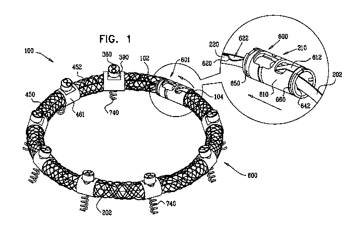
Reference is now made to Fig. 1, which is a schematic illustration of an
annuloplasty structure 100, e.g., at least one elongate segment or tubular
element,
comprising a plurality of compressible subunits 450 and a plurality of anchor
mounts 461,
in accordance with an embodiment of the present invention. Structure 100
comprises a
modular annuloplasty structure in which the plurality of compressible subunits
450 are
alternately disposed with respect to the plurality of anchor mounts 461.
Typically,
structure 100 comprises an implant shaped to define a tubular structure having
a cross-
section of any suitable shape, e.g., circular or elliptical. Compressible
subunits 450 are
shaped to define a hollow lumen and comprise a braided mesh 452 (e.g., wire or
polyester), by way of illustration and not limitation. For example,
compressible subunits
450 may comprise a plurality of coils, braided structures, stent-shaped
struts, or
accordion- or bellows-shaped structures. A ratchet mechanism 600 (described
hereinbelow with reference to Figs. 6A-B) is disposed within the hollow lumen
of
structure 100. Ratchet mechanism 600 comprises a ratchet body 202 having a
fixed end
210 and a dynamic end 220. Although ratchet mechanism 600 is shown as being
used in
combination with structure 100, it is to be noted that any of the ratchet
mechanisms
described herein may be used in combination with structure 100.
Typically compressible subunits 450 and anchor mounts 461 comprise a
biocompatible material, e.g., nitinol, ePTFE, PTFE, stainless steel, platinum
iridium,
titanium, or cobalt chrome. In some embodiments, compressible subunits 450 and
anchor
mounts 461 are coated with PTFE (Polytetrafluoroethylene). In some
embodiments,
compressible subunits 450 function as accordion- or bellows-shaped
compressible
structures which facilitate proper cinching of the annulus when structure 100
is
contracted. The configuration of the annulus of the mitral valve differs from
patient to
patient. Compressible subunits 450, when compressed, e.g., typically along a
longitudinal
axis of structure 100, enable respective portions of annuloplasty structure
160 to
independently conform to the configuration of each portion of the annulus that
is in
alignment with a given portion of the annuloplasty structure.
It is to be noted that for some applications, annuloplasty structure 100 is
shaped to
define a single tubular structure independently of the plurality of anchor
mounts 461. In
such an embodiment, the single tubular structure comprises an elongate sheath
of
33
CA 02728078 2010-12-14
WO 2010/004546 PCT/IL2009/000593
compressible material, as described hereinabove with respect to compressible
subunits
450.
A contracting wire (not shown) is disposed within the lumen of structure 100
generally alongside ratchet body 202. Typically, pulling on the contracting
wire controls
the structural configuration of ratchet body 202 which in turn controls the
structural
configuration of structure 100, as will be described hereinbelow. In response
to the
pulling of the wire, an inward radial force is applied to structure 100, and a
perimeter of
structure 100 is modulated, i.e., reduced.
The contracting wire comprises a flexible and/or superelastic material, e.g.,
nitinol,
polyester, PTFE, ePTFE, stainless steel, or cobalt chrome, and is configured
to reside
chronically within structure 100. In some embodiments, the contracting wire
comprises a
braided polyester suture (e.g., Ticron). In some embodiments, the contracting
wire is
coated with polytetrafluoroethylene (PTFE). In some embodiments, the
contracting wire
comprises a plurality of wires that are intertwined to form a rope structure.
Typically, structure 100 is shaped to provide at least one longitudinal lumen
for
passage therethrough of ratchet body 202 and the contracting wire. In some
embodiments, structure 100 is shaped to provide a first longitudinal lumen
passage
therethrough of the contracting wire and a second longitudinal lumen for
passage
therethrough of ratchet body 202.
Fixed end 210 is fixed within a substantially tubular ratchet-coupling housing
610,
while dynamic end 220 slides through housing 610 along a track 642 in the
direction as
indicated by the arrow. Ratchet body 202 is shaped to define a plurality of
first engaging
structures, e.g., first grooves 620, which are engageable by a tooth 612 of
housing 610.
As dynamic end 220 is slid away from fixed end 210 (i.e., in the direction as
indicated by
the arrow), grooves 620 are engaged by a second engaging structure, e.g.,
tooth 612,
thereby allowing ratchet body 202 to slide in only one direction, i.e., the
direction in
which dynamic end 220 is first fed through housing 610 and as indicated by the
arrow.
As dynamic end 220 advances beyond fixed end 210, dynamic end 220 slides
alongside
the portion of body 202 that is adjacent to fixed end 210.
Each anchor mount 461 is shaped to provide at least one longitudinal anchor
mount lumen having an axis that is parallel with the longitudinal axis of the
annuloplasty
structure. The anchor mount lumen facilitates passage therethrough of ratchet
body 202
34
CA 02728078 2010-12-14
WO 2010/004546 PCT/IL2009/000593
and the contracting wire. In some embodiments, each anchor mount 461 is shaped
to
provide a first longitudinal lumen passage therethrough of the contracting
wire and a
second longitudinal lumen for passage -therethrough of ratchet body 202.
Each anchor mount 461 is shaped to provide an anchor channel for passage
therethrough of a helical anchor 740. As will be described hereinbelow, the
channel is
shaped to define a lumen having a channel axis that is disposed at a non-zero
angle, e.g.,
transverse, with respect to a longitudinal axis of the longitudinal lumen of
the anchor
mount through which ratchet body 202 and the contracting wire pass. As such,
in
response to pulling of the contracting wire, the resultant sliding of portions
of the
contracting wire and of ratchet body 202 through the longitudinal lumen mount
461, does
not interfere with the anchor channel and anchor 740 disposed therein. The
angle of the
anchor channel with respect to the longitudinal lumen of anchor mount 461
facilitates
corkscrewing of the anchor into the annulus of the valve of the patient at an
angle as
defined by the intersecting axes of the anchor channel and the longitudinal
lumen of
mount 461, as described hereinbelow with reference to Fig. 8.
Typically, for embodiments in which annuloplasty structure 100 comprises a
plurality of anchor mounts 461, the respective angles defined by the
intersecting axes of
each anchor channel with the respective axis of the longitudinal lumen of each
mount 461
is identical for all mounts 461. Alternatively, a first portion of the
plurality of anchor
mounts 461 has an angle that differs from the angle of a second portion of the
plurality of
anchor mounts. For example, a portion of anchor mounts 461 designated to be
anchored
to the anterior portion of the annulus has an angle that is different from a
portion of
anchor mounts 461 designated to be anchored to the posterior portion of the
annulus.
Thus, the anchors may be anchored to different portions of the annulus at
different angles
in response to a need therefor.
It is to be noted that although helical anchors 740 are used in combination
with
structure 100, any anchor described herein may be used in combination with
structure
100.
For embodiments in which structure 100 is implanted during an open-heart or
minimally-invasive procedure, structure 100 is advanced toward the valve in a
closed
configuration (e.g., substantially ring-shaped or "D"-shaped), as shown. It is
to be noted
that structure 100 may be advanced toward the valve of the patient in a linear
CA 02728078 2010-12-14
WO 2010/004546 PCT/IL2009/000593
configuration during an open-heart or minimally-invasive valve repair
procedure. In such
an embodiment, once structure 100 is properly positioned within the left
atrium of the
heart, the contracting wire (not shown) is pulled and-first and second ends
102- and 104-of
annuloplasty structure 100 are drawn toward each other such that structure 100
assumes
its closed configuration.
For embodiments in which structure 100 is advanced during a percutaneous valve
repair procedure, structure 100 is manufactured having a first end 102 that is
typically
coupled to, e.g., welded to, housing 610 and a second end 104 that is not
coupled to
housing 610 during the advancing. Thus, structure 100, in such an embodiment,
is
advanced toward the left atrium of the patient in a generally linear
configuration thereof.
For embodiments in which structure 100 is advanced toward the valve in a
linear
configuration, second end 104 is coupled to an engaging structure configured
to engage
housing 610 as structure 100 is made to assume its closed configuration. In
some
embodiments, the engaging structure coupled to second end 104 comprises a tube
having
a diameter that is smaller than an inner diameter of housing 610 and is
configured to slide
within housing 610 as structure 100 is drawn into its closed configuration.
Housing 610 comprises first and second coupling sites 650 and 660, for
coupling
of first end 102 and second end 104 of structure 100, respectively, to housing
610.
It is to be noted that annuloplasty structure 100 may be used independently of
ratchet mechanism 600. For example, annuloplasty structure 100 may comprise
only the
contracting wire passing through the lumen of structure 100. In such an
embodiment,
once annuloplasty structure 100 is deployed from its linear state, the
respective ends of the
contracting wire are: (1) pulled such that the annuloplasty structure assumes
its closed
configuration, and (2) locked together in order to maintain the closed
configuration.
As described herein, structure 100 typically comprises a braided mesh in
embodiments in which sutures pass through structure 100 and facilitate
anchoring or
suturing of structure 100 to the annulus. For embodiments in which
annuloplasty
structure 100 is positioned using an open-heart procedure, the mesh
facilitates suturing of
structure 100 to the annulus of the patient. In such an embodiment, the
physician passes
the suture through the mesh at a first location thereof, through tissue of the
annulus, and
subsequently, through a second location of the mesh, thereby suturing
structure 100 to the
annulus. In some embodiments, the suturing is performed following placement of
the
36
CA 02728078 2010-12-14
WO 2010/004546 PCT/IL2009/000593
annuloplasty structure along the annulus. In some embodiments, a plurality of
sutures are
sutured to the annulus of the patient and the annuloplasty structure is slid
along the
sutures and-toward the annulus. - In such an -embodiment, respective ends of
each of the
plurality of sutures are threaded through the mesh prior to the sliding, and
are knotted
together and clipped following the sliding. The knotting of the sutures
maintains the
positioning of the annuloplasty structure along the annulus.
For some embodiments, the mesh facilitates anchoring of the annuloplasty
structure to the annulus of the patient. In such an embodiment, the physician
passes the
anchor through the mesh at a first location thereof and then through tissue of
the annulus.
It is to be understood that the braided mesh may be used independently of or
in
combination with the compressible subunits and/or with the anchor mounts. For
example,
the mesh may surround at least compressible subunits 450 of structure 100.
Alternatively,
the braided mesh may be used independently of compressible subunits 450 and/or
anchor
mounts 461. In such an embodiment, structure 100 may comprise only ratchet
mechanism 600 and/or the contracting wire surrounded by a sheath of braided
mesh.
Reference is now made to Fig. 2A, which is a schematic illustration of a flat-
ribbon ratchet mechanism 200, in accordance with an embodiment of the present
invention. Typically, ratchet mechanism 200 is used in combination with
annuloplasty
structure 100 as described hereinabove with reference to Fig. 1, in accordance
with an
embodiment of the present invention. It is to be noted that ratchet mechanism
200 may be
used in combination with any of the annuloplasty structures described herein.
Ratchet
mechanism 200 comprises a ratchet body 202 defining a flat ribbon having a
proximal
fixed end 210 and a distal dynamic end 220. Although Fig. 1 shows ratchet
mechanism
600 disposed within annuloplasty structure 100, it is to be noted that ratchet
mechanism
200 may be disposed within annuloplasty structure 100. Ratchet mechanism 200
is
disposed within the lumen of structure 100 such that fixed end 210 is disposed
within the
lumen of structure 100 in the vicinity of first end 102 thereof, and dynamic
end 220 is
disposed within the lumen of structure 100 in the vicinity of second end 104
thereof.
As described hereinabove, in some embodiments, structure 100 is advanced
toward the left atrium of the patient in a generally linear configuration.
Although ratchet
body 202 is shown in a linear configuration, it is to be noted that ratchet
body 202 is later
drawn into a closed configuration (e.g., substantially ring-shaped or "D"-
shaped
37
CA 02728078 2010-12-14
WO 2010/004546 PCT/IL2009/000593
configuration) simultaneously with structure 100 assuming its closed
configuration (e.g.,
substantially ring-shaped or "D"-shaped configuration). As the contracting
wire is pulled
and -first-and second ends 102 and 104 of - annuloplasty structure 100 are
drawn toward -
each other such that structure 100 assumes its closed configuration, dynamic
end 220 is
advanced past fixed end 210 such that ratchet body 202 assumes its closed
configuration
as well. As dynamic end 220 advances beyond fixed end 210, dynamic end 220 and
the
distal portion of body 202 are slid alongside fixed end 210 and the proximal
portion of
body 202. Dynamic end 220 and fixed end 210 are able to meet each other due to
the
sliding of ratchet body 200 along a track within the a respective lumen of
each anchor
mount 461 of structure 100, as will be described hereinbelow.
Ratchet body 202 is shaped to define a plurality, e.g., at least two as shown,
of
first engaging structures, e.g., first windows 204, in the vicinity of dynamic
end 220 and a
plurality of second windows 206 in the general vicinity of the middle of
ratchet body 202.
It is to be noted that the number of second windows 206 is shown by way of
illustration
and not limitation. Fixed end 210 is shaped to define a second engaging
structure, e.g., a
tooth 230, which projects angularly away from a longitudinal axis of ratchet
body 202 and
is configured to engage the first engaging structures, e.g., windows 204 and
206. Fixed
end 210 is shaped to define a slit 240 surrounding tooth 230. As ratchet
mechanism 200
is initially drawn into its closed configuration, dynamic end 220 slides
alongside tooth
230 and slit 240 of fixed end 210.
Ratchet body 202 provides a portion 222 disposed between first windows 204 and
second windows 206. Typically, portion 222 provides a smooth surface for
unobstructed
back and forth sliding of dynamic end 220 past fixed end 210 and enables the
physician to
adjust the size/perimeter of the annuloplasty structure before it is
positioned along the
annulus. Additionally, portion 222 enables the physician to adjust the
size/perimeter of
the ratchet mechanism 200 prior to being locked in place in response to the
engaging of
second windows 206 by tooth 230. Typically, portion 222 has a distance Di3
that is
between 30 mm and 70 mm, e.g., 50 mm.
For embodiments in which ratchet mechanism 200 is disposed within structure
100, ratchet mechanism 200 is typically disposed alongside the portion of
contracting wire
110 which is disposed within the lumen of structure 1,00. As, structure 100.
is pulled into.
its closed configuration in response to the pulling of contracting wire 110,
dynamic end
220 is pulled toward fixed end 210. Dynamic end 220 is passively advanced
alongside
38
CA 02728078 2010-12-14
WO 2010/004546 PCT/IL2009/000593
fixed end 210 due to the compression force applied by structure 100 in
response to the
pulling of contracting wire 110. That is, dynamic end 220 is not pulled by
contracting
wire _ 1-10, - rather it is- passively -pushed - in = response to the pulling
of wire - 110.
Additionally, wire 110 is aligned alongside an external surface of ratchet
body 202 and at
an external perimeter thereof. In response to pulling of contracting wire 110,
contracting
wire 110 pushes against the external surface of ratchet body 202 and applies a
compression force thereto. Responsively to the compression force of wire 110
on the
external surface of ratchet body 202, ratchet body 202 passively compresses.
Further
additional pulling of wire 110 reduces the perimeter of ratchet mechanism 200,
and
thereby of structure 100.
In response to continued pulling of contracting wire 110, structure 100
radially
contracts and, in turn, applies an additional compression force to ratchet
mechanism 200.
In response to the compression force to the ratchet mechanism by structure
100, ratchet
body 202 radially contracts as dynamic end 220 is passively slid further
distally away
from fixed end 210 thereby drawing second windows 206 closer toward tooth 230
of fixed
end 210. Dynamic end 220 is slid distally away from fixed end 210 until tooth
230
engages a first window 208 of second windows 206. Tooth 230 remains locked in
position with respect to first window 208 until an additional compression
force is applied
to ratchet body 202 in response to additional pulling of contracting wire 110.
This
additional force slides dynamic end 220 even further away from fixed end 210
until tooth
230 engages a second window 209 of second windows 206. Tooth 230 prevents
ratchet
body 202 from sliding in an opposite direction with respect to the direction
by which
dynamic end 220 is fed beyond fixed end 210. Thus, second windows 206 maintain
respective ratcheted perimeters of the now substantially ring-shaped or "D"-
shaped ratchet
body 202, and thereby maintain respective ratcheted perimeters of structure
100.
Alternatively, for some embodiments, dynamic end 220 is shaped to define one
or
more holes configured for looping of contracting wire 110 therethrough. In
such an
embodiment, dynamic end 220 is pulled in response to tensile force applied to
contracting
wire 110 as it is pulled. Additional force applied to wire 110 pulls ratchet
mechanism 200
into a closed configuration, e.g., a substantially ring-shaped configuration.
For embodiments in. which structure is advanced toward .the. left atrium. in.
its.
closed configuration, prior to the advancing, the physician forms structure
100 into a
closed configuration by advancing dynamic end 220 beyond fixed end 210 until
first
39
CA 02728078 2010-12-14
WO 2010/004546 PCT/IL2009/000593
windows 204 are in alignment with tooth 230 and ratchet body 202 locks in
place. At this
stage, structure 100 defines a generally ring-shaped structure having a
relatively large
perimeter. As described hereinabove, -once positioned 'along -the -annulus of
the patient,- -
the physician pulls wire 110 and dynamic end 220 slides and is pushed further
away from
fixed end 210 until second windows 206 lock and maintain a reduced perimeter
of ratchet
body 202, and thereby, structure 100.
It is to be noted that the plurality of second windows 206 are provided such
that
ratchet body 202, and thereby structure 100, can lock in place and maintain
respective
ratcheted perimeters thereof. Thus, the length of ratchet mechanism 200 in its
linear
configuration, the locking mechanism of ratchet mechanism 200, and
compressible
subunits 450 described hereinabove are provided so as to enable annuloplasty
structure
100 to accommodate various sizes of dilated annuli of given patients.
Additionally,
ratchet mechanism 200 facilitates: (1) positioning and anchoring structure 100
along the
dilated annulus while body 202 (and thereby structure 100) has a first
perimeter thereof,
(2) contracting the dilated annulus in response to the contracting of body 202
(and thereby
structure 100), and (3) maintaining the contracted state of the annulus while
body 202
(and thereby structure 100) has a second perimeter thereof that is typically
smaller than
the first perimeter.
It is to be further noted that ratchet mechanism 200 is described herein as
being
used in combination with structure 100 by way of illustration and not
limitation. For
example, ratchet mechanism 200 may be surrounded by a tubular sheath
comprising a
braided mesh, e.g., metal or fabric such as polyester. The braided mesh
facilitates passage
of sutures or longitudinal guide members through the sheath in order to anchor
or suture
the sheath to the annulus. In some embodiments, during expansion of the
sheath, by
pulling on opposite ends thereof, the braided mesh is longitudinally pulled
such that the
mesh decreases in diameter, i.e., the transverse cross-sectional diameter that
is
perpendicular with respect to the longitudinal axis of structure 100. During
contraction of
the sheath from its relaxed state, the mesh is compressed such that the
diameter of the
mesh closely resembles the diameter of the mesh in its relaxed state.
Fig. 2B shows ratchet mechanism 200 as described hereinabove with respect to
Fig. 2A, with the exception. that fixed end 2.10 is. shaped to define a
housing 250, in
accordance with an embodiment of the present invention. Typically, housing 250
of fixed
end 210 is shaped to define tooth 230 and slit 240 and is configured to
receive dynamic
CA 02728078 2010-12-14
WO 2010/004546 PCT/IL2009/000593
end 220 in a manner as described hereinabove with respect to Fig. 2A.
Typically, housing
250 is configured to provide stability to mechanism 200 during the aligning of
windows
204 and 206 with tooth-230 of fixed end 210.
During the initial contraction of structure 100, dynamic end 220 is fed into
housing 250. As described hereinabove, ratchet body 202 assumes a closed
configuration
as dynamic end 220 is initially locked in place when tooth 230 of housing 250
engages
first windows 204. A compression force is further applied to ratchet body 202
(e.g., a
radial force or a tensile force applied in response to pulling the contracting
wire, as
described hereinabove) which further advances dynamic end 220 away from
housing 250.
Fig. 3 shows a system 300 comprising ratchet body 202 passing through a first
one
of anchor mounts 461 of annuloplasty structure 100, in accordance with an
embodiment
of the present invention. Anchor mount 461 comprises a lateral-aperture anchor
mount
341 which comprises a substantially hollow, tubular element 463 configured for
passage
therethrough of ratchet body 202 and contracting wire 110. The anchor mount
shown is
configured to fix in place fixed end 210 of ratchet body 202. It is to be
noted that anchor
mount 341 may fix in place any of the ratchet bodies described herein.
Additionally,
anchor mount 341 is shaped to define an aperture 340 configured for passage
therethrough
of an anchor, as will be described hereinbelow. In some embodiment, a tubular
channel
(configuration shown hereinbelow with reference to Fig. 4) for passage of an
anchor is
coupled to, e.g., welded to, mount 341 along portions of mount 341 which
define aperture
340. As shown, aperture 340 is provided at a location along mount 461 such
that passage
of a tissue anchor therethrough (e.g., directly or indirectly through a
channel coupled to
portions of mount 341 defining aperture 340), does not interfere with
contracting wire 110
and/or ratchet body 202 disposed within the annuloplasty structure.
It is to be noted that only one anchor mount 341 is shown for clarity of
illustration.
For example, ratchet mechanism 200 may be coupled to a plurality of anchor
mounts 341
which are disposed at various sites with respect to ratchet body 202. It is to
be further
noted that a respective compressible subunit 450 may be coupled to either end
of anchor
mount 341. As shown, anchor mount 461 is shaped to define a first coupling
site 302 and
a second coupling site 304. For embodiments in which ratchet mechanism 300 is
used in
combination with. compressible subunits 450, as described hereinabove with
reference-to
Fig. 1, a respective compressible subunit 450 is coupled to coupling sites 302
and 304.
41
CA 02728078 2010-12-14
WO 2010/004546 PCT/IL2009/000593
Reference is now made to Fig. 4, which is a schematic illustration of system
300
comprising a tissue anchor 360 coupled to anchor mount 341, in accordance with
an
embodiment of the present invention. Anchor- mount 341 fixes in place fixed
end 210 of
ratchet body 202 as described herein. Ratchet body 202 of Fig. 3 is shown in
an open,
linear configuration thereof, i.e., dynamic end 220 is not aligned alongside
fixed end 210.
An anchor 360 is shown coupled to mount 461. In some embodiments, a tube-
channel
1200 (as described in more detail hereinbelow with reference to Fig. 11) is
coupled to
mount 461 portions of mount 341 defining aperture 340. In some embodiments,
channel
1200 is welded to mount 461 during the manufacturing of mount 341.
In some embodiments, tube-channel 1200 is not welded to mount 341 but rather
is
advanced toward mount 341 together with, e.g., surrounding, anchor 360. In
such an
embodiment, channel 1200 is free to rotate with respect to aperture 340 along
the
longitudinal axis of mount 341.
As shown, anchor 360 is shaped to define a helix having a pointed distal end
370
which punctures through tissue of the annulus of the heart. It is to be noted
that a helical
anchor is shown by way of illustration and not limitation, and that any
suitable anchor
may be used to anchor the annuloplasty structure to the annulus. For
embodiments in
which a helical anchor is used, tube-channel 1200 may comprise a bar, as
described in US
Provisional Patent Application 61/001,013, PCT Patent Application
PCT/IL07/001503,
and US Patent Application 11/950,930 to Gross et al., entitled, "Segmented
ring
placement". This bar is configured to restrict continued corkscrewing of
helical anchor
360 into the tissue of the annulus beyond a predetermined distance, e.g.,
between 3 mm
and 10 mm. Additionally, the bar functions as a nut providing a thread for the
helical
anchor to be advanced distally and corkscrewed around the bar and into the
tissue of the
annulus.
As shown, helical anchor 360 is coupled at a proximal end thereof (i.e., the
portion
of anchor 360 that is not configured to be advanced into the annulus tissue)
to a head
portion 380. Typically, a distal end of head portion 380 has a diameter that
is larger than
a diameter of tube-channel 1200. Once anchor 360 is advanced distally through
tube-
channel 1200, the distal portion of head portion 380 abuts a proximal portion
of tube-
channel -1200 and prevents continued distal. motion of anchor .360.. Even when
head
portion 380 abuts tube-channel 1200, anchor 360 is allowed to continue
rotational motion.
This continued rotational motion draws tissue of the annulus toward the
annuloplasty
42
CA 02728078 2010-12-14
WO 2010/004546 PCT/IL2009/000593
structure. In the event that a gap between the annulus tissue and the
annuloplasty
structure is created during the initial anchoring of the structure to the
annulus of the valve,
the continued rotation of anchor 360 minimizes and substantially eliminates
the gap-. , As
shown, head portion 380 is shaped to define one or more, e.g., two as shown,
engaging
elements, e.g., holes, 390. In some embodiments, engaging elements 390 are
configured
for coupling and/or passage therethrough of an actuation means by way of
illustration and
not limitation, and the anchoring means is configured to corkscrew the anchor
into the
tissue of the annulus.
It is to be noted that engaging elements 390 are shown as being circular by
way of
illustration and not limitation, and that elements 390 may be shaped to define
any suitable
shape, e.g., rectangles, ovals, etc.
Typically, head portion 380 prevents continued distal motion of anchor 360
into
the annulus with respect to the distal surface of the anchor mount, i.e., the
portion of the
mount designated to align with and contact the annulus. For embodiments in
which tube-
channel 1200 is advanced together with anchor 360, the tube-channel 1200
rotates within
aperture 340 along the longitudinal axis of mount 461 together with the
rotating of anchor
360.
Reference is now made to Figs. 5A-C, which are schematic illustrations of
system
300 as described hereinabove with reference to Fig. 4, with the exception that
anchor
mount 461 comprises a transverse-lumen anchor mount 342 comprising a tubular
element
465 shaped to define an anchor lumen 501 having an longitudinal axis 502
thereof, in
accordance with an embodiment of the present invention. Tubular element 465
fixes in
place fixed end 210 of ratchet body 202 as described hereinabove with
reference to Fig.
2A. Typically, anchor mount 461 provides at least one longitudinal anchor
mount lumen
having an axis that is parallel with the longitudinal axis of the annuloplasty
structure.
Anchor mount lumen facilitates passage therethrough of ratchet mechanism 200
and
contracting wire 110. Longitudinal axis 502 of anchor lumen 501 is at a non-
zero angle,
e.g., transverse, with respect to the longitudinal axis of the anchor mount
lumen of anchor
mount 461. Transverse lumen 501 is shaped to facilitate passage therethrough
of tube-
channel 1200, as described hereinabove with reference to Fig. 4. As shown,
transverse
lumen 501 does not interfere with ratchet body 202 and contracting wire 110..
.
43
CA 02728078 2010-12-14
WO 2010/004546 PCT/IL2009/000593
Reference is now made to Figs. 5A-B. Anchor mount 461 is coupled at either end
thereof to a respective stabilizing structure 310. Typically, since anchor
mount 461
comprises hollow tubular element 465, anchor-mount 461 has a tendency to pivot
laterally
with respect to ratchet body 202. Stabilizing structure 310 is shaped to
define mounts 312
which are configured to surround and lock in place a portion of anchor mount
461 and to
prevent swiveling thereof. Ratchet body 202 passes through aperture 330 of
stabilizing
structure 310 and through the longitudinal anchor mount lumen. Passing of
ratchet body
202 through structure 310 and then through mount 461 locks in place
stabilizing structure
310 which, in turn, locks in place anchor mount 461 and prevents it from
pivoting
laterally. Additionally, aperture 330 of stabilizing structure 310 provides a
suitable track
for advancement of ratchet body 202 along a defined path. For example, this
track
enables the proper positioning of dynamic end 220 with respect to fixed end
210.
Typically, aperture 330 has a major axis 331 and has a longitudinal axis 332
that is
transverse with respect to major axis 331. Major axis 331 of aperture 330 is
typically
disposed at a non-zero angle with respect to axis 502 of anchor lumen 501. A
portion of
ratchet body 202 passes through aperture 330 along longitudinal axis 332
thereof.
Typically, ratchet body 202 passes through aperture 330 of a first stabilizing
structure
310, through the lumen of anchor mount 461, and subsequently through aperture
330 of a
second stabilizing structure 310. Prior to the coupling of mount 461 to a pair
of structures
310, mount 461, and thereby lumen 501, is allowed to pivot laterally.
Following the
coupling of structures 310 to mount 461, structures 310 restrict the lateral
pivoting of
mount 461.
During the manufacture of structure 310, aperture 330 is created such that
major
axis 331 is disposed at a desired angle with respect to axis 502 of anchor
lumen 501 when
coupled to mount 461. A portion of ratchet body 202 is then passed through
mount 461
and subsequently through aperture 330, thereby fixing the angle of the major
axis of
aperture 330 with respect to axis 502 of anchor lumen 501. Typically, (a)
longitudinal
axis 332 of aperture 330 is substantially parallel with respect to a plane of
the annulus and
parallel with the longitudinal axis of the annuloplasty structure, and (b)
axis 502 of anchor
lumen 501 is at a non-zero angle with respect to major axis 331 of the
aperture 330. Thus,
the angle of anchor lumen 501 with respect to longitudinal axis 332
facilitates
corkscrewing of the tissue anchor into the annulus at an angle as defined by
the
44
CA 02728078 2010-12-14
WO 2010/004546 PCT/IL2009/000593
intersecting axes 502 of lumen 501 and major axis 331 of aperture 330 (shown
in Fig.
5C).
For embodiments in which system 300 comprises a plurality of anchor mounts
461, the respective pairs of structures 310 coupled on either end of each
mount 461 may
be manufactured differently. For example, (1) a first pair of structures 310
may be shaped
to define apertures 330 having a major axis at a first desired angle with
respect to axis 502
of anchor lumen 501 of a first anchor mount 461, and (2) a second pair of
structures 310
may be shaped to define apertures 330 having a major axis at a second desired
angle with
respect to the longitudinal axis of anchor lumen 501 of a second anchor mount
461. Thus,
the respective anchors configured to be passed through each of the first and
second anchor
mounts are anchored to the tissue at the desired first and second angles,
respectively. In
some embodiments, the anchors which pass through the anchor mounts positioned
along
the annulus in alignment with the base of the posterolateral leaflet may be
anchored at an
angle that is different from an angle at which the anchors which pass through
the anchor
mounts positioned along the annulus in alignment with the base of the
anteromedial leaflet
are anchored.
Fig. 5C shows a perspective view of system 300 from an opposite view than that
shown in Fig. 5A. Ratchet body 202 passes unobstructed alongside anchor lumen
501 of
anchor mount 461. As described hereinabove, anchor mount 461 may also function
as a
housing for fixed end 210 of ratchet body 202. Anchor mount 461 is shaped to
define a
slit 520 which engages and fixes in place a portion 212 of fixed end 210.
Typically,
portion 212 projects away perpendicularly from a longitudinal axis of ratchet
body 202.
Reference is now made to Figs. 3 and 5B-C. Anchor mount 461 is flanked by
stabilizing structures 310. Fig. 5B shows a stabilizing unit 500 having a
stabilizing
structure 310 is shaped to define: (1) a hole 320 configured for passage
therethrough of
contracting wire 110, and (2) a longitudinal aperture 330 configured for
passage
therethrough of ratchet body 202, in accordance with an embodiment of the
present
invention. Typically, aperture 330 has a width L7 of between 0.3 mm and 0.8
mm. Such
a width facilitates passage therethrough of at least a portion of ratchet body
202. For
embodiments in which a first portion of body 202 is slid alongside a second
portion of
body 202 (e.g., dynamic end 220 slides alongside- fixed end 210), width L7
accommodates.
for the widths of both the first and second portions of ratchet body 202 and
facilitates
passage therethrough of both portions.
CA 02728078 2010-12-14
WO 2010/004546 PCT/IL2009/000593
Fig. 3 shows ratchet body 202 in a closed configuration thereof. It-is to be
noted
that ratchet body 202 assumes a substantially circular configuration thereof
and that only a
portion of ratchet body 202 is shown. Typically, dynamic end 220 is passively -
fed
through aperture 330 alongside fixed end 210. As such, a portion of body 202
distal to
fixed end 210 aligns alongside a portion proximal to dynamic end 220, as shown
in Fig. 3.
Thus, width L7 of aperture 330 accommodates for the widths of: (1) the portion
of body
202 distal to fixed end 210, and (2) the portion of body 202 proximal to
dynamic end 220.
Reference is now made to Figs. 6A-B which are schematic illustrations of a
ratchet
mechanism 600, in accordance with an embodiment of the present invention.
Ratchet
body 202 is shaped to define dynamic distal end 220 and fixed proximal end
210. As
shown, ratchet body 202 is shaped to define a plurality of first engaging
structures, e.g.,
grooves 622, configured to be engaged by a second engaging structure, a tooth
612, at
fixed end 210. Fixed end 210 is coupled to a substantially tubular ratchet-
coupling
housing 610 which is shaped to define a first coupling site 650 and a second
coupling site
660. For embodiments in which ratchet mechanism 600 is used in combination
with
compressible subunits 450 as described hereinabove with reference to Fig. 1, a
respective
compressible subunit 450 is coupled to coupling sites 650 and 660.
As described hereinabove with reference to Fig. 1, ratchet mechanism 600 is
disposed within the lumen of structure 100 such that fixed end 210 is disposed
within the
lumen of structure 100 in the vicinity of first end 102 thereof and dynamic
end 220 is
disposed within the lumen of structure 100 in the vicinity of second end 104
thereof.
Although ratchet body 202 is shown in a linear configuration, it is to be
noted that ratchet
body 202 is drawn into its closed configuration simultaneously with structure
100
assuming its closed configuration. As contracting wire 110 is pulled and first
and second
ends 102 and 104 of annuloplasty structure 100 are drawn toward each other
such that
structure 100 assumes its closed configuration, dynamic end 220 is fed into
housing 610
and is advanced past fixed end 210 such that ratchet body 202 assumes its
closed
configuration as well. As dynamic end 220 advances beyond fixed end 210,
dynamic end
220 and the portion of body 202 that is proximal to end 220 are slid alongside
fixed end
210 and the portion of body 202 that is distal to fixed end 210. As shown,
housing 610 is
coupled to an insert 640 that is shaped to define a longitudinal track 642. As
dynamic end
220 is fed into housing 610 of fixed end 210, dynamic end slides along track
642. Thus,
46
CA 02728078 2010-12-14
WO 2010/004546 PCT/IL2009/000593
dynamic end 220 and fixed end 210 are able to meet each other due to the
sliding dynamic
end 220 along track 642 within the lumen housing 610.
Ratchet body 202 is shaped to define a plurality, e.g., at least two as shown,
of
first grooves 620 in the vicinity of dynamic end 220 and a plurality of second
grooves 630
in the general vicinity of the middle of ratchet body 202. It is to be noted
that the
respective numbers of first grooves 620 and second grooves 630 are shown by
way of
illustration and not limitation. As ratchet mechanism 600 is initially drawn
into its closed
configuration, dynamic end 220 slides alongside track 642 and tooth 612
engages
respective grooves 622 of ratchet body 202.
Ratchet body 202 provides a portion 222 disposed between first grooves 620 and
second grooves 630. Typically, portion 222 provides a smooth surface for
unobstructed
back and forth sliding through fixed end 210 and enables the physician to
adjust the
size/perimeter of the annuloplasty structure before it is positioned along the
annulus.
Additionally, portion 222 enables the physician to adjust the size/perimeter
of ratchet
mechanism 600 prior to the locking of second grooves 630 by tooth 612.
Typically,
portion 222 has a distance that is between 30 mm and 70 mm, e.g., 50 mm.
It is to be noted that ratchet mechanism 600 may be anchored to the annulus
independently of annuloplasty structure 100 described hereinabove with
reference to Fig.
1 and with reference to ratchet mechanism 200 described hereinabove with
reference to
Figs. 2A-B. Alternatively, for embodiments in which ratchet mechanism 600 is
disposed
within structure 100, ratchet mechanism 600 is typically disposed alongside
the portion of
contracting wire 110 which is disposed within the lumen of structure 100. As
structure
100 is pulled into its closed configuration in response to the pulling of
contracting wire
I10, dynamic end 220 is pulled toward fixed end 210. Dynamic end 220 is
passively
advanced within housing 610, typically alongside fixed end 210, due to the
compression
force applied by structure 100 in response to the pulling of contracting wire
110.
In response to continued pulling of contracting wire 110, structure 100
radially
contracts and, in turn, applies an additional compression force to ratchet
mechanism 600.
As described hereinabove, in response to the compression force, ratchet body
202 radially
contracts as dynamic end 220 is passively slid further distally away from
fixed end 210
thereby drawing second grooves 630 closer toward tooth 612 of housing 610.
Dynamic
end 220 is slid distally away from fixed end 210 until tooth 612 engages a
first groove
47
CA 02728078 2010-12-14
WO 2010/004546 PCT/IL2009/000593
624 of second grooves 630. Tooth 612 remains locked in position with respect
to first
groove 624 until an additional compression force of structure 100 is applied
to ratchet
body 202 (i.e., in response to the pulling of contracting wire 1 10). This
additional force
slides dynamic end 220 even further away from fixed end 210 until tooth 612
engages a
second groove 626 of second grooves 630. Tooth 612 prevents body 202 of
mechanism
600 from sliding in an opposite direction with respect to the direction by
which dynamic
end 220 is fed beyond fixed end 210. Thus, second grooves 630 maintain
respective
ratcheted perimeters of the now closed ratchet body 202, and thereby maintain
respective
ratcheted perimeters of structure 100.
For embodiments in which structure is advanced toward the left atrium in its
closed configuration (e.g., during an open-heart procedure or during a
minimally-invasive
procedure), dynamic end 220 is advanced past fixed end 210 until first grooves
620 are in
alignment with tooth 612 and ratchet body 202 is locked in an expanded
configuration
thereof and has a relatively large perimeter. As described hereinabove, once
positioned
along the annulus of the patient, the dynamic end 220 is pushed further
distally away (i.e.,
in the direction as indicated by the arrow in Fig. 6B) from fixed end 210
until locking
groves 630 lock and fix a perimeter of body 202, and thereby, fix a perimeter
of structure
100.
It is to be noted that the plurality of second grooves 630 is provided such
that
ratchet body 202, and thereby structure 100, can lock in place and maintain
respective
ratcheted perimeters thereof. Thus, the length of ratchet mechanism 600 in its
linear
configuration, the locking mechanism of ratchet mechanism 600, and
compressible
subunits 450 described hereinabove are provided so as to enable annuloplasty
structure
100 to accommodate various sizes of dilated annuli of given patients.
Additionally,
ratchet mechanism 600 facilitates: (1) positioning and anchoring structure 100
along the
dilated annulus while body 202 (and thereby structure 100) has a first
perimeter thereof,
(2) contracting the dilated annulus in response to the contracting of body 202
(and thereby
structure 100), and (3) maintaining the contracted state of the annulus while
body 202
(and thereby structure 100) has a second perimeter thereof that is typically
smaller than
the first perimeter.
It is to be further noted that ratchet mechanism. 600 is described as being
used in
combination with structure 100 by way of illustration and not limitation. For
example,
48
CA 02728078 2010-12-14
WO 2010/004546 PCT/IL2009/000593
ratchet mechanism 600 may be surrounded by a tubular sheath comprising a
braided
mesh, e.g., metal or fabric such as polyester.
Fig. 6B shows dynamic end 220 having already passed through housing 610 of
fixed end 210. As such, ratchet body 202 assumes a closed configuration
(partially shown
for clarity of illustration). As shown, dynamic end 220 is shaped to define
one or more
holes 613 configured for looping of the contracting wire therethrough. In such
an
embodiment, dynamic end 220 is pushed in response to tensile force applied to
the
contracting wire as it is pulled. As described hereinabove, additional force
applied to the
contracting wire pushes ratchet mechanism 200 into a closed configuration,
e.g., a
substantially ring-shaped configuration. Further additional pulling of the
contracting wire
reduces the perimeter of ratchet mechanism 600, and thereby of the
annuloplasty
structure.
Fig. 7 shows ratchet mechanism 600 as described hereinabove with reference to
Figs. 6A-B, with the exception that housing 610 provides a tooth 712 is shaped
to define a
window 714, in accordance with an embodiment of the present invention. Tooth
712 is
coupled to housing 610 along a junction and bends along the junction. As tooth
712
engages groove 620 of ratchet body 202, window 714 surrounds a portion 772 of
an upper
surface 770 of ratchet body 202 which defines groove 620. Window 714 thus
enables
tooth 712 to advance distally and bend as far as possible within groove 620
without being
obstructed by portion 772 of upper surface 770 which defines groove 620. Tooth
712
engages groove 620 and locks ratchet body 202 in place until an additional
inward, radial
pushing force is applied thereto, e.g., typically, in response to the pulling
of contracting
wire 110 described herein. In response to the additional inward, radial force
applied to
ratchet body 202, (a) dynamic end 220 is slid further away from housing 610 in
the same
direction in which dynamic end 220 was initially fed into housing 610 (i.e.,
the direction
as indicated by the arrow), and (b) tooth 712 slides along upper surface 770
of ratchet
body 202 until tooth 712 engages another groove 620 of ratchet body 202.
Dynamic end 220 is shaped to define one or more holes 613 configured for
looping of the contracting wire therethrough. In such an embodiment, dynamic
end 220 is
pulled in response to tensile force applied to the contracting wire as it is
pulled.
Additional force applied. to the contracting wire pulls ratchet -mechanism 600
into the
closed configuration. Further additional pulling of the contracting wire
reduces the
perimeter of ratchet mechanism 600, and thereby of the annuloplasty structure.
49
CA 02728078 2010-12-14
WO 2010/004546 PCT/IL2009/000593
It is to be noted that ratchet body 202 may be pulled by contracting wire 110
in
some embodiments. Ratchet body 202 is typically pushed in response to the
radial,
compressing force applied to body 202 by the annuloplasty structure in
response to the
pulling of contracting wire 110.
Reference is now made to Figs. 6A-B and 7. Fixed end 210 of ratchet body 202
is
shaped to define a protrusion 722 (not shown in Figs. 6A-B). Housing 610 is
shaped to
define a slit (not shown for clarity of illustration) for passage therethrough
of protrusion
722 in order to fix fixed end 210 in place with respect to housing 610.
Fig. 8 shows an anchor mount system 900 comprising an anchor mount 461
comprising a double-lumen anchor mount 343 that is shaped to define a channel
460 and a
lumen 920, or channel, in accordance with an embodiment of the present
invention.
Anchor mount 461 is shaped to define a lateral wall 467 having a first portion
464 and a
second portion 466 generally at opposite sites of mount 461 when viewed in
cross-section
(e.g., at 12 o'clock and 6 o'clock). Typically, first portion 464 is shaped to
define an
opening thereof, and second portion 466 is shaped to define an opening
thereof. Channel
460 extends from the opening of first portion 464, through the anchor mount,
to the
opening in second portion 466. As described hereinabove with reference to Fig.
1, anchor
mount 461 is configured for facilitating passage therethrough any anchor
described herein
in order to facilitate anchoring of an annuloplasty structure (e.g., any
annuloplasty
structure comprising mount system 900) to the annulus of the patient. Channel
460 has a
diameter between about 0.8 mm and 2.5 mm, e.g., 1.8 mm, that is sized to
facilitate
passage therethrough of any one of the anchors, anchoring structures, or
anchoring
systems described herein. Typically, the anchors described herein are
configured for
passage through channel 460 have a diameter of between about 0.5 mm and 2.4
mm, e.g.,
1.6 mm.
First portion 464 of lateral wall 467 of mount 461 is shaped to define a
tapered
opening 950 above channel 460. Opening 950 has a diameter that is typically
larger than
a diameter D2 of channel 460. Typically, during the anchoring of the
annuloplasty
structure to the annulus, an anchor is coupled to an advancement structure,
e.g., a tube or a
rod, at a distal end thereof and is advanced via the advancement structure
toward channel
460. In some embodiments, a portion of the distal end of the advancement
structure has a
diameter that is slightly larger than the proximal end of channel 460, i.e.,
opening 950 of
anchor mount 461. Thus, the advancement of the advancement structure is
restricted from
CA 02728078 2010-12-14
WO 2010/004546 PCT/IL2009/000593
passage through channel 460 beyond the portion of the distal end of the tube
that has a
diameter larger than the diameter of channel 460. This restriction helps
ensure that the
anchor is not advanced too deeply within tissue of the annulus:
In some embodiments, a proximal portion (e.g., the portion of the anchor that
is
coupled to the distal end of the advancement structure) of the anchor is
configured to
expand. In such an embodiment, the proximal portion of the anchor is
compressed within
an overtube during the advancement of the anchor toward the annulus of the
valve. Once
the anchor is positioned properly within channel 460 and is initially anchored
to the
annulus of the valve, the overtube is slid proximally from the proximal end of
the anchor
and the proximal portion is allowed to expand. In such an embodiment, the
expanded
portion of the anchor has a diameter that is (a) larger than diameter D2 of
channel 460 and
(b) smaller than the diameter at the distal end of opening 950. Thus, the
expanded,
proximal portion of the anchor rests within the proximal end of opening 950
and functions
as a cap which restricts further distal advancement of the anchor into the
tissue of the
annulus.
Anchor mount 461 is shaped to provide an anchor mount and ratchet body lumen
920 for passage of ratchet body 202 of any of the ratchet mechanisms described
herein.
Ratchet body lumen 920 has (a) a longitudinal axis 942 that is substantially
parallel with
respect to the plane of the annulus and parallel with the longitudinal axis of
the
annuloplasty structure, and (b) an axis 940 that is typically at a non-zero
angle, e.g.,
transverse, with respect to longitudinal axis 942. Channel 460 has a first
axis 930 is
typically at a non-zero angle, e.g., transverse, with respect to longitudinal
axis 942.
Typically, lumen 920 is disposed with respect to channel 460 such that axis
940 of lumen
920 is disposed at an angle theta, with respect to axis 930 of channel 460.
Typically, the
anchor is anchored at angle theta with respect to axes 940 and 920 and the
plane of the
annulus of the valve. It is to be noted angle theta may range between 10
degrees and 70
degrees, typically 30 degrees.
Typically, for embodiments in which the annuloplasty structure comprises a
plurality of anchor mount systems 900, angle theta is identical for all mounts
461.
Alternatively, a first portion of the plurality of anchor mount systems 900
has an angle
theta that differs from the angle theta of a. second portion .of the plurality
of anchor mount.
systems 900. For example, a portion of anchor mount systems 900 designated to
be
anchored to the anterior portion of the annulus has an angle theta that is
different from a
51
CA 02728078 2010-12-14
WO 2010/004546 PCT/IL2009/000593
portion of anchor mount systems 900 designated to be anchored to the posterior
portion of
the annulus. Thus, the anchors may be anchored to different portions of the
annulus at
different angles in response to a need therefor.
In some embodiments, the contracting wire described herein passes through
lumen
920 alongside ratchet body 202. In some embodiments, mount 461 of system 900
is
shaped to provide an additional distinct lumen configured for passage
therethrough of the
contracting wire (configuration not shown).
Anchor mount 461 comprises first and second coupling sites 960 and 970
configured for coupling, e.g., wrapping therearound or welding, respective
ends of one or
more compressible subunits 450 as described hereinabove.
Fig. 9 shows an anchor mount system 1000 comprising an anchor mount 461
having a curved lateral surface 1100 that is coupled to an anchor channel 350
for passage
of an anchor therethrough, in accordance -with an embodiment of the present
invention.
Anchor mount 461 is configured for use in combination with any of the
annuloplasty
structures described herein. Mount 461 and is shaped to define a first lumen
1010
configured for passage therethrough of the contracting wire and a second lumen
1020 for
passage therethrough of the ratchet body of any one of the ratchet mechanisms
described
herein. Lumens 1010 and 1020 facilitate unobstructed passage of the
contracting wire and
the ratchet body, respectively, with respect to the passage of an anchor
through channel
350.
As described hereinabove with respect to Fig. 8, lumen 1020 has a first axis
1022
and channel 350 has a second axis 1030 which is disposed at an angle theta
(e.g., between
10 degrees and 70 degrees, typically 30 degrees) with respect to first axis
1022. As such,
the anchor passed through channel 350 is anchored to the annulus at -angle
theta with
respect to the ratchet body disposed within lumen 1020.
Anchor mount 461 comprises first and second coupling sites 1110 and 1112
configured for coupling, e.g., wrapping therearound or welding, respective
ends of one or
more compressible subunits 450 as described hereinabove.
Fig. 10 shows an anchor mount system 1111 comprising an anchor mount 461
comprising lateral-aperture anchor mount 341 which is shaped to define an
aperture 340
configured for passage therethrough of an anchor, as described hereinabove
with
reference to Fig. 3, in accordance with an embodiment of the present
invention. In some
52
CA 02728078 2010-12-14
WO 2010/004546 PCT/IL2009/000593
embodiments, the anchor is slid through aperture 340 and rests against
portions 1142 of
mount 461 which define aperture 340. Typically, portions 1142 provide
horizontal
surfaces 1 140 which function as- shelves impeding continued distal motion of
an anchor
configured to be advanced through aperture 340. In some embodiment, a channel
for
passage of the anchor is welded to mount 461 along portions 1142 of mount 461.
In some
embodiments, the channel is advanced toward mount 461 together with the
anchor. In
such an embodiment, the channel is free to rotate with respect to aperture 340
along the
longitudinal axis of mount 461.
Anchor mount 461 comprises a substantially tubular element 463 which defines a
longitudinal anchor mount lumen. Aperture 340 is created at a location of
mount 461
such that passage of an anchor via aperture 340, directly or indirectly, does
not interfere
with the contracting wire and/or ratchet body disposed within the longitudinal
lumen of
mount 461.
Reference is now made to Figs. 5C and 10. Anchor mount 461 also functions as a
housing for fixed end 210 of ratchet body 202. Anchor mount 461 is shaped to
define slit
520 which engages and locks portion 212 of fixed end 210.
Anchor mount 461 comprises first and second coupling sites 112 and 114
configured for coupling, e.g., wrapping therearound or welding, respective
ends of one or
more compressible subunits 450.
Reference is now made to Fig. 11, which is a schematic illustration an anchor
tube-channel 1200 configured to be used in combination with any one of anchor
mounts
461 described herein, in accordance with an embodiment of the present
invention. In
some embodiments, anchor channel 1200 is configured to be advanced through
lumen 501
of anchor mount 461 shown in Figs. 5A and 5C. In some embodiments, channel
1200 is
welded to anchor mount 461, shown in Figs. 3, 4, and 10, via aperture 340. In
some
embodiments, during the manufacture of mount 461, channel 1200 is welded via
surface
1100 to anchor mount 461, shown in Fig. 9, in place of channel 350.
Channel 1200 has (a) a proximal end 1250 which provides a passageway for
passage of an anchor through a channel 1210 of channel 1200, and (b) a distal
end 1260
which typically rests against the annulus of the valve when the annuloplasty
structure is
positioned along the annulus. Proximal end 1250 of channel 1200 is shaped to
define an
external ring 1220 having a diameter larger than the diameter of proximal end
1250 of
53
CA 02728078 2010-12-14
WO 2010/004546 PCT/IL2009/000593
channel 1200. For embodiments in which channel 1200 is configured to be
advanced
distally through lumen 501 of anchor mount 461 shown in Figs. 5A and 5C, ring
1220
functions - to -impede continued distal-motion of channel-,1200- beyond, a
predetermined
depth, as limited by ring 1220 abutting a proximal opening of channel 501 of
anchor
mount 461. In such an embodiment, channel 1200 is free to rotate with respect
to aperture
340 along the longitudinal axis of mount 461.
Channel 1200 is shaped to define one or more (e.g., two, as shown) lateral
slits
1230 and 1240. In some embodiments, a longitudinal bar (not shown) is
configured to be
welded between slits 1230 and 1240. Slits 1230 and 1240 enable the bar to be
welded to
channel 1200 in any given configuration, e.g., substantially perpendicularly
to or
diagonally with respect to slits 1230 and 1240, and at any angle with respect
to slits 1230
and 1240. For embodiments in which the bar is welded diagonally with respect
to slits
1230 and 1240, a first end of the bar may be coupled to a portion of channel
1200 defining
proximal end 1231 of slit 1230 while a second end of the bar is coupled to a
portion of
channel 1200 defining distal end 1242 of slit 1240, by way of illustration and
not
limitation. For example, in some embodiments, the first end of the bar may be
coupled to
proximal end 1231 of slit 1230 while the second end of the bar is coupled to a
portion
defining slit 1240 that is between proximal end 1241 and distal end 1242
thereof. For
embodiments in which the bar is welded substantially perpendicularly with
respect to slits
1230 and 1240, the first and second ends of the bar may be coupled to: (1)
proximal end
1231 of slit 1230 and proximal end 1241 of slit 1240, respectively, (2) distal
end 1232 of
slit 1230 and distal end 1242 of slit 1240, respectively, or (3) parallel
portions of slits
1230 and 1240 that are between the respective distal and proximal ends of
slits 1230 and
1240.
Typically, the bar provides a reference force to help corkscrew the anchor
into
tissue of the annulus during the initial corkscrewing thereof. Even when the
bar restricts
further distal motion of the anchor beyond a predetermined distance (e.g., a
predetermined
distance from that lateral surface of mount 461 which rests against tissue of
the annulus),
the anchor is allowed to resume rotational motion together with rotational
motion of
channel 1200 for embodiments in which channel 1200 is not welded to anchor
mount 461.
In the event that a gap is created between the annulus tissue and the
annuloplasty structure
during the initial anchoring of the structure to the annulus of the valve,
this continued
rotational motion draws tissue of the annulus toward the annuloplasty
structure. Such
54
CA 02728078 2010-12-14
WO 2010/004546 PCT/IL2009/000593
proximal drawing of the tissue thereby minimizes and substantially eliminates
the gap.
Techniques for use with a helical anchor and the bar as described herein may
be used in
combination- with- techniques described in US Provisional Application-
61/001,013- to
Gross et al., entitled, "Segmented ring placement," filed October 29, 2007,
which is
incorporated herein by reference.
Fig. 12 is a schematic illustration of an anchoring structure 1800 comprising
a
tapered, conical helical element 1802 comprising a plurality of coils 1810, in
accordance
with an embodiment of the present invention. Typically, the plurality of coils
1810
comprises a pointed distal end 1820 which punctures tissue of the annulus and
allows for
coils 1810 to be corkscrewed distally into the tissue of the annulus. A
proximal surface of
element 1802 is coupled to a head portion 1830 comprising raised surfaces 1832
having a
defined conformation. In some embodiments, head portion 1830 functions to
prevent
distal screwing of structure 1800 into the annulus of the patient beyond a
predetermined
depth as defined by the combined length of head portion 1830 and coils 1810.
Although
structure 1800 is not able to be advanced further distally, continued rotation
of structure
1800 draws tissue proximally with respect to the annuloplasty structure,
thereby
substantially minimizing or eliminating a gap that may be created between the
annuloplasty structure and the tissue of the annulus.
Typically, an anchor advancement structure, e.g., a tube or a rod, (not shown)
is
coupled at a distal end thereof to structure 1800 via raised surfaces 1832. In
such an
embodiment, the distal end of the advancement device is shaped to define
recessed
portions which are similar in shape to the define conformation of raised
surfaces 1832.
The advancement device is coupled to structure 1800 when the recessed portions
of the
device accommodate the conformation of raised surfaces 1832 by surrounding and
locking in place surfaces 1832 with respect to the recessed portions of the
advancement
device. The advancement device is locked together with structure 1800 when a
rotational
force is applied to the advancement force in a rotational direction as
indicated by the
arrow. Once the advancement device facilitates the anchoring of structure 1800
to the
annulus of the patient, a rotational force is applied to the anchor
advancement structure in
a direction opposite to the direction indicated by the arrow which detaches
the
advancement device from structure 1800 by sliding the recessed portions of the
advancement device away from raised structures 1832.
CA 02728078 2010-12-14
WO 2010/004546 PCT/IL2009/000593
For embodiments in which structure 1800 is used to percutaneously anchor an
annuloplasty structure to the annulus, the anchor advancement structure
comprises an
advancement structure,- e.g., a tube or a rod, which -is typically coupled to
head-portion
1830 prior to being transcatheterally advanced toward the annuloplasty
structure. For
embodiments in which anchor structure 1800 is used to anchor the annuloplasty
structure
to the annulus during an open-heart procedure, an external anchoring device
(e.g., an
advancement tube, an advancement rod, or a screw-driving system) is used in
order to
facilitate anchoring of structure 1800 to the annulus.
In either embodiment, once the anchor advancement structure advances the
anchor
toward the annuloplasty structure,.the anchor advancement structure is rotated
in order to
facilitate corkscrewing of anchoring structure 1800 into the annulus of the
patient. For
embodiments in which the compressible subunits of the annuloplasty structure
comprise a
braided mesh, as described hereinabove, structure 1800 may ' be advanced
through the
mesh and anchor the annuloplasty structure to the annulus via the mesh. For
embodiments in which the compressible subunits of the annuloplasty structure
comprise a
coiled structure, coils 1810 of structure 1800 are coiled around a portion of
coils of the
coiled compressible subunits of the annuloplasty structure and subsequently
through the
tissue of the annulus of the patient. During the coiling of coils 1810 of
structure 1800
around the portion of coils of the coiled compressible subunits of the
annuloplasty
structure, a longitudinal axis 1801 of structure 1800 is at a non-zero angle,
e.g.,
perpendicular, with respect to a longitudinal axis of the annuloplasty
structure. Such
intercoiling of coils 1810 with the coils of the coiled compressible subunits
of the
annuloplasty structure facilitates the coupling of the annuloplasty structure
with anchoring
structure 1800 during the corkscrewing of structure 1800 into the tissue of
the annulus.
For embodiments in which the annuloplasty structure comprises at least one
anchor mount, as described hereinabove, structure 1800 is advanced through the
anchor
mount and into the annulus of the patient.
Reference is now made to Figs. 5A, 5C, and 12. Typically, head portion 1830
has
a diameter that is larger than the inner diameter of lumen 501 of anchor mount
461. As
anchoring structure 1800 is advanced through lumen 501, a distal surface of
head portion
1830 abuts a proximal opening. of lumen 501. and inhibits continued distal
motion of
structure 1800 through the tissue of the annulus beyond the predetermined
depth.
56
CA 02728078 2010-12-14
WO 2010/004546 PCT/IL2009/000593
Reference is now made to Figs. 8 and 12. Typically, the diameter of head
portion
1830 is larger than diameter D2 of channel 460 defined by anchor mount 461. As
structure 1800 is advanced through channel 460, the distal surface of head
portion 1830
abuts proximal opening 950 and inhibits continued distal motion of structure
1800
through the tissue of the annulus beyond the predetermined depth.
Reference is now made to Figs. 9 and 12. Typically, the diameter of head
portion
1830 is larger than the inner diameter of channel 350 coupled to anchor mount
461. As
structure 1800 is advanced through channel 350, the distal surface of head
portion 1830
abuts a proximal opening of channel 350 and inhibits continued distal motion
of coils
1810 through the tissue of the annulus beyond the predetermined distance.
Reference is now made to Figs. 10 and 12. As structure 1800 is advanced
through
channel 350, the distal surface of head portion 1830 abuts horizontal surfaces
1140
defining aperture 340 and inhibits continued distal motion of coils 1810
through the tissue
of the annulus beyond the predetermined distance.
Reference is now made to Figs. 11 and 12. As structure 1800 is advanced
through
channel 1210 of channel 1200, the distal surface of head portion 1830 abuts
proximal end
1250 of channel 1200 and inhibits continued distal motion of coils 1810
through the tissue
of the annulus.
Reference is again made to Fig. 12. The proximal coil of helical element 1802
has
a diameter that is larger than the diameter of the distal coil of element
1802. The
diameters of the coils of helical element 1802 are gradually reduced in each
successive
coil from the proximal coil to the distal coil. The distal coil is corkscrewed
into the tissue
of the annulus following the puncturing of the annulus by pointed distal end
1820. As the
distal coil is corkscrewed distally through the tissue of the annulus, the
distal coil pushes
against the surrounding tissue, thereby exerting a radial force against
surrounding tissue of
the annulus. Each successive proximal coil of helical element 1802 enters an
opening
defined by the distal coil adjacent thereto. The diameter of the opening is
smaller than the
diameter of the successive proximal coil. Thus, each successive proximal coil
of exerts an
outward, radial force on surrounding tissue corresponding to the diameter of
successive
proximal coil. Thus, the proximal coil exerts a greater force on the
surrounding tissue
than does the distal coil. It is to be noted that the ratio between the
diameter of the
57
CA 02728078 2010-12-14
WO 2010/004546 PCT/IL2009/000593
proximal coil to the diameter of the distal coil is shown by way of
illustration and not
limitation. For example, the ratio may be smaller than the ratio that appears
in Fig. 12.
In some embodiments, the proximal coil of helical element 1802 has a diameter
that is smaller than the diameter of the distal coil of element 1802
(configuration not
shown). The diameters of the coils of helical element 1802 are gradually
increased in
each successive coil from the proximal coil to the distal coil. The distal
coil is
corkscrewed into the tissue of the annulus following the puncturing of the
annulus by
pointed distal end 1820. As the distal coil is corkscrewed distally through
the tissue of the
annulus, the distal coil pushes against the surrounding tissue, thereby
exerting a radial
force against surrounding tissue of the annulus. Each successive proximal coil
of the
helical element enters an opening defined by the distal coil adjacent thereto.
Thus, the
frictional force of the cardiac tissue on the anchor is reduced. The diameter
of the
opening is larger than the diameter of the successive proximal coil. Thus,
each successive
proximal coil of exerts an inward, radial force on tissue disposed within the
lumen of the
successive proximal coil corresponding to the diameter of the successive coil.
Thus, the
proximal coil exerts a greater force tissue disposed within the lumen defined
by helical
element 1802 than does the distal coil. Additionally, each coil of helical
element 1802
exerts an inward, radial force on tissue disposed within a lumen of helical
element 1802
corresponding to the diameter of each respective coil.
Figs. 13A-B show an anchor 1900 comprising a distal barb 1930 and body portion
1910 which assume first and second configurations, respectively, in accordance
with an
embodiment of the present invention. Anchor 1900 has a proximal end 1920 and a
distal
pointed tip 1940 that punctures tissue of the patient. Body portion 1910 is
shaped to
define a narrow distal portion 1950 which is proximal to distal barb 1930.
Typically,
anchor 1900 comprises a shape-memory alloy, e.g., nitinol, which enables
structure to
transition between the configuration shown in Fig. 13A to the configuration
shown in Fig.
13B.
During advancement toward the cardiac tissue, anchor 1900 is typically
surrounded by an overtube (not shown) which maintains anchor 1900 in a
generally
straight configuration (shown in Fig. 13A). A distal end of the overtube
contacts tissue of
the patient and anchor -1900 is. slightly, pushed. distally -so. that barb .
1930 emerges from-
within the tube and is able to puncture the tissue. Anchor 1900 is further
pushed distally
from within the overtube such that anchor 1900 further penetrates the tissue
and is
58
CA 02728078 2010-12-14
WO 2010/004546 PCT/IL2009/000593
allowed to gradually assume its resting configuration (i.e., the configuration
anchor 1900
has a tendency to assume, as shown in Fig. 13B) commensurate with the extent
of distal
pushing of anchor 1900.
For embodiments in which anchor 1900 is used to anchor the annuloplasty
structure comprising the braided mesh described hereinabove, anchor 1900 is
initially
passed through the mesh prior to being advanced through the tissue of the
patient. In such
an embodiment, prior to anchoring the annuloplasty structure to the annulus of
the patient,
anchor 1900 anchors itself to the annuloplasty structure by being entwined by
the mesh.
In some embodiments, prior to being advanced through tissues of the annulus,
anchor
1900 is advanced through, and in some embodiments, coupled to, anchor mounts
461
described herein.
In some embodiments, as anchor 1900 assumes its bent configuration (shown in
Fig. 13B), the proximal bending of body, 1910 pushes proximally tissue of. the
annulus
that is disposed between anchor 1900 and the annuloplasty structure positioned
at the
surface of the annulus. Thus, annulus tissue is pushed proximally toward the
annuloplasty
structure. For instances in which a gap is created between the annuloplasty
structure and
the tissue of the annulus, the proximal pushing of the annulus tissue toward
the
annuloplasty structure in response to the bending of anchor 1900,
substantially minimizes
or eliminates the gap.
Figs. 13C-D show anchor 1900 as described hereinabove with reference to Figs
13A-B with the exception that body 1910 is not shaped to provide narrow distal
portion
1950, in accordance with an embodiment of the present invention.
Fig. 13E is a cross-sectional illustration of anchor 1900 anchored within
tissue
1960, in accordance with an embodiment of the. present =invention. For
embodiments in
which anchor 1900 is used in combination with an annuloplasty structure, the
annuloplasty structure is positioned at a surface 1962 of tissue 1960. In such
an
embodiment, proximal end 1920 is coupled to (e.g., disposed within) the
annuloplasty
structure at a first location thereof, body portion 1910 of anchor 1900 is
disposed within
tissue 1960 in a "U"-shaped configuration thereof, and distal barb 1930 is
exposed from
within tissue 1960 and is coupled to the annuloplasty structure at a second
location
thereof.
59
CA 02728078 2010-12-14
WO 2010/004546 PCT/IL2009/000593
For embodiments in which the annuloplasty structure comprises the braided
mesh,
barb 1930 is first passed through the braided, mesh at the first location of
the annuloplasty
structure, through tissue 1960, then through the-braided mesh at the second
location of the
annuloplasty structure, thereby anchoring the structure to the annulus while
additionally
coupling anchor 1900 to the annuloplasty structure.
Figs. 14A-B which are schematic illustrations of an anchor 2000 having a
substantially rigid body portion 2010, a distal pointed tip 2032, and a flap
2050 proximal
to distal tip 2032 which assume first and second positions, respectively, in
accordance
with an embodiment of the present invention. Body portion 2010 has a proximal
end
2020 and is shaped to define a slit 2040 between a distal portion of body 2010
and flap
2050. Slit 2040 enables flap 2050 to transition between the configuration of
flap 2050
shown in Fig. 14A to the configuration of flap 2050 shown in Fig. 14B.
Typically, anchor
2000 comprises a shape-memory alloy, e.g., nitinol, which enables flap 2050 to
transition
along a junction 2030 between flap 2050 and body portion 2010 between the
configuration shown in Fig. 14A to its resting configuration (i.e., the
configuration flap
has a tendency to assume, as shown in Fig. 14B).
Anchor 2000 is typically surrounded by a sheath or sleeve (not shown) that is
typically rectangular and defines a lumen for surrounding anchor 2000, and
enables flap
2050 to maintain a generally straight configuration (shown in Fig. 14A) as it
is advanced
toward the tissue of the patient. A distal end of the sheath contacts tissue
of the patient
and anchor 2000 is slightly pushed distally so that distal pointed end 2032
emerges from
within the tube and is able to puncture the tissue. Anchor 2000 is further
pushed distally
from within the overtube such that anchor 2000 further penetrates the tissue.
Structure is
then distally advanced to a desired depth and is then pulled proximally
enabling flap 2050
to gradually bend along junction 2030 away from a longitudinal axis of body
portion
2010. Anchor 2000 assumes its relaxed, or bent, position (shown in Fig. 14B)
commensurate with the extent of proximal pulling of anchor 2000. A proximal
end of
flap 2050 is shaped to define a pointed tip 2052. As flap 2050 assumes its
relaxed, or
bent, configuration, tip 2052 punctures surrounding tissue in order to further
anchor
anchor 2000 to tissue of the patient. In its relaxed, or bent, configuration,
flap 2050
defines a surface 2051 that is aligned angularly with respect to the
longitudinal axis of
body portion 2010. Surface 2051 defined by flap 2050 is configured to restrict
further
proximal motion of anchor 2000.
CA 02728078 2010-12-14
WO 2010/004546 PCT/IL2009/000593
For embodiments in which anchor 2000 is used to anchor the annuloplasty
structure comprising the braided mesh described hereinabove, the sheath or
sleeve
surrounding anchor 2000 is - initially passed through the mesh. In some
embodiments, - .
prior to being advanced through tissues of the annulus, anchor 2000 is
advanced through,
and in some embodiments, coupled to, anchor mounts 461 described herein. For
embodiments in which anchor 2000 is advanced through anchor mounts 461, the
channel
provided by the anchor mount functions to maintain the generally straightened
configuration as structure is advanced through the anchor mount toward the
tissue of the
annulus.
Fig. 15 shows an anchor 2100 having a proximal end 2120, a substantially
rigid,
cylindrical body portion 2110, and a distal end 2130 shaped to define distal
prongs 2140
each having pointed distal end 2142, in accordance with an embodiment of the
present
invention. Each prong 2140 is shaped to define a tapered body portion and a
distal barb
2150 shaped to define distal pointed end 2142 and proximal pointed ends 2152.
Typically, anchor 2100 comprises a shape-memory alloy, e.g., nitinol, which
enables
prongs 2140 to transition from the substantially straight configuration, as
shown, to a
curved configuration in which pointed distal ends 2142 curve proximally such
each prong
2140 assumes a substantially "U"-shaped configuration. It is to be noted that
anchor 2100
is shown as comprising three prongs 2140 by way of illustration and not
limitation, and
that any suitable number or prongs may be used.
During advancement toward the cardiac tissue, anchor 2100 is typically
surrounded by an overtube (not shown) which maintains prongs 2140 in a
generally
straight configuration (as shown). A distal end of the overtube contacts
tissue of the
patient and anchor 2100 is slightly pushed distally so that distal pointed
ends 2142 emerge
from within the tube and puncture the tissue. Anchor 2100 is further pushed
distally from
within the overtube such that anchor 2100 further penetrates the tissue and
prongs 2140
are allowed to gradually bend away from a longitudinal axis of body portion
2110 in order
to assume their respective bent configurations (shown in Fig. 16B)
commensurate with
the extent of distal pushing of anchor 2100. As prongs 2140 assume their
respective bent
configurations, proximal pointed ends 2152 puncture surrounding tissue in
order to further
anchor anchor 2100 to tissue of the patient. In its expanded, bent
configuration, anchor
2100 is configured to restrict proximal motion of anchor 2100 through the
tissue.
61
CA 02728078 2010-12-14
WO 2010/004546 PCT/IL2009/000593
For embodiments in which anchor 2100 is used to anchor the annuloplasty
structure comprising the braided mesh described hereinabove, the overtube is
initially
passed-through the mesh until ' it contacts cardiac tissue underlying the '
annuloplasty-
structure. In such an embodiment, prior to anchoring the annuloplasty
structure to the
annulus of the patient anchor 2100 is anchored to the annuloplasty structure
by being
entwined in the braided mesh. Once the distal end of the overtube contacts
tissue of the
annulus, anchor 2100 is pushed distally from within the overtube and into
tissue of the
annulus. In some embodiments, prior to being advanced through tissues of the
annulus,
anchor 2100 is advanced through, and in some embodiments, coupled to, anchor
mounts
461 described herein.
For embodiments in which anchor 2100 is advanced through anchor mounts 461
described herein, the channel provided by the anchor mount functions to
maintain the
generally straightened configuration as anchor 2100 is advanced through the
anchor
mount toward the tissue of the annulus.
In some embodiments, as prongs 2140 of anchor 2100 assume their respective
bent configurations (shown in Fig. 16B), the proximal bending of prongs 2140
pushes
proximally tissue of the annulus that is disposed between anchor 2100 and the
annuloplasty structure. Thus, annulus tissue is pushed proximally toward the
annuloplasty structure. For instances in which a gap is created between the
annuloplasty
structure and the tissue of the annulus, the proximal pushing of the annulus
tissue toward
the annuloplasty structure in response to the bending of prongs 2140 of anchor
2100,
substantially minimizes or eliminates the gap.
Anchor 2100 is shaped to define an opening 2160 in a vicinity of proximal end
2120 of anchor 2100. Typically, an anchoring advancement device, an
advancement tube,
and advancement rod, or a suture, is removably coupled to anchor 2100 by being
looped
through opening 2160.
It is to be noted that anchor 2100 is shaped to define opening 2160 by way of
illustration and not limitation. For example, anchor 2100 may be manufactured
without
opening 2160. For either embodiment in which anchor 2100 is shaped to define
opening
2160 or in which anchor 2100 is not shaped to define opening 2160, an anchor
advancement structure, as described herein, may be coupled to anchor 2100 via
a lumen
defined by cylindrical body portion 2110 of anchor 2100.
62
CA 02728078 2010-12-14
WO 2010/004546 PCT/IL2009/000593
Figs. 16A-B show an anchor delivery system 2200 comprising stationary finger-
engaging rings 2220, a displaceable finger-engaging ring 2222, and a tubular
housing
2210 configured to advance and facilitate anchoring of anchor 2100, in
accordance with
an embodiment of the present invention. System 2200 comprises a pushing rod
2224
which is coupled at a distal end thereof to displaceable finer-engaging ring
2222 and is
slidably displaced through tubular housing 2210. A distal end of pushing rod
2224 is
coupled to a proximal end of a secondary pushing rod 2226 which is configured
to slide
within a lumen defined by a distal tubular element 2228.
Typically, one or more anchors 2100 are preloaded within distal tubular
element
2228. In response to distal displacement of ring 2222, pushing rod 2224
applies a force to
secondary pushing rod 2226, which in turn slides in part within element 2228
and applies
a force to the at least one anchor 2100 disposed therein. In response to the
applied force,
anchor 2100 is pushed from within element 2228, and ultimately distally to a
distal end
2230 of element 2228. As it is pushed, anchor 2100 is advanced into tissue of
the patient,
as described hereinabove with reference to Fig. 15.
In some embodiments, distal tubular element 2228 may be attachable to rod 2226
by being slidable around a distal portion of rod 2226. In such an embodiment,
one or
more anchors are preloaded within tubular element 2228 and subsequently,
element 2228
is slid around the distal portion of rod 2226.
As shown in Fig. 16A, anchor 2100 is preloaded within tubular element 2228 of
system 2200 in a compressed state thereof. A proximal end of anchor 2100 is
coupled to
a cap 2170 comprising at least one expandable projection 2172 which is
compressed
within tube 2228. When anchor 2100 is expanded (shown in Fig. 16B),
projections 2172
impede continued distal advancement of anchor 2100 within tissue of the
patient beyond a
predetermined depth that is defined by the combined height of anchor 2100 and
a portion
of cap 2170 between a distal end thereof and a distal end of projection 2172
in an
expanded state thereof.
Fig. 16B shows ring 2222 pushed distally, as indicated by the arrow. A length
of
an exposed portion of secondary pushing rod 2226 is shorter than the length of
the
exposed portion of rod 2226, as shown in Fig. 16A, indicating that a distal
portion of rod
2226 has been pushed within tubular element 2228, which thereby pushes anchor
2100
distally from within tubular element 2228. Once exposed from within element
2228,
63
CA 02728078 2010-12-14
WO 2010/004546 PCT/IL2009/000593
anchor 2100 is allowed to assume its relaxed, predetermined configuration, as
shown in
Fig. 16B, in which prongs 2140 are allowed to curl proximally, as described
hereinabove
with reference -to Fig. 15. Additionally, -projections 2172 are allowed to
assume- their
respective relaxed configurations, in which projections 2172 project laterally
from cap
2170.
In some embodiments, in response to continued pushing of ring 2222, a distal
portion of ring 2222 abuts a proximal portion of tubular housing 2210 and
impedes
continued distal motion of rod 2226.
Typically, system 2200 is used during an open-heart procedure in order to
anchor
an annuloplasty device to the annulus of the patient. For embodiments in which
the
annuloplasty structure comprises a braided mesh as described herein, distal
end 2230 of
system 2200 is advanced through the braided mesh until it abuts against the
lateral surface
of the annuloplasty structure, i.e., the surface with is in contact with the
annulus. Distal
displacement of ring 2222 advances the at least one anchor 2100 distally to
distal end
2230 of system 2200, through a portion of the braided mesh, and subsequently
into tissue
of the patient. Anchor 2100 is coupled to the braided mesh when projections
2172
engage, e.g., are entangled with, at least a portion of the mesh.
For embodiments in which the annuloplasty structure comprises at least one
anchor mount, as described herein, distal end 2230 of system 2200 may be
advanced at
least in part through the anchor mount. Ring 2222 is distally displaced and
anchor 2100 is
advanced distally to distal end 2230 of system 2200 through the channel of the
anchor
mount, and subsequently into tissue of the patient. As the anchor is advanced
through the
channel of the mount, the wall defining the channel maintains the straight
configuration of
the anchor. As cap 2170 is advanced distally, and projections 2172 emerge from
within
tubular element 2228, projections 2172 expand. Typically, a diameter defined
by
expanded projections 2172 is larger than the diameter of the channel of the
anchor mount.
As such, the distal ends of projections 2172 abut against the proximal opening
of the
channel and impede continued distal advancement of the anchor through the
tissue of the
patient.
For embodiments in which a plurality of anchors are housed within tubular
element 2228, system 2200 comprises a baffle mechanism 'or a ratchet mechanism
in
64
CA 02728078 2010-12-14
WO 2010/004546 PCT/IL2009/000593
order to ensure that distal displacement of ring 2222 will advance only one
anchor at a
time out of tubular element 2228.
It is to be noted that the scope of the present invention includes use of
system 2200
for advancement and anchoring of any of the anchors or anchoring structures
described
herein. For embodiments in which system 2200 is used in order to anchor the
helical
anchors described herein, system 2200 may be rotated along a longitudinal axis
of
housing 2210.
Reference is now made to Figs. 17A-F, which are schematic illustrations of a
system 400 for repairing a mitral valve 30, being advanced into a left atrium
of a patient,
in accordance with an embodiment of the present invention. Typically, a
catheter 404
(Fig. 17B) is advanced into the left atrium of the patient using a
percutaneous
endovascular approach typically combined with monitoring by electromagnetic
and/or
sound waves, e.g., fluoroscopy, transesophageal echo, transthoracic echo,
and/or
echocardiography, to maintain real-time orientation of a distal tip of the
catheter within
the heart of the patient. Typically, catheter 404 is transseptally advanced
into the left
atrium.
Catheter 404 typically comprises a 13 F catheter, although another size may be
appropriate for a given patient. In some embodiments, catheter 404 is advanced
through
vasculature of the patient and into the right atrium using a suitable point of
origin
typically determined for a given patient. For example:
(1) Catheter 404 is introduced into the femoral vein of the patient, through
the
inferior vena cava, into the right atrium of the heart, transseptally, e.g.,
typically, through
the fossa ovalis, and finally into the left atrium;
(2) Catheter 404 is introduced into the basilic vein, through the subclavian
vein to
the superior vena cava, into the right atrium, transseptally, e.g., typically,
through the
fossa ovalis, and finally into the left atrium; or
(3) Catheter 404 is introduced into the external jugular vein, through the
subclavian vein to the superior vena cava, into the right atrium,
transseptally, e.g.,
typically, through the fossa ovalis, and finally into the left atrium.
In some embodiments, catheter 404 is advanced through aninferior vena cava 22
of the patient (as shown) and into the right atrium using a suitable point of
origin typically
determined for a given patient.
CA 02728078 2010-12-14
WO 2010/004546 PCT/IL2009/000593
Fig. 17A shows a guide wire 402 being advanced into the right atrium of the
patient. Advancement of wire 402 typically precedes advancement of catheter
404 into
the right atrium of the patient. 'Wire 402 comprises a semi-rigid wire which
provides a
guide for the subsequent advancement of catheter 404 therealong and into the
right atrium
of the patient, as shown in Fig. 17B. Once catheter 404 has entered the right
atrium, guide
wire 402 is retracted and extracted from within the body of the patient (Fig.
17C). In Fig.
17D, catheter 404 is pushed distally until it reaches the interatrial septum
of heart 20 of
the patient.
(In this context, in the specification and in the claims, "proximal" means
closer to
the orifice through which catheter 404 is originally placed into the
vasculature of the
patient, and "distal" means further from this orifice.)
As shown in Fig. 17E, a resilient needle 406 and a dilator (not shown) are
advanced through catheter 404 and into heart 20 of the patient. In order to
advance
catheter 404 transseptally, into the left atrium, the dilator is advanced to
the septum, and
the needle 406 is pushed from within the dilator and is allowed to puncture
the septum of
heart 20 such that an opening is created which facilitates passage of the
dilator and
subsequently catheter 404 therethrough and into the left atrium. Subsequently,
the dilator
is through the hole in the septum of heart 20 created by needle 406.
Typically, the dilator
is shaped to define a hollow shaft for passage along needle 406, the hollow
shaft being
shaped to define a tapered distal end. This tapered distal end is first
advanced through the
hole created by needle 406. The hole is enlarged when the gradually increasing
diameter
of the distal end of the dilator is pushed through the hole in the septum. The
advancement
of catheter 404 through the septum and into the left atrium is followed by the
extraction of
the dilator and needle 406 from within catheter 404 (Fig. 17F).
Fig. 17G is a schematic illustration of a first discrete segment 430 and a
second
discrete segment 440 of an annuloplasty structure 408, e.g., at least one
elongate segment,
typically two as shown, being advanced along catheter 404, in accordance with
an
embodiment of the present invention. Segments 430 and 440 are disposed within
catheter
404 in a substantially linear configuration, thereby having a longitudinal
axis thereof.
Segments 430 and 440 are configured to be chronically implanted within heart
20 along
an. annulus 40. of mitral valve 3Ø Typically, segments .430. and 440
comprise a
biocompatible material, e.g., ePTFE, PTFE, nitinol, stainless steel, platinum
iridium,
titanium, or cobalt chrome. In some embodiments, segments 430 and 440 are
coated with
66
CA 02728078 2010-12-14
WO 2010/004546 PCT/IL2009/000593
PTFE (Polytetrafluoroethylene). Compressible subunits 450 are illustrated as
coils, by
way of illustration and not limitation, and facilitate bending of the segments
into a
suitable configuration and compressing of the segments when they are later
drawn toward
one another. For example, compressible subunits 450 may be shaped as struts of
a stent,
as a bellows, or as an accordion, or may comprise a braided mesh (as shown in
Fig. 1). In
some embodiments, a braided mesh comprising an elastic material, e.g., metal
or fabric
such as polyester, surrounds segments 430 and 440.
In some embodiments of the present invention, segments 430 and 440 comprise
coils made of stainless steel, e.g., type 316 LVM. Suitable coil shapes
include round wire
coils or flat wire coils.
It is to be noted that any one of ratchet mechanisms (e.g., ratchet mechanism
200,
ratchet mechanism 600, or tubular ratchet mechanism 3101) described herein may
be
disposed within the longitudinal lumen of structure 408.
Prior to advancing segments 430 and 440 into the left atrium of the patient,
segments 430 and 440 are loaded into an advancement catheter 410 in a
substantially
linear configuration, as shown in Fig. 17G. The linear configuration defines a
longitudinal axis of segments 430 and 440 of structure 408. Segments 430 and
440 are
typically advanced into the left atrium of the patient during a single
transcatheter
advancement.
During advancement of segment 430 within advancement catheter 410, segment
430 has a length L1 between about 20 mm and about 60 mm, e.g., 30 mm.
Typically,
segment 430 is configured for positioning along a portion of annulus 40 at the
junction
between annulus 40 and the base of the anteromedial leaflet of valve 30.
Similarly,
second segment 440 is designated to be anchored to. annulus 40 at the base of
the
posterolateral leaflet, and thus is sized in accordance therewith. For
example, segment
440 may have a length L2 of between about 30 mm and about 100 mm, e.g., 50 mm.
The
respective lengths of segments 430 and 440 enable the segments to dynamically
support
the mitral valve in accordance with the relative motion of the anteromedial
and
posterolateral leaflets. Typically, segments 430 and 440 each have a diameter
L3 of
between about 2.0 mm and about 4.0 mm, typically between about 2.5 mm and
about 3.5
MITI.
67
CA 02728078 2010-12-14
WO 2010/004546 PCT/IL2009/000593
Typically, segments 430 and 440 are each shaped to define a lateral wall that
has
at least one flexible hollow lumen configured for sliding advancement of at
least one
control wire therethrough. As shown, a first control wire.480 and a second-
control wire-
490 are disposed within both the first and second segments 430 and 440.
Typically, wires
480 and 490 function to position and adjust a relative disposition and
configuration of
segments 430 and 440 with respect to a configuration of annulus 40 of valve
30. Such
functions of wires 480 and 490 are described hereinbelow. As such, a diameter
of control
wires 480 and 490 (e.g., between about 0.2 mm and about 0.4 mm, typically,
between
0.25 mm and 0.3 mm) provides the wires with the strength to control structure
408.
Typically, control wires 480 and 490 provide a pulling and/or pushing force to
segments
430 and 440.
Control wires 480 and 490 comprise a flexible, resilient, and superelastic
material,
e.g., nitinol, polyester, ePTFE, stainless steel, or cobalt chrome, and are
configured to
reside chronically within structure 100. In some embodiments, control wires
480 and 490
comprise a braided polyester suture (e.g., Ticron). In some embodiments,
control wires
480 and 490 are coated with polytetrafluoroethylene (PTFE). In some
embodiments,
control wires 480 and 490 each comprise a plurality of wires that are
intertwined to form a
rope structure.
In some embodiments, first and second control tubes are disposed within both
the
first and second segments. Typically, the first and second control tubes are
configured to
function similarly to control wires 480 and 490 described herein.
Typically, each segment 430 and 440 comprises a plurality of compressible
subunits 450 and a plurality of anchor mounts 461 which are disposed
alternately with
respect to one another. It is to be noted, however, that segments 430 and 440
may each
comprise a single elongate structure comprising compressible material and do
not
comprise anchor mounts 461.
Typically, each anchor mount 461 is shaped to define a lateral wall that is
shaped
to provide a first portion 464 and a second portion 466 generally at opposite
sites of
mount 461 when viewed in cross-section (e.g., at 12 o'clock and 6 o'clock).
Anchor
mounts 461 of annuloplasty structure 408 each comprise at least one channel
460.
Channel 460 is configured to extend from first portion 464, through the given
segment, to
second portion 466. A respective flexible and longitudinal guide member 470 is
coupled,
68
CA 02728078 2010-12-14
WO 2010/004546 PCT/IL2009/000593
e.g., welded, looped through, or soldered, at a distal end thereof to a
portion of lateral wall
462 of mount 461 and is used to facilitate anchoring of annuloplasty structure
408 to the
annulus of the patient, as will be described hereinbelow: . . . .
It is to be noted that although anchor mount 461 is shaped to define channel
460
by way of illustration and not limitation. For example, anchor mount 461 may
comprise
any one of the anchor mounts described herein with reference to Figs. 1, 3, 4,
5A, 5C, 8,
9, and 10. It is to be noted that a respective anchor channel 1200 described
in Fig. 11 may
be used in combination with one or more anchor mounts 461.
Typically, guide member 470 is configured to facilitate guiding of an
anchoring
system toward channel 460 (as will be described hereinbelow). Typically, guide
member
470 comprises a flexible, superelastic metal wire, e.g., nitinol or PTFE. In
some
embodiments, guide member 470 comprises a fiber, e.g., nylon, polypropylene,
Kevlar,
Teflon, or polyester. Typically, each guide member 470 has a diameter of
between about
0.05 mm and about 0.3 mm, e.g., 0.1 mm. Prior to advancing segments 430 and
440 into
the left atrium of the patient, advancement catheter 410 is preloaded with
segments 430
and 440, with control wires 480 and 490, with guide members 470, and with a
multilumen
catheter 420 which is disposed proximally to segments 430 and 440. Thus,
segments 430
and 440 are simultaneously conveyed toward heart 20, during a single
transcatheter
advancement. Typically, advancement catheter 410 comprises a 12 F catheter,
although
other sizes may be appropriate depending on the size of catheter 404.
In some embodiments of the present invention, multilumen catheter 420 is
shaped
to provide a primary lumen and at least one secondary lumen. Typically,
multilumen
catheter 420 is configured to advance therethrough and into the left atrium an
anchor
coupled to an anchor-advancement structure, e.g., a tube or a rod. In some
embodiments,
the multilumen catheter is disposed proximally to the annuloplasty structure
and is
configured to push the segments through the advancement catheter.
Figs. 17H-I show deployment of first segment 430 of the segmented annuloplasty
ring, in accordance with an embodiment of the present invention. Segments 430
and 440
are disposed in a linear configuration within advancement catheter 410 when
catheter 410
is advanced within catheter 404 and initially enters the left atrium. As shown
in Fig. 17H,
a distal end of catheter 410 emerges from within catheter 404. Segment 430
maintains its
linear configuration as it is initially pushed from within catheter 410.
69
CA 02728078 2010-12-14
WO 2010/004546 PCT/IL2009/000593
Anchor mount 461 is coupled to a bar 710, as described hereinabove with
reference to Fig. 11. It is to be noted that anchor mount 461 is coupled to
bar 710 by way
of illustration and not limitation. For example, anchor mount-461 may not be
coupled to
bar 710, as described hereinabove. Typically, bar 710 is disposed within
channel 460
angularly, e.g., substantially perpendicular, with respect to an axis 477 (as
shown in Fig.
17G) of channel 460, i.e., the axis that is transverse with respect to the
longitudinal axis of
structure 408, and substantially parallel to the longitudinal axis of
annuloplasty structure
408.
Typically, first and second segments 430 and 440 of structure 408 are
ultimately
made to assume a somewhat round configuration that resembles an annuloplasty
ring in
structure and function.
As shown in Fig. 171, control wires 480 and 490 are tightly pulled proximally,
applying a force to segment 430 and compressing segment 430 so that it is made
to
assume a curved configuration. The curved configuration is thus achieved as
compressible subunits 450 are compressed in response to the pulling of control
wires 480
and 490. Typically, compressible subunits 450 are compressed generally in
parallel with
the longitudinal axis of segment 430. Such a curved configuration minimizes
the
possibility for segment 430 to prematurely contact walls of heart 20: (1)
during
deployment of system 400 within the left atrium, and (2) prior to positioning
segments
430 and 440 along annulus 40.
It is to be noted that in some embodiments, segments 430 and 440 of
annuloplasty
structure 408 comprise a shape-memory alloy, e.g., nitinol. In some
embodiments,
segments 430 and 440 are introduced within catheter 410 in a straight
configuration, and
are each biased to assume a generally semi-circular configuration once
expanded from
within catheter 410. Annuloplasty structure 408 thus assumes a somewhat round
configuration typically independently of the application of a proximal force
to control
wires 480 and 490. In such an embodiment, control wires 480 and 490 are used
instead to
expand the segments by separating at least a part of segment 430 from at least
a part of
segment 440.
Fig. 17J is a schematic illustration of system 400 comprising annuloplasty
structure 408 and multilumen catheter 420, in accordance with an embodiment of
the
present invention. Each control wire 480 and 490 is coupled to a respective
adjustment
CA 02728078 2010-12-14
WO 2010/004546 PCT/IL2009/000593
wire 482 and 492 by way of illustration and not limitation. Adjustment wires
482 and 492
are configured to contribute to adjusting a relative disposition of segments
430 and 440
once inside the left atrium of heart-20. The functions of wires 482 and 492
are-described
in more detail hereinbelow.
Typically, multilumen catheter 420 is shaped to define a primary lumen 426 and
secondary lumens 422 and 424. The distal end of each guide member 470 is
coupled to a
respective anchor mount 461 and the proximal end of each guide member 470 is
manipulated or controlled from outside the body of the patient proximally to
catheter 410,
while a majority of the remaining portion of guide member 470 (i.e., the
portion of guide
member 470 disposed between the proximal and distal ends thereof) is disposed
within
primary lumen 426.
In some embodiments, multilumen catheter 420 comprises a plurality of
secondary
lumens for passage of guide members 470 therethrough. In some embodiments,
multilumen catheter 420 provides a respective lumen for each guide member 470.
In such
an embodiment, catheter 420 prevents tangling of guide members 470 as they are
disposed therein. In some embodiments, two or more guide members 470 may be
disposed within a single secondary lumen of multilumen catheter 420.
In some embodiments, a handle assembly (not shown) is coupled to a proximal
end of catheter 410. The handle assembly may be disposable. Respective
proximal ends
of guide members 470 are accessible and controllable from the handle assembly.
For
example, a respective proximal end of each guide member 470 may be coupled to
a
respective switch which independently controls the guide member. Additionally,
respective ends of control wires 480 and 490 are accessible and controllable
from the
handle assembly. Further additionally, a proximal end of lumen 426 and of
catheter 421
disposed therein are accessible from the handle assembly in order to advance
an anchor
through catheter 421 and toward the annuloplasty structure (as will be
described
hereinbelow).
Each guide member 470 is reversibly coupled to a flexible, steerable catheter
421
which is disposed within primary lumen 426 of multilumen catheter 420. In some
embodiments, a distal portion of each guide member 470 is disposed alongside
an external
surface of at least a portion, e.g., a distal portion, of catheter 421, e.g.,
typically, when
catheter 421 is pushed distally from within multilumen catheter 420. Catheter
421 is
71
CA 02728078 2010-12-14
WO 2010/004546 PCT/IL2009/000593
steerable by guide members 470 in response to a pulling force applied to a
respective one
of guide members 470 (as will be described hereinbelow). Catheter 421 is
shaped to
define -a- lumen configured for passage therethrough ' of an anchor coupled to
an anchor
advancement system. Catheter 421 is typically steered toward a given anchor
mount 461
in response to the pulling of a given guide member 470 attached thereto.
Catheter 421
comprises a tapered distal end 429 which is positioned within channel 460 of
anchor
mount 461. Once end 429 is positioned within channel 460, the anchor disposed
within
catheter 421 is advanced therefrom distally toward the annulus. Since, a
respective
anchor or anchoring structure is advanced through the lumen of catheter 421,
the lumen of
catheter 421 typically has a diameter D7 of between about 1.0 mm to about 4.0
mm (e.g.,
2.0 mm). Diameter D7 of catheter 421 allows passage therethrough of at least
one anchor
at a given time.
Typically, once segments 430 and 440 are initially pushed from within catheter
410, and prior to pushing of steerable catheter 421 from within multilumen
catheter 420,
one or more guide members 470 functions to position and adjust a relative
disposition and
configuration of segments 430 and 440 with respect to a configuration of
annulus 40 of
valve 30. For example, pulling on one or more guide members 470 may lift
proximally
from the annulus a portion of the segment to which it is coupled, while the
remaining
portions of the segment are disposed in a spatial orientation that is distal
with respect to
the portion of the segment being raised.
Typically, in order to accommodate for the combined diameters of catheter 421
and the plurality of guide members 470, primary lumen 426 of multilumen
catheter 420
has a diameter D1 of between 1.2 mm and 4.5 mm, e.g., 2.5 mm.
Catheter 421 comprises an external ring 427 disposed proximally to distal end
429
and facilitates coupling of respective distal portions of guide members 470 to
the external
surface of catheter 421. As shown in the cross-section of ring 427, ring 427
is shaped to
define a plurality of lumens 431 for passage therethrough of a respective one
of guide
members 470. In such an embodiment, guide members 470 are prevented from being
tangled together. In some embodiments, two or more guide members 470 pass
through a
single lumen 431. In such an embodiment, lumen 431 maybe circular, oval, or
any other
suitable shape. It is to be noted that the. side and shape of. lumen 431 are.
shown by way of . .
illustration and not limitation and that the size and shape of lumens 431 may
be larger
than they appear in Fig. 17J. Typically, ring 427 is allowed to rotate with
respect to the
72
CA 02728078 2010-12-14
WO 2010/004546 PCT/IL2009/000593
longitudinal axis of catheter 421. Such freedom of movement of ring 427 with
respect to
catheter 421 facilitates unobstructed steering of catheter 421 in response to
pulling of a
given -longitudinal guide member -470. - Additionally, the freedom of movement
reduces
any resistance in pulling of the given guide member 470.
First and second portions of control wire 490 and a portion of adjustment wire
482
are disposed within secondary lumen 422 (as shown) of multilumen catheter 420,
while
first and second portions of control wire 480 and a portion of adjustment wire
492 are
disposed within secondary lumen 424 (as shown) of multilumen catheter 420.
Multilumen catheter 420 separates and isolates control wire 480 from control
wire 490
and separates and isolates adjustment wire 482 from adjustment wire 492,
thereby
enabling the physician to distinguish between each of control wires 480 and
490 and
between adjustment wires 482 and 492. Thus, catheter 420 helps facilitate
independent
control by the physician of each of the wires which ultimately determine the
relative
positioning of structure 408 within the left atrium of heart 20.
In some embodiments, control wires 480 and 490 and adjustment wires 482 and
492 may be disposed within in the same secondary lumen of multilumen catheter
420 and
are coupled to the handle (described hereinabove) in such a manner so as to
prevent
tangling and to allow proper control of each of the wires.
Typically, steerable catheter 421 pushes segments 430 and 440 distally within
advancement catheter 410.
Figs. 18A-B are schematic perspective views of system 400 comprising
annuloplasty structure 408 which is coupled to annulus 40 of mitral valve 30,
in
accordance with an embodiment of the present invention. As shown, guide
members 470
are coupled at respective distal ends thereof to - respective anchor mounts
461 of
annuloplasty structure 408. Respective portions of guide members 470 pass
through ring
427 and alongside catheter 421, and ultimately through advancement catheter
410. As
shown, advancement catheter 410 comprises a radiopaque marking 411 at a distal
portion
thereof, and marking 411 helps the physician locate the distal end of catheter
410 with
respect to structure 408. In some embodiments, and during initial positioning
of the distal
end of advancement catheter 410 within the left atrium of heart 20, at least
one steering
wire 413, e.g., one as shown, is coupled at a distal end thereof to a distal
portion of
73
CA 02728078 2010-12-14
WO 2010/004546 PCT/IL2009/000593
catheter 410. A proximal end of steering wire 413 is disposed at a site
outside the body of
the patient, enabling the physician to steer the distal end of catheter 410.
Control wires 480 and 490 are shown disposed within at least one hollow lumen
of
both first and second segments 430 and 440 of annuloplasty structure 480,
thereby
coupling the segments. In some embodiments, each of segments 430 and 440 is
shaped to
provide a first lumen configured for sliding advancement therethrough of wire
480, and a
second lumen configured for sliding advancement of wire 490 (configuration not
shown).
First and second portions of control wire 480 emerge from within segments 430
and 440
at respective first ends 432 and 442 of segments 430 and 440. The first and
second
portions of control wire 480 are disposed within secondary lumen 424 of
multilumen
catheter 420 such that first and second ends of wire 480 are exposed and
controllable from
outside the body of the patient. Similarly, first and second portions of
control wire 490
emerge from within segments 430 and 440 at respective second ends 434 and 444
of
segment 430 and 440. The first and second portions of control wire 490 are
disposed
within secondary lumen 422 of multilumen catheter 420, such that first and
second ends
of wire 490 are exposed and controllable from outside the body of the patient.
In some embodiments, multilumen catheter 420 is shaped to provide additional
secondary lumens (not shown for clarity of illustration). Typically, the
additional
secondary lumens are provided for passage of supplementary instruments, e.g.,
for suction
and/or irrigation, therethrough and into the left atrium of the patient.
Following the deployment, segments 430 and 440 are expanded by being
separated in accordance with the shape of the dilated annulus. In some
embodiments,
adjustment wires 482 and 492, shown in Fig. 17J, help facilitate the
separation of
segments 430 and 440. Techniques for use with annuloplasty structure 408 and
adjustment wires (referred to hereinabove as 482 and 492) may be used in
combination
with techniques described in US Provisional Application 61/001,013 to Gross et
al.,
entitled, "Segmented ring placement," filed October 29, 2007.
The separating of segments 430 and 440 occurs when the physician pushes
control wires 480 and 490. In some embodiments, during the pushing of control
wires
480 and 490, the physician simultaneously pushes while pushing the adjustment
wires
which provide an auxiliary pushing force which helps expand segments 430 and
440.
Such pushing of the control wires feeds greater portions of control wires 480
and 490 into
74
CA 02728078 2010-12-14
WO 2010/004546 PCT/IL2009/000593
segments 430 and 440. The relaxed configuration of control wires 480 and 490
is shown
in Figs. 18A-B. Typically, segments 430 and 440 expand laterally as increasing
lengths
of control wires 480 and 490 are pushed and fed into segments 430 and 440.
Control wires 480 and 490 enable the physician to independently control a
relative
disposition of second ends 434 and 444 and first ends 432 and 442 of segments
430 and
440, respectively. For example, distal pushing of the first and second ends of
control wire
480 distances second ends 434 and 444 of segments 430 and 440, respectively.
Similarly,
distal pushing of the first and second ends of control wire 490 distances
first ends 432 and
442 of segments 430 and 440, respectively. It is to be noted that the use of
two discrete
control wires allows for independent control of the distance that separates
first ends 432
and 442 and the distance that separates second ends 434 and 444 of segments
430 and
440.
Additionally, pulling on respective ends of control wires 480 and 490 shapes
segments 430 and 440 in accordance with the curved structural conformation of
annulus
40 at a given site destined for anchoring of a respective one of the segments
thereto. For
example, pulling on a first end of control wire 490 and on a first end of
control wire 480
curves segment 430 by drawing together second end 432 and first end 434,
respectively,
of segment 430. Thus, segment 430 is compressed at least in part, and is made
to assume
a shape according to the curvature of the annulus at the base of the
anteromedial leaflet.
In some embodiments of the present invention, structure 408 is optionally
rotated
as appropriate about an axis of annulus 40. Guided by fluoroscopy and/or
echocardiography, the physician assesses the relative disposition of segments
430 and 440
with respect to annulus 40 of heart 20. Multilumen catheter 420 is configured
to be
rotatable 360 degrees about a longitudinal axis thereof. By rotating
multilumen catheter
420, the segments are positioned properly with respect to the annulus. That
is, segment
440 is positioned above a portion of annulus 40 at the base of the
posterolateral leaflet,
while segment 430 is positioned above a portion of annulus 40 at the base of
the
anteromedial leaflet.
Following the deployment and expansion of annuloplasty structure 408, catheter
421 is pushed distally from within advancement catheter 410, thereby exposing
a distal
end of steerable catheter 421. Additionally, in some embodiments; multilumen
catheter
420 is retracted slightly within advancement catheter 410. Retracting
multilumen catheter
CA 02728078 2010-12-14
WO 2010/004546 PCT/IL2009/000593
420 frees the lumen of the distal end of catheter 410, thereby restoring
flexibility to the
distal end of catheter 410 and enabling proper steering thereof, e.g., in
response to pulling
steering wire 413. Structure 408 is pushed toward annulus 40 by pushing on,
both catheter-
410 and on wires 480 and 490. Additionally, the structure is properly aligned
with
annulus 40 by steering and/or rotating the distal tip of catheter 410, and by
steering and/or
rotating the distal tip of multilumen catheter 420.
As shown, segment 440 is aligned against the base of posterolateral leaflet 32
at
the annulus, and segment 430 is aligned against the base of anteromedial
leaflet 34 at the
annulus. Segments 430 and 440 are shown prior to anchoring thereof to annulus
40.
Reference is now made to Fig. 19A, which is a schematic illustration of
catheter
421 of system 400 being steered toward a given anchor mount 461 of structure
408 and
facilitating anchoring of structure 408 to annulus 40, in accordance with an
embodiment
of the present invention.
Once advancement catheter 410 and multilumen catheter 420 have positioned
segments 430 and 440 in their proper orientation with respect to annulus 40,
steerable
catheter 421 is pushed from within advancement catheter 410, thereby exposing
a distal
portion of steerable catheter 421. The physician pulls on the proximal end of
a first guide
member 472 of the plurality of guide members 470. In response to the pulling,
catheter
421 is steered toward the distal end of guide member 472, and thereby toward
segment
440 and toward an anchor mount 461 which is coupled to the distal end of guide
member
472. As the physician pulls the proximal end of guide member 472, he releases
the
respective proximal ends of guide members 470 not being pulled in order to
provide slack
to members 470 such that they do not resist movement of catheter 421 toward
anchor
mount 461. In conjunction with the steering of catheter 421, the physician
pushes on a
proximal end of catheter 421 so as to push catheter 421 distally toward the
location along
segment 440 to which it is being steered. As the distal end of catheter 421 is
steered
toward anchor mount 461, portions of members 470 that are coupled to ring 427
of
catheter 421 are also drawn toward anchor mount 461. When the distal end of
catheter
421 has been sufficiently steered toward anchor mount 461, catheter 421 is
further pushed
distally such that distal tapered end 429 of catheter 421 slides partially
within channel 460
of anchor mount 461.
76
CA 02728078 2010-12-14
WO 2010/004546 PCT/IL2009/000593
At a site proximal to catheter 404, and outside the body of the patient, the
physician slides a first anchoring system through the lumen of catheter 421.
The anchor is
advanced -via the anchoring - system through the lumen of catheter 421 ,
toward structure
408, through a lumen of distal tapered end 429, and subsequently inserted, in
part, into
channel 460 of anchor mount 461. For embodiments in which catheter 410 is
coupled to
the handle assembly, as described hereinabove, the anchor is introduced within
the lumen
of catheter 421 from a proximal opening within the handle which provides an
access to
the lumen of catheter 421. In some embodiments, the handle comprises a
hemostatic
valve at the opening. The anchor of the anchoring system is ultimately further
advanced
through tissue of annulus 40. As shown, the anchor of the anchoring system
comprises a
helical anchor 740 having a pointed distal tip 750 configured to puncture
tissue of annulus
40. Anchor 740 is corkscrewed into tissue of annulus 40. It is to be noted
that helical
anchor 740 is shown by way of illustration and not limitation. For example,
any anchor
described herein as well as any suitable tissue anchor known in the art may be
passed
through the lumen of catheter 421 and used to anchor structure 408 to annulus
40 of mitral
valve 30.
Fig. 19B shows catheter 421 being advanced toward anchor mount 461 of segment
440, in accordance with an embodiment of the present invention. Guide member
472 is
pulled such that it is made taught and enables steering of catheter 421 toward
anchor
mount 461 to which guide member 472 is coupled. Guide members 470 that are not
being
pulled are shown as being in a relaxed, passive, slackened state. Typically,
at least a
distal portion of catheter 421 comprises a plurality of compressible subunits,
e.g.,
accordion- or bellow-shaped structures, a braided mesh, or a plurality of
coils, which
enable steering and maneuvering of catheter 421 in the direction of the guide
member 470
being pulled.
In some embodiments, once catheter 421 has been steered toward anchor mount
461 in response to pulling guide member 472, guide member 472 is further
pulled and
catheter 421 is pushed distally, in the direction as indicated by the arrow,
in order to
advance distal tapered end 429 of catheter 421 toward channel 460 of anchor
mount 461.
Reference is now made to Figs. 19C-E, which are schematic illustrations of an
anchoring system 2600, in accordance with an embodiment of.the.present
invention.. Fig.
19C shows a bar 710 disposed within channel 460. Typically, bar 710 is
disposed
angularly with respect to an axis of channel 460, and at the base of the
channel. It is to be
77
CA 02728078 2010-12-14
WO 2010/004546 PCT/IL2009/000593
noted that bar 710 is disposed substantially in parallel with the longitudinal
axis of
segment 440 (or segment 430) by way of illustration and not limitation. For
example, bar
710 may be disposed perpendicularly to the axis of segment 440, i.e., the axis
which runs
from the first and second openings in the lateral wall of segment 440 between
which
channel 460 extends.
Anchoring system 2600 comprising an anchor advancement structure 2620, e.g., a
rod or a tube, which is reversibly coupled to anchor 740 via an applicator
741. Typically,
anchor 740 comprises a helical element whose proximal end is tightly wrapped
around a
distal projection 743 of applicator 741 coupled to a distal end of advancement
structure
2620. In some embodiments, anchor 740 has a tendency to expand radially. By
being
advanced through the lumen of catheter 421, radial expansion of anchor 740 is
inhibited
as anchor 740 is advanced therein. Anchoring system 2600 is advanced partially
within
channel 460, as shown in Fig. 19C.
It is to be noted that applicator 741 is shown by way of illustration and not
limitation, and that that scope of the present invention includes the use of
anchor 740
independently of applicator 741. In such an embodiment, the proximal end of
anchor 740
is tightly wrapped around a distal end of advancement structure 2620 and is
decoupled
therefrom in a manner as will be described hereinbelow with reference to the
decoupling
of anchor 740 from projection 743 of applicator 741.
Reference is now made to Fig. 19D. Anchoring of anchor 740 begins when the
physician rotates advancement structure 2620 about a longitudinal axis
thereof, as
indicated by the arrow. Such rotation corkscrews a distal portion of the
helical element
around and beyond bar 710 and subsequently into annulus 40 of the patient.
Reference is again made to Fig. 19C. As described hereinabove, channel 460 has
a diameter between about 0.8 mm and 2.5 mm, typically 1.8 mm. Diameter is thus
sized
in order to enable passage of anchor 740 through channel 460. Typically,
anchor 740
configured for passage through channel 460 has a diameter D3 of between about
0.5 mm
and 2.4 mm, e.g., 1.6 mm. Typically, each coil of the coiled, helical element
has a
diameter D4 of between about 0.2 mm and 0.6 mm, e.g., 0.3 mm.
Typically, the helical element of anchor 740 is shaped to define at least two
adjacent distal rotational subunits 720 and at least two adjacent proximal
rotational
subunits 730. A distance Dil (e.g., between about 0.3 mm and about 2.0 mm)
between
78
CA 02728078 2010-12-14
WO 2010/004546 PCT/IL2009/000593
adjacent distal rotational subunits 720 is typically greater than a distance
Di2 (e.g.,
between about 0 mm and about 0.6 mm) between adjacent proximal rotational
subunits
730. --Typically, a diameter of bar 710 is less than distance Dil -and greater
'than distance
M. Distance Di l enables distal rotational subunits 720 to be corkscrewed
around and
beyond bar 710 and subsequently into annulus 40 of the patient. Distance Di2
is typically
less than a diameter of bar 710, and therefore restricts proximal rotational
subunits 730
from being corkscrewed fully around bar 710 and into annulus 40.
During an attempt to corkscrew proximal rotational subunits 730 around bar
710,
bar 710 restricts the rotation of subunits 730 therearound and applies a
counterforce to a
torque applied by rotation of structure 2620. The counterforce applied by bar
710
expands proximal subunits 730 radially such that subunits 730 are no longer
wrapped
tightly around the projection 743 of applicator 741. Following the expansion
of subunits
730, anchor 740 is released from projection 743 of applicator 741, typically
by pulling on
structure 2620 while continuing to apply a rotational, helix-expanding force
to proximal
subunits 730. Structure 2620 and applicator 741 coupled thereto is then pulled
proximally
within the lumen of catheter 421 and extracted from within the body of the
patient, as
shown in Fig. 19E. During the removal of structure 2620 from heart 20, guide
member
470 typically remains within system 400, and it is later decoupled from anchor
mount
461.
In some embodiments of the present invention, a few coils of the helical
element
are wrapped around projection 743, while the remaining coils extend distally
from a distal
end of projection 743. Typically, a smaller number of coils are wrapped around
projection 743 than the number of coils that extend distally from the distal
end of
projection 743 and are not wrapped around projection 743. As shown by way of
illustration and not limitation, three coils are wrapped around projection
743, while four
coils are disposed distally to the distal end of projection 743. The coils
wrapped around
projection 743 generally provide enough frictional force to maintain their
position around
projection 743 of applicator 741.
In some embodiments, a protrusion (not shown) is typically disposed along
projection 743 adjacent to the proximal-most tip of the helical element of
anchor 740.
During initial implantation of the anchor within annulus 40, of the patient
(i.e., as structure
2620 is rotated), the protrusion applies a circumferentially-directed pushing
force to the
proximal-most tip of the helical element. By pushing on the proximal-most tip
of the
79
CA 02728078 2010-12-14
WO 2010/004546 PCT/IL2009/000593
helical element, the protrusion typically adds to the frictional force
described above, in
order to rotate anchor 740. One or both of these forces enable a distal end of
anchor 740
to puncture annulus 40. As anchor 740. is advanced into tissue of annulus 40,
a portion of - -
proximal rotational subunits of anchor 740 slides distally along projection
743 and away
from the protrusion.
Following implantation within annulus 40 of distal rotational subunits 720,
the
distal end of projection 743 is impeded by bar 710. The physician continues to
rotate
structure 2620 such that the proximal-most tip of anchor 740 continues to
slide distally
from the protrusion while the entire anchor 740 continues to be advanced
distally within
tissue of annulus 40.
During the continued rotation of structure 2620, fewer rotational subunits are
wrapped around projection 743, thereby reducing friction between anchor 740
and
projection 743. After a sufficient number of rotations, the minimal friction
between
anchor 740 and projection 743 enables the physician to pull on structure 2620
in order to
applicator 741 from anchor 740.
As shown in Fig. 19E, once anchor 740 has been implanted within tissue of the
annulus, catheter 421 is moved away from anchor mount 461 responsively to the
pulling
on a different guide member 470, as will be described hereinbelow, and to the
proximal
retracting of catheter 421.
Reference is now made to Figs. 20A-B, which are perspective schematic
illustrations of catheter 421 of system 400 anchoring annuloplasty structure
408 to
annulus 40, in accordance with respective embodiments of the present
invention. Catheter
421 is advanced toward anchor mount 461 of segment 430 in order to anchor
segment 430
to annulus 40 at the base of anteromedial leaflet 34. A second guide member
474 of the
plurality of guide members 470 is pulled in order to steer catheter 421 toward
anchor
mount 461 coupled to guide member 474. Once distal tapered portion 429 is
advanced
partially within channel 460 of anchor mount 461, an anchoring system advances
an
anchor through the lumen of catheter 421, through the lumen of distal tapered
tip 429,
through channel 460, and subsequently into tissue of the annulus of the
patient, as
described hereinabove with reference to Fig. 19A-E.
As guide member 474 is pulled, the remaining guide members 470 that are not
being pulled are released, in order to provide catheter 421 freedom to move
toward guide
CA 02728078 2010-12-14
WO 2010/004546 PCT/IL2009/000593
member 474 and anchor mount 461 coupled thereto. As shown in Fig. 20A,
portions of
guide members 470 not being pulled and that are disposed distally to and in
the vicinity of
ring 427 are pulled toward anchor mount . 461 coupled to, -guide member 470. -
.In-
conjunction with the steering of catheter 421, catheter 421 is pushed distally
in order to be
advanced distally toward the anchor mount to which it is being steered.
Fig. 20B shows segments 430 and 440 anchored to annulus 40. A respective
anchor 740 has been passed through each channel 460 of each anchor mount 461.
In
order to anchor structure 408 to annulus 40, catheter 421. is steered toward
each anchor
mount 461 by pulling on the respective guide member 470 coupled to each anchor
mount.
When distal end 429 of catheter 421 is positioned at a given anchor mount, an
anchor is
passed through the lumen of catheter 421 from a site outside the body of the
patient and is
advanced through catheter 421 by an anchor advancement system.
Catheter 421 may be steered toward the anchor mounts in any sequence thereof.
For example, by pulling on a guide member coupled to an anchor mount of
segment 440,
catheter 421 may be steered first toward segment 440 in order to anchor
structure 408 to
annulus 40 at the base of posterolateral leaflet 32. The physician may then
want to anchor
structure 408 to annulus 40 at the base of anteromedial leaflet 34 by pulling
on a guide
wire coupled to an anchor mount of segment 430. In some embodiments, each
guide
member 470 is colorized in order to enable the physician to determine toward
which.
anchor mount, and thus, to which location along annulus 40, catheter 421 is
being steered
in response to the pulling of a given guide member.
For some embodiments in which system 400 comprises a handle assembly coupled
to advancement catheter 410, as described hereinabove, the proximal ends of
each guide
member 470 are pulled and released by at least one switch. mechanism coupled
to the
handle. In some embodiments, each guide member 470 is controlled by a
respective
switch, and each switch is labeled with a suitable label indicating a position
along
structure 408 to which the guide member is coupled. For example, guide members
470
coupled to segment 440 may be labeled Pi to P,,, and guide members 470 coupled
to
segment 430 may be labeled Al to A.
In some embodiments, catheter 421 is preloaded with a plurality of anchors,
e.g.,
helical anchors or anchors as shown-herein, or any other-suitable anchor. When
distal end' -
429 is steered toward each anchor mount 461, a pushing rod pushes on the
proximal-most
81
CA 02728078 2010-12-14
WO 2010/004546 PCT/IL2009/000593
anchor in order to apply a force to the distal-most anchor disposed within the
lumen of
catheter 421 until the distal-most anchor is pushed through channel 460 of the
respective
anchor mount 461.
Typically, following anchoring of structure 408 to the annulus by implanting
every
anchor within the annulus, a cutting means is advanced through catheter 421.
Catheter
421 is steered toward each anchor mount 461 (i.e., in a manner as described
hereinabove)
and the cutting means cuts the respective guide member coupled to each mount
toward
which catheter 421 is steered. As such, each guide member 470 is decoupled
from the
respective anchor mount 461.
In some embodiments, catheter 421 is extracted from within the body of the
patient, and an overtube comprising a cutting means disposed therein is slid
along each
one of guide members 470 and toward the respective anchor mount to which the
guide
member is coupled. The cutting means then cuts the guide member, and the
cutting
means and the guide member are then extracted from within the body of the
patient.
Subsequently, the overtube is then reintroduced within the body of the patient
by being
slid along a second one of the guide members in order to decouple that guide
member
from the annuloplasty structure.
In some embodiments, once catheter 421 has been steered to a first location of
the
annuloplasty structure by pulling on a first one of guide members 470, and the
anchor
advancement structure (a) advances the anchor through catheter 421 and toward
the
annulus, (b) facilitates anchoring of the annuloplasty structure to the
annulus, and (c) is
decoupled from the anchor, the anchor advancement structure is extracted from
within
catheter 421. Subsequently, the cutting means is introduced within catheter
421 and is
advanced through catheter 421 toward the anchor mount coupled to the first
guide
member. The cutting means cuts the guide member coupled to the anchor mount
and is
then extracted from within catheter 421 together with the cut guide member.
Catheter 421
is then steered toward a second location of the annuloplasty structure by
pulling on a
second guide member 470. A second anchor is advanced to the second location
and
anchors the annuloplasty structure to the annulus at the second location.
Following the
anchoring, the second guide member is cut as described hereinabove. As such,
each guide
member 470 is systematically cut. following implanting of the respective
anchor in the
vicinity of the location along the annuloplasty structure to which the
respective guide
member is coupled.
82
CA 02728078 2010-12-14
WO 2010/004546 PCT/IL2009/000593
In some embodiments, a respective distal portion of each guide member 470
(i.e.,
a portion of guide member 470 that is proximal to the portion of guide member
470 that is
coupled to anchor mount 461) comprises a material configured to dissolve.
after being-
exposed within heart 20 of the patient for a period of time, e.g., between 15
minutes and
90 minutes. In such an embodiment, following anchoring of structures 740 to
annulus 40
as described hereinabove, the respective distal portions of each guide member
470
dissolves, thereby decoupling guide member 470 from the respective anchor
mount 461.
Each guide member 470 is then pulled from its proximal end until its distal
end is
extracted from within the body of the patient.
In some embodiments, after anchoring annuloplasty structure 408 to annulus 40,
one of control wires 480 or 490, e.g., control wire 480, is extracted from
within segments
430 and 440 when the physician pulls on a first end of wire 480. Subsequently,
the
physician replaces control wire 490 with a contracting wire, e.g., a tensile
suture, (not
shown) by (a) tying a first end of the contracting wire to a first end of wire
490, and then
(b) pulling on a second end of wire 490. The physician holds onto a second end
of the
contracting wire and pulls wire 490 until the first end of the contracting
wire has replaced
control wire 490 in segments 430 and 440, e.g., until the second end of the
contracting
wire is once again exposed outside the body of the patient. An intracorporeal
portion of
the contracting wire remains disposed within both segments 430 and 440. The
contracting
wire comprises a flexible and/or superelastic material, e.g., nitinol,
polyester, ePTFE,
PTFE, stainless steel, or cobalt chrome, and is configured to reside
chronically within
segments 430 and 440. In some embodiments, the contracting wire is coated with
polytetrafluoroethylene (PTFE). In some embodiments, the contracting wire
comprises a
braided polyester suture (e.g., Ticron). Additionally, the contracting wire is
configured to
withstand cardiac forces and constant motion of segments 430 and 440 that
result from the
motion of annulus 40. As such, the contracting wire typically has a relatively
thick
diameter of between about 0.1 mm and about 1.0 mm, typically between about 0.2
mm
and about 0.4 mm.
In some embodiments, two contracting wires reside chronically within segments
430 and 440. In such an embodiment, a first tensile suture replaces control
wire 480, and
a second tensile suture replaces control wire 490. Control wires 480 and 490
are replaced
as described hereinabove.
83
CA 02728078 2010-12-14
WO 2010/004546 PCT/IL2009/000593
In any embodiment, using tactile feedback, or echocardiography, and optionally
in
combination with fluoroscopic imaging, first and second ends of the
contracting wire(s)
are pulled to an extent that is based on (a) the level of dilation of the
preoperative mitral
valve, and/or (b) real-time monitoring of regurgitation minimization.
Typically, for embodiments in which a contracting wire is used, a lock is
advanced
around the first and second ends of the contracting wire and secures together
the ends of
the contracting wire, and thereby secures segments 430 and 440 of annuloplasty
structure
408, thereby defining its final configuration within annulus 40 of mitral
valve 30. The
excess portions of the contracting wire are clipped proximally to the lock and
are
extracted from the body via catheter 404. Following clipping, first and second
clipped
ends of the contracting wire remain accessible for future tightening together
of segments
430 and 440 upon need therefor. In some embodiments, the first and second ends
of the
contracting wire are located using fluoroscopy or any other method described
herein.
Reference is now made to Figs. 17G-J, 18A-B, 19A-E, and 20A-B. It is to be
noted that two annuloplasty ring segments 430 and 440 are shown by way of
illustration
and not limitation. For example, annuloplasty structure 408 may comprise only
one
segment of segments 430 and 440. In some embodiments, annuloplasty structure
408
may comprise one elongate segment having a length of the combined lengths L1
and L2
(shown in Fig. 17H) of segments 430 and 440, respectively, or any other
suitable length
according to the needs of a given patient, e.g., according to the extent of
dilation of the
annulus of the mitral valve.
It is to be additionally noted that use of a helical anchor 740 is described
herein by
way of illustration and not limitation, and that the scope of the present
invention includes
the use of other apparatus for anchoring annuloplasty structure 408 to annulus
40. For
example, anchor 740 may comprise a screw, harpoon, barb, or any other
anchoring
structure or anchor known in the art. In some embodiments, anchor 740
comprises a wire
configured to penetrate annulus 40 in a generally straight configuration and
to
subsequently assume a curved configuration once inside tissue of annulus 40.
It is to be
noted that any anchoring structure, anchor and/or anchoring system described
herein with
reference to Figs. 1, 4, 5A, 5C, 12, 13A-E, 14A-B, and 15 may be used to
anchor structure
408 independently of or in combination with bar 710 shown in Figs. 19B-E. It
is to be
noted that anchor mount 461 shown in Figs. 19A-E may be used independently of
or in
combination with bar 710. In some embodiments, channel 1200 described
hereinabove
84
CA 02728078 2010-12-14
WO 2010/004546 PCT/IL2009/000593
with reference to Fig. 11 may be used independently of or in combination with
anchor
mount 461 shown in Figs. 19A-E. It is to be further noted that anchor mounts
461 shown
in ..Figs. 17G-J, 18A-B, 19A-E, and 20A-B may, comprise any one of anchor
mounts. 461.
shown in Figs. 3-4, 5A-C, and 8-10.
It is to be further noted that segments 430 and 440 are shown as comprising
mounts 461 by way of illustration and not limitation. For example, segments
430 and 440
may each comprise only one elongate compressible subunit 450, and each guide
member
470 may be coupled to segments 430 and 440 at any respective suitable location
along the
compressible subunit 450.
By reducing a circumference of annulus 40, leaflets 32 and 34 are drawn toward
one another to prevent recurring dilation of mitral valve 30, restore leaflet
coaptation, and
reduce mitral regurgitation.
It is to be noted that in some embodiments of the present invention, guide.
members 470 comprise a screw at a distal end thereof. In such an embodiment,
each
guide member 470 is screwed in to a respective anchor mount 461. Following the
steering of catheter 421 toward the anchor mount and the anchoring of the
annuloplasty
structure to the annulus of the patient, the guide member is decoupled from
the anchor
mount by rotating the proximal end of the guide member from outside the body
of the
patient. The guide member is then extracted from the body of the patient via
catheter 404.
It is to be noted that anchor mount 461 shown in Figs. 1, 3, 4, 5A, 5C, and 8-
10
may be used in combination with any of the annuloplasty structures described
herein. In
some embodiments, a given annuloplasty structure may comprise a plurality of
identical
anchor mounts 461. In some embodiments, a given annuloplasty structure may
comprise
a plurality of various types of anchor mounts 461 described herein.
It is to be noted that the scope of the present invention is not limited to
minimally-
invasive procedures (e.g., transcatheter procedures such as percutaneous or
intercostal
penetration procedures), and includes applications in which system 400 is
applied in
invasive procedures such as open-heart surgery.
It is to be noted that the annuloplasty structures described herein may be
advanced
toward the annulus using a percutaneous approach, a minimally-invasive
approach and/or
an open-heart approach.
CA 02728078 2010-12-14
WO 2010/004546 PCT/IL2009/000593
Reference is again made to Figs. 17A-J, 18A-B, 19A-E, and 20A-B. It is to be
noted that system 400 is shown as being used in a percutaneous transcatheter
access to the
left atrium of the patient by way of illustration and not limitation. It is to
be noted that-
system 400 may be used for anchoring annuloplasty structure 408 to annulus 40
during an
open-heart procedure. For example, the left atrium may be exposed following an
incision
in a wall of heart 20. As mitral valve 30 is exposed, the patient is connected
to a
cardiopulmonary bypass pump which maintains the circulation of blood and the
oxygen
content of the patient's body during the exposing of valve 30. Catheter 404 is
placed in
the left atrium and segments 430 and 440 are pushed from within advancement
catheter
410. In some embodiments, segments 430 and 440 are disposed externally to
catheter 410
prior to placing catheter 404 in the left atrium. Segments 430 and 440 are
then anchored
to annulus 40 as described hereinabove. The wall of heart 20 is sutured around
catheter
404, typically using a purse stitch, and the patient is disconnected from the
cardiopulmonary bypass pump in order to- restore function to heart 20. In such
an
embodiment, the physician is able to reduce the circumference of valve 30 in
response to
feedback from fluoroscopic and/or ultrasound real-time imaging of the function
of valve
30 in a beating heart. Typically, the physician reduces the circumference
while viewing
the mitral regurgitation in real-time and tightens structure 408 responsively
to the extent
to which the regurgitation is reduced. For embodiments in which a minimally-
invasive
approach is used, system 400 may be introduced into the heart either through
an
intercostal access from the left side of the patient or through an intercostal
access from the
right side of the patient.
Reference is again made to Figs. 17A-J, 18A-B, 19A-E, and 20A-B. In some
embodiments, a distal end of each guide member 470 may be fixedly coupled to a
distal
portion of catheter 421, while a distal portion of each guide member 470
(i.e., a portion of
guide member 470 proximal to the distal end thereof) is reversibly coupled to
respective
segments 430 and 440 by being looped within respective portions of segments
430 and
440 that are typically adjacent to channel 460 of each respective anchor mount
461. Such
looping of the guide member creates a channel for slidable motion of the guide
member.
Remaining portions of the respective guide members 470 are disposed (a) within
catheter
410 and run proximally alongside catheter 421, or in some embodiments, (b)
within
respective secondary lumens of multilumen catheter 420. In some embodiments,
the
remaining portions of guide members 470 are passed through respective channels
within
86
CA 02728078 2010-12-14
WO 2010/004546 PCT/IL2009/000593
ring 427 of catheter 421. It is to be noted that in such an embodiment,
catheter 421 may
be used independently of ring 427.
Iii such an embodiment, catheter 421 is steered toward a first location along
either
segment in response to pulling of a, guide member 470 coupled to the segment
at the first
location (as described hereinabove). As the guide member is pulled, the distal
portion of
guide member 470 slides within the channel thereby (a) allowing the remaining
portions
of guide member 470 to be fed proximally within catheter 410, and (b) pulling
the distal
end of guide member 470, and thereby catheter 421, toward the first location.
An anchor
is then passed through catheter 421, as described hereinabove, and catheter
421 facilitates
anchoring of structure 408 to the annulus at the first location.
Once catheter 421 has facilitated anchoring of annuloplasty structure 408 to
the
annulus using a plurality of anchors, catheter 421 is extracted from within
the body of the
patient by being pulled proximally. As catheter 421 is pulled, the physician
released the
proximal ends of guide members 470, and guide members 470, coupled at distal
ends
thereof to catheter 421, are pulled together with catheter 421. As catheter
421 is pulled,
the proximal ends of guide members 470 are fed into advancement catheter 410
and
toward the annuloplasty structure. The proximal ends of the guide members then
trail the
distal ends of the guide members as they are looped through the annuloplasty
structure
and then fed back through advancement catheter 410. As guide members 470 are
pulled,
they are slid from within their respective channels, and are thereby decoupled
from
structure 408.
Figs. 21-22 are schematic illustrations of a handle assembly 2800 configured
for
use in an open-heart and/or a minimally-invasive procedure to deliver
annuloplasty
structure 100 as described hereinabove with reference to Fig. 1, in accordance
with an
embodiment of the present invention. Handle assembly 2800 comprises a handle
2802
and semi-flexible multitube portion 2808 coupled at a proximal end thereof to
a distal end
of handle 2802. Multitube portion 2808 comprises a plurality of tubes 2810
coupled and
bound together by stabilizing rings 2812 and 2820. In some embodiments, a
sheath
surrounds tubes 2810 and is hermetically sealed at a distal end thereof to
ring 2820 and at
a proximal end thereof to a distal end of handle 2802. A respective distal end
of each tube
2810 is coupled to structure 100 via a respective anchor mount 461. As such,
the
respective distal portions of tubes 2810 are flexible such that each tube 2810
branches
radially. It is to be noted that a contracting wire is disposed within
structure 100 (as
87
CA 02728078 2010-12-14
WO 2010/004546 PCT/IL2009/000593
described hereinabove with reference to Fig. 1), and is not shown for clarity
of
illustration. In some embodiments, handle assembly 2800 is disposable.
As shown in Fig. 21, a distal end 2840 of each tube 2810 is positioned against
a
first lateral surface of a respective anchor mount 461 in alignment with a
proximal
opening of channel 460 of anchor mount 461. Typically, a longitudinal axis of
channel
460 is transverse with respect to the longitudinal axis of anchor mount 461.
Fig. 22 shows
contracting wire 110 of annuloplasty structure 100 coupled to tubes 2810. It
is to be
noted that compressible units 450 and anchor mounts 461 (shown in Fig. 21) are
not
shown for clarity of illustration. Each distal end 2840 of tubes 2810 is
coupled to a
contracting wire coupling element 2830, i.e., an extension or projection, at a
proximal end
thereof. Each contracting wire coupling element 2830 is shaped to define a
hole at a distal
portion thereof configured for slidable passage therethrough of at least a
portion of
contracting wire 110. As shown in Fig. 21, each contracting wire coupling
element 2830
passes through an opening (e.g., a second channel, a hole, or a groove that is
distinct from
channel 460 and has a longitudinal axis that is transverse with respect to the
longitudinal
axis of anchor mount 461) in a respective anchor mount 461. Each contracting
wire
coupling element 2830 is configured to surround contracting wire 110 passing
through
mount 461 and enables slidable advancement therethrough of contracting wire
110.
As shown in Fig. 22, tubes 2810 and distal ends 2840 thereof are shaped to
define
a hollow lumen 2805 configured for passage of a respective anchor through each
tube
2810, through distal end 2805, through channel 460 of anchor mount 461, and
subsequently into tissue of the patient. Fig. 21 shows helical anchors 740
coupled to
structure 100 via mounts 461. A cross-sectional illustration of proximal end
2801 of
handle 2801 (Fig. 22) shows proximal end 2801 being shaped to define a
plurality of
proximal openings lumens 2803. Handle 2802 is shaped to define a plurality of
lumens
2803 whose distal ends are accessed by respective proximal ends of tubes 2810.
In some
embodiments, each lumen 2803 is labeled at proximal end 2801 with a suitable
label
indicating to which portion of the annulus the anchor passed through a given
lumen will
be anchored. For example, lumens 2803 that are configured to deliver
respective anchors
to the annulus at the base of the anteromedial leaflet, are labeled Al-An, in
accordance
with the number of desired anchoring sites along the annulus at the base of
the
anteromedial leaflet. Similarly, lumens 2803 that are configured to deliver
respective
anchors to the annulus at the base of the posterolateral leaflet, are labeled
P1-Ps, in
88
CA 02728078 2010-12-14
WO 2010/004546 PCT/IL2009/000593
accordance with the number of desired anchoring sites along the annulus at the
base of the
posterolateral leaflet.
An anchor is advanced into each lumen 2803 through a respective opening in
proximal end 2801 of handle 2802. An anchor advancement system, e.g., a rod as
described hereinabove, advances each anchor through a respective lumen 2803,
through
tube 2810 accessing lumen 2803, and toward anchor mount 461 coupled to that
tube. In
some embodiments, tubes 2810 are preloaded with a respective anchor, and once
annuloplasty structure 100 is positioned at the annulus, an anchor advancement
rod is
advanced through each lumen in order to facilitate advancing of the anchor
into tissue of
the patient. In some embodiments, tubes 2810 are each preloaded with a
respective
anchor and a respective rod coupled at a distal end thereof to each anchor. A
proximal
end of each rod is accessible from proximal end 2801 of handle 2802 by a
physician who
is able to push and/or rotate the rod in order to facilitate advancing of the
anchor into
tissue of the patient.
A portion of contracting wire 110 is configured to be disposed within a lumen
of
structure 100, as described hereinabove. The remaining portions of contracting
wire 110
are slidably disposed within (a) housing 610, (b) a tube 2811 of tubes 2810,
and (c) handle
2802. Handle 2802 comprises first, second, and third rotating rings 2804,
2806, and 2807,
respectively. Typically, a portion, e.g., an end, of a first end of
contracting wire 110 is
coupled to second rotating ring 2806, and a portion, e.g., an end, of a second
end of
contracting wire 110 is coupled to third rotating ring 2807. Once anchors 740
have been
anchored to tissue of the patient, and structure 100 has been anchored thereby
to the
annulus, a portion of contracting wire 110 is pulled in order to reduce the
perimeter/size
of the portion of contracting wire 110 that is disposed within structure 100.
Contracting
wire 110 is pulled when the first and/or second ends thereof are drawn
proximally in
response to rotating rings 2806 and/or 2807. For example, as ring 2806 is
rotated, a
portion of the first end of contracting wire 110 is wrapped around a threaded
element (not
shown) disposed within handle 2802 and pulls contracting wire 110 proximally.
As wire
110 is pulled proximally, the portion of wire 110 disposed within the lumen of
structure
100 slides through the holes of contracting wire coupling elements 2830, and a
portion of
the portion of wire 110 that was originally disposed within the lumen of
structure 100
slides proximally out of the lumen of structure 100 and toward handle 2802. In
some
89
CA 02728078 2010-12-14
WO 2010/004546 PCT/IL2009/000593
embodiments, ring 2806 may be rotated as ring 2807 remains stationary, or vice
versa. In
some embodiments, rings 2806 and 2807 are rotated opposite directions.
Typically, ring ' 2804 locks rings 2806 and 2807 in place, thereby locking
contracting wire 110 in a given perimeter as defined by the rotating of rings
2806 and
2807. It is to be noted that three rings 2804, 2806, and 2807 are shown by way
of
illustration and not limitation.
Using tactile feedback, or echocardiography, and optionally in combination
with
fluoroscopic imaging, the first and second ends of contracting wire 110 are
pulled to an
extent that is based on (a) the level of dilation of the preoperative mitral
valve, and/or (b)
real-time monitoring of regurgitation minimization. For embodiments in which
structure
100 comprises a ratchet mechanism, as described hereinabove with reference to
Figs. 1,
2A-B, 3, 4, 5A-C, 6A-B, and 7, the ratchet mechanism maintains the ratcheted
perimeter
of structure 100 following the pulling of wire 110. Contracting wire 110 is
then pulled
from within the lumen of structure 100 by cutting a first portion of wire 110
and then
pulling on a first end of contracting wire 110, e.g., by pulling proximally on
assembly
2800.
In some embodiments, the first and second ends of contracting wire 110 are
exposed proximally to rings 2806 and 2807, respectively. In such an
embodiment,
following the adjustment of annuloplasty structure 100 by rotating rings 2806
and 2807,
ring 2804 is rotated in order to unlock rings 2806 and 2807 which are, in
turn, allowed to
rotate so as to unwind the portion of contracting wire 110 from the threaded
element in
handle 2802. One of the ends of the contracting wire is then pulled in order
to remove
contracting wire 110 from structure 100. A first end of contracting wire 110
is pulled
such that the second end of the contracting wire is pulled (a) distally
through tube 2811,
(b) through housing 610, (c) through each hole of contracting wire coupling
elements
2830, (d) back through housing 610, (e) pulled proximally back through tube
2811, until
the second end of contracting wire 110 is exposed outside the body of the
patient.
In some embodiments, the first and second ends of wire 110 are fixedly coupled
to
rings 2806 and 2807. In such an embodiment, in order to remove contracting
wire 110
from within structure 100, tube 2811 is cut together with at least one portion
of wire 110,
and wire 110 is then pulled from within the lumen of structure 100: By pulling
on wire
CA 02728078 2010-12-14
WO 2010/004546 PCT/IL2009/000593
110 and freeing wire 110 from within structure 110 and from contracting wire
coupling
elements 2830, handle assembly 2800 is decoupled from structure 100.
Once contracting wire 110 is removed from within the holes of contracting wire
coupling elements 2830, tubes 2810 are decoupled from structure 100 by pulling
handle
2802 and/or tubes 2810 proximally such that contracting wire coupling elements
2830 are
pulled from within anchor mounts 461. Handle assembly 2800 is pulled
proximally
leaving structure 100 coupled to the annulus of the patient.
In some embodiments, compressible subunits 450 comprise a coil, and the anchor
used to anchor structure 100 to the annulus comprises a helical coil
comprising coils
which are coiled around a portion of coils of tubular, compressible subunits
450 of the
annuloplasty structure and subsequently through the tissue of the annulus of
the patient.
In such an embodiment, the annuloplasty structure does not comprise anchor
mounts 461,
and the distal ends of tubes 2810 are positioned at a first lateral surface of
compressible
subunits 450 of the annuloplasty structure. During the manufacture of assembly
2800, the
annuloplasty structure is coupled to each tube 2810 by passing a respective
contracting
wire coupling element 2830 between adjacent coils of compressible subunits
450.
Contracting wire 110 is then fed through the respective holes defined by each
contracting
wire coupling element 2830. Following the coiling of the coils of the anchor
around a
portion of coils of compressible subunits 450, the contracting wire is pulled
from within
the lumen of the annuloplasty structure, and from within each hole of
contracting wire
coupling elements 2830. Handle assembly 2800 is thereby detached from the
annuloplasty structure and can be pulled proximally therefrom.
It is to be noted that although helical anchors 740 are shown, the scope of
the
present invention includes the use of any anchor described herein.
In some embodiments, annuloplasty structure 100 does not comprise anchor
mounts 461 but rather comprises a braided mesh. In either embodiment in which
structure
comprises or lacks anchor mounts 461, prior to advancement of structure 100 by
handle
assembly 2800, a plurality of sutures are sutured at respective locations
along the annulus
of the valve. Respective ends of each of the sutures are then threaded at
respective
locations through structure 100. Structure 100 is then slid along the sutures
and toward
the annulus of the valve by being pushed by -handle assembly 2800.' Once
positioned at
the annulus, the sutures are locked in place at the exposed lateral surface of
structure 100.
91
CA 02728078 2010-12-14
WO 2010/004546 PCT/IL2009/000593
In some embodiments, a bead is slid distally along each suture, and is secured
in place by
crimping, an adhesive, or a ratcheting mechanism, thereby locking the suture
in place
proximal to structure 100. The remaining portions of the suture are then cut
proximally to
the bead. In some embodiments, respective portions of one suture or of two
adjacent
sutures are knotted together in order to lock the suture(s) in place. The
remaining portions
of the suture(s) are then cut proximally to the knot.
It is to be noted that although structure 100 is shown as being coupled to
handle
assembly 2800, the scope of the present invention includes the use of handle
assembly
2800 to advance structure 408 as described hereinabove with reference to Figs.
17G-J,
18A-B, 19A-E, and 20A-B. For example, handle assembly 2800 may advance
segments
430 and/or 440.
For embodiments in which a minimally-invasive approach is used, system 2800
may be introduced into the heart either through an intercostal access from the
left side of
the patient or through an intercostal access from the right side of the
patient.
It is to be noted that handle assembly 2800 (Figs. 21 and 22) may be used for
anchoring the annuloplasty structures described herein to the annulus during
an open-heart
procedure. For example, the left atrium may be exposed following an incision
in a wall of
the heart. As the mitral valve is exposed, the patient is connected to a
cardiopulmonary
bypass pump which maintains the circulation of blood and the oxygen content of
the
patient's body during the exposing of the valve. Once the annuloplasty
structure is
positioned along the annulus of the valve and anchored thereto, the wall of
the heart is
sutured around the tubular portions of handle assembly 2800 (i.e., multitube
portion 2808
of assembly 2800), typically using a purse stitch, and the patient is
disconnected from the
cardiopulmonary bypass pump in order to restore function to the heart. The
physician is
able to reduce the perimeter of the annulus in response to feedback from
fluoroscopic
and/or ultrasound real-time imaging of the function of the valve in a beating
heart.
Typically, the physician reduces the perimeter while viewing the mitral
regurgitation in
real-time and tightens the annuloplasty structure responsively to the extent
to which the
regurgitation is reduced.
Figs. 23A-B are schematic illustrations of an annuloplasty structure system
3100
comprising a tubular ratchet mechanism 3101; in-accordance with an embodiment
of the
present invention. Typically, ratchet mechanism 3101 is surrounded by a
compressible,
92
CA 02728078 2010-12-14
WO 2010/004546 PCT/IL2009/000593
tubular surrounding 450. Ratchet mechanism 3101 comprises a first tubular
element 3102
and a second tubular element 3106 spaced apart from each other at first ends
thereof.
Tubular element 3102 is coupled at a - second end thereof .to a first tubular
coupling
member 3105, and tubular element 3106 is coupled at a second end thereof to a
second
tubular coupling member 3107. As shown in Fig. 23B, first tubular coupling
member
3105 comprises a first coupling site 3122 configured for coupling thereto a
first end of
compressible, tubular surrounding 450 (Fig. 23A), and second tubular coupling
member
3107 comprises a second coupling site 3124 configured for coupling thereto a
second end
of compressible, tubular surrounding 450 (Fig. 23A).
During the manufacture of system 3100, while holding a first end of
contracting
wire 110 in place outside system 3100, a second end of contracting wire 110 is
fed
through (a) a hole 3120 defined by second tubular coupling member 3107, (b)
second
tubular coupling member 3107, (c) tubular element 3106, (d) tubular member
3102, (e)
first tubular coupling member 3105, (f) a portion of second tubular coupling
member
3107, and finally back through hole 3120. Typically, contracting wire 110 is
configured
for slidable advancement within system 3100.
Typically, during open-heart and minimally-invasive procedures, system 3100 is
advanced toward the annulus of the mitral valve of the patient in the
configuration shown
in Fig. 23A, i.e., first and second ratchet tubular coupling members 3105 and
3107,
respectively, are coupled together. For embodiments in which system 3100 is
used during
a percutaneous procedure (and in some embodiments, during open-heart and
minimally-
invasive procedures), system 3100 is disposed within an advancement catheter
in a linear
configuration thereof. That is, (a) compressible, tubular surrounding 450 is
disposed
linearly, thereby defining a longitudinal axis thereof, (b) tubular members
3102 and 3106
are disposed coaxially along the longitudinal axis, (c) first and second
tubular coupling
members 3105 and 3107, respectively, are not coupled together, but rather are
disposed at
opposite ends of system 3100 along the longitudinal axis, and (d) contracting
wire 110
extends longitudinally within the advancement catheter between first and
second tubular
coupling members 3105 and 3107 while respective first and second ends of
contracting
wire 110 are disposed outside the body of the patient.
In. such an embodiment, system 3.100 is transcatheterally advanced toward the
left
atrium in a linear configuration thereof while first and second ends of
contracting wire
110 are disposed outside the body of the patient. As system 3100 is pushed
from within
93
CA 02728078 2010-12-14
WO 2010/004546 PCT/IL2009/000593
the advancement catheter and is disposed within the left atrium of the
patient, the first and
second ends of contracting wire 110 are pulled, thereby pulling first and
second tubular
coupling members 3105 and 31.07 toward each other, In response to continued
pulling of
contracting wire 110, first and second tubular coupling members 3105 and 3107
are
coupled and locked together, and system 3100 assumes a substantially circular
configuration, as shown in Fig. 23A.
Typically, first tubular element 3102 has a diameter that is larger than a
diameter
of second tubular element 3106 such that second tubular element 3106 is
allowed to slide
through first tubular element 3102. First tubular element 3102 is shaped to
define a
plurality of first engaging elements (e.g., teeth) 3110 at a receiving portion
3104. Second
tubular element 3106 is shaped to define a plurality of second engaging
elements (e.g.,
indented portions 3112) at a feeding portion 3108 thereof. Typically, in
response to
continued pulling of contracting wire 110, as feeding portion 3108 (i.e., the
first end, of
second tubular element 3106) is initially fed through receiving portion 3104
(i.e., the first
end, of first tubular portion 3102), a first indented portion of indented
portions 3112 is
slid through receiving portion 3104 until it is aligned and locks in place
with a first one of
teeth 3110 of receiving portion 3104.
In response to additional force applied to tubular elements 3102 and 3106 by
continued pulling of contracting wire 110, the first indented portion of
indented portions
3112 is disengaged from the first tooth of teeth 3110 and is advanced toward
the second
tooth of teeth 3110. Typically, pulling on contracting wire 110 controls the
spatial
relationship between tubular structures 3102 and 3106 which, in turn, control
the
structural configuration of system 3100. Thus, a perimeter of system 3100 is
modulated,
i.e., reduced, in response to the compression of surrounding 450 by the
inward, radial
force applied due to the pulling of contracting wire 110.
It is to be noted that the plurality of teeth 3110 is provided such that
tubular
elements 3102 and 3106 of ratchet mechanism 3101, and thereby compressible,
tubular
surrounding 450, lock in place and maintain respective ratcheted perimeters
thereof. Such
a locking mechanism is applied so as to enable system 3100 to accommodate
various sizes
of dilated annuli of given patients. Additionally, ratchet mechanism 3101
facilitates: (1)
positioning and anchoring of structure system 3100 to the dilated annulus
while
compressible surrounding 450 has a first perimeter thereof, (2) contracting of
the dilated
annulus in response to the contracting of ratchet mechanism 3101, and (3)
maintaining of
94
CA 02728078 2010-12-14
WO 2010/004546 PCT/IL2009/000593
the contracted state of the annulus while tubular elements 3102 and 3106 (and.
thereby
surrounding 450) have a second perimeter thereof that is typically smaller
than the first
perimeter. -
Typically, compressible, tubular surrounding 450 comprises a coil, and the
anchor
used to anchor system 3100 to the annulus comprises a helical coil comprising
coils which
are coiled around a portion of coils of compressible, tubular surrounding 450
and
subsequently through the tissue of the annulus of the patient, as described
hereinabove.
In some embodiments, compressible, tubular surrounding 450 comprises a braided
mesh, e.g., metal or fabric such as polyester. In such an embodiment, any
anchor
described herein may be passed through the braided mesh, and subsequently
through the
tissue of the annulus, thereby (a) anchoring system 3100 to the annulus, and
(b) coupling
system 3100 to the anchor. Alternatively, a plurality of sutures may be used
to anchor
system 3100 to the annulus of the patient.
Once system 3100 is anchored to the annulus of the patient, using real-time
monitoring, tactile feedback, or echocardiography, and optionally in
combination with
fluoroscopic imaging, contracting wire 110 is pulled. Consequently, the
leaflets are
drawn toward one another in accordance with the level of dilation of the
preoperative
mitral valve. Thus, generally, the normal structural configuration is returned
to the
leaflets, effecting a reduction in mitral valve perimeter/size and
regurgitation. As
contracting wire 110 is pulled, ratchet mechanism 3101 locks system 3100 in
place so that
system 3100, and thereby the annulus of the patient, assumes and maintains a
desired
perimeter. While a first end of contracting wire 110 is freed, a second end of
wire 110 is
then pulled from a site outside the body of the patient until contracting wire
110 is
removed from system 3100 and from the body of the patient.
It is to be noted that anchors described herein for passage through the
braided
mesh of the annuloplasty structure, or configured for coiling around a portion
of coils of
coiled compressible subunits 450, have a diameter of between 0.5 mm and 3.5
mm, e.g.,
1.6 mm.
It is to be further noted that systems described herein for treatment of
dilated
mitral valves may be used to treat valves other than mitral valve 30, mutatis
mutandis.
For example, system 400 and structures 100 and 408 may be used to treat an
aortic valve
CA 02728078 2010-12-14
WO 2010/004546 PCT/IL2009/000593
of the patient or a tricuspid valve. In some embodiments, systems described
herein for
use with a dilated annulus may be applied in order to treat dilated venous
valves.
It is to be still further noted that systems described herein for treatment of
mitral'-
valves may be used to treat other annular muscles within the body of the
patient. For
example, the systems described herein may be used in order to treat a
sphincter muscle
within a stomach of the patient.
It is also to be noted that the scope of the present invention include the use
of the
anchors described herein in order to anchor intrabody apparatus other than
annuloplasty
structures.
The scope of the present invention includes embodiments described in US Patent
Application 11/950,930 to Gross et al., filed December 5, 2007, entitled,
"Segmented ring
placement," which is assigned to the assignee of the present patent
application and is
incorporated herein by reference.
Additionally, the scope of the present invention includes embodiments
described
in one or more of the following:
= PCT Publication WO 06/097931 to Gross et al., entitled, "Mitral Valve
treatment techniques," filed March 15, 2006;
= US Provisional Patent Application 60/873,075 to Gross et al., entitled,
"Mitral valve closure techniques," filed December 5, 2006;
= US Provisional Patent Application 60/902,146 to Gross et al., entitled,
"Mitral valve closure techniques," filed on February 16, 2007;
= US Provisional Patent Application 61/001,013 to Gross et al., entitled,
"Segmented ring placement," filed October 29, 2007;
= . PCT Patent Application PCT/IL07/001503 to Gross et at., entitled,
"Segmented ring placement," filed on December 5, 2007;
= US Provisional Patent Application 61/132,295 to Gross et al., entitled,
"Annuloplasty devices and methods of delivery therefor," filed on June 16,
2008;
= US Patent Application 12/341,960, to Cabin, entitled, "Adjustable partial
annuloplasty ring and mechanism therefor," filed on December 22, 2008;
96
CA 02728078 2010-12-14
WO 2010/004546 PCT/IL2009/000593
= US Provisional Patent Application 61/207,908, to Miller et al., entitled,
"Actively-engageable movement-restriction mechanism for use with an
annuloplasty structure," filed on February 17,-2009;
= US Patent Application 12/435,291 to Maisano et al., entitled: "Adjustable
repair chords and spool mechanism therefor," filed May 4, 2009; and
= US Patent Application 12/437,103 to Zipory et al., entitled, "Annuloplasty
ring with intra-ring anchoring," Filed on May 7, 2009.
All of these applications are incorporated herein by reference. Techniques
described herein can be practiced in combination with techniques described in
one or
more of these applications.
For some applications, techniques described herein are practiced in
combination
with techniques described in one or more of the references cited in the
Background
section and Cross-References section of the present patent application. All
references
cited herein, including patents, patent applications, and articles, are
incorporated herein by
reference.
It will be appreciated by persons skilled in the art that the present
invention is not
limited to what has been particularly shown and described hereinabove. Rather,
the scope
of the present invention includes both combinations and subcombinations of the
various
features described hereinabove, as well as variations and modifications
thereof that are not
in the prior art, which would occur to persons skilled in the art upon reading
the foregoing
description.
97