Note: Descriptions are shown in the official language in which they were submitted.
CA 02728111 2013-07-16
RADIATION THERAPY SYSTEM
Cross-Reference To Related Application
[0001] This application claims priority from United States Provisional
Patent
Application Serial No. 61/129,411 filed June 24, 2008.
Field of the Invention
[0002] The present invention relates generally to radiation therapy and
in particular
to a radiation therapy system in which a linear accelerator is immersed in and
oriented with
respect to the magnetic field of an MRI apparatus to expose the linear
accelerator to magnetic
force that directs electrons therein along a central axis thereof.
Back2round of the Invention
[0003] Image guidance for radiation therapy is an active area of
investigation and
technology development. Current radiotherapy practice utilizes highly
conformal radiation
portals that are directed at a precisely defined target region. This target
region consists of the
Gross Tumour Volume (GTV), the Clinical Target Volume (CTV) and the Planning
Target
Volutne (PTV). The GTV and CTV consist of gross tumour disease and the
subclinical
microscopic extension of the gross disease. During radiation treatments, these
volumes must
be irradiated at a sufficient dose in order to give an appropriate treatment
to the patient.
Because of the uncertainty in identifying this volume at the time of
treatment, and due to
unavoidable patient and tumour motion, an enlarged, the PTV is typically
irradiated.
100041 Because a volume that is larger than the biological extent of the
disease is
typically irradiated, there is an increased risk of normal tissue
complications due to the
unnecessary irradiation of healthy tissue. Thus, it is desirable to conform
the radiation beam
to the GTV and CTV only, and to provide an imaging method to assist in the
placement of the
radiation beam on this volume at the time of treatment. This technique is
known as Image
Guided Radiation Therapy (IGRT).
[0005] Commercially available techniques that are available for IGRT
typically use
x-ray or ultrasound imaging technology to produce planar x-ray, computed
tomography, or
3D ultrasound images. Furthermore, fiducial markers can bc used in conjunction
with these
imaging techniques to improve contrast. However, fiducial markers must be
placed using an
invasive technique, and are thus less desirable. IGRT techniques based on x-
rays or
ultrasound are not ideally suited to IGRT: x-rays suffer from low soft tissue
contrast and are
not ideally suited to imaging tumours; ultrasound cannot be utilized in all
locations of the
body. Further, x-ray based techniques use ionizing radiation and thus deposit
supplemental
CA 02728111 2010-12-15
WO 2009/155700
PCT/CA2009/000873
- 2 -
dose to the patient. Finally, x-ray and ultrasound based IGRT techniques are
difficult to
integrate into a linear accelerator such that they can provide images in any
imaging plane in
real time at the same moment as the treatment occurs.
[0006) In order to overcome these difficulties, it has been proposed
to integrate
radiotherapy systems with a Magnetic Resonanee Imaging (MRI) device. As is
well known,
MRI offers excellent imaging of soft tissues, and can image in any plane in
real time.
(0007j An MRI functions by providing a homogeneous, ancl strong
magnetic field
that aligns the nuclear magnetic moments of target nuclei; hydrogen nuclei
(protons) are the
raost common imaging target in MM. In the presence of the magnetic field, the
magnetic
moments of the nuclei align with the homogeneous magnetic field and oscillate
at a frequency
determined by the field strength; this frequency is known AS the Larmor
frequency. This
alignment can be perturbed using a radiofrequency (RF) pulse, such that the
magnetization
flips from the direction of the magnetic field (I30 field) to a perpendicular
direction, and thus
exhibits transverse raagnetization. When the nuclei reverts back to its
original state, the
transverse magnetic moment decays to zero, while the longitudinal magnetic
moment
increases to its original value. Different soft tissues exhibit different
transverse and
longitudinal relaxation times. A specific magnetic field strength is applied
to a small sample
of tissue utilizing gradient magnetic coils, and images of these soft tissues
can be formed by
generating a specific sequence of pertorbing RF pulses and analyzing the
signals that are
emitted by the miclei as they return to their original magnetization state
after being perturbed
by the first RF pulse.
[00081 A medical linear accelerator fluactions by using a cylindrical
waveguide that
is excited in a TMois mode such that the electric field lies upon the central
axis of the
waveguide. The phase velocity of the structure is controlled by introducing
septa into the
waveguide which form cavities. The septa have small holes at their centre to
allow passage of
an electron beam. Septa have the further advantage that they intensify the
electric field at the
center of the waveguide such that field gradients in the MeV/ra range are
available for RF
input power that is in the MW range. Electrons are introduced into one emd of
the accelerating
structure, and are then accelerated to MeV energies by the central electric
field of the
accelerating waveguide. These electrons are alined at a high atomic number
target, and the
electronic energy is converted in high energy x-rays by the brerasstrablung
process_ The
waveguide is typically mounted on a C-arm gantry such the central axis of the
waveguide is
parallel to the ground. This waveguide rotates around a patient, which lies at
the central axis
of rotation. The medical accelerator utilizes a system employing a 270*
bending magnet such
CA 02728111 2010-12-15
WO 2009/155700
PCT/CA2009/000873
- 3 -
that the radiation beam generated by the waveguide is focused at a point on
the central axis of
rotation known as the isocentre.
[0009] As is known, there are several significant technological
challenges associated
with the integration of a linear accelerator with an MRI device.
[00010j For example, if the linear accelerator is physically close to
the MR1, the large
magnetic field of the IVIRI magnet can affect the acceleration of electrons in
the accelerating
wavcguide since electrons are charged particles, and are thus influeneed by
the Lorentz force,
F q(v x B), where v x B is the cross product between the electron
velocity v, and the
magnetic flux density B. If the direction of the electron motion is
perpendicular to the
magnetic field direction, the deflection of the electron's path will be a
maximum, and it will
very often result in electrons tending to collide with the side wall of the
linear accelerator,
which will stop the particle accelerating process.
[000111 A further challenge is due to the pulsed power nature of the
linear accelerator.
In order to supply sufficient RF power (on the order of Mega-Watts MWs) to the
accelerating
waveguide, medical linear accelerators operate in a pulsed power mode where
high voltage is
converted to pulsed power using a pulse forming network (PPN). The process of
generating
high voltage pulses involves sudden starting and stopping of large currents in
the modulation
process, and these in turn can give rise to radiofrequency emissions whose
spectrurn can
overlap the Larmor frequency of the hydrogen nuclei within the imaging
subject. This would
thus interfere with the signals emitted by these nuclei as they relax, and
would thus deteriorate
the image forming process of the MRI.
(00012j A further challenge that exists when integrating an MRI with a
medical linear
accelerator involves the orientation of the MRI magnet with respect to the
accelerating
waveguide such that the waveguide can be directed at the patient without
obstruction from the
magnet.
[00013] A further challenge relates to the dose deposition pattern
obtained when a
patient is exposed to high energy x-ray used in radiotherapy in the presence
of a strong
magnetic field used for imaging by 1R1_ The dose deposited by the MRI is due
to electrons
scattered by the incoming photons by the photoelectric, Compton, or pair
production
processes. These electrons are charged particles, and are also subject to the
Lorentz force. If
the direction of the magnetic flux density is perpendicular to the incident
direction of the x-
ray beam, this produces perturbations to the dose deposition pattern that are
significant, and '
increase in magnitude as the magnetic flux density increases.
[00014] U.S. Patent No. 6,30,798 to Green discloses a method of
mounting an open,
bi-planar magnet on a conventional C-arm medical linear accelerator. The
design of the
CA 02728111 2010-12-15
=
WO 2009/155700
PCT/CA2009/000873
- 4 -
linear accelerator is not changed from that built by the patent assignee
(Varian), and much of
the patent describes methods of retrofitting an Is/12.1 magnet to an existing
design of linear
accelerator. Several configurations of an MRI magnet are described. For
example, the
magnet can be mounted independently of the C-arm accelerator, and thus remain
stationary.
hi this configuration the raagnct has a wide enough opening to allow
irradiation lime several
angles. Alternatively, the MRI magnet is mounted on the C-arm gantry, and
rotates with the
gantry to provide rotational therapy. Green relies on the MRI magnet being
small enough to
be able to be added to an existing medical accelerator manufactured by Varian
Medical
Systeins.
I00015] Further, according to Ore= the MRI magnet is positioned so that
the
radiation beam itself is parallel or perpendicular to the direction of the
main magnetic field.
Several magnet orientations are described, and include coils with a central
opening (for
passage of the patient or the radiation beam) or no central opening.
[00016] To avoid interference between electron acceleration or the
linear accelerator's
270 bending magnet and the MRI, Green suggests a low magnetic field that is
only just
sufficient to provide the lowest quality image to align a beam with a
specified region of
tissue. As well, Relive shielding methods to reduce the magnetic field from
the MRI magnet
at the accelerating waveguide of the medical linear accelerator are employed.
Green does not
comtemplate a solution to RF interference problems described above.
1000171 The Green document also suggests a mechanism whereby an x-ray
beam
would cause spectral changes in the NMR. spectra of the tissues being
irradiated, and further
describes a method that would image the region of tissue being irradiated
based on the
suggested ?MR spectral changes.
[00018) PCT Patent Application Publication No. WO 2004/024235 to
Lagendijk
discloses a cylindrical, solenoid shaped MRI magnet that is combined with a
linear
accelerator that is mounted perpendicular to this naagnet (at its mid-point)
and points to the
central axis of the magnet. The patient lies on the central axis of the
cylindrical magnet, with
the magnetic field in the cranial-caudal direction. The radiation beam is
perpendicular to the
direction of magnetic field. The magnet is designed with active shielding such
that the
magnetic held where the accelerating waveguide is located is reduced to a low
value. The
radiation beam must penetrate the solenoidal magnet to reach the patient
located inside the
solenoid, and is thus attenuated by the magnet. Filters are described to
compensate for the
effects of the solenoidal magnet on the quality of the x-ray beam. As well an
embodiment
whereby the solenoidal magnet is split such that an unattenuated x-ray beam
reaches the
CA 02728111 2010-12-15
WO 2009/155700
PCT/CA2009/000873
- 5 -
patient is also described. No solutions to the RF interference, or
perturbation of the dose
distribution by the tiofield are described.
[00019] U.S. Patent Application No. 6,862,469 to Buchelz et al.
discloses a method to
combine a proton beam with an MRI system. This invention is indirectly related
to the
current disclosure since it relates to proton therapy, and does not discuss
methods of bringing
a medical linear accelerator close to an MRI magnet. This disclosure describes
a photon beam
that impinges through an aperture of an MRI megnet in the same direction as
the Bo field, and
is thus not deflected by the Bo field since the vector product v x B is 0 in
this case. A
limitation of this disclosure is the small aperture size in the magnet.
1000201 Specific discussion about interference between the manufacture
of the proton
beam MRI operation is not discussed. Typically, in proton irradiators, the
proton beam is
accelerated to the desired energy far from the patient. It is thus implied
that the proton
acceleration process does not produce magnetic interference with the MRI.
[00021] A significant part of the Bucholz et aL disclosure relates to
feedback methods
whereby the MRI imaging information is used to position the proton beam at the
suitable
position on the patient.
[00022) Bucholz et al. describe a system where the patient is rotated
is for rotational
therapy; however gantry rotation of the proton beam is briefly mentioned. For
a rotating
gantry, Bucholz describes a stationary ivifil magnet where beam access through
the magnet
gap is proposed.,
[00023] Bucholz et al. further briefly mention other magnetic and RE
interference,
and suggests that shielding methods can be used to remove these, if needed.
[000241 PCT Patent Application Publication No. WO 2006/136865 to Kruip
et al.
discloses a MRI system that can be combined with proton therapy. A
sophisticated magnet
design is described that allows the proton beam to be in the same direction as
the Bo field of
the MRI, but with a large opening that would allow for translation of the
proton beam. The
magnet design is complicated, and involves non-circular and complex coils. As
in Bucholz et
al., the proton source is far from the magnet, and the two devices are assumed
not to interfere
with each other magnetically. Furthermore, no discussion of rotation therapy
is described.
100025] U.S, Patent Application Publication No, 2005/0197564 to Dempsey
discloses
a method of delivering radiotherapy using a radionuclide as the source of
ionizing radiation in
combination with an open solenoid MRI. The patient is place in the bore of the
MRI magnet
such that the magnetic field is parallel to cranial-caudal direction. The
radionuclide used is
60Co and it is placed such the patimt is irradiated through the opening of the
MRI solenoid,
CA 02728111 2010-12-15
WO 2009/155700
PCT/CA2009/000873
- 6 -
and so the magnetic field is perpendicular to the direction of the x-ray beam.
60Co is
radioactive, and emits photons with a mean energy of 1.25 MeV.
[00026] In the Dempsey design, no accelerating waveguicle is used and
so the
problems of an electron beam deflection in the accelerating waveguide by the
Ba field of the
IVER.1 are not encountered. RF interference between a medical linear
accelerator and an MRI
are also avoided since 6 Co does not use a PFN. However, this method
introduces a new
problem in that 6 Co is ferromagnetic, and will thus introduce inhomogeneities
in the Ba field
of the MRI. When the Co source is rotated, these inhomogeneities will degrade
the MRI
image quality, which necessitates the use of novel techniques to recover the
image quality. As
well, 60Co has a finite dose rate for a given source activity, which reduces
in time due to the
half life of 6 Co. This dose nate is generally lower than that of a medical
linear accelerator and
is thus undesirable. As well, 6 Co source size is large enough such that the
focal paint of the
radiation source is larger than that of a rne4ica1 linear accelerator. This
reduces the x-ray
beam quality of the 6 Co source as compared to that of the medical linear
accelerator.
[00027] Dempsey describes in scane detail the perturbation effects on
the dose
distribution in the patient due to the roagoetic field. He suggests that at
1.5 T, these
perturbations are significant for perpendicular irradiation, but are
considerably reduced, if not
eliminated, when a low 'field MRI, such as 0.3 T, is used in perpendicular
irradiation.
[000281 Dempsey also describes the use of alternate irradiation sources
such as
protons or =tons.
[00029] PCT Patent Application Publication No WO 2007/045076 to Falb:me
et al.,
assigned to the assigaee of the present application, and the contents of which
are incorporated
herein by reference, describes a medical linear accelerator that is combined
with a bi-planar
permanent magnet suitable for ICU.
[00030] While the documents described above provide various
advancements, there
are technological problems that are yet to be resolved. For example, several
of the
configurations described above propose a reliance on the usc of magnetic
shielding, or
shimming. Such shielding is strategically placed on or around the system to
mitigate the
magnetic effect of the MRI on the linear accelerator as or to compensate for
the effect of the
ferromagnetic 6 Co on the MRI. As a result of this reliance on magnetic
shielding/shimming,
these systems tend to be designed so as to provide as large a distance as
possible between the
MRI and linear accelerator or 60Co source. Unfortunately, increasing the
distance between the
MRI and linear accelerator lowers the photon dose rate seen at the MRI
isocentre. As a result,
the tre,annent tirae for providing necessary dosage is prolonged. Another
significant
drawback to the larger distance required between the linear accelerator and
the isocentre is the
CA 02728111 2010-12-15
= WO
2009/155700 PC T/CA2009/000873
- 7 -
according increase in the physical size of the combined MR1 ¨ linear
accelerator. Such
increases in size lead to difficulties in ensuring integrated MR1-fine
devices can be installed
in standard-size radiation therapy suites. As would be understood, a
configuration that relies
far less or not at all on magnetic shieldinWsbimming for reducing magnetic
interference
would pose fewer restrictions on the relative placement of the linac and the
KRT. As a result,
such a configuration would enable size reduction and dose rate increases
without undesirable
magnetic interference.
[00031] A second difficulty that is common to the above is that
these produce dose
distributions hi the patient that are perturbed from die case where there is
no magnetic field.
This perturbation is due to the Lorentz force on the scattered electrons that
originate when
photons interact with the biological material of the patient. One of more of
the above
proposals use a device arrangement where the photon bean3 is perpendicular to
the Bo field of
the MRI imager, and so in this case the Lorentz force on the scattered
electrons is greatest. A
device where the Bo field was parallel to the direction of the photon beam
would produce
scattered electrons of which a great majority have a small angle of travel
with that of the BO
field, and would thus have a minimum Lorentz force on the scattered electrons.
This will
produce only a small perturbation to the dose distribution received by the
patient. This effect
has been studied, and is described by Bielajew, Med. Phys, vol 20, no. 4, pp
1171 ¨ 1179
(1991).
[000321 The above-described patent to Green however, suggests an
embodiment
where the Bo field of the MR1 and the direction of the x-ray beam are
parallel. A defining
feature of disclosure Green, however, is that it uses a standard linear
accelerator configuration
where the accelerating waveguide is mounted on a C-arm gantry, and the
accelerating
waveguide is parallel to the floor, and rotates about an axis that is also
parallel to the floor.
Further, the layout of the MR1 and accelerating waveguide described in Green
is such that the
linear accelerator uses a 270 bending magnet to direct the photon beam toward
the MR1.
While contemplated, it would be tuiderstood by the skilled worker that such an
embodiment
is, however, highly unpractical. For example, an MR1 that produces images of
human
subjects with a field of view that is large enough and has sufficient contrast
to be useful in
image guided radiotherapy is far larger than those that can fit directly under
a standard linear
accelerator as is suggested by Green. This is simply because the size of the
MRI magnet is
strongly related to the desired field of view size, and contrast is directly
related to magnetic
field strength. In other words, systems built according to the proportions
shown in the
Figures of Green would simply not be capable of producing images useful for
guiding
radiotherapy because it could not support magnets required to do so.
CA 02728111 2015-07-09
-8-
1000331 A further difficulty with Green is that it clearly relies on a low
magnetic field
strength to reduce magnetic interference between the MRI and linear
accelerator. Further,
Green suggests methods whereby NMR spectral techniques are used to visualize a
radiation
beam. However, those skilled in the art, knowing that the magnetic field
strength is a limiting
factor when producing high contrast imaging, would immediately recognize that
one cannot
rely on a low magnetic field to produce MRI images that are in any way
suitable for guiding
radiotherapy. Further, it is well known that NMR spectroscopy functions well
only at high
magnetic field strengths.
[00034] It is therefore an object of the invention to at least mitigate
the disadvantages
encountered when integrating a linear accelerator and an MRI for image guided
radiotherapy.
Summary of the Invention
[00035] In accordance with an aspect, there is provided a radiation
therapy system
comprising:
a magnetic resonance imaging (MRI) apparatus; and
a linear accelerator capable of generating a beam of radiation, the linear
accelerator immersed in and oriented with respect to the MRI magnetic field to
expose the
linear accelerator to magnetic force that directs electrons therein along a
central axis thereof.
[00036] In accordance with another aspect, there is provided a radiation
therapy
system comprising:
a magnetic resonance imaging (MRI) apparatus; and
a linear accelerator capable of generating a beam of radiation, the linear
accelerator immersed in and oriented with respect to the MRI magnetic field to
expose the
linear accelerator to magnetic force that focuses the electron beam therein.
[00036a) In accordance with another aspect, there is provided a radiation
therapy
system comprising
a magnetic resonance imaging (MRI) apparatus configured to generate an
MRI magnetic field, the MRI apparatus comprising at least a first magnet and a
second
magnet spaced apart to define a first gap there between, wherein the first
magnet includes an
opening therein having a central axis; and wherein the first magnet and the
second magnet are
co-axially aligned along the central axis so that the central axis extends
from the first magnet
to the second magnet; and
a linear accelerator configured to generate a beam of radiation, the linear
accelerator coupled to the MRI apparatus so that an axis of particle travel in
an accelerating
waveguide of the linear accelerator is co-axially aligned with the central
axis and configured
CA 02728111 2015-07-09
8a -
to expose particles being accelerated therein to the MRI magnetic field so
that magnetic force
directs the particles along the axis of particle travel.
100036b1 In accordance with another aspect, there is provided a radiation
therapy
system comprising
a magnetic resonance imaging (MRI) apparatus configured to generate an
MRI magnetic field, the MRI apparatus comprising at least a first magnet and a
second
magnet spaced apart to define a first gap there between; and
a linear accelerator attached to the MRI apparatus, the linear accelerator
comprising:
a transmission waveguide configured to generate a beam of radiation,
the transmission waveguide operably coupled to a radiofrequency (RF) power
supply
located remote from the radiation therapy system.
1000371 The above-described radiation therapy system is advantageous
particularly
because the linear accelerator is positioned so as to be close to the MRI
apparatus but not
necessarily require magnetic shielding to limit the interference by the MRI
magnet with the
linear accelerator. Thus, design considerations for limiting the interference
are reduced.
[00038I A further advantage of the design shown in this disclosure is
that, for a
practical system the magnetic flux density of the MRI magnet that can be
combined with the
linear accelerator can be relatively high. This is simply because the
direction of electron
travel in the linear accelerator is always parallel to the direction of the
magnetic field. Thus,
the Lorentz force on the electrons is close to or is equal to zero in this
case, and no deflections
of the electron trajectory are produced by the MRI magnetic field. Thus, if
higher magnetic
CA 02728111 2010-12-15
WO 2009/155700
PCT/CA2009/000873
- 9 -
field strengths are required to improve the MR1 image quality, the present
disclosure can still
be used to combine the hal magnet and the linear accelerator.
[00039) A further advantage of the design shown in this disclosure is
that the
perturbation of the dose distribution in a patient is more desirable. In some
prior proposals,
the direction of the x-ray beam is perpendicular to the MRI magnet Bo field.
In such cases,
the patient dose distribution is perturbed in an undesirable manner, and it
can produce
considerable hot or cold regions of dose. This effect has been described, for
example, in
Kirkby et al, Med. Phys. Vol 35, no. 3, pp. 1019-1027, (2008). The effect is
directly due to
the orientation of the x-ray beam and the direction of the MR I Bo field, and
cannot be
removed_ The effect intensifies at higher magnetic field strengths, and could
limit the
usefulness of the combined NM-linear accelerator. However, as described in
Bielajew (Med.
Phys, vol 20, no. 4, pp 1171 ¨ 1179 (1993)), when the x-ray beam is in the
same direction as
an external B field, the lateral electron scatter is reduced. On the central
axis of a large field
size, Bielajew showed that there is no change to the dose distribution as
compared to the zero
B field case. On a lateral profile, there will be a change to the dose
distribution, since them is
reduced lateral electron scattered due to the focusing effect of the Lorentz
force. This change
results iu a sharper dose drop off in the penumbra region, and is thus
benefieial in
radiotherapy. Thus the dose distribution for the Mill-linear accelerator
system described here
will have superior qualities to those in previously disclosed systems.
[00040] Another advantage provided is that due to the focusing of the
particles, the
dose distribution has a sharper dose drop off in the penumbra region, and thus
exhibits
improved beans sharpness at the beam edges.
1000411 A further advantage of the design shown in this disclosure is
that it allows for
a more compact size since the linear accelerator can be placed closer to the
MR1 magnet
isocentre without any Added complexity of magnetic shielding. This is a
significant
advantage since the practical usefulness of the device may otherwise he
linaited by its size.
Firstly, the x-ray dose rate at isoc,enter is inversely proportional to the
square of the distance
from the linear accelerator target, and so an increased distance between the
MR1 and the
linear accelerator means a dose rate reduction proportional to 1 divided by
the square of the
distance. Secondly, a combined MRI-linear accelerator that is too large to fit
into a standard
radiotherapy suite may be technologically feasible but commercially
impractical since
hospitals may not be able to modify available room dimensions within existing
radiotherapy
departments. A compact device which can be fitted into an existing suite will
be less
expensive to install and operate than a larger device which requires a new
facility to be built,
and thus offers economical and commercial advantages.
CA 02728111 2010-12-15
WO 2009/155700
PCT/CA2009/000873
- 10 -
[00042] A further advantage of the design shown in this disnlosure is that
it allows
radiotherapy treatmeets using an electron beam as well as a photon beam. For
example,
electron therapy is obtained simply by removing the high Z target front the
exit of the
accelerating waveguide, and instead using the electron beam directly for
cancer therapy. In
the present disclosure, the direction of electron motion is parallel to the
magnetic field on the
central axis, and so the electron trajectory will not be subject to a Lorentz
force. Away from
the central axis there will be a small Lorentz force on the electron motion.
This force will
cause a spiraling motion of the electron path about the direction of the
magnetic field with a
radius that is proportional to the electron's transverse momentum, and
inversely proportional
to the magnetic field strength. However, this motion will preserve the
electron's longitudinal
momentum, and thus it will not prevent the electron beam from reaching the
patient. While
one consideration is that such spiraling electron motion could cause
synchrotron radiation, it
has been shown by Bielajew (Med. Phys, vol. 20, no. 4, pp 1171 ¨ 1179 (1993))
that, at
magnetic field strengths relevant to MAI and electron energies relevant to
radiotherapy, such
effects are minimal and have little, if any, detrimental consequences.
[00043] A further advantage of this of the design shown in this disclosure
is that the
calculation of dose in the patient is facilitated. In radiotherapy, the
radiation dose is
calculated by perfomiing a convolution of the photon impulse finiction. This
impulse
function is the response of a primary photon at a single point, and is also
known as a dose
spread kernel. The dose spread kernel represents the spreading of the energy
released during
a photon interaction. The spreading is due to the subsequent random photon and
electron
interactions. As described by Bielajew (Med. Phys, vol 2O, no. 4, pp 1171 ¨
1179 (1993)),
the spreading of the electronic component of the dose spread fimation will be
reduced in the
presence of a parallel roa.gnetic field, and will thus be physically smaller.
Dosiraetric
computations due to a smaller dose spread kernel are easier to execute since
the convolution
required for accurate calculations will be less complex, and thus dose
computations will be
simplified in this case.
Brief Deseri_ption of the Drawines
_ [000441 Embodiments will now be described more fully with reference to
the
accompanying drawings in which:
1000451 Figure 1 is a side view of a radiation therapy systetn, according
to an
embodiment;
[000461 Figure 2 is a front view of the radiation therapy system of Figure
1;
(00047) Figure 3 is a top view of the radiation therapy system of Figure 1;
and
CA 02728111 2010-12-15
WO 2009/155700
PCT/CA2009/000873
- 11 -
[00048] Figure 4 shows two plots of magnetic flux density versus
position for two
different parameterizations of coil configurations of tbe radiation therapy
system of Figure L
Detailed ])esetlption &the Exnbodiments
100049] The present disclosure describes a device which addresses one
or more of the
problems set out above. ln order to reduce the degree to which ele,ctrons
stray from the
central axis of the accelerating waveguide, and to thereby focus electrons on
the accelerator's
target, a linear accelerator can be immersed in a magnetic tield that is
parallel to the direction
of electron travel in the accelerating waveguide, and advantageously and
surprisingly the
magnetic field can be provided by the MR1 magnet itself.
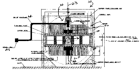
[00050] Figure 1 is a side view of a radiation therapy system 10
according to an
embodiment, that can fit entirely within a room that is three (3) metres in
height. Radiation
therapy system 10 comprises an open-bore, 0.2 Testa MRI assembly 12 integrated
with a
standard S-band standing wave 61vIV linear accelerator 14. Radiation therapy
system 10 is
configured to provide a 30 cm diameter imaging voltune 50, and comprises two
sets of
fourteen (14) circular coils 20 of diiferent diameters that are concentrically
positioned about a
common central axis 23. The sets are in a fixed spaced relationship thereby to
provide a
space therehetween for entry of a patient. In this embodiment, the smallest of
the circular
coils 20 has an inner diameter of 25 centimetres, which is sufficient for
receiving the linear
accelerator 14. The largest of the circular coils 20 has a diameter of 230
centimetres. The
naagnet cylinder thicknesses are five (5) centimeters, and there is a 2.5
centimetre separation
between coils 20. This separation is provided to enable connection to the
support structure,
and the interspersing of coolant such as water or a cryogenic coolant_ As can
be seen in
Figure 1, the inner two cylinders are shorter in order to accomodate the
dimensions of a multi-
leaf collimator (ML,C) 40 for beam shaping, and a beain stop 42.
1000511 In Figure 1, the patient enters the systera 10 from the right
hand side, and lies
hori2ontally between the two sets of coils 20. The radiation therapy system 10
rotates about a
horizontal axis 32 that is aligned with the cranial-caudal axis of the
patient. The gap between
inward facing surfaces of the two sets of coils 20 is 80 cm. This gap
dimension
aCCoranlOdateS two five (5) centimetre stainless steel plates 34 to be used to
anchor the sets of
coils 20, while permitting a 70 centimetre space between the sets of coils 20.
The 70
centimetres space is sufficient to accommodate the addition of gradient
magnets (not shown),
while still allowing enough room to rotate the entire two sets of coils 20
around the patient.
100052] A front view of the radiation therapy system 10 is shown in
Figure 2. In this
view, the axis of rotation 32 is perpendicular to the page, and the patient
enters the system by
CA 02728111 2010-12-15
WO 2009/155700
PCT/CA2009/000873
- 12 -
going in a direction that is into the page. A gantry ring supporting the
system 10 is clearly
. seen in this view, as are support posts to give the assembly the required
stiffness so as not to
sag or warp when rotating.
[00053] A top view of the radiation therapy system 10 is shown in
Figure 3. In this
view, the concentric magnet coils are clearly displayed, showing the linear
accelerator
'positioned along the central axis 23.
[00054] Advantageously, the radiation therapy system 10 rotates as a
unit about
1,,¨;=,antal *Irk 24. The transmission RF waveguide 44 that is operably
connected to the
linear accelerator 14 is, in this embodiment, of type 284. The transmission RF
wavegtucte 44
goes to a rotary joint 46 that rotates about axis 32. Rotary joint 46 in turn
provides coupling
to a RF power supply dm is, in this embodiment, located remote from the
radiation therapy
system 10. With this configuration, the RF power supply does not rotate.
[00055] Advantageously, the axis of particle travel within the linear
accelerator 14 is
also coincident with central axis 23. In this embodiment, linear accelerator l
4 is positioned
along central axis 23 partly within the 25 centimetre hole that is formed by
the smallest of the
circular coils 20, so as to provide a linear accelerator isocentre to target
distance of 75
centimetres, which permits application of a high dose rate during treatment_
In this position,
linear accelerator 14 is inmiersed in and oriented with respect to the
magnetic field produced
by the MRI toils 20 to expose the linear accelerator 14 to magnetic force that
dfrects (or
"focuses") particles therein along the central axis 23 of the linear
accelerator 14_
[00056] The mechanism of focusing is due to the presence of both
longitudinal and
radial magnetic fields at the central axis 23 at which the linear accelerator
14 has been
positioned. Particles that enter the linear accelerator 14 away from its
central axis 23 will
experience a Lorentz force in the asimuthal direction due to the radial
magnetic field of the
MRI magnets. The resulting angular motion of the particles will then cause an
inward radial
Lorentz force due to interaction with the longitudinal magnetic field of the
MR1 magnets.
The net result is a confinement of the particles to the central axis 23 of the
linear accelerator
14. Thus, this configuration provides Lorentz force that assists the linear
accelerator 14 by
redirecting any stray particles back to the central axis 23 of the linear
accelerator 14.
[00057] It would be understood that positioning of the linear
accelerator 14 as
described above places the linear accelerator within a region where the
magnetic field has a
reasonable level of homogeneity. This positioning thereby ensures a radial
magnetic field that
is advantageous for focusing of the particle beam within the linear
accelerator 14.
[00058] While due to their use of RF fields for providing particle
focusing, some
modem standing wave linear accelerators 14 do not necessarily require
additional focusing as
CA 02728111 2010-12-15
WO 2009/155700 PCT/CA2009/000873
- 13 -
described above, the positioning of the linear accelerator 14 as described
above providing
supplemental focusing will not harm the accelerator functionality, and will of
course provide
the compactness, permrbation-reduction, and increased dose rate advantages
tbat have been
described above. Furthermore, augmented performance of the linear accelerator
14 may
result due to any additional particle focusing provided by the advantageous
placement of the
linear accelerator 14 relative to the MRI magnetic field. Such additional
focusing would
serve to reduce the beam spot size, among other things, thereby increasing the
accuracy of the
radiation therapy.
1000591
As an example, the Varian 2100 and 2300 series medical accelerators
a standing wave accelerating structure and also a waveguide focusing magnetic
coil. Thus,
this type of focusing magnetic field can be applied to any linear accelerator,
and can be
exploited to combine a linear accelerator with an
100060] An example of a selected set of performance parameters is given
in Tables 1
and 2, and the resultingcentral axis field plot in Figure 4.
[000611 Table 1 shows parameters representing the coils 20 for rmdiation
therapy
system 10, where the coils 20 produce a somewhat uniform magnetic field in a
30 cm sphere-
shaped imaging volt= at the magnet isocentre, with about a 71 ppm
nonuniforrnity. In this
embodiment, the gauge of the copper wirc. for the coils 20 is 18 AWG; the
total coil weight is
17,363 kilograms; and the total power dissipated is 1207 kW.
current resistance voltage power weight Number
coil #
(A) (ca) (kV) (kW) (kg) of turns
1 4.231 279.6 1.183 5.006 84.1 12173
2 -2.919 419.4 -1.224 3.573 126.2 12173
3 0.648 1016.8 0.659 0.427 305.2 22090
4 -1.657 1271.0 -2.107 3.492 381.6 22090
2928 1525.3 4.465 13.073 457.9 22090
6 -1.399 1779.5 -2.489 3.481 534.2 22090
7 0.848 2033.7 1.724 1.462 610.5 22090
8 0.185 2287.9 0.423 0.078 686.9 22090
9 -5.344 2542.1 -13.586 72.606 763.2 22090
-2351 2796.3 -7133 18.193 839.5 22090
11 7355 3050.5 22.437 165.032 915.8 22090
12 0.349 3304.7 1.153 0.402 992.2 22090
13 3.147 3558.9 11.201 35.255 1068.5 22090
CA 02728111 2010-12-15
WO 2009/155700
PCT/CA2009/000873
-14-
14 9.599 3050.5 29.281 281.066 915.9 17672
Table 1
[00062] Table 2 shows parameters representing the coils 20 for
radiation therapy
system 10, where the coils 20 produce an acceptably homogenous magnetic field
in a 30 cm
sphere-s.hapcd imaging volume at the magnet isocentrc, with about a 80 ppm
nonb.omoegenity. In this embodiment, the gauge of the copper wire for the
coils 20 is 8
AWG; the total coil weight is 18, 093 kilograms; and the total power
dissipated is 1486
kiloWatts (kW).
Number
current resistance voltage power weight
coil # of
(A) (o) (kV) (kW) (k48)
turas
1 -56.548 2.845 -0.161 9.10 87.31 1245
2 51.683 4.267 0.221 11.40 131.03 1245
3 -1.813 10.345 -0.019 0.03 317.92 2265
4 -2.999 12.932 -0.039 0_12 397.47 2265
15.246 15.518 0.237 3.61 477.01 2265
6 -13.339 18.104 -0.241 3.22 556.56 2265
7 24.029 20.690 0.497 11.95 636.10 2265
8 43_366 23.277 -1.009 43.77 715.64 2265
9 -68.877 25.863 -1.781 122.70 795.19 2265
44.061 28.449 1.253 55.23 874.73 2265
11 94.814 31.036 2.943 279.00 954.28 2265
12 -21.832 33.622 -0.734 16.03 1033.82 2265
13 67.934 36.208 2.460 167.10 1113.36 2265
14 25.124 31.036 0.780 19.59 955.91 1815
__________________________________________________ - _____
Table 2
[00063J Figure 4 is a plot of the longitudinal magnetic flux density
along the central
axis of the 14 magnet coils arranged as shown in Figures 1 to 3, with the
operating parameters
in Tables 1 and 2. The top plot represents the data in Table 1, whereas the
bottom plot
represents the data in Table 2.
[00064] It is preferred that the linear accelerator is located in the
region between 75
and 115 cm from the magnet isocenter, and thus sees a magnetic flux density
that ranges from
between these values. (a) 18 AWG copper wire: -0.225 T (target end) to -0.12 T
(gun end);
and (b) 8 AWG copper wire 0.11 T (target end) to 0.145 T (gun end). At these
field strengths,
CA 02728111 2010-12-15
WO 2009/155700
PCT/CA2009/000873
- 15 -
focusing of the electron beam within the linear accelerator is excellent. This
focusing will
allow exceptional performance of the linear accelerator.
[00065] While the above has provided embodiments having particular
parameters, it
will be understood that the magnet coils can be configured in a variety of
manners. For
example, those skilled in the art will recopin that there are many other coil
turn and current
combinations that can produce an acceptably homogeneous magnetic field over
different
imaging volinnes and shapes_ For example, according to an alternative
embodiment where
magnetic fields larger than 0.2 T are desired, the coils 20 could be cooled to
superconducting
temperatures using a cryogenic coolant, and thus superconducting coils can
also be used. In
such an embodiment, magnetic flux densities on the order of 1 T or higher
could be generated
while positioning the linear accelerator 14 as has been described above,
without adversely
affecting the operation of the linear accelerator 14 due to its advantageous
positioning. The
degree of homogeneity (80 ppm of non-homogeneity or less) is sufficient for
many MR1
imaging applications, and the 30 centimetres iniaging volume is large enough
to be useful in
image guided radiotherapy.
[000661 For the configurations described above, power supplies and
cooling systems
that are stable enough to be useful in MR1 are available comanercially
(Danfysic,
Copenhagen, Denmark).
[000671 In the configurations summarized in Tables 1 and 2, a resistive
coil was used.
[00068] As would be recognized, The MR1 assembly shown in Figures 1 to
3 does not
have any active coils to limit the extent of the fringe magnetic field lines.
This is not optimal
in MR1 since magnetic field lines that go far from the magnet isocentre
present a safety
hazard, and are thus undesirable. It is typical to use active shielding
methods to limit the
extent of the MR1 B0 field. Although the example of the integrated MR1magnet
and linear
accelerator shown in Figures 1 to 3 does not include active shielding, those
skilled in the art
will recognize that active shielding techniques can still be used while
keeping the radial
component of the roagnetie field to zero on the central axis of the magnets
assembly. This is
consistent with the present disclosure, and this will not prevent the
functionality of this
disclosure.
[00069] Those skilled in the art will also recognize that the
arrangement shown in
Figure 1 to 3 is only one of several arrangements that are possible where a
linear accelerator
can be integrated with an MRI magnet assembly where the long axis of the
linear accelerator
is in a direction parallel to the B0 field of the MR1. Other embodiments may
also utilize
noncircular current carrying coils or permanent magnets.
CA 02728111 2010-12-15
WO 2009/155700=
PCT/CA2009/000873
- 16 -
[00070] Although embodiments have been described, those of skill in the
art will
appreciate that variations and modifications may be made without departing
from the purpose
and scope thereof as defined by the appended clpims