Note: Descriptions are shown in the official language in which they were submitted.
CA 02728347 2010-12-16
WO 2010/004438 PCT/IB2009/007082
1
LM-1 ANTIBODIES, FUNCTIONAL FRAGMENTS, LM-1 TARGET
ANTIGEN, AND METHODS FOR MAKING AND USING SAME
Related Applications
This application claims the benefit of priority of application serial no.
61/143,351, filed January 8, 2009, and application serial no. 61/061,881,
filed
June 16, 2008, all of which applications are expressly incorporated herein by
reference in their entirety.
Field of the Invention
The invention relates to an antibody, known as LM-1 and a target,
known as LM-1 Target or Antigen. The antibody denoted LM-1 is an IgM
and binds to different types of neoplasia, cancer, tumor and metastasis. LM-1
inhibits growth of various types of cancer cells and stimulates or induces
apoptosis of various types of cancer cells. LM-1 also reduces formation or
establishment of metastases at one or more sites arising from a primary
neoplasia, tumor or cancer, or growth or proliferation of a metastasis that
has
formed or been established at one or more other sites.
Introduction
Metastatic disease at sites peripheral to the primary cancer potentially
contribute to cancer progression and relapse. Consequently, inhibiton of
establishment or formation of metastasis, or reduction or decrease of
metastasis growth, proliferation of or progression metastatic tumors that have
been established is likely to reduce or inhibit cancer progression and
relapse.
The invention addresses this need and provides related benefits.
Summary
The invention provides isolated and purified antibodies and functional
fragments that compete for binding to a cell or to an antigen that LM-1
antibody, as represented by antibody produced by a cell line DSMZ Deposit
No. DSM ACC 2623, or represented by heavy and light chain sequences set
forth as SEQ ID NOs:1, 3, 5, 7 or 9, and 11 or 13, binds. In one embodiment,
an antibody or functional fragment competes with LM-1 antibody, as
represented by antibody produced by a cell line DSMZ Deposit No. DSM
50035!532vt
CA 02728347 2010-12-16
WO 2010/004438 PCT/IB2009/007082
2
ACC 2623, or represented by heavy and light chain sequences set forth as
SEQ ID NOs: 1, 3, 5, 7 or 9, and 11 or 13, for binding to an neoplastic, tumor
or cancer or a metastatic cell. In another embodiment, an antibody or
functional fragment thereof competes for binding of LM-1 to NONO/nmt55
protein.
In particular aspects, an antibody or functional fragment competes with
LM-1 antibody, as represented by antibody produced by a cell line DSMZ
Deposit No. DSM ACC 2623, or represented by heavy and light chain
sequences set forth as SEQ ID NOs:1, 3, 5, 7 or 9, and 11 or 13, for binding
to
an antigen (e.g., NONO/nmt55 protein) on one or more of a stomach
adenocarcinoma (e.g., diffuse or intestinal), colorectal cancer such as
adenocarcinoma, ovarian carcinoma, lung cancer, such as lung
adenocarcinomas, squamous cell lung carcinoma and small cell lung
carcinoma, melanoma, lobular and ductal mammary carcinomas, breast cancer
such as invasive ductal or lobular cancer, gastric cancer, pancreatic cancer,
such as pancreatic adenocarcinomas (e.g., ductal), sarcomas, gastrointestinal
cancer such as a stomach cancer, nervous tissue or brain tumor such as a
glioma, esophageal cancer such as esophageal squamous cell carcinomas and
adenocarcinomas, osteosarcoma, fibrosarcomas, urinary bladder cancer,
prostate cancer such as prostate adenocarcinomas, kidney cancer such as renal
carcinoma, ovarian cancer such as adenocarcinomas, testicular cancer,
endometrial cancer, cervical cancer such as squamous cell and
adenocarcinomas, uterine cancers such as adenocarcinomas, Hodgkin's
disease, lymphomas, and leukemias. Such polypeptides are particularly useful
for the detection and treatment of stomach adenocarcinoma (e.g., diffuse or
intestinal), colorectal cancer such as adenocarcinoma, ovarian carcinoma, lung
cancer, such as lung adenocarcinomas, squamous cell lung carcinoma and
small cell lung carcinoma, melanoma, lobular and ductal mammary
carcinomas, breast cancer such as invasive ductal or lobular cancer, gastric
cancer, pancreatic cancer, such as pancreatic adenocarcinomas (e.g., ductal),
sarcomas, gastrointestinal cancer such as a stomach cancer, nervous tissue or
brain tumor such as a glioma, esophageal cancer such as esophagial squamous
cell carcinomas and adenocarcinomas, osteosarcoma, fibrosarcomas, urinary
bladder cancer, prostate cancer such as prostate adenocarcinomas, kidney
500351832v1
CA 02728347 2010-12-16
WO 2010/004438 PCT/IB2009/007082
3
cancer such as renal carcinoma, ovarian cancer such as adenocarcinomas,
testicular cancer, endometrial cancer, cervical cancer such as squamous cell
and adenocarcinomas, uterine cancers such as adenocarcinomas, Hodgkin's
disease, lymphomas, and leukemias.
In another embodiment, an antibody or functional fragment competes
with LM-1 antibody, as represented by antibody produced by a cell line
DSMZ Deposit No. DSM ACC 2623, or represented by heavy and light chain
sequences set forth as SEQ ID NOs:1, 3, 5, 7 or 9, and 11 or 13, for binding
to
a stomach adenocarcinoma (e.g., diffuse or intestinal), colorectal cancer such
as adenocarcinoma, ovarian carcinoma, lung cancer, such as lung
adenocarcinomas, squamous cell lung carcinoma and small cell lung
carcinoma, melanoma, lobular and ductal mammary carcinomas, breast cancer
such as invasive ductal or lobular cancer, gastric cancer, pancreatic cancer,
such as pancreatic adenocarcinomas (e.g., ductal), sarcomas, gastrointestinal
cancer such as a stomach cancer, nervous tissue or brain tumor such as a
glioma, esophageal cancer such as esophagial squamous cell carcinomas and
adenocarcinomas, osteosarcoma, fibrosarcomas, urinary bladder cancer,
prostate cancer such as prostate adenocarcinomas, kidney cancer such as renal
carcinoma, ovarian cancer such as adenocarcinomas, testicular cancer,
endometrial cancer, cervical cancer such as squamous cell and
adenocarcinomas, uterine cancers such as adenocarcinomas, Hodgkin's
disease, lymphoma, or leukemia. In an additional embodiment, an antibody or
functional fragment competes with LM- 1 antibody, as represented by antibody
produced by a cell line DSMZ Deposit No. DSM ACC 2623, or represented
by heavy and light chain sequences set forth as SEQ ID NOs:1, 3, 5, 7 or 9,
and 11 or 13, for binding to one of lung adenocarinoma cell line Colo-699
(DSMZ accession number ACC 196), lung adenocarinoma cell line DV-90
(DSMZ accession number ACC 307), epidermoid lung carcinoma cell line
EPLC-272H (DSMZ accession number ACC 383), lung squamous cell
carcinoma cell line LOU-NH91 (DSMZ accession number ACC 393), HT-29
(ATCC Accession No. HTB-38; DSMZ Accession No. ACC 299), A549
(DSMZ Accession No. ACC 107) or BXPC-3 (ATCC Accession No. CRL-
1687) cells. In a further embodiment, an antibody or functional fragment
thereof inhbits or reduces proliferation, or stimulates or induces apoptosis,
of
3
500351832v1
CA 02728347 2010-12-16
WO 2010/004438 PCT/IB2009/007082
4
one or more of a stomach adenocarcinoma (e.g., diffuse or intestinal),
colorectal cancer such as adenocarcinoma, ovarian carcinoma, lung cancer,
such as lung adenocarcinomas, squamous cell lung carcinoma and small cell
lung carcinoma, melanoma, lobular and ductal mammary carcinomas, breast
cancer such as invasive ductal or lobular cancer, gastric cancer, pancreatic
cancer, such as pancreatic adenocarcinomas (e.g., ductal), sarcomas,
gastrointestinal cancer such as a stomach cancer, nervous tissue or brain
tumor
such as a glioma, esophageal cancer such as esophagial squamous cell
carcinomas and adenocarcinomas, osteosarcoma, fibrosarcomas, urinary
bladder cancer, prostate cancer such as prostate adenocarcinomas, kidney
cancer such as renal carcinoma, ovarian cancer such as adenocarcinomas,
testicular cancer, endometrial cancer, cervical cancer such as squamous cell
and adenocarcinomas, uterine cancers such as adenocarcinomas, Hodgkin's
disease, lymphoma, or leukemia, or one of lung adenocarinoma cell line Colo-
699 (DSMZ accession number ACC 196), lung adenocarinoma cell line DV-
90 (DSMZ accession number ACC 307), epidermoid lung carcinoma cell line
EPLC-272H (DSMZ accession number ACC 383), or lung squamous cell
carcinoma cell line LOU-NH91 (DSMZ accession number ACC 393) cells.
The invention also provides isolated and purified antibodies and
functional fragments thereof that bind to cells or to an antigen (e.g.,
NONO/nmt55 protein) that LM-1 antibody, as represented by antibody
produced by a cell line DSMZ Deposit No. DSM ACC 2623, or represented
by heavy and light chain sequences set forth as SEQ ID NOs:1, 3, 5, 7 or 9,
and 11 or 13, binds. In one embodiment, an antibody or functional fragment
binds to an adenocarcinoma cell or a squamous cell carcinoma to which LM-1
antibody, as represented by antibody produced by a cell line DSMZ Deposit
No. DSM ACC 2623, or represented by heavy and light chain sequences set
forth as SEQ ID NOs:1, 3, 5, 7 or 9, and 11 or 13, binds. In particular
aspects,
an antibody or functional fragment binds to one or more of stomach
adenocarcinoma (e.g., diffuse or intestinal), colorectal cancer such as
adenocarcinoma, ovarian carcinoma, lung cancer, such as lung
adenocarcinomas, squamous cell lung carcinoma and small cell lung
carcinoma, melanoma, lobular and ductal mammary carcinomas, breast cancer
such as invasive ductal or lobular cancer, gastric cancer, pancreatic cancer,
4
500351832v1
CA 02728347 2010-12-16
WO 2010/004438 PCT/IB2009/007082
such as pancreatic adenocarcinomas (e.g., ductal), sarcomas, gastrointestinal
cancer such as a stomach cancer, nervous tissue or brain tumor such as a
glioma, esophageal cancer such as esophagial squamous cell carcinomas and
adenocarcinomas, osteosarcoma, fibrosarcomas, urinary bladder cancer,
5 prostate cancer such as prostate adenocarcinomas, kidney cancer such as
renal
carcinoma, ovarian cancer such as adenocarcinomas, testicular cancer,
endometrial cancer, cervical cancer such as squamous cell and
adenocarcinomas, uterine cancers such as adenocarcinomas, Hodgkin's
disease, lymphoma, or leukemia, to which LM-1 antibody, as represented by
antibody produced by a cell line DSMZ Deposit No. DSM ACC 2623, or
represented by heavy and light chain sequences set forth as SEQ ID NOs:1, 3,
5, 7 or 9, and 11 or 13, binds. In another embodiment, an antibody or
functional fragment binds to a stomach adenocarcinoma (e.g., diffuse or
intestinal), colorectal cancer such as adenocarcinoma, ovarian carcinoma, lung
cancer, such as lung adenocarcinomas, squamous cell lung carcinoma and
small cell lung carcinoma, melanoma, lobular and ductal mammary
carcinomas, breast cancer such as invasive ductal or lobular cancer, gastric
cancer, pancreatic cancer, such as pancreatic adenocarcinomas (e.g., ductal),
sarcomas, gastrointestinal cancer such as a stomach cancer, nervous tissue or
brain tumor such as a glioma, esophageal cancer such as esophagial squamous
cell carcinomas and adenocarcinomas, osteosarcoma, fibrosarcomas, urinary
bladder cancer, prostate cancer such as prostate adenocarcinomas, kidney
cancer such as renal carcinoma, ovarian cancer such as adenocarcinomas,
testicular cancer, endometrial cancer, cervical cancer such as squamous cell
and adenocarcinomas, uterine cancers such as adenocarcinomas, Hodgkin's
disease, lymphoma, or leukemia to which LM-1 antibody, as represented by
antibody produced by a cell line DSMZ Deposit No. DSM ACC 2623, or
represented by heavy and light chain sequences set forth as SEQ ID NOs: 1, 3,
5, 7 or 9, and 11 or 13, binds. In an additional embodiment, an antibody or
functional fragment binds to one of lung adenocarinoma cell line Colo-699
(DSMZ accession number ACC 196), lung adenocarinoma cell line DV-90
(DSMZ accession number ACC 307), epidermoid lung carcinoma cell line
EPLC-272H (DSMZ accession number ACC 383), or lung squamous cell
carcinoma cell line LOU-NH91 (DSMZ accession number ACC 393) cells
5
500351832v1
CA 02728347 2010-12-16
WO 2010/004438 PCT/IB2009/007082
6
that LM-1 antibody, as represented by antibody produced by a cell line DSMZ
Deposit No. DSM ACC 2623, or represented by heavy and light chain
sequences set forth as SEQ ID NOs: 1, 3, 5, 7 or 9, and 11 or 13, binds.
The invention further provides isolated and purified antibodies and
functional fragments that include a heavy or light chain variable region
sequence with about 60% or more identity to a heavy or light chain sequence
variable regions of LM-1 antibody, as represented by antibody produced by a
cell line DSMZ Deposit No. DSM ACC 2623, or represented by heavy and
light chain sequences set forth as SEQ ID NOs: 1, 3, 5, 7 or 9, and 11 or 13,.
In one embodiment, an antibody or subsequence thereof includes a sequence at
least 60 % or more (e.g., 65%, 70%, 75%, 80%, 85%, 90%, 95%, etc.)
identical to a heavy chain variable region sequence set forth as SEQ ID NO:1,
3, 5, 7 or 9, or heavy chain of antibody produced by a cell line DSMZ Deposit
No. DSM ACC 262, or a sequence at least 60% or more (e.g., 65%, 70%,
75%, 80%, 85%, 90%, 95%, etc.) identical to a light chain variable region
sequence set forth as SEQ ID NO:9 or light chain of antibody produced by a
cell line DSMZ Deposit No. DSM ACC 262. In another embodiment, an
antibody or subsequence includes a sequence at least 60 % or more (e.g., 65%,
70%, 75%, 80%, 85%, 90%, 95%, etc.) identical to a heavy chain variable
region sequence set forth as SEQ ID NO: 1, 3, 5, 7 or 9, or heavy chain of
antibody produced by a cell line DSMZ Deposit No. DSM ACC 262, and a
sequence at least 60% or more (e.g., 65%, 70%, 75%, 80%, 85%, 90%, 95%,
etc.) identical to a light chain variable region sequence set forth as SEQ ID
NO:9 or light chain of antibody produced by a cell line DSMZ Deposit No.
DSM ACC 262. In a further embodiment, an antibody or subsequence
includes a sequence at least 80-85%, 85-90%, 90-95%, or 95-100% identical
to one or more CDRs in heavy chain variable region sequence set forth as SEQ
ID NO: 1, 3, 5, 7 or 9 (e.g., amino acids 24-35, 52-67, or 100-118), or one or
more CDRs in a heavy chain variable region of antibody produced by a cell
line DSMZ Deposit No. DSM ACC 262, or a sequence at least 80-85%, 85-
90%, 90-95%, or 95-100% identical to one or more CDRs in a light chain
variable region sequence set forth as SEQ ID NO:9 (e.g., amino acids 23-35,
51-58 or 90-101 of SEQ ID NO:11 or 13), or one or more CDRs in a light
6
500351832v1
CA 02728347 2010-12-16
WO 2010/004438 PCT/IB2009/007082
7
chain variable region of antibody produced by a cell line DSMZ Deposit No.
DSM ACC 262.
The invention further provides isolated and purified antibodies and
functional fragments thereof that have one or more amino acid additions,
deletions or substitutions of LM-1 antibody, as represented by antibody
produced by a cell line DSMZ Deposit No. DSM ACC 2623, or represented
by heavy and light chain sequences set forth as SEQ ID NOs:1, 3, 5, 7 or 9,
and 11 or 13. In particular aspects, an antibody or functional fragment has
sequence at least 80-85%, 85-90%, 90-95%, or 95-100% identical to a heavy
chain variable region sequence set forth as SEQ ID NO: 1, 3, 5, 7 or 9, and 11
or 13, or a sequence at least 80-85%, 85-90%, 90-95%, or 95-100% identical
to a light chain variable region sequence set forth as SEQ ID NO:9. In further
aspects, an antibody or functional fragment has a heavy or light chain
sequence with 100% identity to one or more CDRs in a heavy or light chain
variable region sequence set forth as SEQ ID NOs: 1, 3, 5, 7 or 9, and 11 or
13
(e.g., amino acids 24-35, 52-67, 100-118 of SEQ ID NO: 1, 3, 5 or 7, or amino
acids 23-35, 51-58, 90-101, of SEQ ID NO:11), and has less than 100%
identity to a region outside of the CDRs in a heavy or light chain variable
region sequence set forth as SEQ ID NOs:1, 3, 5, 7 or 9, and 11 or 13. Such
variants can bind to antigen (e.g., NONO/nmt55 protein) or epitope of an
antigen to which a reference antibody (e.g., LM-1) binds.
The invention also provides antibodies and functional fragments
thereof that have a binding affinity within about 1-5000 fold of the binding
affinity of LM-1 antibody, as represented by antibody produced by a cell line
DSMZ Deposit No. DSM ACC 2623, or represented by heavy and light chain
sequences set forth as SEQ ID NOs:1, 3, 5, 7 or 9, and 11 or 13 for binding to
an antigen (e.g., NONO/nmt55 protein) or a cell (e.g., a neplastic, cancer,
tumor or metastatic cell). In various embodiments, antibodies and functional
fragments have a binding affinity within about 1-5000 fold of the binding
affinity of LM-1 antibody, as represented by antibody produced by a cell line
DSMZ Deposit No. DSM ACC 2623, or represented by heavy and light chain
sequences set forth as SEQ ID NOs: 1, 3, 5, 7 or 9, and 11 or 13 for binding
to
an antigen (e.g., NONO/nmt55 protein), or stomach adenocarcinoma (e.g.,
diffuse or intestinal), colorectal cancer such as adenocarcinoma, ovarian
7
500351832v1
CA 02728347 2010-12-16
WO 2010/004438 PCT/IB2009/007082
8
carcinoma, lung cancer, such as lung adenocarcinomas, squamous cell lung
carcinoma and small cell lung carcinoma, melanoma, lobular and ductal
mammary carcinomas, breast cancer such as invasive ductal or lobular cancer,
gastric cancer, pancreatic cancer, such as pancreatic adenocarcinomas (e.g.,
ductal), sarcomas, gastrointestinal cancer such as a stomach cancer, nervous
tissue or brain tumor such as a glioma, esophageal cancer such as esophagial
squamous cell carcinomas and adenocarcinomas, osteosarcoma,
fibrosarcomas, urinary bladder cancer, prostate cancer such as prostate
adenocarcinomas, kidney cancer such as renal carcinoma, ovarian cancer such
as adenocarcinomas, testicular cancer, endometrial cancer, cervical cancer
such as squamous cell and adenocarcinomas, uterine cancers such as
adenocarcinomas, Hodgkin's disease, lymphoma, or leukemia. In additional
embodiments, an antibody or functional fragment has a binding affinity within
about 1-5000 fold of the binding affinity of LM-1 antibody, as represented by
antibody produced by a cell line DSMZ Deposit No. DSM ACC 2623, or
represented by heavy and light chain sequences set forth as SEQ ID NOs: 1, 3,
5, 7 or 9, and 11 or 13 for binding to an antigen (e.g., NONO/nmt55 protein),
or one of lung adenocarinoma cell line Colo-699 (DSMZ accession number
ACC 196), lung adenocarinoma cell line DV-90 (DSMZ accession number
ACC 307), epidermoid lung carcinoma cell line EPLC-272H (DSMZ
accession number ACC 383), or lung squamous cell carcinoma cell line LOU-
NH91 (DSMZ accession number ACC 393) cells. In further embodiments, an
antibody or functional fragment has a binding affinity within about KD 10-5 M
to about KD 10-13 M for binding to an antigen (e.g., NONO/nmt55 protein), or
one or more cells or cell lines set forth herein (e.g., stomach
adenocarcinoma,
colorectal cancer such as adenocarcinoma, ovarian carcinoma, lung cancer,
such as lung adenocarcinomas, squamous cell lung carcinoma and small cell
lung carcinoma, melanoma, lobular and ductal mammary carcinomas, breast
cancer such as invasive ductal or lobular cancer, gastric cancer, pancreatic
cancer, such as pancreatic adenocarcinomas (e.g., ductal), sarcomas,
gastrointestinal cancer such as a stomach cancer, nervous tissue or brain
tumor
such as a glioma, esophageal cancer such as esophagial squamous cell
carcinomas and adenocarcinomas, osteosarcoma, fibrosarcomas, urinary
bladder cancer, prostate cancer such as prostate adenocarcinomas, kidney
8
500351832v1
CA 02728347 2010-12-16
WO 2010/004438 PCT/IB2009/007082
9
cancer such as renal carcinoma, ovarian cancer such as adenocarcinomas,
testicular cancer, endometrial cancer, cervical cancer such as squamous cell
and adenocarcinomas, uterine cancers such as adenocarcinomas, Hodgkin's
disease, lymphoma, or leukemia, etc., or one of lung adenocarinoma cell line
Colo-699 (DSMZ accession number ACC 196), lung adenocarinoma cell line
DV-90 (DSMZ accession number ACC 307), epidermoid lung carcinoma cell
line EPLC-272H (DSMZ accession number ACC 383), or lung squamous cell
carcinoma cell line LOU-NH91 (DSMZ accession number ACC 393) cells.
Antibodies of the invention include IgG, IgA, IgM, IgE and IgD. In
various aspects, an IgG is an IgG1, IgG2, IgG3, or IgG4.
Antibody functional fragments and subsequences of the invention
include functional fragments and subsequences of the various antibodies set
forth herein. In a particular embodiment, a functional fragment of LM-1
antibody, as represented by antibody produced by a cell line DSMZ Deposit
No. DSM ACC 2623, or represented by heavy and light chain sequences set
forth as SEQ ID NOs: 1, 3, 5, 7 or 9, and 11 or 13 that competes with LM-1
antibody, as represented by antibody produced by a cell line DSMZ Deposit
No. DSM ACC 2623, or represented by heavy and light chain sequences set
forth as SEQ ID NOs:1, 3, 5 or 7, and 9 for binding to a cell or an antigen
(e.g., NONO/nmt55 protein), or that retains at least partial binding to a cell
or
antigen to which LM-1 antibody, as represented by antibody produced by a
cell line DSMZ Deposit No. DSM ACC 2623, or represented by heavy and
light chain sequences set forth as SEQ ID NOs:1, 3, 5, 7 or 9, and 11 or 13
binds, is provided. In particular aspects, a functional fragment or a
subsequence is an Fab, Fab', F(ab')2, Fv, Fd, single-chain Fv (scFv),
disulfide-
linked Fvs (sdFv), VL, VH, trispecific (Fab3), bispecific (Fab2), diabody ((VL-
VH)2 or (VH-VL)2), triabody (trivalent), tetrabody (tetravalent), minibody
((scFv-CH3)2), bispecific single-chain Fv (Bis-scFv), IgGdeltaCH2, scFv-Fc
and (scFv)2-Fc. In additional aspects, a functional fragment or a subsequence
of a full length antibody heavy or light chain, or a heavy or light chain
variable
region, includes one or more CDRs of a heavy or light chain sequence of LM-
1 antibody, as represented by antibody produced by a cell line DSMZ Deposit
No. DSM ACC 2623, or represented by heavy and light chain sequences set
forth as SEQ ID NOs:1, 3, 5, 7 or 9, and 11 or 13 (e.g., amino acids 24-35, 52-
9
500351832v1
CA 02728347 2010-12-16
WO 2010/004438 PCT/IB2009/007082
67 or 100-118 of SEQ ID NO:1, 3, 5, 7 or 9, or amino acids 23-35, 51-58 or
90-101 of SEQ ID NO: 11). In further aspects, a functional fragment or a
subsequence of a full length antibody heavy or light chain, or a heavy or
light
chain variable region, has a length from about 20-30, 30-50, 50-100, 100-150,
5 150-200, 200-250, 250-300, 300-400, 400-500, amino acid residues.
The invention also provides antibodies and subsequences that include a
heterologous domain. In one embodiment, a heterologous domain includes a
detectable label, tag or cytotoxic agent. In particular aspects, a detectable
label or tag is an enzyme, enzyme substrate, ligand, receptor, radionuclide, a
10 T7-, His-, myc-, HA- or FLAG-tag, electron-dense reagent, energy transfer
molecule, paramagnetic label, fluorophore, chromophore, chemi-luminescent
agent, or a bio-luminescent agent.
The invention moreover provides nucleic acid sequences that encode
antibodies and functional fragments thereof. In one embodiment, a nucleic
acid sequence is at least 75-100% complementary or identical to a nucleic acid
sequence that encodes a heavy or a light chain variable region sequence of
LM-1 antibody, as represented by antibody produced by a cell line DSMZ
Deposit No. DSM ACC 2623, or represented by heavy and light chain
sequences set forth as SEQ ID NOs:1, 3, 5, 7 or 9, and 11 or 13 or a
subsequence thereof (e.g, amino acids 24-35, 52-67 or 100-118 of SEQ ID
NO:1, 3, 5 or 7, or amino acids 23-35, 51-58 or 90-101 of SEQ ID NO:9). In
another embodiment, a nucleic acid encodes a subsequence of LM-1 antibody,
as represented by antibody produced by a cell line DSMZ Deposit No. DSM
ACC 2623, or represented by heavy and light chain sequences set forth as
SEQ ID NOs: 1, 3, 5, 7 or 9, and 11 or 13 (e.g., amino acids 24-35, 52-67 or
100-118 of SEQ ID NO:1, 3, 5, 7 or 9, or amino acids 23-35, 51-58 or 90-101
of SEQ ID NO: 11 or 13). In particular aspects, a nucleic acid sequence has a
length from about 10-20, 20-30, 30-50, 50-100, 100-150, 150-200, 200-250,
250-300, 300-400, 400-500, or 500-1000 nucleotides. In additional aspects, a
nucleic acid sequence specifically hybridizes to a nucleic acid that encodes
SEQ ID NO:1, 3, 5, 7 or 9, and 11 or 13, or a subsequence thereof, or
specifically hybridizes to a nucleic acid sequence complementary to a nucleic
acid that encodes SEQ ID NO:1, 3, 5, 7 or 9, and 11 or 13 or a subsequence
SEQ ID NO:1, 3, 5, 7 or 9, and 11 or 13. In further aspects, a nucleic acid is
500351832v1
CA 02728347 2010-12-16
WO 2010/004438 PCT/IB2009/007082
11
an antisense polynucleotide, a small interfering RNA, or a ribozyme nucleic
acid that specifically hybridizes to a nucleic acid sequence encoding or
complementary to SEQ ID NO: 1, 3, 5, 7 or 9, and 11 or 13 or a subsequence
thereof. Antisense polynucleotides, small interfering RNA, and ribozyme
polynucleotides can have a length from about 10-20, 20-30, 30-50, 50-100,
100-150, 150-200, 200-250, 250-300, 300-400, 400-500, 500-1000, 1000-
2000 nucleotides, and be at least 90% complementary or identical to a nucleic
acid sequence that encodes SEQ ID NOs:I, 3, 5, 7 or 9, and 11 or 13 or a
subsequence thereof (e.g., 24-35, 52-67 or 100-118 of SEQ ID NO:1, 3, 5, 7 or
9, or amino acids 23-35, 51-58 or 90-101 of SEQ ID NO: 11 or 13). In still
further aspects, nucleic acid sequence can include an expression control
sequence or a vector (e.g., a viral, bacterial, fungal or mammalian vector).
The invention additionally provides isolated and purified cells as well
as transformed host cells that express an antibody or subsequence thereof that
includes a sequence at least 60% or more (e.g., 65%, 70%, 75%, 80%, 85%,
90%, 95%, etc.) identical to a heavy or light chain variable region sequence
set
forth as SEQ ID NO:1, 3, 5, 7 or 9, and 11 or 13 or a sequence at least 60% or
more (e.g., 65%, 70%, 75%, 80%, 85%, 90%, 95%, etc.) identical to a heavy
or light chain variable region sequence of LM-1 antibody, as represented by
antibody produced by a cell line DSMZ Deposit No. DSM ACC 2623, or
represented by heavy and light chain sequences set forth as SEQ ID NOs:1, 3,
5, 7 or 9, and 11 or 13. Such cells include eukaryotic and non-eukaryotic
cells, which can stably or transiently express antibody or subsequence
thereof,
or be stably or transiently transformed with the nucleic acid or vector that
encodes antibody or subsequence thereof or.
The invention further provides kits. In various embodiments, a kit
includes an antibody or functional fragment thereof that competes with LM-1
antibody, as represented by antibody produced by a cell line DSMZ Deposit
No. DSM ACC 2623, or represented by heavy and light chain sequences set
forth as SEQ ID NOs:1, 3, 5, 7 or 9, and 11 or 13 for binding to an antigen
(e.g., NONO/nmt55 protein) or to a cell (e.g., a neoplastic, cancer, tumor or
metastatic cell). In particular aspects, a kit includes an antibody or
functional
fragment thereof that competes with LM-1 antibody, as represented by
antibody produced by a cell line DSMZ Deposit No. DSM ACC 2623, or
I1
500351832v1
CA 02728347 2010-12-16
WO 2010/004438 PCT/IB2009/007082
12
represented by heavy and light chain sequences set forth as SEQ ID NOs: 1, 3,
5, 7 or 9, and 11 or 13 for binding to a stomach adenocarcinoma (e.g., diffuse
or intestinal), colorectal cancer such as adenocarcinoma, ovarian carcinoma,
lung cancer, such as lung adenocarcinomas, squamous cell lung carcinoma and
small cell lung carcinoma, melanoma, lobular and ductal mammary
carcinomas, breast cancer such as invasive ductal or lobular cancer, gastric
cancer, pancreatic cancer, such as pancreatic adenocarcinomas (e.g., ductal),
sarcomas, gastrointestinal cancer such as a stomach cancer, nervous tissue or
brain tumor such as a glioma, esophageal cancer such as esophageal squamous
cell carcinomas and adenocarcinomas, osteosarcoma, fibrosarcomas, urinary
bladder cancer, prostate cancer such as prostate adenocarcinomas, kidney
cancer such as renal carcinoma, ovarian cancer such as adenocarcinomas,
testicular cancer, endometrial cancer, cervical cancer such as squamous cell
and adenocarcinomas, uterine cancers such as adenocarcinomas, Hodgkin's
disease, lymphoma, or leukemia. In an additional embodiment, a kit includes
an antibody or functional fragment thereof that competes with LM-1 antibody,
as represented by antibody produced by a cell line DSMZ Deposit No. DSM
ACC 2623, or represented by heavy and light chain sequences set forth as
SEQ ID NOs:1, 3, 5, 7 or 9, and 11 or 13 for binding to an antigen (e.g.,
NONO/nmt55 protein), or one of lung adenocarinoma cell line Colo-699
(DSMZ accession number ACC 196), lung adenocarinoma cell line DV-90
(DSMZ accession number ACC 307), epidermoid lung carcinoma cell line
EPLC-272H (DSMZ accession number ACC 383), or lung squamous cell
carcinoma cell line LOU-NH91 (DSMZ accession number ACC 393) cells.
Kits of the invention also include antibodies and functional fragments
that bind to cells or an antigen (e.g., NONO/nmt55 protein) that LM-1
antibody, as represented by antibody produced by a cell line DSMZ Deposit
No. DSM ACC 2623, or represented by heavy and light chain sequences set
forth as SEQ ID NOs:1, 3, 5, 7 or 9, and 11 or 13 binds. In one embodiment, a
kit includes an antibody or functional fragment that binds to an
adenocarcinoma cell or a squamous cell carcinoma to which LM-1 antibody,
as represented by antibody produced by a cell line DSMZ Deposit No. DSM
ACC 2623, or represented by heavy and light chain sequences set forth as
SEQ ID NOs:1, 3, 5, 7 or 9, and 11 or 13 binds, such as a stomach
12
500351832v1
CA 02728347 2010-12-16
WO 2010/004438 PCT/IB2009/007082
13
adenocarcinoma (e.g., diffuse or intestinal) cell, a lung adenocarcinoma cell,
a
pancreas adenocarcinoma cell, a colon adenocarcinoma cell, a breast
adenocarcinoma cell, an esophagus squamous cell carcinoma, to which LM-1
antibody, as represented by antibody produced by a cell line DSMZ Deposit
No. DSM ACC 2623, or represented by heavy and light chain sequences set
forth as SEQ ID NOs:1, 3, 5, 7 or 9, and 11 or 13 binds. In another
embodiment, a kit includes an antibody or functional fragment binds to a
stomach adenocarcinoma (e.g., diffuse or intestinal), colorectal cancer such
as
adenocarcinoma, ovarian carcinoma, lung cancer, such as lung
adenocarcinomas, squamous cell lung carcinoma and small cell lung
carcinoma, melanoma, lobular and ductal mammary carcinomas, breast cancer
such as invasive ductal or lobular cancer, gastric cancer, pancreatic cancer,
such as pancreatic adenocarcinomas (e.g., ductal), sarcomas, gastrointestinal
cancer such as a stomach cancer, nervous tissue or brain tumor such as a
glioma, esophageal cancer such as esophagial squamous cell carcinomas and
adenocarcinomas, osteosarcoma, fibrosarcomas, urinary bladder cancer,
prostate cancer such as prostate adenocarcinomas, kidney cancer such as renal
carcinoma, ovarian cancer such as adenocarcinomas, testicular cancer,
endometrial cancer, cervical cancer such as squamous cell and
adenocarcinomas, uterine cancers such as adenocarcinomas, Hodgkin's
disease, lymphoma, or leukemia to which LM-1 antibody, as represented by
antibody produced by a cell line DSMZ Deposit No. DSM ACC 2623, or
represented by heavy and light chain sequences set forth as SEQ ID NOs:1, 3,
5, 7 or 9, and 11 or l3binds. In an additional embodiment, a kit includes an
antibody or functional fragment that binds to lung adenocarinoma cell line
Colo-699 (DSMZ accession number ACC 196), lung adenocarinoma cell line
DV-90 (DSMZ accession number ACC 307), epidermoid lung carcinoma cell
line EPLC-272H (DSMZ accession number ACC 383), or lung squamous cell
carcinoma cell line LOU-NH91 (DSMZ accession number ACC 393) cells
that LM-1 antibody, as represented by antibody produced by a cell line DSMZ
Deposit No. DSM ACC 2623, or represented by heavy and light chain
sequences set forth as SEQ ID NOs:1, 3, 5, 7 or 9, and 11 or 13 binds.
Kits of the invention further include antibodies and functional
fragments that include a heavy or light chain variable region sequence with
13
500351832v!
CA 02728347 2010-12-16
WO 2010/004438 PCT/IB2009/007082
14
about 60% or more identity to a heavy or light chain sequence variable regions
of LM-1 antibody, as represented by antibody produced by a cell line DSMZ
Deposit No. DSM ACC 2623, or represented by heavy and light chain
sequences set forth as SEQ ID NOs:1, 3, 5, 7 or 9, and 11 or 13. In one
embodiment, a kit includes an antibody or subsequence thereof with a
sequence at least 60 % or more (e.g., 65%, 70%, 75%, 80%, 85%, 90%, 95%,
etc.) identical to a heavy chain variable region sequence set forth as SEQ ID
NO:1, 3, 5, 7 or 9, or to a sequence at least 60% or more (e.g., 65%, 70%,
75%, 80%, 85%, 90%, 95%, etc.) identical to a light chain variable region
sequence set forth as SEQ ID NO: I I or 13. In another embodiment, a kit
includes an antibody or subsequence with a sequence at least 60 % or more
(e.g., 65%, 70%, 75%, 80%, 85%, 90%, 95%, etc.) identical to a heavy chain
variable region sequence set forth as SEQ ID NO: 1, 3, 5, 7 or 9, and to a
sequence at least 60% or more (e.g., 65%, 70%, 75%, 80%, 85%, 90%, 95%,
etc.) identical to a light chain variable region sequence set forth as SEQ ID
NO: 11 or 13. In further embodiments, a kit includes an antibody or
subsequence with a sequence at least 80-85%, 85-90%, 90-95%, 95-100%
identical to one or more CDRs in heavy chain variable region sequence set
forth as SEQ ID NO:1, 3, 5, 7 or 9, (e.g., amino acids 24-35, 52-67 or 100-118
of SEQ ID NO:1, 3, 5, 7 or 9), or one or more CDRs in a heavy chain variable
region of antibody produced by a cell line DSMZ Deposit No. DSM ACC 262,
or a sequence at least 80-85%, 85-90%, 90-95%, 95-100% identical to one or
more CDRs in a light chain variable region sequence set forth as SEQ ID
NO:11 or 13 (e.g., amino acids 23-35, 51-58 or 90-101 of SEQ ID NO:11), or
one or more CDRs in a light chain variable region of antibody produced by a
cell line DSMZ Deposit No. DSM ACC 262.
In additional embodiments, a kit also includes an anti-cell proliferative
or immune enhancing treatment or therapeutic agent, or an anti-neoplastic,
anti-cancer or anti-tumor or anti-metastatic agent, or an article of
manufacture
(e.g., for delivering the antibody, anti-cell proliferative or immune
enhancing
treatment or therapy into a subject locally, regionally or systemically). In
particular aspects, the instructions are for treating undesirable cell
proliferation
or a cell proliferative disorder (e.g., a neoplasia, tumor cancer or
metastasis).
14
50035I832vI
CA 02728347 2010-12-16
WO 2010/004438 PCT/IB2009/007082
The invention yet additionally provides pharmaceutical compositions.
In one embodiment, a composition includes an antibody or functional fragment
and a pharmaceutically acceptable carrier or excipient. In another
embodiment, a composition includes an antibody that competes with LM-1
5 antibody, as represented by antibody produced by a cell line DSMZ Deposit
No. DSM ACC 2623, or represented by heavy and light chain sequences set
forth as SEQ ID NOs:1, 3, 5, 7 or 9, and 11 or 13 for binding to a cell or an
antigen (e.g., NONO/nmt55 protein), or that binds to a cell or an antigen
(e.g.,
NONO/nmt55 protein) to which LM-1 antibody, as represented by antibody
10 produced by a cell line DSMZ Deposit No. DSM ACC 2623, or represented
by heavy and light chain sequences set forth as SEQ ID NOs:1, 3, 5, 7 or 9,
and 11 or 13 binds, or that includes a heavy or light chain variable region
sequence with about 60% or more identity to a heavy or light chain sequence
variable regions as set forth in SEQ ID NOs:1, 3, 5, 7 or 9, and 11 or 13 or a
15 sequence at least 80-85%, 85-90%, 90-95%, 95-100% identical to one or more
CDRs in a heavy chain or light chain variable region sequence set forth as
SEQ ID NO:1, 3, 5, 7 or 9, and 11 or 13 (e.g., amino acids 24-35, 52-67 or
100-118 of SEQ ID NO:1, 3, 5 or 7, or amino acids 23-35, 51-58 or 90-101 of
SEQ ID NO: 11), and a pharmaceutically acceptable carrier or excipient. In a
further embodiment, a composition includes an antigen (e.g., NONO/nmt55
protein) and a pharmaceutically acceptable carrier or excipient
Antibodies, functional fragments and antigen (e.g., NONO/nmt55
protein), modified forms are useful for treating a subject in need of
treatment.
The invention therefore provides methods of using antibodies, functional
fragments an antigen (e.g., NONO/nmt55 protein) in treatment (e.g.,
therapeutic or prophylactic) of a subject having or at risk of having
undesirable cell proliferation, such as a cell proliferative or
hyperproliferative
disorder. In one embodiment, a method includes administering an antibody or
functional fragment (e.g., a LM-1 antibody, as represented by antibody
produced by a cell line DSMZ Deposit No. DSM ACC 2623, or represented
by heavy and light chain sequences set forth as SEQ ID NOs:1, 3, 5, 7 or 9,
and 11 or 13) or an antigen (e.g., NONO/nmt55 protein) to a subject having or
at risk of having undesirable cell proliferation (e.g., a cell proliferative
disorder) an amount effective to treat undesirable cell proliferation. In
50035!832v1
CA 02728347 2010-12-16
WO 2010/004438 PCT/IB2009/007082
16
particular aspects, a cell proliferative disorder is a metastatic or non-
metastatic, solid or liquid neoplasia, malignancy, tumor or cancer. In various
aspects, undesirable cell proliferation (e.g., a cell proliferative disorder)
affects
or is present at least in part in brain, head or neck, breast, esophagus,
mouth,
nasopharynx, nose or sinuses, stomach, duodenum, ileum, jejunum, lung,
liver, pancreas, kidney, adrenal gland, thyroid, bladder, colon, rectum,
prostate, uterus, endometrium, cervix, ovary, bone marrow, lymph, blood,
bone, testes, skin or muscle, or hematopoetic system. In additional aspects,
undesirable cell proliferation (e.g., a cell proliferative disorder) includes
a
neoplasia, tumor, cancer or metastasis that affects or is at least in part
present
in breast, lung, thyroid, head and neck, nasopharynx, nose or sinuses, brain,
spine, adrenal gland, lymph, gastrointestinal tract, mouth, esophagus,
stomach,
duodenum, ileum, jejunum, small intestine, colon, rectum, genito-urinary
tract,
uterus, endometrium, ovary, cervix, bladder, testicle, penis, prostate,
kidney,
pancreas, adrenal gland, liver, bone, bone marrow, lymph, blood, muscle, skin
or is hematopoetic. In further particular aspects, a neoplasia, tumor, cancer
or
metastasis is a sarcoma, carcinoma, adenocarcinoma, melanoma, myeloma,
blastoma, glioma, lymphoma or leukemia. In additional particular aspects, a
neoplasia, tumor or cancer is a stomach adenocarcinoma (e.g., diffuse or
intestinal), colorectal cancer such as adenocarcinoma, ovarian carcinoma, lung
cancer, such as lung adenocarcinomas, squamous cell lung carcinoma and
small cell lung carcinoma, melanoma, lobular and ductal mammary
carcinoma, breast cancer such as invasive ductal or lobular cancer, gastric
cancer, pancreatic cancer, such as pancreatic adenocarcinoma (e.g., ductal),
sarcoma, gastrointestinal cancer such as a stomach cancer, nervous tissue or
brain tumor such as a glioma, esophageal cancer such as esophagial squamous
cell carcinoma or adenocarcinoma, osteosarcoma, fibrosarcoma, urinary
bladder cancer, prostate cancer such as prostate adenocarcinoma, kidney
cancer such as renal carcinoma, ovarian cancer such as adenocarcinoma,
testicular cancer, endometrial cancer, cervical cancer such as squamous cell
or
adenocarcinoma, uterine cancer such as adenocarcinoma, or Hodgkin's
disease, or a metastasis thereof.
In another embodiment, a method includes administering an antibody
or functional fragment (e.g., a LM-1 antibody, as represented by antibody
16
500351832v1
CA 02728347 2010-12-16
WO 2010/004438 PCT/IB2009/007082
17
produced by a cell line DSMZ Deposit No. DSM ACC 2623, or represented
by heavy and light chain sequences set forth as SEQ ID NOs:I, 3, 5, 7 or 9,
and 11 or 13) or an antigen (e.g., NONO/nmt55 protein) to a subject having or
at risk of having a metastasis an amount effective to reduce or inhibit spread
or
dissemination of a tumor, cancer or neoplasia to other sites, locations or
regions within the subject. In various aspects, a method reduces or inhibits
metastasis of a primary tumor or cancer to one or more other sites, the
formation or establishment of a metastasis at one or more other sites, thereby
inhibiting or reducing tumor or cancer relapse or tumor or cancer progression.
In further aspects, a method reduces or inhibits growth, proliferation,
mobility
or invasiveness of tumor or cancer cells that potentially or do develop, form
or
establish metastases; reduces or inhibits formation or establishment of
metastases arising from a primary tumor or cancer to one or more other sites,
locations or regions distinct from the primary tumor or cancer; reduces or
inhibits growth or proliferation of a metastasis at one or more other sites,
locations or regions distinct from the primary tumor or cancer after the
metastasis has formed or has been established; or reduces or inhibits
formation
or establishment of additional metastasis after the metastasis has been formed
or established.
In further particular aspects, a neoplasia, tumor or cancer, or metastasis
is progressively worsening or is in remission. In still additional aspects,
treatment results in alleviating or ameliorating one or more adverse physical
symptoms associated with a cell proliferative disorder, or a neoplasia, tumor
or cancer, or reduces or decreases neoplasia, tumor or cancer volume, inhibits
or prevents an increase in neoplasia, tumor or cancer volume, inhibits
neoplasia, tumor or cancer progression or worsening, stimulates neoplasia,
tumor or cancer cell lysis or apoptosis, or inhibits, reduces or decreases
neoplasia, tumor or cancer proliferation or metastasis, or prolongs or extends
lifespan of the subject, or improves the quality of life of the subject.
Methods include administration to a subject locally, regionally, or
systemically. Exemplary subjects (e.g., mammals such as humans) include
candidates for, and those undergoing, or having undergone an anti-cell
proliferative or anti-hyperproliferative disorder (e.g., anti-neoplastic, anti-
17
500351832v1
CA 02728347 2010-12-16
WO 2010/004438 PCT/IB2009/007082
18
tumor, anti-cancer or anti-metastasis) or immune-enhancing treatment or
therapy.
The invention yet also provides combined methods for treating a
disorder in a subject in need of treatment. In one embodiment, a method
includes administering to a subject an antibody that competes with LM-1
antibody, as represented by antibody produced by a cell line DSMZ Deposit
No. DSM ACC 2623, or represented by heavy and light chain sequences set
forth as SEQ ID NOs:1, 3, 5, 7 or 9, and 11 or 13 for binding of to an antigen
(e.g., NONO/nmt55 protein) or cell, or binds to a cell to which LM-1
antibody, as represented by antibody produced by a cell line DSMZ Deposit
No. DSM ACC 2623, or represented by heavy and light chain sequences set
forth as SEQ ID NOs:1, 3, 5, 7 or 9, and 11 or 13 binds and an anti-cell
proliferative or immune-enhancing treatment or therapy to a subject (e.g.,
prior to, substantially contemporaneously with or following each other). In
another embodiment, an antigen (e.g., NONO/nmt55 protein) to which LM-1
antibody binds, and an anti-cell proliferative or immune-enhancing treatment
or therapy to a subject (e.g., prior to, substantially contemporaneously with
or
following each other). In various aspects, an anti-cell proliferative or
immune-enhancing treatment or therapy includes surgical resection,
radiotherapy, radiation therapy, chemotherapy, immunotherapy, hyperthermia,
an alkylating agent, anti-metabolite, plant extract, plant alkaloid,
nitrosourea,
hormone, nucleoside or nucleotide analogue, a lymphocyte, plasma cell,
macrophage, dendritic cell, NK cell or B-cell, an antibody, a cell growth
factor, a cell survival factor, a cell differentiative factor, a cytokine, an
interferon or a chemokine.
Antibodies and functional fragments thereof are useful for detecting,
screening for and identifying the presence of cells or an antigen (e.g.,
NONO/nmt55 protein) that binds to LM-1 antibody, as represented by
antibody produced by a cell line DSMZ Deposit No. DSM ACC 2623, or
represented by heavy and light chain sequences set forth as SEQ ID NOs: 1, 3,
5, 7 or 9, and 11 or 13 or antigen that binds to LM-1 antibody, as represented
by antibody produced by a cell line DSMZ Deposit No. DSM ACC 2623, or
represented by heavy and light chain sequences set forth as SEQ ID NOs:1, 3,
5, 7 or 9, and 11 or 13. The invention therefore provides methods for
l8
500351832v1
CA 02728347 2010-12-16
WO 2010/004438 PCT/IB2009/007082
19
detecting or screening for cells and antigens (e.g., NONO/nmt55 protein) that
bind to LM-1 antibody, as represented by antibody produced by a cell line
DSMZ Deposit No. DSM ACC 2623, or represented by heavy and light chain
sequences set forth as SEQ ID NOs:1, 3, 5, 7 or 9, and 11 or 13 methods for
identifying a subject that is amenable to treatment in accordance with the
methods of the invention. In one embodiment, a method includes contacting a
biological material or sample with an antibody or functional fragment under
conditions allowing binding between antibody or functional fragment and cell
or antigen that binds to LM-1 antibody, as represented by antibody produced
by a cell line DSMZ Deposit No. DSM ACC 2623, or represented by heavy
and light chain sequences set forth as SEQ ID NOs:1, 3, 5, 7 or 9, and 11 or
13
and assaying for binding of the antibody or functional fragment to a cell or
antigen that binds to LM-1 antibody, as represented by antibody produced by a
cell line DSMZ Deposit No. DSM ACC 2623, or represented by heavy and
light chain sequences set forth as SEQ ID NOs:1, 3, 5, 7 or 9, and 11 or 13.
The binding of the antibody or functional fragment to a cell or antigen that
binds to LM-1 antibody, as represented by antibody produced by a cell line
DSMZ Deposit No. DSM ACC 2623, or represented by heavy and light chain
sequences set forth as SEQ ID NOs: 1, 3, 5, 7 or 9, and 11 or 13 indicates
that
the biological material contains the cell or antigen that binds to LM- 1
antibody, as represented by antibody produced by a cell line DSMZ Deposit
No. DSM ACC 2623, or represented by heavy and light chain sequences set
forth as SEQ ID NOs:1, 3, 5, 7 or 9, and 11 or 13. In another embodiment, a
method includes analyzing a biological sample for the presence of an antigen
to which LM-1 antibody binds (e.g., NONO/nmt55). The presence of a cell or
antigen (e,g, NONO/nmt55) that binds to LM-1 identifies a subject that is
amenable to treatment in accordance with the methods of the invention. In
one aspect, the biological material or sample is obtained from a mammalian
(e.g., primate, such as a human) subject.
The invention moreoever provides methods for diagnosing a subject
having or at increased risk of having undesirable cell proliferation or a cell
proliferative disorder (e.g., neoplasia, tumor or cancer, or metastasis),
methods
of determining or ascertaining the presence or extent of undesirable or
aberrant cell proliferation or a cellular hyperproliferative disorder (e.g.,
19
500351832vi
CA 02728347 2010-12-16
WO 2010/004438 PCT/IB2009/007082
neoplasia, tumor or cancer, or metastasis), as well as methods of identifying
a
subject appropriate for treatment with an LM-1 antibody, or an antibody that
binds to an LM-1 antigen (e.g., NONO/nmt55). In various embodiments, a
method includes contacting a biological material or sample from a subject
5 with an antibody or functional fragment that competes with LM- 1 antibody,
as
represented by antibody produced by a cell line DSMZ Deposit No. DSM
ACC 2623, or represented by heavy and light chain sequences set forth as
SEQ ID NOs:1, 3, 5, 7 or 9, and 11 or 13 for binding or an antibody or
functional fragment that binds to a cell or antigen (e.g., NONO/nmt55) to
10 which LM-1 antibody, as represented by antibody produced by a cell line
DSMZ Deposit No. DSM ACC 2623, or represented by heavy and light chain
sequences set forth as SEQ ID NOs:1, 3, 5, 7 or 9, and 11 or 13 binds, or an
antibody or functional fragment that includes a heavy or light chain variable
region sequence with about 60% or more identity to a heavy or light chain
15 sequence variable regions of LM-1 antibody, as represented by antibody
produced by a cell line DSMZ Deposit No. DSM ACC 2623, or represented
by heavy and light chain sequences set forth as SEQ ID NOs:1, 3, 5, 7 or 9,
and 11 or 13 under conditions allowing binding of the antibody or functional
fragment, and assaying for binding of the antibody to a cell or antigen (e.g.,
20 NONO/nmt55) that binds to LM-1 antibody, as represented by antibody
produced by a cell line DSMZ Deposit No. DSM ACC 2623, or represented
by heavy and light chain sequences set forth as SEQ ID NOs: 1, 3, 5, 7 or 9,
and 11 or 13. The presence or amount of a cell or an LM-1 antigen (e.g.,
NONO/nmt55) can be correlated with the presence or extent of a neoplasia,
tumor or cancer, thereby diagnosing a subject having or at increased risk of
having undesirable cell proliferation or a cell proliferative disorder (e.g.,
neoplasia, tumor or cancer, or metastasis), or establishing the presence or
extent of a neoplasia, tumor or cancer. The presence or amount of a cell or an
antigen (e.g., NONO/nmt55) can also identify a subject appropriate for
treatment with an LM-1 antibody, or an antibody that binds to an LM-1
antigen (e.g., NONO/nmt55), due to an increased probability of responding to
treatment. In particular aspects, the methods for diagnosing a subject
identify
those that have (e.g., the presence or extent) or are at increased risk of
having
undesirable cell proliferation or a cell proliferative disorder (e.g.,
neoplasia,
500351832v1
CA 02728347 2010-12-16
WO 2010/004438 PCT/IB2009/007082
21
tumor or cancer, or metastasis). In one aspect, the biological material or
sample is obtained from a mammalian (e.g., primate, such as a human)
subject. In additional aspects, the biological material or sample comprises a
biopsy, such as a lung, pancreas, stomach, breast, esophageal, ovarian or
uterine biopsy.
Description of Drawings
Figure 1 shows a graph depicting the functional analysis of antibody
LM-1 in vitro. The consequences of antibody treatment on the proliferation of
different carcinoma cell lines were measured using a 3-(4,5-dimethylthiazol-2-
yl)-2,5-diphenyltetrazolium bromide ("MTT") proliferation assay, which
shows a concentration dependent inhibition of cell proliferation of LM-1 on
lung pancreas carcinoma cell line LOU-NI91. The control for these studies
was depleted cell culture supernatant with an unrelated IgM antibodies added
at similar concentrations.
Figures 2A and 2B show a series of graphs of the results of MTT
reduction assays for mitochondrial dehydrogenase activity showing that the
LM-1 monoclonal antibody inhibits cell proliferation and decreases survival,
or induces apoptosis of EPLC-272H epidermoid cell carcinoma of the lung
cells after A) 24 hours of incubation; and B) 48 hours of incubation.
Figure 3 shows a graph showing that the LM-1 antibody induces
apoptosis. In these studies, apoptosis of lung carcinoma cell line LOU-NH91
was detected using the Cell Death Detection ELISA<sup>PLUS</sup> apoptosis
assay. The control in these studies was depleted cell culture supernatant at a
similar concentration.
Figures 4A and 4B show a series of graphs of the results of a cell
death ELISA showing that the LM-1 monoclonal antibody induces apoptosis
of LOU-NH91 cells after A) 24 hours of incubation; and B) 48 hours of
incubation.
Figures 5A-5F show A) MALDI-TOF spectra for LM- 1; and the
results of screening for antibody LM-1 binding to B) mono-saccharides, C) di-
saccharides, D) tri-saccharides, E) tetra-saccharides and F) oligosaccharides.
21
50035I832v1
CA 02728347 2010-12-16
WO 2010/004438 PCT/IB2009/007082
22
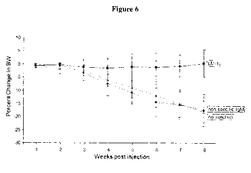
Figure 6 shows body weight of LM-1 injected mice, which was
maintained for 8 weeks post injection. Body weight in the no injection control
and non-specific IgM injected control was reduced by almost 20%, due to
poor health from liver metastasis.
Figure 7 shows data indicating that LM- I antibody can reduce tumor
metastasis establishment, formation, or proliferation (growth).
Figure 8A-8B show BxPC3 cell membrane preparation analysis by 2D
polyacrylamide gel electrophoresis (PAGE). A) Fractionated proteins
transferred to PVDF membrane and stained with LM-1 antibody; and B) Spots
on the PVDF membrane that bound to LM-1, which were suerimposed on a
silverstained PAGE, and the corresponding spots excised from the gel and
subjected to MALDI-TOF analysis.
Figures 9A-9D show identification of LM- 1 Target. A) Gel-
chromatography of BXPC-3 extracts; B)-D) fractions 9 and 10 selected and
subjected to anion-exchange chromatography and subsequent blotting with
LM-1 antibody.
Figures l0A-IOC show siRNA transfected BxPc-3 cells to
downregulate expression of NONO/nmt55 and reduced binding of LM-1. A)
siRNA downregulated NONO/nmt55 expression; B) Binding of LM-1 to
siRNA transfected cells was reduced (arrow) and C) Load Control.
Figures 11A-11D show immunoprecipitation of MKN cells with anti
nmt55 antibody, and subsequent staining with A) anti NONO/nmt55 Mouse
mAb / anti mouse IgG HRP; B), anti mouse IgG HRP; C), LM-1 / anti human
IgM HR; and D), anti human IgM HRP. The top (higher molecular weight)
arrow is NONO, and the bottom (lower molecular weight) arrow is mouse
heavy chain.
Figures 12A-12B show immunoprecipitation of BxPC-3 cells with anti
nmt55, and subsequent staining with A) LM-1; and B) anti NONO/nmt55.
Arrows indicate mnt55 and mouse IgG heavy chain.
Figures 13A-13B show immunoprecipitation of A549 cells with anti
nmt55 and subsequent staining with A) anti NONO/nmt55; and B) LM-1.
Arrows indicate positions of nmt55 and mouse IgG heavy chain.
22
50035I832v1
CA 02728347 2010-12-16
WO 2010/004438 PCT/IB2009/007082
23
Figures 14A-14B show data indicative of LM-1 binding to
recombinantly expressed NONO/nmt55-6xHis protein.
Figure 15 show data indicative of LM-1 binding to bacterially
expressed NONO/nmt55-6xHis protein.
Figures 16A-16C show a polyacrylamide gel electrophoresis (PAGE)
analysis of A) nmt55 expression Lane 1) Novex Sharp molecular weight
marker, Lane 2) To sample showing baseline level of protein expression, Lane
3) TFINAL showing level of nmt55 expression post heat induction; B) nmt55
following ProfinaTM purification Lane 1) Novex Sharp molecular weight
marker, Lane 2) Purified and concentrated nmt55 from periplasmic
expression; and C) a western blot of nmt55 following ProfinaTM purification
Lane 1) Novex Sharp molecular weight marker, Lane 2) Purified and
concentrated nmt55 detected using LM-lopt scFv.
Detailed Description
The invention is based, at least in part, on antibodies that bind to
various neoplastic, cancer, tumor and metastatic cells. A non-limiting
exemplary antibody is designated LM-1 antibody, as represented by antibody
produced by a cell line DSMZ Deposit No. DSM ACC 2623, deposited on
November 6, 2003 at the German Collection of Microorganisms and Cell
Cultures ("DSMZ" - Deutsche Sammlung von Mikroorganismen and
Zellkulturen GmbH, Mascheroder Weg lb, 38124 Braunschweig, Germany)
under the terms of the Budapest Treaty, or represented by heavy and light
chain sequences set forth as SEQ ID NOs: 1, 3, 5, 7 or 9, and 11 or 13. LM-1
antibody, represented by antibody produced by a cell line DSMZ Deposit No.
DSM ACC 2623, or represented by heavy and light chain sequences set forth
as SEQ ID NOs:1, 3, 5, 7 or 9, and 11 or 13 is a human IgM antibody that
specifically binds to various neoplastic, cancer, tumor and metastatic cells.
LM-1 therefore binds to an antigen expressed on various neoplastic, cancer,
tumor and metastatic cells. LM-1 is able to inhibit or reduce proliferation of
various neoplastic, cancer, tumor and metastatic cells. LM-1 is also able to
stimulate or induce apoptosis of various neoplastic, cancer, tumor and
metastatic cells.
23
500351832v1
CA 02728347 2010-12-16
WO 2010/004438 PCT/IB2009/007082
24
The invention is also based, at least in part, on identification of a target
of LM-1, i.e., an antigen that binds to LM-1. As disclosed herein, LM-1
antibody binds to non-pou domain-containing octamer-binding protein
(NONO), also known as 54 kDa nuclear RNA- and DNA-binding protein
(p54nrb) and 55 kDa nuclear protein (nmt55). NONO/nmt55 can be target for
treatment of a neoplasia, cancer, tumor or metastasis. NONO/nmt55 can be a
diagnostic indicator of a neoplasia, cancer, tumor or metastasis. For example,
detection of NONO/nmt55 on cell surface can indicate the presence of a
neoplasia, cancer, tumor or metastasis. NONO/nmt55 can be also be a
vaccine. For example, NONO/nmt55 can be adminstered to a subject with a
neoplasia, cancer, tumor or metastasis that expresses cell surface
NONO/nmt55 in order to elicit an immune response against the a neoplasia,
cancer, tumor or metastasis.
Antibodies of the invention include polyclonal and monoclonal
antibodies. Antibodies are proteins which include amino acids, or "residues,"
covalently linked by an amide bond or equivalent. The term "monoclonal,"
when used in reference to an antibody refers to an antibody that is based
upon,
obtained from or derived from a single clone, including any eukaryotic,
prokaryotic, or phage clone. A "monoclonal" antibody is therefore defined
herein structurally, and not the method by which it is produced.
Antibodies of the invention can belong to any antibody class, IgM,
IgG, IgE, IgA, IgD, or subclass. Exemplary subclasses for IgG are IgGI, IgG2,
IgG3 and IgG4.
Antibodies of the invention can have kappa or lambda light chain
sequences, either full length as in naturally occurring antibodies, mixtures
thereof (i.e., fusions of kappa and lambda chain sequences), and
subsequences/fragments thereof. Naturally occurring antibody molecules
contain two kappa or two lambda light chains.
The amino acid sequences and nucleic acid sequences of LM-1
antibody, as represented by various heavy and light chain variable region
sequences, SEQ ID NOs:1-10, are as follows:
24
500351832v1
CA 02728347 2010-12-16
WO 2010/004438 PCT/IB2009/007082
The heavy chain variable region of the human monoclonal antibody
LM-1, as represented by amino acid sequences (SEQ ID NOs:1, 3, 5, 7 and 9)
and nucleic acid sequences (SEQ ID NOs:2, 4, 6, 8 and 10), respectively, with
differences shown in bold, are as follows:
5 Amino acid sequence of LM-1 heavy chain variable (VH) region
sequence, as represented by SEQ ID NO:1:
QVQLQESGPGLV KPSPTLSLTCAVSGGSISSGGYYWS WIRQHPGKGLE
WIGYIYYSGSTYYNPSLKSRVTIS VDTSKNQFSLKLSS VTAADTAVYY
CARVDARYDYVWGSYRYDAFDIWGQGTMVTVSS
10 Amino acid sequence of LM-1 heavy chain variable (VH) region
sequence, as represented by SEQ ID NO:3 (1BTA1.16VH):
QVQLQESGPGLVKPSQTLSLTCAVSGGSISSGGYYWSWIRQHPGKGLE
WIGYIYYSGSTYYNPSLKSRVTIS VDTS KNQFSLKLSS VTAADTAVYYC
ARVDARYDYVWGSYRYDAFDIWGQGTMVTVSS
15 Amino acid sequence of LM-1 heavy chain variable (VH) region
sequence, as represented by SEQ ID NO:5 (1BTA1.7 VH):
QVQLQESGPGLVKPSPTLSLTCAVSGGSISSGGYYWSWIRQHPGKGLE
WIGYIYYSGSTYYNPSLKSRVTISVDTS KNQFSLKLSSVTAADTAVYYC
ARVDARYDYVWGSYRFDAFDIWGQGTMVTVSS
20 Amino acid sequence of LM-1 heavy chain variable (VH) region
sequence, as represented by SEQ ID NO:7 (1BTA2.5 VH):
QLQLQES GPGL V KPS QTLS LTCT V S GGS IS S GGYYW S W IRQHPG KGLE
WIGYIYYSGSTYYNPSLKSRVTIS VDTSKNQFSLKLSS VTAADTAVYYC
ARVDARYDYVWGSYRYDAFDIWGQGTMVTVSS
25 Amino acid sequence of LM-1 heavy chain variable (VH) region
sequence, as represented by SEQ ID NO:9 (VHLlopt):
EVQLVESGGGLV QPGGSLRLSCAV SGGSISSGGYYWS WIRQAPGKGL
EWVIGYIYYSGSTYYADS VKGRFTISRDNSKNTLYLQMNSLRAEDTA
VYYCARVDARYDYV WGSYRYDAFDIWGQGTLVTV S S
500351832vl
CA 02728347 2010-12-16
WO 2010/004438 PCT/IB2009/007082
26
Nucleotide sequence of LM-1 heavy chain variable (VH) region
sequence, as represented by SEQ ID NO:2:
CCGACCCTGT CCCTCACCTG CGCTGTCTCT GGTGGCTCCA TCAGCAGTGG
TGGTTACTAC 60
TGGAGCTGGA TCCGCCAGCA CCCAGGGAAG GGCCTGGAGT GGATTGGGTA
CATCTATTAC 120
AGTGGGAGCA CCTACTACAA CCCGTCCCTC AAGAGTCGAG TTACCATATC
AGTAGACACG 180
TCTAAGAACC AGTTCTCCCT GAAGCTGAGC TCTGTGACTG CCGCGGACAC
GGCCGTGTAT 240
TACTGTGCGA GAGTTGATGC GCGATATGAT TACGTTTGGG GGAGTTATCG
TTATGATGCT 300
TTTGATATCT GGGGCCAAGG AACCCTGGTC ACCGTCTCTT CA 333
Nucleotide sequence of LM-1 heavy chain variable (VH) region
sequence, as represented by SEQ ID NO:4 (1BTA1. 16VH):
CAGGTGCAGCTGCAGGAGTCGGGCCCAGGACTGGTGAAGCCTTCA
CAGACCCTGTCCCTCACCTGCGCTGTCTCTGGTGGCTCCATCAGCA
GTGGTGGTTACTACTGGAGCTGGATCCGCCAGCACCCAGGGAAGG
GCCTGGAGTGGATTGGGTACATCTATTACAGTGGGAGCACCTACTA
CAACCCGTCCCTCAAGAGTCGAGTTACCATATCAGTAGACACGTCT
AAGAACCAGTTCTCCCTGAAGCTGAGCTCTGTGACTGCCGCGGACA
CGGCCGTGTATTACTGTGCGAGAGTTGATGCGCGATATGATTACGT
TTGGGGGAGTTATCGTTATGATGCTTTTGATATCTGGGGCCAAGGG
ACAATGGTCACCGTCTCTTCA
Nucleotide sequence of LM-1 heavy chain variable (VH) region
sequence, as represented by SEQ ID NO:6 (1BTA1.7 VH):
CAGGTGCAGCTGCAGGAGTCGGGCCCAGGACTGGTGAAGCCTTCA
CCGACCCTGTCCCTCACCTGCGCTGTCTCTGGTGGCTCCATCAGCA
GTGGTGGTTACTACTGGAGCTGGATCCGCCAGCACCCAGGGAAGG
GCCTGGAGTGGATTGGGTACATCTATTACAGTGGGAGCACCTACTA
CAACCCGTCCCTCAAGAGTCGAGTTACCATATCAGTAGACACGTCT
26
500351832v1
CA 02728347 2010-12-16
WO 2010/004438 PCT/IB2009/007082
27
AAGAACCAGTTCTCCCTGAAGCTGAGCTCTGTGACTGCCGCGGACA
CGGCCGTGTATTACTGTGCGAGAGTTGATGCGCGATATGATTACGT
TTGGGGGAGTTATCGTTTTGATGCTTTTGATATCTGGGGCCAAGGG
ACAATGGTCACCGTCTCTTCA
Nucleotide sequence of LM-1 heavy chain variable (VH) region
sequence, as represented by SEQ ID NO:8 (1BTA2.5 VH):
CAGCTGCAGCTGCAGGAGTCGGGCCCAGGACTGGTGAAGCCTTCA
CAGACCCTGTCCCTCACCTGCACTGTCTCTGGTGGCTCCATCAGCA
GTGGTGGTTACTACTGGAGCTGGATCCGCCAGCACCCAGGGAAGG
GCCTGGAGTGGATTGGGTACATCTATTACAGTGGGAGCACCTACTA
CAACCCGTCCCTCAAGAGTCGAGTTACCATATCAGTAGACACGTCT
AAGAACCAGTTCTCCCTGAAGCTGAGCTCTGTGACTGCCGCGGACA
CGGCCGTGTATTACTGTGCGAGAGTTGATGCGCGATATGATTACGT
TTGGGGGAGTTATCGTTATGATGCTTTTGATATCTGGGGCCAAGGG
ACAATGGTCACCGTCTCTTCA
Nucleotide sequence of LM-1 heavy chain variable (VH) region
sequence, as represented by SEQ ID NO: 10 (VHLIopt):
GAGGTGCAGCTGGTCGAGAGCGGGGGAGGCCTGGTGCAGC
CAGGGGGATCTCTGAGACTGAGCTGCGCCGTGAGCGGCGGATCTAT
TTCCAGCGGGGGATATTATTGGTCTTGGATCAGACAGGCTCCCGGA
AAGGGGCTGGAATGGGTCATCGGCTACATCTACTACAGCGGCAGC
ACCTACTACGCCGACAGCGTGAAGGGCCGGTTCACCATCAGCCGG
GACAACAGCAAGAACACCCTGTACCTGCAGATGAACAGCCTGCGG
GCCGAGGACACCGCGGTGTACTACTGCGCCAGAGTGGACGCCAGA
TACGACTACGTGTGGGGCAGCTACAGATACGACGCCTTCGACATCT
GGGGCCAGGGCACCCTGGTGACCGTGTCTTCT
The light chain variable regions of the human monoclonal antibody
LM-1, as represented by amino acid sequences (SEQ ID NO:11 and 13) and
nucleic acid sequences (SEQ ID NO:12 and 14), respectively, are as follows:
Amino acid sequence of LM-1 light chain variable (VL) region
sequence, as represented by SEQ ID NO: 11:
QSVLTQPPSVSAAPGQKVTISCSGSSSNIGNNYVSWYQQLPGTAPKLLI
27
500351832v!
CA 02728347 2010-12-16
WO 2010/004438 PCT/IB2009/007082
28
YDNNKRPSGIPDRFS GS KSGTSATLGITGLQTGDEADYYCGTWDS SLS
AGWVFGGGTKLTVLGQ
Amino acid sequence of LM-1 light chain variable (VL) region
sequence, as represented by SEQ ID NO:13 (VKLlopt):
DIQMTQSPSSLSASVGDRVTITCRSGSSSNIGNNYVSWYQQKPGKAPK
LLIYDNNKEPSGVPSRFSGS GSGTDFTLTISSLQPEDFATYYCQGT WD
SSLSAGWVFGQGTKVEIKR
Amino acid sequence of LM-1 light chain (L) sequence, as represented
by SEQ ID NO:15:
MACPGFLWALVISTCLEFSMASWAQSVLTQPPSVSAAPGQKVTISCSG
SS SNIGNNY V S WYQQLPGTAPKLLIYDNNKRPSGIPDRFS GS KSGTSAT
LGITGLQTGDEADYYCGTWDS SLSAGW VFGGGTKLTVLGQPKAAPS V
TLFPPS
SEELQANKATLVCLISDFYPGAVTVAWKADSSPVKAGVETTTPSKQSN
N KYAASSYLSLTPEQWKSHKSYSCQVTHEGSTVEKTVAPTECS
Nucleotide sequence of LM-1 light chain variable (VL) region
sequence, as represented by SEQ ID NO:12 (1BTAl.16 VL):
CAGTCTGTGTTGACGCAGCCGCCCTCAGTGTCTGCGGCCCCAGGAC
AGAAGGTCACCATCTCCTGCTCTGGAAGCAGCTCCAACATTGGGAA
TAATTATGTATCCTGGTACCAGCAGCTCCCAGGAACAGCCCCCAAA
CTCCTCATTTATGACAATAATAAGCGACCCTCAGGGATTCCTGACC
GATTCTCTGGCTCCAAGTCTGGCACGTCAGCCACCCTGGGCATCAC
CGGACTCCAGACTGGGGACGAGGCCGATTATTACTGCGGAACATG
GGATAGCAGCCTGAGTGCTGGTTGGGTGTTCGGCGGAGGGACCAA
GCTGACCGTCCTAGGTCAG
Nucleotide sequence of LM-1 light chain variable (VL) region
sequence, as represented by SEQ ID NO: 14 (VKLlopt):
GACATCCAGATGACCCAGAGCCCCAGCAGCCTGAGCGCCAGCGTG
GGCGACAGAGTGACCATCACCTGCAGAAGCGGCAGCAGCAGCAAC
ATCGGCAACAATTATGTCTCTTGGTATCAGCAGAAACCTGGCAAGG
CCCCCAAGCTGCTGATCTACGACAACAACAAAGAACCCAGCGGCG
TGCCCAGCCGGTTTAGCGGCAGCGGCTCCGGCACCGACTTCACCCT
GACCATCAGCAGCCTGCAGCCCGAGGATTTCGCCACCTACTACTGT
28
500351832v1
CA 02728347 2010-12-16
WO 2010/004438 PCT/IB2009/007082
29
CAGGGGACATGGGATAG
CAGCCTGTCCGCCGGCTGGGTGTTCGGCCAGGGAACAAAG
GTGGAGATCAAGAGA
Predicted CDRs, of which there are three in each of heavy and light
chain, are conveniently denoted herein as HC-CDR I, HC-CDR2 and HC-
CDR3; and LC-CDR I, LC-CDR2 and LC-CDR3. CDR positions were based
upon upon definitiosn of Kabat (e.g., Sequences of Proteins of Immunological
Interest, 4th Ed. US Department of Health and Human Services. Public Health
Service (1987), and Kabat et al., Sequences of Proteins of Immunological
Interest, 5th Ed. US Department of Health and Human Services, Public Health
Service (1991)), except numbering of CDRs is based upon amino acid residue
number of the sequences set forth herein beginning from the amino-terminus
and does not follow the Kabat numbering system. Heavy chain variable
region CDR placement was modeled after herceptin antibody variable region
(PDB file 1N8Z) due to 95% sequence identity with the framework residues,
and light chain variable region CDR placement was modeled after PDB file
2RHEa due to 82% sequence identity with the framework residues.
Predicted CDR sequences of exemplary heavy variable region chain
are CDR I; VSGGSISSGGYY, CDR2; YIYYSGSTYYNPSLKS, and CDR3;
VDARYDYVWGSYRYDAFDI. CDRI of heavy chain spans nucleotides 72-
105 which encode amino acids 24-35, CDR2 spans nucleotides 156-201 which
encode amino acids 52-67, and CDR3 spans nucleotides 300-354 which
encode amino acids 100-118.
Predicted CDR sequences of exemplary light variable region chain are
90-101, CDRI; SGSSSNIGNNYVS, CDR2; DNNKRPSG, and CDR3;
GTWDSSLSAGWV. CDR1 of lambda light chain spans nucleotides 69-105
which encode amino acids located at positions 23-35. CDR2 spans nucleotides
153-174 which encode amino acids 51-58 and CDR3 spans nucleotides 270-
303 and encode amino acids 90-101.
In accordance with the invention, there are provided isolated and
purified antibodies and functional (e.g., cell or antigen binding) fragments
structurally and/or functionally related to LM- 1 antibody, as represented by
antibody produced by a cell line DSMZ Deposit No. DSM ACC 2623, or
29
500351832v1
CA 02728347 2010-12-16
WO 2010/004438 PCT/IB2009/007082
represented by heavy and light chain sequences set forth as SEQ ID NOs:1, 3,
5, 7 or 9, and 11 or 13, respectively. In various embodiments, antibodies and
functional fragments compete with LM-1 antibody, as represented by antibody
produced by a cell line DSMZ Deposit No. DSM ACC 2623, or represented
5 by heavy and light chain sequences set forth as SEQ ID NOs:1, 3, 5, 7 or 9,
and 11 or 13 for binding to a cell or antigen (e.g., NONO/nmt55). In
additional embodiments, antibodies and functional fragments compete with
LM-1 antibody, as represented by antibody produced by a cell line DSMZ
Deposit No. DSM ACC 2623, or represented by heavy and light chain
10 sequences set forth as SEQ ID NOs:1, 3, 5, 7 or 9, and 11 or 13 for binding
to
an antigen (e.g., NONO/nmt55), or an adenocarcinoma cell or a squamous cell
carcinoma.
In accordance with the invention, there are also provided isolated and
purified antibodies and functional (e.g., cell or antigen binding) fragments
that
15 bind to NONO/nmt55 protein. In various embodiments, antibodies and
functional fragments bind to an N-terminal NONO-nmt55 amino acid
sequence region (e.g., amino acids 1-300 of NONO-nmt55). In one aspect,
NONO-nmt55 includes a sequence set forth as:
1 MQSNKTFNLE KQNHTPRKHH QHHHQQQHHQ QQQQQPPPPP IPANGQQASS
20 51 QNEGLTIDLK NFRKPGEKTF TQRSRLFVGN LPPDITEEEM RKLFEKYGKA
101 GEVFIHKDKG FGFIRLETRT LAEIAKVELD NMPLRGKQLR VRFACHSASL
151 TVRNLPQYVS NELLEEAFSV FGQVERAVVI VDDRGRPSGK GIVEFSGKPA
201 ARKALDRCSE GSFLLTTFPR PVTVEPMDQL DDEEGLPEKL VIKNQQFHKE
251 REQPPRFAQP GSFEYEYAMR WKALIEMEKQ QQDQVDRNIK EAREKLEMEM
25 301 EAARHEHQVM LMRQDLMRRQ EELRRMEELH NQEVQKRKQL ELRQEEERRR
351 REEEMRRQQE EMMRRQQEGF KGTFPDAREQ EIRMGQMAMG GAMGINNRGA
401 MPPAPVPAGT PAPPGPATMM PDGTLGLTPP TTERFGQAAT MEGIGAIGGT
451 PPAFNRAAPG AEFAPNKRRR Y
30 In another aspect, NONO-nmt55 includes a sequence set forth as:
1 MQSNKTFNLE KQNHTPRKHH QHHHQQQHHQ QQQQQPPPPP IPANGQQASS
51 QNEGLTIDLK NFRKPGEKTF TQRSRLFVGN LPPDITEEEM RKLFEKYGKA
101 GEVFIHKDKG FGFIRLETRT LAEIAKVELD NMPLRGKQLR VRFACHSASL
151 TVRNLPQYVS NELLEEAFSV FGQVERAVVI VDDRGRPSGK GIVEFSGKPA
201 ARKALDRCSE GSFLLTTFPR PVTVEPMDQL DDEEGLPEKL VIKNQQFHKE
251 REQPPRFAQP GSFEYEYAMR WKALIEMEKQ QQDQVDRNIK EAREKLEMEM
In further embodiments, antibodies and functional fragments compete
with LM-1 antibody, as represented by antibody produced by a cell line
DSMZ Deposit No. DSM ACC 2623, or represented by heavy and light chain
500351832x1
CA 02728347 2010-12-16
WO 2010/004438 PCT/IB2009/007082
31
sequences set forth as SEQ ID NOs: 1, 3, 5, 7 or 9, and 11 or 13 for binding
of
to one or more of a stomach adenocarcinoma (e.g., diffuse or intestinal),
colorectal cancer such as adenocarcinoma, ovarian carcinoma, lung cancer,
such as a lung adenocarcinoma, squamous cell lung carcinoma and small cell
lung carcinoma, melanoma, lobular and ductal mammary carcinoma, breast
cancer such as invasive ductal or lobular cancer, gastric cancer, pancreatic
cancer such as pancreatic adenocarcinoma (e.g., ductal), sarcoma,
gastrointestinal cancer such as a stomach cancer, nervous tissue or brain
tumor
such as a glioma, esophageal cancer such as esophagial squamous cell
carcinoma and adenocarcinoma, osteosarcoma, fibrosarcoma, urinary bladder
cancer, prostate cancer such as prostate adenocarcinoma, kidney cancer such
as renal carcinoma, ovarian cancer such as adenocarcinoma, testicular cancer,
endometrial cancer, cervical cancer such as squamous cell and
adenocarcinoma, uterine cancer such as adenocarcinoma, Hodgkin's disease,
lymphoma, and leukemia. In yet additional embodiments, antibodies and
functional fragments compete with LM-1 antibody, as represented by antibody
produced by a cell line DSMZ Deposit No. DSM ACC 2623, or represented
by heavy and light chain sequences set forth as SEQ ID NOs:1, 3, 5, 7 or 9,
and 11 or 13 for binding to an antigen (e.g., NONO/nmt55), or to one or more
of a stomach adenocarcinoma (e.g., diffuse or intestinal), colorectal cancer
such as adenocarcinoma, ovarian carcinoma, lung cancer, such as a lung
adenocarcinoma, squamous cell lung carcinoma and small cell lung
carcinoma, melanoma, lobular and ductal mammary carcinoma, breast cancer
such as invasive ductal or lobular cancer, gastric cancer, pancreatic cancer
such as pancreatic adenocarcinoma (e.g., ductal), sarcoma, gastrointestinal
cancer such as a stomach cancer, nervous tissue or brain tumor such as a
glioma, esophageal cancer such as esophagial squamous cell carcinoma and
adenocarcinoma, osteosarcoma, fibrosarcoma, urinary bladder cancer, prostate
cancer such as prostate adenocarcinoma, kidney cancer such as renal
carcinoma, ovarian cancer such as adenocarcinoma, testicular cancer,
endometrial cancer, cervical cancer such as squamous cell and
adenocarcinoma, uterine cancer such as adenocarcinoma, Hodgkin's disease,
lymphoma, and leukemia. In still further embodiments, antibodies and
functional fragments compete with LM- 1 antibody, as represented by antibody
31
500351832v1
CA 02728347 2010-12-16
WO 2010/004438 PCT/IB2009/007082
32
produced by a cell line DSMZ Deposit No. DSM ACC 2623, or represented
by heavy and light chain sequences set forth as SEQ ID NOs:1, 3, 5, 7 or 9,
and 11 or 13 for binding to lung adenocarinoma cell line Colo-699 (DSMZ
accession number ACC 196), lung adenocarinoma cell line DV-90 (DSMZ
accession number ACC 307), epidermoid lung carcinoma cell line EPLC-
272H (DSMZ accession number ACC 383), or lung squamous cell carcinoma
cell line LOU-NH91 (DSMZ accession number ACC 393) cells. In particular
aspects, antibodies and functional fragments competitively inhibit binding of
LM-1 antibody, as represented by antibody produced by a cell line DSMZ
Deposit No. DSM ACC 2623, or represented by heavy and light chain
sequences set forth as SEQ ID NOs: 1, 3, 5, 7 or 9, and 11 or 13 to a cell or
an
antigen (e.g., NONO/nmt55) by at least 30%, 40%, 50%, 60%, 70%, 80%,
90% or more.
In accordance with the invention, there are also provided antibodies
and functional fragments that bind to a cell or an antigen (e.g., NONO/nmt55)
that LM-1 antibody, as represented by antibody produced by a cell line DSMZ
Deposit No. DSM ACC 2623, or represented by heavy and light chain
sequences set forth as SEQ ID NOs:1, 3, 5, 7 or 9, and 11 or 13 binds. In one
embodiment, an isolated or purified antibody or functional fragment thereof
binds to a cell or an antigen (e.g., NONO/nmt55) that LM-1 antibody, as
represented by antibody produced by a cell line DSMZ Deposit No. DSM
ACC 2623, or represented by heavy and light chain sequences set forth as
SEQ ID NOs:1, 3, 5, 7 or 9, and 11 or 13 binds. In particular aspects, the
antibody or functional fragment thereof binds to a cell or an antigen (e.g.,
NONO/nmt55) present on an adenocarcinoma cell or a squamous cell
carcinoma to which LM-1 antibody, as represented by antibody produced by a
cell line DSMZ Deposit No. DSM ACC 2623, or represented by heavy and
light chain sequences set forth as SEQ ID NOs:1, 3, 5, 7 or 9, and 11 or 13
binds. In additional particular aspects, the antibody or functional fragment
thereof binds to one or more of a stomach adenocarcinoma (e.g., diffuse or
intestinal), colorectal cancer such as adenocarcinoma, ovarian carcinoma, lung
cancer, such as a lung adenocarcinoma, squamous cell lung carcinoma and
small cell lung carcinoma, melanoma, lobular and ductal mammary
32
500351832v1
CA 02728347 2010-12-16
WO 2010/004438 PCT/IB2009/007082
33
carcinoma, breast cancer such as invasive ductal or lobular cancer, gastric
cancer, pancreatic cancer such as pancreatic adenocarcinoma (e.g., ductal),
sarcoma, gastrointestinal cancer such as a stomach cancer, nervous tissue or
brain tumor such as a glioma, esophageal cancer such as esophagial squamous
cell carcinoma and adenocarcinoma, osteosarcoma, fibrosarcoma, urinary
bladder cancer, prostate cancer such as prostate adenocarcinoma, kidney
cancer such as renal carcinoma, ovarian cancer such as adenocarcinoma,
testicular cancer, endometrial cancer, cervical cancer such as squamous cell
and adenocarcinoma, uterine cancer such as adenocarcinoma, Hodgkin's
disease, lymphoma, and leukemia, to which LM-1 antibody, as represented by
antibody produced by a cell line DSMZ Deposit No. DSM ACC 2623, or
represented by heavy and light chain sequences set forth as SEQ ID NOs:1, 3,
5, 7 or 9, and 11 or 13 binds. In further particular aspects, the antibody or
functional fragment thereof binds to a cell or an antigen (e.g., NONO/nmt55)
present on a lung adenocarinoma cell line Colo-699 (DSMZ accession number
ACC 196), lung adenocarinoma cell line DV-90 (DSMZ accession number
ACC 307), epidermoid lung carcinoma cell line EPLC-272H (DSMZ
accession number ACC 383), or lung squamous cell carcinoma cell line LOU-
NH91 (DSMZ accession number ACC 393) cell, to which LM-1 antibody, as
represented by antibody produced by a cell line DSMZ Deposit No. DSM
ACC 2623, or represented by heavy and light chain sequences set forth as
SEQ ID NOs:1, 3, 5, 7 or 9, and 11 or 13 binds.
The term "bind," or "binding," when used in reference to an antibody
or functional fragment, means that the antibody or functional fragment
interacts at the molecular level with a corresponding epitope (antigenic
determinant) present on a cell or an antigen (e.g., NONO/nmt55). Epitopes of
antigens that comprise amino acids typically include relatively short
sequences, e.g. about five to 15 amino acids in length. Epitopes can be
contiguous or non-contiguous. A non-contiguous amino acid sequence
epitope forms due to protein folding. Techniques for identifying epitopes are
known to the skilled artisan and include screening overlapping oligopeptides
for binding to antibody (for example, U.S. Patent No. 4,708,871), phage
display peptide library kits, which are commercially available for epitope
33
500351832vl
CA 02728347 2010-12-16
WO 2010/004438 PCT/IB2009/007082
34
mapping (New England BioLabs). Epitopes may also be identified by
inference when epitope length peptide sequences are used to immunize
animals from which antibodies that bind to the peptide sequence are obtained
and can be predicted using computer programs, such as BEPITOPE (Odorico
et al., J. Mol. Recognit. 16:20 (2003)).
The invention further provides antibodies and functional fragments that
inhibit, decrease or reduce cell growth or proliferation, or stimulate or
induce
cell death, lysis or apoptosis. In particular embodiments, binding of LM-1
antibody, as represented by antibody produced by a cell line DSMZ Deposit
No. DSM ACC 2623, or represented by heavy and light chain sequences set
forth as SEQ ID NOs:1, 3, 5, 7 or 9, and 11 or 13 to a neoplastic, tumor or
cancer, or metastasis cell inhibits, decreases or reduces cell growth or
proliferation, or stimulates or induces cell death, lysis or apoptosis. In
another
embodiment, binding of LM-1 antibody, as represented by antibody produced
by a cell line DSMZ Deposit No. DSM ACC 2623, or represented by heavy
and light chain sequences set forth as SEQ ID NOs:1, 3, 5, 7 or 9, and 11 or
13
to a stomach adenocarcinoma (e.g., diffuse or intestinal), colorectal cancer
such as adenocarcinoma, ovarian carcinoma, lung cancer, such as a lung
adenocarcinoma, squamous cell lung carcinoma and small cell lung
carcinoma, melanoma, lobular and ductal mammary carcinoma, breast cancer
such as invasive ductal or lobular cancer, gastric cancer, pancreatic cancer
such as pancreatic adenocarcinoma (e.g., ductal), sarcoma, gastrointestinal
cancer such as a stomach cancer, nervous tissue or brain tumor such as a
glioma, esophageal cancer such as esophagial squamous cell carcinoma and
adenocarcinoma, osteosarcoma, fibrosarcoma, urinary bladder cancer, prostate
cancer such as prostate adenocarcinoma, kidney cancer such as renal
carcinoma, ovarian cancer such as adenocarcinoma, testicular cancer,
endometrial cancer, cervical cancer such as squamous cell and
adenocarcinoma, uterine cancer such as adenocarcinoma, Hodgkin's disease,
lymphoma, or leukemia inhibits, decreases or reduces cell growth or
proliferation, or stimulates or induces cell death, lysis or apoptosis. In a
further embodiment, binding of LM-I antibody, as represented by antibody
produced by a cell line DSMZ Deposit No. DSM ACC 2623, or represented
34
500351832v!
CA 02728347 2010-12-16
WO 2010/004438 PCT/IB2009/007082
by heavy and light chain sequences set forth as SEQ ID NOs:1, 3, 5, 7 or 9,
and 11 or 13 to lung adenocarinoma cell line Colo-699 (DSMZ accession
number ACC 196), lung adenocarinoma cell line DV-90 (DSMZ accession
number ACC 307), epidermoid lung carcinoma cell line EPLC-272H (DSMZ
5 accession number ACC 383), or lung squamous cell carcinoma cell line LOU-
NH91 (DSMZ accession number ACC 393) cells.
The invention moreover provides of antibodies and functional
fragments that are structurally and/or functionally related to LM-1 antibody,
as
represented by antibody produced by a cell line DSMZ Deposit No. DSM
10 ACC 2623, or represented by heavy and light chain sequences set forth as
SEQ ID NOs:1, 3, 5, 7 or 9, and 11 or 13 which includes a heavy or light chain
variable region sequence that exhibits a degree of identity to SEQ ID NOs: 1,
3, 5, 7 or 9, and 11 or 13 or that exhibits a degree of identity to a sequence
within SEQ ID NOs: 1, 3, 5, 7 or 9, and 11 or 13 (e.g., one or more CDRs, such
15 as amino acids 24-35, 52-67, or 100-118 of SEQ ID NO:1, 3, 5 or 7, or amino
acids 23-35, 51-58 or 90-101 of SEQ ID NO:11). In particular embodiments,
antibodies and functional fragments include a heavy or a light chain variable
region sequence with about 60% or more identity to a heavy or light chain
sequence variable region of LM-1 antibody, as represented by antibody
20 produced by a cell line DSMZ Deposit No. DSM ACC 2623, or represented
by heavy and light chain sequences set forth as SEQ ID NOs: 1, 3, 5, 7 or 9,
and 11 or 13 or a sequence within LM-1 antibody, as represented by antibody
produced by a cell line DSMZ Deposit No. DSM ACC 2623, or represented
by heavy and light chain sequences set forth as SEQ ID NOs: 1, 3, 5, 7 or 9,
25 and 11 or 13 (e.g., one or more CDRs, such as amino acids 24-35, 52-67 or
100-118 of SEQ ID NO:1, 3, 5, or 7, or amino acids 23-35, 51-58 or 90-101 of
SEQ ID NO: 11). In other particular embodiments, antibodies or functional
fragments include a heavy or a light chain with at least 65%, 70%, 75%, 80%,
85%, 90%, 95%, or more identity to a heavy chain variable region sequence of
30 LM-1 antibody, as represented by antibody produced by a cell line DSMZ
Deposit No. DSM ACC 2623, or represented by heavy and light chain
sequences set forth as SEQ ID NOs:1, 3, 5, 7 or 9, and 11 or 13 or a sequence
within LM-1 antibody, as represented by antibody produced by a cell line
500351832v!
CA 02728347 2010-12-16
WO 2010/004438 PCT/IB2009/007082
36
DSMZ Deposit No. DSM ACC 2623, or represented by heavy and light chain
sequences set forth as SEQ ID NOs:1, 3, 5, 7 or 9, and 11 or 13 (e.g., one or
more CDRs, such as amino acids 24-35, 52-67, or 100-118 of SEQ ID NO:1,
3, 5 or 7, or amino acids 23-35, 51-58 or 90-101 of SEQ ID NO:11). In
additional particular embodiments, antibodies or functional fragments include
a heavy or a light chain variable region sequence with at least 80-85%, 85-
90%, 90-95%, 95-100% identity to one or more CDRs in LM-1 antibody, as
represented by antibody produced by a cell line DSMZ Deposit No. DSM
ACC 2623, or represented by heavy and light chain sequences set forth as
SEQ ID NOs:1, 3, 5, 7 or 9, and 11 or 13 (e.g., amino acids 24-35, 52-67, or
100-118 of SEQ ID NO:1, 3, 5 or 7, or amino acids 23-35, 51-58 or 90-101 of
SEQ ID NO:11). In a particular aspect, an antibody or a functional fragment
thereof includes a heavy or a light chain variable region sequence with 95-
100% identity to one, two or three CDRs in each heavy or light chain variable
region sequences in LM-1 antibody, as represented by antibody produced by a
cell line DSMZ Deposit No. DSM ACC 2623, or represented by heavy and
light chain sequences set forth as SEQ ID NOs:1, 3, 5, 7 or 9, and 11 or 13
(e.g., amino acids 24-35, 52-67, or 100-118 of SEQ ID NO:1, 3, 5 or 7, or
amino acids 23-35, 51-58 or 90-101 of SEQ ID NO: 11).
Antibodies and functional fragments of the invention therefore include
those with at least partial sequence identity to LM-1 antibody, as represented
by antibody produced by a cell line DSMZ Deposit No. DSM ACC 2623, or
represented by heavy and light chain sequences set forth as SEQ ID NOs: 1, 3,
5, 7 or 9, and 11 or 13. The percent identity of such antibodies and
functional
fragments can be as little as 60%, or can be more (e.g., 65%, 70%, 75%, 80%,
85%, 90%, 95%, etc.).
The percent identity can extend over the entire sequence length of LM-
1 antibody, as represented by antibody produced by a cell line DSMZ Deposit
No. DSM ACC 2623, or represented by heavy and light chain sequences set
forth as SEQ ID NOs:1, 3, 5, 7 or 9, and 11 or 13 or a contiguous region or
area within LM-1 antibody, as represented by antibody produced by a cell line
DSMZ Deposit No. DSM ACC 2623, or represented by heavy and light chain
sequences set forth as SEQ ID NOs:1, 3, 5, 7 or 9, and 11 or 13. In particular
36
500351832v1
CA 02728347 2010-12-16
WO 2010/004438 PCT/IB2009/007082
37
aspects, the length of the sequence sharing the percent identity is 5 or more
contiguous amino acids, e.g., 5, 6, 7, 8, 9, 10, 11, 12, 13, 14, 15, 16, 17,
18,
19, 20, 21, 22, 23, 24, 25, etc. contiguous amino acids. In additional
particular
aspects, the length of the sequence sharing the percent identity is 25 or more
contiguous amino acids, e.g., 26, 27, 28, 29, 30, 31, 32, 33, 34, 35, etc.
contiguous amino acids. In further particular aspects, the length of the
sequence sharing the percent identity is 35 or more contiguous amino acids,
e.g., 35, 36, 37, 38, 39, 40, 41, 42, 43, 44, 45, 45, 47, 48, 49, 50, etc.,
contiguous amino acids. In yet additional particular aspects, the length of
the
sequence sharing the percent identity is 50 or more contiguous amino acids,
e.g., 50-55, 55-60, 60-65, 65-70, 70-75, 75-80, 80-85, 85-90, 90-95, 95-100,
100-110, etc. contiguous amino acids. In yet further particular aspects, the
length of the sequence sharing the percent identity is equal to the length of
any
CDR of a variable region sequence (e.g., amino acids 24-35, 52-67, or 100-
118 of SEQ ID NO: 1, 3, 5, 7 or 9, or amino acids 23-35, 51-58 or 90- 101 of
SEQ ID NO: 11), or a region outside the CDRs but within the variable region
of a heavy or light chain sequence, such as LM-1 antibody, as represented by
antibody produced by a cell line DSMZ Deposit No. DSM ACC 2623, or
represented by heavy and light chain sequences set forth as SEQ ID NOs: 1, 3,
5 or 7, and 9.
The term "identity" and grammatical variations thereof, mean that two
or more referenced entities are the same. Thus, where two antibody sequences
are identical, they have the same amino acid sequence, at least within the
referenced region or portion. Where two nucleic acid sequences are identical,
they have the same polynucleotide sequence, at least within the referenced
region or portion. The identity can be over a defined area (region or domain)
of the sequence. An "area of identity" refers to a portion of two or more
referenced entities that are the same. Thus, where two protein or nucleic acid
sequences are identical over one or more sequence regions they share identity
within that region. Exemplary identity are antibodies and functional
fragments with an amino acid sequence with 50%, 60%, 70%, 75%, 80%,
85%, 90%, 95%, 96%, 97%, 98%, or more sequence identity to a reference
antibody or functional fragment, for example, LM-1 antibody, as represented
37
500351832v1
CA 02728347 2010-12-16
WO 2010/004438 PCT/IB2009/007082
38
by antibody produced by a cell line DSMZ Deposit No. DSM ACC 2623, or
represented by heavy and light chain sequences set forth as SEQ ID NOs: 1, 3,
5, 7 or 9, and 11 or 13 or a subsequence thereof.
The terms "homologous" or "homology" mean that two or more
referenced entities share at least partial identity over a given region or
portion.
"Areas, regions or domains" of homology or identity mean that a portion of
two or more referenced entities share homology or are the same. Thus, where
two antibody sequences are identical over one or more sequence regions they
share identity in these regions. "Substantial homology" means that a molecule
is structurally or functionally conserved such that it has or is predicted to
have
at least partial structure or function of one or more of the structures or
functions (e.g., a biological function) of the reference molecule, or
relevant/corresponding region or portion of the reference molecule to which it
shares homology. An antibody or functional fragment with substantial
homology has or is predicted to have at least partial activity or function as
the
reference antibody. For example, in a particular embodiment, a LM-1
antibody, as represented by antibody produced by a cell line DSMZ Deposit
No. DSM ACC 2623, or represented by heavy and light chain sequences set
forth as SEQ ID NOs:1, 3, 5, 7 or 9, and 11 or 13 with one or more
modifications (e.g., substitutions, deletions or additions of LM-1 antibody,
as
represented by antibody produced by a cell line DSMZ Deposit No. DSM
ACC 2623, or represented by heavy and light chain sequences set forth as
SEQ ID NOs:1, 3, 5, 7 or 9, and 11 or 13) retain the ability to at least
partially
compete for binding of LM-1 antibody, as represented by antibody produced
by a cell line DSMZ Deposit No. DSM ACC 2623, or represented by heavy
and light chain sequences set forth as SEQ ID NOs:1, 3, 5, 7 or 9, and 11 or
13
to a cell or an antigen (e.g., NONO/nmt55), or at least retains partial
binding
to a cell or antigen to which LM-1 antibody, as represented by antibody
produced by a cell line DSMZ Deposit No. DSM ACC 2623, or represented
by heavy and light chain sequences set forth as SEQ ID NOs:1, 3, 5, 7 or 9,
and 11 or 13 binds is considered to have substantial homology to LM-I
antibody, as represented by antibody produced by a cell line DSMZ Deposit
38
500351832v1
CA 02728347 2010-12-16
WO 2010/004438 PCT/IB2009/007082
39
No. DSM ACC 2623, or represented by heavy and light chain sequences set
forth as SEQ ID NOs: 1, 3, 5, 7 or 9, and 11 or 13.
The extent of identity (homology) between two sequences can be
ascertained using a computer program and mathematical algorithm known in
the art. Such algorithms that calculate percent sequence identity (homology)
generally account for sequence gaps and mismatches over the comparison
region or area. For example, a BLAST (e.g., BLAST 2.0) search algorithm
(see, e.g., Altschul et al., J. Mol. Biol. 215:403 (1990), publicly available
through NCBI) has exemplary search parameters as follows: Mismatch -2;
gap open 5; gap extension 2. For polypeptide sequence comparisons, a
BLASTP algorithm is typically used in combination with a scoring matrix,
such as PAM100, PAM 250, BLOSUM 62 or BLOSUM 50. FASTA (e.g.,
FASTA2 and FASTA3) and SSEARCH sequence comparison programs are
also used to quantitate the extent of identity (Pearson et al., Proc. Natl.
Acad.
Sci. USA 85:2444 (1988); Pearson, Methods Mol Biol. 132:185 (2000); and
Smith et al., J. Mol. Biol. 147:195 (1981)). Programs for quantitating protein
structural similarity using Delaunay-based topological mapping have also been
developed (Bostick et al., Biochem Biophys Res Commun. 304:320 (2003)).
Antibodies and functional fragments of the invention include those that
retain at least one or more partial activities or functions of LM-1 antibody,
as
represented by antibody produced by a cell line DSMZ Deposit No. DSM
ACC 2623, or represented by heavy and light chain sequences set forth as
SEQ ID NOs:1, 3, 5, 7 or 9, and 11 or 13. As disclosed herein, the antigen
(e.g., NONO/nmt55) to which LM-1 antibody, as represented by antibody
produced by a cell line DSMZ Deposit No. DSM ACC 2623, or represented
by heavy and light chain sequences set forth as SEQ ID NOs: 1, 3, 5, 7 or 9,
and 11 or 13 binds is expressed on malignant and non-malignant, neoplastic,
tumor and cancer cells. Non-limiting examples of cells that bind to LM-1
antibody, as represented by antibody produced by a cell line DSMZ Deposit
No. DSM ACC 2623, or represented by heavy and light chain sequences set
forth as SEQ ID NOs:1, 3, 5, 7 or 9, and 11 or 13 and therefore express a
target antigen of LM-1 include one or more of a stomach adenocarcinoma
(e.g., diffuse or intestinal), colorectal cancer such as adenocarcinoma,
ovarian
39
500351832vI
CA 02728347 2010-12-16
WO 2010/004438 PCT/IB2009/007082
carcinoma, lung cancer, such as a lung adenocarcinoma, squamous cell lung
carcinoma and small cell lung carcinoma, melanoma, lobular and ductal
mammary carcinoma, breast cancer such as invasive ductal and lobular cancer,
gastric cancer, pancreatic cancer such as pancreatic adenocarcinoma (e.g.,
5 ductal), sarcoma, gastrointestinal cancer such as a stomach cancer, nervous
tissue or brain tumor such as a glioma, esophageal cancer such as esophagial
squamous cell carcinoma and adenocarcinoma, osteosarcoma, fibrosarcoma,
urinary bladder cancer, prostate cancer such as prostate adenocarcinoma,
kidney cancer such as renal carcinoma, ovarian cancer such as
10 adenocarcinoma, testicular cancer, endometrial cancer, cervical cancer such
as
squamous cell and adenocarcinoma, uterine cancer such as adenocarcinoma,
Hodgkin's disease, lymphoma, and leukemia, or lung adenocarinoma cell line
Colo-699 (DSMZ accession number ACC 196), lung adenocarinoma cell line
DV-90 (DSMZ accession number ACC 307), epidermoid lung carcinoma cell
15 line EPLC-272H (DSMZ accession number ACC 383), or lung squamous cell
carcinoma cell line LOU-NH91 (DSMZ accession number ACC 393) cells.
Thus, in various embodiments, an antibody or functional fragment binds to an
antigen (e.g., NONO/nmt55), or one or more cells, such as a stomach
adenocarcinoma (e.g., diffuse or intestinal), colorectal cancer such as
20 adenocarcinoma, ovarian carcinoma, lung cancer, such as a lung
adenocarcinoma, squamous cell lung carcinoma or small cell lung carcinoma,
melanoma, lobular or ductal mammary carcinoma, breast cancer such as
invasive ductal or lobular cancer, gastric cancer, pancreatic cancer such as
pancreatic adenocarcinoma (e.g., ductal), sarcoma, gastrointestinal cancer
such
25 as a stomach cancer, nervous tissue or brain tumor such as a glioma,
esophageal cancer such as esophagial squamous cell carcinoma or
adenocarcinoma, osteosarcoma, fibrosarcoma, urinary bladder cancer, prostate
cancer such as prostate adenocarcinoma, kidney cancer such as renal
carcinoma, ovarian cancer such as adenocarcinoma, testicular cancer,
30 endometrial cancer, cervical cancer such as squamous cell and
adenocarcinoma, uterine cancer such as adenocarcinoma, Hodgkin's disease,
lymphoma, or leukemia, or lung adenocarinoma cell line Colo-699 (DSMZ
accession number ACC 196), lung adenocarinoma cell line DV-90 (DSMZ
accession number ACC 307), epidermoid lung carcinoma cell line EPLC-
500351832v1
CA 02728347 2010-12-16
WO 2010/004438 PCT/IB2009/007082
41
272H (DSMZ accession number ACC 383), or lung squamous cell carcinoma
cell line LOU-NH91 (DSMZ accession number ACC 393) cells.
Antibodies and functional fragments that bind to a cell or an antigen
(e.g., NONO/nmt55) to which LM-1 antibody, as represented by antibody
produced by a cell line DSMZ Deposit No. DSM ACC 2623, or represented
by heavy and light chain sequences set forth as SEQ ID NOs:1, 3, 5, 7 or 9,
and 11 or 13 binds can have greater or less relative binding affinity for a
cell
or an antigen (e.g., NONO/nmt55) than LM-1 antibody, as represented by
antibody produced by a cell line DSMZ Deposit No. DSM ACC 2623, or
represented by heavy and light chain sequences set forth as SEQ ID NOs:1, 3,
5, 7 or 9, and 11 or 13. Additional antibodies and functional fragments of the
invention therefore include those that have greater than, about the same or
less
than the binding affinity of LM-1 antibody, as represented by antibody
roduced by a cell line DSMZ Deposit No. DSM ACC 2623, or represented by
heavy and light chain sequences set forth as SEQ ID NOs:1, 3, 5, 7 or 9, and
11 or 13 for binding to a cell or an antigen (e.g., NONO/nmt55). For example,
an antibody or functional fragment of the invention may have an affinity
greater or less than 2-5, 5-10, 10-100, 100-1000 or 1000-10,000-fold affinity,
or any numerical value or range within or encompassing such values, than
LM-1 antibody, as represented by antibody produced by a cell line DSMZ
Deposit No. DSM ACC 2623, or represented by heavy and light chain
sequences set forth as SEQ ID NOs: 1, 3, 5, 7 or 9, and 11 or 13. In one
embodiment, an antibody or a functional thereof has a binding affinity within
about 1-5000 fold of the binding affinity of LM-1 antibody, as represented by
antibody produced by a cell line DSMZ Deposit No. DSM ACC 2623, or
represented by heavy and light chain sequences set forth as SEQ ID NOs:1, 3,
5, 7 or 9, and 11 or 13 for binding to an antigen (e.g., NONO/nmt55), or to a
neplastic, cancer, tumor or metastatic cell. In another embodiment, an
antibody or a functional thereof has a binding affinity within about 1-5000
fold of the binding affinity of LM-1 antibody, as represented by antibody
produced by a cell line DSMZ Deposit No. DSM ACC 2623, or represented
by heavy and light chain sequences set forth as SEQ ID NOs:1, 3, 5, 7 or 9,
and 11 or 13 for binding to an antigen (e.g., NONO/nmt55), or to a stomach
41
500351832vl
CA 02728347 2010-12-16
WO 2010/004438 PCT/IB2009/007082
42
adenocarcinoma (e.g., diffuse or intestinal), colorectal cancer such as
adenocarcinoma, ovarian carcinoma, lung cancer, such as a lung
adenocarcinoma, squamous cell lung carcinoma or small cell lung carcinoma,
melanoma, lobular or ductal mammary carcinoma, breast cancer such as
invasive ductal or lobular cancer, gastric cancer, pancreatic cancer such as
pancreatic adenocarcinoma (e.g., ductal), sarcoma, gastrointestinal cancer
such
as a stomach cancer, nervous tissue or brain tumor such as a glioma,
esophageal cancer such as esophagial squamous cell carcinoma or
adenocarcinoma, osteosarcoma, fibrosarcoma, urinary bladder cancer, prostate
cancer such as prostate adenocarcinoma, kidney cancer such as renal
carcinoma, ovarian cancer such as adenocarcinoma, testicular cancer,
endometrial cancer, cervical cancer such as squamous cell and
adenocarcinoma, uterine cancer such as adenocarcinoma, Hodgkin's disease,
lymphoma, or leukemia. In a further embodiment, an antibody or a functional
thereof has a binding affinity within about 1-5000 fold of the binding
affinity
of LM-1 antibody, as represented by antibody produced by a cell line DSMZ
Deposit No. DSM ACC 2623, or represented by heavy and light chain
sequences set forth as SEQ ID NOs:1, 3, 5, 7 or 9, and 11 or 13 for binding to
lung adenocarinoma cell line Colo-699 (DSMZ accession number ACC 196),
lung adenocarinoma cell line DV-90 (DSMZ accession number ACC 307),
epidermoid lung carcinoma cell line EPLC-272H (DSMZ accession number
ACC 383), or lung squamous cell carcinoma cell line LOU-NH91 (DSMZ
accession number ACC 393) cells. In the foregoing embodiments binding
affinity can be 1-5000 fold greater or less than the binding affinity of LM- 1
antibody, as represented by antibody produced by a cell line DSMZ Deposit
No. DSM ACC 2623, or represented by heavy and light chain sequences set
forth as SEQIDNOs:1,3,5,7or 9,and 11or 13.
Binding affinity can be determined by association (Ka) and dissociation
(Kd) rate. Equilibrium affinity constant, K, is the ratio of Ka/Kd.
Association
(Ka) and dissociation (Kd) rates can be measured using surface plasmon
resonance (SPR) (Rich and Myszka, Curr. Opin. Biotechnol. 11:54 (2000);
Englebienne, Analyst. 123:1599 (1998)). Instrumentation and methods for real
time detection and monitoring of binding rates are known and are
42
50035I832vI
CA 02728347 2010-12-16
WO 2010/004438 PCT/IB2009/007082
43
commercially available (BiaCore 2000, Biacore AB, Upsala, Sweden; and
Malmqvist, Biochem. Soc. Trans. 27:335 (1999)).
Additional specific non-limiting antibodies and functional fragments
have binding affinity for a cell or an antigen (e.g., NONO/nmt55) to which
LM-1 antibody, as represented by antibody produced by a cell line DSMZ
Deposit No. DSM ACC 2623, or represented by heavy and light chain
sequences set forth as SEQ ID NOs: 1, 3, 5, 7 or 9, and 11 or 13 within about
Kd 10-2 M to about Kd 10-15 M, or within about Kd 10-6 M to about Kd 10-12 M.
In particular embodiments, binding affinity for is less than 5x10.2 M, 10-2 M,
5x 10⁻³ M, 10-3 M 5x 10⁻⁴ M, 10 4 M 5x 10⁻⁵ M, 10-5 M RIO M, 106 M 5x 10'7
M, 10-7 M 5x10⁸ M, 10-8 M 5x 10⁻⁹ M, 10-9 M 5x101 M, 1010 M 5x 10-" M,
5x10-12 M, 10"12 M 5x10-13 M, 10"13 M 5x10¹⁴ M, 10-14 M 5x10"15 M, and 1015
M. In particular embodiments, an antibody or functional fragment has a
binding affinity within about Kd 105 M to about Kd 10-13 M for binding to an
antigen (e.g., NONO/nmt55), or to a neplastic, cancer, tumor or metastatic
cell. In additional particular embodiments, an antibody or functional fragment
has a binding affinity within about Kd 10-5 M to about Kd 10-13 M for binding
to a stomach adenocarcinoma (e.g., diffuse or intestinal), colorectal cancer
such as adenocarcinoma, ovarian carcinoma, lung cancer, such as a lung
adenocarcinoma, squamous cell lung carcinoma or small cell lung carcinoma,
melanoma, lobular or ductal mammary carcinoma, breast cancer such as
invasive ductal or lobular cancer, gastric cancer, pancreatic cancer such as
pancreatic adenocarcinoma (e.g., ductal), sarcoma, gastrointestinal cancer
such
as a stomach cancer, nervous tissue or brain tumor such as a glioma,
esophageal cancer such as esophagial squamous cell carcinoma or
adenocarcinoma, osteosarcoma, fibrosarcoma, urinary bladder cancer, prostate
cancer such as prostate adenocarcinoma, kidney cancer such as renal
carcinoma, ovarian cancer such as adenocarcinoma, testicular cancer,
endometrial cancer, cervical cancer such as squamous cell or adenocarcinoma,
uterine cancer such as adenocarcinoma, Hodgkin's disease, lymphoma, or
leukemia. In further particular embodiments, an antibody or functional
fragment has a binding affinity within about Kd 10-5 M to about Kd 1013 M for
binding to lung adenocarinoma cell line Colo-699 (DSMZ accession number
43
500351832v1
CA 02728347 2010-12-16
WO 2010/004438 PCT/IB2009/007082
44
ACC 196), lung adenocarinoma cell line DV-90 (DSMZ accession number
ACC 307), epidermoid lung carcinoma cell line EPLC-272H (DSMZ
accession number ACC 383), or lung squamous cell carcinoma cell line LOU-
NH91 (DSMZ accession number ACC 393) cells.
Antibodies and functional fragments that bind to a cell or an antigen
(e.g., NONO/nmt55) to which LM-1 antibody, as represented by antibody
produced by a cell line DSMZ Deposit No. DSM ACC 2623, or represented
by heavy and light chain sequences set forth as SEQ ID NOs:I, 3, 5, 7 or 9,
and 11 or 13 binds, or that compete with LM-1 antibody, as represented by
antibody produced by a cell line DSMZ Deposit No. DSM ACC 2623, or
represented by heavy and light chain sequences set forth as SEQ ID NOs:l, 3,
5, 7 or 9, and 11 or 13 for binding to a cell or to an antigen (e.g.,
NONO/nmt55), can have greater or less relative cell proliferation inhibiting
or
reducing activity, or greater or less relative cell apoptosis inducing or
stimulating activity than LM-1 antibody, as represented by antibody produced
by a cell line DSMZ Deposit No. DSM ACC 2623, or represented by heavy
and light chain sequences set forth as SEQ ID NOs: 1, 3, 5, 7 or 9, and 11 or
13. Antibodies and functional fragments of the invention therefore include
those that bind to a cell or an antigen (e.g., NONO/nmt55) to which LM-1
antibody, or compete with LM-1 antibody for binding to a cell or an antigen
(e.g., NONO/nmt55), and have greater or less relative cell proliferation
inhibiting or reducing activity, or greater or less relative cell apoptosis
inducing or stimulating activity than LM-1 antibody, as represented by
antibody produced by a cell line DSMZ Deposit No. DSM ACC 2623, or
represented by heavy and light chain sequences set forth as SEQ ID NOs: 1, 3,
5, 7 or 9, and 11 or 13.
Invention antibodies therefore include those that have a sequence
distinct from LM-1 antibody, as represented by antibody produced by a cell
line DSMZ Deposit No. DSM ACC 2623, or represented by heavy and light
chain sequences set forth as SEQ ID NOs:1, 3, 5, 7 or 9, and 11 or 13 but that
retain one or more activities or functions, at least in part, of LM-1
antibody, as
represented by antibody produced by a cell line DSMZ Deposit No. DSM
ACC 2623, or represented by heavy and light chain sequences set forth as
44
500351832v1
CA 02728347 2010-12-16
WO 2010/004438 PCT/IB2009/007082
SEQ ID NOs:1, 3, 5, 7 or 9, and 11 or 13. Exemplary activities and functions
include, for example, binding to a cell to which LM-1 antibody binds; binding
to an an antigen (e.g., NONO/nmt55) to which LM-1 antibody binds;
competing with LM-1 antibody for binding to a cell or to an antigen (e.g.,
5 NONO/nmt55); inhibiting or reducing cell growth or proliferation, or
stimulating or inducing cell death, lysis or apoptosis (e.g., a neoplastic,
tumor
or cancer, or metastasis cell); binding to one or more of a stomach
adenocarcinoma (e.g., diffuse or intestinal), colorectal cancer such as
adenocarcinoma, ovarian carcinoma, lung cancer, such as a lung
10 adenocarcinoma, squamous cell lung carcinoma and small cell lung
carcinoma, melanoma, lobular and ductal mammary carcinoma, breast cancer
such as invasive ductal or lobular cancer, gastric cancer, pancreatic cancer
such as pancreatic adenocarcinoma (e.g., ductal), sarcoma, gastrointestinal
cancer such as a stomach cancer, nervous tissue or brain tumor such as a
15 glioma, esophageal cancer such as esophageal squamous cell carcinoma and
adenocarcinoma, osteosarcoma, fibrosarcoma, urinary bladder cancer, prostate
cancer such as prostate adenocarcinoma, kidney cancer such as renal
carcinoma, ovarian cancer such as adenocarcinoma, testicular cancer,
endometrial cancer, cervical cancer such as squamous cell and
20 adenocarcinoma, uterine cancer such as adenocarcinoma, Hodgkin's disease,
lymphoma, and leukemia; inhibiting lung adenocarinoma cell line Colo-699
(DSMZ accession number ACC 196), lung adenocarinoma cell line DV-90
(DSMZ accession number ACC 307), epidermoid lung carcinoma cell line
EPLC-272H (DSMZ accession number ACC 383), or lung squamous cell
25 carcinoma cell line LOU-NH91 (DSMZ accession number ACC 393) cell
growth or proliferation, or stimulating or inducing lung adenocarinoma cell
line Colo-699 (DSMZ accession number ACC 196), lung adenocarinoma cell
line DV-90 (DSMZ accession number ACC 307), epidermoid lung carcinoma
cell line EPLC-272H (DSMZ accession number ACC 383), or lung squamous
30 cell carcinoma cell line LOU-NH91 (DSMZ accession number ACC 393) cell
death, lysis or apoptosis, etc.
Thus, in accordance with the invention there are also provided
modified antibodies and functional fragments provided that the modified form
500351832v1
CA 02728347 2010-12-16
WO 2010/004438 PCT/IB2009/007082
46
retains, at least a part of an activity or function of unmodified or reference
antibody, or functional fragment. In one embodiment, an antibody or a
functional fragment thereof includes a heavy or a light chain variable region
sequence with one or more amino acid additions, deletions or substitutions of
LM-1 antibody, as represented by antibody produced by a cell line DSMZ
Deposit No. DSM ACC 2623, or represented by heavy and light chain
sequences set forth as SEQ ID NOs: 1, 3, 5, 7 or 9, and 11 or 13 provided said
antibody or functional fragment retains at least partial activity or function
of
intact full length LM-1 antibody, as represented by antibody produced by a
cell line DSMZ Deposit No. DSM ACC 2623, or represented by heavy and
light chain sequences set forth as SEQ ID NOs:1, 3, 5, 7 or 9, and 11 or 13.
In
one aspect, an antibody or a functional fragment with one or more amino acid
additions, deletions or substitutions of LM-1 antibody, as represented by
antibody produced by a cell line DSMZ Deposit No. DSM ACC 2623, or
represented by heavy and light chain sequences set forth as SEQ ID NOs:1, 3,
5, 7 or 9, and 11 or 13 competes for binding to a cell or an antigen (e.g.,
NONO/nmt55) to which LM-1 antibody, as represented by antibody produced
by a cell line DSMZ Deposit No. DSM ACC 2623, or represented by heavy
and light chain sequences set forth as SEQ ID NOs:1, 3, 5, 7 or 9, and 11 or
13
binds. In another aspect, an antibody or a functional fragment with one or
more amino acid deletions, substitutions or additions of LM-1 antibody, as
represented by antibody produced by a cell line DSMZ Deposit No. DSM
ACC 2623, or represented by heavy and light chain sequences set forth as
SEQ ID NOs:1, 3, 5, 7 or 9, and 11 or 13 binds to a cell or an antigen (e.g.,
NONO/nmt55) to which LM-1 antibody binds. In an additional aspect, an
antibody or a functional fragment with one or more amino acid deletions,
substitutions or additions of LM-1 antibody, as represented by antibody
produced by a cell line DSMZ Deposit No. DSM ACC 2623, or represented
by heavy and light chain sequences set forth as SEQ ID NOs: 1, 3, 5, 7 or 9,
and 11 or 13 inhibits or reduces proliferation of a cell in which LM-1
antibody
inhibits or reduces proliferation. In a further aspect, an antibody or a
functional fragment with one or more amino acid deletions, substitutions or
additions of LM-1 antibody, as represented by antibody produced by a cell
line DSMZ Deposit No. DSM ACC 2623, or represented by heavy and light
46
500351832v1
CA 02728347 2010-12-16
WO 2010/004438 PCT/IB2009/007082
47
chain sequences set forth as SEQ ID NOs:1, 3, 5, 7 or 9, and 11 or 13
stimulates or induces death, lysis or apoptosis of a cell in which LM-1
antibody stimulates or induces death, lysis or apoptosis. In still further
particular aspects, cell growth or proliferation is inhibited, decreased or
reduced at least 20%, 30%, 40%, 50%, 60%, 75%, or more relative to a
control (untreated) cell, or any numerical value or range within or
encompassing such percent values. In yet further particular aspects, cell
death,
lysis or apoptosis is at least 20%, 30%, 40%, 50%, 60%, 75%, or more relative
to a control (untreated) cell, or any numerical value or range within or
encompassing such percent values.
As used herein, the term "modify" and grammatical variations thereof,
means that the composition deviates from a reference composition. Such
modified proteins, nucleic acids and other compositions may have greater or
less activity than or a distinct function from a reference unmodified protein,
nucleic acid, or composition.
Modifications, which include substitutions, additions and deletions,
can also be referred to as "variants." Specific non-limiting examples of amino
acid variants include LM-1 antibody, as represented by antibody produced by
a cell line DSMZ Deposit No. DSM ACC 2623, or represented by heavy and
light chain sequences set forth as SEQ ID NOs: 1, 3, 5, 7 or 9, and 11 or 13
fragments and subsequences. Exemplary LM-1 antibody subsequences and
fragments include a portion of the LM- 1 antibody, as represented by antibody
produced by a cell line DSMZ Deposit No. DSM ACC 2623, or represented
by heavy and light chain sequences set forth as SEQ ID NOs:1, 3, 5, 7 or 9,
and 11 or 13 that at least partially competes with LM-1 antibody for binding
to
a cell or an antigen (e.g., NONO/nmt55), or that retains at least partial
binding
activity to a cell or an antigen (e.g., NONO/nmt55) to which LM-1 antibody,
as represented by antibody produced by a cell line DSMZ Deposit No. DSM
ACC 2623, or represented by heavy and light chain sequences set forth as
SEQ ID NOs:1, 3, 5, 7 or 9, and 11 or 13 binds, or that retains an ability to
inhibit or reduce proliferation of a cell in which LM-1 antibody inhibits or
reduces proliferation, or that retains an ability to stimulate or induce
death,
47
500351832v!
CA 02728347 2010-12-16
WO 2010/004438 PCT/IB2009/007082
48
lysis or apoptosis of a cell in which LM-1 antibody stimulates or induces
death, lysis or apoptosis.
As used herein, the term "fragment" or "subsequence" means a portion
of the full length molecule. Thus, a fragment or subsequence of an antibody
has one or more less amino acids than a full length intact reference antibody
(e.g. one or more internal or terminal amino acid deletions from either amino
or carboxy-termini of heavy or light chain variable or constant regions). A
nucleic acid fragment has at least one less nucleotide than a full length
comparison nucleic acid sequence. Fragments therefore can be any length up
to the full length native molecule.
The terms "functional fragment" and "functional subsequence" when
referring to an antibody refers to a portion of an antibody with a function or
activity. For example, a functional fragment can retain one or more partial
functions or activities as an intact reference antibody, e.g., a function or
activity of LM-1 antibody, as represented by antibody produced by a cell line
DSMZ Deposit No. DSM ACC 2623, or represented by heavy and light chain
sequences set forth as SEQ ID NOs: 1, 3, 5, 7 or 9, and 11 or 13. For example,
a LM-1 antibody subsequence that competes for binding of full length intact
LM-1 antibody, as represented by antibody produced by a cell line DSMZ
Deposit No. DSM ACC 2623, or represented by heavy and light chain
sequences set forth as SEQ ID NOs:1, 3, 5, 7 or 9, and I1 or 13 to a cell or
to
an antigen (e.g., NONO/nmt55), or that binds to a cell or an antigen (e.g.,
NONO/nmt55) to which full length intact LM-1 antibody binds is considered a
functional subsequence.
Antibody fragments, including single-chain antibodies, can include all
or a portion of heavy or light chain variable region(s) (e.g., one or more
CDRs,
such as CDR I, CDR2 or CDR3, respectively amino acids 24-35, 52-67, or
100-118 of SEQ ID NO: 1, 3, 5, 7 or 9, and amino acids 23-35, 51-58 or 90-
101 of SEQ ID NO:1 1) alone or in combination with all or a portion of one or
more of the following: hinge region, CH1, CH2, and CH3 domains. Also
included are antigen-binding subsequences of any combination of of heavy or
light chain variable region(s) (e.g., one or more CDRs, such as CDR I, CDR2
48
500351832v1
CA 02728347 2010-12-16
WO 2010/004438 PCT/IB2009/007082
49
or CDR3, respectively amino acids 24-35, 52-67, or 100-118 of SEQ ID NO:
1, 3, 5, 7 or 9, and amino acids 23-35, 51-58 or 90-101 of SEQ ID NO:11)
with a hinge region, CH1, CH2, and CH3 domains.
Exemplary antibody subsequences and fragments of the invention
include Fab, Fab', F(ab')2, Fv, Fd, single-chain Fv (scFv), disulfide-linked
Fvs
(sdFv), VL, VH, trispecific (Fab3), bispecific (Fab2), diabody ((VL-VH)2 or
(VH-
VL)2), triabody (trivalent), tetrabody (tetravalent), minibody ((scFv-CH3)2),
bispecific single-chain Fv (Bis-scFv), IgGdeltaCH2, scFv-Fc and (scFv)2-Fc.
Such subsequences and fragments can have binding affinity as the full length
antibody, the binding specificity as the full length antibody, or one or more
activities or functions of as a full length antibody, e.g., a function or
activity of
LM-1 antibody, as represented by antibody produced by a cell line DSMZ
Deposit No. DSM ACC 2623, or represented by heavy and light chain
sequences set forth as SEQ ID NOs: 1, 3, 5, 7 or 9, and 11 or 13.
Antibody subsequences and fragments can be combined. For example,
a VL or VH subsequences can be joined by a linker sequence thereby forming a
VL-VH chimera. In particular, a heavy chain variable sequence of LM-1
antibody, as represented by antibody produced by a cell line DSMZ Deposit
No. DSM ACC 2623, or represented by heavy and light chain sequences set
forth as SEQ ID NOs: 1, 3, 5, 7 or 9, and 11 or 13, can be combined with a
light chain variable sequence of LM-1 antibody, as represented by antibody
produced by a cell line DSMZ Deposit No. DSM ACC 2623, or represented
by heavy and light chain sequences set forth as SEQ ID NOs: 1, 3, 5, 7 or 9,
and 11 or 13. The invention therefore provides: 1) heavy chain variable
sequence of LM- 1 antibody, as represented by antibody produced by a cell
line DSMZ Deposit No. DSM ACC 2623, or represented by heavy chain
variable sequence set forth as SEQ ID NO: 1, 3, 5, 7 or 9) light chain
variable
sequence of LM-1 antibody, as represented by antibody produced by a cell
line DSMZ Deposit No. DSM ACC 2623, or represented by light chain
variable sequence set forth as SEQ ID NO:9 alone and in combination with
each other. A combination of single-chain Fvs (scFv) subsequences can be
joined by a linker sequence thereby forming a scFv - scFv chimera. Antibody
49
500351832vI
CA 02728347 2010-12-16
WO 2010/004438 PCT/IB2009/007082
subsequences and fragments include single-chain antibodies or variable
region(s) alone or in combination with all or a portion of other subsequences.
Modified proteins further include amino acid substitutions.
Substitutions can be conservative or non-conservative and may be in a
5 constant or variable (e.g., hypervariable, such as CDR or FR) region of LM-1
antibody, as represented by antibody produced by a cell line DSMZ Deposit
No. DSM ACC 2623, or represented by heavy and light chain sequences set
forth as SEQ ID NOs:1, 3, 5, 7 or 9, and 11 or 13. In particular embodiments,
a modified LM-1 antibody, as represented by antibody produced by a cell line
10 DSMZ Deposit No. DSM ACC 2623, or represented by heavy and light chain
sequences set forth as SEQ ID NOs:1, 3, 5, 7 or 9, and 11 or 13 has one or a
few conservative or non-conservative amino acid substitutions.
Antibody structural determinants that contribute to antigen binding,
such as complemetarity determining regions (CDR, of which there are three in
15 each heavy and light chain sequence, conveniently denoted as HC-CDR 1, HC-
CDR2 and HC-CDR3; and LC-CDR1, LC-CDR2 and LC-CDR3; respectively
amino acids 24-35, 52-67 or 100-118 of SEQ ID NO:1, 3, 5, 7 or 9, and amino
acids 23-35, 51-58 or 90-101 of SEQ ID NO:11) within hypervariable regions
are known to the skilled artisan. The location of additional regions, such as
D-
20 and J-regions are also known to the skilled artisan. Antibodies and
subsequences thereof in which one or more CDR sequences have sufficient
sequence identity to LM- I antibody, as represented by antibody produced by a
cell line DSMZ Deposit No. DSM ACC 2623, or represented by heavy and
light chain sequences set forth as SEQ ID NOs:1, 3, 5, 7 or 9, and 11 or 13 so
25 as to retain at least partial function or activity of LM-1 antibody, as
represented by antibody produced by a cell line DSMZ Deposit No. DSM
ACC 2623, or represented by heavy and light chain sequences set forth as
SEQ ID NOs:1, 3, 5, 7 or 9, and 11 or 13 e.g., cell or antigen (e.g.,
NONO/nmt55) binding, binding affinity (e.g., Kd), cell proliferation
inhibition,
30 or stimulating or inducing cell apoptosis, etc.
Accordingly, amino acid substitutions in constant or variable regions
of LM-1 antibody, as represented by antibody produced by a cell line DSMZ
500351832v1
CA 02728347 2010-12-16
WO 2010/004438 PCT/IB2009/007082
51
I
Deposit No. DSM ACC 2623, or represented by heavy and light chain
sequences set forth as SEQ ID NOs:1, 3, 5, 7 or 9, and 11 or 13 are likely to
be
tolerated. One, a few or several substitutions in a variable region outside of
a
CDR of LM-1 antibody, as represented by antibody produced by a cell line
DSMZ Deposit No. DSM ACC 2623, or represented by heavy and light chain
sequences set forth as SEQ ID NOs: 1, 3, 5, 7 or 9, and 11 or 13 is also
likely
to be tolerated at least to the extent that at least partial cell or antigen
binding
activity is retained, or partial cell proliferation inhibiting or apoptosis
stimulating or inducing activity is retained. One or a few conservative
substitutions in a CDR of LM-1 antibody, as represented by antibody
produced by a cell line DSMZ Deposit No. DSM ACC 2623, or represented
by heavy and light chain sequences set forth as SEQ ID NOs:1, 3, 5, 7 or 9,
and 11 or 13 (e.g., amino acids 24-35, 52-67 or 100-118 of SEQ ID NO: 1, 3,
5, 7 or 9, and amino acids 23-35, 51-58 or 90-101 of SEQ ID NO:11), is also
likely to be tolerated at least to the extent that at least partial cell or
antigen
(e.g., NONO/nmt55) binding activity is retained (i.e., cell or antigen binding
is
not destroyed), or partial cell proliferation inhibiting or apoptosis
stimulating
or inducing activity is retained. Non-conservative substitution of many amino
acids in hypervariable regions (e.g., CDRs) of LM-1 antibody, as represented
by antibody produced by a cell line DSMZ Deposit No. DSM ACC 2623, or
represented by heavy and light chain sequences set forth as SEQ ID NOs: 1, 3,
5, 7 or 9, and 11 or 13 is likely to affect one or more of cell or antigen
(e.g.,
NONO/nmt55) binding activity, binding affinity (e.g., Kd), or antibody
function or activity, such as cell proliferation inhibition, stimulating or
inducing cell apoptosis, etc.
A "conservative substitution" is the replacement of one amino acid by
a biologically, chemically or structurally similar residue. Biologically
similar
means that the substitution does not destroy a biological activity, e.g., cell
binding or cell proliferation inhibiting or apoptosis inducing or stimulating
activity. Structurally similar means that the amino acids have side chains
with
similar length, such as alanine, glycine and serine, or a similar size.
Chemical
similarity means that the residues have the same charge or are both
hydrophilic or hydrophobic. Particular examples include the substitution of
51
500351832v1
CA 02728347 2010-12-16
WO 2010/004438 PCT/IB2009/007082
52
one hydrophobic residue, such as isoleucine, valine, leucine or methionine for
another, or the substitution of one polar residue for another, such as the
substitution of arginine for lysine, glutamic for aspartic acids, or glutamine
for
asparagine, serine for threonine, and the like.
In particular embodiments, a heavy or light chain hypervariable region
sequence or a region therein, such as a CDR (CDR 1, CDR2 or CDR3; amino
acids 24-35, 52-67 or 100-118 of SEQ ID NO: 1, 3, 5, 7 or 9, or amino acids
23-35, 51-58 or 90-101 of SEQ ID NO: 11) or FR will have 1-10, 1-5, 1-3 or
fewer (e.g., 1 or 2) amino acid substitutions. In an additional embodiment, an
amino acid substitution within a heavy or light chain hypervariable region
sequence is not within more than one CDR. In an additional embodiment, a
substitution within a heavy or light chain hypervariable region sequence is
not
within a CDR. In another embodiment, a substitution within a hypervariable
region sequence is not within an FR.
The effect of a given modification can be readily assayed in order to
identify antibodies and functional fragments retaining at least a part of the
cell
or antigen (e.g., NONO/nmt55) binding activity, affinity or antibody function
or activity of unmodified antibody, e.g., LM-1 antibody, produced by a cell
line DSMZ Deposit No. DSM ACC 2623, or represented by heavy and light
chain sequences set forth as SEQ ID NOs:1, 3, 5, 7 or 9, and 11 or 13. For
example, an amino acid substitution in a variable region (e.g., within or
outside of CDR1, CDR2 or CDR3) of LM-1 antibody, as represented by
antibody produced by a cell line DSMZ Deposit No. DSM ACC 2623, or
represented by heavy and light chain sequences set forth as SEQ ID NOs:1, 3,
5, 7 or 9, and 11 or 13 can be assayed for cell or antigen (e.g., NONO/nmt55)
binding, cell proliferation inhbiting or reducing activity, inducing or
stimulating cell death, lysis or apoptosis, etc.
Regional mutability analysis can be used to predict the effect of
particular substitutions in complementarity determining regions (CDR) and
framework regions (FR) (Shapiro et al., .Ilmmunol. 163:259 (1999)). In brief,
sequence comparison indicates a hierarchy of mutability among di- and
trinucleotide sequences located within Ig intronic DNA, which predicts
52
500351832v1
CA 02728347 2010-12-16
WO 2010/004438 PCT/IB2009/007082
53
regions that are more or less mutable. Quantitative structure-activity
relationship (QSAR) can be used to identify the nature of the antibody
recognition domain and, therefore, amino acids that participate in ligand
binding. Predictive models based upon OSAR can in turn be used to predict
the effect of substitutions (mutations). For example, the effect of mutations
on
the association and dissociation rate of an antibody interacting with its
antigen
has been used to construct quantitative predictive models for both kinetic (Ka
and Kd) constants, which in turn is used to predict the effect of other
mutations
on the antibody (De Genst et al., J Biol Chem. 277:29897 (2002)). The skilled
artisan can therefore use such analysis to identify amino acid substitutions
of
antibodies and functional fragments that are likely to result in an antibody
or
functional fragment that retains at least partial activity or function of non-
susbtituted antibody or functional fragment.
Another method for identifying residues or regions for mutagenesis is
called "alanine scanning mutagenesis" which is described, for example, by
Cunningham and Wells (Science 244:1081 (1989)). A residue or group of
target residues are identified (e.g., charged residues such as arg, asp, his,
lys,
and glu) and replaced by a neutral or negatively charged amino acid (most
desirably alanine or polyalanine) to affect the interaction of the amino acids
with the surrounding aqueous environment in or outside the cell. The domains
demonstrating functional sensitivity to the substitutions then are refined by
introducing further or other variants at or for the sites of substitution.
Thus,
while the site for introducing an amino acid sequence variation is
predetermined, the mutation need not be predetermined. For instance, to
optimize the performance of a mutation at a given site, alanine scanning or
random mutagenesis may be conducted at the target codon or region and the
expressed variants are screened for antigen or cell binding, or the ability to
induce apoptosis or inhibit proliferation of a neoplastic, tumor, cancer or
metastatic cell.
Amino acid substitutions may be with the same amino acid, except that
a naturally occurring L-amino acid is substituted with a D-form amino acid.
Modifications therefore include one or more D-amino acids substituted for L-
amino acids, or mixtures of D-amino acids substituted for L-amino acids.
53
500351832v1
CA 02728347 2010-12-16
WO 2010/004438 PCT/IB2009/007082
54
Modifications also include structural and functional analogues, for example,
peptidomimetics having synthetic or non-natural amino acids or amino acid
analogues and derivatized forms.
Modified forms further include derivatized sequences, for example,
amino acids in which free amino groups form amine hydrochlorides, p-toluene
sulfonyl groups, carbobenzoxy groups; the free carboxy groups from salts,
methyl and ethyl esters; free hydroxl groups that form O-acyl or O-alkyl
derivatives, as well as naturally occurring amino acid derivatives, for
example,
4-hydroxyproline, for proline, 5-hydroxylysine for lysine, homoserine for
serine, ornithine for lysine, etc. Modifications can be produced using methods
known in the art (e.g., PCR based site-directed, deletion and insertion
mutagenesis, chemical modification and mutagenesis, cross-linking, etc.).
Modified forms include additions and insertions. For example, an
addition can be the covalent or non-covalent attachment of any type of
molecule to a protein (e.g., antibody), nucleic acid or other composition.
Typically additions and insertions confer a distinct function or activity.
Additions and insertions include fusion (chimeric) polypeptide or
nucleic acid sequences, which is a sequence having one or more molecules not
normally present in a reference native (wild type) sequence covalently
attached to the sequence. A particular example is an amino acid sequence of
another protein (e.g., antibody) to produce a multifunctional protein (e.g.,
multispecific antibody).
In accordance with the invention, there are provided antibodies,
nucleic acids, and other compositions that include a heterologous domain.
Thus, a heterologous domain can consist of any of a variety of different types
of small or large functional moieties. Such moieties include nucleic acid,
peptide, carbohydrate, lipid or small organic compounds, such as a drug (e.g.,
a cell anti-proliferative agent), metals (gold, silver), etc. A heterologous
domain can be an amino acid addition or insertion.
Particular non-limiting examples of heterologous domains include, for
example, tags, detectable labels and cytotoxic agents. Specific examples of
54
50035I832vI
CA 02728347 2010-12-16
WO 2010/004438 PCT/IB2009/007082
tags and detectable labels include enzymes (horseradish peroxidase, urease,
catalase, alkaline phosphatase, beta-galactosidase, chloramphenicol
transferase); enzyme substrates; ligands (e.g., biotin); receptors (avidin);
radionuclides (e.g., c14, S35P32, P33 H3,1125 I131, gallium-67 and 68,
5 scantium-47, indium-111, radium-223); T7-, His-, myc-, HA- and FLAG-tags;
electron-dense reagents; energy transfer molecules; paramagnetic labels;
fluorophores (fluorescein, fluorscamine, rhodamine, phycoerthrin,
phycocyanin, allophycocyanin); chromophores; chemi-luminescent
(imidazole, luciferase, acridinium, oxalate); and bio-luminescent agents.
10 Specific examples of cytotoxic agents (cytotoxins) include diptheria,
toxin,
cholera toxin and ricin.
Additional examples of heterologous domains include, for example,
anti-cell proliferative agents (e.g., anti-neoplastic, anti-tumor or anti-
cancer, or
anti-metastasis agents). Specific non-limiting examples of anti-cell
15 proliferative agents (e.g., anti-neoplastic, anti-tumor or anti-cancer, or
anti-
metastasis agents, cytotoxins, etc.) are disclosed herein and known in the
art.
Linker sequences may be inserted between the protein (e.g., antibody),
nucleic acid, or other composition and the addition or insertion (e.g.,
heterologous domain) so that the two entities maintain, at least in part, a
20 distinct function or activity. Linker sequences may have one or more
properties that include a flexible structure, an inability to form an ordered
secondary structure or a hydrophobic or charged character which could
promote or interact with either domain. Amino acids typically found in
flexible protein regions include Gly, Asn and Ser. Other near neutral amino
25 acids, such as Thr and Ala, may also be used in the linker sequence. The
length of the linker sequence may vary (see, e.g., U.S. Patent No. 6,087,329).
Linkers further include chemical cross-linking and conjugating agents, such as
sulfo-succinimidyl derivatives (sulfo-SMCC, sulfo-SMPB), disuccinimidyl
suberate (DSS), disuccinimidyl glutarate (DSG) and disuccinimidyl tartrate
30 (DST).
Further examples of additions include glycosylation, fatty acids, lipids,
acetylation, phosphorylation, amidation, formylation, ubiquitinatation, and
500351832v1
CA 02728347 2010-12-16
WO 2010/004438 PCT/IB2009/007082
56
derivatization by protecting/blocking groups and any of numerous chemical
modifications. Other permutations and possibilities will be readily apparent
to
those of ordinary skill in the art, and are considered to be within the scope
of
the invention.
The term "isolated" used as a modifier of a composition means that the
composition is made by the hand of man or is separated from one or more
other components in their naturally occurring in vivo environment. Generally,
compositions so separated are substantially free of one or more materials with
which they normally associate with in nature, for example, one or more
protein, nucleic acid, lipid, carbohydrate, cell membrane. Thus, an isolated
composition is substantially separated from other biological components in the
cell of the organism in which the composition naturally occurs, or from the
artificial medium in which it is produced (e.g., synthetically or through cell
culture). For example, an isolated polypeptide is substantially separated from
other polypeptides and nucleic acid and does not include a library of
polypeptides or polynucleotides present among millions of polypeptide or
nucleic acid sequences, such as a polypeptide, genomic or cDNA library, for
example. An isolated nucleic acid is substantially separated from other
polypeptides and nucleic acid and does not include a library of polypeptides
or
polynucleotides present among millions of polypeptide or nucleic acid
sequences, such as a polypeptide, genomic or cDNA library, for example. The
term "isolated" does not exclude alternative physical forms of the
composition, for example, an isolated protein could include protein multimers,
post-translational modifications (e.g., glycosylation, phosphorylation) or
derivatized forms.
The term "purified" used as a modifier of a composition refers to a
composition free of most or all of the materials with which it typically
associates with in nature. Thus, a protein separated from cells is considered
to be substantially purified when separated from cellular components by
standard methods while a chemically synthesized nucleic acid sequence is
considered to be substantially purified when separated from its chemical
precursors. Purified therefore does not require absolute purity. Furthermore,
a
56
500351832v1
CA 02728347 2010-12-16
WO 2010/004438 PCT/IB2009/007082
57
"purified" composition can be combined with one or more other molecules.
Thus, the term "purified" does not exclude combinations of compositions.
"Purified" proteins and nucleic acid include proteins and nucleic acids
produced by standard purification methods. The term also includes proteins
and nucleic acids produced by recombinant expression in a host cell as well as
chemical synthesis. "Purified" can also refer to a composition in which the
level of contaminants is below a level that is acceptable to a regulatory
agency
for administration to a human or non-human animal, for example, the Food
and Drug administration (FDA).
Substantial purity can be at least about 60% or more of the molecule
by mass. Purity can also be about 70% or 80% or more, and can be greater,
for example, 90% or more. Purity can be less, for example, in a
pharmaceutical carrier the amount of a molecule by weight % can be less than
60% but the relative proportion of the molecule compared to other
components with which it is normally associated with will be greater. Purity
can be determined by any appropriate method, including, for example, UV
spectroscopy, chromatography (e.g., HPLC, gas phase), gel electrophoresis
(e.g., silver or coomassie staining) and sequence analysis (peptide and
nucleic
acid).
Methods of producing polyclonal and monoclonal antibodies are
known in the art. For example, LM-1 antigen (e.g., NONO/nmt55) or an
immunogenic fragment thereof, optionally conjugated to a carrier such as
keyhole limpet hemocyanin (KLH) or ovalbumin (e.g., BSA), or mixed with
an adjuvant such as Freund's complete or incomplete adjuvant, and used to
immunize an animal. Using conventional hybridoma technology, splenocytes
from immunized animals that respond to LM-1 antigen (e.g., NONO/nmt55)
can be isolated and fused with myeloma cells. Monoclonal antibodies
produced by the hybridomas can be screened for reactivity with LM-1 antigen
(e.g., NONO/nmt55), for example, via ELISA. Additional non-limiting
particular methods of antibody and functional fragment screening and
selection include phage display, protein-mRNA link via ribosome and mRNA
display, display on yeast, bacteria, mammalian cells or retroviruses,
microbead
57
500351832v1
CA 02728347 2010-12-16
WO 2010/004438 PCT/IB2009/007082
58
via in vitro compartmentalization, protein-DNA display, growth selection via
yeast 2-hybrid, protein fragment complementation (Hoogenboom, R., Nature
Biotechnol. 23:1105 (2005)).
Antibodies that compete with LM-1 antibody, as represented by
antibody produced by a cell line DSMZ Deposit No. DSM ACC 2623, or
represented by heavy and light chain sequences set forth as SEQ ID NOs: 1, 3,
5, 7 or 9, and 11 or 13 for binding to a cell or antigen (e.g., NONO/nmt55)
can
be screened and identified using a conventional competition binding assays.
Screened antibodies are selected based upon an ability to compete with LM-1
antibody, as represented by antibody produced by a cell line DSMZ Deposit
No. DSM ACC 2623, or represented by heavy and light chain sequences set
forth as SEQ ID NOs:1, 3, 5, 7 or 9, and 11 or 13 for binding to a cell or an
antigen (e.g., NONO/nmt55). The ability of an antibody to compete with
LM-1 antibody for binding to a cell or an antigen (e.g., NONO/nmt55), or to
inhibit, prevent or block binding of LM-1 antibody to a cell or an antigen
(e.g.,
NONO/nmt55), can be determined by various assays know in the art,
including enzyme linked immunosorbent assay (ELISA).
Proteins and antibodies, subsequences and fragments thereof, as well
as other modified sequences can be produced by genetic methodology. Such
techniques include expression of all or a part of the gene encoding the
protein
or antibody into a host cell such as Cos cells or E. coli. Such host cells can
express full length or a fragment, for example, an scFv (see, e.g., Whitlow et
al., In: Methods: A Companion to Methods in Enzymology 2:97 (1991), Bird
et al., Science 242:423 (1988); and U.S. Patent No. 4,946,778). Antibodies
and functional fragments, and nucleic acid sequences can also be produced by
chemical synthesis using methods known to the skilled artisan, for example,
an automated peptide synthesis apparatus (see, e.g., Applied Biosystems,
Foster City, CA).
Cells or antigen (e.g., NONO/nmt55) suitable for generating antibodies
can be produced by any of a variety of standard protein purification or
recombinant expression techniques known in the art. For example, LM-1
antigen (e.g., NONO/nmt55) can be transfetced into cells, including bacteira
58
500351832v1
CA 02728347 2010-12-16
WO 2010/004438 PCT/IB2009/007082
59
or eukoryotic cells (e.g., yeast). Lm-1 is also present on cells, such as Colo-
699 (DSMZ accession number ACC 196), lung adenocarinoma cell line DV-
90 (DSMZ accession number ACC 307), epidermoid lung carcinoma cell line
EPLC-272H (DSMZ accession number ACC 383), or lung squamous cell
carcinoma cell line LOU-NH91 (DSMZ accession number ACC 393) cells.
Accordingly, recombinant LM-1 antigen (e.g., NONO/nmt55), whole cells, or
cell preparations, cell extracts or fractions of such cells can be used to
immunize animals in order to produce antibodies that compete with LM-1
antibody, as represented by antibody produced by a cell line DSMZ Deposit
No. DSM ACC 2623, or represented by heavy and light chain sequences set
forth as SEQ ID NOs:1, 3, 5, 7 or 9, and 11 or 13 for binding of to a cell or
antigen (e.g., NONO/nmt55), or that bind to a cell or an antigen (e.g.,
NONO/nmt55) to which LM-1 antibody, as represented by antibody produced
by a cell line DSMZ Deposit No. DSM ACC 2623, or represented by heavy
and light chain sequences set forth as SEQ ID NOs:1, 3, 5, 7 or 9, and 11 or
13
binds, for example.
Animals that may be immunized include mice, rats, rabbits, goats,
sheep, cows or steer, guinea pigs or primates. Initial and any optional
subsequent immunization may be through intravenous, intraperitoneal,
intramuscular, or subcutaneous routes. Subsequent immunizations may be at
the same or at different concentrations of LM-1 antigen (e.g., NONO/nmt55)
preparation, and may be at regular or irregular intervals.
Animals include those genetically modified to include human IgG gene
loci, which can therefore be used to produce human antibodies. Transgenic
animals with one or more human immunoglobulin genes that do not express
endogenous immunoglobulins are described, for example in, U.S. Patent
No. 5,939,598. Additional methods for producing human polyclonal
antibodies and human monoclonal antibodies are described (see, e.g., Kuroiwa
et al., Nat. Biotechnol. 20:889 (2002); WO 98/24893; WO 92/01047; WO
96/34096; WO 96/33735; U.S. Patent Nos. 5,413,923; 5,625,126; 5,633,425;
5,569,825; 5,661,016; 5,545,806; 5,814,318; 5,885,793; 5,916,771; and
5,939,598). An overview of the technology for producing human antibodies is
described in Lonberg and Huszar (Int. Rev. Immunol. 13:65 (1995)).
59
500351832v1
CA 02728347 2010-12-16
WO 2010/004438 PCT/IB2009/007082
Antibodies can also be generated using other techniques including
hybridoma, recombinant, and phage display technologies, or a combination
thereof (see U.S. Patent Nos. 4,902,614, 4,543,439, and 4,411,993; see, also
Monoclonal Antibodies, Hybridomas: A New Dimension in Biological
5 Analyses, Plenum Press, Kennett, McKearn, and Bechtol (eds.), 1980, and
Harlow et at., Antibodies: A Laboratory Manual, Cold Spring Harbor
Laboratory Press, 2nd ed. 1988).
Antibody subsequences and fragments can be prepared by proteolytic
hydrolysis of the antibody, for example, by pepsin or papain digestion of
10 whole antibodies. Antibody subsequences and fragments produced by
enzymatic cleavage with pepsin provide a 5S fragment denoted F(ab')2. This
fragment can be further cleaved using a thiol reducing agent to produce 3.5S
Fab' monovalent fragments. Alternatively, an enzymatic cleavage using
pepsin produces two monovalent Fab' fragments and the Fc fragment directly
15 (see, e.g., U.S. Patent Nos. 4,036,945 and 4,331,647; and Edelman et al.,
Methods Enymol. 1:422 (1967)). Single-chain Fvs and antibodies can be
produced as described in U.S. Patent Nos. 4,946,778 and 5,258,498; Huston et
al., Methods Enzymol. 203:46 (1991); Shu et at., Proc. Natl. Acad. Sci. USA
90:7995 (1993); and Skerra et al., Science 240:1038 (1988). Other methods of
20 cleaving antibodies, such as separation of heavy chains to form monovalent
light-heavy chain fragments, further cleavage of fragments, or other enzymatic
or chemical may also be used.
Modified antibodies and functional fragments having altered
characteristics, such as increased binding affinity, can be produced using
25 methods known to the skilled artisan art. For example, affinity maturation
techniques can be used to improve antibody binding affinity (US
2004/0162413 Al; U.S. Patent Nos. 6,656,467, 6,531,580, 6,590,079 and
5,955,358; Fiedler et al., Protein Eng. 15:931 (2002); Pancook et al., Hybrid.
Hybridomics 20:383 (2001); Daugherty et al., Protein Eng. 11:825 (1998); Wu
30 et al., Proc. Nat'l Acad. Sci. USA 95:6037 (1998); and Osbourn et al.,
Immunotechnology 2:181 (1996)).
500351832vI
CA 02728347 2010-12-16
WO 2010/004438 PCT/IB2009/007082
61
Antibodies can be humanized using a variety of techniques known in
the art including, for example, CDR-grafting (EP 239,400; W091/09967; U.S.
Patent Nos. 5,225,539; 5,530,101; and 5,585,089), veneering or resurfacing
(EP 592,106; EP 519,596; Padlan, Molecular Immunol. 28:489 (1991);
Studnicka et at., Protein Engineering 7:805 (1994); Roguska. et al., Proc.
Nat'l. Acad. Sci. USA 91:969 (1994)), and chain shuffling (U.S. Patent No.
5,565,332). Human consensus sequences (Padlan, Mol. Immunol. 31:169
(1994); and Padlan, Mol. Immunol. 28:489 (1991)) have previously used to
produce humanized antibodies (Carter et al., Proc. Natl. Acad. Sci. USA
89:4285 (1992); and Presta et al., J. Immunol. 151:2600 (1993)).
Methods for producing chimeric antibodies are known in the art (e.g.,
Morrison, Science 229:1202 (1985); Oi et al., BioTechniques 4:214 (1986);
Gillies et al., J. Immunol. Methods 125:191 (1989); and U.S. Patent Nos.
5,807,715; 4,816,567; and 4,816,397). Chimeric antibodies in which a
variable domain from an antibody of one species is substituted for the
variable
domain of another species are described, for example, in Munro, Nature
312:597 (1984); Neuberger et al., Nature 312:604 (1984); Sharon et al.,
Nature 309:364 (1984); Morrison et al., Proc. Nat'l. Acad. Sci. USA 81:6851
(1984); Boulianne et al., Nature 312:643 (1984); Capon et al., Nature 337:525
(1989); and Traunecker et al., Nature 339:68 (1989).
Suitable techniques that additionally may be employed in antibody
methods include affinity purification, non-denaturing gel purification, HPLC
or RP-HPLC, size exclusion, purification on protein A column, or any
combination of these techniques. The antibody isotype can be determined
using an ELISA assay, for example, a human Ig can be identified using mouse
Ig-absorbed anti-human Ig.
In accordance with the invention, further provided are methods of
producing antibodies and functional fragments. In one embodiment, a method
includes administering a LM-1 antigen (e.g., NONO/nmt55), or cell
expressing a LM-1 antigen (e.g., NONO/nmt55), to an animal, screening the
animal for expression of an antibody that binds to the LM-1 antigen (e.g.,
NONO/nmt55) or cell expressing a LM-1 antigen (e.g., NONO/nmt55),
61
500351832v!
CA 02728347 2010-12-16
WO 2010/004438 PCT/IB2009/007082
62
selecting an animal that produces an antibody that binds to LM-1 antigen
(e.g.,
NONO/nmt55) or cell expressing a LM-1 antigen (e.g., NONO/nmt55), and
isolating the antibody from the selected animal. In another embodiment, a
method includes administering LM-1 antigen (e.g., NONO/nmt55) or cell
expressing a LM-1 antigen (e.g., NONO/nmt55) to an animal capable of
expressing a human immunoglobulin; isolating spleen cells from an animal
that produces antibody that binds to the LM-1 antigen (e.g., NONO/nmt55) or
cell expressing a LM-1 antigen (e.g., NONO/nmt55), fusing the spleen cells
with a myeloma cell to produce a hybridoma, and screening the hybridoma for
expression of an antibody that binds to LM-1 antigen (e.g., NONO/nmt55) or
cell expressing an LM-1 antigen (e.g., NONO/nmt55).
In accordance with the invention, there are provided host cells that
express antibodies and functional fragments of the antibodies as set forth
herein. In particular embodiments, host cells are purified or isolated, and
optionally have not been transformed with a nucleic acid that encodes the
expressed antibody or functional fragment. In additional embodiments, a host
cell expresses an antibody or functional fragment that includes a heavy or
light
chain sequence with 50%, 60%, 70%, 75%, 80%, 85%, 90%, 95%, 96%, 97%,
98%, or more sequence identity to LM-1 antibody, as represented by antibody
produced by a cell line DSMZ Deposit No. DSM ACC 2623, or represented
by heavy and light chain sequences set forth as SEQ ID NOs:1, 3, 5, 7 or 9,
and 11 or 13. In further embodiments, a host cell expresses a heavy or light
chain sequence with at least 80-85%, 85-90%, 90-95%, 95-100% identity to
one or more CDRs in heavy chain variable region sequence or light chain
variable region sequence of LM-1 antibody, as represented by antibody
produced by a cell line DSMZ Deposit No. DSM ACC 2623, or represented
by heavy and light chain sequences set forth as SEQ ID NOs:1, 3, 5, 7 or 9,
and 11 or 13 (e.g., amino acids 24-35, 52-67 or 100-118 of SEQ ID NO: 1, 3,
5, 7 or 9, and amino acids 23-35, 51-58 or 90-101 of SEQ ID NO:11).
In accordance with the invention, there are provided isolated and
purified nucleic acids. Nucleic acids of the invention include, among other
things, nucleic acid sequences 1) encoding antibodies and functional
fragments that are structurally or functionally related to LM-1 antibody,
62
500351832v1
CA 02728347 2010-12-16
WO 2010/004438 PCT/IB2009/007082
63
produced by a cell line DSMZ Deposit No. DSM ACC as represented by
antibody, or represented by heavy and light chain sequences set forth as SEQ
ID NOs: 1, 3, 5, 7 or 9, and 11 or 13; 2) encode LM-1 antibody, as represented
by antibody produced by a cell line DSMZ Deposit No. DSM ACC 2623, or
represented by heavy and light chain sequences set forth as SEQ ID NOs:1, 3,
5, 7 or 9, and 11 or 13 or antibodies and functional fragments that include
all
or a portion of a sequence of SEQ ID NOs:1, 3, 5, 7 or 9, and 11 or 13 (e.g.,
one or more CDRs, amino acids 24-35, 52-67 or 100-118 of SEQ ID NO: 1, 3,
5, 7 or 9, or amino acids 23-35, 51-58 or 90-101 of SEQ ID NO:11) that
exhibit a degree of complementarity or identity with nucleic acid sequences
encoding antibodies and functional fragments with sequence identity to LM-1
antibody, as represented by antibody produced by a cell line DSMZ Deposit
No. DSM ACC 2623, or represented by heavy and light chain sequences set
forth as SEQ ID NOs:1, 3, 5, 7 or 9, and 11 or 13; and 4) that hybridize to
sequences encoding antibodies and functional fragments that have sequence
identity to LM-1 antibody, as represented by antibody produced by a cell line
DSMZ Deposit No. DSM ACC 2623, or represented by heavy and light chain
sequences set forth as SEQ ID NOs:1, 3, 5, 7 or 9, and 11 or 13.
In particular embodiments, a nucleic acid sequence encodes a heavy or
light chain sequence of LM-1 antibody, as represented by antibody produced
by a cell line DSMZ Deposit No. DSM ACC 2623, or represented by heavy
and light chain sequences set forth as SEQ ID NOs:1, 3, 5, 7 or 9, and 11 or
13
or a functional fragment thereof. In another embodiment, a nucleic acid
sequence is 75-100% complementary or identical to a nucleic acid sequence
that encodes SEQ ID NO:1, 3, 5, 7 or 9, and 11 or 13. In a further
embodiment, a nucleic acid sequence is 75-100% complementary or identical
to a nucleic acid sequence that encodes SEQ ID NO:9.
Proteins, such as antibodies that include amino acid substitutions,
additions or deletions can be encoded by a nucleic acid. Consequently,
nucleic acid sequences encoding proteins that include amino acid
substitutions, additions or deletions are also provided.
63
500351832v1
CA 02728347 2010-12-16
WO 2010/004438 PCT/IB2009/007082
64
The terms "nucleic acid" and "polynucleotide" and the like refer to at
least two or more ribo- or deoxy-ribonucleic acid base pairs (nucleotides)
that
are linked through a phosphoester bond or equivalent. Nucleic acids include
polynucleotides and polynucleosides. Nucleic acids include single, double or
triplex, circular or linear, molecules. Exemplary nucleic acids include but
are
not limited to: RNA, DNA, cDNA, genomic nucleic acid, naturally occurring
and non naturally occurring nucleic acid, e.g., synthetic nucleic acid.
Nucleic acids can be of various lengths. Nucleic acid lengths typically
range from about 20 nucleotides to 20 Kb, or any numerical value or range
within or encompassing such lengths, 10 nucleotides to 10Kb, 1 to 5 Kb or
less, 1000 to about 500 nucleotides or less in length. Nucleic acids can also
be
shorter, for example, 100 to about 500 nucleotides, or from about 12 to 25, 25
to 50, 50 to 100, 100 to 250, or about 250 to 500 nucleotides in length, or
any
numerical value or range or value within or encompassing such lengths. In
particular embodiments, a nucleic acid sequence has a length from about 10-
20, 20-30, 30-50, 50-100, 100-150, 150-200, 200-250, 250-300, 300-400, 400-
500, 500-1000, 1000-2000, nucleotides, or any numerical value or range
within or encompassing such lengths. In additional embodiments, nucleic acid
sequences range in length to encode any of SEQ ID NOs:1, 3, 5, 7 or 9, and 11
or 13, or a subsequence thereof, such as nucleotides 72-105, 153-201 or 300-
354 of SEQ ID NO:2 and nucleotides 69-105, 153-174, or 270-303 of SEQ ID
NO: 12. Shorter polynucleotides are commonly referred to as
"oligonucleotides" or "probes" of single- or double-stranded DNA. However,
there is no upper limit to the length of such oligonucleotides.
Polynucleotides include L- or D-forms and mixtures thereof, which
additionally may be modified to be resistant to degradation when administered
to a subject. Particular examples include 5' and 3' linkages resistant to
endonucleases and exonucleases present in various tissues or fluids of a
subject.
In accordance with the invention there are provided nucleic acid
sequences that hybridize to a nucleic acid that encodes all or a fragment of a
LM-1 antibody, as represented by antibody produced by a cell line DSMZ
64
500351832v1
CA 02728347 2010-12-16
WO 2010/004438 PCT/IB2009/007082
Deposit No. DSM ACC 2623, or represented by heavy and light chain
sequences set forth as SEQ ID NOs:1, 3, 5, 7 or 9, and 11 or 13. In one
embodiment, a nucleic acid sequence specifically hybridizes to a nucleic acid
encoding SEQ ID NO: 1, 3, 5, 7 or 9, or a portion thereof (e.g., nucleotides
72-
5 105, 153-201, or 300-354 of SEQ ID NO:2). In another embodiment, a
nucleic acid sequence specifically hybridizes to a nucleic acid encoding SEQ
ID NO:11 or 13 or a portion thereof (e.g., nucleotide positions corresponding
to the light chain variable region CDRs, 69-105, 153-174, or 270-303 of SEQ
ID NO:12). In a further embodiment, a nucleic acid sequence is at least 75-
10 100% complementary or homologous to a nucleic acid sequence that encodes
all or a subsequence or fragment of LM-1 antibody, as represented by
antibody produced by a cell line DSMZ Deposit No. DSM ACC 2623, or
represented by heavy and light chain sequences set forth as SEQ ID NOs:1, 3,
5,7or9,and11or13.
15 The term "hybridize" and grammatical variations thereof refer to the
binding between nucleic acid sequences. Hybridizing sequences will
generally have more than about 50% homology (e.g., 50%, 60%, 70%, 80%,
90%, or more identity) to a reference nucleic acid or a sequence
complementary to a reference sequence. Hybridizing sequences that are
20 100% or fully complementary to a reference sequence, for example, to a
nucleic acid that encodes an amino acid sequence of a reference sequence,
exhibit 100% base pairing with no mismatches. The hybridization region
between hybridizing sequences typically is at least about 12-15 nucleotides,
15-20 nucleotides, 20-30 nucleotides, 30-50 nucleotides, 50-100 nucleotides,
25 100 to 200 nucleotides or more, or any numerical value or range within or
encompassing such lengths.
In accordance with the invention, there are further provided antisense
polynucleotides, small interfering RNA, and ribozyme nucleic acid. In one
embodiment, an antisense polynucleotide, small interfering RNA, or ribozyme
30 nucleic acid specifically hybridizes to a nucleic acid sequence encoding LM-
1
antibody, as represented by antibody produced by a cell line DSMZ Deposit
No. DSM ACC 2623, or represented by heavy and light chain sequences set
forth as SEQ ID NOs:1, 3, 5, 7 or 9, and 11 or 13 or SEQ ID NO: 1, 3, 5, 7 or
500351832v1
CA 02728347 2010-12-16
WO 2010/004438 PCT/IB2009/007082
66
9, and 11 or 13 or a portion thereof, and optionally reduces expression of LM-
1 antibody, as represented by antibody produced by a cell line DSMZ Deposit
No. DSM ACC 2623, or represented by heavy and light chain sequences set
forth as SEQ ID NOs: 1, 3, 5, 7, 9, 11 or 13. In another embodiment, an
antisense polynucleotide, small interfering RNA, or ribozyme nucleic acid is
at least 60% or more (e.g., 65%, 70%, 75%, 80%, 85%, 90%, 95%, etc.)
complementary or homologous to a nucleic acid sequence that encodes LM-1
antibody, as represented by antibody produced by a cell line DSMZ Deposit
No. DSM ACC 2623, or represented by heavy and light chain sequences set
forth as SEQ ID NOs:1, 3, 5, 7, 9, 11 or 13 or a subsequence thereof (e.g.,
nucleotides 72-105, 153-201 and 300-354 of SEQ ID NO:2, or nucleotides 69-
105, 153-174, or 270-303 of SEQ ID NO: 12). Antisense polynucleotides can
have a length from about 10-20, 20-30, 30-50, 50-100, 100-150, 150-200, 200-
250, 250-300, 300-400, 400-500, 500-1000, 1000-2000 nucleotides, or any
numerical value or range within or encompassing such lengths.
As used herein, the term "antisense" refers to a polynucleotide or
peptide nucleic acid capable of binding to a specific DNA or RNA sequence.
Antisense includes single, double, triple or greater stranded RNA and DNA
polynucleotides and peptide nucleic acids (PNAs) that bind RNA transcript or
DNA. Particular examples include RNA and DNA antisense that binds to
sense RNA. For example, a single stranded nucleic acid can target a protein
transcript that participates in metabolism, catabolism, removal or degradation
of glycogen from a cell (e.g., mRNA). Antisense molecules are typically 95-
100% complementary to the sense strand but can be "partially"
complementary, in which only some of the nucleotides bind to the sense
molecule (less than 100% complementary, e.g., 95%, 90%, 80%, 70% and
sometimes less), or any numerical value or range within or encompassing such
percent values.
Triplex forming antisense can bind to double strand DNA thereby
inhibiting transcription of the gene. Oligonucleotides derived from the
transcription initiation site of the gene, e.g., between positions -10 and +10
from the start site, are one particular example.
66
50035I832v1
CA 02728347 2010-12-16
WO 2010/004438 PCT/IB2009/007082
67
Short interfering RNA (referred to as siRNA or RNAi) for inhibiting
gene expression is known in the art (see, e.g., Kennerdell et al., Cell
95:1017
(1998); Fire et al., Nature, 391:806 (1998); WO 02/44321; WO 01/68836; WO
00/44895, WO 99/32619, WO 01/75164, WO 01/92513, WO 01/29058, WO
01/89304, WO 02/16620; and WO 02/29858). RNAi silencing can be induced
by a nucleic acid encoding an RNA that forms a "hairpin" structure or by
expressing RNA from each end of an encoding nucleic acid, making two RNA
molecules that hybridize.
Ribozymes, which are enzymatic RNA molecules that catalyze the
specific cleavage of RNA can be used to inhibit expression of the encoded
protein. Ribozymes form sequence-specific hybrids with complementary
target RNA, which is then cleaved. Specific examples include engineered
hammerhead motif ribozyme molecules that can specifically and efficiently
catalyze endonucleolytic cleavage of sequences encoding a protein that
participates in metabolism, catabolism, removal or degradation of glycogen,
for example.
Antisense, ribozymes, RNAi and triplex forming nucleic acid are
referred to collectively herein as "inhibitory nucleic acid" or "inhibitory
polynucleotides." Such inhibitory nucleic acid or polynucleotides can inhibit
or reduce expression of the sequence to which it binds or targets, and
consequently, encoded protein as appropriate.
Inhibitory polynucleotides do not require expression control elements
in order to function in vivo. Inhibitory polynucleotides can be absorbed by
the
cell or enter the cell via passive diffusion. Inhibitory polynucleotides can
optionally be introduced into a cell using a vector. Inhibitory
polynucleotides
may be encoded by a nucleic acid so that it is transcribed. Furthermore, a
nucleic acid encoding an inhibitory polynucleotide may be operatively linked
to an expression control element for sustained or increased expression of the
encoded antisense in cells or in vivo. Inhibitory nucleic acid can be designed
based upon protein and nucleic acid sequences disclosed herein or available in
the database.
67
500351832v1
CA 02728347 2010-12-16
WO 2010/004438 PCT/IB2009/007082
68
Nucleic acid sequences further include nucleotide and nucleoside
substitutions, additions and deletions, as well as derivatized forms and
fusion/chimeric sequences (e.g., encoding recombinant polypeptide). For
example, due to the degeneracy of the genetic code, nucleic acids include
sequences and subsequences degenerate with respect to nucleic acids that
encode , modified forms and variants thereof. Other examples are nucleic
acids complementary to a sequence that encodes
Nucleic acid deletions (subsequences and fragments) can have from
about 10 to 25, 25 to 50 or 50 to 100 nucleotides. Such nucleic acids are
useful for expressing polypeptide subsequences, for genetic manipulation (as
primers and templates for PCR amplification), and as probes to detect the
presence or an amount of a sequence encoding a protein (e.g., via
hybridization), in a cell, culture medium, biological sample (e.g., tissue,
organ,
blood or serum), or in a subject.
Nucleic acids can be produced using various standard cloning and
chemical synthesis techniques. Techniques include, but are not limited to
nucleic acid amplification, e.g., polymerase chain reaction (PCR), with
genomic DNA or cDNA targets using primers (e.g., a degenerate primer
mixture) capable of annealing to antibody encoding sequence. Nucleic acids
can also be produced by chemical synthesis (e.g., solid phase phosphoramidite
synthesis) or transcription from a gene. The sequences produced can then be
translated in vitro, or cloned into a plasmid and propagated and then
expressed
in a cell (e.g., a host cell such as yeast or bacteria, a eukaryote such as an
animal or mammalian cell or in a plant).
In accordance with the invention, there are further provided vectors
that comprise nucleic acid sequences of the invention. In one embodiment, a
vector includes a nucleic acid sequence encoding an antibody or functional
fragment as set forth herein. In another embodiment, a vector includes a
nucleic acid sequence encoding
Vectors include viral, prokaryotic (bacterial) and eukaryotic (plant,
fungal, mammalian) vectors. Vectors can be used for expression of nucleic
acids in vitro or in vivo. Such vectors, referred to as "expression vectors,"
are
68
500351832v1
CA 02728347 2010-12-16
WO 2010/004438 PCT/IB2009/007082
69
useful for introducing nucleic acids, including nucleic acids that encode LM-1
antibody, as represented by antibody produced by a cell line DSMZ Deposit
No. DSM ACC 2623, or represented by heavy and light chain sequences set
forth as SEQ ID NOs:1, 3, 5, 7 or 9, and 11 or 13 subsequences and fragments
thereof, nucleic acids that encode modified forms or variants of LM-1
antibody, as represented by antibody produced by a cell line DSMZ Deposit
No. DSM ACC 2623, or represented by heavy and light chain sequences set
forth as SEQ ID NOs:1, 3, 5, 7 or 9, and 11 or 13 nucleic acids that encode
inhibitory nucleic acid, and expressing the encoded protein or inhibitory
nucleic acid (e.g., in solution or in solid phase), in cells or in a subject
in vivo.
Vectors can also be used for manipulation of nucleic acids. For
genetic manipulation "cloning vectors" can be employed, and to transcribe or
translate the inserted nucleic acid.
A vector generally contains an origin of replication for propagation in
a cell in vitro or in vivo. Control elements, including expression control
elements, present within a vector, can be included to facilitate transcription
and translation, as appropriate.
Vectors can include a selection marker. A "selection marker" is a gene
that allows for the selection of cells containing the gene. "Positive
selection"
refers to a process in which cells that contain the selection marker survive
upon exposure to the positive selection. Drug resistance is one example of a
positive selection marker-cells containing the marker will survive in culture
medium containing the selection drug, and cells lacking the marker will die.
Selection markers include drug resistance genes such as neo, which confers
resistance to G418; hygr, which confers resistance to hygromycin; and puro,
which confers resistance to puromycin. Other positive selection marker genes
include genes that allow identification or screening of cells containing the
marker. These genes include genes for fluorescent proteins (GFP and GFP-
like chromophores, luciferase), the lacZ gene, the alkaline phosphatase gene,
and surface markers such as CD8, among others. "Negative selection" refers
to a process in which cells containing a negative selection marker are killed
upon exposure to an appropriate negative selection agent. For example, cells
69
500351832v!
CA 02728347 2010-12-16
WO 2010/004438 PCT/IB2009/007082
which contain the herpes simplex virus-thymidine kinase (HSV-tk) gene
(Wigler et al., Cell 11:223 (1977)) are sensitive to the drug gancyclovir
(GANC). Similarly, the gpt gene renders cells sensitive to 6-thioxanthine.
Viral vectors include those based upon retroviral (lentivirus for
5 infecting dividing as well as non-dividing cells), foamy viruses (U.S.
Patent
Nos. 5,624,820, 5,693,508, 5,665,577, 6,013,516 and 5,674,703; W092/05266
and W092/14829), adenovirus (U.S. Patent Nos. 5,700,470, 5,731,172 and
5,928,944), adeno-associated virus (AAV) (U.S. Patent No. 5,604,090), herpes
simplex virus vectors (U.S. Patent No. 5,501,979), cytomegalovirus (CMV)
10 based vectors (U.S. Patent No. 5,561,063), reovirus, rotavirus genomes,
simian virus 40 (SV40) or papilloma virus (Cone et al., Proc. Natl. Acad. Sci.
USA 81:6349 (1984); Eukaryotic Viral Vectors, Cold Spring Harbor
Laboratory, Gluzman ed., 1982; Sarver et al., Mot. Cell. Biol. 1:486 (1981);
U.S. Patent No. 5,719,054). Adenovirus efficiently infects slowly replicating
15 and/or terminally differentiated cells and can be used to target slowly
replicating and/or terminally differentiated cells. Additional viral vectors
useful for expression include parvovirus, Norwalk virus, coronaviruses,
paramyxo- and rhabdoviruses, togavirus (e.g., sindbis virus and semliki forest
virus) and vesicular stomatitis virus (VSV).
20 A nucleic acid can be expressed when the nucleic acid is operably
linked to an expression control element. As used herein, the term "operably
linked" refers to a physical or a functional relationship between the elements
referred to that permit them to operate in their intended fashion. Thus, an
expression control element "operably linked" to a nucleic acid means that the
25 control element modulates nucleic acid transcription and as appropriate,
translation of the transcript.
The term "expression control element" refers to nucleic acid that
influences expression of an operably linked nucleic acid. Promoters and
enhancers are particular non-limiting examples of expression control elements.
30 A "promoter sequence" is a DNA regulatory region capable of initiating
transcription of a downstream (3' direction) sequence. The promoter sequence
includes nucleotides that facilitate transcription initiation. Enhancers also
500351832v1
CA 02728347 2010-12-16
WO 2010/004438 PCT/IB2009/007082
71
regulate gene expression, but can function at a distance from the
transcription
start site of the gene to which it is operably linked. Enhancers function at
either 5' or 3' ends of the gene, as well as within the gene (e.g., in introns
or
coding sequences). Additional expression control elements include leader
sequences and fusion partner sequences, internal ribosome binding sites
(IRES) elements for the creation of multigene, or polycistronic, messages,
splicing signal for introns, maintenance of the correct reading frame of the
gene to permit in-frame translation of mRNA, polyadenylation signal to
provide proper polyadenylation of the transcript of interest, and stop codons.
Expression control elements include "constitutive" elements in which
transcription of an operably linked nucleic acid occurs without the presence
of
a signal or stimuli. Expression control elements that confer expression in
response to a signal or stimuli, which either increase or decrease expression
of
operably linked nucleic acid, are "regulatable." A regulatable element that
increases expression of operably linked nucleic acid in response to a signal
or
stimuli is referred to as an "inducible element." A regulatable element that
decreases expression of the operably linked nucleic acid in response to a
signal
or stimuli is referred to as a "repressible element" (i.e., the signal
decreases
expression; when the signal is removed or absent, expression is increased).
Expression control elements include elements active in a particular
tissue or cell type, referred to as "tissue-specific expression control
elements."
Tissue-specific expression control elements are typically more active in
specific cell or tissue types because they are recognized by transcriptional
activator proteins, or other transcription regulators active in the specific
cell or
tissue type, as compared to other cell or tissue types.
Tissue-specific expression control elements include promoters and
enhancers active in hyperproliferative cells, such as cell proliferative
disorders
including neoplasias, tumors and cancers, and metastasis. Particular non-
limiting examples of such promoters are hexokinase II, COX-2, alpha-
fetoprotein, carcinoembryonic antigen, DE3/MUC1, prostate specific antigen,
C-erB2/neu, telomerase reverse transcriptase and hypoxia-responsive
promoter.
71
50035!832v1
CA 02728347 2010-12-16
WO 2010/004438 PCT/IB2009/007082
72
For bacterial expression, constitutive promoters include T7, as well as
inducible promoters such as pL of bacteriophage A,, plac, ptrp, ptac (ptrp-lac
hybrid promoter). In insect cell systems, constitutive or inducible promoters
(e.g., ecdysone) may be used. In yeast, constitutive promoters include, for
example, ADH or LEU2 and inducible promoters such as GAL (see, e.g.,
Ausubel et al., In: Current Protocols in Molecular Biology, Vol. 2, Ch. 13,
ed.,
Greene Publish. Assoc. & Wiley Interscience, 1988; Grant et al., In: Methods
in Enzymology, 153:516-544 (1987), eds. Wu & Grossman, 1987, Acad.
Press, N.Y.; Glover, DNA Cloning, Vol. II, Ch. 3, IRL Press, Wash., D.C.,
1986; Bitter, In: Methods in Enzymology, 152:673-684 (1987), eds. Berger &
Kimmel, Acad. Press, N.Y.; and, Strathern et al., The Molecular Biology of
the Yeast Saccharomyces eds. Cold Spring Harbor Press, Vols. I and II
(1982)).
For mammalian expression, constitutive promoters of viral or other
origins may be used. For example, SV40, or viral long terminal repeats
(LTRs) and the like, or inducible promoters derived from the genome of
mammalian cells (e.g., metallothionein IIA promoter; heat shock promoter,
steroid/thyroid hormone/retinoic acid response elements) or from mammalian
viruses (e.g., the adenovirus late promoter; mouse mammary tumor virus
LTR) are used.
In accordance with the invention, there are provided host cells
transformed or transfected with nucleic acids and vectors of the invention. In
one embodiment, a cell is stably or transiently transformed with a nucleic
acid
that encodes an antibody, a functional fragment, a heavy or light chain
sequence, or a portion of a heavy or light chain sequence (e.g., a variable
region, or one or more CDRs, amino acids 24-35, 52-67 or 100-118 of SEQ ID
NO: 1, 3, 5, 7 or 9, or amino acids 23-35, 51-58 or 90-101 of SEQ ID NO: 11).
In another embodiment, a host cell is stably or transiently transformed with
an
antisense or inhibitory nucleic acid.
Host cells include but are not limited to prokaryotic and eukaryotic
cells such as bacteria, fungi (yeast), plant, insect, and animal (e.g.,
mammalian, including primate and human) cells. The cells may be a primary
72
500351832v1
CA 02728347 2010-12-16
WO 2010/004438 PCT/IB2009/007082
73
cell isolate, cell culture (e.g., passaged, established or immortalized cell
line),
or part of a plurality of cells, or a tissue or organ ex vivo or in a subject
(in
vivo). For example, bacteria transformed with recombinant bacteriophage
nucleic acid, plasmid nucleic acid or cosmid nucleic acid expression vectors;
yeast transformed with recombinant yeast expression vectors; plant cell
systems infected with recombinant virus expression vectors (e.g., cauliflower
mosaic virus, CaMV; tobacco mosaic virus, TMV) or transformed with
recombinant plasmid expression vectors (e.g., Ti plasmid); insect cell systems
infected with recombinant virus expression vectors (e.g., baculovirus); and
animal cell systems infected with recombinant virus expression vectors (e.g.,
retroviruses, adenovirus, vaccinia virus), or transformed animal cell systems
engineered for stable expression.
The term "transformed" or "transfected" when use in reference to a
cell (e.g., a host cell) or organism, means a genetic change in a cell
following
incorporation of an exogenous molecule, for example, a protein or nucleic acid
(e.g., a transgene) into the cell. Thus, a "transfected" or "transformed" cell
is
a cell into which, or a progeny thereof in which an exogenous molecule has
been introduced by the hand of man, for example, by recombinant DNA
techniques.
The nucleic acid can be stably or transiently transfected or transformed
(expressed) in the cell and progeny thereof. Host cells therefore include
those
that stably or transiently express antibody, functional fragment or nucleic
acid.
The cell(s) can be propagated and the introduced antibody expressed, or
nucleic acid transcribed. A progeny of a transfected or transformed cell may
not be identical to the parent cell, since there may be mutations that occur
during replication.
Typically, cell transfection or transformation employs a "vector,"
which refers to a plasmid, virus, such as a viral vector, or other vehicle
known
in the art that can be manipulated by insertion or incorporation of a nucleic
acid.
A viral particle or vesicle can be designed to be targeted to particular
cell types (e.g., hyperproliferating cells) by inclusion of a protein on the
73
500351832v1
CA 02728347 2010-12-16
WO 2010/004438 PCT/IB2009/007082
74
surface that binds to a target cell ligand or receptor. Alternatively, a cell
type-
specific promoter and/or enhancer can be included in the vector in order to
express the nucleic acid in target cells. Thus, the viral particle or vesicle
itself,
viral vector, or a protein on the viral surface can be made to target cells
for
transfection or transformation in vitro, ex vivo or in vivo.
Introduction of compositions (e.g., protein and nucleic acid) into target
cells (e.g., host cells) can also be carried out by methods known in the art
such
as osmotic shock (e.g., calcium phosphate), electroporation, microinjection,
cell fusion, etc. Introduction of nucleic acid and polypeptide in vitro, ex
vivo
and in vivo can also be accomplished using other techniques. For example, a
polymeric substance, such as polyesters, polyamine acids, hydrogel, polyvinyl
pyrrolidone, ethylene-vinylacetate, methylcellulose, carboxymethylcellulose,
protamine sulfate, or lactide/glycolide copolymers, polylactide/glycolide
copolymers, or ethylenevinylacetate copolymers. A nucleic acid can be
entrapped in microcapsules prepared by coacervation techniques or by
interfacial polymerization, for example, by the use of hydroxymethylcellulose
or gelatin-microcapsules, or poly (methylmethacrolate) microcapsules,
respectively, or in a colloid system. Colloidal dispersion systems include
macromolecule complexes, nano-capsules, microspheres, beads, and lipid-
based systems, including oil-in-water emulsions, micelles, mixed micelles, and
liposomes.
Liposomes for introducing various compositions into cells are known
in the art and include, for example, phosphatidylcholine, phosphatidylserine,
lipofectin and DOTAP (e.g., U.S. Patent Nos. 4,844,904, 5,000,959,
4,863,740, and 4,975,282; and GIBCO-BRL, Gaithersburg, Md). Piperazine
based amphilic cationic lipids useful for gene therapy also are known (see,
e.g., U.S. Patent No. 5,861,397). Cationic lipid systems also are known (see,
e.g., U.S. Patent No. 5,459,127). Polymeric substances, microcapsules and
colloidal dispersion systems such as liposomes are collectively referred to
herein as "vesicles." Accordingly, viral and non-viral vector means of
delivery
into cells, tissue or organs, in vitro, in vivo and ex vivo are included.
74
500351832v1
CA 02728347 2010-12-16
WO 2010/004438 PCT/IB2009/007082
The invention includes in vivo methods. For example, a cell such as an
undesirably proliferating cell or cell proliferative disorder to which LM-1
antibody or functional fragment binds can be present in a subject, such as a
mammal (e.g., a human subject). A subject having such cells, or cell surface
5 expressed LM-1 antigen (e.g., NONO/nmt55) may therefore be treated by
administering, for example, an antibody, or subsequence or fragment thereof,
that binds to such cells, or by administering an antigen (e.g., NONO/nmt55).
In accordance with the invention, there are provided methods of
treating undesirable cell proliferation or a cell proliferative or cellular
10 hyperproliferative disorder in a subject. Such methods can be praticed with
any of the antibodies, functional fragments, modified and variant forms set
forth herein. In one embodiment, a method includes administering to a subject
an amount of LM-1 antibody, as represented by antibody produced by a cell
line DSMZ Deposit No. DSM ACC 2623, or represented by heavy and light
15 chain sequences set forth as SEQ ID NOs:1, 3, 5, 7 or 9, and 11 or 13
effective
to treat the undesirable cell proliferation or a cell proliferative or cell
hyperproliferative disorder in the subject. In another embodiment, a method
includes dministering to a subject an amount of an antibody that binds to an
LM-1 antigen (e.g., NONO/nmt55) effective to treat the undesirable cell
20 proliferation or a cell proliferative or cell hyperproliferative disorder
in the
subject.
As used herein, the terms "cell proliferative disorder" and "cellular
hyperproliferative disorder" and grammatical variations thereof, when used in
reference to a cell, tissue or organ, refers to any undesirable, excessive or
25 abnormal cell, tissue or organ growth, proliferation, differentiation or
survival.
A hyperproliferative cell denotes a cell whose growth, proliferation, or
survival is greater than desired, such as a reference normal cell, e.g., a
cell that
is of the same tissue or organ but is not a hyperproliferative cell, or a cell
that
fails to differentiate normally. Undesirable cell proliferation and
30 hyperproliferative disorders include diseases and physiological conditions,
both benign hyperplastic conditions characterized by undesirable, excessive or
abnormal cell numbers, cell growth, cell proliferation, cell survival or
500351832x1
CA 02728347 2010-12-16
WO 2010/004438 PCT/IB2009/007082
76
differentiation in a subject. Specific examples of such disorders include
metastatic and non-metastatic neoplasia, tumors and cancers (malignancies).
In various embodiments, a method includes administering to a subject
a LM- 1 antibody, as represented by antibody produced by a cell line DSMZ
Deposit No. DSM ACC 2623, or represented by heavy and light chain
sequences set forth as SEQ ID NOs:1, 3, 5, 7 or 9, and 11 or 13, or an
antibody that binds to an LM-1 antigen (e.g., NONO/nmt55), in an amount
effective to treat the cell proliferative or cellular hyperproliferative
disorder in
the subject. In particular aspects, the disorder is a neoplasia, tumor or
metastatic or non-metastatic cancer (malignancy). In additional aspects, the
disorder affects or is present in part at least in breast, lung, thyroid, head
and
neck, nasopharynx, nose or sinuses, brain, spine, adrenal gland, thyroid,
lymph, gastrointestinal (mouth, esophagus, stomach, duodenum, ileum,
jejunum (small intestine), colon, rectum), genito-urinary tract (uterus,
ovary,
endometrium, cervix, bladder, testicle, penis, prostate), kidney, pancreas,
adrenal gland, liver, bone, bone marrow, lymph, blood, muscle, skin, or the
hematopoetic system.
The terms "tumor," "cancer" and "neoplasia" are used interchangeably
and refer to a cell or population of cells whose growth, proliferation or
survival is greater than growth, proliferation or survival of a normal
counterpart cell, e.g. a cell proliferative or differentiative disorder.
Typically,
the growth is uncontrolled. The term "malignancy" refers to invasion of
nearby tissue. The term "metastasis" refers to spread or dissemination of a
tumor, cancer or neoplasia to other sites, locations or regions within the
subject, in which the sites, locations or regions are distinct from the
primary
tumor or cancer.
Invention methods can be used to reduce or inhibit metastasis of a
primary tumor or cancer to other sites, or the formation or establishment of
metastatic tumors or cancers at other sites distal from the primary tumor or
cancer thereby inhibiting or reducing tumor or cancer relapse or tumor or
cancer progression. Thus, methods of the invention include, amoung other
things, 1) reducing or inhibiting growth, proliferation, mobility or
invasiveness
76
500351832v1
CA 02728347 2010-12-16
WO 2010/004438 PCT/IB2009/007082
77
of tumor or cancer cells that potentially or do develop metastases (e.g,
disseminated tumor cells, DTC); 2) reducing or inhibiting formation or
establishment of metastases arising from a primary tumor or cancer to one or
more other sites, locations or regions distinct from the primary tumor or
cancer; 3) reducing or inhibiting growth or proliferation of a metastasis at
one
or more other sites, locations or regions distinct from the primary tumor or
cancer after a metastasis has formed or has been established; and 4) reducing
or inhibiting formation or establishment of additional metastasis after the
metastasis has been formed or established.
Neoplasias, tumors and cancers include a sarcoma, carcinoma,
adenocarcinoma, melanoma, myeloma, blastoma, glioma, lymphoma or
leukemia. Exemplary cancers include, for example, carcinoma, sarcoma,
adenocarcinoma, melanoma, neural (blastoma, glioma), mesothelioma and
reticuloendothelial, lymphatic or haematopoietic neoplastic disorders (e.g.,
myeloma, lymphoma or leukemia). In particular aspects, a neoplasia, tumor or
cancer includes a lung adenocarcinoma, lung carcinoma, diffuse or interstitial
gastric carcinoma, colon adenocarcinoma, prostate adenocarcinoma,
esophagus carcinoma, breast carcinoma, pancreas adenocarcinoma, ovarian
adenocarcinoma, adenocarcinoma of the adrenal gland, adenocarcinoma of the
endometrium or uterine adenocarcinoma.
Neoplasia, tumors and cancers include benign, malignant, metastatic
and non-metastatic types, and include any stage (I, II, III, IV or V) or grade
(GI, G2, G3, etc.) of neoplasia, tumor, or cancer, or a neoplasia, tumor,
cancer
or metastasis that is progressing, worsening, stabilized or in remission.
Neoplasias, tumors and cancers can arise from a multitude of primary
tumor types, including but not limited to breast, lung, thyroid, head and
neck,
nasopharynx, nose or sinuses, brain, spine, adrenal gland, thyroid, lymph,
gastrointestinal (mouth, esophagus, stomach, duodenum, ileum, jejunum
(small intestine), colon, rectum), genito-urinary tract (uterus, ovary,
endometrium, cervix, bladder, testicle, penis, prostate), kidney, pancreas,
adrenal gland, liver, bone, bone marrow, lymph, blood, muscle, skin, and the
hematopoetic system, and may metastasize to secondary sites.
77
500351832v!
CA 02728347 2010-12-16
WO 2010/004438 PCT/IB2009/007082
78
A "solid neoplasia, tumor or cancer" refers to neoplasia, tumor or
cancer (e.g., metastasis) that typically aggregates together and forms a mass.
Specific examples include visceral tumors such as melanomas, breast,
pancreatic, uterine and ovarian cancers, testicular cancer, including
seminomas, gastric or colon cancer, hepatomas, adrenal, renal and bladder
carcinomas, lung, head and neck cancers and brain tumors/cancers.
Carcinomas refer to malignancies of epithelial or endocrine tissue, and
include respiratory system carcinomas (lung, small cell lung),
gastrointestinal
system carcinomas, genitourinary system carcinomas, testicular carcinomas,
breast carcinomas, prostatic carcinomas, endocrine system carcinomas, and
melanomas. The term also includes carcinosarcomas, e.g., which include
malignant tumors composed of carcinomatous and sarcomatous tissues.
Adenocarcinoma includes a carcinoma of a glandular tissue, or in which the
tumor forms a gland like structure. Melanoma refers to malignant tumors of
melanocytes and other cells derived from pigment cell origin that may arise in
the skin, the eye (including retina), or other regions of the body. Additional
carcinomas can form from the uterine/cervix, endometrium, lung, head/neck,
colon, pancreas, testes, adrenal gland, kidney, esophagus, stomach, liver and
ovary.
Sarcomas refer to malignant tumors of mesenchymal cell origin.
Exemplary sarcomas include for example, lymphosarcoma, liposarcoma,
osteosarcoma, chondrosarcoma, leiomyosarcoma, rhabdomyosarcoma and
fibrosarcoma.
Neural neoplasias include glioma, glioblastoma, meningioma,
neuroblastoma, retinoblastoma, astrocytoma, oligodendrocytoma
Specific non-limiting examples of neoplasias, tumors and cancers
amenable to treatment include malignant and non-malignant neoplasias,
tumors and cancers, and metastasis. In particular, a neoplasia, tumor, cancer
or metastasis of any stage (e.g., stages IA, IB, IIA, 1113, IIIA, 11113 or IV)
or
grade (e.g., grades GI, G2 or G3). Additional non-limiting examples include
a stomach adenocarcinoma (e.g., diffuse or intestinal), colorectal
adenocarcinoma, squamous cell lung carcinoma, lung adenocarcinoma,
78
50035I832v1
CA 02728347 2010-12-16
WO 2010/004438 PCT/IB2009/007082
79
squamous cell carcinoma of the esophagus, adenocarcinoma of the pancreas,
urothel carcinoma of the urinary bladder, renal carcinoma of the kidney,
adenocarcinoma of the prostate, ductal carcinoma of the breast, lobular
carcinoma of the breast, adenocarcinoma of the ovary, adenocarcinoma of the
adrenal gland, adenocarcinoma of the endometrium or a uterus
adenocarcinoma.
A "liquid neoplasia, tumor or cancer" refers to a neoplasia, tumor or
cancer of the reticuloendothelial or hematopoetic system, such as a lymphoma,
myeloma, or leukemia, or a neoplasia that is diffuse in nature. Particular
examples of leukemias include acute and chronic lymphoblastic, myeolblastic
and multiple myeloma. Typically, such diseases arise from poorly
differentiated acute leukemias, e.g., erythroblastic leukemia and acute
megakaryoblastic leukemia. Specific myeloid disorders include, but are not
limited to, acute promyeloid leukemia (APML), acute myelogenous leukemia
(AML) and chronic myelogenous leukemia (CML); lymphoid malignancies
include, but are not limited to, acute lymphoblastic leukemia (ALL), which
includes B-lineage ALL and T-lineage ALL, chronic lymphocytic leukemia
(CLL), prolymphocytic leukemia (PLL), hairy cell leukemia (HLL) and
Waldenstrom's macroglobulinemia (WM). Specific malignant lymphomas
include, non-Hodgkin lymphoma and variants, peripheral T cell lymphomas,
adult T cell leukemia/lymphoma (ATL), cutaneous T-cell lymphoma (CTCL),
large granular lymphocytic leukemia (LGF), Hodgkin's disease and Reed-
Sternberg disease.
As used herein, the terms "treat," "treating," "treatment" and
grammatical variations thereof mean subjecting an individual patient to a
protocol, regimen, process or remedy, in which it is desired to obtain a
physiologic response or outcome in that patient. Since every treated patient
may not respond to a particular treatment protocol, regimen, process or
remedy, treating does not require that the desired physiologic response or
outcome be achieved in each and every patient or patient population.
Accordingly, a given patient or patient population may fail to respond or
respond inadequately to treatment.
79
500351832v1
CA 02728347 2010-12-16
WO 2010/004438 PCT/IB2009/007082
Methods of the invention may be practiced by any mode of
administration or by any route, systemic, regional and local administration.
Exemplary administration routes include intravenous, intrarterial,
intradermal,
intramuscular, subcutaneous, intra-pleural, transdermal (topical),
5 transmucosal, intra-cranial, intra-spinal, intra-ocular, rectal, oral
(alimentary)
and mucosal.
Methods of the invention include, among other things, methods that
provide a detectable or measurable improvement in a condition of a given
subject, such as alleviating or ameliorating one or more adverse (physical)
10 symptoms or consequences associated with the presence of a cell
proliferative
or cellular hyperproliferative disorder, neoplasia, tumor or cancer, or
metastasis, i.e., a therapeutic benefit or a beneficial effect.
A therapeutic benefit or beneficial effect is any objective or subjective,
transient, temporary, or long-term improvement in the condition or pathology,
15 or a reduction in onset, severity, duration or frequency of an adverse
symptom
associated with or caused by cell proliferation or a cellular
hyperproliferative
disorder such as a neoplasia, tumor or cancer, or metastasis. A satisfactory
clinical endpoint of a treatment method in accordance with the invention is
achieved, for example, when there is an incremental or a partial reduction in
20 severity, duration or frequency of one or more associated pathologies,
adverse
symptoms or complications, or inhibition or reversal of one or more of the
physiological, biochemical or cellular manifestations or characteristics of
cell
proliferation or a cellular hyperproliferative disorder such as a neoplasia,
tumor or cancer, or metastasis. A therapeutic benefit or improvement
25 therefore be a cure, such as destruction of target proliferating cells
(e.g.,
neoplasia, tumor or cancer, or metastasis) or ablation of one or more, most or
all pathologies, adverse symptoms or complications associated with or caused
by cell proliferation or the cellular hyperproliferative disorder such as a
neoplasia, tumor or cancer, or metastasis. However, a therapeutic benefit or
30 improvement need not be a cure or complete destruction of all target
proliferating cells (e.g., neoplasia, tumor or cancer, or metastasis) or
ablation
of all pathologies, adverse symptoms or complications associated with or
caused by cell proliferation or the cellular hyperproliferative disorder such
as a
500351832v!
CA 02728347 2010-12-16
WO 2010/004438 PCT/IB2009/007082
81
neoplasia, tumor or cancer, or metastasis. For example, partial destruction of
a
tumor or cancer cell mass, or a stabilization of the tumor or cancer mass,
size
or cell numbers by inhibiting progression or worsening of the tumor or cancer,
can reduce mortality and prolong lifespan even if only for a few days, weeks
or months, even though a portion or the bulk of the tumor or cancer mass, size
or cells remain.
Specific non-limiting examples of therapeutic benefit include a
reduction in neoplasia, tumor or cancer, or metastasis volume (size or cell
mass) or numbers of cells, inhibiting or preventing an increase in neoplasia,
tumor or cancer volume (e.g., stabilizing), slowing or inhibiting neoplasia,
tumor or cancer progression, worsening or metastasis, stimulating, inducing or
increasing neoplasia, tumor or cancer cell lysis or apoptosis or inhibiting
neoplasia, tumor or cancer proliferation, growth or metastasis. An invention
method may not take effect immediately. For example, treatment may be
followed by an increase in the neoplasia, tumor or cancer cell numbers or
mass, but over time eventual stabilization or reduction in tumor cell mass,
size
or numbers of cells in a given subject may subsequently occur after cell lysis
or apoptosis of the neoplasia, tumor or cancer, or metastasis.
Additional adverse symptoms and complications associated with
neoplasia, tumor, cancer and metastasis that can be inhibited, reduced,
decreased, delayed or prevented include, for example, nausea, lack of
appetite,
lethargy, pain and discomfort. Thus, a partial or complete decrease or
reduction in the severity, duration or frequency of an adverse symptom or
complication associated with or caused by a cellular hyperproliferative
disorder, an improvement in the subjects well being, such as increased energy,
appetite, psychological well being, are all particular non-limiting examples
of
therapeutic benefit. A therapeutic benefit or improvement therefore can also
include a subjective improvement in the quality of life of a treated subject.
In various embodiments, a method reduces or decreases neoplasia,
tumor or cancer, or metastasis size or volume, inhibits or prevents an
increase
in neoplasia, tumor or cancer, metastasis size or volume, inhibits or delays
neoplasia, tumor or cancer progression or worsening, stimulates neoplasia,
81
500351832v!
CA 02728347 2010-12-16
WO 2010/004438 PCT/IB2009/007082
82
tumor or cancer, or metastasis cell lysis or apoptosis, or inhibits, reduces,
decreases or delays neoplasia, tumor or cancer proliferation or metastasis. In
an additional embodiment, a method prolongs or extends lifespan of the
subject. In a further embodiment, a method improves the quality of life of the
subject.
Examination of a biopsied sample containing a neoplasia, tumor or
cancer, or metastasis (e.g., blood or tissue sample), can establish
neoplastic,
tumor or cancer, or metastasis cell volume or cell numbers, and therefore
whether a reduction or stabilization in mass or numbers or volume of
neoplastic, tumor or cancer or metastatic cells or inhibition of neoplasia,
tumor, cancer or metastatic cell establishment, formation, proliferation,
growth
or survival (apoptosis) has occurred. For a solid neoplasia, tumor or cancer,
invasive and non-invasive imaging methods can ascertain neoplasia, tumor or
cancer size or volume. Examination of blood or serum, or bone marrow, for
example, for populations, numbers and types of cells (e.g., hematopoetic
cellular hyperproliferative disorders, disseminated tumor cells) can establish
whether a reduction or stabilization in mass or numbers of neoplastic, tumor,
cancer or metastasis cells or inhibition of neoplastic, tumor, cancer or
metastasis establishment, formation, proliferation, growth or survival
(apoptosis) has occurred.
Invention compositions and methods can be combined with any other
treatment or therapy that provides a desired effect. In particular, treatments
and therapies that have been characterized as having an anti-cell
proliferative
activity or function are applicable. Exemplary treatments and therapies
include anti-cell proliferative or immune enhancing agents or drugs.
The treatments and therapies can be performed prior to, substantially
contemporaneously with any other methods of the invention, for example, an
anti-cell proliferative or anti-cellular hyperproliferative disorder (e.g., a
neoplasia, tumor or cancer, or metastasis).
The invention therefore provides combination methods in which the
methods of the invention, in which any of the antibodies, functional
fragments,
and modified and variant forms, are used in a combination with any
82
500351832v1
CA 02728347 2010-12-16
WO 2010/004438 PCT/IB2009/007082
83
therapeutic regimen, treatment protocol or composition, such as an anti-cell
proliferative rotocol, agent or drug set forth herein or known in the art. In
one
embodiment, a method includes administering LM-1 antibody, as represented
by antibody produced by a cell line DSMZ Deposit No. DSM ACC 2623, or
represented by heavy and light chain sequences set forth as SEQ ID NOs:1, 3,
5, 7 or 9, and 11 or 13 and an anti-cell proliferative or immune enhancing
treatment, agent or drug. In another embodiment, a method includes
administering an antibody that binds to an LM-1 antigen (e.g., NONO/nmt55),
and an anti-cell proliferative or immune enhancing treatment, agent or drug.
The anti-cell proliferative or immune enhancing treatment, agent or drug can
be administered prior to, substantially contemporaneously with or following
administration of LM-1 antibody, as represented by antibody produced by a
cell line DSMZ Deposit No. DSM ACC 2623, or represented by heavy and
light chain sequences set forth as SEQ ID NOs:1, 3, 5, 7 or 9, and 11 or 13,
or
antibody that binds to an LM-1 antigen (e.g., NONO/nmt55).
As used herein, an "anti-cell proliferative," "anti-neoplastic," "anti-
tumor," or "anti-cancer" treatment, therapy, activity or effect means any
therapy, treatment regimen, agent, drug, protocol or process that is useful in
treating pathologies, adverse symptoms or complications associated with or
caused by abnormal or undesirable cell proliferation (cell
hyperproliferation),
a cellular hyperproliferative disorder, neoplasia, tumor or cancer, or
metastasis. Particular therapies, treatment regimens, agents, drugs, protocol
or
processes can inhibit, decrease, slow, reduce, delay, or prevent cell
proliferation, cell growth, cellular hyperproliferation, neoplastic, tumor, or
cancer (malignant) growth, proliferation, survival or metastasis. Such
treatments, therapies, regimens, protocols, agents and drugs, can operate by
disrupting, reducing, inhibiting or delaying cell cycle progression or cell
proliferation or growth; increasing, stimulating or enhancing cell apoptosis,
lysis or death; inhibiting nucleic acid or protein synthesis or metabolism;
reducing, decreasing, inhibiting or delaying cell division; or decreasing,
reducing or inhibiting cell survival, or production or utilization of a cell
survival factor, growth factor or signaling pathway (extracellular or
intracellular).
83
500351832v1
CA 02728347 2010-12-16
WO 2010/004438 PCT/IB2009/007082
84
Examples of anti-cell proliferative treatments and therapies include
chemotherapy, immunotherapy, radiotherapy (ionizing or chemical), local or
regional thermal (hyperthermia) therapy and surgical resection.
Specific non-limiting classes of anti-cell proliferative agents and drugs
include alkylating agents, anti-metabolites, plant extracts, plant alkaloids,
nitrosoureas, hormones (steroids), nucleoside and nucleotide analogues.
Specific non-limiting examples of microbial toxins include bacterial cholera
toxin, pertussis toxin, anthrax toxin, diphtheria toxin, and plant toxin
ricin.
Specific examples of drugs include cyclophosphamide, azathioprine,
cyclosporin A, melphalan, chlorambucil, mechlorethamine, busulphan,
methotrexate, 6-mercaptopurine, thioguanine, 5-fluorouracil, 5-fluorouridine,
cytosine arabinoside, 6-thioguanine, 6-mercatopurine, AZT, 5-azacytidine (5-
AZC) and 5-azacytidine related compounds, pentostatine, gemcitabine,
cytarabine, bleomycin, actinomycin D, dactinomycin, mithramycin,
mitomycin C, carmustine, calicheamicin, lomustine, semustine, streptozotocin,
teniposide, etoposide, hydroxyurea, nitrosourea, cisplatin, carboplatin,
levamisole, ifosfamide, mitotane, mitoxantrone, procarbazine, dacarbazine,
taxol, vinblastine, vincristine, vindesine, doxorubicin, daunorubicin,
epirubicin, idarubicin, daunomycin and dibromomannitol. Specific non-
limiting examples of hormones include prednisone, prednisolone,
diethylstilbesterol, fluoxymesterone, flutamide, leuprolide, toremifene,
triamcinolone, zoladex, and gonatrophin releasing hormone antagonists.
Radiotherapy includes internal or external delivery to a subject. For
example, alpha, beta, gamma and X-rays can administered to the subject
externally without the subject internalizing or otherwise physically
contacting
the radioisotope. Specific examples of X-ray dosages range from daily doses
of 50 to 200 roentgens for prolonged periods of time (3 to 5/week), to single
doses of 2000 to 6000 roentgens. Dosages vary widely, and depend on
duration of exposure, the half-life of the isotope, the type of radiation
emitted,
the cell type and location treated and the progressive stage of the disease.
Specific non-limiting examples of radionuclides include, for example, 47Sc
67Cu 72Se 88Y 90Sr 90Y, 97Ru 99TC 1058 I11 In 1251, 1311 149Th 153Sm 186Re
188Re 19405 203Pb 211At 212Bi 213Bi 212Pb, 223Rd 225AC 227Ac, and 228Th.
84
500351832v1
CA 02728347 2010-12-16
WO 2010/004438 PCT/IB2009/007082
Antibodies that bind to tumor cells are a particular example of an anti-
cell proliferative treatment or therapy. Anti-tumor antibodies include, for
example, M195 antibody which binds to leukemia cell CD33 antigen (U.S.
Patent No. 6,599,505); monoclonal antibody DS6 which binds to ovarian
5 carcinoma CA6 tumor-associated antigen (U.S. Patent No. 6,596,503); human
1131312 monoclonal antibody which binds to epithelial cell surface H antigen
(U.S. Patent No. 4,814,275); and BR96 antibody which binds to Le'
carbohydrate epitope expressed by colon, breast, ovary, and lung carcinomas.
Additional anti-tumor antibodies that can be employed include, for example,
10 Herceptin (anti-Her-2 neu antibody), Rituxan , Zevalin, Bevacizumab
(Avastin), Bexxar, Campath , Oncolym, 17-1A (Edrecolomab), 3F8 (anti-
neuroblastoma antibody), MDX-CTLA4, IMC-C225 (Cetuximab) and
Mylotarg.
As used here, the term "immune enhancing," when used in reference to
15 a treatment, therapy, agent or drug means that the treatment, therapy,
agent or
drug provides an increase, stimulation, induction or promotion of an immune
response, humoral or cell-mediated. Such therapies can enhance immune
response generally, or enhance immune response to a specific target, e.g., a
cell proliferative or cellular hyperproliferative disorder such as a
neoplasia,
20 tumor or cancer, or metastasis.
Specific non-limiting examples of immune enhancing agents include
antibody, cell growth factors, cell survival factors, cell differentiative
factors,
cytokines, interferons and chemokines. Additional examples of immune
enhancing agents and treatments include immune cells such as lymphocytes,
25 plasma cells, macrophages, dendritic cells, NK cells and B-cells that
either
express antibody against the cell proliferative disorder or otherwise are
likely
to mount an immune response against the cell proliferative disorder.
Cytokines that enhance or stimulate immunogenicity include IL-2, IL-la, IL-
1P, IL-3, IL-6, IL-7, granulocyte-macrophage-colony stimulating factor
30 (GMCSF), IFN-y, IL-12, TNF-a, and TNF(3, which are also non-limiting
examples of immune enhancing agents. Chemokines including MIP-la, MIP-
1(3, RANTES, SDF-1, MCP-1, MCP-2, MCP-3, MCP-4, eotaxin, eotaxin-2, I-
500351832v1
CA 02728347 2010-12-16
WO 2010/004438 PCT/IB2009/007082
86
309/TCA3, ATAC, HCC-1, HCC-2, HCC-3, PARC, TARC, LARCIMIP-3a,
CKO, CK(36, CK(37, CK(38, CK(39, CK(311, CK(312, C 10, IL-8, ENA-78,
GROG, GRO3, GCP-2, PBP/CTAPIIIf3-TG/NAP-2, Mig, PBSF/SDF-1, and
lymphotactin are further non-limiting examples of immune enhancing agents.
Methods of the invention also include, among other things, methods
that result in a reduced need or use of another treatment protocol or
therapeutic regimen, process or remedy. For example, for a neoplasia, tumor
or cancer, or metastasis, a method of the invention has a therapeutic benefit
if
in a given subject it results in a less frequent or reduced dose or
elimination of
an anti-cell proliferative (e.g., anti-neoplastic, anti-tumor, anti-cancer or
anti-
metastatic) or immune enhancing treatment or therapy, such as a
chemotherapeutic drug, radiotherapy, immunotherapy, or surgery for
neoplasia, tumor or cancer, or metastasis treatment or therapy.
In accordance with the invention, methods of reducing need or use of
an anti-cell proliferative (e.g., anti-neoplastic, anti-tumor, anti-cancer or
anti-
metastasis) treatment or therapy are provided. In one embodiment, a method
includes administering to a subject LM-1 antibody, as represented by antibody
produced by a cell line DSMZ Deposit No. DSM ACC 2623, or represented
by heavy and light chain sequences set forth as SEQ ID NOs:1, 3, 5 or 7, and 9
in an amount effective to treat a cellular hyperproliferative disorder (e.g.,
a
neoplasia, tumor or cancer, or metastasis), and to reduce or eliminate need
for
an anti-cell proliferative (anti-neoplasia, anti-tumor or anti-cancer, or anti-
metastasis) or immune-enhancing therapy. In another embodiment, a method
includes administering to a subject an antibody that binds to an LM-1 antigen
(e.g., NONO/nmt55) in an amount effective to treat a cellular
hyperproliferative disorder (e.g., a neoplasia, tumor or cancer, or
metastasis),
and to reduce or eliminate need for an anti-cell proliferative (anti-
neoplasia,
anti-tumor or anti-cancer, or anti-metastasis) or immune-enhancing therapy.
The methods can be performed prior to, substantially contemporaneously with
or following administration of an anti-neoplastic, -tumor, -cancer or -
metastasis, or immune-enhancing therapy.
86
50035!832v1
CA 02728347 2010-12-16
WO 2010/004438 PCT/IB2009/007082
87
The doses or "amount effective" or "amount sufficient" in a method of
treatment or therapy in which it is desired to achieve a therapeutic benefit
or
improvement includes, for example, any objective or subjective alleviation or
amelioration of one, several or all pathologies, adverse symptoms or
complications associated with or caused by the target (e.g., cellular
hyperproliferative disorder), to a measurable or detectable extent, although
preventing, inhibiting or delaying a progression or worsening of the target
(e.g., cellular hyperproliferative disorder) pathology, adverse symptom or
complication, is a satisfactory outcome. Thus, in the case of a cellular
hyperproliferative disorder, the amount will be sufficient to provide a
therapeutic benefit to a given subject or to alleviate or ameliorate a
pathology,
adverse symptom or complication of the disorder in a given subject. Single or
multiple doses may be administered or the dose may be proportionally
increased or reduced as indicated by the status of treatment or therapeutic
target (e.g., cellular hyperproliferative disorder) or any side effect(s) of
the
treatment or therapy.
Exemplary non-limiting amounts (doses) are in a range of about 0.1
mg/kg to about 100 mg/kg, and any numerical value or range or value within
such ranges. Greater or lesser amounts (or doses) can be administered, for
example, 0.01-500 mg/kg, and any numerical value or range or value within
such ranges. Additional exemplary non-limiting amounts (or doses) range
from about 0.1-50 mg/kg, 0.5-50 mg/kg, 1.0-25 mg/kg, 1.0-10 mg/kg, and any
numerical value or range or value within such ranges.
Methods of the invention may be practiced one or more times (e.g., 1-
10, 1-5 or 1-3 times) per day, week, month, or year. The skilled artisan will
know when it is appropriate to delay or discontinue administration. An
exemplary non-limiting dosage schedule is 1-7 times per week, for 1, 2, 3, 4,
5, 6, 7, 8, 9, 10, 15, 20 or more weeks, and any numerical value or range or
value within such ranges.
Of course, as is typical for any treatment or therapy, different subjects
will exhibit different responses to treatment and some may not respond or
respond inadequately to a particular treatment protocol, regimen or process.
87
500351832v1
CA 02728347 2010-12-16
WO 2010/004438 PCT/IB2009/007082
88
Amounts effective or sufficient will therefore depend at least in part upon
the
disorder treated (e.g., cell proliferation, benign hyperplasia or a neoplasia,
tumor or cancer and the type or stage, e.g., the tumor or cancer grade and if
it
is advanced, late or early stage), the therapeutic effect desired, as well as
the
individual subject (e.g., the bioavailability within the subject, gender, age,
etc.) and the subject's response to the treatment based upon genetic and
epigenetic variability (e.g., pharmacogenomics).
Cell toxicity and viability (cell apoptosis, lysis, growth proliferation,
etc.) can be measured in a variety of ways on the basis of colorimetric,
luminescent, radiometric, or fluorometric assays known in the art.
Colorimetric techniques for determining cell viability include, for example,
Trypan Blue exclusion. In brief, cells are stained with Trypan Blue and
counted using a hemocytometer. Viable cells exclude the dye whereas dead
and dying cells take up the blue dye and are easily distinguished under a
light
microscope. Neutral Red is adsorbed by viable cells and concentrates in cell
lysosomes; viable cells can be determined with a light microscope by
quantitating numbers of Neutral Red stained cells.
Fluorometric techniques for determining cell viability include, for
example, propidium iodide, a fluorescent DNA intercalating agent. Propidium
iodide is excluded from viable cells but stains the nucleus of dead cells.
Flow
cytometry of propidium iodide labeled cells can then be used to quantitate
viable and dead cells. Release of lactate dehydrogenase (LDH) indicates
structural damage and death of cells, and can be measured by a
spectrophotometric enzyme assay. Bromodeoxyuridine (BrdU) is
incorporated into newly synthesized DNA and can be detected with a
fluorochrome-labeled antibody. The fluorescent dye Hoechst 33258 labels
DNA and can be used to quantitate proliferation of cells (e.g., flow
cytometry). Quantitative incorporation of the fluorescent dye
carboxyfluorescein diacetate succinimidyl ester (CFSE or CFDA-SE) can
provide cell division analysis (e.g., flow cytometry). This technique can be
used either in vitro or in vivo. 7-aminoactinomycin D (7-AAD) is a
fluorescent intercalator that undergoes a spectral shift upon association with
DNA, and can provide cell division analysis (e.g., flow cytometry).
88
500351832v1
CA 02728347 2010-12-16
WO 2010/004438 PCT/IB2009/007082
89
Radiometric techniques for determining cell proliferation include, for
example, [3H]-Thymidine, which is incorporated into newly synthesized DNA
of living cells and frequently used to determine proliferation of cells.
Chromium (51Cr)-release from dead cells can be quantitated by scintillation
counting in order to quantitate cell viability.
Luminescent techniques for determining cell viability include, for
example, the CellTiter-Glo luminescent cell viability assay (Promega Madison
WI). This technique quantifies the amount of ATP present to determine the
number of viable cells.
Commercially available kits for determining cell viability and cell
proliferation include, for example, Cell Proliferation Biotrak ELISA
(Amersham Biosciences Piscataway, NJ); the Guava ViaCountTM Assay,
which provides rapid cell counts and viability determination based on
differential uptake of fluorescent reagents (Guava Technologies, Hayward,
CA); the CyQUANT Cell Proliferation Assay Kit (Molecular Probes, Inc.,
Eugene, OR); and the CytoLux Assay Kit (PerkinElmer Life Sciences Inc.,
Boston, MA). The DELFIA Assay Kits (PerkinElmer Life Sciences Inc.,
Boston, MA) can determine cell proliferation and viability using a time-
resolved fluorometric method. The QuantosTM Cell Proliferation Assay is a
fluorescence-based assay that measures the fluorescence of a DNA-dye
complex from lysed cells (Stratagene, La Jolla, CA). The CellTiter-Glo cell
viability assay is a luminescent assay for measuring cell viability (Promega,
Madison WI).
The terms "subject" and "patient" are used interchangeably herein and
refer to animals, typically mammals, such as humans, non-human primates
(gorilla, chimpanzee, orangutan, macaque, gibbon), domestic animals (dog
and cat), farm and ranch animals (horse, cow, goat, sheep, pig), laboratory
and
experimental animals (mouse, rat, rabbit, guinea pig). Subjects include
disease
model animals (e.g., such as mice, rats and non-human primates) for studying
in vivo efficacy (e.g., a neoplasia, tumor or cancer, or metastasis animal
model). Human subjects include children, for example, newborns, infants,
toddlers and teens, between the ages of I and 5, 5 and 10 and 10 and 18 years,
89
50035I832vt
CA 02728347 2010-12-16
WO 2010/004438 PCT/IB2009/007082
adults between the ages of 18 and 60 years, and the elderly, for example,
between the ages of 60 and 65, 65 and 70 and 70 and 100 years.
Subjects include mammals (e.g., humans) in need of treatment, that is,
they have undesirable or aberrant cell proliferation (cell hyperproliferation)
or
5 a cellular hyperproliferative disorder. Subjects also include those at risk
of
having a undesirable cell proliferation or a cellular hyperproliferative
disorder.
Subjects further include a subject in need of an anti-cell proliferative or
immune enhancing treatment or therapy due to a lab or clinical diagnosis
warranting such treatment, subjects undergoing an anti-cell proliferative or
10 immune enhancing therapy, and subjects having undergone an anti-cell
proliferative or immune enhancing therapy and are at risk of relapse or
recurrence.
At risk subjects include those with a family history, genetic
predisposition, or who have suffered a previous affliction with a cell
15 proliferative or cellular hyperproliferative disorder (e.g., a benign
hyperplasia,
neoplasia, tumor or cancer, or metastasis), and are at risk of relapse or
recurrence. At risk subjects further include environmental exposure to
carcinogens or mutagens, such as smokers, or those in an occupational
(industrial, chemical, agricultural) setting. Such subjects at risk for
20 developing a cell proliferative or cellular hyperproliferative disorder
such as
neoplasia, tumor or cancer can be identified with genetic screens for tumor
associated genes, gene deletions or gene mutations. Subjects that lack Brcal
are at risk for developing breast cancer, for example. Subjects at risk for
developing colon cancer have deleted or mutated tumor suppressor genes,
25 such as adenomatous polyposis coli (APC), for example. At risk subjects
having particular genetic predisposition towards cell proliferative disorders
are
known (see, e.g., The Genetic Basis of Human Cancer 2 d ed. by Bert
Vogelstein (Editor), Kenneth W. Kinzler (Editor) (2002) McGraw-Hill
Professional; The Molecular Basis of Human Cancer. Edited by WB Coleman
30 and GJ Tsongalis (2001) Humana Press; and The Molecular Basis of Cancer.
Mendelsohn et al., WB Saunders (1995)).
500351832v1
CA 02728347 2010-12-16
WO 2010/004438 PCT/IB2009/007082
91
At risk subjects can therefore be treated in order to inhibit or reduce
the likelihood of developing a cell proliferative or cellular
hyperproliferative
disorder, or after having been cured of a cell proliferative disorder,
suffering a
relapse or recurrence of the same or a different cell proliferative or
cellular
hyperproliferative disorder. The result of such treatment can be to reduce the
risk of developing a cell proliferative or cellular hyperproliferative
disorder, or
to prevent a cell proliferative or cellular hyperproliferative disorder, or a
pathology, adverse symptom or complication thereof in the treated at risk
subject.
The invention further provides kits, including antibodies, functional
fragments, modified and variants forms, nucleic acids, agents, drugs and
pharmaceutical formulations, packaged into suitable packaging material,
optionally in combination with instructions for using the kit components,
e.g.,
instructions for performing a method of the invention. In one embodiment, a
kit includes an LM-1 antibody, as represented by antibody produced by a cell
line DSMZ Deposit No. DSM ACC 2623, or represented by heavy and light
chain sequences set forth as SEQ ID NOs:1, 3, 5, 7 or 9, and 11 or 13, or an
antibody that binds to an LM-1 antigen (e.g., NONO/nmt55). In one aspect,
the instructions are for treating undesirable cell proliferation or
hyperproliferation, or a cellular hyperproliferative disorder. In another
aspect,
the instructions are for treating a neoplasia, tumor or cancer, or metastasis.
In
a further embodiment, a kit includes an LM-1 antibody, as represented by
antibody produced by a cell line DSMZ Deposit No. DSM ACC 2623, or
represented by heavy and light chain sequences set forth as SEQ ID NOs:1, 3,
5, 7 or 9, and 11 or 13, or an antibody that binds to an LM-1 antigen (e.g.,
NONO/nmt55), and instructions for treating undesirable cell proliferation or
hyperproliferation, or a cellular hyperproliferative disorder, and an anti-
cell
proliferative or immune enhancing treatment, agent or drug. In various
aspects, a kit includes an anti-neoplastic, anti-cancer or anti-tumor agent.
In
still a further aspects, a kit includes an article of manufacture, for
example, an
article of manufacture for delivering the antibody or nucleic acid, anti-cell
proliferative or immune enhancing treatment, agent or drug into a subject
locally, regionally or systemically.
91
500351832v1
CA 02728347 2010-12-16
WO 2010/004438 PCT/IB2009/007082
92
The term "packaging material" refers to a physical structure housing
the components of the kit. The packaging material can maintain the
components sterilely, and can be made of material commonly used for such
purposes (e.g., paper, corrugated fiber, glass, plastic, foil, ampules, etc.).
The
label or packaging insert can include appropriate written instructions, for
example, practicing a method of the invention, e.g., treating a cell
proliferative
or cellular hyperproliferative disorder, an assay for screening for, detecting
or
identifying a LM-1 antigen (e.g., NONO/nmt55), or a cell to which LM-1
antibody, as represented by antibody produced by a cell line DSMZ Deposit
No. DSM ACC 2623, or represented by heavy and light chain sequences set
forth as SEQ ID NOs:1, 3, 5, 7 or 9, and 11 or 13 binds, etc. Thus, in
additional embodiments, a kit includes a label or packaging insert including
instructions for practicing a method of the invention in solution, in vitro,
in
vivo, or ex vivo.
Instructions can therefore include instructions for practicing any of the
methods of the invention described herein. For example, invention
pharmaceutical compositions can be included in a container, pack, or
dispenser together with instructions for administration to a subject to treat
a
cell proliferative or cellular hyperproliferative disorder, such as a
neoplasia,
tumor or cancer, or metastasis. Instructions may additionally include
indications of a satisfactory clinical endpoint or any adverse symptoms or
complications that may occur, storage information, expiration date, or any
information required by regulatory agencies such as the Food and Drug
Administration for use in a human subject.
The instructions may be on "printed matter," e.g., on paper or
cardboard within the kit, on a label affixed to the kit or packaging material,
or
attached to a vial or tube containing a component of the kit. Instructions may
comprise voice or video tape and additionally be included on a computer
readable medium, such as a disk (floppy diskette or hard disk), optical CD
such as CD- or DVD-ROM/RAM, magnetic tape, electrical storage media
such as RAM and ROM and hybrids of these such as magnetic/optical storage
media.
92
500351832v1
CA 02728347 2010-12-16
WO 2010/004438 PCT/IB2009/007082
93
Invention kits can additionally include a buffering agent, a
preservative, or a protein/nucleic acid stabilizing agent. The kit can also
include control components for assaying for activity, e.g., a control sample
or
a standard. Each component of the kit can be enclosed within an individual
container or in a mixture and all of the various containers can be within
single
or multiple packages.
Antibodies (e.g., LM-1 antibody, as represented by antibody produced
by a cell line DSMZ Deposit No. DSM ACC 2623, or represented by heavy
and light chain sequences set forth as SEQ ID NOs:1, 3, 5, 7 or 9, and 11 or
13, or antibody that binds to an LM-1 antigen (e.g., NONO/nmt55)), nucleic
acids, and other compositions (e.g., LM-1 antigen (e.g., NONO/nmt55)) and
methods of the invention can be included in or employ pharmaceutical
formulations. Such pharmaceutical formulations are useful for treatment of, or
administration or delivery to, a subject in vivo or ex vivo.
Pharmaceutical formulations include "pharmaceutically acceptable"
and "physiologically acceptable" carriers, diluents or excipients. As used
herein the terms "pharmaceutically acceptable" and "physiologically
acceptable" include solvents (aqueous or non-aqueous), solutions, emulsions,
dispersion media, coatings, isotonic and absorption promoting or delaying
agents, compatible with pharmaceutical administration. Such formulations can
be contained in a liquid; emulsion, suspension, syrup or elixir, or solid
form;
tablet (coated or uncoated), capsule (hard or soft), powder, granule, crystal,
or
microbead. Supplementary compounds (e.g., preservatives, antibacterial,
antiviral and antifungal agents) can also be incorporated into the
formulations.
Pharmaceutical formulations can be made to be compatible with a
particular local, regional or systemic administration or delivery route. Thus,
pharmaceutical formulations include carriers, diluents, or excipients suitable
for administration by particular routes. Specific non-limiting examples of
routes of administration for compositions of the invention are parenteral,
e.g.,
intravenous, intrarterial, intradermal, intramuscular, subcutaneous, intra-
pleural, transdermal (topical), transmucosal, intra-cranial, intra-spinal,
intra-
93
500351832v1
CA 02728347 2010-12-16
WO 2010/004438 PCT/IB2009/007082
94
ocular, rectal, oral (alimentary), mucosal administration, and any other
formulation suitable for the treatment method or administration protocol.
Solutions or suspensions used for parenteral application can include: a
sterile diluent such as water for injection, saline solution, fixed oils,
polyethylene glycols, glycerine, propylene glycol or other synthetic solvents;
antibacterial agents such as benzyl alcohol or methyl parabens; antioxidants
such as ascorbic acid or sodium bisulfite; chelating agents such as
ethylenediaminetetraacetic acid; buffers such as acetates, citrates or
phosphates and agents for the adjustment of tonicity such as sodium chloride
or dextrose. pH can be adjusted with acids or bases, such as hydrochloric acid
or sodium hydroxide.
Pharmaceutical formulations for injection include sterile aqueous
solutions (where water soluble) or dispersions and sterile powders for the
extemporaneous preparation of sterile injectable solutions or dispersion. For
intravenous administration, suitable carriers include physiological saline,
bacteriostatic water, Cremophor ELTM (BASF, Parsippany, NJ) or phosphate
buffered saline (PBS). The carrier can be a solvent or dispersion medium
containing, for example, water, ethanol, polyol (for example, glycerol,
propylene glycol, and liquid polyetheylene glycol, and the like), and suitable
mixtures thereof. Fluidity can be maintained, for example, by the use of a
coating such as lecithin, by the maintenance of the required particle size in
the
case of dispersion and by the use of surfactants. Antibacterial and antifungal
agents include, for example, parabens, chlorobutanol, phenol, ascorbic acid
and thimerosal. Isotonic agents, for example, sugars, polyalcohols such as
mannitol, sorbitol, sodium chloride can be included in the composition.
Including an agent which delays absorption, for example, aluminum
monostearate or gelatin can prolong absorption of injectable compositions.
Sterile injectable formulations can be prepared by incorporating the
active composition in the required amount in an appropriate solvent with one
or a combination of above ingredients. Generally, dispersions are prepared by
incorporating the active composition into a sterile vehicle containing a basic
dispersion medium and any other ingredient. In the case of sterile powders for
94
500351832v1
CA 02728347 2010-12-16
WO 2010/004438 PCT/IB2009/007082
the preparation of sterile injectable solutions, methods of preparation
include,
for example, vacuum drying and freeze-drying which yields a powder of the
active ingredient plus any additional desired ingredient from a previously
prepared solution thereof.
5 For transmucosal or transdermal administration, penetrants appropriate
to the barrier to be permeated are used in the formulation. Such penetrants
are
known in the art, and include, for example, for transmucosal administration,
detergents, bile salts, and fusidic acid derivatives. Transmucosal
administration can be accomplished through the use of nasal sprays, inhalation
10 devices (e.g., aspirators) or suppositories. For transdermal
administration, the
active compounds are formulated into ointments, salves, gels, creams or
patches.
The pharmaceutical formulations can be prepared with carriers that
protect against rapid elimination from the body, such as a controlled release
15 formulation or a time delay material such as glyceryl monostearate or
glyceryl
stearate. The formulations can also be delivered using articles of manufacture
such as implants and microencapsulated delivery systems to achieve local,
regional or systemic delivery or controlled or sustained release.
Biodegradable, biocompatible polymers can be used, such as ethylene
20 vinyl acetate, polyanhydrides, polyglycolic acid, collagen,
polyorthoesters,
and polylactic acid. Methods for preparation of such formulations are known
to those skilled in the art. The materials can also be obtained commercially
from Alza Corporation (Palo Alto, CA). Liposomal suspensions (including
liposomes targeted to cells or tissues using antibodies or viral coat
proteins)
25 can also be used as pharmaceutically acceptable carriers. These can be
prepared according to known methods, for example, as described in U.S.
Patent No. 4,522,811.
Additional pharmaceutical formulations appropriate for administration
are known in the art (see, e.g., Gennaro (ed.), Remington: The Science and
30 Practice of Pharmacy, 201h ed., Lippincott, Williams & Wilkins (2000);
Ansel
et al., Pharmaceutical Dosage Forms and Drug Delivery Systems, 7`h ed.,
Lippincott Williams & Wilkins Publishers (1999); Kibbe (ed.), Handbook of
500351832v1
CA 02728347 2010-12-16
WO 2010/004438 PCT/IB2009/007082
96
Pharmaceutical Excipients American Pharmaceutical Association, 3`d ed.
(2000); and Remington's Pharmaceutical Principles of Solid Dosage Forms,
Technonic Publishing Co., Inc., Lancaster, Pa., (1993)).
The compositions used in accordance with the invention, including
proteins (antibodies), nucleic acid (inhibitory), treatments, therapies,
agents,
drugs and pharmaceutical formulations can be packaged in dosage unit form
for ease of administration and uniformity of dosage. "Dosage unit form" as
used herein refers to physically discrete units suited as unitary dosages
treatment; each unit contains a quantity of the composition in association
with
the carrier, excipient, diluent, or vehicle calculated to produce the desired
treatment or therapeutic (e.g., beneficial) effect. The unit dosage forms will
depend on a variety of factors including, but not necessarily limited to, the
particular composition employed, the effect to be achieved, and the
pharmacodynamics and pharmacogenomics of the subject to be treated.
The invention provides cell-free (e.g., in solution, in solid phase) and
cell-based (e.g., in vitro or in vivo) methods of screening, detecting and
identifying a cell or antigen (e.g., NONO/nmt55) to which LM-1 antibody, as
represented by antibody produced by a cell line DSMZ Deposit No. DSM
ACC 2623, or represented by heavy and light chain sequences set forth as
SEQ ID NOs:1, 3, 5, 7 or 9, and 11 or 13binds. The methods can be
performed in solution, in vitro using a biological material or sample, and in
vivo, for example, using neoplastic, tumor or cancer, or metastasis cells,
tissue
or organ (e.g., a biopsy) from an animal.
In accordance with the invention, there are provided methods of
identifying, detecting or screening for a cell or an antigen (e.g.,
NONO/nmt55)
to which LM-1 antibody, as represented by antibody produced by a cell line
DSMZ Deposit No. DSM ACC 2623, or represented by heavy and light chain
sequences set forth as SEQ ID NOs:1, 3, 5, 7 or 9, and 11 or 13 binds. In one
embodiment, a method includes contacting a biological material or sample
with a LM-1 antibody, as represented by antibody produced by a cell line
DSMZ Deposit No. DSM ACC 2623, or represented by heavy and light chain
sequences set forth as SEQ ID NOs: 1, 3, 5, 7 or 9, and 11 or 13, or an
96
500351832vI
CA 02728347 2010-12-16
WO 2010/004438 PCT/IB2009/007082
97
antibody that binds to an LM-1 antigen (e.g., NONO/nmt55) under conditions
allowing binding of the antibody to a cell or antigen; and assaying for
binding
of the antibody to the cell or antigen. The binding of the antibody to a cell
or
antigen detects their presence. In one aspect, the biological material or
sample
is obtained from a mammalian subject. In a further aspect, the antibody that
binds to the cell or antigen (e.g., NONO/nmt55) is distinct from LM-1
antibody, produced by a cell line DSMZ Deposit No. DSM ACC 2623, or
represented by heavy and light chain sequences set forth as SEQ ID NOs:1, 3,
5, 7 or 9, and 11 or 13 binds.
The invention also provides cell-free (e.g., in solution, in solid phase)
and cell-based (e.g., in vitro or in vivo) methods of diagnosing and
monitoring
progression of a subject having or at increased risk of having undesirable or
aberrant cell proliferation or a cellular hyperproliferative disorder (e.g.,
neoplasia, tumor or cancer, or metastasis), the presence or extent of
undesirable or aberrant cell proliferation or a cellular hyperproliferative
disorder (e.g., neoplasia, tumor or cancer, or metastasis), as well as
identifying
a subject appropriated for treatment with an LM- I antibody, or an antibody
that binds to an LM-1 antigen (e.g., NONO/nmt55), due to increased
probability of responding to treatment. The methods can be performed in
solution, in vitro using a biological material or sample, for example, a
biopsy
of suspicious cells that may comprise or be indicative of neoplastic, tumor or
cancer, or metastasis cells, tissue or organ. The methods can also be
performed in vivo, for example, in an animal.
In accordance with the invention, there are provided methods of
diagnosing and monitoring progression of a subject having or at increased risk
of having undesirable or aberrant cell proliferation or a cellular
hyperproliferative disorder (e.g., neoplasia, tumor or cancer, or metastasis),
methods of determining the presence or extent of undesirable or aberrant cell
proliferation or a cellular hyperproliferative disorder (e.g., neoplasia,
tumor or
cancer, or metastasis), and methods of identifying a subject appropriate for
treatment with an LM-1 antibody, or an antibody that binds to an LM-1
antigen (e.g., NONO/nmt55). In one embodiment, a method includes
contacting a biological material or sample (e.g., from a subject) with an LM-1
97
500351832v1
CA 02728347 2010-12-16
WO 2010/004438 PCT/IB2009/007082
98
antibody, produced by a cell line DSMZ Deposit No. DSM ACC 2623, or
represented by heavy and light chain sequences set forth as SEQ ID NOs:1, 3,
5, 7 or 9, and 11 or 13, or an antibody that binds to an LM-1 antigen (e.g.,
NONO/nmt55), under conditions allowing binding of the antibody to a cell or
antigen; and assaying for binding of the antibody to the cell or antigen. The
binding of the antibody to the cell or antigen can be used to ascertain the
presence or amount of cell or antigen, which can be correlated with increased
risk of having undesirable or aberrant cell proliferation or a cellular
hyperproliferative disorder (e.g., neoplasia, tumor or cancer, or metastasis),
or
the presence or extent of undesirable or aberrant cell proliferation or a
cellular
hyperproliferative disorder (e.g., neoplasia, tumor or cancer, or metastasis),
thereby diagnosing the subject. The presence or amount of cell or antigen
(e.g., NONO/nmt55) can also identify a subject appropriate for treatment, as
such subjects will have a greater probability of favorably responding to
treatment of a hyperproliferative disorder (e.g., neoplasia, tumor or cancer,
or
metastasis), for example, treatment with an with an LM-1 antibody, produced
by a cell line DSMZ Deposit No. DSM ACC 2623, or represented by heavy
and light chain sequences set forth as SEQ ID NOs:1, 3, 5, 7 or 9, and 11 or
13, or an antibody that binds to an LM-I antigen (e.g., NONO/nmt55). In one
aspect, a biological material or sample is obtained from a human. In another
aspect, a biological material or sample comprises a biopsy (e.g., a biopsy of
lung, pancreas, stomach, breast, esophagus, ovary or uterus). Methods of
monitoring progression of undesirable or aberrant cell proliferation or a
cellular hyperproliferative disorder (e.g., neoplasia, tumor or cancer, or
metastasis) can be performed at a regular or irregular intervals, for example,
daily, bi-weekly, weekly, bi-monthly, monthly, quaterly, semi-or bi-annually,
annually, etc., as appropriate.
Identifying, detecting, screening and diagnostic assays of the invention
can be practiced by analysis of potential or suspect hyperproliferating cells,
for
example, a cell of a cellular hyperproliferative disorder or an appropriate
sample. Cells include hyperproliferating, immortalized, neoplastic, tumor and
cancer cell lines and primary isolates derived from breast, lung, thyroid,
head
and neck, nasopharynx, nose or sinuses, brain, spine, adrenal gland, thyroid,
98
500351832v1
CA 02728347 2010-12-16
WO 2010/004438 PCT/IB2009/007082
99
lymph, gastrointestinal (mouth, esophagus, stomach, duodenum, ileum,
jejunum (small intestine), colon, rectum), genito-urinary tract (uterus,
ovary,
endometrium, cervix, bladder, testicle, penis, prostate), kidney, pancreas,
adrenal gland, liver, bone, bone marrow, lymph, blood, muscle, skin, and the
hematopoetic system, and metastasis or secondary sites.
The term "contacting," when used in reference to a composition such
as a protein (e.g., antibody), material, sample, or treatment, means a direct
or
indirect interaction between the composition (e.g., protein such as an
antibody) and the other referenced entity. A particular example of direct
interaction is binding. A particular example of an indirect interaction is
where
the composition acts upon an intermediary molecule, which in turn acts upon
the referenced entity. Thus, for example, contacting a cell (e.g., that
comprises a cellular hyperproliferative disorder) with an antibody includes
allowing the antibody to bind to the cell, or allowing the antibody to act
upon
an intermediary (e.g., antigen) that in turn acts upon the cell.
The terms "assaying" and "measuring" and grammatical variations
thereof are used interchangeably herein and refer to either qualitative or
quantitative determinations, or both qualitative and quantitative
determinations. When the terms are used in reference to binding, any means
of assessing the relative amount, affinity or specificity of binding is
contemplated, including the various methods set forth herein and known in the
art. For example, antibody binding can be assayed or measured by an ELISA
assay, Western blot or immunoprecipitation assay.
The term "correlating" and grammatical variations thereof refers to a
relationship or link between two or more entities. For example, LM-1 antigen
(e.g., NONO/nmt55), or cells that express LM-1 antigen (e.g., on the cell
surface) are associated with various tumors, neoplasias, cancers and
metastasis. Thus, because of this relationship between cell surface expressed
LM-1 antigen (e.g., NONO/nmt55) and cancer, they correlate with each other.
Thus, correlating the quantity of LM-1 antigen (e.g., NONO/nmt55) or cells
expressing LM-1 antigen (e.g., on the cell surface) can indicate the presence
and/or extent of a tumor, neoplasia, cancer or metastasis in a subject.
99
500351832v1
CA 02728347 2010-12-16
WO 2010/004438 PCT/IB2009/007082
100
Unless otherwise defined, all technical and scientific terms used herein
have the same meaning as commonly understood by one of ordinary skill in
the art to which this invention relates. Although methods and materials
similar or equivalent to those described herein can be used in the practice or
testing of the invention, suitable methods and materials are described herein.
All publications, patents, Genbank accession numbers and other
references cited herein are incorporated by reference in their entirety. In
case
of conflict, the present specification, including definitions, will control.
As used herein, singular forms "a", "and," and "the" include plural
referents unless the context clearly indicates otherwise. Thus, for example,
reference to an "antibody" includes a plurality of antibodies and reference to
"a treatment or therapy" can include multiple simultaneous, consecutive or
sequential doses, treatments or therapies, and so forth.
As used herein, all numerical values or numerical ranges include whole
integers within or encompassing such ranges and fractions of the values or the
integers within or encompassing ranges unless the context clearly indicates
otherwise. Thus, for example, reference to a range of 90-100%, includes any
numerical value or range within or encompassing such values, such as 91%,
92%, 93%, 94%, 95%, 95%, 97%, etc., as well as 91.1%, 91.2%, 91.3%,
91.4%, 91.5%, etc., 92.1%, 92.2%, 92.3%, 92.4%, 92.5%, etc., and any
numerical range within such a range, such as 90-92%, 90-95%, 95-98%, 96-
98%, 99-100%, etc. In an additional example, reference to a range of 1-5,000
fold includes 1, 2, 3, 4, 5, 6, 7, 8, 9, 10, 11, 12, 13, 14, 15, 16, 17, 18,
19, 20,
fold, etc., as well as 1.1, 1.2, 1.3, 1.4, 1.5, fold, etc., 2.1, 2.2, 2.3,
2.4, 2.5, fold,
etc., and any numerical range within such a range, such as 1-2, 5-10, 10-50,
50-100, 100-500, 100-1000, 500-1000, 1000-2000, 1000-5000, etc. In a
further example, reference to a range of KD 10-5 M to about KD 10-13 M
includes any numerical value or range within or encompassing such values.
The invention is generally disclosed herein using affirmative language
to describe the numerous embodiments. The invention also specifically
includes embodiments in which particular subject matter is excluded, in full
or
in part, such as substances or materials, method steps and conditions,
100
500351832v1
CA 02728347 2010-12-16
WO 2010/004438 PCT/IB2009/007082
101
protocols, procedures, assays or analysis. Thus, even though the invention is
generally not expressed herein in terms of what the invention does not
include,
aspects that are not expressly included in the invention are nevertheless
disclosed.
A number of embodiments of the invention have been described.
Nevertheless, it will be understood that various modifications may be made
without departing from the spirit and scope of the invention. Accordingly, the
following examples are intended to illustrate but not limit the scope of
invention described in the claims.
Examples
Example 1
This example includes a description of materials and methods.
Cell Culture: Human lung squamous cell carcinoma cell line LOU-NH91 was
cultured in RPMI-1640 media (PAA, Vienna, Austria) supplemented with
20% fetal calf serum (FCS), 2 mM glutamine and penicillin/streptomycin
(both 1%) and incubated in a humidified, 5% CO2 atmosphere at 37 C. For
the assays described, cells were grown to sub-confluency, detached with
trypsin/EDTA and washed twice with phosphate buffered saline (PBS) before
use.
Producing Hybridomas: We immortalized lymphocytes by fusing them to
the HAB-1 heteromycloma. In brief, the HAB-1 heteromyeloma cells were
washed twice with RPMI 1640 (PAA, Vienna, Austria) without additives and
centrifuged the cells for 5 minutes at 1500 rpm. We then thawed frozen
lymphocytes obtained from either the spleen or the lymph nodes and we
washed these cells twice with RPMI 1640 without additives and centrifuged
these cells at 1500 rpm for 5 minutes. Both the HAB-1 and the lymphocyte
cell pellets were resuspended in 10 ml RPMI 1640 without additives and were
counted in a Neubauer cell counting chamber. We washed the cells again,
added the HAB-1 cells and the lymphocytes together in a ratio of 1:2 to 1:3,
mixed them, and centrifuged the mixture for 8 minutes at 1500 rpm. We pre-
warmed Polyethylene Glycol 1500 (PEG) to 37 C. and carefully let the PEG
101
500351832v1
CA 02728347 2010-12-16
WO 2010/004438 PCT/IB2009/007082
102
run drop-wise onto the pellet while slightly rotating the 50 ml tube. Next, we
gently resuspended the pellet and rotated the tube for exactly 90 seconds in a
37 C. water bath. We washed the cells twice with a full 10 ml pipette of
RPMI without additives and centrifuged the cells for 5 minutes at 1500 rpm.
We added 1 ml of RPMI 1640 with HAT supplement (PAA, Vienna, Austria)
and 10% FCS, 1% glutamine, and 1% penicillin/streptomycin ("RPMI 1640
HAT") into each well of a 24-well plate. The cell pellet was dissolved in
RPMI 1640 HAT and 0.5 ml of the cells was added to each well of the 24-well
plate. We then placed the 24-well plates into a 37 C. incubator and changed
the RPMI 1640 HAT medium weekly. After four to six weeks, the cell culture
supernatants were screened for antibody production in an enzyme-linked
immunosorbent assay (ELISA).
Using this protocol, approximately 80% to 90% of the triomas generated are
viable and approximately 50% secrete immunoglobulins. Positive clones were
tested immunohistochemically on autologous tumor tissue sections and clones
that showed a positive reaction were subsequently re-cloned.
cDNA Synthesis and RT-PCR: To obtain the sequence of the antibody, we
isolated whole RNA from the trioma using the RNASE Kit from Qiagen.
Total RNA may also be prepared using methods standard in the art, e.g., those
described in Krenn et al. (Clip. Exp. Immunol. 115:168-175, 1999). cDNA
synthesis from total RNA obtained from hybridoma cell line LM-1 (DSMZ
Accession No. DSM ACC2623) was performed with 5 pg total RNA using
Gibco BRL (Eggenstein, Germany) M-MLV Reverse Transcriptase according
to the manufacturer's instructions. The amplification of VH and VL genes was
carried out in a 25 Al volume with 1.75 mM MgC12, 0.4 pM primer, 200 pM of
each dNTP, and IU Taq polymerase (MBI Fermentas, St. Leon-Rot,
Germany). The PCR-products were amplified using the following cycle
profiles: 95 C. for 2 min, followed by 35 cycles of 94 C. for 30 sec; 65 C.
for 30 sec (for VH3 and VH4 primers), 60 C. for VH1, VH2, VHS, VH6 and
52 C. for VL primers respectively; a final extension at 72 C. for 4 min.
Sequencing the Antibody: The PCR products were purified using gel
electrophoresis through 2% agarose (Roth, Karlsruhe, Germany) followed by
102
500351832v!
CA 02728347 2010-12-16
WO 2010/004438 PCT/IB2009/007082
103
gel extraction of the PCR product using a Jetsorb gel extraction kit (Genomed,
Bad Oeynhausen, Germany). The PCR products were then cloned using the
pCR-Script Amp SK+ cloning kit (Stratagene, Heidelberg, Germany). Ten
positive clones were sequenced using the DyeDeoxy termination cycle
sequencing kit (Applied Bio-Systems Inc., Weiterstadt, Germany) and
analysed with an ABIPrism373 automated DNA sequencer (both strands were
sequenced using T3 and T7 primers). The sequences were analysed using the
DNASIS for Windows sequence comparison software and the GenBank and
IMGT/V-QUEST databases. The International Immunogenetics ("IMGT")
database is coordinated by Marie-Paule Lefranc at the Universite Montpellier,
Montpellier, France.
Immunohistochemical Staining of Paraffin Sections: Paraffin-embedded
human tissues were sectioned (2 m). Paraffin was removed by two xylene
washes for 5 minutes each, two 100% ethanol washes for 5 minutes each, one
methanol (70 ml) and H202 (500 l) wash for 5 minutes, two 90% ethanol
washes for 3 minutes each, two 80% ethanol washes for 3 minutes each, two
70% ethanol washes for 3 minutes each, and washing in Tris/NaC1(3 grams
Tris, 40.5 grams NaCl in 5 litres of distilled H2O and pH adjusted to 7.4 with
HCI).
Slides containing the tissue sections were incubated in 300 ml distilled H2O
and citric acid (pH 5.5) in a pressure cooker at 100 C. for 5 minutes. The
slides were blocked for 15 minutes with 150 pl of 0.5% Bovine Serum
Albumin Fraction V ("BSA;" Roth, Karlsruhe, Germany) in phosphate
buffered saline ("PBS") per slide, and washed once with Tris/NaCI.
The sections were incubated with the primary antibody (e.g., LM-1, unrelated,
human monoclonal IgM antibodies (ChromPure IgM, Dianova, Hamburg,
Germany, 10 g/ml), CK8 antibody, or mouse CAM 5.2 antibody) diluted
1:50 (CAM 5.2 diluted 1:10) with BSA/PBS (Dako, Hamburg, Germany) for
2.5 hours in a humidified incubator at 37 C. The sections were then washed
three times with Tris/NaC1(3 grams Tris, 40.5 grams NaCl in 5 litres of
distilled H2O and pH adjusted to 7.4 with HCI), followed by incubation with
the secondary antibody (e.g., peroxidase-labeled rabbit anti-human or rabbit
103
500351832v1
CA 02728347 2010-12-16
WO 2010/004438 PCT/IB2009/007082
104
anti-mouse conjugate (Dako)) diluted 1:50 in PBS containing 30% rabbit
serum at room temperature ("RT") for 1 hour. After washing three times with
Tris/NaCI the tissue sections were incubated in PBS for 10 minutes before
staining with 150 l diaminobenzidine (0.05%)-hydrogen peroxide (0.02%)
for 10 minutes at RT. The reaction was stopped using running tap water (10-
minutes) and the sections counterstained with hematoxylin. After
mounting with glycerol-gelatin, the sections were analyzed using light
microscopy.
Immunohistochemical Staining of Cryo-Sections from Autologous
10 Tumors: Frozen human tissues were sectioned (4 m) air-dried for two
hours, fixed in acetone, air-dried for 30 minutes, and washed with Tris/NaCl
(3 grams Tris, 40.5 grams NaCl in 5 litres of distilled H2O and pH adjusted to
7.4 with HCI). The cryosections were then blocked with PBS containing 3%
milk powder for 15-30 minutes at RT. After washing three times with
15 Tris/NaCl the sections were incubated with LM-1 human IgM antibodies,
unrelated human monoclonal IgM (Chrompure IgM, Dianova, 10 pg/ml), CK8
(diluted 1:50 with BSA/PBS; Dako) or mouse CAM 5.2 antibody (diluted 1:10
with BSA/PBS) for 30 minutes at RT. The sections were washed three times
with Tris/NaCI, followed by incubation with secondary antibodies
(peroxidase-labeled rabbit anti-human or rabbit anti-mouse conjugate 1:50 in
70% PBS and 30% human serum) for 30 minutes at RT. After washing three
times with Tris/NaCl and incubation in PBS for 10 minutes, the sections were
stained with diaminobenzidine (0.05%)-hydrogen peroxide (0.02%) for 10
minutes at RT. The reaction was stopped under running tap water and the
sections counterstained with hematoxylin. After mounting with glycerol-
gelatin, the sections were analyzed using light microscopy.
Preparation of Tumor Cell Membrane Extracts: Isolation of membrane
proteins from tumor cells was performed as described using standard methods
in the art, as described, for example, in Hensel et al. (Int. 7. Cancer 81:229-
235, 1999). In particular, confluent tumor cells (e.g., LOU-NH91 cells) were
washed twice with PBS, harvested with a cell scraper, centrifuged, and
resuspended in hypotonic buffer (20 mM HEPES, 3 mM KCI, 3 mM MgCl,,)
and incubated for 15 minutes on ice. The cells were then sonicated for 5
104
50035I832vI
CA 02728347 2010-12-16
WO 2010/004438 PCT/IB2009/007082
105
minutes and the nuclei were pelleted by centrifugation at 10,000xg for 10 min.
The supernatant was centrifuged for 40 minutes at 100,000xg in a swing-out
rotor to pellet the membranes. After washing the pellet with hypotonic buffer,
the pellet was resuspended in membrane lysis buffer (50 mM HEPES pH 7.4,
0.1 mM EDTA, 10% glycerol, and 1% Triton X-100). Complete protease
inhibitor (Boehringer, Mannheim, Germany) also was added to all solutions.
Western Blotting: Western blots were performed using standard techniques
as described, for example, in Hensel et al. (Int. 7. Cancer 81:229-235, 1999).
In short, blotted nitrocellulose membranes were blocked with PBS containing
3% low fat milk powder, followed by incubation for 1 hour with 20-40 pg of
LM-1 human IgM antibodies or unrelated human control IgM (ChromPure
IgM, Dianova). The secondary antibody (peroxidase-coupled rabbit anti-
human IgM antibody 1:1,000, Dianova) was detected with the
SUPERSIGNAL chemiluminescence kit from Pierce (KMF, St. Augustin,
Germany).
Cytospin Preparation: The adherent growing cells were detached by adding
Trypsin/EDTA (PAA, Vienna, Austria) followed by a 5 minute incubation in
an humidified incubator (37 C., 5% C02) and centrifugation for 5 minutes at
1,500 rpm. The cells then were washed twice with 10 ml of RPMI-1640 cell
culture medium (PAA, Vienna, Austria). The cell number was adjusted to a
density of lx105 cells/ml. From this solution, 100 l were centrifuged onto
microscope slides with a cytospin centrifuge (CYTOSPIN 2, Shandon, UK)
for 2 minutes at 50 rpm. The resultant cytospins were dried for at least 2
hours and stained as specified below.
Immunoperoxidase Staining of Cytospins and Cryosections: Cytospins
were dried for at least two hours at room temperature or cryosections were
dried for at least two hours after they were cut. The sections or cytospins
were
then fixed for 10 minutes in acetone. The fixed cryosections/cytospins were
dried for 30 minutes at room temperature, washed three times with Tris-NaCI
(3 grams Tris, 40.5 grams NaCl in 5 litres of distilled H2O and pH adjusted to
7.4 with HCI), and placed into Tris/NaCI for 5 minutes. The
cryosections/cytospins were blocked for 15-30 minutes with 3% milk powder
105
500351832v!
CA 02728347 2010-12-16
WO 2010/004438 PCT/IB2009/007082
106
in PBS (100 pl per cryosection/cytospin) and washed three times with Tris-
NaCl. The cryosections/cytospins were incubated in 100 p1 of primary
antibody per cryosection/cytospin (e.g., at 20 pg/ml in 0.5% BSA/PBS; CK 8
at 1:50 in BSA/PBS; CAM 5.2 at 1:10 in BSA/PBS; or RPMI 1640 media
(PAA, Vienna, Austria) as a negative control) for 30 minutes in a humidified
chamber at room temperature. Following the incubation, the
cryosections/cytospins were washed three times with Tris-NaCl.
The cryosections/cytospins were then incubated in 100 p1 of a solution
containing the secondary antibody (70% PBS+30% rabbit or human
serum+e.g., 1:50 rabbit anti-mouse antibody, peroxidase coupled or 1:50
rabbit anti-human IgM antibody, peroxidase coupled; Dako, Hamburg,
Germany) per cryosection/cytospin for 30 minutes in a humidified chamber at
room temperature and washed three times with Tris-NaC1 and placed into PBS
for 10 minutes. The cryosections/cytospins where then incubated for 10
minutes in 100 pl of a solution containing 0.05% diaminobenzidine and 0.02%
hydrogen peroxide (Sigma, Taufkirchen (Munchen), Germany). Following
the incubation, the cryosections/cytospins were washed with distilled H2O and
placed into a hematoxylin staining solution (Roth, Karlsruhe, Germany) for 5
minutes. The cryosections/cytospins were then rinsed for 15 minutes under
running tap water, washed with distilled H2O cover with prewarmed glycerol-
gelatin.
106
500351832v1
CA 02728347 2010-12-16
WO 2010/004438 PCT/IB2009/007082
107
Glycosidase Assay: Membrane extracts of BXPC-3 cells, prepared by
differential centrifugation, were used for glycosylation studies. To cleave
all
types of N- and Winked carbohydrate chains, the membrane extract was
denaturated in buffer containing 1% sodiumdodecylsulfate and 1% B-
mercaptoethanol for 3 min at 95 C. The denaturated extract was diluted with
reaction buffer (PBS pH 7.4, 1 % nonidet NP-40, 1 % 8-mercaptoethanol) to the
final protein concentration of 0.5 mg/ml. For deglycosylation of 0- and N-
linked carbohydrates, aliquots of l00 l were incubated either with IOU N-
glycosidase F (Roche Applied Science, Mannheim, Germany) or 5mU 0-
glycosidase (Roche Applied Science, Mannheim, Germany) at 37 C over
night. Untreated extract in reaction buffer served as control. The extent of
deglycosylation was analyzed by SDS-Page and Western Blotting procedure.
Example 2
This example includes a description of materials and methods used in the
studies decribed in Examples 15 and 16.
Materials: RPMI 1640 and FCS (PAA), silent Fect (BioRAD), Cell
dissociation solution (Sigma C5789), Si GENOME siRNA (Dharmacon),
IMACS, MColumns, and Protein G Micro Beads and anti human IgM Micro
Beads (Miltenyi Biotec).
PBS: 8 g NaCl, 0.2 g KCI, 1.44 g Na2HPO4, and 0.24 g KH2PO4
in 800 ml of distilled H20.
PBS-Tween: 8 g NaCl, 0.2 g KCI, 1.44 g Na2HPO4, 0.24 g KH2PO4
in 800 ml of distilled H2O, add 400 pl Tween 20.
Lysis buffer 1: 1% Triton, 150 mM NaCl, 50 mM Tris, pH 8.
Hypoton Buffer: 20 mM HEPES pH 7.4, 3 mM KCI, 3 mM MgC12.
Running buffer: 25 mM Tris, 250 mM Glycin, 0.1% SDS
Transfer buffer: 48 mM Tris, 39 mM Glycine, 20% MeOH pH 9.2, add one
mini tablet protease inhibitor per 10 ml (Roche).
107
500351832v1
CA 02728347 2010-12-16
WO 2010/004438 PCT/IB2009/007082
108
Loading dye: 250 mM Tris, 10% SDS, 0.5% Bromophenolblue, 50%
Glycerol, 210 mM DTT.
Lysis Buffer for membrane Preparation (2): 150 mM NaCl, 50 mM Tris,
1.5% Triton, 0.5% Na-DOC, 10% Glycerin, 1 mM EDTA, pH 8. 0, add one
mini tablet protease inhibitor per 10 ml (Roche).
Transfection: BxPc-3 cells were plated in 6-well plates at 2x 10⁵ cells/well
the
day before. At day of transfection two solutions were prepared (per well): 125
pl of serum free medium (SFM) with 5 I silent Feet, and l25 1 of SFM and
22.5 p1 of a 5 M RNA solution (50nM final concentration). Both solutions
are mixed and incubated for 30 min at RT, and the mix was added dropwise to
the cells. Medium was changed after 5-6 hrs. After 48 hrs, cells were once
washed with 1xPBS. Cells were harvested in 200 pI lysis buffer 1.
Preparation of whole cell lysate: 9 x l00cells (MKN, BxPC, A549) were
washed three times with pre-cooled PBS. Cell pellet was resuspended in 1 mL
lysis buffer (150 mM NaCl. 1 % Triton X-100, 50 mM Tris HCI, pH 8.0).
After 30 minutes incubation on ice with occasional mixing cells were
centrifuged at 10,000xg at 4 C to sediment the cell debris. The supernatant
was transferred to a fresh 1.5 rnL tube.
Preparation of cell membrane extracts: After medium was removed from
culture dish, cells were washed three times with pre-cooled PBS. 5mL pre-
cooled PBS was added to a 15 cm culture dish and cells were scraped using a
cell scraper. Cell suspension was transferred to a 50 niL tube and centrifuged
at 1,300 rpm for 5 minutes. Cell pellets from different culture dishes were
collected and again washed with PBS. After centrifugation with 1,300 rpm for
5 minutes cells were resuspended in hypoton buffer (10 mL / 1 g cell pellet).
The cell suspension was incubated for 30 minutes on ice with vortexing every
5 minutes and then freezed and thawed on liquid nitrogen 5 times. To
sediment the cell debris the suspension was centrifuged for 10 minutes at
13,000 rpm at 4 C. The resulting supernatant was then centrifuged in an
ultracentrifuge for 45 minutes at 125,000xg at 4 C. The resulting pellet of 4
g
of suspension was resuspended in 1 mL lysis buffer and solved with a short
pulse (2 see) sonification. The suspension was centrifuged for 10 minutes at
108
500351832v1
CA 02728347 2010-12-16
WO 2010/004438 PCT/IB2009/007082
109
13,000 rpm at 4 C and the supernatant containing the membrane fraction
transferred to a fresh 1.5 mL tube.
Immunoprecipitation: Immunopurification was performed with p Columns
and pMACS Separator (Miltenyi Biotec). To 150-300 pL membrane
preparation or 300-400pL full lysate, 1.5 pL of monoclonal mouse antibody
(anti-nmt55) and 50 pL of Protein G Micro Beads (magnetic labeled) were
added and was filled with lysis buffer to a total volume of 800 pL.
The suspension was incubated for 30 minutes rotating with 16 rpm at 4 C.
Miltenyi pColumns were placed in the magnetic field of the pMACS
Separator. Columns were prepared by rinsing with 200 pL of lysis buffer. Cell
tysate was applied onto the column. After the lysate ran through the columns
were washed with 5x200 pL lysis buffer. For elution 20 pL of pre-heated (95
C) lx SDS gel loading buffer (50 mM Tris HCI. pH 6.8; 50 mM DTT: 1%
SDS; 0.005% bromphenol blue; 10% glycerol) was applied onto the column
and incubated for 5 minutes at room temperature. A fresh collection tube was
placed under the column and the column was eluted with another 50 pL of
pre-heated (95 C) Ix SDS gel loading buffer.
SDS-PAGE: Samples were applied to a 10% SDS-PAGE after addition of
15 1 Loading buffer to 35 l of lysed cells. 14 l were loaded per lane, gel ran
for 45 min at 40 mA.
Western Blot: The gel was blotted in a wet blotting chamber (BioRad) on a
PVDF-membrane (Millipore) for lhour at 350 mA. Blots were blocked in 5%
dry milk in PBS-Tween for one hour. First antibodies anti-grp78 (1:2000),
anti-nmt55 (1:2500) and anti-vimentin (1:1250) were applied for 1 hour in 5%
dry milk in PBS-Tween. LM1 C7 (40 Vg/ml) was applied for 2 hours. Blots
were washed with PBS-Tween three times for 5 minutes and Peroxidase-
coupled secondary antibody was applied for one hour. Blots were washed 3
times for 15 minutes and were developed with Pierce ECL Super Signal West
Pico solutions.
FACS: Cells were always kept on ice, and ice cold and sterile filtered PBS
were used. Cells from transfection are dissolved with cell dissociation
109
500351832v1
CA 02728347 2010-12-16
WO 2010/004438 PCT/IB2009/007082
110
solution for 10 minutes, then resuspended in complete growth medium,
centrifuged down at 800g for 5 minutes, cells resuspended in fresh complete
medium and counted, cells adjusted to 2x10⁵ /ml in complete medium and
incubated on ice for 30 min. Dispense 1 ml cells per Eppendorf-tube, wash
with PBS, centrifuge down at 800g at 4 C, resuspend in 500 pl PBS, spin
down and then add the first antibody: LM1 at 25 resp. 100 g/ml, or anti-
nmt55 at 1:50 resp 1:25-1:100. Negative controls were isotype control
antibody and, without the 1st antibody. After antibody incubation, cells were
washed once with PBS and secondary antibody (anti-human IgM-FITC
(DAKO, F0317) or anti-mouse IgG FITC (dianova, 115-095-008)-Diluted
1:50 in 2O0 1, added, and incubated for 30 min (in the dark). Afterwards,
cells were washed 2 times with PBS, transferred to a FACS tube in 250 l
PBS. FACS was performed with a FACS Scan (Beckmann-Coulter), and data
analysed with the free software WinMDI 2.8.
Cell culture: A549 (carcinomic human alveolar basal epithelial cells) and
HEK293 (human embryonal kidney cells) were obtained from ECACC
(European collection of cell culture). The growth medium for A549 and
HEK293 cells was RPMI 1640 without glutamine (PAA) supplemented with
10% fetal bovine serum, 1% 1-glutamine and antibitotics. Cells were cultured
at 37 C, 95% air and 5% C02, and passaged every 2 to 3 days.
Generation of Nono-sense and -antisense construct: The human Nono
cDNA was amplified by polymerase chain reaction (PCR) using the human
pancreatic cancer cell line BxPC. cDNA as a template using the following
primer set: 5'primer-ATG CAG AGT AAT AAA ACT TTT AAC-3', and 3'-
primer, 5'-GTA TCG GCG ACG TTT GTT TGG GGC-3'. This fragment was
ligated by TA cloning into pcDNA3.1-V5-His TOPO TA vector (Invitrogen)
yielded into a 50:50 ratio of Nono-sense and -antisense plasmids. Several
Nono-sense and -antisense constructs were identified by polymerase chain
reaction (PCR) called "pcDNA3.1-V5-His-Nono-6xHis-Anti"and
"pcDNA3.1-V5-His-Nono-6xHis". The sequence direction of PCR product
into expression construct was confirmed by DNA sequencing (Qiagen).
110
50035I832vI
CA 02728347 2010-12-16
WO 2010/004438 PCT/IB2009/007082
111
Construction of stable cell line: To generate a stable cell line, 5 g of
pcDNA3.1-V5-His-Nono-6xHis-Anti or pcDNA3.1-V5-His-Nono-6xHis
plasmid was transfected, which confers neomycin resistance, into A549 cells
using TransPass Transfection reagent (BioLabs) according to the
manufacture's instruction. Two days after transfection, cells were selected in
1
mg/ml G418 (PAA) for 2 weeks. Next, G418-resistant clones were selected
and analysed of reduced or increased Nono protein expression by western blot
using mnt 55-antibody (Dianova) or penta-His antibody (Qiagen). Several
positive clones were identified and expanded.
Protein production in bacteria (E.Coli): BL21 StarTM(DE2) E.Coli were
transformed with a plasmid encoding full length transcript of the human Nono
gene called "pEXP5-CT-Nono-6xHis." Transformants were selected on LB
plates containing ampicillin and colonies were then grown overnight at 37 C
in LB medium supplemented with 100mg/ml amipicillin with shaking at 250
rpm. The overnight culture was diluted 25-fold with fresh LB medium
complemented with ampicillin and cultured at 37 C until an OD600 of 0.9 was
reached. Protein expression was then induced by addition of ImM IPTG
(isopropyl-l-thio-B-D-galacto-pyranoside, Invitrogen) and incubation at 37 C
for 3h. Bacteria were collected by centrifugation and lysed by sonification in
a
buffer containing 20mM NaCl, 0.1 M Tris-HC1 pH7.8, 10 mM MgC12, 1/1000
NP-40, 0.5mM EDTA, 1/10 gycerol, 50 M PMSF and 0.2 mg/ml lysozyme
and antiprotease agents (Complete, Roche). After removal of cell debris by
centrifugation (10min 5000g), the protein was checked by western blot
analysis.
Ill
500351832v]
CA 02728347 2010-12-16
WO 2010/004438 PCT/IB2009/007082
112
Example 3
This example includes a description of the generation of a cell line
expressing
LM-1 monoclonal antibody. The following studies were carried out using the
materials and methods in Example 1. A description of LM-1 monoclonal
antibody heavy and light chain variable region sequences is also included.
As described above, the LM-1 monoclonal antibody expressing hybridoma
was obtained by fusing lymphocytes obtained from the lymph nodes of a
cancer patient with the heteromyeloma cell line HAB-1 (taller, et al., Br. J.
Cancer 62:595-598, 1990). The lymphoid sources were not pre-selected in
terms of the age or sex of the patient. The resultant cell is a type of
hybridoma
known as a trioma, as it is the fusion of three cells. Like normal B-
lymphocytes, this trioma has the ability to produce antibodies. The
specificity
of the antibody is determined by the specificity of the original lymphocyte
from the patient that was used to generate the trioma.
The hybridoma supernatants were screened for antibody production using an
ELISA assay. Following ELISA, antibodies were primarily tested
immunohistochemically against their autologous tumor for tumor specific
reactivity. The LM-1 antibody was generated from the lymphocytes of a
patient with lung adenocarcinoma.
Predicted CDRs, of which there are three in each of heavy and light chain, are
conveniently denoted herein as LC-CDR1, LC-CDR2 and LC-CDR3; and HC-
CDR I, HC-CDR2 and HC-CDR3.
Predicted CDR sequences of LM-1 heavy variable region chain are CDR1,
VSGGSISSGGYY, CDR2, YIYYSGSTYYNPSLKS, and CDR3,
VDARYDYVWGSYRYDAFDI. CDR1 of the LM-1 heavy chain spans
nucleotides 72-105 which encode amino acids 24-35, CDR2 spans nucleotides
153-201 which encode amino acids 52-67, and CDR3 spans nucleotides 300-
354 which encode amino acids 100-118.
Predicted CDR sequences of light variable region chain are CDR1;
SGSSSNIGNNYVS, CDR2; DNNKRPSG, and CDR3; GTWDSSLSAGWV.
112
500351832v1
CA 02728347 2010-12-16
WO 2010/004438 PCT/IB2009/007082
113
CDR1 of lambda light chain spans nucleotides 69-105 which encode amino
acids located at positions 23-35. CDR2 spans nucleotides 153-174 which
encode amino acids 51-58 and CDR3 spans nucleotides 270-303 and encode
amino acids 90-101.
Example 4
This example includes a description of immunohistochemical characterization
of an LM-1 antibody. The following studies were carried out using the
materials and methods in Example 1.
To characterize the monoclonal antibody secreted by a hybridoma, we tested
the antibody against a panel of normal and tumor tissues using an
immunoperoxidase assay as described in the materials and methods. This
assay provided us with an overview of which tissues were stained by the
antibody and of the distribution of the antigen.
Antibodies that are specific for tumor cells and not for normal tissue were
further characterized. First, we tested these antibodies against the same
types
of tumors from different patients. We then tested these antibodies against
tumors of other organs and, finally, against normal tissues. Using these
assays, we identified the human LM-1 monoclonal antibody. This tumor
reactive antibody is of the IgM/X isotype (Table 1).
TABLE 1
Origin of the LM-1 Monoclonal IgM Antibody and Clinical Data of Cancer
Patients
Tumour Tumour Source of Ig
Antibody Organ Tumour type stage grade Age Sex Lymphocytes Class
LM-1 Lung Adenocarcinoma T2N1 G3 45 M Lymph Node IgMl2,
To investigate the genetic origin of this human monoclonal IgM antibodies the
VH gene was amplified, cloned and sequenced. The sequence was compared
with germ-line sequences in the IMGT/V-QUEST database to identify the
most homologous germ-line genes and to detect somatic mutations. The
results are represented in Table 2. The degree of identity of the nucleotide
113
500351832v1
CA 02728347 2010-12-16
WO 2010/004438 PCT/IB2009/007082
114
sequences of the VH segment to those of the closest reported germ-line VH
genes was approximately 99.6% as summarized in Table 2.
TABLE 2
Characterization of Variable Heavy Region
of Monoclonal IgM Antibody LM-1
Heavy chain
Germ-line Homology R/S R/S
Antibody gene (%) Frame CDR
LM-1 IGHV 99.6 1/0 0/0
4-30.01/4-31
*01
Genes of the VH4 gene family expressed the LM-1 antibody. The high
homology of the VH regions to the germ-line genes and the low R/S ratio,
which is an indicator for affnity maturation of antibodies, indicates that
none
of the antibodies underwent affnity maturation by somatic mutation due to
antigen contact. The data indicate that the LM-1 antibody belongs to the
family of naturally occurring, non-affnity matured antibodies.
After initial testing on autologous tumors, the reaction patterns of the
antibodies were investigated in greater detail using immunohistochemical
staining on a variety of paraffin- and cryo-embedded carcinomas and normal
tissues. LM-1 antibody exhibited no detectable binding activity with normal
tissues (Table 3).
TABLE 3
Reaction Pattern of the Monoclonal
1gM Antibody LM-I on Normal Tissues
CAM M6 (IgM-
Tissue LM-1 5.2 Control)
Stomach - + -
Colon - + -
Lung - - -
Esophagus - - -
Urinary bladder - - -
Prostate - - -
114
500351832vi
CA 02728347 2010-12-16
WO 2010/004438 PCT/IB2009/007082
115
Breast - - -
Pancreas - -
Small Intestine - + -
In contrast, LM-1 antibody stains a variety of different tumor tissues (Table
4).
TABLE 4
Reaction Pattern of the Monoclonal
1gM Antibody LM-1 on Tumor Tissues
LM-1 M6 (IgM-
Tissue Carcinoma type +/- CAM 5.2 Control)
Stomach Adeno/diffuse 5/0 + -
Adeno/intestinal 2/1 + -
Colon Adeno 3/0 + -
Lung Adeno 5/1 + -
Squamos cell 6/0 +(CK5/6) -
Esophagus Squamos cell 3/0 +(CK5/6) -
Adeno (Barrett) 4/0 + -
Pancreas Adeno 6/0 + -
Urinary bladder Urothel 1/0 + -
Kidney Renal cell 1/0 - -
Prostrate Adeno 7/0 + -
Breast Invasive (ductal) 4/0 + -
Invasive (lobular) 4/0 +
Ovary Adeno 3/0 +
Uterus Adeno 3/0 +
LM-1 antibody gave a broad staining pattern on a variety of tumor tissues that
were tested. The positive control antibodies used in these studies were a
mouse monoclonal antibody against human cytokeratin 5/6 ("CK 5/6;" Dako
A/S, Denmark) for squamous cell carcinoma of the lung and esophagous and a
mouse monoclonal antibody against human cytokeratin ("CAM 5.2;" Becton
Dickinson, New Jersey). Additional positive control anti-bodies (AEI/AE3 for
adenocarcinoma of the colon and antibody CK8 for invasive ductal carcinoma
of the breast) were used in the studies.
To examine the antigen recognized by the LM-1 antibody, Western blots were
performed with membrane extracts of established lung carcinoma cell line
LOU-NH9 1. Antibody LM-1 reacted with an antigen with an approximate
molecular weight of 70 kDa. To rule out non-specific binding of IgM
115
500351832x1
CA 02728347 2010-12-16
WO 2010/004438 PCT/IB2009/007082
116
antibodies to membrane extracts, unrelated human control IgM was used as
control.
Moreover, the LM-1 monoclonal antibody also specifically stains a number of
carcinoma cell lines. In particular, the LM-1 antibody specifically binds lung
adenocarinoma cell line Colo-699 (DSMZ accession number ACC 196), lung
adenocarinoma cell line DV-90 (DSMZ accession number ACC 307),
epidermoid lung carcinoma cell line EPLC-272H (DSMZ accession number
ACC 383), and lung squamous cell carcinoma cell line LOU-NH91 (DSMZ
accession number ACC 393). Slides of these cells were stained according to
the cytospin protocol described in the materials and methods section.
Example 5
This example includes a description of cell proliferation inhibition studies
of
an LM-1 antibody. The following studies were carried out using the materials
and methods in Example 1.
Cell proliferation may be assayed by a number of methods that are standard in
the art, for example, by the reduction of tetrazolium salts. The yellow
tetrazolium salt 3-(4,5-dimethylthiazol-2-yl)-2,5-diphenyltertrazolium
bromide ("MTT") (Sigma, St. Louis, MO.), is reduced by metabolically active
cells, in part by the action of mitochondrial dehydogenase enzymes to generate
reducing equivalents such as NADH and NADPH. The resulting intracellular
purple formazan can be solubilized and quantified by spectrophotometric
means. The MTT cell proliferation assay measures the rate of cell
proliferation and, when metabolic events lead to apoptosis, the reduction in
cell viability.
For the MTT assay, we trypsinized cells and resuspended the cells in 10 ml of
RPMI-1460 medium containing 10% Fetal Calf Serum ("FCS") (20% FCS for
LOU-NH91), 1% glutamine, and 1% penicillin/streptomycin (complete
medium). The cells were then counted and diluted to 1x10⁶ cells/ml. 50 l of
this suspension were pipetted into wells of a 96-well plate, resulting in
approximately 5x10⁴ cells/well. The first row of wells was left empty. We
then added 50 l of the antibody diluted in complete medium to each well.
The 96-well plate was then incubated for 24 or 48 hours in a 37 C. incubator.
116
500351832v1
CA 02728347 2010-12-16
WO 2010/004438 PCT/IB2009/007082
117
After the incubation period, 50 l MTT solution (5 mg/ml in PBS) were added
to each well. The 96-well plate was incubated for 30 minutes at 37 C. and
centrifuged for 5 minutes at 800xg. The supernatant was aspirated, 150 pl of
dimethylsuiphoxide (DMSO) were added to each well, and the cell pellet was
resuspended. Absorption was determined at a wavelength of 540 nm and at a
reference wavelength of 690 nm in an ELISA reader.
As shown in Figure 1, after 24 hours, monoclonal antibody LM-1 inhibited
cell proliferation of lung carcinoma cell line LOU-NH91. In these studies,
LOU-NH91 cells were incubated with the LM-1 monoclonal antibody, with
depleted supernatant, or without an antibody for 24 hours. The y-axis shows
the difference in absorbance at 540 rim and 690 pm (A540.-A(,9o). As is
evident
from these graphs, incubation with LM-1 monoclonal antibody resulted in a
decrease in cell proliferation and cell viability after both a 24 hour and a
48
hour incubation period.
Further exemplary results of such studies are depicted in Figures 2A and 2B.
After 24 or 48 hours, monoclonal antibody LM-1 inhibited cell proliferation of
human epidermoid cell carcinoma cell line EPLC-272H in a concentration-
dependent manner, while the controls with depleted cell culture supernatant
remained unchanged (Figure 2A and 2B).
Example 6
This example includes a description of cell apoptosis studies of an LM-1
antibody. The following studies were carried out using the materials and
methods in Example 1.
A number of assays standard in the art maybe used to determine if an antibody
induces apoptosis of a cell. For example, we used the CELL DEATH
DETECTION ELISA PLUS (Roche, Mannheim, Germany) to analyze the extent
to which the LM-1 antibody induce apoptosis. The cell death detection
ELISA is based on a quantitative sandwich-enzyme-immunoassay principle
using mouse monoclonal antibodies directed against DNA and histones,
respectively. This assay allows the specific determination of mono- and oligo-
nucleosomes which are released into the cytoplasm of cells which die from
apoptosis.
117
500351832v1
CA 02728347 2010-12-16
WO 2010/004438 PCT/IB2009/007082
118
In particular, lx104 tumor cells (LOU-NH91) were plated on 96-well plates
and incubated in presence of different concentrations of the human IgM-
antibody LM- 1 for 24 hours at 37 C and 7% CO2 in an CO2 incubator.
Depleted cell culture supernatant with unrelated IgM antibodies served as
negative control.
After the incubation period, the cells were centrifuged for 10 minutes at
200xg and the supernatants were removed. The resulting cell pellets were
then incubated with 200 pl lysis-buffer for 30 minutes at room temperature.
After centrifugation, 20 l the supernatants were transferred into a
streptavidin-coated microtiter plate (MTP) and 80 l immunoreagent (a
mixture of 10% Anti-Histone-Biotin, 10% Anti-DNA-peroxidase (Anti-DNA
POD) and 80% incubation buffer) added before incubation for 2 hours at room
temperature on a MTP shaker at 250 rpm. Following the incubation period,
unbound components were removed by a washing step with incubation buffer.
POD was determined photometrically with ABTSTM as a substrate (1 ABTSTM
(2,2'-Azino-di[3-ethyl-'bent-thiazolin-sufonate]) tablet in 5 ml substrate
buffer). Antibody-induced apoptosis was measured by determining the color
intensity of the green precipitate that it formed as a result of this reaction
using
an ELISA reader at a wavelength of 415 nm in comparison to ABTSTM
solution as a blank (reference wavelength of approximately 490 l). Based on
this color intensity, we calculated the level of the antibody-induced
apoptosis.
These studies clearly showed that antibody LM-1 induces apoptosis in LOU-
NH91 carcinoma cells after 24 hours of incubation (Figures 3 and 4A) and
after 48 hours of incubation (Figure 4B). The Y-axis in these figures is the
difference between the absorbance at 415 nm and at the 490 nm reference
wavelength (A415-A490) and the medium control is RPMI 1460 medium. The
concentration of the LM-1 antibody was either 25 g or 50 pg/ml in
supernatant.
Example 7
This example includes a description of in vivo neoplasm imaging by using an
LM-1 antibody.
118
500351832v1
CA 02728347 2010-12-16
WO 2010/004438 PCT/IB2009/007082
119
A patient suspected of having a neoplasm, such as a lung carcinoma, may be
given a dose of radioiodinated LM-1 antibody, or another tumor-specific
polypeptide, and radiolabeled unspecific antibody using the methods described
herein. Localization of the tumor for imaging may be effected according to
the procedure of Goldenberg et al. (N. Engl. 7. Med., 298:1384, 1978). By
I.V. an infusion of equal volumes of solutions of 131I-LM-1 antibody and Tc-
99m-labeled unspecific antibody may be administered to a patient. Prior to
administration of the reagents I.V., the patient is typically pre-tested for
hypersensitivity to the antibody preparation (unlabeled) or to antibody of the
same species as the antibody preparation. To block thyroid uptake of 1311,
Lugol's solution is administered orally, beginning one or more days before
injection of the radioiodinated antibody, at a dose of 5 drops twice or three-
times daily. Images of various body regions and views may be taken at 4, 8,
and 24 hours after injection of the labeled preparations. If present, the
neoplasm, e.g., a colorectal carcinoma, is detected by gamma camera imaging
with subtraction of the Tc-99m counts from those of 131I, as described for
131I-
labeled anti-CEA antibody and Tc-99m-labeled human serum albumin by
DeLand et al. (Cancer Res. 40:3046, 1980). At 8 hours after injection,
imaging is usually clear and improves with time up to the 24 hour scans.
Example 8
This example includes a description of neoplasm treatment by using an LM-1
antibody, such as labeled antibody mixtures.
A patient diagnosed with a neoplasm, for example, a lung carcinoma, may be
treated with the polypeptides of the invention as follows. Lugol's solution
may be administered, e.g., 7 drops 3 times daily, to the patient.
Subsequently,
a therapeutic dose of 131I-LM-1 antibody may be administered to the patient.
For example, a 1311 dose of 50 mCi may be given weekly for 3 weeks, and then
repeated at intervals adjusted on an individual basis, e.g., every three
months,
until hematological toxicity interrupts the therapy. The exact treatment
regimen is generally determined by the attending physician or person
supervising the treatment. The radioiodinated antibodies may be administered
as slow I.V. infusions in 50 ml of sterile physiological saline. After the
third
injection dose, a reduction in the size of the primary tumor and metastases
119
500351832v1
CA 02728347 2010-12-16
WO 2010/004438 PCT/IB2009/007082
120
may be noted, particularly after the second therapy cycle, or 10 weeks after
onset of therapy.
Example 9
This example includes a description of neoplasm treatment by using
conjugated antibodies.
A patient diagnosed with a neoplasm, for example, a patient with a lung
carcinoma that has metastasized to the chest, may be treated with solutions of
13'I-LM-1, '0B-LM-1, and a Tc-99m labeled unspecific antibody. An amount
of131I-labeled LM-1 antibody (in 50 ml of sterile physiological saline)
sufficient to provide 100 MCi of 131I activity based on a 70 kg patient weight
may be administered to the patient. This dosage is equal to 3.3 mg of an
antibody having 40-80 Boron atoms and 8-16 Boron-10 atoms per antibody
molecule. The neoplasm is first precisely localized using the procedure of
Example 6. In addition, Lugol's solution should be continuously administered
to the patient, as in the previous example. A well-collimated beam of thermal
neutrons may then be focused on the defined tumor locations. Irradiation with
an external neutron beam dose of 400-800 rads, delivered in a period of from
8-20 min, is effected for each tumor locus, and is optionally repeated with
administration of the tumor-locating antibody, with or without the radiolabel,
at intervals adjusted on an individual basis, but usually not exceeding a
total
dose of 3200 rads unless simultaneous external irradiation therapy is
indicated.
If desired, in addition to this therapy, an anti-tumor agent, such as a
chemotherapeutic agent, may also be administered to the patient.
Example 10
This example includes a description of additional immunohistochemical
characterization of LM-1 antibody.
Immunohistochemistry analysis revealed that LM-1 antibody binds to various
forms of cancer. For example, LM-1 binds to all grades and stages of lung
adenocarcinoma tested, and no differences between males or females were
detected. In particular, LM-1 antibody binds to lung adenocarcinoma at UICC
stages of IA, IB, IIB, IIIA and IIIB (e.g., pT1pNoG1, pTIG2, pTIpNxG2,
pTIpNoG2, pTIpNoG3, pT2pNo, pT2pNoG1, pT2G2, pT2pNoG2,
120
500351832v1
CA 02728347 2010-12-16
WO 2010/004438 PCT/IB2009/007082
121
pT2pNIG2, pT2pNoG3, pT2pNIGl, pT2pN1G3, pT3pNxG3, pT3pNIG3,
pT1pN2G2, pT2pN2G2, pT2pN2G3, pTIpN3G1 and pT4pNoG2) with a high
percentage of cells of each stage staining positive for LM-1 (greater than
40%,
50% or 60%, typically 90% or greater).
LM-1 also binds to all grades and stages of lung squamous cell carcinoma
tested, and no differences between males or females were detected. In
particular, LM-1 antibody binds to lung squamous cell carcinoma at UICC
stages of IA, IIA, IB, IIB, IIIA, IIIB and IV (e.g., pT1G2, pT1pNoG1,
pTlpNoG2, pTIpNoMoG3, pT1pNoG3, pTIpN1G3, pTIpN2G3, pT2pNoG2,
pT2pNxG2, pT2pNIG2, pT2G3, pT2pNoG3, pT2pNJG3, pT2pN2G2,
pT2pN2G3, pT3pNoG2, pT3pN1G2, pT3pN2G3, pT4pN1G3,
pT2pNOpMIG2 and pT3pNOpMIG2,) with a high percentage of cells of each
stage staining positive for LM-1 (greater than 30%, typically 90% or greater).
LM- I further binds to all grades and stages of colon adenocarcinoma tested,
and no differences between males or females were detected. In particular,
LM-1 antibody binds to colon adenocarcinoma at UICC stages of I, IIA, IIB,
IIIA, IIIB and IIIC (e.g., pTlpNoGl, pTlpNoG2, pT2pNo, pT2pNoG2,
pT2pNoG3, pT3pNoG2, pT3pNoG3, pT4pNoG3, pT2pNIG2, pT4pN1G2,
pT3pN1G2, pT3pN2G2, pT3pNIG3, pT3pN1G2, pT2pN2G2 and pT3pN2G2)
with a high percentage of cells of each stage staining positive for LM-1
(greater than 30%, typically 90% or greater).
LM-1 additionally binds to all grades and stages of pancreas adenocarcinoma
tested, and no differences between males or females were detected. In
particular, LM-1 antibody binds to pancreas adenocarcinoma at UICC stages
of I, II, III, IVA and IVB (e.g., pTIpNo, pT3pNoG2, pT3pNoG3, pT2pN1G3,
pT3pN1G2, pT3pNIG3, pT3pNlaG2, pT3pN1aG2-3, pT3pNlbGl,
pT3pNlbG2, pT3pNlbG2-3, pT3pNlbG3, pT4pNlb, pT3pNopMlG2,
pT4pNIbpMIG2 and pT4pNIbpM1G3), and on endocrine tumors, with a high
percentage of cells of each stage staining positive for LM-1 (greater than
30%,
typically 90% or greater).
Thus, LM-I antigen is therefore ubiquitously expressed on all grades and
stages of lung adenocarcinoma, lung squamous cell carcinoma, colon
121
500351832v1
CA 02728347 2010-12-16
WO 2010/004438 PCT/IB2009/007082
122
adenocarcinoma and pancreas adenocarcinoma of both males and females.
LM-1 antigen is therefore a target and LM-1 antibodies and functional
fragments thereof a therapy for treating all stages of lung adenocarcinoma,
lung squamous cell carcinoma, colon adenocarcinoma and pancreas
adenocarcinoma in both males and females.
Immunohistochemistry analysis also revealed that LM-1 antibody binds to
various metastatic forms of cancer. In particular, LM-1 binds to lymph node
and brain metastasis arising from lung adenocarcinoma and lung squamous
cell carcinoma. LM-1 also binds to lymph node metastasis arising from breast
invasive ductal and invasive lobular cancer. LM-1 further binds to liver
metastasis, lung metastasis and lymph node metastasis arising from colon
adenocarcinoma. LM-1 additionally binds to lymph node metastasis arising
from stomach adenocarcinoma (intestinal and diffuse), arising from pancreas
adenocarcinoma and arising from head and neck squamous cell carcinoma.
LM-1 antigen is therefore a target and LM-1 antibodies and functional
fragments thereof a therapy for reducing or inhibiting establishment or
formation of metastatic tumors, or growth of established metastatic tumors,
arising from these and other cancers, and reducing the risk of cancer relapse
or
progression to metastatic tumor formation or establishment, or growth or
proliferation of established or formed metastasis.
Immunohistochemistry analysis also revealed that LM-1 antibody did not
detectably bind to various healthy non-cancerous tissues. In particular, LM-1
did not detectably binds to stomach (glandular), colon (epithelial), lung
(glandular or alveolar), esophagus (epithelial), pancreas (glandular), liver
(glandular), kidney (epithelial), prostate (glandular), testis (germinal),
breast
(glandular), uterus (epithelial), ovary (glandular), small intestine
(epithelial),
bladder (epithelial), or adrenal gland (endocrine). LM-1 also did not
detectably bind to cerebellum, cerebral cortex, endothelium, retina, fallopian
tube, heart, kidney, lymph node, pancreas, thyroid, parathyroid, pituitary,
placenta, skin, spleen, muscle or thymus.
Example 11
122
500351832v(
CA 02728347 2010-12-16
WO 2010/004438 PCT/IB2009/007082
123
This example includes a description of studies profiling carbohydrate (N-
glycan) present on LM- 1 antibody, as represented by an antibody produced by
a hybridoma depsoied as DSMZ Depsoit No. DSM ACC2623, deposited on
November 6, 2003, or as represented by an antibody having heavy and light
chain variable region sequences set forth as SEQ ID NOs: 1, 3, 5 or 7, and 11.
In brief, LM-1 obtained from human/mouse hybridomas was evaluated for
presence of typical non-human glycan structures. Human and non-human
mammalian glycan structures can be distinguished based upon sialylated
glycans: Sialylated glycans of human origin contain only Neu5Ac as sialic
acid, while rodents and most other mammals also integrate Neu5Gc into
sialylated structures.
MALDI-TOF MS analysis of liberated permethylated N-glycans was used to
determine the structures that are present on the antibodies. Replacement of
one
Neu5Ac residue by Neu5Gc in a permethylated glycan introduces a mass shift
of 30 Da. For MALDI-TOF analysis of permethylated glycans, 1.4 mg of
LM-1 was digested with PNGase F and liberated glycans purified. After
permethylation, glycans were analysed by MALDI-TOF MS.
The MALDI-TOF spectra for LM-1 is shown in Figure 5A. Man5GleNAc2 to
Man8GIcNAc2 represent high mannose type glycans. The other structures
correspond to:
NeuAcIG1: NeuAc1Gal IGIcNAcIMan5GIcNAc2
NeuGCI G1: NeuGc 1 Gal l GICNAc I Man5GlcNAc2
NeuAc 1 G2: NeuAc I Gal2GIcNAc2Man3GIcNAc2
NeuGCIG2: NeuGCIGal2GICNAc2Man3GIcNAc2
NeuAcIG3: NeuAc1.Gal3GICNAc2Man3GlcNAc2
NeuGe1G3: NeuGc1Ga13GIcNAc2Man3GIcNAc2
NeuAc2G3: NeuAc2Gal3GlcNAc2Man3GlcNAc2
NeuAc1NeuGcIG3: NeuAc1NeuGclGa13GICNAc2Man3GIcNAc2
NeuGc2G3: NeuGc2Gal3G1cNAc2Man3GIcNAc2
NeuAe2G4: NeuAc2Gal4GIcNAc3Man3GIcNAc2
N euAc 1 NeuGc1G4: NeuAc I NeuGcIGal4GIcNAc3Man3GIcNAc2
NeuGc2G4: NeuGc2Gal4GICNAc3Man3GIcNAc2
123
500351832v1
CA 02728347 2010-12-16
WO 2010/004438 PCT/IB2009/007082
124
Only the most dominant glycan structures are indicated in Figure 5A. All
identified glycan structures are listed in TABLE 5:
Glycan structure m/z LM-1
Arnold et at.
Man5GlcNAc2 1580.5 +++
5.6%
Man6GlcNAc2 1784.5 ++++
9.5%
Man7GlcNAc2 1989.5 +++
3.2%
Man8GlcNAc2 2193.5 +++
4.1%
Man9GlcNAc2 396.6 +
1%
G1cNAclMan3G1cNAc2 1417.5 +
0.9%
G1cNAc1Man4GlcNAc2 1621.5 +
G1cNAc1Man6GlcNAc2 2029.5 +
-
G1cNAc1Man7G1cNAc2 2233.5 +
Gal1G1cNAc2Man3GlcNAc2 1621.5 +
1.1%
Ga11G1cNAc2Man4GIcNAc2 1825.6 +
0.7%
Gal2GIcNAc2Man3GICNAc2 2070.5 +
Man4GlcNAc2Fucl 1550.4 +
-
Man6GlcNAc2Fucl 1995.5 +
G1cNAc1Man3G1cNAc2Fucl 1591.5 +
GIcNAc2Man3G1cNAc2Fucl (GOF) 1836.6 +
0.2%
GallGlcNAc1Man3GlcNAc2Fuc1 1795.5 +
GallGlcNAc2Man3GlcNAc2Fucl (G1F) 2040.5 +
0.4%
Gal2GlcNAc2Man3GlcNAc2Fucl (G2F) 2245.6 +
1.1%
Gal3GlcNAc3Man3GlcNAc2Fuc1 2695.6 +
Gal4GlcNAc3Man3GlcNAc2Fuc2 3072.6 +
NeizAcIGa12G1cNAc1Man3G1cNAc2 2186.5 ++
1.9%
NeuGc I Gal2GIcNAc I Man3GlcNAc2 2217.5 ++
124
500351832v1
CA 02728347 2010-12-16
WO 2010/004438 PCT/IB2009/007082
125
NeuAc I Gal lGlcNAclMan5GIcNAc2 2391.6 ++
NeuGc1GallGlcNAclMan5GIcNAc2 2421.6 ++
NeuAc IGal2GlcNAc2Man3GlcNAc2 2432.7 +++
1.7%
NeuGc1Gal2GlcNAc2Man3G1cNAc2 2462.7 ++
NeuAc1Gal3GlcNAc2Man3GlcNAc2 2636.6 ++++
NeuGcIGal3GlcNAc2Man3GlcNAc2 2696.6 +++
NeuAc I Gal4GlcNAc2Man3GlcNAc2 2840.6 ++
NeuGc1Gal4G1cNAc2Man3GIcNAc2 2870.6 ++
NeuAc2Gal3GlcNAc2Man3GlcNAc2 2997.6 +++
NeuAcINeuGc1Gal3GIcNAc2Man3GIcNAc2 3027.6 ++++
NeuGc2Gal3GIcNAc2Man3GIeNAc2 3057.6 +++
NeuAc2Gal4G1cNAc3Man3GlcNAc2 3243.7 ++
NeuAe1NeuGCIGal4GIcNAc3Man3GIcNAc2 3273.7 +++
NeuGc2Gal4GICNAc3Man3GICNAc2 3303.7 ++
NeuAc2Gal4GlcNAc3Man3GIcNAc2 3447.7 + +
NcuAcINeuGc1Ga14G1cNAc3Man3GIcNAc2 3477.7 ++
NeuGc2Gal4GlcNAc3Man3GIcNAc2 3507.7 +
NeuAc2Gal2G1cNAc2Man3GlcNAc2Fuc1 2967.6 ++
2.6%
Nei iAc l NeuGc I Gal2GICNAc2Man3GIcNAc2Fuc 1 2997.6 +++
NeuGc2Gal2GICNAc2Man3GIcNAc2Fuc1 3027.6 +++
++++)>5%;+++)2-5%;++)1-2%;+<1%
Detailed analysis of N-glycans on LM-1 was compared to data published in
Arnold et al. (J. Biol. Chem. 280:29080 (2005)). Glycoprofiling of LM-1
antibody shows a complex glycan pattern. There is a significant content of
high mannose structures (Man5 to Man9) and also a significant amount of
sialylated glycans. Galactosylated and fucosylated glycans (e.g., G1, G2,
GOF, G1F and G2F) constitute only a minor fraction of glycans. The glycan
composition of LM-1 is comparable to data published by Arnold et al. on the
glycosylation of human serum IgM. There are however differences in
glycoforms.
The MALDI-TOF MS analysis (Figure 5A) demonstrates the presence of both
NeuAc and NeuGc on the different sialylated N-glycans. For each of the
sialylated glycans, isoforms with NeuAc, NeuGc or both sialic acids are
present.
125
500351832v!
CA 02728347 2010-12-16
WO 2010/004438 PCT/IB2009/007082
126
Example 12
This example includes data that appears to show binding of LM-1 to a
carbohydrate target antigen. This example also includes a description of
binding studies with LM-1 antibody against a library of carbohydrates, and
blood group antigens.
LM-1 antibody binds to a number of various tumor cells, such as one or more
of a lung adenocarinoma cell line Colo-699 (DSMZ accession number ACC
196), lung adenocarinoma cell line DV-90 (DSMZ accession number ACC
307), epidermoid lung carcinoma cell line EPLC-272H (DSMZ accession
number ACC 383), and a lung squamous cell carcinoma cell line LOU-NH91
(DSMZ accession number ACC 393). Tumor cells were treated with N-
glycosidase and then analyzed for binding of LM-1 to the cells, as described
in
Example 1.
Although the data indciate that LM-1 binding to N-glycosidase treated cells
was significantly reduced, suggesting possible involvement of a carbohydrate
moiety in the eptiope to which LM-1 binds, subsequent data described in
Example 16 indicates that LM-1 binds to a bacterially expressed antigen,
meaning that carbohydrates are not necessary for LM-1 binding to antigen.
A panel of carbohydrates was screened for binding of LM-1 antibody. In
particular, the panel included mono-, di-, tri, tetra- and oligosaccharides
conjugated to a paolyacrylamide spacer. The carbohydrates, number of
saccharides in the carbohydrates and spacer type are listed in TABLE 6.
Table 6: Carbohydrate conjugates
Spacers: 1 = carbohydrate-HOCH2CH2CH2NH2-; 2 = carbohydrate-
HOCH2CH2NH-; 3 = carbohydrate-NHCOCH2NH-; and 4 = carbohydrate-
O(CH2)3NHCO(CH2)5NH-
number carbohydrate structure short name number spacer
saccharides
0 HOCH2(HOCH)4CH2NH- aminoglucitol
126
500351832v1
CA 02728347 2010-12-16
WO 2010/004438 PCT/IB2009/007082
127
number carbohydrate structure short name number spacer
saccharides
1 Ga1NAc(31-4GlcNAc(3 Lac-di-Nac di 1
2 G1cNAc(31-3Gal(3- GlcNac(33GaI di 2
3 G1cNAc(31-6(G1cNAcP 1-3)Galp l - Tk tetra 2
4 GaINAc31-4Ga1(31-4G1c(3- GAl tri 1
GaINAcal-3(Fuca 1-2)Ga1R 1- A type 2 tetra 1
4GIcNAc(3-
6 Gala 1-3(Fuca 1-2)Gal(31- B type 2 tetra 1
4GlcNAc(3-
7 Gala 1-3Ga1(31-4Glc(3- Gala 1-3'Lac tri 2
8 GlcNAc(31-2Ga1(31-3GalNAca- GIcNAc(31-2'TF tri 1
9 Galal-4GIcNAc(3- Gala4GlcNAc di l
Neu5Ac(3- (3-N-acetylneuraminic mono 3
acid
11 Glca 1-4GIc(3- maltose di 1
12 Glca- a-D-glucose mono 1
13 Glc(3- (3-D-glucose mono I
14 Gala- a-D-galactose mono 1
Gal(3- (3-D-galactose mono 1
16 Mana- a-D-mannose mono 1
17 6-H2PO3Mana- a-D-mannose-6- mono I
18 Fuca- a-L-fucose mono 1
127
500351832v1
CA 02728347 2010-12-16
WO 2010/004438 PCT/IB2009/007082
128
number carbohydrate structure short name number spacer
saccharides
19 (3-D-G1cNAc- (3-N-acetyl-D- mono 1
glucosamine
20 a-D-GaINAc- a-N-acetyl-D- mono 1
21 P-D-Ga1NAc- (3-N-acetyl-D- mono 1
22 Mana 1-3 (Mana 1-6)Mana- Mani tri 1
23 3-O-su-Gal(3- (3-D-galactose-3-sulfate mono 1
24 Neu5Aca- a-N-acetylneuraminic mono 3
acid
25 Neu5Aca2-3Galal-4GIcNAc(3- 3'SLN tri 1
26 Gala 1-4Ga1(31-4GIc(3- Pk, Gb3 tri 2
27 Galal-3Ga1NAc(3- Tap di 1
28 Gal(31-3Ga1(3- Ga1p3Ga1 di 1
29 Gal[31-3(Fuca1-4)G1cNAc(3- Lea tri 1
30 Fuca l -2Ga1(31-3(Fuca I - Leb tetra 1
4)GIcNAcf3-
31 Fuca l-2Ga1p 1-3G1cNAc(3- Led, H type 1 tri 1
32 Gal(31-3GIcNAcp- Lec di 1
33 Gal(31-4(Fuca1-3)GIcNAc(3- Lex tri 1
34 Fuca l-2Gal(31-4(FucaI- Les' tetra 1
3)G1cNAc(3-
35 Galp 1-4GIc(3- Lac di 1
CA 02728347 2010-12-16
WO 2010/004438 PCT/IB2009/007082
129
number carbohydrate structure short name number spacer
saccharides
36 Gal(31-4GIcNAc(3- LacNAc di 1
37 Gal(31-3Ga1NAca- TF di 1
38 Fucal-3GIcNAcI3- Fuca3GIcNAc di 1
39 Fuca 1-4GIcNAc(3- Fuca4GlcNAc, Le di 1
40 Ga1NAca 1-3GaINAcP- Fs-2 di 1
41 GalNAca t -3Ga1NAca- core 5 di 1
42 Gala 1-3Ga1NAca- Taa di l
43 Neu5Aca2-3Ga1(31-3G1cNAc(3- 3'-SiaLec tri 1
44 Galal-2Ga1(3- Gala2Gal di 1
45 Gal(31-3Ga1NAc(3- T(3(3 di 1
46 G1cNAcI3I -4GIcNAc[3- (GIcNAc)2 di 1
47 Neu5Aca2-6GalNAca- sTn di 1
48 Fuca l-2Ga1(31-3Ga1NAca- H type 3 tri 1
49 Neu5Aca2-3Ga1(31-4G1c(3- 3'-SL tri 3
50 Neu5Aca2-3Ga1(31-3(Fuca1- sLea tetra 1
4)G1cNAc(3-
51 Neu5Aca2-3Gal(31-4(Fuca1- sLe' tetra 1
3)GlcNAc(3-
52 Neu5Aca2-6GaI(31-4G1c(3- 6'-SL tri 3
53 6-O-su-GIcNAc(3- (3-N-acetyl-D- mono I
CA 02728347 2010-12-16
WO 2010/004438 PCT/IB2009/007082
130
number carbohydrate structure short name number spacer
saccharides
glucosamine-
54 O-su-3Gall31-3(Fuca I-4)G1cNAc(3- 3'-O-su-Lea tri 1
55 O-su-3Gal(31-4(Fucal-3)GlcNAc(3- 3'-O-su-Le" tri 1
56 3'-O-su-LacNAc - 3'-su-LacNAc di 1
57 3'-O-su-Ga1131-3GLcNAc(3- 3'-su-Lei di 4
58 Galal-6Glc(3- melibiose di 1
59 Gala 1-3Ga1(31-4GIcNAc(3- Gala 1-3'LacNAc tri 1
60 GlcNAca 1-3Galf 31-3Ga1NAca- GlcNAca l -3'TF tri 1
61 Neu5Aca2-8Neu5Aca2 (Sia)2 di 1
62 Neu5Aca2-8Neu5Aca2- (Sia)3 tri 1
8Neu5Aca2
63 G1cNAc(31-3Ga1(3I-3Ga1NAca- G1cNAc(3I-3'TF tri 1
64 Gal(31-2Ga1(3- Ga12pGal di 1
65 Gal(31-4(6-O-su)G1cNAc(3- 6-O-su-LacNAc di 1
66 Gal(31-3(GIcNAc131-6)Ga1NAca- core 2 tri 1
67 Fuca 1-2Gal131-3Ga1NAc(3- H type 4 tri 1
68 Gal(31-3G1cNAc(31-3Ga1(31- LNT tetra 2
4GIcNAc(3-
69 Gal(31-4G1cNAc(3I-3Ga1P 1- LNnT tetra 2
4G1cNAc(3-
CA 02728347 2010-12-16
WO 2010/004438 PCT/IB2009/007082
131
number carbohydrate structure short name number spacer
saccharides
70 Neu5Aca2-3Ga13- GM4 di 1
71 Neu5Aca2-6Ga1p- Neu5Ac6Gal di 1
72 Ga1NAca1-3(Fuca1-2)Gal(3- Afr; tri 4
73 Gala 1-3(Fuca 1-2)Gal(3- B tri 1
74 Ga1NAca1-3Gal(3- Adi di 1
75 Gala 1-3Ga1(3- Bd; di 1
76 Fucal-2Ga1(31-4G1cNAc(3- H type 2 tri 1
77 6'-su-LacNAc(3- 6'-O-su-LacNAc di 1
78 Fucal-2Ga1(3- Hdi di 1
79 3'-O-su-Gal(31-3Ga1NAca- 3'-O-su-TF di 1
80 G1cNAc(31-3Gal(31-4G1cNAcP GlcNAc(31-3'LacNAc tri 2
81 GaINAc131-3Ga1NAc(3- di-Ga1NAc 3 di 1
82 Neu5Aca2-3Ga1(31-3Ga1NAca- 3'-SiaTF tri 1
83 G1cNAc(31-3Ga1NAca- core 3 di 1
84 GIcNAc(31-6Ga1NAca- core 6 di 1
85 GIcNAc(31-3(G1cNAcp 1-6) core 4 tri 1
GalNAca-
86 Neu5Aca2-6(Neu5Aca2-3Ga1J31- Sia2TF tetra 1
88 Neu5Aca2-6Ga1(31-4G1cNAc(31- YDS oligo 3
2Mana 1-6(Neu5Aca2-6Gal(31-
CA 02728347 2010-12-16
WO 2010/004438 PCT/IB2009/007082
132
number carbohydrate structure short name number spacer
saccharides
4GIcNAc(31-2Mana 1-3)Man(31-
4G1cNAc(31-4GIcNAc(3-
89 Gal(31-4G1cNAc(31-2Mana1- 9-OS oligo 3
6(Gal(31-4G1cNAc(31-2Mana 1-
6)Man(31-4G1cNAc(31-4GIcNAc(3-
90 GIcNAc(31-2Mana1-6(G1cNAc(31- 7-OS oligo 3
2Mana 1-3)Man(31-4G1cNAc(3 l -
4GIcNAc(3-
91 Neu5Aca2-3(Neu5Aca2- 3,6-SiaTn tri 1
6)GalNAca-
93 Neu5Aca2-3Ga1(3I-4G1cNAc(3- 6'-SLN tri 1
Screening was performed by ELISA. Briefly, carbohydrate conjugates were
immobilized on a 96 well microtiter plate, blocked with 2% bovine serum
albumin and incubated in a first step with the primary antibody. Detection was
performed by incubating with the POD-labelled secondary rabbit anti-human
IgM antibody (Dianova, code 309-035-095), development with TMB-
microwell peroxidase substrate (tebu-bio, code TMB-500) for 15 minutes, and
OD measurement at 450nm against 630nm as a reference wave length. Each
probe was investigated in duplicate.
First studies were performed against a panel of 6 conjugates to find the
individual antibody dilutions for the test, because positive signals should be
in
the OD range of 0.5 to 1Ø Moreover, two types of control studies were
performed to proove the carbohydrate specificity of the obtained signals and
to
exclude unspecific artefacts: (i) the use of the aminoglucitol-conjugate
(number 0) as a chemically comparable but non-carbohydrate antigen, and (ii)
a preincubation of the carbohydrate conjugate with periodate according to
Woodward (Woodward et al., J.Immunol.Meth 78:143 (1985)), which alters
CA 02728347 2010-12-16
WO 2010/004438 PCT/IB2009/007082
133
the carbohydrate structure. A decreased signal intensity therefore detects a
carbohydrate specificity of antibody binding.
Additionally, a commercially available human IgM antibody that does not
bind carbohydrates was investigated against a panel of 6 conjugates as a
negative control. The studies included blank controls for each conjugate on
each individual microtiter plate. Blanks represent wells without incubation
with the primary antibody.
Screening was performed with a single concentration, 0.5 g/ml LM-1, 2 g/ml
hIgM negative control, and a 1:20 dilution for the Nemod-TF2 control
antibody which represents a concentration of less than 0.1 [tg/ml. The NM-TF2
antibody was tested against the GalBl-3Ga1NAca-conjugate (Table 6, number
37) which is strongly recognized by this antibody. Signal intensity was
always measured to a OD of more than 4.
Signal intensity of blank probes was always measured at OD<0.020. Figures
5B-5F show results of screening for antibody LM-1 are illustrated with respect
to binding to mono-(Figure 5B), di-(Figure 5C), tri-(Figure 5D), tetra-(Figure
5E) and oligosaccharide-(Figure 5F) conjugates, as listed in Table 6. Control
bars are in order, the blank, the non-carbohydrate conjugate (Table 6, number
"0"), and the positive control Nemod-TF2.
Screening revealed a low signal intensity despite high antibody concentrations
compared to the positive control antibody Nemod-TF2. Moreover relatively
high binding was measured for the non-carbohydrate control (0), which was
not the case for the Nemod-TF2. Nevertheless binding to charged
carbohydrate conjugates seemed to be enhanced as compared to uncharged
carbohydrate conjugates. However, again, the data in Example 16 indicate
that carbohydrates are not necessary for LM-1 binding to antigen.
To check the specificity of the signals as well as to discriminate from a high
background three additional studies were performed. The conjugate
6OsuLacNAc (Table 6, number 77) was selected for these studies because the
signal intensity against this conjugate was enhanced. The positive control
CA 02728347 2010-12-16
WO 2010/004438 PCT/IB2009/007082
134
antibody Nemod-TF2 was additionally investigated against the Ga1131-
3GalNAca-conjugate (Table 6, number 37):
(i) Binding of antibody in a series of different concentrations against the
conjugate 6OsuLacNAc (Table 6, number 77), starting with a concentration
where all coated antigen is bound (saturation concentration). Measurement of
concentration dependent binding was started with antibody concentrations
which result in maximum binding under the ELISA conditions used. LM-1
(20 g/ml) was used for the binding to the conjugate 6OsuLacNAc. The
starting concentration of Nemod-TF2 was <0.1 g/ml (binding to conjugate
number 37, Table 6). A linear relation of OD and concentration indicates an
unspecific adsorption meaning there was no specific binding to this
carbohydrate.
(ii) comparison of antibody binding against 6OsuLacNAc (Table 6, number
77) and the non-carbohydrate conjugate (Table 6, number 0) at four
concentrations (dilution steps 1:2). Concentration dependent binding was
investigated in a smaller concentration range against the conjugate
6OsuLacNAc (Table 6, number 77) as well as against the non-carbohydrate
conjugate (Table 6, number 0). Measurement was not performed with control
antibody Nemod-TF2, because the antibody does not bind to the non-
carbohydrate conjugate. A comparison of the binding to the carbohydrate- and
non-carbohydrate conjugate, may indicate to some extent a specific
carbohydrate recognition.
(iii) binding to 6OsuLacNAc (Table 6, number 77) and the non-carbohydrate
conjugate (Table 6, number 0) with or without periodate incubation. Mild
periodate oxidation at acid pH cleaves carbohydrate vicinal hydroxyl groups
(Woodward et al., J.Immunol.Meth 78:143 (1985), and is therefore a tool to
check the specificity of a carbohydrate mediated antibody binding.
Preincubation of the carbohydrate antigen with periodate would decrease the
signal intensity of antibody binding. Binding of theLM-1 antibody was not
sensitive to periodate, as compared to the Nemod-TF2 control antibody, in
which a decreased signal was measured. Moreover, an increased binding was
not only measured to the carbohydrate coated wells but to the non-
CA 02728347 2010-12-16
WO 2010/004438 PCT/IB2009/007082
135
carbohydrate coated wells too, which supports the interpretation of high
background binding, possibly mediated by contamination(s) within the
samples.
The data indicates absence of specific binding to any of the sugars. Thus, it
seems unlikely that the carbohydrates tested reflect the target, and that a
protein part may be part of the epitope.
For analysis of blood group antigens, LM-1 antibody was screened for binding
to Al, A2, B and 0 blood groups using a standard hemagglutination assay.
Hemagglutination involves red blood cells (RBCs) and can be used to identify
RBC surface antigens (with known antibodies) or to screen for antibodies
(with RBCs with known surface antigens). The results indicate that LM-1 did
not detectably bind to any of the Al, A2, B and 0 blood group antigens.
For analysis of LM-1 antibody binding to lymphocytes and granulocytes. In
brief, venous blood was collected from a volunteer and a Ficoll gradient was
prepared to separate the blood components. Ficoll is part of Ficoll-Paque
which is used in to separate blood into its components (e.g., erythrocytes,
leukocytes etc.). Ficoll-Paque is placed at the bottom of a column, and blood
is
then slowly layered above Ficoll-Paque. After centrifugation, the following
layers will be visible in the column, from top to bottom: plasma and other
constituents, mono-nuclear cells (PBMC/MNC, e.g. lymphocytes), Ficoll-
Paque, and erythrocytes & granulocytes which should be present in pellet
form. After separation with Ficoll gradient, the different cell populations
(lymphocytes, granulocytes) were washed and used for FACS analysis. Cells
(2 x 10⁵) were subsequently incubated on ice with LM-1 antibody in a final
concentration of 10O 1g/ml or human isotype-matched control antibody
(Chrompure human IgM, Dianova, Hamburg, Germany) in the same
concentration for 15 minutes on ice, washed with PBS containing 0.01%
sodium azide, and then incubated with a FITC-labeled rabbit anti-human IgM
antibody (1:50, Dianova) for 15 minutes on ice. Antibodies were optimally
diluted in PBS containing 0.01% sodium azide and cells were analyzed by
flow cytometry (FACScan; Becton Dickinson, USA). The results indicate that
LM-1 did not detectably bind to lymphocytes or granulocytes.
CA 02728347 2010-12-16
WO 2010/004438 PCT/IB2009/007082
136
Example 13
This example includes a description of studies showing LM-1 antibody
fragment retains cell proliferation inhibiting activity.
LM-1 antibody, as represented by an antibody produced by a hybridoma
deposited as DSMZ Depsoit No. DSM ACC2623, deposited on November 6,
2003, or as represented by an antibody having heavy and light chain variable
region sequences set forth as SEQ ID NOs:1, 3, 5, or 7 and 11 was subjected
to a buffer exchange with 100 mM sodium citrate (pH 3.5) using NAP" - 10
columns (Amersham Pharmacia Biotech) prior to pepsin digestion. For each
milligram of antibody, 5 pg pepsin (Sigma Aldrich, Taufkirchen, Germany)
was added, followed by incubation for 10-15 min in a 37 C water bath. The
reaction was terminated by adding 1/10 volume of 3.0 M Tris (pH 8.8)
followed by centrifuging at 10,000 g for 30 min. Pepsin digestion was also
done with an unrelated control Human IgM antibody (Chrompure IgM,
Dianova, Hamburg, Germany). Prior to apoptosis studies F,, fragment and
human control IgM fragment were dialyzed against PBS. SDS gel
electrophoresis and Western blotting confirmed pepsin cleavage of both
antibodies. The MTT cell proliferation assay described in Example 4 was
used to study the effect on proliferation of BXPC-3 and MKN-45 cells.
The data indicate that LM-1 Fõ fragment inhibits cell proliferation of BXPC-3
and MKN-45 cells. The foregoing results indicate that LM-1 antibody
fragments retain the ability to inhibit or reduce cell proliferation.
Example 14
This example includes a description of in vivo studies of LM-1 antibody. The
data indicate that LM-1 antibody is effective and can reduce size of various
tumors, including colon carcinoma, lung cancer, pancreatic cancer, a tumor
resistant to chemotherapy, as well as tumor metastasis.
HT-29 human colon carcinoma cells are considered a model for metastasis in
humans. This colon carcinoma metastasizes to liver in mice.
CA 02728347 2010-12-16
WO 2010/004438 PCT/IB2009/007082
137
In brief, HT-29 cells (1 x 10⁶) were injected intraportally into three groups
of
athymic mice (10 week old Balb/c nu/nu mice, Charles River GmbH, Sulzfeld,
Germany). On Days 7, 9, 11, 13 and 15 after inoculation, mice were
administered 260 g PAT-LM1 (-10.4 mg/kg, Group 1) and 260 g non-
specific IgM (Group 2) qod. Additional control animals (Group 3) did not
receive any treatment (no injection). The number of animals with either
macroscopic or microscopic tumor lesions on the liver at Day 60 were
determined for each group.
Body weight of the LM-1 injected mice was maintained for 8 weeks post
injection of HT-29 cells. In contrast, body weight in the no injection control
mice and non-specific IgM injected control mice declined by almost 20%
during the 60 day observation period, due to poor health from liver metastasis
(Figure 6).
Multiple macroscopic and microscopic lesions on the liver occurred in about
80% of the no injection control mice, and in about 70% of the non-specific
IgM control mice. In contrast, only about 20% of the LM-1 injected mice had
either macroscopic or microscopic tumor lesions on the liver (Figure 7). The
foregoing results therefore indicate that LM-1 antibody can reduce tumor
metastasis establishment, formation, or proliferation (growth) of metastatic
cells.
The foregoing results indicate that LM-1 antibody reduces the number of
metastasis in liver. LM-1 antibody treatment conserves the initial body weight
of the animals after tumor cell injection, which may indicate systemic anti-
tumor activity of LM- 1.
A-549 is a human cell line that forms lung carcinoma in animals, and therefore
can be used as an animal model of human lung carcinoma (e.g., non-small cell
lung carcinoma, NSCLC). A-549 lung carcinoma cells (2.0x10⁶) were
injected s.c. at day 0 into mice (C.B-17/IcrHanHsd-scid, age 6-8 weeks, n=10
per group). LM-1 antibody (200 g) or control mAb (Chrompure human IgM),
or vehicle control (NaCI) was administered i.p. at days 1, 3, 5, 7 and 9 to
mice.
Mice were sacrificed at day 17 and tumor weights and volumes were
determined.
CA 02728347 2010-12-16
WO 2010/004438 PCT/IB2009/007082
138
Average tumor size in mice injected with LM-1 antibody was significantly
less than in mice injected with control mAb. The reduction in tumor size in
mice injected with LM-1 antibody was 71%, compared to mice injected with
control mAb.
Histological analysis revealed that in LM- I treated mice, tumor lesions
exhibited evidence of tumor cell apoptosis and tumor growth inhibtion and
regression. In LM-1 treated mice, tumor lesions also exhibited evidence of
necrosis.
BXPC-3 is a human cell line that forms pancreatic cancer (carcinoma) in
animals, and therefore can be used as an animal model of human pancreatic
cancer. BXPC-3 cells (2.0x10⁶) were administered s.c. at day 0 to mice (C.B-
17/IcrHanHsd-scid, age 6-8 weeks, n=10 per group). LM-1 antibody (200 g)
or control mAb (Chrompure human IgM) was administered i.p. every second
day after the tumor became established (palpable) at day 8 (5 doses) and in
week 4 (4 doses). Mice were sacrificed at day 24 and tumor volumes
determined.
Size of the established pancreatic tumor in mice injected with LM-1 antibody
was significantly less than in mice injected with control mAb. The reduction
in tumor size in mice injected with LM-1 antibody was 44%, compared to
mice injected with control mAb.
To determine LM-1 activity in an established tumor model, established A-549
cell lung carcinoma in mice was subjected to treatment with LM-1 antibody.
A-549 lung carcinoma cells (2.0x10⁶) were administered s.c. at day 0 to mice
(NMRI nude mice, age 6-8 weeks, n=10 per group). LM-1 antibody (1 mg/kg,
3 mg/kg, 9 mg/kg, or 27 mg/kg) or control saline or IgM mAb (Chrompure
human IgM, 675 g) was administered i.p. qod six times to mice with
established tumors (14 days after lung carcinoma cell administration) every
second day after tumor became established (palpable, =7mm2). At day 14,
average tumor volume was about 200mm3. Mice were sacrificed at day 27
and tumor volume determined.
CA 02728347 2010-12-16
WO 2010/004438 PCT/IB2009/007082
139
Reduction of established tumor volume was dose dependent with the greatest
reduction in tumor volume observed at a dose of 27 mg/kg. Tumor volume
appeared to stabilize at doses of 1 and 3 mg/kg. At the 9 mg/kg dose, there
was no apparent reduction of tumor volume.
The foregoing results indicate that LM-1 antibody can reduce the size of
various tumor types, including colon carcinoma, lung cancer, pancreatic
cancer, a tumor resistant to chemotherapy, as well as tumor cell metastasis
establishment, formation, and proliferation (growth). The foregoing results
also indicate that LM-1 antibody can reduce the number, size of tumors or
metastasis, or stabilize the number or size of various established tumors.
Example 15
This example includes a description of LM-1 target identification and
verification. The data indicate that LM-1 antibody can apparently bind to non-
pou domain-containing octamer-bindign protein (NONO), also known as 54
kDa nuclear RNA- and DNA-binding protein (p54nrb) and 55 kDa nuclear
protein (nmt55).
In brief, a membrane preparation of BxPC3 cells was analyzed by 2D
polyacrylamide gel electrophoresis (PAGE) (Proteome Factory, Berlin, DE).
Fractionated proteins (Figure 8A) were transferred to a PVDF membrane and
subsequently stained with LM-1 antibody. The indicated spots on the PVDF
membrane (that bound to LM-1; Figure 8B) were suerimposed on a
silverstained PAGE, and the 8 corresponding spots were excised from the gel
and subjected to MALDI-TOF analysis (Proteome Factory, Berlin, DE).
The results of the MALDI-TOF, which lists the proteins from each spot in
order of probability, after comparison of identified fragments with the
sequence database, were Spot la, Vimentin, Desmin, tubulin, alpha, keratin,
and Neurofilament; Spot lb, Vimentin, Desmin, peripherin, keratin, and
Neurofilament; Spot lc, Vimentin, Desmin, peripherin, keratin, neurofilament,
and alpha-internexin; Spot 2, cytokeratin 8; Spot 3, Not identified, Gastric
inhibitory polypeptide, Glucose-dependent insulinotropic peptide,
phospholipase C-alpha, and protein disulfide isomerase; Spot 4, cytokeratin
18, and ATP-binding cassette protein; Spot 5, beta actin; Spot 6, 54 kDa
CA 02728347 2010-12-16
WO 2010/004438 PCT/IB2009/007082
140
protein, non-POU domain containing (NONO), and human splicing factor;
Spot 7, Not identified, fucosyltransferase, Tumor necrosis factor receptor
superfamily member 5, cystic fibrosis transmembrane conductance regulator,
HECT domain containing 1, and Alu subfamily SB 1; Spot 7, Not identified,
fucosyltransferase, Tumor necrosis factor receptor superfamily member 5,
cystic fibrosis transmembrane conductance regulator, HECT domain
containing 1, and Alu subfamily SB 1; Spot 8, Not identified, serine/arginine
repetitive matrix 2, ras guanyl releasing protein, unnamed protein product,
and
mKIAA0232 protein.
LM-1 target was also identified by Gel-chromatography of BXPC-3 extracts
(Figure 9A) and fractions 9 and 10 selected and subjected to anion-exchange
chromatography and subsequent blotting with LM-1 antibody (Figures 9B-
9D). Appropriately sized stained LM-1 target proteins from the corresponding
gel (58 and 65 kDa) were excised and sequenced.
In brief, protein bands were reducesand alkylated, and then digested with the
protease trypsin. The resulting peptides were measured with MALDI MS in a
range of 800Da-4500Da for obtaining a peptide mass fingerprint. A database
search with the program ProFound was done against the NCBI database.
Band 1: The protein was identified as calnexin [67.9 kDa / p14.6 / giJ179832
/ Homo sapiens].
Band 2: Glutamate dehydrogenase 1 [56.3 kDa / pI 6.7 / giJ4885281 / Homo
sapiens] was identified in band 2.
Band 3: The protein was identified as calnexin [67.9 kDa / pI 4.6 / giJ179832
/ Homo sapiens].
Band 4: The protein of band 4 was identified as non-POU domain
containing, octamer-binding, [54.3 kDa / pI 9.1 / gif34932414 / Homo
sapiens].
Band 5: Identification of this band is not really clear, perhaps due to low
protein concentration in the gel band. But there is evidence that the protein
band consists of non-POU domain containing, octamer-binding, [54.3 kDa
/ pI 9.1 / gif34932414 / Homo sapiens].
Band 6: A mix of keratin 9 [62.3 kDa / pI 5.1 / gif55956899 / Homo sapiens]
CA 02728347 2010-12-16
WO 2010/004438 PCT/IB2009/007082
141
and chaperonin [61.2 kDa / pI 5.7 / giJ31542947 / Homo sapiens] could be
found in the protein band. Keratin 9 might be a contamination.
The amino acid sequence of wild type NONO (SEQ ID NO: 16) with
Sequences identified in LM-1 target that are identical to wild type are marked
in bold (first identification).
1 mqsnktfnle kqnhtprkhh qhhhqqqhhq qqqqqppppp ipangqqass qnegltidlk
61 nfrkpgektf tqrsrlfvgn Ippditeeem rklfekygka gevfihkdkg fgfirletrt
121 laeiakveld nmplrgkqlr vrfachsasl tvrnlpqyvs nelleeafsv fgqveravvi
181 vddrgrpsgk givefsgkpa arkaldrcse gsfllttfpr pvtvepmdgl ddeeglpekl
241 viknggfhke reqpprfaqp gsfeyeyamr wkaliemekq ggdgvdrnik
eareklemem
301 eaarhehqvm lmrgdlmrrq eelrrmeelh nqevqkrkql elrqeeerrr reeemrrgge
361 emmrrqqegf kgtfpdareq eirmgqmamg gamginnrga mppapvpagt
pappgpatmm
421 pdgtlgltpp tterfgqaat megigaiggt ppafnraapg aefapnkrrr y
The amino acid sequence of wild type NONO with Sequences identified in
LM-1 target that are identical to wild type are marked in bold (second
identification).
1 mqsnktfnle kqnhtprkhh ghhhgqqhhq qqqqqppppp ipangqqass qnegltidlk
61 nfrkpgektf tqrsrlfvgn lppditeeem rklfekygka gevfihkdkg fgfirletrt
121 laeiakveld nmplrgkqlr vrfachsasl tvmlpgyvs nelleeafsv fgqveravvi
181 vddrgrpsgk givefsgkpa arkaldrcse gsfllttfpr pvtvepmdgl ddeeglpekl
241 viknggfhke reqpprfaqp gsfeyeyamr wkaliemekq ggdqvdrnik eareklemem
301 eaarhehqvm lmrgdlmrrq eelrrmeelh ngevgkrkql elrqeeerrr reeemrrqqe
361 emmrrgqegf kgtfpdareq eirmggmamg gamginnrga mppapvpagt
pappgpatmm
421 pdgtlgltpp tterfgqaat megigaiggt ppafnraapg aefapnkrrr y
The foregoing data indicate that LM-1 binds to a new cancer target, namely a
membrane bound isoform of NON-POU DOMAIN-CONTAINING
OCTAMER-BINDING PROTEIN (NONO), also known as 54 kDa nuclear
RNA- and DNA-binding protein (p54nrb) and 55 kDa nuclear protein
(nmt55). This protein is expressed on cancer, tumor and malignant cells, and
LM-1 binding induces apoptosis of the cells to which it binds.
The nmt55 gene has been reported to be mapped to chromosome Xg 13.1
(70,420,158-70,437,743). Analysis of expression of 33 X-linked genes in 8
mouse/human somatic cell hybrids that contained either the human active (3
CA 02728347 2010-12-16
WO 2010/004438 PCT/IB2009/007082
142
hybrids) or inactive (5 hybrids) X chromosome was reported to reveal that the
nmt55 gene was expressed only in those hybrids with the active human X.
The gene spans about 18 kb and consists of 12 exons ranging in size from 40
to 1,227 bp, the start codon has been reported to be in exon 3 and the stop
codon in exon 12.
In order to verify the identity of LM-1 Target, siRNA transfection was
performed to downregulate expression of NONO/nmt55. Binding of LM1
should disappear if NONO/nmt55 is the target.
In brief, BxPc-3 cells were transiently transfected with specific siRNA
(dharmacon siGENOME Smart Pool) and were harvested 48h after
transfection. Cells were lysed and samples run on a 10% PAGE and blotted on
PVDF-Membrane. The membrane blot was stained either with a-nmt55 or
LM-1 IgM.
The siRNA downregulated NONO/nmt55 expression (Figure 10A). Binding
of LM-1 in siRNA transfected cells was reduced (Figure lOB, arrow). These
studies corroborate that LM-1 binds to NONO/nmt55.
To further verify the identity of LM-1 Target, immunoprecipitation studies of
MKN, BxPC-3 and A549 cell membrane preparations were performed with a
commercial antibody that binds to NONO/nmt55 (Dianova, MA3-2024). The
results are illustrated in Figures 11-13.
Figures 11A-11D illustrate the results with MKN cells. In brief, cell
membrane extracts were immunoprecipitated with anti nmt55, and
subsequently stained with anti NONO/nmt55 Mouse mAb / anti mouse IgG
HRP (Figure I IA), anti mouse IgG HRP (Figure I IB), LM-1 / anti human
IgM HRP (Figure 11C), anti human IgM HRP (Figure 11D). The top (higher
molecular weight) arrow is NONO, and the bottom (lower molecular weight)
arrow is mouse heavy chain.
Figures 12A-12B illustrate the results with BxPC-3 cells. In brief, cell
membrane extracts were immunoprecipitated with anti nmt55, and
subsequently stained with LM-1 (Figure 12A), or stained with anti
CA 02728347 2010-12-16
WO 2010/004438 PCT/IB2009/007082
143
NONO/nmt55 (Figure 12B), Arrows indicate nmt55 and mouse IgG heavy
chain. The results indicate that anti nmt55 antibody can precipitate the
protein
from the BxPC-3 cell membrane extract.
Figures 13A-13B illustrate the results with A549 cells. In brief, cell
membrane extracts were immunoprecipitated with anti nmt55, and
subsequently stained with anti NONO/nmt55 (Figure 13A), or stained with
LM-1 (Figure 13B), Arrows indicate nmt55 and mouse IgG heavy chain. The
results indicate that anti nmt55 antibody can precipitate the protein from the
A549 cell membrane extract.
To further verify the identity of LM-1 Target, FACS analysis of A549, BxPC-
3 and MKN cells with antibody that binds to NONO/nmt55 (Dianova, MA3-
2024) or LM-1 were performed. These studies revealed that NONO/nmt55 is
expressed on the cell surface of A549, BxPC-3 and MKN cells.
A549 cells were stably transfected with NONO antisense, and analyzed by
FACS analysis with antibody that binds to NONO/nmt55 and LM-1. To
produce vectors, human Nono cDNA (1422 bp) was amplified by polymerase
chain reaction (PCR) using the human pancreatic cancer cell line BxPC. After
PCR amplification, Nono full length transcript was inserted into pcDNA3.1-
V5-His by TA cloning reaction (Invitrogen). This method is an undirected
cloning strategy and therefore the ratio between Nono-sense (pcDNA3.1-V5-
His-Nono-6xHis) and -antisense (pcDNA3.1-V5-His-Nono-6xHis-Anti)
plasmids are around 50:50. After screening by PCR, several clones with Nono
in sense (NONO-sense) and antisense (NONO-anti) direction under the
control of the cytomegalovirus promoter (CMV) were identified.
A549 cells were transfected with NONO-anti using TransPass transfection
reagent and cells selected in 1 mg/ml G418 for 2-3 weeks. Seven stable cell
lines with this antisense were established. Western blot analysis of
endogenous Nono protein expression in these cells revealed significant
reduction of protein levels compared to levels in control cells.
FACS studies of A549 cells transfected with NONO-anti revealed weaker
binding of both NONO/nmt55 and LM-1 to the cells. The studies indicate that
CA 02728347 2010-12-16
WO 2010/004438 PCT/IB2009/007082
144
downregulation of NONO/nmt55 expression on the cell surface of A549 cells
reduced binding of anti NONO/nmt55 and LM-1 to the cells.
NONO-sense was transfected into HEK293 using TransPass transfection
reagent and cells selected in 1 mg/ml G418 for 2-3 weeks. Ten stable cell
clones were established that overexpressed NONO. Western blot analysis of
these cells showed a stable overexpression of recombinant Nono-6xHis fusion
protein. These stable cell lines can be used for FACS studies or, after Nono-
6xHis protein purification, generation of specific target ELISA's.
To confirm that LM-1 binds to HEK293 cell transfected with NONO, 40 g of
HEK293-Nono-6xHis-A9 and HEK293"'t cell lysate was used for a western
blot. After blotting immunoreactive proteins were detected using LM-1 (IgM)
and a horseradish peroxidase-conjugated secondary antibody (anti-human-
IgM-HRP) (Figure 14A). To show that the LM-1-Nono interaction are target
specific, the same protein probes were analyzed with a control IgM (Figure
14B). The analysis revealed a specific interaction of LM-1 and Nono-6xHis
fusion protein. Different size of fusion and endogenous protein is an artifact
of
the recombinant protein expression in pcDNA3.1-V5-His.
Example 16
This example includes a description of studies to determine whether
glycosylation of NONO protein is required for LM-1 binding.
To determine if NONO glycosylation is required for LM-1 binding, human
NONO gene (pEXP5-CT) was transformed into BL21 (DE3) bacteria. After
transformation, several positive bacteria clones were expressed in LB-medium
induced by 1 mM IPTG. Western blot analysis demonstrated that LM-1 (IgM)
bind to the bacteria expressed Nono-6xHis fusion protein (Figure 15). The
fact that bacteria don't glycosylate proteins excludes the thesis that the LM-
1
antibody-antigen (target) interaction requires antigen (target) glycosylation.
Example 17
CA 02728347 2010-12-16
WO 2010/004438 PCT/IB2009/007082
145
This example includes a description of various LM-1 antibody variants, and
binding studies. The binding studies indicate that the LM-1 antibody sequence
variants retain binding capability to target antigen nmt55.
Various LM-1 antibodies were expressed as a recombinant scFv (single-chain)
antibody and analyzed for binding to the target cells (A549, BxPC-3, HT-29,
Hela, CRL1424 and HDFa cells). Single amino acid changes in the protein
sequence of the heavy and/or light chain V (variable) domains can affect cell
expression level, and possibly the affinity of the antibody to the target
antigen.
Nevertheless, all variants detectably bind to cells expressing target as well
as
bacterially expressed NONO/nmt55 as determined by an ELISA assay.
The different LM-1 scFv antibodies studies for binding are LM-1 scFv (new)-
represented by SEQ ID NOs:3 and 11, 1BTA1.16VH and 1BTA1.16VL), LM-
1 scFv (as represented by SEQ ID NOs:I and 11), LM-1 scFv opt (as
represented by SEQ ID NOs:9 and 13), a hybridoma derived LM-1 IgM, and
LM-1 IgM (SEQ ID NOs:3 and 11) produced in a perC.6TM cell line
(Percivia).
Binding analysis was performed with living cell populations of human cancer
cell lines (A549, BxPC-3, HT-29, Hela, CRL1424 and HDFa cells) grown to a
consistent cell density. Human antibodies were added to the cells and if they
display a target antigen recognised by the antibodies then binding will occur.
A secondary antibody with a fluorescent tag (FITC) is added which is then
detected by FACS (Fluorescent Activated CeilSorting). We use mouse anti-
FLAG FITC for the LM-1 scFv proteins and mouse anti-human IgM FITC for
the IgM protein.
Binding is reflected by a population shift to the right, which is considered
positive, providing that this shift is greater than the negative controls.
Several
events can cause a "false positive" shift. Cells alone auto fluoresce, so they
need to be measured, as some cell lines do this more than others. The addition
of just the primary antibody causes the cells a shift as does the addition of
just
the secondary antibody. All of these events are considered as negative
controls. Binding of the antibody to a cell line (HDFa- dermal cell line)
negative for the target antigen is also performed.
CA 02728347 2010-12-16
WO 2010/004438 PCT/IB2009/007082
146
A549, BxPC-3, HT-29, Hela, CRL1424 and HDFa cell lines were compared
for LM-1 binding. A concentration of 10,000 cells was used for each cell line.
Antibody was added at 100ug/ml but the total amount of protein used per
reaction is 20ug. The data iundciate that all forms of the LM- I antibody
studied bind to the 5 cell lines that express the target antigen.
Next, LM-1 target antigen, nmt55, was expressed in bacteria and purified
using an anti-HIS resin. The Coomassie stained SDS-gel showed several
bands in the 50-60 Kda region, which is the expected size. The 55 Kda band
was the strongest, but not the only band. These other bands will contribute to
the overall protein concentration. ELISA assays were performed. Briefly,
nmt55 is coated onto the plate, blocked, then probed with the antibodies then
the relevant secondary antibody-HRP is added and detected. The data indciate
that all forms of the LM-1 antibody studied bind to bacterially expressed
nmt55 target antigen.
Example 18
This example includes a description of generation of isotype switched LM1
IgG antibody and binding studies.
In brief, the VH region of LM1 was amplified by polymerase chain reaction
(PCR) using the human LM1 IgM hybridoma. cDNA as a template using the
following primer set: 5'primer-AGA TCT GCC GCC ACC ATG GCA TGC
CCT GGC TTC-3', and 3'-primer, 5'-TGA AGA GAC GGT GAC CAT TGT
CCC. The CH region of LMI was amplified by polymerase chain reaction
(PCR) using the expression vector pFUSE-CHIg-hGl as template. Following
primer set was used: 5'primer-AGC ACC AAG GGC CCA TCG GTC TTC-
3', and 3'-primer, 5-CTC GAG TCA TTT ACC CGG AGA CAG GGA
GAG.
To produce LM1 heavy chain, the VH- and CH-region was ligated by T4
DNA ligase (New England Biolabs) and amplified by polymerase chain
reaction (PCR). Following primer set was used: 5'primer- AGA TCT GCC
GCC ACC ATG GCA TGC CCT GGC TTC -3', and 3'-primer, 5'- CTC
GAG TCA TTT ACC CGG AGA CAG GGA GAG. This fragment was
CA 02728347 2010-12-16
WO 2010/004438 PCT/IB2009/007082
147
ligated by TA cloning into pEXP5-CT/TOPO TA vector (Invitrogen). After
TA cloning, LM1 heavy chain was cut out of pEXP5-CT/TOPO TA vector by
BglII/XhoI and transferred into first expression cassette of pVitro2-neo-mcs
(InvivoGen).
The light chain of LM1 was amplified by polymerase chain reaction (PCR)
using the human LM1 IgM hybridoma. cDNA as a template using the
following primer set: 5'primer-GAT ATC TCC GCC ACC ATG GCA TGC
CCT GGC TTC-3', and 3'-primer, 5'-GTC GAC CTA TGA ACA TTC TGT
AGG GGC CAC. This PCR fragment was ligated by TA cloning into pEXP5-
CT/TOPO TA vector (Invitrogen). After TA cloning, LM1 light chain was cut
out of pEXP5-CT/TOPO TA vector by EcoRV/Sal I and transferred into
second expression cassette of pVitro2-neo-mcs (InvivoGen). The sequence of
all PCR products was confirmed by DNA sequencing (Qiagen).
To confirm that the IgG form retains binding activity, antibody was produced
by expression in HEK293 cells, and antibody binding to A549 and BxPc-3
cells was evaluated. The materials used for these studies included RPMI 1640
(PAA, E15-039), 10% Fetal bovine serum (PAA, A15-151), 1% Glutamin
(PAA, MI 1-004), Cell dissociation solution (Sigma C5789), Anti-human
IgG-FITC, dianova, 109-095-003, and 1xPBS.
The expression plasmid "pVitro2-LMI-HC-LC" with a stock concentration of
2.5 g/ l and a A260/A280 ratio between 1.75 and 1.78 was utilized. The
transfection procedure was performed in serum-containing medium (DMEM
I% FBS) on a surface of around 2500 cm2 (16x 154 cm2 poly-D lysine coated
well plates). In brief, HEK293 cells cultured under serum-containing
conditions and collected in the middle of the exponential growth phase, were
counted 24 h before to perform the transfection procedure. They were diluted
to a concentration of about 2.5x10⁷ cells/plate and incubated until next day.
Cells were counted again and when they nearly doubled their concentration
(between 5.0 x 10⁷ cells/plate (cell density around 80%), they were
transfected
as described below.
For the preparation of the transfection complex, 1.5mg endofree DNA was
diluted in 40m1 fresh serum-free medium (sufficient for sixteen 150 cm2
CA 02728347 2010-12-16
WO 2010/004438 PCT/IB2009/007082
148
plate). After pipetting up and down to mix the solution, 10mi PEI was added,
immediately vortex-homogenized and incubated for 8 min at RT. After
addition of 324m1 of serum-containing Medium (10%), the DNA:PEI complex
was added to the cells and incubated at 37 C for one hour. To remove the PEI
from cells, plates are washed three times with PBS. Then, plates are filled
with
70m1 serum-containing Medium (1% FBS) and incubated overnight. To
remove dead cells, medium was changed and then all plates were incubated
normally for 6 days at 37 C. The supernatants were stored at 4 C for later
protein purifications.
For binding studies, A549 and BxPc-3 cells cells were trypsinized with cell
dissociation solution, resuspended in complete medium and set on 2x10⁵/ml.
After 30 minutes on ice, cells were dispersed at lml per FACS-tube and
washed once with ice-cold PBS by centrifugation with 500g and 4 C.
Staining was done with indicated concentrations of IgG antibody or without
antibody in 200 l PBS. Antibodies were incubated 30 minutes on ice, then
washed with ice cold PBS and secondary antibody was applied at a dilution of
1:50 in 200p l per tube. After another 30 minutes of incubation in the dark,
cells were washed twice with PBS and applied to FACS.
FACS analysis revealed that the LM1 IgG antibody binds to both A549 and
BxPc-3 cells, indicating that the antibody retains antigen/epitope
specificity.
Example 19
This example includes a description of additional binding studies indicating
that LM-1 binds to target antigen, NONO/nmt55, at a portion that includes the
N-termial 300 amino acid sequence of NONO/nmt55.
In brief, nmt-55 was cloned into pPOW vector for secretion into E. coli
periplasm. Primers were designed to amplify the NONO gene from the human
NONO gene cloned in pEXP5-CT (described in Example 16) and introduced
to the bacterial expression vector pPOW which allows secretion into the E.coli
periplasm via the pe1B secretion signal.
CA 02728347 2010-12-16
WO 2010/004438 PCT/IB2009/007082
149
To express nmt-55 pPOW, a 50m1 starter culture was inoculated with freshly
transformed E. coli BL21 (DE3) containing pPOW plasmid, encoding for full
length nmt55, was grown in yeast tryptone (YT) ampicillin medium at 33 C
with shaking, approximately 250rpm. Following overnight incubation, 25 ml
of this culture was added to 175 ml of terrific broth (TB) containing
ampicillin
in a 2 liter Erlenmeyer flask. Culture was grown at 33 C with shaking,
approximately 250 rpm, until it reached an optical density (0D600) of -4.000
(~3h), a lml aliquot and stored at 4 C for later comparison (T ). Culture was
then shifted to a 42 C Incubators and continued to be grown for an additional
3 hours. Progress was checked periodically after 3 hours, once OD600 had
stabilized induction was stopped. A lml aliquot was taken post-induction
(TFINAL) and stored at 4 C for later comparison.
Cultures were transferred to 250m1 polycarbonate bottles (Nalgene) and
centrifuged at 4000 x g, 4 C for 20 minutes to pellet E. coli cells. The media
was them decanted and the pellets stored at -20 C for protein extraction and
purification using ProfiniaTM anti-His purification.
To extract nmt-55 pPOW, thawed pellets were thouroughly re-suspended, by
pipetting and vortexing, in 15 ml of Profinia denaturing IMAC lysis buffer
(Bio-Rad Laboratories) to lyse the cells. The lysate was then sonicated on
ice,
using a probe sonicator (10 m amplitude), at 30 sec intervals for 4 minutes.
The lysate was clarified by centrifugation (20,230 x g; Eppendorf minifuge
542) in 2m1 microfuge tubes (Eppendorf) and filtered through 0.45 m filter
under vacuum. Lysate was then transferred a 50 ml sample tube (Falcon; BD)
for purification.
Purification was performed via His-tag using ProfiniaTM denaturing
immobilised metal affinity chromatography (IMAC). In brief, extracted nmt-
55 was purified via ProfiniaTM automated IMAC protein purification. The
buffers used for purification were supplied by Bio-Rad as 1.4x concentrates.
Urea was added to these concentrates to obtain a final concentration of 6M
urea. An IMAC (Ni-NTA) was installed into the system and the instrument
set up as per manufacturer's recommendations. Profinia IMAC protocol:
Step Function Buffer mUmin Column Time
CA 02728347 2010-12-16
WO 2010/004438 PCT/IB2009/007082
150
Volumes (rains)
1 Water Wash Di Water 20 - 0.2
2 Equilibrate column Di Water 2 2 1
3 Equilibrate column Buffer 1 2 5 2.5
4 Load sample N/A 2 X X
Wash column 1 Buffer 1' 2 6 3
6 Wash column 2 Buffer 2 2 6 3
7 Elute I* Buffer 3 2 - 3.1
8 Elute 2 Buffer 3' 2 4 2
9 Clean column Buffer 5 2 5 2.5
Clean column Buffer 6 2 5 2.5
11 Water wash Di Water 2 5 2.5
12 Store column Buffer 7 2 7 3.5
13 Water wash Di Water 20 - 0.2
14 Clean pump seals Di Water 20 - 0.2
Clean pause - - -
16 Clean sample port Di Water 20 0.2
1 Buffer number as per Profinia position number
Buffer Exchange was performed via Amicon Ultra-15 devices. In brief, 15m1
of 1 x PBS (300mM NaCl, pH6.5) was added to an Amicon Ultra-15
5 centrifugal concentrator and spun at 4000rpm (4 C) for 15 minutes to remove
any preservative on the membranes. Entire eluted sample volume (4ml) was
added to the membrane reservoir and made to 15m1 with 1 x PBS (300mM
NaCl, pH6.5) and spun at 4000rpm (4 C) for 30 minutes. Once sample was at
-1.0ml the flowthrough was discarded and an additional 14 ml of 1 x PBS
10 (300mM NaCl, pH6.5) was added to the reservoir and the process repeated.
Repeat an additional 2 times to ensure urea concentration is sufficiently low.
Recover protein sample and measure concentration via OD280.
Polyacrylamide (PAGE) Gel Electrophoresis was carried out on To, TFINAL
using the Invitrogen Novex NuPAGE system as per manufacturer's
15 recommendations.
Western Blot was performed using an Invitrogen iB1otTM Gel Transfer System
as per manufacturer's recommendations. Post transfer membrane was blocked
using 5% skim milk TBS-T for 1 hour at room temperature. Blocking reagent
was discarded and primary antibody (LM-lopt scFv, a single chain Fv variant
of LM-1 with a FLAG and His tags at the C-terminus) was added at a ratio of
1:500 in 5% skim milk TBS-T (5ml total volume) and incubated for 1 hour at
room temperature (rocking). Primary antibody was then discarded and the
CA 02728347 2010-12-16
WO 2010/004438 PCT/IB2009/007082
151
membrane washed with 3 x changes of 5ml TBS-T at 5 minute intervals.
Secondary antibody (anti-FLAG HRP conjugate) was added at a ratio of
1:1000 in 5% skim milk TBS-T and incubated for a further hour (rocking).
The membrane was again washed with 3 x changes of 5ml TBS-T at 5 minute
intervals and for an additional 5 minutes with TBS. Detection was via Metal
Enhanced DAB Substrate colorimetric detection (Thermo Scientific).
Mass Spec Analysis of In-gel Digestion of Silver Stained Proteins was then
performed. In brief, the gel band of interest from the purified nmt55 gel
(Figure 16B) was excised and placed in a 1.5 ml microfuge tube (Eppendorf)
and washed twice with 300 l of Milli Q quality water for approx 15 minutes.
The gel plug was then washed 2 times with 50 1 of 50% acetonitrile/50mM
ammonium bicarbonate (pH8.0) to remove all stain from gel plug. Each wash
was approximately 30 minutes in length.
The gel plug was then dehydrated with 100 pl of acetonitrile until it turned
opaque, and then the liquid was decanted and the plug dried in vacuum
centrifuge. The plug was then rehydrated and incubated in 1OmM
dithiothreitol (DTT) in 25mM ammonium bicarbonate at 56 C for 1 hour,
then allowed to cool to room temperature. An equal volume of 55mM
iodoacetamide in 25 mM ammonium bicarbonate was then added to the
sample and incubated in the dark for 45 minutes. Following this the
DTT/iodoacetamide solution was decanted and the sample washed with 25
mM ammonium bicarbonate solution for 10 minutes followed by a 10 minute
dehydration with 100% acetonitrile. Again the sample was rehydrated with 25
mM ammonium bicarbonate solution for 15 minutes. This liquid was then
decanted and replaced with 100% acetonitrile for 10 minutes. The liquid then
decanted and the plug dried via vacuum centrifuge.
Sequence grade, modified porcine Trypsin (Promega) solution was prepared
fresh by dissolving 20 to 25 g in 200pl 1mM HCl or 1% Acetic acid so that
final concentration is between 100 ng/pl to 125 ng/.tL. Working trypsin was
made by diluting stock solution 1:10 with 25 mM ammonium bicarbonate
such that final concentration = 10 to 12.5 ng/pl.
CA 02728347 2010-12-16
WO 2010/004438 PCT/IB2009/007082
152
Following vacuum centrifuge the dried plug was rehydrated in 20 it of
working trypsin solution for 20 minutes. Excess trypsin solution was removed
with a pipette and the sample digested at 37 C for 4 hrs. The digest was
stopped by the additional of 25 l of 10 % formic acid and allowed to stand
for 15 minutes. Supernatant was recovered and the gel plug was further
extracted with 15 1 of 50% acetonitrile/50mM ammonium bicarbonate for 15
minutes to recover additional peptides and pooled with initial supernatant, at
this point the plug was discarded. The sample was then concentrated to 5 l in
a vacuum centrifuge and loaded directly onto an LC-MSD-ToF Mass
Spectrometer for analysis.
Figure 16A shows PAGE analysis of nmt55 expresion. Gel was stained using
Invitrogen SimplyBlue SafeStain. Lane 1) Novex Sharp molecular weight pre-
stained protein marker. Lane 2) To sample showing baseline level of protein
expression. Lane 3) TFINAL showing level of nmt55 expression post heat
induction at 42 .
Figure 16B shows PAGE analysis of nmt55 following ProfiniaTM purification.
Gel was stained using Invitrogen SimplyBlue SafeStain. Lane 1) Novex Sharp
molecular weight pre-stained protein marker. Lane 2) Purified and
concentrated nmt55 from periplasmic expression in E coli. Although the
predicted MW of NMT55 is 55 KDa, a protein band of about 30KDa was
observed suggesting a cleavage product.
Figure 16C shows Western blot of nmt55 following ProfiniaTM purification.
Lane 1) Novex Sharp molecular weight pre-stained protein marker. Lane 2)
Purified and concentrated nmt55 detected using LM-lopt scFv as primary
antibody with an anti-FLAG monoclonal antibody conjugated with horse-
radish peroxidase (HRP) as secondary antibody. A protein band of about
30KDa was detected by the LM-lopt scFv.
The faint 30 KDa protein band from the Coomassie stained gel (Figure 16B)
was cut out and treated for mass spectral analysis as described. From the mass
spectra and computer database search, several hits were identified.
SLYD ECOLI FKBP-type peptidyl-prolyl cis-trans isomerase slyD OS=Escherichia
coli
NONO HUMAN Non-POU domain-containing octamer-binding protein OS=Homo sapiens
CA 02728347 2010-12-16
WO 2010/004438 PCT/IB2009/007082
153
DEODECOHS Purine nucleoside phosphorylase deoD-type OS=Escherichia coli
DEOD2_VIBCH Purine nucleoside phosphorylase deoD-type 2 OS=Vibrio cholerae
DEOD_SALCH Purine nucleoside phosphorylase deoD-type OS=Salmonella
choleraesuis
Probability Based Mowse Score was performed on the protein hits. Ions score
is -10*Log(P), where P is the probability that the observed match is a random
event.
Individual ions scores > 51 indicate identity or extensive homology (p<0.05).
Protein scores are derived from ions scores as a non-probabilistic basis for
ranking protein hits.
The sequence coverage for nmt55 was 11%. The corresponding peptides
(bold, underligned) of NONO identified from the mass spectrometer and
database search correspond to peptide sequences in the N-terminal region of
the recombinant NMT55. Thus the epitope to which LM-1 binds is present at
least in the N-terminal 30KDa domain, or from amino acids 1-300 of
NONO/nmt55.
1 MQSNKTFNLE KQNHTPRKHH QHHHQQQHHQ QQQQQPPPPP
IPANGQQASS
51 QNEGLTIDLK NFRKPGEKTF TQRSRLFVGN LPPDITEEEM
RKLFEKYGKA
101 GEVFIHKDKG FGFIRLETRT LAEIAKVELD NMPLRGKQLR
VRFACHSASL
151 TVRNLPQYVS NELLEEAFSV FGQVERAVVI VDDRGRPSGK
GIVEFSGKPA
201 ARKALDRCSE GSFLLTTFPR PVTVEPMDQL DDEEGLPEKL
VIKNQQFHKE
251 REQPPRFAQP GSFEYEYAMR WKALIEMEKQ QQDQVDRNIK
EAREKLEMEM
301 EAARHEHQVM LMRQDLMRRQ EELRRMEELH NQEVQKRKQL
ELRQEEERRR
351 REEEMRRQQE EMMRRQQEGF KGTFPDAREQ EIRMGQMAMG
GAMGINNRGA
401 MPPAPVPAGT PAPPGPATMM PDGTLGLTPP TTERFGQAAT
MEGIGAIGGT
451 PPAFNRAAPG AEFAPNKRRR Y