Note: Descriptions are shown in the official language in which they were submitted.
Methods and Systems for Coherent Imaging and Feedback
Control for Modification of Materials
Field
The application relates to coherent imaging, and
to optical modification or measurement of materials, such as
through the use of lasers.
Background of the Invention
Lasers are known to be important tools for
processing a wide range of materials. Example processes
include welding, drilling, cutting, routing, perforating,
sintering and surface treatment. Materials can include
metals, semiconductors, dielectrics, polymers, as well as
hard and soft biological tissue. By focusing a beam, it can
be possible to achieve improved precision of the laser's
action in a direction transverse to the beam axis. However,
localizing the laser's action in the axial direction of the
beam can be difficult.
Common to many laser processes, are metrology
techniques to guide a processing system and obtain quality
assurance data before, during and/or after the laser action.
Aspects of the laser interaction and practical limitations
can interfere with the standard techniques. Some examples
of such aspects include plasma generation/electrical
interference, high aspect ratio holes, blinding by the
processing laser, fast moving material, unpredictable
-1-
CA 2728950 2017-06-15
CA 02728950 2011-01-21
73674-17
geometries, material relaxation and potential damage to the
metrology instrumentation by the processing laser.
Control of laser cut depth is a major enabler for
the use of lasers in a variety of microsurgeries. In
particular, there exists an enormous demand for spinal
surgeries (one third of neurosurgery cases in some
hospitals). Current mechanical tools are archaic and
difficult to use safely and efficiently except by
experienced surgeons. It would be desirable to use lasers
because of their high transverse control, no tool wear and
non-contact operation (infection control). There are other
benefits from laser use such as flexible coagulation control
and a natural aseptic effect. However, lasers have very boor
axial control (meaning, the beam continues in the axial
direction). This means that if the point of perforation is
not controlled with extreme precision, unintended injury to
surrounding soft tissue is almost certain. Thus, the use of
lasers has so far been precluded in a vast number of cases.
Current laser systems are mainly used on soft
tissue and rely on an assumption of constant material
removed/given amount of exposure. However, this assumption
is not always a good one and furthermore, one often does not
know exactly how much tissue needs to be removed a priori.
Precision cutting or ablation at interfaces of tissue with
vastly different optical, mechanical, and thermal properties
is of particular interest to neurological, orthopedic, ear-
nose-throat, and laparoscopic surgeons. Unlike corneal laser
surgery, these surgical specialties are mainly concerned
with non-transparent, optically turbid tissue types with
heterogeneous tissue properties on the microscopic scale,
where detailed and precise a priori opto-thermal
characterization is not feasible. The resultant non-
2
CA 02728950 2011-01-21
73674-17
deterministic tissue cutting/ablation process greatly
hinders the use of lasers during such surgeries. For
example, several authors have recently highlighted that
practical laser osteotomy (surgical procedure to cut bone)
is limited by a lack of laser depth control. The potential
benefit of precise removal of tissue may provide significant
clinical impact in this and other areas of surgical oncology
and implantation..
In industrial applications, laser processing has
the advantage that a single laser can be used to clean, weld
and/or machine different materials without mechanical
adjustment or changing chemical treatments. Although laser
ablation of heterogeneous or multi-layered samples has been
accomplished, these processes require tremendous amounts of
development and rely on uniform sample characteristics or
models with limited applicability and varied success. Laser
welding and cleaning, too, typically require extensive
multi-parameter optimization. This problem of achieving a
specific set of processing objectives (feature aspect ratio,
heat affected zone, etc.) within the available parameter
space (encompassing feed rate, pulse energy, pulse duration,
wavelength, assist gas, spot size and focal position) is
compounded by characteristics of the material (melt and
ablation threshold and polymer molecular weight).
Accordingly, industrial laser process development requires
significant time and financial investment, and may demand
fine tolerance feedstock to ensure reliability. Laser
process monitoring and control of welding and drilling has
used sensors to measure the metal temperature, reflectivity
and plasma temperature near the area being processed. These
forms of metrology do not provide an accurate measurement of
laser beam penetration depth.
3
CA 02728950 2011-01-21
73674-17
Laser welding is an industrial process that is
particularly well suited fo automated and high volume
manufacturing. The diverse applications for laser welding
have in common a process of controlled heating by a laser to
create a phase change localized to the bond region.
Controlling this phase change region (PCR) is important to
control the quality of the weld and the overall productivity
of the welding system. The high spatial coherence of laser
light allows superb transverse control of the welding
energy. Axial control (depth of the PCR) and subsequent
thermal diffusion are problematic in thick materials. In
these applications, the depth of the PCR is extended deep
into the material (-mm) using a technique widely known as
"keyhole welding". Here, the beam intensity is sufficient to
melt the surface to open a small vapor channel (also known
as a capillary or "the keyhole") which allows the optical
beam to penetrate deep into the material. Depending on the
specific application, the keyhole is narrow (<mm) but
several millimetres deep and sustained with the application
of as much as -104 W of optical power. As a result, the
light-matter interaction region inside the PCR can be
turbulent, unstable and highly stochastic. Unfortunately,
instability of keyhole formation can lead to internal voids
and high weld porosity resulting in weld failure, with
potential catastrophic consequences. Weld quality
verification is usually required, often using expensive ex
situ and destructive testing. Welding imaging solutions are
offered but are limited in their capabilities and usually
monitor regions either before or after of the PCR, to track
the weld joint, or record the top surface of the cooled weld
joint.
4
CA 02728950 2011-01-21
73674-17
Summary
According to a broad aspect of the invention, there
is provided an apparatus comprising: a material processing
beam source that produces a material processing beam that is
applied to a sample location in a material modification
process; an imaging optical source that produces imaging
light; an optical interferometer that produces an
interferometry output using at least a component of the
imaging light_ that is delivered to the sample, the
interferometry output based on at least one optical path
length to the sample compared to another optical path
length; and a feedback controller that controls at least one
processing parameter of the material modification process
based on the interferometry output.
According to another broad aspect of the
invention, there is provided feedback control apparatus for
use with a material processing system that implements a
material modification process, the material processing
system having an optical access port, the apparatus
comprising: an imaging optical source that produces imaging
light; an input-output port that outputs a first component
of the imaging light to the optical access port of the
material processing system and that receives a reflection
component of the imaging light in return; an optical
combiner that combines the reflection component and another
component of the imaging light to produce an interferometry
output, the interferometry output based on a path length
taken by the first component and the reflection component
compared to a path length taken by the another component of
5
CA 02728950 2011-01-21
73674-17
the imaging light; a feedback controller that_ generates at
least one signal that influences at least one processing
parameter of the material modification process based on the
interferometry output.
According to another broad aspect of the
invention, there is provided an apparatus for producing and
processing an interferometry output, the apparatus
comprising: a memory that stores a pre-calculated
synthesized interferogram for a target result; an
interferometer for producing an interferometry output; a
signal detector that produces a measured interferogram from
the interferometry output; an interferogram processor that
processes the measured interferogram together with the pre-
calculated expected interferogram to produce a correlation
result; and a thresholder configured to determine when the
result meets a threshold.
According to another broad aspect of the
invention, there is provided a method for controlling at
least one processing parameter of a material modification
process, the method comprising: generating imaging light
with an imaging optical source; producing an interferometry
output using at least a component of the imaging light that
is delivered to a sample, the interferometry output based on
at least one optical path Length to the sample compared to
another optical path length; and automatically controlling
at least one processing parameter of a material modification
process based on the interferometry output.
According to another broad aspect of the
invention, there is provided a method for producing and
processing an interferometry output, the method comprising:
storing a pre-calculated synthesized interferogram for a
target result in memory; producing an interferometry output;
6
CA 02728950 2011-01-21
73674-17
detecting a measured interferogram from the interferometry
output; processing the measured interferogram together with
the pre-calculated expected interferogram to produce a
correlation result; and determining when the result meets a
threshold.
According to another broad aspect of the
invention, there is provided an apparatus that generates a
record of a material modification process, the apparatus
comprising: a material processing beam source that produces
a material processing beam that is applied to a sample
location in the material modification process; an imaging
optical source that produces imaging light; an optical
interferometer that produces an interferometry output using
at least a component of the imaging light that is delivered
to the sample, the interferometry output based on at least
one optical path length to the sample compared to another
optical path length; and a record generator that generates a
record of the material modification process based on the
interferometry output at a plurality of times.
According to another broad aspect of the
invention, there is provided a method of generating a record
of a material modification process, the method comprising:
applying a material processing beam to a sample location as
part of the material modification process; generating
imaging light with an imaging optical source; producing an
interferometry output using at least a component of the
imaging light that is delivered to the sample, the
interferometry output based on at least one optical path
length to the sample compared to another optical path
length; and generating a record of the material modification
process based on the interferometry output at a plurality of
times.
7
CA 02728950 2011-01-21
*
73674-17
According to another broad aspect of the present
invention, there is provided a computer readable storage
medium having stored thereon a record of a material
modification process that is based on an interferometry
output at a plurality of times.
Brief Description of the Drawings
Figure 1 is a block diagram of a material
processing system featuring feedback control from an inline
coherent imaging system provided by an embodiment of the
invention;
Figure 2 is a block diagram of an example
implementation of the feedback controller of Figure 1;
Figures 3 to 5 are block diagrams of material
processing systems featuring feedback control from an inline
coherent imaging system;
Figures 6 and 7 are block diagrams of one and two
channel material processing systems featuring feedback
control from an inline coherent imaging system and a
balanced photodetector;
Figure 8 is a block diagram of an apparatus for
processing an interferometry output using a pre-calculated
synthesized interferogram;
Figure 9 shows an example of M-mode OCT imaging of
laser cutting of bovine rib bone in which subsurface
structure appears static during exposure to the initial
1.43x10⁵ pulses, followed by a sudden onset of machining with
an approximately linear etch rate;
Figure 10 shows an example of the material etch
rate and removal efficiency in bovine rib bone due to
8
CA 02728950 2011-01-21
73674-17
exposure from a ns-duration fiber laser (constant average
power 23 W);
Figure 11 is an example of M-mode OCT imaging of
laser cutting of a multilayer sample;
Figure 12 is an example of in situ B-mode OCT
image of bone before (left) and after (right) drilling;
=
Figure 13 is an example of a real-time M-mode
image of percussion drilling in steel;
Figure 14 is a block diagram of another example
imaging system provided by an embodiment of the invention;
Figure 15 is a depiction of a fully processed M-
mode image from the system of Figure 14 with a line
superimposed at the selected filter depth (top), and showing
the response from the homodyne filter exhibiting a sharp
peak as the machining front crosses the selected depth
(bottom);
Figure 16 is a flowchart of a method of feedback
control using the homodyne filter-based approach;
Figure 17 is a block diagram of another system
with inline coherent imaging;
Figure 18 is a block diagram of a laser surgery
system featuring the ICI system of Figure 17;
Figure 19 is a block diagram of a welding system
featuring the ICI system of Figure 17;
Figure 20 is a plot comparing Homodyne filtering
to standard (cubic spline resampling, FFT) processing.
Detailed Description
9
CA 02728950 2011-01-21
73674-17
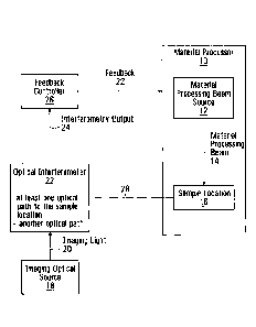
Figure 1 is a logical block diagram of a material
processing system featuring coherent imaging and feedback
control, in accordance with an embodiment of the invention.
The system has a material processor 10 that implements a
material modification process. The material processor 10
has a material processing beam source 12 that produces a
material processing beam 14 that, in turn, modifies a sample
located, at a sample location 16. Also shown is an imaging
optical source 18 that produces imaging light 20, at least a
component of which is input to an optical interferometer 22.
The interferometer 24 produces an interferometry output 24
that is input to a feedback controller 26. The feedback
controller 26 generates feedback 28 that is input to the
material processor to control at least one processing
parameter of the material modification process.
The optical interferometer 22 produces the
interferometry output using at least a component of the
imaging light 20 that is delivered to the sample location
16. Line 28 is a logical representation of the interaction
between the optical interferometer 22 and the sample
location 16 The interferometry output 24 is based on a
length of at least one optical path to the sample location
compared to a length of another optical path. The optical
paths are not depicted in the Figure in the interest of
clarity, but various examples are described later. The
sample location is the location from which the reflected
imaging light is collected. The sample location can be
selected from various options to achieve different imaging
objectives. For example, in some embodiments, the sample
location is at the physical location of a material sample
being processed. In some
embodiments, the sample location
is near the physical location of a material sample being
processes. In some embodiments, the sample location is a
CA 02728950 2011-01-21
73674-17
position chosen to yield meaningful information about the
material processing.
In some embodiments, the interferometry output at
multiple instances is processed to identify changes in
interferometry output in respect of a material being
processed. In some embodiments, at least some of the
feedback control is a function of such changes. In some
embodiments, changes in the interferometry data are used to
provide an indication of modification/sample motion "speed"
or other rates of change.
In a specific example of processing the
interferometry data to identify changes, in some
embodiments, the feedback controller is further configured
to determine if the interferometry output initially
comprises substantially only light reflected
along a reference path (this reference path may be along a
reference arm if there is one or along the sample arm) after
which the interferometry output is based on the path
length of a sample path(s) compared to the path length of
the reference path. This might occur, for example, when the
sample location initially has only one reflective
surface/subsurface (in no reference arm case) or no
reflective surface/subsurface (in reference arm case), and
then after material has been modified and/or moved relative
to the imaging optics, at some point there is an additional
reflective surface/sub-surface detected.
In some embodiments, the feedback controller is
further configured to determine when the interferometry
output makes a transition from comprising substantially only
light reflected along a reference path (this reference path
may be along a reference arm if there is one or along the
11
CA 02728950 2011-01-21
73674-17
sample arm) after which the interferometry output is based
on the path length of a sample path compared to the path
length of the reference path. The feedback controller
generates at least one signal that influences at least one
processing parameter of the material modification process
based on the interferometry output taking into account the
transition.
In some embodiments, the feedback controller 26 is
a real-time controller that controls the processing
parameter of the material modification process during the
process. In another embodiment, the feedback controller
controls at least one processing parameter during intervals
between successive processes.
in some embodiments, the material modification
processing beam source is a laser, such as a solid state,
fiber or gas laser.
In some embodiments, the material modification
processing beam source generates an ion beam and/or an
electron beam.
The material being processed by such a system may,
for example, be one or more of: metal, semiconductor,
dielectric, hard biological tissue, soft biological tissue,
plastic, rubber, wood, composite. Other materials are
possible.
In some embodiments, the interferometer has a
combiner, and two distinct arms, referred to as a reference
arm, and a sample arm. A first component of the imaging
light is applied to an input of the reference arm resulting
in an output signal of the reference arm. A second
12
CA 02728950 2011-01-21
73674-17
component of the imaging light is applied to the sample arm
resulting man output signal of the sample arm. At least a
component of the output signal of the sample arm includes
reflections of the component of the imaging light from the
sample location. The combiner combines the output signal of
the reference arm and the output signal of the sample arm to
produce a combined signal which functions as the
interferometry output. Depending on the implementation, the
combiner may be a coupler, a circulator, or a splitter; any
component that performs the combining function can be used.
In some embodiments, the system also has a signal
detector that produces an interferogram from the
interferometry output. In some embodiments, the signal
detector is in the form of an array of detector elements. A
specific example is a line camera. Other examples of such a
signal detector are described later in the context of
specific detailed example implementations.
Another example of a signal detector that produces
an interferogram from the interferometry output is an
amplified balanced photodiode pair. Other examples of such
a signal detector are described later in the context of
specific detailed example implementations.
In some embodiments, there are multiple sample
arms, and a respective interferogram is generated for each
sample arm, reference arm combination.
In some embodiments, there are multiple reference
arms, and a respective interferogram is generated for each
sample arm, reference arm combination.
In some embodiments, there are multiple reference
arms and multiple sample arms, and a respective
13
CA 02728950 2011-01-21
73674-17
interferogram is generated for each sample arm, reference
arm combination.
There may be multiple sample arms, for example,
where there are multiple reflectors at the sample location.
Such sample arms may share common optical components in
delivering reflections from the sample to the combiner, but
the optical path lengths will be different. Some of the
sample arms may be to subsurface reflectors.
For any cases where multiple interferograms are
generated, these multiple interferograms are then used by
the feedback controller 26 in generating the feedback 28 to
control the material processor 10.
Recall that the interferometry output is based on
a length of at least one optical path to the sample location
compared to a length of another optical path. In some
embodiments, the "another optical path" is simply a
different optical path to the sample. Effectively, the two
paths being compared by the interferometer in this case are
two paths to different reflectors of the same sample. In
this case, the imaging light will traverse the same optical
path but for small differences between the locations of the
reflectors at the sample location.
In some embodiments, the at least one path length
is at least two path lengths to respective reflectors at the
sample location, and the another path length is along a
reference arm.
In some embodiments, the feedback controller is
further configured to determine if the interferometry output
initially comprises substantially only light reflected
along a reference path (this reference path may be along a
11
CA 02728950 2011-01-21
73674-17
reference arm if there is one or along the sample arm) after
which the.interferometry output is based on the path
length of a sample path compared to the path length of the
reference path. This might occur, for example, when the
sample location initially has only one reflective
surface/subsurface (in no reference arm case) or no
reflective surface/subsurface (in reference arm case), and
then after material has been removed, at some point there is
an additional reflective surface/sub-surface.
In some embodiments, the feedback controller is
further configured to determine when the interferometry
output makes a transition from comprising substantially only
light reflected along a reference path (this reference path
may be along a reference arm if there is one or along the
sample arm) after which the interferometry output is based
on the path length of a sample path compared to the path
length of the reference path. The feedback controller
generates at least one signal that influences at least one
processing parameter of the material modification process
based on the interferometry output taking into account the
transition.
In some embodiments, the feedback processor
performs an analysis based on the interferometry output to
produce a depth measurement reflecting how deep the material
processing beam has penetrated at the sample location. In
some such embodiments, the feedback controller controls at
least one processing parameter of the material modification
process based on the depth measurement.
In some embodiments, the feedback controller
performs an analysis based on the interferometry output and
generates feedback control that controls depth cutting
CA 02728950 2011-01-21
73674-17
relative to an interface that is closest to the cutting
laser.
In some embodiments, the feedback controller
performs an analysis based on the interferometry output and
generates feedback control that controls depth cutting
relative to an interface that is beyond the current cut
depth.
It is to be understood that any processing
parameter of the material modification process may be
controlled by the feedback controller. Specific examples
include:
on/off state of the material processing beam;
the average power of the material processing beam;
the pulse duration of the material processing
beam;
the peak intensity of the material processing
beam;
the density of the material processing beam;
the energy of the material processing beam;
the particle species of the material processing
beam;
the wavelength of the material processing beam;
the pulse repetition rate of the material
processing beam;
the pulse energy of the material processing beam;
the pulse shape of the material processing beam
= 16
CA 02728950 2011-01-21
73674-17
the scan speed of the material processing beam;
the focal diameter of the material processing
beam;
the focal position of the material processing
beam;
the spatial pattern of the material processing
beam on the sample;
the material feed rate;
the cooling media flow rate;
the cover/assist gas flow rate;
the cover/assist gas pressure;
the cover/assist gas blend;
the arc welding process parameters (such as
voltage, current and wire feed rate);
and
the additive material feed rate.
In a specific example, the feedback controller
controls at least one processing parameter of the material
modification process based on the depth measurement by
controlling the material modification source beam to be off
when the depth measureme= indicates a specified depth.
In some embodiments, the feedback controller has
an interferogram processor that performs an analysis based
on the interferometry output to produce an indication of
when the material modification source beam has penetrated to
a specified depth that may, for example be absolute, or
17
CA 02728950 2011-01-21
73674-17
relative to a surface or interface associated with the
material. In some such embodiments, the feedback controller
controls the material processing beam source to turn off the
material processing beam based on the indication of when the
laser has penetrated to the specified depth.
In some embodiments, the feedback controller has
an interferogram processor that performs an analysis based
on the interferometry output to produce an indication of the
proximity of the region of the material currently being
modified to other regions of the material.
In some embodiments, the feedback controller has
an interferogram processor that performs an analysis based
on the interferometry output to produce an indicaticn of the
remaining amount of material to be penetrated.
In some embodiments, an interferogram processor
performs analysis based on the interferometry ouLput to
produce an indication of when material is present at a
specified depth, and the feedback controller controls the
material processing beam source to turn on the material
processing beam based on said indication. Figures 6 and 7
are two specific examples of such a system which features an
optical circulator and balanced photodetector. These
figures are described below.
Figure 2 shows a partial example implementation of
a feedback controller. Shown is a signal detector 30 that
receives the interferoMetry output 18 and generates a
measured interferogram 32. An interferogram processor 34
receives the measured interferogram 32. A memory 36 is
provided in which is stored a pre-calculated synthesized
interferogram 37 for a target result. The interferogram
processor 34 processes the measured interferogram together
8
CA 02728950 2011-01-21
73674-17
with the pre-calculated synthesized interferogram 37 to
produce a correlation result 38. The feedback controller
controls at least one processing parameter of the material
modification process based on the correlation result that is
a measure of similarity of the measured interferogram 32 and
the synthesized interferogram 37.
The pre-calculated synthesized interferogram for a
target result is pre-calculated such that it is immediately
available for correlation with the measured interferogram.
It is synthesized in the sense that it is determined from
calculations alone; no optical signals are involved in its
generation.
In some embodiments, the pre-calculated
synthesized interferogram for a target result is an estimate
of what is expected when a specified depth is reached by the
material processing beam.
In some embodiments, the interferogram processor
produces the correlation result by multiplying the measured
interferogram by the pre-calculated interferogram on a
detector element basis and then summing.
In some embodiments, at least one of the pre-
calculated synthesized interferogram and the measured
interferogram is shaped to compensate for at least one of:
spectrometer alignment;
spectrometer grating angle nonlinearity;
imaging distortion from imaging optics in the
spectrometer;
wavelength to wave number/frequency re-sampling;
19
CA 02728950 2011-01-21
73674-17
finite size of detector active area;
spectral envelope shape;
dispersion mismatch; and
another non-ideality contained in the
interferogram that degrades image quality.
Compensation may, for example, be achieved through
a controlled modulation of the complex phase and amplitude
of the individual elements of the synthesized interferogram.
The amount of modulation can be determined from at least one
of experimental calibration of apparatus, mathematical
modelling of optical propagation, theoretical analysis of
system response, and a combination of the above. The exact
method depends on the specific non-ideality to be
compensated for.
A specific example is dispersion. For a fixed
dispersive element, the relative phase lag/advance of each
wavelength arising from the dispersive terms of the material
can be added to each element in the synthesized
interferogram. Progressive dispersion (i.e., dispersion
intrinsic in the sample) can also be compensated for because
the synthetic interferogram can be calculated differently
for each depth to be measured.
In some embodiments, the correlation result is
processed to identify when a specified depth has been
reached by the material processing beam. This can, for
example, be achieved by determining when the correlation
result exceeds a threshold.
CA 02728950 2011-01-21
73674-17
In some embodiments, the system further includes
an interferogram synthesizer that synthesizes the pre-
calculated synthesized interferogram.
Another embodiment provides a feedback control
system for use with a material processing system that
implements a material modification process, the material
processing system having a camera port. Such a feedback
control system comprises the functionality of figure I, not
including the material processor. In this case, the optical
interferometer 22 interacts with the material processor 10
through a camera port, not shown. The feedback 28 is
provided from the feedback controller 26 to another input of
the material processor 10.
The embodiments described above can, for example,
be used to measure the geometry, morphology, optical
scattering and/or composition of a material before, during
and/or after processing by a material modification beam,
such as a laser. In some embodiments, feedback information
about the geometry/morphology/composition of the material
may be provided (such as, hole, cut, static or dynamic
subsurface features, and/or melt pool depth) and such
information may be used, either directly or indirectly, to
control a material modification process, such as a laser
modification process.
In some materials, the systems described herein
may sense elements of the geometry of the material being
worked on and their position in relation to other material
geometry elements that are below the surface with which the
modification beam is interacting. In some embodiments this
information is used to guide the modification to within
prescribed margins of subsurface geometry, even where the
precise location of said geometry may have been previously
21
CA 02728950 2011-01-21
73674-17
unknown and/or uncharacterized. In some embodiments, the
'depth of a laser cut into bone is measured such that laser
modification may be ceased some distance before it
penetrates a subsurface layer of bone of interest. This may
be useful for providing safe margins in laser surgery. In
some embodiments, such margins/feedback are achieved using
analysis of the metrology data, in some embodiments, using
techniques that are manual, automatic or some combination of
the two.
In other embodiments, apparatus, methods and
systems are provided that sense changes at the subsurface
level, such as but, not limited to, temperature changes,
state changes, fluid flow, and/or pressure waves, that can,
in some embodiments, be further used to inform the laser
exposure process. In some embodiments, these changes are
determined based on a comparison/analysis of multiple
measured interferograms. The phase of the interferogram is
sensitive to movement in the sample on the order of a few
nanometers. Slight temperature, pressure, flow and state
changes cause movements of the tissue that change this
phase. Also, coherent images have a characteristic "speckle
pattern" that is the partial result of the
microscopic/nanoscopic components of the sample creating an
internal interference pattern. This speckle pattern is also
extremely sensitive to the changes mentioned above. In some
embodiments, subsurface changes are observed during laser
processing of varying rates by looking at the frequency of
the change in speckle pattern.
In an embodiment, the apparatus described is used
to track elements of the melt pool in the process of laser
welding. Persons of skill in the art will appreciate that
melt pool (and/or keyhole) sLability and penetration depth
22
CA 02728950 2011-01-21
73674-17
can be an indicator of the quality of a laser weld. Some
embodiments are used to measure these and/or other
indicators and, in some embodiments, for the purposes of
disciplining the welding process, aiding welding process
development or to produce quality assurance data for the
whole or part of the process.
In some embodiments, the imaging light source is a
light source with a spectrum centered at a wavelength, Ao,
that in some embodiments may be between 300 and 15000 nm and
may have a width, nA, that can provide an axial resolution,
oz, that may be represented by the following relationship:
21112).-
2
7 AA
In some embodiments, the imaging light source may
be: superluminescent diodes, laser diodes, light emitting
diodes, ultrafast optical oscillators, semiconductor optical
amplifiers and halogen lamps; however, persons of ordinary
skill will understand that other appropriate light sources
may be used. In other embodiments, the light source may
include a superluminescent diode, in some embodiments having
an emission spectrum ranging from 1100 nm to 1400 nm or, in
alternative embodiments a Ti:A103 oscillator, in some
embodiments having an emission spectrum ranging from 750 nm
to 900 nm. In some embodiments, depending on the subsequent
detector technology chosen, a light source that has a narrow
instantaneous linewidth that is rapidly swept across the
spectral band defined by 1,.,and nx may be used instead of or
together with the other sources mentioned.
In other embodiments, additional light sources may
be included for material modification. In some embodiments,
these sources may have spectra in the region of 200 nm to
23
CA 02728950 2011-01-21
73674-17
15000 nm and can, in some embodiments, be continuous or, in
other embodiments, be pulsed in their emission. In
embodiments having pulsed emissions, pulse energies ranging
from 1 nJ to 1 NJ and pulse durations ranging from 1 fs to
30 minutes may be used.
In some embodiments a signal detector (which may
be a single detector or combination of detectors) senses the
intensities of the different wavelengths of light of
interest. This may involve the use of diffractive elements
to disperse the spectrum spatially over a detector array.
Alternatively, the signal detector may be a balanced or
unbalanced photodetector where the timing of the arrival of
components of the spectrum may be known to be simultaneous
or dispersed in time.
Electronics may be included that can measure and
interpret the detected signal. At this point the signal is
not optical anymore. In some embodiments, these may
include, but are not limited to, on-board camera hardware,
frame grabbers, field programmable gate arrays, application-
specific integrated circuits, personal computers, data
acquisition cards. The electronics hardware may be chosen
to complement the feedback schema and methods or algorithms
employed.
Some embodiments include software and/or hardware
stored on an appropriate computer readable storage medium
implementing methods or algorithms capable of identifying
the position bottom of the hole and/or subsurface interfaces
and/or changes of interest in the imaging data and can
calculate metrics and control parameters based on their
positions, for example their absolute or relative positions.
24
CA 02728950 2011-01-21
73674-17
Figure 3 is a block diagram of a first detailed
implementation. In this embodiment, a modification laser
(FL) 100 also serves as the imaging light source. This
results in the imaging and processing beam alignment being
automatic. In the embodiment shown, the apparatus, methods
and systems use a free-space Michelson interferometer that
includes a beam splitter (BS) 102, dispersion compensation
(DC) 104, a reference mirror (RM) 106, galvanometer mirrors =
(GM) 108 and an objective 110 to focus the light onto the
sample 112. Detection is accomplished by a spectrometer
comprising a grating (GR) 114, lens (ASL) 116 and
photodetector array (IGALC) 118. The PC 122 and frame
grabber (FG) 120 implement the electronics and algorithm
components of the apparatus, methods and systems described
herein. The PC 122 controls the modification laser 100
and/or another aspect of the modification process through
feedback path 124, and in this case functions as the
feedback controller.
Figure 4 is a block diagram of a second detailed
implementation. In this embodiment, separate modification
(ML) 200 and imaging (SLD) 204 light sources are shown. In
this embodiment, the two light paths are combined by a
dichroic or other combining optic (DM) 206 after independent
focal objectives 208,210. In this embodiment, the
interferometer can be built in single or, in other
embodiments, in multi-mode optical fibre. Detection is
accomplished by means of a high speed spectral detector
(HSS) 212. While the embodiment shown displays a 50:50
power splitting ratio 2-14 between sample arm 216 and
reference arm 218, in other embodiments other splitting
ratios in the interferometer are possible and may depend on
the availability of optical power and/or the need for
detection sensitivity. In some embodiments, other
CA 02728950 2011-01-21
73674-17
interferometer configurations e.g. Mach-Zehnder, Sagnac,
common path, etc. may be possible. While, in this
embodiment, DM 206 is shown to reflect the imaging light and
transmit the modification light, the reverse can
additionally be possible. Additionally, in some
embodiments, the combination of the beams via polarization-
sensitive or neutral reflection optics can occur. A skilled
=
person will understand that detection, processing and
feedback electronics are omitted from the embodiment shown
in this figure. Feedback controller 214 receives the output
of the HSS 212 and controls the modification laser 206
and/or some other aspect of the material modification
process.
Figure 5 is a block diagram of a third detailed
implementation. In this embodiment, a high power broadband
source is created by coupling short, dispersion-optimized
pulses output by broadband source 300 into a length of
single mode optical fiber 310. This results in an expansion
of spectral bandwidth, in some embodiments, on the order of
a factor of 6, though in other embodiments, more or less
broadening is possible. The embodiment shown here features
a Ti:A103 laser source 301 that operates in the region of 650
to 1100 nm. In other embodiments, spectral ranges from 300
to 3000 nm are possible. In this embodiment, a Clan-Taylor
polarizer (GTP) 302, Faraday optical isolator (ISO) 304,
half-lambda waveplate polarization control 305 and Fork
prism dispersion compensation 306 are shown. In other
embodiments, other broadband sources (such as
superluminescent diodes, other lasers and/or other
broadening methods) may be substituted for the broadened
Ti:A103 laser source.
26
CA 02728950 2011-01-21
73674-17
In this embodiment, the modification (ML) 320 and
imaging beams can be combined by an optic component (UM) 312
before they are focused by a common focal objective 314. In
such embodiments, the lens may be achromatic, aspheric
and/or conical (i.e. axicon). This beam combination may be
focused through an optional nozzle 316 that can be used to
apply assisting fluids to the modification process. The
nozzle spray may also be independent from the optical beam;
i.e. the two are delivered to the sample from different
points. The Michelson interferometer includes the 50:50
splitter 322 and reference mirror 326. Also shown are
polarization controllers 324,325,330. The spectral
detection in this embodiment involves a fiber-coupled
reflective grating spectrometer 318. In some embodiments,
an additional mirror in front of the lens (ASL) 320 can
allow the beam to approach and leave the reflective grating
318 as close to the Littrow configuration as possible,
improving diffraction efficiency. In some embodiments, a
transmission grating and/or multi-grating, and/or Fabry-
Perot spectrometer may be used. A silicon line camera 330
produces an interferogram that is passed to image processing
electronics 332, the output of which is passed to feedback
controller 334. Feedback controller 334 produces a feedback
336 to control the modification laser 320 or some other
aspect of the modification process.
Proper alignment and beam shaping of the
modification and imaging light can be beneficial to the
quality and usefulness of the imaging data and feedback
control. In some embodiments, it can be desirable to image
down into a high aspect ratio fealure such as a hole being
drilled. In such cases, an alignment method (in some
embodiments using a dichroic mirror beam combiner for
imaging and modification light) provides that, the two beams
27
CA 02728950 2011-01-21
73674-17
meet on the reflective surface of the combiner at
substantially the same point. In such embodiments, adequate
beam control of the two beams (one or more mirrors) is
beneficial. With the two beams emanating from the same
point of the combining optic, they can then be focused
through a suitably achromatic (or other design) lens. In
some embodiments, the use of an array detector or a pinhole
(in some embodiments made by the modification laser itself)
located at the focal plane of the lens can aid the
adjustment of the combining optic, so that both beams focus
on substantially the same spot. This can, in some
embodiments, be used to match the reference arm length of
the interferometer to place the center of the focal volume
at a desired position in the imaging field of view. This
position may be selected on the basis of the modification
application at hand and may additionally be adjusted
throughout the modification process. In other embodiments,
such as those where a common focal lens is not used, it may
be beneficial to have the central ray for all beams
coincident on the combining optic. It may additionally be
desirable to shift the focal positions of the imaging and
modification beams independently from one another, to more
efficiently image/modify depths of choice. In some
embodiments, this may be accomplished by adjusting the
divergence of the imaging or modification beams before they
reach the common focusing lens.
The focal spot size of the imaging and
modification beams can have an impact on the quality of the
imaging results. A careful consideration of morphology
aspect ratio and imaging beam numerical aperture should be
made. In embodiments where an imaging beam is much smaller
than the hole transversely, the resulting imaging data may
give a clear signature of the bottom of the hole and
28
CA 02728950 2011-01-21
73674-17
interfaces below it. However, in such embodiments, the
practical imaging range may be limited by the short Raleigh
range present in a high numerical aperture beam. In some
embodiments, a numerical aperture is employed to reject
signals that emanate from the sidewalls of the hole. In
such embodiments, if portions of a hole/incision periphery
are illuminated in a sample that is (quasi)transparent and
captured by the imaging system, the corresponding signals
may complicate the imaging data and may make it more
difficult for an automatic algorithm to use the data for
feedback. However, in embodiments where the sample is
nontransparent, it may be beneficial to have some
illumination of the sidewalls as such a signal can provide
information about cut width, recast deposition and the depth
of the bulk material.
In some embodiments, the optical components are
matched (in some embodiments the group delay and higher
order dispersion terms) in the sample and reference arms to
reduce any dispersion mismatch between the two arms. This
may improve axial imaging resolution. It may also be
beneficial to change this dispersion compensation in the
reference arm to match additional dispersion caused by
material present in the sample.
When imaging into a sample, the degree of
carbonization that may be created by the modification laser
can be a consideration. Lasers that cause large amounts of
charring can reduce the imaging depth (and the advance
notice for perforation etc.). Selecting lasers with reduced
carbonization (ultrashort pulses, center wavelengths of 3000
nm, 9600 nm etc.) may be beneficial.
Methods and algorithms may be used to process the
raw data and/or provide feedback parameters, and may include
29
CA 02728950 2011-01-21
73674-17
steps of background spectrum subtraction,
resampling/interpolation between the spectrometer pixels,
wavelength and/or frequency space, noise floor equalization,
fast Fourier transformation, Kasai autocorrelation/Doppler
shifting and/or other calculations based on the phase and/or
separation of interference fringes. Such methods may be
implemented in hardware and/or software. In some
embodiments an analysis of a speckle pattern and/or changes
thereof is employed to indicate tissue differentiation,
temporal heating dynamics and/or other characteristics of
the sample. These analyses may, for example, be performed by
calculating the spatial or temporal variation of the speckle
and its amplitude. Such methods and algorithms are in some
embodiments used to assess the depth of thermal damage that
has occurred, is occurring and/or will occur in the future.
Methods of signal extraction that forgo many of the previous
steps are also possible. In one embodiment, a set of
homodyne or heterodyne waveforms can be pre-calculated based
on one or a plurality of simulated optical path length
differences, nonlinearities/nonidealities in the
spectrometer, wavelength to wavenumber/frequency
conversions, single or multi-order dispersion mismatch in
the interferometer, Doppler shifts, non-ideal spectral
shapes and other adjustments to the imaging data. Sets of
such homodyne/heterodyne waveforms can be multiplied against
the data collected by the hardware or software Lo determine
imaging information at one or more of the voxels in the
imaging space. This result may be obtained due to the
orthogonality and/or quasi-orthogonality of the different
interference fringe frequencies present in the acquired
data. Detailed examples of this approach are described
below. In some embodiments, methods and algorithms may
provide computational savings when compared to other methods
CA 02728950 2011-01-21
73674-17
that use, for example, fast Fourier transformation. This
may be desirable for real-time feedback applications where a
fast response generally provides improved outcomes from the
process. Processing can, in some embodiments, use the full
spectrum data set, or, in other embodiments, use a
subsection of the data set. In embodiments using a
subsection of the data set, this can reduce processing time,
and can provide lower axial resolution, which may be useful
for a variety of feedback purposes. Homodyne/heterodyne
filtering can also have applications in general image
processing in the Fourier domain variants of Optical
Coherence Tomography where the large number of post-
processing and/or real-time calculations (including
interpolation, digital dispersion compensation, spectral
shaping etc.) may encumber the computational efficiency of
the system. Though not limited to this case, such
embodiments may be useful in situations where imaging is
targeting a subsection of the full depth of field.
In some embodiments, it is beneficial to obtain
the homodyne waveform(s) by measuring a real interferogram
when an interface is at specific depth(s) in the image. The
complex homodyne waveform(s) may be obtained by shifting the
interface optomechanically by moving the interface,
optically with phase shifting optics and/or through digital
processing, which may use Hilbert transforms and other
methods. Additional shaping steps (which may include
denoising, averaging, envelope shaping) may then be applied
to further optimize these waveforms. In some embodiments,
the spectral profile is shaped through digital, optical
(including, but not limited to mechanical blocking,
polarization adjustment, neutral density filtering,
interference filtering, Fabry-Perot elements) or other
methods to change the effective point spread function of the
31
CA 02728950 2011-01-21
73674-17
algorithm to be more optimal for feedback use. For example,
in one embodiment, a non-Gaussian spectral profile may be
applied digitally to the homo/heterodyne waveform to create
additional lobes in the point spread function. These lobes
may be engineered to provide "early warning" signals or
structured local/global minima and maxima for the feedback
algorithm to settle in.
In embodiments where the sample is transparent or
semitransparent material, the space originally occupied by
the sample bulk can be filled with air as material is
removed by a modification laser. In embodiments where the
sample has an optical Index of refraction that is greater
than air, as material is removed, the optical path length to
any subsurface reflectors may be reduced. This has the
effect of changing apparent depth of said reflectors (in
some embodiments, closer to and, in other embodiments,
further from, the zero delay point) at a rate that is
generally related to the linear removal rate of material and
the optical index. In embodiments using an M-mode image
("motion-mode", shown in later examples), the superficial
interface and the subsurface interface trend towards each
other with continuing material removal until their eventual
meeting at the point of perforation. Sensing the separation
of the two interfaces and using such separation as an input
into a feedback method or algorithm may be used to represent
a surgical margin to be preserved/monitored. In the Fourier
domain, these two interfaces may appear as two separate
frequencies that are approaching each other. Apparatus and
systems implementing methods and algorithms that sense the
change in frequency difference between the two signals can
communicate such information to a process controller and/or
user that can control the cut.
32
CA 02728950 2011-01-21
73674-17
Measuring the relative slopes can measure the
effective optical index of refraction of the material being
removed. This can be an indicator of the material's
composition which can be useful information to feed back.
In some embodiments, it may be possible to detect when the
modification laser has perforated one material and started
on the next by tracking a change in the relative slope.
These same principles may also be applied to
situations where the material that fills the hole is water
and/or materials other than air.
In some embodiments, a circulator is added to the
interferometer between the source and the fiber splitter.
In some embodiments, a balanced photodetector (in addition
to or instead of the spectrometer) is used to detect the
interference fringes that are created as the interface
arrives at the zero delay point of the interferometer. In
such embodiments, the balanced photodetector may have higher
measurement rates than an array of detectors or the sweep
rate of a Fourier Domain Mode Locked laser (or other swept
source), and improve feedback response. This can provide
fast, simple and inexpensive feedback to detect the arrival
of an interface at a certain depth. In some embodiments,
this can be used to detect when material is present at a
certain distance away from the system optics. It is known to
those skilled in the art that the effectiveness of a focused
laser beam may depend on the distance between the focus and
the material to be modified. This embodiment could be used
to provide feedback to the material processing system with
picosecond accuracy. In some embodiments, this feedback may
be used to permit emission of modification energy only when
material is present in a prescribed depth zone (PDZ) Lhat
may, in some embodiments, be related to the focal zone of
33
CA 02728950 2011-01-21
73674-17
the modification laser. The PDZ position and thickness may
be tuned through control of the imaging light source
spectrum and the reference arm length. This tuning may be
factory set and/or may be dynamically set by the operator.
In some embodiments, the imaging and modification beams may
be coupled to a handpiece and the PDZ configured to be co-
located with the focus of the modification beam some
distance away from the distal end of the handpiece. In this
way, the handpiece acts as an optical analogue to the
traditional surgical scalpel. The PDZ would be analogous to
the edge of the tip of the scalpel blade and may be used to
incise material that is located at the PDZ.
This may have a number of advantages including,
but not limited to providing a tactile interface that is
familiar to surgeons, reducing total laser energy use,
reducing total laser exposure to the material and/or
patient. It is known to those skilled in the art that some
kinds of laser modification of materials may generate plasma
above the material that scatters and/or absorbs laser
energy. While such plasma is present, further applied energy
may not have the desired modification effect and may
contribute to larger heat affected zones. In some
embodiments, the plasma may block imaging light, thus
preventing reflections from the material from triggering the
feedback system until said plasma has dissipated. This
provides the advantage of limiting modification application
energy from being applied unless the plasma conditions near
the sample are favourable.
In some embodiments, the feedback control may be
used in conjunction with an operator switch (such as a foot
pedal) such that the operator can indicate his/her consent
34
CA 02728950 2011-01-21
73674-17
to emit modification energy when the optoelectronic feedback
conditions are met.
In some embodiments, the feedback control may be
effected on the modification energy source by way of optical
pulse picker, digital seed pulse control, pump modulation,
shutter, electro-optic modulator, Pockles cell and/or
acousto-optic modulator.
A specific example is depicted in Figure 6 which
shows optical circulator 350 and balanced photodetector 352.
The output of the balanced photodetector 352 goes to
feedback controller 354 which controls the modification beam
source.
A two channel version is depicted in Figure 7.
The path length down the sample arm of one channel is
approximately the same as that of the reference arm, but
very different from their counterparts in channel 2 (and
further channels if present) to avoid cross talk in the
interference signal.
The embodiments of Figures 6 and 7 are examples of
systems that can be used to detect when material is present
at a specific depth. (10a). Reflections of imaging light
eminating from the sample and captured by the system optics
will generate an interference signal at the (balanced)
photodetector when the reference and sample optical path
lengths are matched.
Optical dispersion induced by a sample being
measured can have an adverse effect on the axial resolution
of coherent images. In some embodiments, the sample can
induce a wavelength dependent phase shift on the
interference pattern that may be dependent on the depth that
CA 02728950 2011-01-21
73674-17
the light has propagated in the sample. A
homodyne/heterodyne algorithm, for example, as described
above, can be used to compensate for these effects. The
dispersion coefficients of the materials in the sample can,
in some embodiments, be calculated a priori or, in other
embodiments, be determined iteratively. One may begin by
assuming that the phase shifts induced by the sample
increase linearly with increasing penetration into the
sample. In this way, each color (i.e. pixel measurement) on
the detector may have a certain phase shift dictated by
which color it is and what depth in the sample the signal is
returning from. If the color measured by each pixel and the
depth associated with each hetero/homodyne waveform can both
be known a priori, this distortion can be estimated and
calculated a priori and may be incorporated into the
heterodyne/homodyne waveforms that are multiplied against
the signal that is measured by the detector(s).
Alternatively, measurement of the optical signal propagating
through the system may also provide dispersion mismatch
information used for compensation. A hetero/homodyne
waveform lookup table can be prepared before the imaging
session. In such embodiments, the dispersion correction can
be applied with zero additional real-time computing load.
Interferogram Correlation Thresholding Apparatus
Referring now to Figure 8, shown is an
interferogram correlation thresholding apparatus provided by
an embodiment of the application. Shown is an
interferometer 46 that produces an interferometry output 48.
There is a signal detector 50 that receives the
interferometry output 48 and generates a measured
interferogram 52. An interferogram processor 54 receives
the measured interferogram. A memory 56 is provided in
36
CA 02728950 2011-01-21
73674-17
which is stored a pre-calculated synthesized interferogram.
The interferogram processor 54 processes the measured
interferogram together with the pre-calculated synthesized
interferogram to produce a correlation result 58. A
thresholder 60 is configured to determine when the
correlation result satisfies a threshold.
The pre-calculated synthesized interferogram for a
target result is pre-calculated such that it is immediately
available for correlation with the measured interferogram.
It is synthesized in the sense that it is determined from
calculations alone; no optical signals are involved in its
generation. Details of how this interferogram can be
adjusted a priori to perform various compensations have been
provided above.
In some embodiments, there is a respective pre-
calculated synthesized interferogram for each of a plurality
of target results. The interferogram processor 54 processes
the measured interferogram together with each of the pre-
calculated synthesized interferogram to produce a respective
correlation result. The thresholder 60 determines when each
correlation result meets a respective threshold.
In some embodiments, the pre-calculated
synthesized interferogram is an interferogram that is an
estimate of what is expected when the target result is
achieved by a material modification beam at a sample
location, and the measured interferogram is in respect of a
sample location. The interferogram processor produces the
correlation result by multiplying the measured interferogram
by the pre-calculated synthesized interferogram on a per
wavelength basis and then summing.
37
CA 02728950 2011-01-21
73674-17
In some embodiments, at least one of the pre-
calculated synthesized interferogram and the measured
interferogram is shaped to compensate for at least one of:
spectrometer alignment;
spectrometer grating angle nonlinearity;
imaging distortion from imaging optics in the
spectrometer;
wavelength to wave number/frequency re-sampling;
finite size of detector active area;
spectral envelope shape;
dispersion mismatch; and
another non-ideality contained in the
interferogram that degrades image quality.
Some embodiments feature an interferogram
synthesizer that calculates the pre-calculated synthesized
interferogram.
In some embodiments, the target result is a
specified depth reached by the material modification beam.
In some embodiments, the apparatus has a feedback
controller that controls a material modification source to
turn off the material modification beam when the correlation
result meets a threshold.
In some embodiments, the apparatus has a feedback
controller that controls a material modification source to
turn on the material modification beam when the correlation
result meets a threshold.
38
CA 02728950 2011-01-21
73674-17
In some embodiments, the apparatus has an
interferogram synthesizer that synthesizes the pre-
calculated synthesized interferogram.
Automatic guidance of laser cutting of hard tissue with
inline coherent imaging
In some embodiments, one or more of the systems
and methods described above, and related Software stored on
computer storage media are configured for automatically
and/or manually guiding the removal of hard tissue by laser
irradiation.
in some embodiments, the basis of the imaging
technology is spectral domain optical coherence tomography,
but in other embodiments, other variants (swept source OCT,
optical frequency domain imaging, time domain OCT etc.) are
employed. It is noted that the motion artifacts generated
in SDOCT are favourable and SDOCT usually has acceptable
rejection of the intense machining light.
In some embodiments, coherent imaging is used to rapidly
measure depth and reflectivity information from a sample
that is being machined with a laser. The imaging beam is
often able to see through the ejecta, plasma, intense
imaging light and beyond the modification zone. This allows
the identification and tracking of subsurface geometry that,
in some embodiments, is then used as a reference to spare
thin layers of tissue.
The combination of imaging and machining light is
accomplished, for example, with a dichroic mirror, but may
also be achieved with polarization and other techniques
known to those skilled in the art. Virtually any
modification laser (250-10600 nT spectra, CW, ps, ns, ps, fs
39
CA 02728950 2011-01-21
73674-17
durations) can be used in this way. This may permit the
tailoring of the machining laser to the application or the
use of existing infrastructure/FDA approvals.
Other useful applications of the imaging system
when integrated into a machining platform are autofocus,
permanent therapeutic records and (with the addition of
scanning optics) pre-treatment planning and post-treatment
confirmation.
Some embodiments employ a streamlined image
processing algorithm that uses a lookup table for
hetero/homodyning in lieu of more complex operations that
require interpolation, digital dispersion compensation, fast
Fourier transforms etc.
Other embodiments feature the inclusion of one or
more of scanning mirrors, more complicated machining
sources, gas assisted cutting, more performant spectrometer
designs, etc.
Coaxial imaging of laser machining processes with
SDOCT provides useful information for measuring critical
parameters for process development, such as etch rate and
morphology relaxation, in industrial materials. In cutting
tissue such as bone, SDOCT has similar benefits. To
demonstrate, an SDOCT system based on a 100 fs mode locked
Ti:A103 oscillator @ 805 nm (Coherent Mira 900) broadened in
single mode optical fiber was used. With a high speed CMOS
spectrometer and fiber based Michelson interferometer, the
imaging system provides < 5 pm axial resolution (in air) and
>100 dB sensitivity measured at 150 pm with a 1.5 ps
(measured) integration time at a maximum line rate of 312
kHz. Images were processed in LabVIEW on 4 cores of a PC
(and/or other software environments) using background
CA 02728950 2011-01-21
73674-17
spectrum subtraction, Gaussian spectral shaping, cubic
spline interpolation, FFT and noise floor equalization.
Other processing techniques and methods (mentioned in this
description) have also been applied.
For machining in these experiments, a 100 - 200 ns
(FWHM) pulsed fiber laser was used (IPG YLP-100-30-30-HC)
with an average power at the sample of 23 W at 1070 nm and
repetition rates from 30-80 kHz. The machining and imaging
beams were aligned via a dichroic mirror and focused
together via a single 50 mm achromatic lens. Fiber
collimators were chosen such that both imaging and machining
focal diameters were approximately 20 pm (1/e2) with depths
of focus of 500 and 340 pm respectively. Having the same
imaging and machining spot sizes reduced sidewall signals
(discussed later) and simplified the images. The imaging and
machining light are were delivered coaxially through a 500
pm diameter gas nozzle orifice (nozzle to sample surface
separation 1 mm) that delivered N, gas (in other cases, other
gases and blends were delivered as well) at 2 bar to provide
cooling, protection of the optics and suppression of
combustion.
Washed and desiccated transverse sections of
bovine ribs served as convenient samples of thick, compact
bone. The imaging system and machining pulse trains were
asynchronously triggered as holes were percussion drilled
into the samples in a direction transverse to the marrow
axis. The M-mode images ("motion-mode" - reflectivity as a
function of depth and time) showed that the cutting behavior
was characterized by initial periods of little to no
material modification followed by a rapid change in the
sample and the sudden onset of cutting at -10 mm/s. While
this behaviour is common to this particular modification
41
CA 02728950 2011-01-21
73674-17
source, it has been seen to be substantially different using
other sources. In Fig. 9, an example section of an M-scan
shows this sudden onset after 143,000 machining pulses and
the approximately linear progression of the hole thereafter.
The number of machining pulses required to
initiate cutting varied from 102 to 106 on the same bone
sample. This is attributed to the large degree of
inhomogeneity in the tissue sample. While this behaviour is
common to this particular modification source, it has been
seen to be substantially different using other sources.
Small variations in absorption and thermal resistance in the
bone (from the presence of blood vessels, etc.) may create
thermal "nucleation" sites where initially slow changes in
residual moisture or carbonization lead to runaway increases
in optical absorption and cutting. The variability in onset
would likely be reduced for an ablation light source
producing a centre wavelength with a short absorption depth
in the tissue. In any case, in situ monitoring of the area
of the sample exposed to machining light provided a direct
readout of the onset of ablation.
Once cutting is initiated, material removal was
approximately linear with pulse number. Several subsurface
interfaces appeared to rise and meet the primary machining
front. OCT measures optical path length and is thus
affected by the index of refraction of the medium. Material
removal above an interface reduces the optical path length
to the stationary subsurface features. The ratio of the
slopes (Equation below, /-apparent depth of subsurface
feature, x-hole depth) gave a direct measure of the
effective index of the material being removed (A). Here n
was found to be 1.5 in close agreement with past reports of
42
CA 02728950 2011-01-21
73674-17
1.530 for similar tissue. These features can provide useful
information for guided cutting as discussed below.
dl ,dx
-n)¨
dt di
Due to the stochastic nature of the onset of
ablation, measuring per pulse or per fluence cut rates using
conventional ex situ methods would be very difficult.
Nevertheless, these parameters are important information for
engineering surgical equipment and procedures. With inline
coherent imaging, these measurements are straightforward and
the information is available immediately after (and, in
fact, during) the process, requiring no further modification
of the samples. As a demonstration, 23 holes were drilled
into ribs at four different repetition rates keeping average
power constant (23 W). Figure 10 shows the material etch
rate and removal efficiency in bovine rib bone due to
exposure from ns-duration fibre laser (constant average
power 23 W). Error bars indicate the standard deviation of
the results. Simple inspection of the M-mode data yields
the resulting cut rates (Fig. 10 with error bars indicating
95% standard deviation confidence intervals). Though
ablation is achieved through thermal processes, material
removal is not simply dependent on average power. For
example, in Fig. 10 (left), etch rate increases by only -30%
when pulse energy is almost tripled. Another way of showing
this result is to consider the efficiency of material
removal per unit incident light. Often it is desirable to
reduce the light exposure without sacrificing cutting speed.
Increased material removal efficiency is observed by
increasing the repetition rate of the ablation laser source.
Explained in simple terms, pulses with half the energy but
twice the repetition rate are more effective at ablation
43
CA 02728950 2011-01-21
73674-17
than pulses with twice the energy but half the repetition
rate. This suggests that intrapulse effects such as
shielding from plasma generation/ejecta is reducing material
removal and greater efficiency could be obtained from
further increasing the repetition rate.
To demonstrate the versatility of the technique in
guiding cutting, a portable ICI system based on a fiber-
coupled superluminescent diode pair (1320 35 nm) and
reflective grating spectrometer with InGaAs photodiode array
was used. Use of this spectral band permits deeper imaging
in bone, at the expense of speed and detector cost. Once
integrated into the micromachining platform, the system has
a 14 pm axial resolution, 30 pm transverse spotsize (1/e2) in
air. The large imaging beam width is used to collect
morphology information from both the bottom of the incision
as well as the surrounding tissue as discussed below.
Thissystem had a 98 dB sensitivity measured at 300 pm with
10 us integration time and 7 mW incident on sample. The
axial line rate is detector limited at 47 kHz. In this
implementation, images were processed in LabVIEW on 4 cores
of a PC using background spectrum subtraction, linear
interpolation, FFT and noise floor equalization.
The machining source used here is a 100 W (maximum
average power) fiber laser (IPG YLR-100-SM) at 1070 nm
focused to 23 pm (1/e2) that is pulsed via TTL command to
emit 300 ns FWHM (measured) duration, 230 pJ pulses incident
on the sample at a repetition rate of 47 kHz. Though the
pulse FWEM is measured to be 300 ns, the shape is highly
asymmetric with a total duration of approximately 3 Ps.
Longer duration pulses that correspond to a simpler pulse
shape were also explored but resulted in degraded cut
quality and reduced reproducibility.
44
CA 02728950 2011-01-21
73674-17
Both imaging and cutting beams were coaxially
aligned via a dichroic mirror and focused together via a
single 50 mm achromatic lens. Imaging was electronically
controlled to trigger asynchronously with laser exposure to
provide the maximum delay between laser exposure and
imaging. Though the tissue had not relaxed to equilibrium
between pulses, the delay improves imaging contrast by
minimizing fringe washout from fast changing interfaces.
The tested sample was cortical bone extracted from
the spinous processes of the bovine lumbar vertebrae. To
create thin sections of bone suitable for this proof of
concept, a 1 mm diameter water cooled drill bit was used to
hollow out small sections of the sample leaving
approximately 600 um of bone sitting above a -1 mm air gap.
The bone/air interface provided an ideal target interface
for machining.
M-mode imaging of the bone during laser exposure
shows the progress of the machining front as a function of
machining pulse. Fig. 11 shows machining where the laser
exposure is controlled to achieve perforation into the air
gap (left) and to stop the incision before perforation
(right). The left part of Figure 11 shows two groups of
1000 pulses causes perforation into air layer, showing next
bone layer (depth 1.7mm). The right part of Figure 11 shows
the application of 7 groups of 200 pulses results in cutting
stopped 150 micron before penetration. Imaging (47 kHz)
continued after cutting to show material relaxation after
drilling. Annotations (intended as guides to the eye): MF-
Machining Front; SI - Subsurface interface; Al - Air
interface; BW - Back wall; P - Point of perforation; LO -
Machining laser off; AG - Air gap; SB Spared bone. The
onset of material removal proved to be highly variable,
CA 02728950 2011-01-21
73674-17
e.g., taking 400 pulses in Fig. 11 left, and only 50 in Fig.
11 right, likely due to the nonuniformity of the top bone
layer, as well as the nondeterministic nature of the onset
of damage in OW machining. Once machining was initiated, it
progressed with a well defined rate until perforation (Fig.
11 left), and the secondary bone layer became visible. Some
obstruction of the imaging beam causes shadowing of
subsurface structure, but tissue striations are clearly
visible, with the most pronounced discontinuity due to the
bone/air interface. As described earlier, the striations
below the machining front appear to move upwards during
material removal.
Scattering from above the machining front is
observed in all images. This comes from scattering from the
sidewalls of the hole. An imaging beam width larger than
the machining beam was used to allow monitoring of sidewall
modifications, thus achieving some degree of transverse
information without lateral scanning. Lateral scanning is
also done in situ (see below) but at the expense of reduced
imaging rate. After laser exposure is terminated (pulse
2000 in Fig. 11 left, pulse 1400 in Fig. 11 right), the
sample relaxes and sidewall and subsurface features become
static. Variation in scattered light during machining arises
due to changes in surface morphology as well as fringe
washout for fast moving interfaces. Note that in SDOCT
interfaces that move more than half the wavelength of light
during the camera integration time will suffer reduced
contrast. This motion-induced artifact is preferable over
time-domain or swept-source variations of OCT where other
fast moving interfaces will appear at incorrect depths, thus
making tracking the incision more difficult.
46
CA 02728950 2011-01-21
73674-17
By translating the sample, B-mode images of the
drilling site before and after processing were obtained.
Since in situ imaging is automatically aligned with the hole
axis, deep imaging in high aspect ratio (>20) holes was
straightforward. Figure 12 shows in situ B-mode OCT image
of bore before (left) and after (right) drilling. The two
clear holes show the lower bone interface, while the middle
hole (corresponding to Fig. 12 right) was drilled to stop
150 pm before the air gap. The spared bone thickness is
highlighted with brace brackets. Back walls seen through
holes corresponding to those in Fig. 12 (left) are labelled
BW. The middle hole clearly shows the spared bone (brace
brackets in Fig. 12 right) above the air gap. The other two
holes are through holes, showing the air gap and scattering
from the lower bone layer. Increased scattering from the
sidewalls of the holes caused by tissue modification in the
thermal cutting process does reduce the penetration depth of
the imaging light, sometimes obscuring deeper features. This
can be minimized by selecting a laser modification process
that causes little or no carbonization of the modification
site.
Applying these forward looking coherent imaging
capabilities may, in some instances, result in tracking of
machining in hard tissue over millimeter length scales with
several orders of magnitude greater temporal resolution than
has previously been reported. It is demonstrated that real-
time imaging permits accurate cutting in tissues in which
little a priori information is available and which may have
a highly stochastic response to machining energy. This
development is an important step towards fine control in
hard tissue surgical procedures, particularly in the
vicinity of sensitive organs such as the nervous system.
47
CA 02728950 2011-01-21
73674-17
Spectral Domain Optical Coherence Tomography
Embodiments described herein use spectral domain
optical coherence tomography and variants. Spectral domain
optical coherence tomography (SDOCT) has been described as
the optical analogue of ultrasound imaging. The measurement
uses a white light, optical fiber interferometer to obtain
the optical path length (OPL) of an object relative to a
fixed reference length. In the spectral domain, the relative
OPL of the sample reflection is encoded in the spacing of
the spectral interference fringes in the output from the
interferometer. Specifically, consider a set of p reflectors
in the sample arm, each with an OPL difference from the
reference length of z1. The resulting spectral interferogram
intensity is approximately:
n 'ref I
1(k)= A(k) E __ + ' +ref 1i cos(2kz .)
1 _
A(k) is the spectral envelope of the imaging light
source and k is wavenumber. The first term is known a priori
and can be subtracted as a background signal. The second
term is typically very small and can be neglected. In the
third term, the weak sample reflection (I,) has its intensity
multiplied by the strong reference signal and appears as a
sinusoidal interference fringe whose spacing (i.e.,
frequency) depends on its depth (zi). Since each depth
corresponds to a different fringe frequency, the signals are
orthogonal and can be monitored independently with no moving
parts. Acquisition speed and signal-to-noise are therefore
limited by the detector and the intensity of the imaging
light. It should be emphasized that ICI can work coaxially
with the machining beam, enabling depth sensing with hole
48
CA 02728950 2011-01-21
73674-17
aspect ratios much higher than would be possible with
triangulation methods.
To extract depth information, the spectral
interferogram (measured with a spectrometer) may be
resampled to units of constant wavenumber by interpolation
and may be transformed to I(z) via FFT. The resulting
function (known as an A-scan or A-line) is a depth-
reflectivity profile of the sample (shown in logarithmic
units relative to the noise floor) with each reflecting
interface in the sample appearing as a point spread function
(PSF) centered about its depth. The PSF full width at half
maximum (FWHM) is usually referred to as the axial
resolution of the system, and for Gaussian A(k) as:
= 2 In 2 .12
jz
7 AA
Thus a short center wavelength (\.) of the light
source and broad spectrum (nx) are desired for high
resolution imaging. Typical axial resolutions in biological
imaging on the order 5-10 pm are achieved with quasi
Gaussian spectra of 830 30 nm FWHM (ophthalmology) or 1310
35 nm FWHM (scattering tissue).
One important imaging artifact may arise due to
the ambiguity between positive and negative OPL (zi and -z-
yield the same interferogram). Since the spectral
interterogram is purely real, the depth-reflected profile
has complex conjugate symmetry about zero. Half of the image
is usually discarded leaving only positive OPLs. However, if
a reflecting interface is located on the negative side of
the reference point, its signature wraps back into the image
as an artifact. Thus, some embodiments are designed with an
adequate depth field of view (FOV) and care is taken to
49
CA 02728950 2011-01-21
73674-17
ensure that all reflecting interfaces are located on only
side of the zero delay.
To create an image, many spectral interferograms
may be acquired serially by the spectrometer, processed into
A-lines ("axial-line"-reflectivity as a function of depth)
and then displayed as a 3D dataset of reflectivity vs. depth
vs. A-line number. In biological imaging, the A-line number
corresponds to transverse position as the imaging beam is
raster scanned. This produces an image of reflectivity as a
function of two spatial dimensions known as a B-mode image
(B - brightness). Alternatively, if the beam is static, the
A-line number corresponds to time and the resulting image is
called an M-mode image (M = motion). This type of image is
useful for observing fast changes in the depth-reflectivity
profile of the sample. For example, coaxial imaging during
the percussion drilling of 304 stainless steel with a 1070
nm center wavelength, 100 ns duration fiber laser (IPG YLP-
1/100/30/30-HC) gives the M-mode image in Figure 13. The
machining front (bright white curve) is seen descending -600
pm into the bulk of the sample. The complete etch depth vs.
pulse number relationship was obtained from drilling a
single hole and required no post-cut material processing.
760 pJ pulses were incident onto a 20 pm e--2
intensity diameter spot at 30 kHz. A coaxial oxygen assist
gas jet at 8.3 bar was used. Imaging rate is 300 kHz. Graph
brightness corresponds to sample reflectivity in logarithmic
scale. The dynamic range shown is - 60 dB.
With acquisition rates of even a few tens of
kilohertz, M-mode images are not only able to directly
measure etch rates but also melt pool flow and other
dynamics of laser drilling/welding processes. Since sensing
below the machining front is possible, M-mode data may also
CA 02728950 2011-01-21
73674-17
be used in conjunction with appropriate feedback hardware to
guide blind hole cutting in a variety of semitransparent
materials including biological tissue even when the exact
sample geometry is not known a priori.
Figure 14 is a schematic diagram of another
imaging system provided by an embodiment of the invention
that will be used as an .example for homodyne mixing.
However, homodyne mixing can be used with any of the systems
described herein. Labels: ISO - Fiber Coupled Optical
Isolator 400; 50:50 - Mode coupler 402; PC - polarization
controller 406; TGR - transmission grating 408; ASL - Air
spaced lens 410; SiLC - Silicon CMOS line camera 412; 50FC -
50 mm fiber collimator 414; 10FC - 10 mm fiber collimator
416. There is a fiber-coupled superluminescent diode (SLD)
418, a custom spectrometer, and fiber optic Michelson
interferometer that can be interfaced to a laser machining
head through a camera port. Imaging light from the SLD
first passes through an optical isolator and/or circulator,
which protects the SLD from back-reflection. The light
continues inio an evanescent mode coupler (beam splitter or
beam combiner) where it is split into the sample and
reference arms, then coupled out of the fiber and into free
space. Some light is retroreflected in both interferometer
arms and the signals are recombined and interfere at the
mode coupler. Polarization controllers correct for
mismatches between the two interferometer arms arising from
polarization effects in single mode fiber and also to
optimize diffraction grating efficiency. Polarization
maintaining fiber may also be used together with or instead
of polarization controllers. A transmission grating is used
in the spectrometer for ease of alignment. Finally, the
camera measures the spectral interferogram and transmits
51
CA 02728950 2011-01-21
73674-17
data via IEEE-1394 to a desktop computer (or other
processing platform, not shown) for processing.
The following is an example measurement of the
performance of the system of Figure 14.
Table 1: Calculated System Performance Characteristics
Axial Resolution (pm) 12
Depth of Field (mm) 5.9
Maximum Line Rate (kHz) 27
Duty Cycle (IEEE-1394
interface limited) 0.73
Sensitivity (dB)* t (35
ps integration) 98
Sensitivity (dB)* t (1 ps
integration) 82
Sensitivity (dB). t (100
ns integration) 67
Max. Dynamic Range (dB)* 66
Based on noise specifications
available for camera operating
at low speed. Actual value is
expected to be lower at full
speed.
Assumes sample arm optics
have -80% efficiency
52
CA 02728950 2011-01-21
73674-17
Some embodiments may have different speed, sensitivity,
resolution and/or dynamic range depending on the choice of
components.
In some embodiments, a complete system would also
include custom interfacing with machining heads for specific
applications. This can generally be accomplished by
modifying a camera port and choosing the correct dichroic
optic to combine the imaging and machining light.
Additionally, an appropriate focused beam diameter for the
imaging beam may be chosen. In some implementations, the
imaging and machining light will be focused by the same
objective (though this is not necessary) whose focal length
is predetermined by existing machining process demands.
Here, the choice and alignment of the sample arm collimator
can be used to give the desired focal characteristics for
imaging. Collimator alignment can also be used to compensate
for focal length variation of the objective between Imaging
and machining light.
As an example application, a machining laser head
with a 100 mm focusing lens is considered. To maintain
uniform imaging over the depth of field, the collimator's
focal length should be chosen so that the focused imaging
beam's Rayleigh range is approximately half the system's
depth of field. For the setup described above, we choose a
10 mm collimating lens and hence, expect a beam waist of 27
pm (1/e2 intensity radius) and a Rayleigh range of 2.8 mm.
Note that to achieve maximum axial resolution, proper
compensation of dispersion mismatch between the sample and
reference arms may be of use.
The design is flexible and can be modified to
improve imaging rate (with an upgraded camera) or axial
resolution. The latter is achieved by selecting a broader
53
CA 02728950 2011-01-21
73674-17
spectrum SLD (or other light source) and a grating with a
reduced line density. This would provide significant
resolution improvement with the drawback of reduced depth of
field but little to no additional monetary cost. For
instance, substituting the current components with an 840
25 nm FWHM light source (Exalos EXS8410-F413) paired with a
1200 lines/mm grating (Edmund Optics NT48-589) could provide
6.2 pm resolution over a maximum range of 3 mm. Note that
with higher spectral bandwidths, proper dispersion mismatch
compensation is important to achieve maximum resolution.
Note that in coherent imaging techniques such as
this, if an interface moves by -X/4 or greater during the
integration time of the detector, the fringe contrast will
be significantly degraded ("washed out"), causing the signal
from that interface to vanish. This corresponds to an upper
limit to the interface speed that can be tracked. However,
it also has the benefit of rejecting certain high-speed
interfaces (e.g. ejecta) that would produce reflections that
complicate the images and make automatic feedback more
difficult. The maximum interface speed depends on the
integration time of the detector, which in turn affects
sensitivity. For an integration time of 35 ps, the system
can track interfaces moving at speeds up to 0.006 m/s. For
faster moving interfaces, integration time can be reduced
(at the expense of sensitivity) to 1 is or 100 ns to give
maximum speeds of 0.21 m/s or 2.1 m/s, respectively. Since
this is faster than typical etch rates in industrial
processes, it is expected that this design will be adequate
for a wide range of applications. The use of line cameras
with shorter integration times, balanced photodetectors
and/or swept sources may allow even faster moving interfaces
to be resolved.
54
CA 02728950 2011-01-21
73674-17
Homodyne Depth Filtering
To use ICI as an automatic feedback method,
processing is preferably able to run at least as fast as
data acquisition. In biological imaging, the interpolation
and EFT operations are necessary to calculate the reflected
intensity from all the depths within the FOV to form an
image. By contrast, in feedback systems, the imaging output
is used to trigger a change in the material modification
process as a function of the imaging output (e.g. terminate
emission), for example once a certain depth has been
reached. In this case, calculating the reflectivity from
all the depths may be excessive. An efficient method for
determining when drilling has penetrated a prescribed depth
is provided.
Starting with a desired depth, z, and using
Equation for 1(k) presented above with calibration data from
the spectrometer, a synthetic interferogram is pre-
calculated, expressed in units of constant camera pixel
number (or the basis that corresponds to the detection
system). This calculation can be completed a priori and does
not contribute to the real-time computing load. Multiple
such pre-calculated synthetic interferograms may be
generated and drawn (individually or otherwise) from a
memory table to be used for different target results, for
example, achieving one of several possible depths, tracking
the approach to a desired depth through a series of
intermediate steps, removal of material from a specified
depth, achieving more material at one depth compared to
another depth, or optimizing change in backscatter from a
target depth.
By homodyne mixing the synthetic interferogram
with the raw data from the camera, the signal from the
CA 02728950 2011-01-21
73674-17
desired depth is extracted which may have significantly
lower spurious side-lobe signal (from interpolation errors)
when compared to other methods known to those skilled in the
art as shown in Figure 20. F. For each imaging output from
the camera, the raw data from the camera is multiplied by
the synthetic interferogram pixel by pixel and then summed.
When the desired depth is reached, the summed result will
have a peak.
Where it is desirable to combine the signal with
multiple synthetic interferograms, a matrix multiplication
approach may be taken.
If data elements are transferred from the detector
serially or quasi-serially (i.e. through multiple camera
taps) then the receiving electronics in some embodiments may
begin calculations on the individual elements as soon as
they become available in order to preserve processing
resources such as memory and/or gates (such as on a field
programmable gate array, FPGA) and to reduce the overall
feedback latency.
To demonstrate, this filter technique is applied
to the spectrometer data used in Figure 15, choosing a 200
pm target depth (indicated by a line 100 in Figure 15). The
filter response shows a clear, high SNR response at the
moment the machining front passes through the depth (Figure
B bottom).
The filter response is used to trigger a feedback
response to stop drilling, or to make some other change to a
parameter of a material modification process. Figure 16 is
a flowchart of a method of automatic feedback control, which
can, for example be used to stop drilling, based upon when a
prescribed depth is reached. More complicated control
56
CA 02728950 2011-01-21
73674-17
systems with feedback from multiple depths and control of
other parameters of the process are also possible. In some
embodiments, a look-up-table is employed to rapidly and
dynamically change the depth(s) of interest (by selecting
different pre-calculated synthetic interferograms).
Depth filtering may achieve computational savings
versus standard processing. The time required to process
multiple blocks of 576 element lines of previously acquired,
raw experimental data with both our standard biological
imaging code (background subtraction, cubic spline
interpolation, FFT, noise floor equalization) and with the
homodyne filter is compared in Table 2. Processing was
conducted with a single thread running MATLAB on a quad-core
Intel desktop CPU in a Microsoft Windows 7 64-bit
environment. The results in Table 3 are expressed in terms
of 103 lines per second (kips) and the relative speed
Increase factor obtained by using the homodyne filter.
Table 2: Comparison of processing speed for 4 x 10⁵ image
lines
Block Interpolation Homoayne Relative
size + FFT (IF) filter speed
(A- speed (kips) (HF)
(HF/IF)
lines) speed
(kips)
2 x 10⁵ 0.77 451.2 588
2 x 10⁴ 5.096 522.2 102
2 x 10³ 4.596 555.6 121
200 1.861 794.0 427
57
CA 02728950 2011-01-21
73674-17
20 0.241 746.0 3097
For very small and very large block sizes, the FFT
method is very slow. This is a result of limitations
specific to the hardware and software environment and not
the computational complexity of the code. As a result, the
best theoretical comparison between the two methods is the
mid-size blocks. Here, even when the FFT produces its best
results, the homodyne filter still outperforms it by two
orders of magnitude.
While the line period limits the raw throughput
rate, it is only a minimum value for the total feedback
latency. Interrupt latency and other delays inherent to
desktop hardware and operating systems are additive and may
ultimately be the dominant terms. For this reason, the full
capabilities of ICI-based feedback potentially will not be
realized without the use of dedicated processing hardware in
the form of a field programmable gate arrays (FPGAs) or
application-specific integrated circuits (ASICs). These
components already exist in many modern cameras, including
the one specified here. The ease of implementation of the
homodyne filter algorithm described here onboard a camera
circumvents the desktop PC bottleneck and allows the camera
itself to discipline the machining system.
Imaging Below Surface
Figure 17 is another example of a system featuring
inline coherent imaging. This implementation features an
optical fiber implementation of ICI. A broadband light
source 500 injects light into the optical fiber 502. An
isolator blocks back reflections from reaching the light
58
CA 02728950 2011-01-21
73674-17
source. Optical coupler 504 splits the light into reference
arm (top) 506 and sample arm 508 (bottom, to laser
processing system). The ratio of splitting depends on the
applications needs. An example would be 50:50 (50% to the
reference arm, 50% to the sample). The reference light
travels along the reference arm and is back reflected. The
path length of the reference arm can be set in coarse
divisions, using various lengths of optical fiber, and fine
divisions using a mirror mounted on a translation stage with
micrometer control. Usually the reference arm length is set
to match the optical path length to the workpiece in the
laser processing system, less approximately two hundred
micrometers. Often it is convenient to put a focussing
objective identical to the one used in the laser processing
platform before the reference mirror (not shown) in order to
match dispersion and control reflected reference arm power.
The reference arm contains optics 510,512 that allow
dispersion and polarization control. Dispersion control is
done so both reference and sample arm are close to
dispersion matched. Polarization control is usually set so
the reference and back reflection from the sample arm have
similar polarization states (for maximum interference). The
reference arm also may include a controllable intensity
attenuator (not shown) to control detector saturation and
imaging dynamic range. This can be accomplished by a
variable neutral density filter, misalignment of a fiber
coupler, or translation of the focussing objective relative
to the end reference mirror (all not shown). The sample arm
fiber exits the inline coherent imaging system and is
connected to an external laser processing platform. Light
backscatters off the workpiece and travels back along the
same fiber. The back-reflected reference light splits at
the optical coupler 504 so part of it is injected into the
59
CA 02728950 2011-01-21
73674-17
fiber connected to the high-speed spectrometer 514 (amount
depending on the coupler splitting ratio). The
backscattered sample light splits at the optical coupler so
part of it is injected into the fiber connected to the high-
speed spectrometer (amount depending on the coupler
splitting ratio). The sample and reference light interfere
in the optical fiber 516. The light is dispersed according
to its wavelength in the spectrometer. The detector may be
a spectrometer that measures intensity as function of
wavelength. The position of the constructive and
destructive peaks contains information about the relative
path length of the sample arm compared to the reference arm.
If light is backscattered simultaneously from more than one
depth in the sample arm (e.g., sides of a laser keyhole),
the strength and relative positions of all the depths is
encoded in the interferogram. The spectral interferogram
(intensity as a function of wavelength) is converted into an
electronic signal by the detector and transmitted to control
electronics 518 for processing. The electronic processing
system controls the spectrometer (e.g., triggering) and
processes the raw detector data. One processing technique
(so-called standard OCT processing) is back subtraction,
cubic spline interpolation for conversion from camera pixel
number to constant frequency step, fast Fourier transform to
yield a graph of backscatter as a function of depth. If
there is only one highly reflecting interface in the sample
arm, the resulting graph will have one strong peak with its
width set by the axial resolution of the system. Axial
resolution is inversely proportional to wavelength bandwidth
measured by the spectrometer (thus the need for a broadband
source to achieve high resolution). Alternatively, the
homodyne filtering approach described above may be used for
faster processing times and improved image quality. In some
CA 02728950 2011-01-21
73674-17
embodiments, a feedback controller (part of or separate from
electronic processing 518) generates feedback to control one
or more processing parameters of the material modification
process. Examples have been provided above in the context
of other embodiments.
Figure 18 is a block diagram of an application of
the ICI to forward-viewing guided laser surgery. Lasers are
useful for tissue ablation because the light can be focused
very tightly, allowing the surgeon to remove tissue in small
volumes. While the light can be delivered with high
precision in the transverse dimensions, it is difficult to
control the final depth of the laser incision. Tissue can
be highly heterogeneous with a large variation in removal
rate, making total energy delivered not a good predictor of
incision depth. Fig. 18 shows a patient treatment area 600
that contains a volume of hard or soft tissue that would
usually be removed by mechanical methods (e.g., drill). The
ICI system measures incision depth as tissue is ablated, and
terminates laser exposure at a predetermined depth. More
importantly, when ICI is implemented using infrared light
(-1300 nm), imaging into the tissue (beyond the ablation
front) is possible. This allows exposure to be terminated
before an interface is penetrated (and before the intense
surgical laser can damage delicate subsurface tissue).
An inline coherent imaging system 602 is provided;
this includes an interferometer, broadband light source and
spectrometer, and an example implementation is depicted in
Figure 17. The patent treatment area is indicated at 600.
There is a surgical laser 604 which generates exposure
controlled by a surgeon, and modified by feedback control.
There is a robotic controlled focussing head 610 (but may be
handheld in some other embodiments) which combines imaging
61
CA 02728950 2011-01-21
73674-17
and surgical beam coaxially and collects imaging light
backscattered from the treatment area. In some embodiments,
imaging and surgical laser light may be combined earlier in
the propagation path of the surgical laser such that imaging
and surgical light arrive pre-combined at the focussing
head. The spectral interferogram data from the ICI system
602 is passed to electronic processing 606 which generates
the electronic feedback control for the surgical laser and
robotic controlled focussing head. In addition, an output
is generated for an image display 608.
The beam from the sample arm of the ICI
interferometer is set to be coaxial with the surgical laser
604. This can be done in free space with an appropriate
dichroic mirror. This guarantees imaging is along the same
line as the surgical beam direction. The reference arm
length is set so sample arm and reference arm are closely
matched. The surgeon can use the image display to image the
target area (and below) before he/she starts the surgical
laser. The imaging system can also be used to fine tune the
position of the surgical laser using co-registration with
other imaging modalities (such as prerecorded MRI or CT).
This would allow the surgeon to look at a small volume of
the treatment area in real-time using the ICI in the context
of larger anatomical features. The electronic processing
would do this co-registration. In addition, the surgeon
could have predefined margins to be removed using the
prerecorded imaging modalities.
Once the surgeon is certain that the surgical
laser will target the right treatment area consistent with
the treatment plan, he/she starts the ablation process. The
system can be programmed to terminate exposure after a
certain depth is cut, or to remain within a certain preset
62
CA 02728950 2011-01-21
73674-17
margin, or to terminate exposure when ablation reaches a
certain distance to a predefined interface. The ICI system
can be used to provide a permanent record of the treatment
procedure, useful for postoperative analysis.
Figure 19 is a block diagram of an application of
the ICI for in situ metrology for laser welding. Laser
welding provides narrow and deep welds, well suited to
automated and high volume manufacturing. The diverse
applications for laser welding have in common a process of
controlled heating by a laser to create a phase change
localized to the bond region. Controlling this phase change
region (PCR) can be used to control the quality of the weld
and the overall productivity of the welding system. The high
spatial coherence of laser light allows superb transverse
control of the welding energy. Axial control (depth of the
PCP) and subsequent thermal diffusion are more problematic
particularly in thick materials. In these applications, the
depth of the PCR is extended deep into the material (-mm)
using a technique widely known as "keyhole welding". Here,
the beam intensity is sufficient to melt the surface to open
a small vapor channel (also known as a capillary or "the
keyhole") which allows the optical beam to penetrate deep
into the material. Depending on the specific application,
the keyhole is narrow (<mm) but several millimetres deep and
sustained with the application of as much as -104 W of
optical power.
In Figure 19, an inline coherent imaging system
702 is provided; this includes an interferometer, broadband
light source and spectrometer, and an example implementation
is depicted in Figure 17. The welding platform is indicated
at 700. There is a welding laser 704 which generates a
welding beam controlled by a welding controller 705, taking
63
CA 02728950 2011-01-21
73674-17
into account feedback control. A focussing objective 703
combines imaging and welding beam for delivery to a welding
work piece 701 and collects imaging light backscattered from
the welding area. There may be additional welding inputs
such as assist gas, an electrical arc, additive material
etc. The spectral interferogram data from the ICI system
702 is passed to electronic processing 706 which generates
the electronic feedback control for the welding controller
704. In addition, an output is generated for an image
display 708. In this case, the ICI system 702 is connected
to a welding platform camera port 718 through a fiber to
free-space coupler 720.
To measure keyhole formation in real time, the
sample arm of the ICI imaging system 702 is set to be
coaxial and/or near coaxial with the welding laser beam, to
be focussed in the PCR. This can be done by collimating the
image beam and directing it into the welding platform camera
port. The ICI system is used to monitor the depth of the
keyhole formed, ensuring that it is appropriate depth for
welding all the workpieces. In pulsed laser welding, the ICI
system can be run at a multiple of the repetition rate of
the welding laser, providing images from before, during and
after laser exposure. This provides direct information on
the creation of the vapour channel, and its subsequent
refilling. With continuous-wave welding sources, the ICI
system can monitor keyhole stability directly. Feedback
from this informar.ion can be used to optimise welding
parameters (such as laser intensity, feed rate, and assist
gas), to increase keyhole stability.
The image display 708 shows the operator real time
information about keyhole penetration and stability as
welding is in process, and provides a permanent record of
64
CA 02728950 2011-01-21
73674-17
the weld creation, situated to the exact region on the
workpiece. This can be important for later quality
assurance.
Another embodiment of the invention provides a
fiber-based ICI in which a common dielectric objective is
used to combine the imaging light and the laser light. Such
an embodiment, optionally, includes a feedback controller,
for example as defined in any of the other embodiments
described previously.
Other embodiments combining, mixing or
interchanging the fundamental design elements described
herein can be possible and will be evident to persons
skilled in the art. These include, but are not limited to,
imaging from other directions (i.e. not in-line with the
modification beam) including the underside of the material
being modified.
Numerous modifications and variations of the
present disclosure are possible in light of the above
teachings. It is therefore to be understood that within the
scope of the appended claims, the disclosure may be
practiced otherwise than as specifically described herein.