Language selection

Search

Patent 2728974 Summary

Third-party information liability

Some of the information on this Web page has been provided by external sources. The Government of Canada is not responsible for the accuracy, reliability or currency of the information supplied by external sources. Users wishing to rely upon this information should consult directly with the source of the information. Content provided by external sources is not subject to official languages, privacy and accessibility requirements.

Claims and Abstract availability

Any discrepancies in the text and image of the Claims and Abstract are due to differing posting times. Text of the Claims and Abstract are posted:

  • At the time the application is open to public inspection;
  • At the time of issue of the patent (grant).
(12) Patent: (11) CA 2728974
(54) English Title: APPARATUS FOR CUTTING A TISSUE PART WITH FOCUSED LASER RADIATION
(54) French Title: DISPOSITIF DE DECOUPE D'UN FRAGMENT DE TISSU A L'AIDE D'UN FAISCEAU LASER FOCALISE
Status: Expired and beyond the Period of Reversal
Bibliographic Data
(51) International Patent Classification (IPC):
  • A61F 9/009 (2006.01)
  • A61F 9/01 (2006.01)
(72) Inventors :
  • DONITZKY, CHRISTOF (Germany)
  • WUELLNER, CHRISTIAN (Germany)
(73) Owners :
  • ALCON INC.
(71) Applicants :
  • ALCON INC. (United States of America)
(74) Agent: KIRBY EADES GALE BAKER
(74) Associate agent:
(45) Issued: 2015-08-25
(86) PCT Filing Date: 2008-06-20
(87) Open to Public Inspection: 2009-12-23
Examination requested: 2011-06-02
Availability of licence: N/A
Dedicated to the Public: N/A
(25) Language of filing: English

Patent Cooperation Treaty (PCT): Yes
(86) PCT Filing Number: PCT/EP2008/005012
(87) International Publication Number: WO 2009152838
(85) National Entry: 2010-12-20

(30) Application Priority Data: None

Abstracts

English Abstract


An apparatus for cutting a tissue part out of a tissue with focused laser
radiation
comprises a light generating device for generating laser radiation to cut the
tissue part, a suction ring with a sealing surface capable of being applied
onto a
tissue surface, devices for generating an underpressure in a cavity delimited
by
the tissue surface and the suction ring, an applanation plate pressed against
the
tissue surface for shaping, and an body opaque to the laser radiation and
arranged below the applanation plate. The body of the apparatus is arranged
onto and rests on a contact plate of the suction ring such that when applying
the sealing surface onto the surface of the tissue, an inner edge of the body
defines an edge of the tissue part to be cut.


French Abstract

L'invention concerne un dispositif de découpe d'un fragment de tissu (10a) dans un tissu (10) à l'aide d'un faisceau laser focalisé. Ce dispositif comprend : - un anneau aspirant (28) ayant une surface d'étanchéité (42) qui peut être appliquée sur la surface (22) du tissu (10), - des équipements (34, 36, 46) destinés à produire une dépression dans un espace vide (38) qui est délimité par la surface d'étanchéité (42), la surface (22) du tissu (10) et l'anneau d'aspiration, - une plaque d'aplatissement (30) qui peut être appliquée à la surface du tissu (10) pour lui donner une forme, et un corps opaque au rayonnement laser (40) qui coopère avec l'anneau d'aspiration (28) et définit par son bord intérieur (40a) un bord (14) du fragment de tissu (10a).

Claims

Note: Claims are shown in the official language in which they were submitted.

-8-
Claims
1. Apparatus for cutting a tissue part out of a tissue by means of focused
laser radiation, comprising:
- a light generating device for generating the laser radiation which
cuts
the tissue part;
- a suction ring which has a sealing surface that is capable of being
applied onto a surface of the tissue,
- devices for generating a reduced pressure in a cavity that is delimited
by the surface of the tissue and by the suction ring,
- an applanation plate that is capable of being pressed against the
surface of the tissue for the purpose of shaping, and
- a body that is opaque to the laser radiation and has an inner edge,
wherein
- the body is arranged onto a contact plate of the suction ring such
that, when applying the sealing surface onto the surface of the tis-
sue, the inner edge of the body defines an edge of the tissue part to
be cut wherein the body rests on the contact plate of the suction ring
that with the sealing surface is capable of being brought into contact
with the tissue and wherein the body is arranged below the applana-
tion plate.
2. Apparatus according to claim 1, wherein the body that is opaque to laser
radiation is annular.
3. Apparatus according to any one of claim 1 or 2, wherein the body that is
opaque to laser radiation at least partly absorbs the laser radiation.
4. Apparatus according to any one of claim 1, 2, or 3, wherein the sealing
surface is capable of being brought into contact with the cornea of an eye
and in that when the sealing surface is applied onto the surface of the tis-
sue, the inner edge of the body that is opaque to the laser radiation de-
fines the edge of a flap that is capable of being folded upwards in the
course of an ophthalmological operation.

Description

Note: Descriptions are shown in the official language in which they were submitted.


CA 02728974 2010-12-20
..
..
Apparatus for cutting a tissue part with focused laser radiation
The invention relates to an apparatus for cutting a tissue part out of a
tissue
with focused laser radiation. In particular, the apparatus relates to the
cutting of
a so-called flap out of the cornea of an eye in the course of refractive
surgery, in
particular LASIK. The invention will be elucidated in more detail in the
following
with regard to refractive surgery, in particular LASIK. But, in addition to
this, the
invention can also be employed generally for the purpose of cutting a tissue
part
out of a tissue by means of focused laser radiation.
In the case of LASIK ¨ that is to say, the now generally known correction of
the
optical imaging properties of the cornea by means of laser radiation ¨ it is
known
firstly to cut a so-called flap out of the anterior region of the cornea,
whereby a
portion of the flap remains connected to the cornea like a hinge, so that the
flap
can be folded aside for the subsequent ablation of corneal tissue by means of
laser radiation. After implementation of the ablation (resection of tissue),
the
flap is folded back, and a relatively rapid healing takes place, the corneal
surface
being largely undamaged.
In the state of the art there are principally two different techniques for the
pro-
duction of the flap.
On the one hand, for the cutting of the flap a mechanical so-called microkera-
tome is employed which cuts into the cornea from the side with a rapidly oscil-
lating cutting edge. In this case a so-called suction ring is mounted onto the
eye,
which fixes the eye by means of vacuum. This is the state of the art in this
re-
gard.
On the other hand, laser radiation is also increasingly being employed for the
cutting of the flap - at present, focused laser radiation with pulse lengths
within
the femtosecond range. In this case the radiation is focused below the
anterior
surface of the cornea, within the tissue, and the focal points are positioned
in
the desired surface in such a way that, as a result, a flap is cut out of the
cor-
nea. This is well-known in the state of the art as femto LASIK.

= CA 02728974 2010-12-20
- 2 -
With femto LASIK there are, in particular, two embodiment variants:
According to a first embodiment, a separate system consisting of suction ring
and lid lock is employed for the purpose of lateral fixation of the eye. A
planar
(flat) optical plate is impressed for the purpose of levelling the cornea. The
stroma is cut two-dimensionally at depth in the manner described above. The
marginal incision (that is to say, the positioning of the focal points in the
mar-
ginal region of the flap) is effected in this case by positioning of the focal
points
out of the plane as far as the corneal surface. In this case, the focal points
reach
into the aforementioned planar optical plate which, as a result, is destroyed
and
consequently constitutes a disposable article.
According to another embodiment of femto LASIK, the planar stromal incision is
likewise carried out using a suction ring with an applanation by suction of
the
eye. In this case, the depth of the incision is defined by a plastic film that
is
transparent to the laser wavelength and that is situated between the
applanation
plate and the cornea. In this case too, the plastic film is a disposable
article. A
special configuration of the edge of the incision is not provided in this
case.
The invention has as its objective to make an apparatus available with which a
tissue part, in particular a flap, can be cut out of a tissue, in particular
the cor-
nea, in simple manner with focused laser radiation. In this connection, in par-
ticular the duration of the operation is to be shortened, and the energy
introduced for the incision into the eye is to be reduced.
For this purpose the invention makes an apparatus available for cutting a
tissue
part out of a tissue by means of focused laser radiation, exhibiting the
following:
- a suction ring which has a sealing surface that is capable of being ap-
plied onto a surface of the tissue,
- devices for generating an underpressure in a cavity that is delimited
by the surface of the tissue and by the suction ring, and
- an applanation plate that is capable of being pressed against the sur-
face of the tissue for the purpose of shaping,
the apparatus being distinguished by

CA 02728974 2010-12-20
- 3 -
- a body that is opaque to the laser radiation and that interacts
with the
suction ring and defines with its inner edge an edge of the tissue part.
According to the invention, in the case of femto LASIK, for example, for each
desired flap diameter and for each desired geometry of the flap a "customised"
body, opaque to the laser radiation, in the above sense is selected and is em-
ployed with the suction ring. In this case the aforementioned body that is
opaque to the laser radiation may have been firmly connected to the suction
ring, so that a specific suction ring is procured for the desired shape of the
flap
in the given case, or the body that is opaque to the laser radiation is
detachably
connected to the suction ring as an interchangeable part.
The 'applanation plate' in the sense of the present invention does not
necessarily
have to be planar (flat) but may also exhibit a curvature, for example a
spherical
curvature, depending on the desired incision.
The aforementioned body that is opaque to laser radiation is preferably
annular.
Another configuration provides that the body that is opaque to the laser radia-
tion either partly absorbs or entirely absorbs the laser radiation.
Another configuration of the invention provides that the body that is opaque
to
laser radiation rests on a contact plate which with a sealing surface is
capable of
being brought into contact with the tissue.
Exemplary embodiments of the invention will be elucidated in more detail in
the
following on the basis of the drawing. Shown are:
Figs. 1A-1C: schematically, the production of a flap in the case of femto
LASIK;
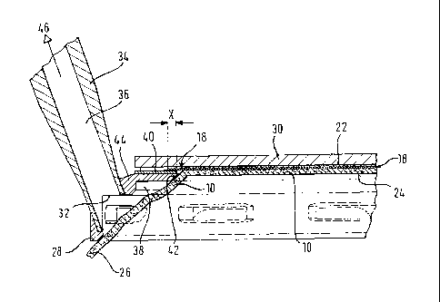
Fig. 2: schematically, an exemplary embodiment of an apparatus for
cutting
a tissue part out of tissue in accordance with the invention;
Fig. 3: a detail from Fig. 2 on an enlarged scale; and
Fig. 4: a top view of a body that is opaque to laser radiation.

CA 02728974 2010-12-20
- 4 -
Figures 1A 1B and 1C show schematically the conditions in the course of the
cutting of a flap 10a in a cornea 10 using focused radiation with pulse
lengths
within the fenntosecond range. The laser wavelength that is used corresponds
to
the state of the art.
Fig. 1A show a top view of the cornea 10, the limbus 12 thereof, the edge 14
of
the flap 10a and the sclera 26. In an articulated part 16 (hinge) the cut flap
10a
remains connected to the cornea 10, so that it can be folded upwards in a man-
ner known as such. Fig. 1B shows the focal plane 18 of the laser ¨ that is to
say,
the plane in which the focal points of the laser radiation are positioned in
such a
way that an incision arises overall. In this connection the term 'plane' which
is
used here is not to be understood in technical language as meaning absolutely
planar but also encompasses curved designs of the surface of the focal points,
as the incision according to Fig. 1B shows, for example.
The circular detail K according to Fig. 1B is represented in Fig. 1C on an
enlarged scale. According to this Figure, the surface in which the laser focal
points are placed is pulled upwards in the marginal region in a lateral
incision 20.
At the points where the incision plane of the focal points intersects the
anterior
surface 22 of the cornea 10 the edge 14 of the cut flap is located. Fig. 1B
also
shows the diameter D1 and the thickness D2 of the flap and the posterior sur-
face 24 of the cornea 10.
The lateral incision 20 and the corresponding guidance of the focal points of
the
laser radiation are elaborate and complicated, which also has consequences on
the probability of imperfect incisions. The lateral incision at the edge 14 is
also
time-consuming and gives rise to a relatively high total energy that is
introduced
into the eye. Both are improved with an apparatus according to Figs. 2 to 4.
Fig. 2 shows a suction ring 28 with an applanation plate 30 which is pressed
onto the anterior surface 22 of the cornea 10. As already stated above, the ap-
planation plate 30 does not have to be absolutely planar (flat) but may also
be
curved, for example spherical.
The suction ring 28 has, in a manner known as such, openings 32 for generating
an effective but not excessive underpressure in the space 38 in such a manner

CA 02728974 2014-11-07
- 5 -
that the eye is laterally fixed in the suction ring 28. The suction ring 28
further
exhibits, in a manner known as such, a connection piece 34, in which a duct 36
leads to a vacuum pump (not shown) which is indicated by reference symbol 46.
The suction ring 28 exhibits a contact plate 44 which bears with a sealing sur-
face 42 against the anterior surface 22 of the cornea 10, so that the space 38
is
sealed against the cornea and in said space an underpressure prevails in
relation
to the external atmosphere.
io The surface 18 in which the laser focal points are positioned is
represented in
Figs. 2 and 3. This focal plane of the laser is totally planar in the
exemplary
embodiment that is represented. This holds, in particular, for the region at
the
edge 14 of the incision. For the purpose of defining the incision edge 14, use
is
made of a body 40 that is opaque to the laser radiation. Fig. 4 shows in top
view
an exemplary embodiment of the body 40 that is opaque to laser radiation.
According to Fig. 2 the body 40 is placed onto the aforementioned suction-ring
contact plate 44. It is situated below the applanation plate 30 and is chosen
in
such a way that its inner edge 40a (see Fig. 4) corresponds to the desired
edge
14 of the flap 10a. Consequently, the shape of the flap 10a can be determined
by selection of the shape of the body 40. According to Fig. 4 the laser pulses
in
region 18a become active ¨ i.e. the laser incision is effected exclusively in
the
surface 18a which in Fig. 4 is drawn in cross-hatched manner, so that the
hinge
region 16 remains uncut. An angled lateral incision 20 with the angle a
according
to Fig. 1C is not necessary here. Rather, in the marginal region the guidance
of
the incision can remain planar. Fig. 2 shows the region 'X' in which the laser
radiation can be guided, without disadvantages for the desired incision, over
the
body 40 that is opaque to laser radiation. This simplifies the control for the
laser
beam.
The exemplary embodiment according to Fig. 2 shows a plane 18 of the focal
points of the laser which is planar in the strict sense, but inside the
incision -
that is to say, outside the region directly around the edge 14 ¨ the guidance
of
the incision may also be other than flat, for example spherical or such like.
The applanation plate 30 remains undamaged in the course of the operation.
The thickness (see Fig. 1B, reference symbol D2) of the flap can be adapted to

= CA 02728974 2010-12-20
- 6 -
the desired shape of the incision by variation of the depth of the focal
points in
the stromal tissue. A change in the position of the focal points of the laser
in the
marginal region in the z-direction (ordinarily, by 'z-direction' here the
direction
perpendicular to the applanation plate is defined - that is to say,
substantially in
the direction of the optical axis) becomes superfluous in the marginal region.
As
a result, the duration of the operation can also be distinctly shortened.

CA 02728974 2010-12-20
-
,
- 7 -
List of Reference Symbols
cornea
10a flap
5 12 limbus
14 edge (of the flap)
16 articulated part ('hinge')
18 focal plane of the laser
18a incision region
10 20 lateral incision
22 anterior surface (of 10)
24 posterior surface (of 10)
26 sclera
28 suction ring
30 applanation plate
32 openings
34 connection piece
36 duct
38 cavity (vacuum)
40 radiation-opaque body
40a inner edge
42 sealing surface
44 suction-ring contact plate
46 vacuum pump
D1 diameter
D2 thickness
a angle
(3 angle
K circle
X region
S stroma

Representative Drawing
A single figure which represents the drawing illustrating the invention.
Administrative Status

2024-08-01:As part of the Next Generation Patents (NGP) transition, the Canadian Patents Database (CPD) now contains a more detailed Event History, which replicates the Event Log of our new back-office solution.

Please note that "Inactive:" events refers to events no longer in use in our new back-office solution.

For a clearer understanding of the status of the application/patent presented on this page, the site Disclaimer , as well as the definitions for Patent , Event History , Maintenance Fee  and Payment History  should be consulted.

Event History

Description Date
Time Limit for Reversal Expired 2022-03-01
Letter Sent 2021-06-21
Letter Sent 2021-03-01
Letter Sent 2020-08-31
Inactive: COVID 19 - Deadline extended 2020-08-19
Inactive: COVID 19 - Deadline extended 2020-08-06
Inactive: COVID 19 - Deadline extended 2020-07-16
Inactive: COVID 19 - Deadline extended 2020-07-02
Inactive: COVID 19 - Deadline extended 2020-06-10
Inactive: Recording certificate (Transfer) 2020-02-04
Common Representative Appointed 2020-02-04
Inactive: Multiple transfers 2019-12-18
Common Representative Appointed 2019-10-30
Change of Address or Method of Correspondence Request Received 2018-01-09
Grant by Issuance 2015-08-25
Inactive: Cover page published 2015-08-24
Inactive: Final fee received 2015-05-20
Pre-grant 2015-05-20
Notice of Allowance is Issued 2015-03-16
Letter Sent 2015-03-16
Notice of Allowance is Issued 2015-03-16
Inactive: Approved for allowance (AFA) 2015-01-19
Inactive: Q2 passed 2015-01-19
Inactive: Office letter 2015-01-08
Appointment of Agent Requirements Determined Compliant 2015-01-08
Revocation of Agent Requirements Determined Compliant 2015-01-08
Inactive: Office letter 2015-01-08
Appointment of Agent Request 2014-12-12
Revocation of Agent Request 2014-12-12
Amendment Received - Voluntary Amendment 2014-11-07
Inactive: S.30(2) Rules - Examiner requisition 2014-05-20
Inactive: Report - No QC 2014-05-08
Amendment Received - Voluntary Amendment 2014-01-29
Inactive: S.30(2) Rules - Examiner requisition 2013-07-31
Amendment Received - Voluntary Amendment 2013-04-22
Inactive: S.30(2) Rules - Examiner requisition 2012-10-22
Letter Sent 2011-06-21
All Requirements for Examination Determined Compliant 2011-06-02
Request for Examination Requirements Determined Compliant 2011-06-02
Request for Examination Received 2011-06-02
Inactive: Notice - National entry - No RFE 2011-03-24
Inactive: Cover page published 2011-02-25
Letter Sent 2011-02-10
Inactive: Notice - National entry - No RFE 2011-02-10
Inactive: First IPC assigned 2011-02-09
Inactive: IPC assigned 2011-02-09
Application Received - PCT 2011-02-09
National Entry Requirements Determined Compliant 2010-12-20
Application Published (Open to Public Inspection) 2009-12-23

Abandonment History

There is no abandonment history.

Maintenance Fee

The last payment was received on 2015-05-27

Note : If the full payment has not been received on or before the date indicated, a further fee may be required which may be one of the following

  • the reinstatement fee;
  • the late payment fee; or
  • additional fee to reverse deemed expiry.

Please refer to the CIPO Patent Fees web page to see all current fee amounts.

Owners on Record

Note: Records showing the ownership history in alphabetical order.

Current Owners on Record
ALCON INC.
Past Owners on Record
CHRISTIAN WUELLNER
CHRISTOF DONITZKY
Past Owners that do not appear in the "Owners on Record" listing will appear in other documentation within the application.
Documents

To view selected files, please enter reCAPTCHA code :


To view images, click a link in the Document Description column. To download the documents, select one or more checkboxes in the first column and then click the "Download Selected in PDF format (Zip Archive)" or the "Download Selected as Single PDF" button.

List of published and non-published patent-specific documents on the CPD .

If you have any difficulty accessing content, you can call the Client Service Centre at 1-866-997-1936 or send them an e-mail at CIPO Client Service Centre.


Document
Description 
Date
(yyyy-mm-dd) 
Number of pages   Size of Image (KB) 
Description 2010-12-20 7 274
Abstract 2010-12-20 1 18
Drawings 2010-12-20 3 74
Claims 2010-12-20 1 38
Representative drawing 2011-02-11 1 13
Cover Page 2011-02-25 1 45
Claims 2010-12-21 1 39
Claims 2013-04-22 1 39
Claims 2014-01-29 1 41
Abstract 2014-01-29 1 19
Drawings 2014-11-07 3 72
Description 2014-11-07 7 271
Claims 2014-11-07 1 38
Representative drawing 2015-07-23 1 13
Cover Page 2015-07-23 2 50
Notice of National Entry 2011-02-10 1 193
Courtesy - Certificate of registration (related document(s)) 2011-02-10 1 103
Notice of National Entry 2011-03-24 1 207
Acknowledgement of Request for Examination 2011-06-21 1 178
Commissioner's Notice - Application Found Allowable 2015-03-16 1 162
Commissioner's Notice - Maintenance Fee for a Patent Not Paid 2020-10-19 1 544
Courtesy - Patent Term Deemed Expired 2021-03-29 1 540
Commissioner's Notice - Maintenance Fee for a Patent Not Paid 2021-08-03 1 542
PCT 2010-12-20 13 549
Fees 2011-04-21 1 37
Correspondence 2014-12-12 8 247
Correspondence 2015-01-08 1 21
Correspondence 2015-01-08 1 24
Correspondence 2015-05-20 1 38