Note: Descriptions are shown in the official language in which they were submitted.
CA 02729160 2015-11-25
WO 2010/002959
PCT/US2009/049349
A CHIMERIC BACTERIOPHAGE LYSIN WITH ACTIVITY
AGAINST STAPHYLOCOCCI BACTERIA
CROSS-REFERENCES TO RELATED APPLICATIONS
[0001] This
application claims the benefit of U.S. Provisional Patent
Application No. 61/078,277 filed on July 3, 2008.
FEDERALLY SPONSORED RESEARCH AND DEVELOPMENT
[0002] This invention
was made with government support under
grant number A M1822 awarded by the National Institutes of Health (NIH).
The U.S. government may retain certain rights to the invention.
TECHNICAL FIELD
[0003] The present
disclosure relates to the identification and use of
chimeric lytic enzymes to rapidly and specifically detect and kill
Staphylococci
bacteria, including certain antibiotic-resistant Staphylococcus aureus
bacterial
strains.
BACKGROUND
[0004] Staphylococcus aureus is an opportunistic pathogen inhabiting
human skin and mucous membranes. S. aureus is the causative agent of
variety of skin and soft tissue infections in humans and serious infections
such
as pneumonia, meningitis, endocarditis, and osteomyelitis. S. aureus
exotoxins also cause disease syndromes such as bullous impetigo, scalded
skin syndrome, and toxic shock syndrome. Additionally, staphylococci are
also among the most common causes of food-borne illness in United States
(Fischetti VA, Novick, R.P., Ferretti, J.J., Portnoy, D.A. and Rood, J.1.,
editor.
2006. Gram-positive pathogens. 2nd ed: ASM Press). S. aureus is also a
major cause of community- and hospital-acquired (nosoconnial) infections. Of
the nearly 2 million cases of nosocomial infections in United States,
approximately 230,000 cases are caused by S. aureus (NNIS. 2003. NNIS
- 1 -
CA 02729160 2010-12-22
WO 2010/002959
PCT/US2009/049349
report, data summary from January 1992 through June 2003, issued August
2003. American Journal of Infection Control 31:481-498.).
[0005] The
global appearance of methicillin- and vancomycin-resistant
clinical isolates of S. aureus has become a serious concern. Currently, 40-
60% of nosocomial infections of S. aureus are resistant to oxacillin (Massey
RC, Horsburgh MJ, Lina G, Hook M, Recker M. 2006. The evolution and
maintenance of virulence in Staphylococcus aureus: a role for host-to-host
transmission? Nat Rev Microbiol 4(12):953-8.) and greater than 60% of the
isolates are resistant to methicillin (Gill SR, Fouts DE, Archer GL, Mongodin
EF, Deboy RT, Ravel J, Paulsen IT, Kolonay JF, Brinkac L, Beanan M and
others. 2005. Insights on evolution of virulence and resistance from the
complete genome analysis of an early methicillin-resistant Staphylococcus
aureus strain and a biofilm-producing methicillin-resistant Staphylococcus
epidermidis strain. J Bacteriol 187(7):2426-38.). Treating infections caused
by the drug-resistant S. aureus has become increasingly difficult and
therefore
is a major concern among healthcare professionals. To
combat this
challenge, development of new and effective antibiotics belonging to different
classes are being aggressively pursued. A number of new antimicrobial
agents such as linezolid, quinupristin¨dalfopristin, daptomycin, tigecyline,
new
glycopeptides and ceftobiprole have been introduced or are under clinical
development (Aksoy DY, Unal S. 2008. New antimicrobial agents for the
treatment of Gram-positive bacterial infections. Clin Microbiol Infect
14(5):411-
20.). However, clinical isolates of MRSA (methicillin-resistant Staphylococcus
aureus) with resistance to these new classes of antibiotics have already been
reported (Tsiodras S, Gold HS, Sakoulas G, Eliopoulos GM, Wennersten C,
Venkataraman L, Moellering RC, Ferraro MJ. 2001. Linezolid resistance in a
clinical isolate of Staphylococcus aureus. Lancet 358(9277):207-8; Mangili A,
Bica I, Snydman DR, Hamer DH. 2005. Daptomycin-resistant, methicillin-
resistant Staphylococcus aureus bacteremia. Clin Infect Dis 40(7):1058-60;
Skiest DJ. 2006. Treatment failure resulting from resistance of
Staphylococcus aureus to daptomycin. J Clin Microbiol 44(2):655-6).
- 2 -
CA 02729160 2010-12-22
WO 2010/002959
PCT/US2009/049349
Consequently, there is an urgent need to develop novel therapeutic agents or
antibiotic alternatives against MRSA.
[0006]
Bacteriophage endolysins (lysins) are one such class of novel
antimicrobial agents that are emerging as novel agents for the prophylactic
and therapeutic treatment of bacterial infections. Lysins
are cell wall
hydrolases that are produced during the infection cycle of double-stranded
DNA bacteriophages (or phages) enabling release of progeny virions.
Typically, lysins have two distinct functional domains consisting of a
catalytic
domain for peptidoglycan hydrolysis and a binding domain for recognition of
surface moieties on the bacterial cell walls. The catalytic domains are
relatively conserved among lysins. The activities of lysins can be classified
into two groups based on bond specificity within the peptidoglycan:
glycosidases that hydrolyze linkages within the aminosugar moieties and
amidases that hydrolyze amide bonds of cross-linking stem peptides. The
binding domains however are not conserved among lysins. Hence the
binding domain imparts species- and strain-specificity because the binding
targets, often carbohydrates associated with the peptidoglycan, display
species- or strain-specific distribution (Fischetti VA, Nelson D, Schuch R.
2006. Reinventing phage therapy: are the parts greater than the sum? Nat
Biotechnol 24(12):1508-11). The
modular architecture of lysins' is an
important feature with respect to their development as antimicrobial agents.
This enables creation of chimeras by swapping lysin domains and thereby
altering binding specificity or enzymatic activity or both (Sheehan MM, Garcia
JL, Lopez R, Garcia P. 1996. Analysis of the catalytic domain of the lysin of
the lactococcal bacteriophage Tuc2009 by chimeric gene assembling. FEMS
Microbiol Lett 140(1):23-8; Lopez R GE, Garcia P, Garcia JL. 1997. The
pneumococcal cell wall degrading enzymes: a modular design to create new
lysins? Microb Drug Res 3:199-211; Croux C, Ronda C, Lopez R, Garcia JL.
1993. Interchange of functional domains switches enzyme specificity:
construction of a chimeric pneumococcal-clostridial cell wall lytic enzyme.
Mol
Microbiol 9(5):1019-25; Donovan DM, Dong S, Garrett W, Rousseau GM,
- 3 -
CA 02729160 2010-12-22
WO 2010/002959
PCT/US2009/049349
Moineau S, Pritchard DG. 2006. Peptidoglycan hydrolase fusions maintain
their parental specificities. Appl Environ Microbiol 72(4):2988-96).
[0007] When
applied exogenously, native or recombinant lysins were able
to degrade the cell wall of susceptible bacteria and cause rapid cell lysis
(Nelson D, Loomis L, Fischetti VA. 2001. Prevention and elimination of upper
respiratory colonization of mice by group A streptococci by using a
bacteriophage lytic enzyme. Proc Natl Acad Sci U S A 98(7):4107-12). Lysins
have been developed against a number of Gram-positive pathogens including
Group A streptococci (Nelson D, Loomis L, Fischetti VA. 2001. Prevention
and elimination of upper respiratory colonization of mice by group A
streptococci by using a bacteriophage lytic enzyme. Proc Natl Acad Sci U S A
98(7):4107-12), S. pneumoniae (Loeffler JM, Nelson D, Fischetti VA. 2001.
Rapid killing of Streptococcus pneumoniae with a bacteriophage cell wall
hydrolase. Science 294(5549):2170-2), Bacillus anthracis (Schuch R, Nelson
D, Fischetti VA. 2002. A bacteriolytic agent that detects and kills Bacillus
anthracis. Nature 418(6900):884-9), enterococci (Yoong P, Schuch R, Nelson
D, Fischetti VA. 2004. Identification of a broadly active phage lytic enzyme
with lethal activity against antibiotic-resistant Enterococcus faecalis and
Enterococcus faecium. J Bacteriol 186(14):4808-12), Group B streptococci
(Cheng Q, Nelson D, Zhu S, Fischetti VA. 2005. Removal of group B
streptococci colonizing the vagina and oropharynx of mice with a
bacteriophage lytic enzyme. Antimicrob Agents Chemother 49(1):111-7), and
Staphylococcus aureus (Rashel M, Uchiyama J, Ujihara T, Uehara Y,
Kuramoto S, Sugihara S, Yagyu K, Muraoka A, Sugai M, Hiramatsu K and
others. 2007. Efficient elimination of multidrug-resistant Staphylococcus
aureus by cloned lysin derived from bacteriophage phi MR11. J Infect Dis
196(8):1237-47). The
activities of most of these lysins have been
demonstrated in vitro and in in vivo models. Several unique characteristics of
lysin make them attractive antibacterial candidates against Gram-positive
pathogens. These include i) rapid antibacterial activity both in vitro and in
vivo; ii) very narrow lytic spectrum (species- and strain-specific); iii) very
strong binding affinity, typically in the nanomolar range; iv) very low
chances
- 4 -
CA 02729160 2010-12-22
WO 2010/002959
PCT/US2009/049349
of developing resistance since the binding epitopes are essential for
viability;
v) safe; and vi) relative ease of modification by genetic engineering
(Fischetti
VA, Nelson D, Schuch R. 2006. Reinventing phage therapy: are the parts
greater than the sum? Nat Biotechnol 24(12):1508-11).
[0008] Although
lysins have been developed against a number of Gram-
positive pathogens, there remains a need for a S. aureus-specific lysin.
Various labs have unsuccessfully attempted to obtain a staphylococcal lysin.
The expression of more than twenty different staphylococcal lysins using a
variety of techniques have been attempted without success. These include
expression of lysin genes in E. coli using different expression vectors and
conditions, expression in Bacillus, yeast and mammalian systems, expression
in the presence of chaperones, expression of truncated versions etc. To our
knowledge, there is only one report of the successful development of S.
aureus-specific lysin called MV-L (Rashel M, Uchiyama J, Ujihara T, Uehara
Y, Kuramoto S, Sugihara S, Yagyu K, Muraoka A, Sugai M, Hiramatsu K and
others. 2007. Efficient elimination of multidrug-resistant Staphylococcus
aureus by cloned lysin derived from bacteriophage phi MR11. J Infect Dis
196(8):1237-47). MV-L lysin is comprised of two catalytic domains (an
endopeptidase and an amidase domain) linked to a single cell wall targeting
(CWT) domain, a type of binding domain. Unless otherwise indicated,
references herein to a "binding domain" herein include a CWT domain. The
MV-L CWT domain, like the staphylolytic enzyme lysostaphin, displays
homology to SH3b-like domains. The SH3b-like domains bind to the peptide
cross-bridge (the penta Glycine) in the staphylococcal cell wall. There are
reports of staphylococcal strains developing resistance at 10-6 frequencies to
lysostaphin by altering their peptide cross-bridges. Therefore, we expect
staphylococci to develop resistance at a higher frequency to lysins containing
SH3b-like CWT domains including MV-L. There is a need for lytic enzymes
capable of specific binding to Staphylococcal bacteria without undesirably
high frequencies of lysostaphin resistance, such as S. aureus ¨ specific
lysins
without SH3b-like CWT domains.
- 5 -
CA 02729160 2010-12-22
WO 2010/002959
PCT/US2009/049349
SUMMARY
[0008] This disclosure describes novel staphylococcal lysins, as well as
methods of making and using the lysin. In one example, the genetic
engineering of a novel chimeric lysin called ClyS (for chimeric lysin for
staphylococci) is described. ClyS is specifically active against susceptible
and drug-resistant staphylococci, and was constructed by fusing the catalytic
domain of a Staphylococcus-specific phage lysin with a unique binding
domain from another Staphylococcus-specific phage lysin that has no known
homologs. ClyS is a soluble Staphylococcal-specific lysin without a SH3b-like
CWT domain, but does contain a CWT domain that is believed to recognize a
staphylococci-specific surface carbohydrate. Consequently, the frequency by
which staphylococcal strains will develop resistance to ClyS may be reduced.
Additionally, biochemical characterization of ClyS revealed that the pH and
salt spectrum of ClyS is very different from conventional lysins thereby
providing unique properties to this chimeric lysin.
[0009] Also included within the scope of the present invention are
methods
of using the binding domain for diagnostic purposes, the method comprising
the steps of contacting a sample with a reporter molecule comprising a cell
wall target domain comprising the amino acid sequence of SEQ ID NO:1 and
a fluorescent reporting moiety bound thereto; and subsequently detecting the
presence of the reporter molecule bound to a staphylococcus bacteria within
the sample. In certain embodiments, the reporter molecule is a green
fluorescent protein.
BRIEF DESCRIPTION OF THE DRAWINGS
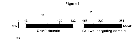
[0010] Figure 1 is a schematic diagram of phiNM3 lysin showing the
putative CHAP ("cysteine- and histidine-dependent
amidohydrolase/peptidase") and CWT domains. The numbers represent the
amino acid positions and the domain limits. The CWT domain of ClyS is
indicated in the diagram.
[0011] Figure 2A is a gel showing the purification of phiNM3 CWT. SDS-
PAGE and coomassie blue stained gel of phiNM3 CWT purified by anion-
exchange chromatography is depicted in the lane marked "CWT." Protein
- 6 -
CA 02729160 2010-12-22
WO 2010/002959
PCT/US2009/049349
molecular weight markers in kilodaltons (kDa) are shown in the lane marked
,cm .11
[0012] Figure 2B shows the amino acid sequence of the phiNM3 CWT
protein (SEQ ID NO:1).
[0013] Figure 3 shows a series of micrographs showing PhiNM3 CWT
binding specifically to staphylococci. Purified phiNM3 CWT was labeled with
FITC and exposed to 1) S. aureus; 2) B. cereus; 3) S. epidermidis; 4) E. coli;
5) Group A Streptococcus and 6) mixed suspension of S. aureus and B.
cereus cells. "P" indicates phase-contrast image and "F" indicates fluorescent
image.
[0014] Figure 4 is a schematic diagram illustrating chimeric lysin
development. In particular, Figure 4 provides schematic diagrams of various
chimeric lysins showing their respective domains and the corresponding
expression and solubility of the protein and activity against S. aureus cells.
Similar domains are depicted in the same shading and labeled. PlyB-cat
indicates catalytic domain of Bacillus-specific lysin PlyB (and is marked with
a
"4" in the figure); Sa-aa indicates 16 amino acid residues specific for
staphylococcal lysins (and is marked with a "Sin the figure); PlyB-CWT
indicates CWT domain of PlyB (and is marked with a "6" in the figure); Twort-
CWT indicates CWT domain of S. aureus phage Twort lysin (and is marked
with a "8" in the figure); Lysostaphin CWt indicates CWT domain of
lysostaphin (and is marked with a "10" in the figure); and Se autolyin amidase
indicates an amidase domain of S. epidermidis autolysin (and is marked with
a "12" in the figure).
[0015] Figure 5A shows the ClyS protein sequence. The predicted protein
sequence of the chimeric protein ClyS showing the Twort endopeptidase
catalytic and the phiNM3 CWT domains.
[0016] Figure 5B shows the amino acid sequence for the AD127 chimeric
molecule, described with respect to Figure 4.
[0017] Figure 5C shows the amino acid sequence for the native
(unmodified) Twort lysin (SEQ ID NO: 12).
- 7 -
CA 02729160 2010-12-22
WO 2010/002959
PCT/US2009/049349
[0018] Figure 6 is a gel showing the purification of ClyS. ClyS was
expressed in E. coli DH5a cells and purified by cation-exchange
chromatography followed by hydroxyapatite chromatography. Purified sample
(10 micrograms) was separated by SDS-PAGE and stained by Coomassie
blue (right hand lane). Protein molecular weight markers in kilodaltons (kDa)
are shown in the left hand lane.
[0019] Figure 7 is a graph showing the activity of ClyS against S.
aureus in
vitro. S. aureus strain 8325-4 cells were resuspended in 20 mM phosphate
buffer (pH 7.4), incubated with 50U of ClyS and 0D600 (filled triangles)
monitored by a spectrophotometer. Control experiments (filled squares) were
performed under the same conditions with buffer alone. Viability (filled
diamonds) of cells, shown as colony-forming units/ml, was determined by
serially diluting and plating the cells.
[0020] Figure 8 is a series of micrographs showing that ClyS causes cell
wall disruption and ultimately lysis of 8325-4 cells. Figures 8A ¨ 8C (A-C)
are
thin-section transmission electron micrographs (bars, 200 nm) of S. aureus 3
minutes after exposure to 50 U of ClyS. The arrows indicate cytoplasmic
membrane extrusions through holes generated in the cell wall by ClyS.
Ultimate lysis results in "cell-ghosts" (D) after the loss of cytoplasmic
contents
(bar, 500 nm).
[0021] Figures 9A and 9B are graphs showing the activity of ClyS in
various pH and salt concentration conditions. Figure 9A is a graph of the
activity of ClyS (50U) tested against S. aureus strain 8325-4 in buffers with
pH
values ranging from 4 and 10 in 15 minute assays. Optical density (filled
squares) and viability (filled diamonds) was measured as described in legend
of Figure 6. Fold killing in the viability assay was calculated by dividing
the
number of viable bacteria after buffer treatment at a particular pH by the
number after exposure to ClyS enzyme at the same pH. Final pH readings for
each reaction are recorded on the x axis. Figure 9B is a graph showing the
activity of ClyS (50 U) tested against S. aureus strain 8325-4 in 20 mM
phosphate buffer (pH 7.4) in the presence of different concentrations of NaCI.
- 8 -
CA 02729160 2010-12-22
WO 2010/002959
PCT/US2009/049349
After 15 minutes samples were assayed for optical density and viability
calculated as above.
[0022] Figure 10 is a bar graph showing that ClyS exerts specific
killing of
staphylococci. Log-phase cultures of different bacteria were exposed to 50 U
of ClyS for 15 minutes. Fold killing was calculated as described in Figure 8
legend.
[0023] Figure 11 depicts a graph of the CFU of MRSA from individual
MRSA infected mice after being administered phosphate buffered saline pH
7.3 (control) or ClyS (630 pg).
[0024] Figure 12 depicts Kaplan Meier Survival Curves showing the effect
of ClyS on preventing death in mice injected with MRSA compared with
phosphate buffer control.
[0025] Figure 13 depicts an isobologram for a checkerboard broth
microdilution study of the effect of vancomycin on VISA (vancomycin-resistant
Staphylococcus aureus) or oxacillin on MRSA with increasing amounts of
ClyS.
[0026] Figure 14 depicts Kaplan Meier Survival Curves showing the effect
of oxacillin alone or in combination with ClyS.
[0027] Figure 15 depicts a photograph of a Coomassie-blue stained SDS-
PAGE gel of a 5-day time-course at 21 C of ClyS in the absence (top left gel)
or presence of 5 mM DTT (top right gel), and pClyS in the absence (bottom
left gel) or presence of 5 mM DTT (bottom right gel). About 20 micrograms of
protein was loaded into each lane of the gel. The bottom right gel shows a
much higher amount of intact pClyS in the presence of 5mM DTT after 5 days
compared to intact ClyS in the presence of 5 mM DTT after 5 days.
DETAILED DESCRIPTION
Definitions
[0028] Unless otherwise indicated, the certain terms used herein and
their
applicability to the present disclosure are defined below.
[0029] The term "isolated" means at least partially purified from a
starting
material. The term "purified" means that the biological material has been
measurably increased in concentration by any purification process, including
- 9 -
CA 02729160 2010-12-22
WO 2010/002959
PCT/US2009/049349
by not limited to, column chromatography, HPLC, precipitation,
electrophoresis, etc., thereby partially, substantially or completely removing
impurities such as precursors or other chemicals involved in preparing the
material. Hence, material that is homogenous or substantially homogenous
(e.g., yields a single protein signal in a separation procedure such as
electrophoresis or chromatography) is included within the meanings of
isolated and purified. Skilled artisans will appreciate that the amount of
purification necessary will depend upon the use of the material. For example,
compositions intended for administration to humans ordinarily must be highly
purified in accordance with regulatory standards.
[0030] The term "lytic enzyme genetically coded for by a bacteriophage"
refers to a polypeptide having at least some lytic activity against the host
bacteria.
[0031] Variants of "chimeric bacteriophage lysin" are included within
the
definition of chimeric bacteriophage lysins, and include a functionally active
chimeric bacteriophage lysin with killing activity against Staphylococcus
aureus having at least 50%, 55%, 60%, 65%, 70%, 75%, 80%, 85%, 90%,
95%, 97%, 98%, 99%, or even at least 99.5% amino acid sequence identity
with a sequence described herein. For example, the present invention
includes chimerical bacteriophage lysins having at least 50%, 55%, 60%,
65%, 70%, 75%, 80%, 85%, 90%, 95%, 97%, 98%, 99%, or even at least
99.5% amino acid sequence identity with the polypeptide sequence of SEQ ID
NO:2.
[0032] "Percent (%) polypeptide sequence identity" with respect to the
lytic
enzyme polypeptide sequences identified here is defined as the percentage of
amino acid residues in a candidate sequence that are identical with the amino
acid residues in the specific lytic enzyme polypeptide sequence, after
aligning
the sequences and introducing gaps, if necessary, to achieve the maximum
percent sequence identity, and not considering any conservative substitutions
as part of the sequence identity. Methods for alignment for purposes of
determining percent amino acid sequence identity are described below.
- 10 -
CA 02729160 2010-12-22
WO 2010/002959
PCT/US2009/049349
Staphylococcal Lysins
[0033] Chimeric bacteriophage lysins with killing activity against S.
aureus
are described herein. Lysins generally occur in a modular structure. Figure 1
is a schematic diagram of phiNM3 lysin showing the putative CHAP domain
110 and the CWT domain 120. The numbers represent the amino acid
positions and the domain limits. The CWT domain of ClyS is shown as
shaded box 120. The N-terminal module consists of a catalytic domain
believed to possess the ability to break down the bacterial cell wall of
certain
bacteria. Enzymatic activities often associated with the catalytic domain are
amidases, endopeptidases, glucosamidases and muramidases. The C-
terminal module consists of a binding domain that is believed to have an
affinity for a carbohydrate epitope on the target bacteria cell wall. The
binding
domain is believed to determine the specificity of the lysin. The peptide
cross-
bridge within the staphylococcal peptidoglycan is believed to function as the
receptor for the CWT domain of lysostaphin, a staphylolytic enzyme produced
by Staphylococcus simulans. The CWT domain of lysostaphin has homology
to the SH3b domain suggesting that such lysins might also utilize the peptide
cross-bridge as its receptor.
[0034] In one embodiment, Staphylococcus-specific binding molecules
comprising a CWT domain within staphylococcal lysins are provided that have
no known domain homologs. In some embodiments, the binding molecules
are lysins. In other embodiments, the binding molecules may be used as
diagnostic tools, for example to identify the presence of Staphylococcus
bacteria. Preferably, such a CWT domain is provided to recognize a different
epitope such as a cell wall-associated carbohydrate instead of the peptide
cross-bridge in the staphylococcal cell wall.
[0035] In a further embodiment, the ClyS lysine can be used to digest
the
cell wall of Staphylococcus aureus bacterial strains, which in turn would
allow
access to the genetic and cytoplasmic material, such as endogenous DNA
and RNA, to further identify and sequence the Staphylococcus aureus
bacterial strain. It will also release membrane-associated and wall-
associated molecules for diagnostic purposes.
-11-
CA 02729160 2010-12-22
WO 2010/002959
PCT/US2009/049349
[0036] Most
preferably, the binding molecule is a soluble binding domain of
a bacterial lysin comprising a polypeptide including an amino acid sequence
providing specific binding to S. aureus, such as SEQ ID NO:1 (phiNM3 CWT
domain). For example, the lysin preferably includes the polypeptide sequence
of S. aureus phage phiNM3 lysin (SEQ ID NO:1) (protein accession number
YP_908849). The phiNM3 lysin CWT domain (SEQ ID NO:1) corresponding
to amino acid residues 158-251 was cloned and expressed. The
approximately 10-kDa protein of SEQ ID NO:1 was highly soluble and was
purified by one-step anion-exchange chromatography to homogeneity. Figure
2A is an anion exchange gel showing the protein of SEQ ID NO:1 in a second
column next to a set of marker proteins in a first column. Figure 2B shows the
amino acid sequence of SEQ ID NO:1. To determine whether the peptide
domain of SEQ ID NO:1 displayed Staphylococcus-specific binding, the
purified protein was labeled with FITC and exposed to log-phase S. aureus, S.
epidermidis and mixed population of S. aureus and Bacillus. Group A
streptococci, E. coli and Bacillus cereus were used as controls. More
preferably, The FITC-labeled phiNM3 CWT domain bound specifically to S.
aureus (Fig 3-1) and S. epidermidis (Fig 3-3) cells when present in single or
mixed populations (Fig 3-6) while binding to streptococci (Fig 3-5), Bacillus
(Figure 3-2) or E. coli (Figure 3-4) was not observed. PhiNM3 lysin
specifically bound to S. aureus (Fig 3-1) and S. epidermidis (Fig 3-2) cells
when present in single or mixed populations (Fig 3-3) while binding to
streptococci (Fig 3-4), Bacillus (Figure 3-5) or E. coli (Figure 3-6) was not
observed.
[0037] In one
embodiment, the binding molecule comprises a CWT binding
domain, such as the amino acid sequence of SEQ ID NO:1, attached to a
reporting portion that is detectable to identify the presence of the binding
molecule bound to Staphylococcal bacteria. For example, the binding
molecule may include the amino acid sequence of SEQ ID NO:1 bound to a
fluorescent reporter group, a radioactive reporter group or a heterologous tag
that is adapted to bind a fluorescent reporter. The phiNM3 (SEQ ID NO:1)
CWT domain may be used as a diagnostic tool for the identification of
-12-
CA 02729160 2010-12-22
WO 2010/002959
PCT/US2009/049349
staphylococcal bacteria. The high affinity binding site may be used in a wide
range of assay techniques to detect S. aureus. Such assay methods include
radioimmunoassays, gold sol radial immune assays, competitive-binding
assays, Western Blot assays and ELISA assays. Such detection assays
advantageously utilize a heterogeneous format wherein a binding reaction
(SEQ ID NO:1) between a conjugated binding agent comprising (SEQ ID
NO:1) and an analyte occurs followed by a wash step to remove unbound
conjugated binding agent. For example, gold sol particles may be prepared
with protein that comprises the binding region with the binding protein
immobilized on the particle surfaces. As binding occurs between the protein
and (staphylococcal) bacteria, the particles merge and form a colored product.
Analogously, the binding protein may be complexed, preferably covalently
with an enzyme such as beta galactosidase, peroxidase, or horseradish
peroxidase. After wash, the remaining bound enzyme can be detected by
adding a substrate such as a fluorogenic or chemilumigenic substrate. The
binding protein may be complexed with any other reagent that can make a
signal such as a rare earth fluor and detected by time resolved fluorescence,
a radioactive material and detected by radioactivity measurement, green
fluorescent protein (GFP) or another fluorescent tag, and detected by
fluorescence.
[0038] For comparison, Figure 5B provides the amino acid sequence of
SEQ ID NO:3, the AD119 sample discussed with respect to Figure 4. AD119
(SEQ ID NO:3) comprises the Twort endopeptidase domain joined to the
Lysostaphin CWT domain. In contrast to the chimeric compound of SEQ ID
NO:2 (AD127), which shares the Twort endopeptidase domain but has the
phiNM3 CWT domain (SEQ ID NO:1) in place of the Lysostaphin CWT
domain, the AD127 compound was insoluble and exhibited little or no killing
activity against S. aureus.
[0039] The conjugation of the binding region with a detectable tag may
be
carried out by synthetic chemistry or a biological process. For example, a
DNA sequence coding for the binding region of SEQ ID NO:1 or of the entire
lysin of SEQ ID NO:2 can be linked to genetic information that encodes a
- 13 -
CA 02729160 2010-12-22
WO 2010/002959
PCT/US2009/049349
detectable marker such as green fluorescent protein (GFP) or an enzyme
such as alkaline phosphatase. This could be accomplished by separating the
DNA for the binding domain by removing the N-terminal catalytic domain and
replacing it in frame with indicator molecules such as green flourescent
protein (GFP) and purifying the expressed fusion molecule for the
identification of S. aureus. Since the binding domain has a similar binding
affinity of an immunoglobulin G molecule, the marked binding domain will
effectively identify Staphylococcus aureus with little false positive
activity.
One also could fuse the GFP molecule or an enzyme at the 5' end of the
whole lysin enzyme if necessary, by doing so the enzymatic domain will be at
least partly inactivated, still allowing the binding domain to function to
bind to
its substrate in the bacillus cell wall. Optionally, the isolated binding
domain
of SEQ ID NO:1 may be separated from the catalytic domain of SEQ ID NO:2
and may be expressed, purified and labeled using a number of fluorescent
molecules such as fluorescein isothiocyanate, rhodamine isothiocyanate and
others known by skilled artisans. The binding domain may be modified with
biotin to allow formation of a biotin-avidin complex after the binding region
adheres to the Staphylococcus aureus for identification.
[0040] In another embodiment, the lysin is a chimeric protein that
comprises an endopeptidase domain of the S. aureus Twort lysin upstream of
the phiNM3 CWT domain (SEQ ID NO:1). The chimeric polypeptide is
preferably sufficiently soluble in phosphate buffered saline (PBS). Preferred
levels of solubility in PBS for the chimeric lysins is at least about 1 mg/ml
and
more preferably at least about 3 mg/mL in PBS. While native staphylococcal
bacteriophage lysins are typically insoluble in PBS, the chimeric lysins
comprising an endopeptidase domain of a first lysin (e.g., Twort S. aureus
lysin) bound to the CWT domain of SEQ ID NO:1 are surprisingly soluble in
PBS (e.g., at least about 1 mg/ml, and typically about 3 mg/ml or greater).
One example of such a lysin is provided in SEQ ID NO:2 (AD127), shown in
Figure 5A and consisting of the Twort lysin endopeptidase domain attached to
the phiNM3 CWT domain (SEQ ID NO:1). The isolated polypeptide of SEQ
ID NO:2 (AD 127) was constructed by engineering S. epidermidis autolysin
-14-
CA 02729160 2010-12-22
WO 2010/002959
PCT/US2009/049349
amidase and Twort lysin endopeptidase domains upstream of phiNM3 CWT
domain, respectively. Chimera AD 126 had no expression or activity but AD
127 was soluble and had very high activity but low expression. To overcome
low expression of AD 127 construct, the entire chimera gene was cloned into
expression vector pJML6 to generate pAD 138. The expression, solubility
and activity of AD 127 from the pAD138 construct was very high. Therefore,
this chimera was named 'ClyS' for Chimeric lysin for Staphylococcus (Figure
5A).
[0041] ClyS (SEQ ID NO:2) contains 280 amino acid residues with a
deduced molecular mass of 31956 Da and a theoretical isoelectric point of
9.17, and was purified by two-step column chromatography to >90%
homogeneity. ClyS had a molecular mass of approximately 31 kDa by
SDS/PAGE (Figure 6) which was confirmed by gel filtrations chromatography,
suggesting that the protein exists as a monomer and is not proteolytically
processed (data not shown).
[0042] The unit activity of ClyS was defined by measuring the
spectrophotometric loss of turbidity, indicative of cell lysis, of S. aureus
8325-
4 cells upon adding serial dilutions of ClyS. In our assays, 5 micrograms of
ClyS corresponded to 1 U of lytic activity. When 50 U of ClyS was added to
exponentially growing 8325-4 cells the 0D600 dropped to baseline within 5
min (Figure 7). To confirm that the observed cell lysis corresponds to cell
death, staphylococcal viability was determined by enumerating aliquots from
the lytic reaction at various time points. A decrease in viability of
approximately 3-logs was observed in 30 min (Figure 7).
[0043] The lytic effect on S. aureus 8325-4 cells exposed to 50U of ClyS
for 1-3 min was visualized by transmission electron microscopy. Typical of
lysin activity observed previously, localized degradation of the cell wall was
observed at single (Figure 8A) or multiple sites (Figure 8B). However, unlike
other lysins, the sites of degradation on the cell was not restricted to the
septal or polar positions but was randomly distributed. This resulted in
extrusions and rupture of the cell membrane (Figure 8C) and subsequent loss
of cytoplasmic contents and formation of cell-ghosts (Figure 8D).
- 15 -
CA 02729160 2010-12-22
WO 2010/002959
PCT/US2009/049349
[0044] The effect of pH on the activity of ClyS was determined by
measuring the drop in 0D600 or cell viability at different pH values. We
observed that ClyS was active over a wide range of pH values but was most
active between pH 9 and 10. However, ClyS retained partial yet significant
activity at physiological pH (Figure 9A). Similarly, the effect of salt
concentration on activity of ClyS was also determined. ClyS displayed activity
in a wide range of salt concentrations (Figure 9B). While its activity
deteriorated above 400 mM NaCI, at physiological concentrations ClyS
functioned well.
[0045] Muralytic activity of ClyS was tested on a number of bacterial
strains representing a variety of species which were divided into sets (Table
1
and Figure 10). Set I consisted of S. aureus strains including methicillin-
sensitive S. aureus (MSSA) and MRSA. ClyS was active against MSSA and
MRSA although differences were observed between S. aureus strains. Set II
consisted of different species of staphylococci including S. epidermidis, S.
simulans and S. sciuri. ClyS was active not only against S. epidermidis
including the biofilm-forming strain RP62A but was also active against S.
simulans and S. sciuri suggesting that ClyS recognizes an epitope in the cell
wall that is present in all staphylococcal cells. Set III consisted of a mix
of
Gram-positive and Gram-negative bacteria including representatives of group
A, B, C and E streptococci, oral streptococcal species including S. gordonii,
and S. salivarius, as well as S. uberis, Bacillus cereus, Pseudomonas
aeruginosa and E. coli. ClyS exhibited no activity against any of these
organisms.
[0046] In another embodiment, a chimeric peptide comprises an isolated
polypeptide comprising an endopeptidase domain of the S. aureus Twort lysin
upstream of the lyphostaphin CWT domain. One example of such a lysin is
provided in SEQ ID NO:3 (AD119).
[0047] In another embodiment, lytic compositions may comprise a mixture
of two or more lysins. The mixture may include a first polypeptide and a
second peptide where one or both of the polypeptides may lack a desired
level of lytic activity, but the mixture provides desirably specific and
effective
- 16 -
CA 02729160 2010-12-22
WO 2010/002959
PCT/US2009/049349
lytic activity against a bacteria of interest. For example, a composition may
include an isolated first polypeptide comprising an endopeptidase domain of
the S. aureus Twort lysin upstream of the lyphostaphin CWT domain
combined with a second isolated polypeptide comprising an S. epidermidis
autolysin amidase domain upstream of the lysostaphin CWT domain. One
example of such a composition comprises a mixture of SEQ ID NO:3
(AD119) and SEQ ID NO:4 (AD112).
[0048] In some examples, the present disclosure pertains to lytic
enzymes
as a prophylactic treatment for preventing infection those who have possibly
been exposed to S. aureus bacteria, or as a therapeutic treatment for those
who have already become ill from the infection. The phage associated lytic
enzymes described herein are specific for S. aureus bacteria and preferably
effectively and efficiently break down the cell wall of the S. aureus
bacteria.
[0049] The chimeric lytic enzyme polypeptides described herein may also
be employed as a therapeutic agent. The lytic enzyme polypeptides of the
present invention can be formulated according to known methods to prepare
pharmaceutically useful compositions, whereby the lytic enzyme product
hereof is combined in admixture with a pharmaceutically acceptable carrier
vehicle. Compositions which may be used for the prophylactic and
therapeutic treatment of a S. aureus bacteria infection also includes the
shuffled and/or chimeric enzyme and a means of application (such as a
carrier system or an oral delivery mode) to the mucosal lining of the oral and
nasal cavity, such that the enzyme is put in the carrier system or oral
delivery
mode to reach the mucosa lining.
[0050] In one preferred embodiment, a Staphylococcus chimeric lysin ,
such as a lysin of SEQ ID NO:2 (ClyS), is administered as an antibacterial
composition in combination with a suitable pharmaceutical carrier. In certain
embodiments, the amount of the chimeric bacteriophase lysin present is a
therapeutically effective amount. "Carriers" as used herein include
pharmaceutically acceptable carriers, excipients, or stabilizers which are
nontoxic to the cell or mammal being exposed thereto at the dosages and
concentrations employed. Often the physiologically acceptable carrier is an
- 17 -
CA 02729160 2010-12-22
WO 2010/002959
PCT/US2009/049349
aqueous pH buffered solution. Examples of physiologically acceptable
carriers include buffers such as phosphate, citrate, and other organic acids;
antioxidants including ascorbic acid; low molecular weight (less than about 10
residues) polypeptide; proteins, such as serum albumin, gelatin, or
immunoglobulins; hydrophilic polymers such as polyvinylpyrrolidone; amino
acids such as glycine, glutamine, asparagine, arginine or lysine;
monosaccharides, disaccharides, and other carbohydrates including glucose,
mannose, or dextrins; chelating agents such as EDTA; sugar alcohols such as
mannitol or sorbitol; salt-forming counterions such as sodium; and/or nonionic
surfactants such as TWEEN TM, polyethylene glycol (PEG), and
PLURONICSTM. These antimicrobial/pharmaceutical compositions may be
administered locally or systemically.
[0051] Routes of administration include topical, ocular, nasal,
pulmonary,
buccal, parenteral (intravenous, subcutaneous, and intramuscular), oral,
parenteral, vaginal and rectal. Also administration from implants is possible.
The compounds of the invention may also be administered topically to the
skin or mucosa, that is, dermally or transdermally. Typical formulations for
this purpose include gels, hydrogels, lotions, solutions, creams, ointments,
dusting powders, dressings, foams, films, skin patches, wafers, implants,
sponges, fibres, bandages and microemulsions. Liposomes may also be
used. Typical carriers include alcohol, water, mineral oil, liquid petrolatum,
white petrolatum, glycerin, polyethylene glycol and propylene glycol.
Penetration enhancers may be incorporated [see, for example, J Pharm Sci,
88 (10), 955-958 by Finnin and Morgan (October 1999).]
[0052] The compounds of the invention may also be administered directly
into the blood stream, into muscle, or into an internal organ. Suitable means
for parenteral administration include intravenous, intraarterial,
intraperitoneal,
intrathecal, intraventricular, intraurethral, intrasternal, intracranial,
intramuscular and subcutaneous. Suitable devices for parenteral
administration include needle (including microneedle) injectors, needle-free
injectors and infusion techniques. The compounds of the invention may also
- 18 -
CA 02729160 2010-12-22
WO 2010/002959
PCT/US2009/049349
be administered intranasally or orally by inhalation, typically in the form of
a
aerosol.
[0053] Suitable antimicrobial preparation forms are, for example
granules,
powders, tablets, coated tablets, (micro) capsules, suppositories, syrups,
emulsions, microemulsions, defined as optically isotropic thermodynamically
stable systems consisting of water, oil and surfactant, liquid crystalline
phases, defined as systems characterized by long-range order but short-
range disorder (examples include lamellar, hexagonal and cubic phases,
either water- or oil continuous), or their dispersed counterparts, gels,
ointments, dispersions, suspensions, creams, aerosols, droplets or injectable
solution in ampule form and also preparations with protracted release of
active compounds, in whose preparation excipients, diluents, adjuvants or
carriers are customarily used as described above. The pharmaceutical
composition may also be provided in bandages or in sutures or the like.
[0054] Many orthopedic surgeons consider that humans with prosthetic
joints should be considered for antibiotic prophylaxis. Late deep infection by
S. aureus is a serious complication sometimes leading to loss of the
prosthetic joint and is accompanied by significant morbidity and mortality. It
may therefore be possible to extend the use of the chimeric bacteriophage
lysin described herein (e.g., SEQ ID NO:2) as a replacement for or for use in
combination with prophylactic antibiotics in this situation. The chimeric
bacteriophage lysin may be administered by injection with a suitable carrier
directly to the site of the orthopedic device in situ to clear the infection,
or on a
surface of the device prior to implantantation. Other injection routes, such
as
subcutaneous, intramuscular, or intraperitoneal, can be used. Alternative
means for administration include transmucosal and transdermal
administration using penetrants such as bile salts or fusidic acids or other
detergents. In addition, if a polypeptide or other compounds of the present
invention can be formulated in an enteric or an encapsulated formulation, oral
administration may also be possible. Administration of these compounds may
also be topical and/or localized, in the form of salves, pastes, gels, and the
like.
- 19 -
CA 02729160 2010-12-22
WO 2010/002959
PCT/US2009/049349
[0055] Prior to, or at the time the enzyme is put in the carrier system
or
oral delivery mode, it may be desirable for a chimeric peptide described
herein to be administered or formulated in a stabilizing buffer environment,
maintaining a pH range between about 5.0 and about 7.5. Prior to, or at the
time the chimeric peptide is put in the carrier system or oral delivery mode,
the enzyme may be in a stabilizing buffer environment for maintaining a
suitable pH range, such as between about 5.0 and about 8.0, including a pH
of about 5.0, 6.0, 7.0, 8.0 or any pH interval of 0.05 therebetween, or any
interval that is a multiple of 0.05 therebetween, including pH values of 5.2,
6.5, 7.4, 7.5 and 8.5.
[0056] There are a number of advantages to using lytic enzymes to treat
bacterial infections. The modular design of lysins, with their distinct
catalytic
and binding domains, makes them ideal for domain swapping experiments in
which bacterial specificities and catalytic activities can be improved or
adapted for use against alternate pathogens. Since the catalytic and binding
targets of lysins (peptidoglycan and associated carbohydrates, respectively)
are largely essential for viability, lysin resistance will be rare.
[0057] "Treatment" refers to both therapeutic treatment and prophylactic
or
preventative measures, wherein the object is to prevent or slow down (lessen)
the targeted pathologic condition or disorder. Those in need of treatment
include those already with the disorder as well as those prone to have the
disorder or those in whom the disorder is to be prevented.
[0058] "Mammal" for purposes of treatment refers to any animal
classified
as a mammal, including humans, domestic and farm animals, and zoo, sports,
or pet animals, such as dogs, cats, cattle, horses, sheep, pigs, goats,
rabbits,
etc. Preferably, the mammal is human.
[0059] The formulations to be used for in vivo administration are
preferably
sterile. This is readily accomplished by filtration through sterile filtration
membranes, prior to or following lyophilization and reconstitution.
Therapeutic
compositions herein generally are placed into a container having a sterile
access port, for example, an intravenous solution bag or vial having a stopper
pierceable by a hypodermic injection needle.
- 20 -
CA 02729160 2010-12-22
WO 2010/002959
PCT/US2009/049349
[0060] The route of administration is in accord with known methods, e.g.
injection or infusion by intravenous, intraperitoneal, intracerebral,
intramuscular, intraocular, intraarterial or intralesional routes, topical
administration, or by sustained release systems. When treating a bacterial
exposure or infection, the lytic enzyme may be administered in any suitable
fashion, including parenterally or through the oral or nasal cavity.
[0061] Dosages and desired drug concentrations of pharmaceutical
compositions of the present invention may vary depending on the particular
use envisioned. The determination of the appropriate dosage or route of
administration is well within the skill of an ordinary physician. Animal-
experiments provide reliable guidance for the determination of effective doses
for human therapy. lnterspecies scaling of effective doses can be performed
following the principles laid down by Mordenti, J. and Chappell, W. "The use
of interspecies scaling in toxicokinetics" In Toxicokinetics and New Drug
Development, Yacobi et al., Eds., Pergamon Press, New York 1989, pp. 42-
96.
[0062] When in vivo administration of a chimeic peptide lysin is
employed,
normal dosage amounts may vary from about 10 ng/kg to up to 1000 mg/kg of
mammal body weight or more per day, or about 1 pg/kg/day to
10000mg/kg/day, depending upon the route of administration. Guidance as to
particular dosages and methods of delivery is also provided below, as well as
in the literature. It is anticipated that different formulations will be
effective for
different treatment compounds and different disorders, that administration
targeting one organ or tissue, for example, may necessitate delivery in a
manner different from that to another organ or tissue.
[0063] The effective dosage rates or amounts of the chimeric peptide to
be
administered parenterally, and the duration of treatment will depend in part
on
the seriousness of the infection, the weight of the patient, the duration of
exposure of the recipient to the infectious bacteria, the seriousness of the
infection, and a variety of a number of other variables. The composition may
be applied anywhere from once to several times a day, and may be applied
for a short or long term period. The usage may last for days or weeks. Any
-21-
CA 02729160 2010-12-22
WO 2010/002959
PCT/US2009/049349
dosage form employed should provide for a minimum number of units for a
minimum amount of time. The concentration of the active units of a chimeric
peptide believed to provide for an effective amount or dosage of enzyme may
be in the range of about 10, 20, 30, 40, 50, 60, 70, 80, 90, or 100 units/ml
up
to about 10,000,000 units/ml of composition, in a range of about 1000 units/ml
to about 10,000,000 units/ml, and from about 10,000 to 10,000,000 units/ml.
[0064] Additionally, a number of methods can be used to assist in
transporting the enzyme across the cell membrane. The enzyme can be
transported in a liposome, with the enzyme be "inserted" in the liposomes by
known techniques. Similarly, the enzyme may be in a reverse micelle. The
enzyme can also be pegylated, attaching the polyethylene glycol to the non-
active part of the enzyme. Alternatively, hydrophobic molecules can be used
to transport the enzyme across the cell membrane. Finally, the glycosylation
of the enzyme can be used to target specific internalization receptors on the
membrane of the cell.
[0065] Another preferred embodiment provides for a composition
comprising a Staphylococcus chimeric lysin bacterial binding protein such as
a lysin of SEQ ID NO:2 (ClyS), with other lytic enzymes which are useful for
sanitizing or decontaminating porous surfaces e.g. textiles, carpeting.
Furthermore, the composition of lytic enzymes may be used to decontaminate
veterinarian surgical or examination areas, where such areas may be thought
to harbor infectious organisms susceptible to the bacteriostatic or
bacteriocidal activity.
[0066] In a further preferred embodiment, a Staphylococcus chimeric
lysin
such as a lysin of SEQ ID NO:2 (ClyS) may be combined with other
bacteriostatic or bacteriocidal agents useful for decontamination of inanimate
solid surfaces suspected of containing infectious bacteria, or for
decontamination of porous surfaces.
EXAMPLES
Example 1: Identification of Specific Binding Peptides and Development of
Chimeric Lysins
-22 -
CA 02729160 2010-12-22
WO 2010/002959
PCT/US2009/049349
[0067] We conducted conserved domain searches of Staphylococcus-
specific phage and prophage lysin protein sequences in the National Center
for Biotechnology Information database. The lysins were classified based on
homology to known domains in the database. We identified several lysins
including the S. aureus phage phiNM3 lysin (protein accession number
YP_908849), S. aureus prophage phi13 amidase (accession number
NP_803402), S. aureus prophage MW2 amidase (accession number
NP_646703.1), etc. that shared 100% sequence identity with each other and
had a conserved CHAP domain within their catalytic domain. However, the C-
terminal domain of these lysins did not display homology to any known
domains in the database (Figure 1).
[0068] Since the attempts to express a native staphylococcal lysin were
unsuccessful, we decided to develop chimeric lysins by taking advantage of
the modular nature of lysins. Traditionally, Bacillus-specific lysins are
expressed at high levels and are soluble in E. coli. Therefore, our first
attempt
was to engineer a 16-amino acid peptide (4) that is conserved in several S.
aureus-specific lysins (Lu JZ, Fujiwara T, Komatsuzawa H, Sugai M, Sakon J.
2006. Cell wall-targeting domain of glycylglycine endopeptidase distinguishes
among peptidoglycan cross-bridges. (Lue et al. (2006) J. Biol. Chem.
281(1):549-58) The catalytic domain of the Bacillus-specific lysin PlyB was
used to generate chimera AD 103 (Figure 4) (SEQ ID NO: 13). The chimeras
were tested for expression, solubility and activity. Then the entire C-
terminal
CWT domain of PlyB (6) was replaced by the putative C-terminal domain of S.
aureus phage Twort lysin (8) to obtain AD 105 (SEQ ID NO:14). This chimera
was not active and so we engineered the lysostaphin CWT domain (10)
downstream of PlyB-catalytic domain (2) to get AD 107 (SEQ ID NO:15).
Although this chimera had expression, the solubility was poor and there was
no activity. The next step was to engineer a S. epidermidis autolysin amidase
domain (12) upstream of the lysostaphin CWT domain (10) which resulted in
AD 112 (SEQ ID NO:4). AD 112 expressed very well and the protein was
also very soluble but there was no lytic activity. However, we observed that
the S. aureus cells clumped when exposed to AD 112. Since the lysostaphin
-23-
CA 02729160 2010-12-22
WO 2010/002959
PCT/US2009/049349
catalytic domain (an amidase) (10) in AD 112 was of bacterial origin, we
attempted to engineer a phage-derived catalytic domain upstream of the
lysostaphin CWT. For this, the endopeptidase domain in Twort lysin (14) was
used to construct chimera AD 119 (SEQ ID NO:3). We observed poor
expression for AD 119 but the chimera was soluble. Although in our lytic
assays AD 119 alone did not show significant activity, when combined with
chimera AD 112 the activity was significantly enhanced. Since we identified
phiNM3 CWT domain from our conserved domains searches and observed
that phiNM3 CWT exhibited Staphylococcus-specific binding, we constructed
chimeras AD 126 (SEQ ID NO:16) and AD 127 (SEQ ID NO:2) by engineering
S. epidermidis autolysin amidase (12) and Twort lysin endopeptidase (14)
domains upstream of phiNM3 CWT domain (2) (SEQ ID NO:1), respectively.
Chimera AD 126 had no expression or activity but AD 127 was soluble and
had very high activity but low expression (Figure 4). To overcome low
expression of AD 127 construct, the entire chimera gene was cloned into
expression vector pJML6 to generate pAD 138. The expression, solubility
and activity of AD 127 from the pAD138 construct was very high. Therefore,
this chimera was named 'ClyS' for Chimeric lysin for Staphylococcus. The
amino acid sequence for ClyS (i.e., SEQ ID NO:2) is provided in Figure 5A.
Example 2: Construction of the ClyS Chimeric Lysin
[0069] Bacterial strains (Table 1) were stored at -80 C routinely grown
at
37 C. Staphylococcal strains used in this study were grown in Trypticase Soy
Broth (TSB) media, streptococcal strains were grown in THY (Todd¨Hewitt
broth, 1% wt/vol yeast extract) media, B. cereus and P. aeruginosa were
grown in BHI (Brain Heart Infusion) media while E. coli was cultivated in LB
(Luria Bertani) media.
[0070] The chimeric lysin was constructed by amplifying and ligating
individual domains from respective genes. For this, the Twort endopeptidase
domain was PCR amplified from plasmid pCR2.1plyTW which contains the
entire lysin (plyTW) gene using primers TW-Endo-Ncol-F: 5'-
CTAGCCATGGAAACCCTGAAACAAGCAG-3' (SEQ ID NO:5) and TW-Endo-
Pstl-R: 5'-ACATGCTGCAGAACCATATTGTAATTAATATTAGTTCTATC-
- 24 -
CA 02729160 2010-12-22
WO 2010/002959
PCT/US2009/049349
3'(SEQ ID NO:6). The cell wall targeting (CWT) domain was PCR amplified
from S. aureus strain 8325 genomic DNA using primers NM3-CBD-Pstl-F: 5'-
ACATGCTGCAGGGTAAATCTGCAAGTAAAATAACAG-3' (SEQ ID NO:7)
and NM3-CBD-Hind-R: 5'-
CCCAAGCTTAAAACACTTCTTTCACAATCAATCTC-3'(SEQ ID NO:8). The
two PCR amplicons were ligated using the Pstl restriction endonuclease site.
The ligated product was cloned into pBAD24 vector using the Ncol-HindlIl
cloning sites to generate recombinant plasmid pAD127. In the second step,
the entire DNA fragment corresponding to clyS was PCR amplified from
pAD124 using primers NM3-Lys-Xba-F: 5'-
CTAGTCTAGAGGTGGAATAATGAAAACATACAGTGAAGCAAG-3' (SEQ ID
NO:9) and primer NM3-CBD-Hind-R(SEQ ID NO:8). The PCR product was
cloned into expression vector pJML6 to generate pAD138. The sequence of
ClyS was confirmed by sequencing. The recombinant plasmid pAD138 was
transformed into E. coli DH5a cells.
Example 3: Overexpression and purification of ClyS
[0071] ClyS was induced overnight from E. coli DH5a (pAD138) cells with
lactose (10g/500m1final concentration) at 30 C. Cells were harvested by
centrifugation, resuspended in buffer A (20 mM phosphate buffer (PB), 1 mM
DTT (dithiothreitol)) and lysed by an EmulsiFlex-05 high pressure
homogenizer (Avestin) at 400C. The lysates were cleared by centrifugation
(2x 50,000xg) for 30 minutes at 4 C and the supernatant applied to a CM-
sepharose column (Amersham Pharmacia, Piscataway, N.J.). ClyS was
eluted with buffer A + 1M NaCI using a linear gradient of 0-50% B in 15
columns volumes. Fractions were analyzed for lytic activity as previously
described (Daniel et al, 2001). Fractions displaying lytic activity were
pooled
and dialyzed overnight against buffer B (PB, 1 mM DTT, 50mM NaCI). The
dialyzed sample was applied to a hydroxylapatite (MacroPrep Type II 40[1m,
BioRad) column and eluted with elution buffer (500 mM PB + 50 mM NaCI+ 1
mM DTT) using a linear gradient of 0-100% B in 20 columns volumes. The
fractions were analyzed by SDS-PAGE and for lytic activity. Active clean
-25-
CA 02729160 2010-12-22
WO 2010/002959
PCT/US2009/049349
fractions of ClyS were pooled and dialyzed against buffer B. Protein
concentration was determined with the BCA method (Sigma, St. Louis, MO).
Example 4: Quantification of ClyS activity
[0072] ClyS activity was measured as previously described (Daniel et al,
2001), with some modifications. Briefly, S. aureus strain 8325-4 was grown to
an 0D600 of 0.25-0.3, centrifuged, and resuspended in PB to a final 0D600 of
0.8-1Ø Two-fold serial dilutions of purified ClyS (100p1) were added to
100[11
of bacterial suspension in 96-well plates (Costar) and the decrease in 0D600
was monitored by a Spectramax Plus 384 spectrophotometer (Molecular
Devices) over 30 min at 37 C. ClyS activity in units per milliliter was
defined
as the reciprocal of the highest dilution of lysin that decreased the
absorbance
by 50% in 15 minutes.
Example 5: Measuring in vitro ClyS activity
[0073] The viability assay of ClyS was tested as previously described
(Nelson et al, 2001). Briefly, logphase cultures of S. aureus strain 8325-4
were resuspended in PB to 0D600 of 0.8-1Ø 50U of ClyS or the
corresponding volume of PB was added to bacterial cells and aliquots were
removed, serially diluted, and plated at 1, 5, 10, 30, and 60 minutes to
assess
the viability of the treated and control cells. All experiments were performed
in triplicate. The activity of ClyS on various bacterial strains was tested as
described previously (Schuch et al, 2002). Briefly, logphase bacterial cells
were treated with 50U of ClyS at 37 C for 15 minutes. The samples were
serially diluted and plated. Control experiments with the addition of
phosphate buffer (pH 7.0) were performed under the same conditions.
Example 6: Measuring ClyS activity as a function of pH and salt profile
[0074] The effect of pH on ClyS activity was determined as previously
described using the universal buffer system pH 4-10 (Yoong et al). Briefly,
logphase 8325-4 cells were resuspended in the universal buffer system and
incubated with 50U of ClyS for 15 minutes. The final pH of each reaction was
- 26 -
CA 02729160 2010-12-22
WO 2010/002959
PCT/US2009/049349
checked by pH paper. The samples were serially diluted and plated. In
controls, PB replaced ClyS.
[0075] Similarly the effect of salt concentration on the lytic activity
of ClyS
was determined by incubating 50U of ClyS with logphase 8325-4 cells in PB
containing NaCI at a final concentration of 25-500 mM for 15 minutes. The
samples were serially diluted and plated to determine the viability counts.
Example 7: Microscopy of ClyS
[0076] S. aureus strain 8325-4 was grown to log-phase, centrifuged and
resuspended in PBS to an absorbance at 600nm of 1Ø The bacterial
suspension was incubated with 50U of ClyS at room temperature. The lytic
reaction was terminated after 1 minute and 5 minutes by adding
glutaraldehyde (final concentration 2.5%). The suspension was pelleted by
centrifugation and overlaid with 2.5% glutaraldehyde in 0.1 M cacodylate
buffer (pH 7.4). The samples were then postfixed in 1% osmium tetroxide,
block stained with uranyl acetate and processed according to standard
procedures by The Rockefeller University Electron Microscopy Service.
[0077] Flourescent labeling and binding analyses were performed on
phiNM3 CWT. S. aureus strain 8325-4 genomic DNA was used to amplify the
putative CWT of phiNM3 lysin using primers NM3-FWD 5'-
CATGCCATGGGTAAATCTGCAAGTAAAATAACAG-3' (SEQ ID NO:10) and
NM 3-REV 5'-CCCAAGCTTAAAACACTTCTTTCACAATCAATCTC-3'(SEQ ID
NO:11). The resulting amplicon was cloned into the arabinose-inducible
expression vector pBAD24. Positive clones containing the insert were
confirmed by sequencing. The approximately 10-kDa phiNM3 CWT protein
was expressed and the protein was purified in one step by cation-exhange
chromatography. The purified protein (1mg/m1) was incubated with 10 pl of
FITC (1mg/m1) for 1 hour. Excess FITC was removed on a desalting column.
The labeled-protein (50 p,g) was incubated with bacterial cells for 10
minutes,
washed 3x with phosphate-buffered saline (pH 7.4) and observed under
fluorescence microscope.
-27-
CA 02729160 2010-12-22
WO 2010/002959
PCT/US2009/049349
Example 8: Measuring in vivo activity of ClyS
[0078] MRSA strain would be grown to log-phase, centrifuged and
resuspended to a predefined titer of about 1010 cfu/ml. For intranasal
infection, 6-wk-old female C57BL/6J, outbread Swiss or BALB/c mice (weight
range 22 to 24 g, Charles River Laboratories, Wilmington, MA) would be
anesthetized with a mixture of ketamine (Fort Dodge Animal Health, Fort
Dodge, IA, 1.2 mg/animal) and xylazine (Miles Inc., Shawnee Mission, KS,
0.25 mg/animal), and inoculated with 15 pl of the bacterial suspension per
nostril (n = 10). The animals would be divided into 2 groups and administered
various concentrations of ClyS or sterile saline intraperitoneally six hours
after
infection and every six hours thereafter for 3 days. The survival rate for
each
group would be observed up to 7 days post infection. For intraperitoneal
infection, mice would be infected intraperitoneally with 100 pl of the
bacterial
suspension (n = 10). The animals would be divided into 2 groups and
administered various concentrations of ClyS or sterile saline
intraperitoneally
six hours after infection and every six hours thereafter for 3 days. The
survival rate for each group would be observed up to 7 days post infection.
Example 9. The linker region by itself does not confer solubility to a
chimera.
[0079] Since the ClyS construct was the only chimera that was highly
soluble and active against staphylococci, we hypothesized that the linker
region comprising of amino acid residues 142 through 185 of ClyS may be
crucial for solubility. We had previously cloned and expressed the native
phiNM3 lysin and observed that the protein was insoluble. To test this
hypothesis, we replaced the endopeptidase domain of ClyS with the amidase
domain of phiNM3 lysin upstream of the linker region of ClyS (ami-link-ClyS)
and expressed the chimera (data not shown). We observed that similar to the
native phiNM3 lysin, the ami-link-ClyS chimera was insoluble and expressed
as inclusion bodies. We also tested the lysates of ami-link-ClyS for activity
against staphylococci and did not observe any lytic activity confirming that
the
protein was insoluble and therefore inactive. Thus, it is the unique
combination of the N and C terminal domains that are the subject of this
- 28 -
CA 02729160 2010-12-22
WO 2010/002959
PCT/US2009/049349
patent that allow for a soluble complex to occur and behave as described
herein.
[0080] Example 10. In vivo Nasal Decolonization of MRSA by ClyS.
Carriage of both MSSA and MRSA in the human anterior nares is the major
reservoir for S. aureus infection. Studies have shown that roughly 80% of the
population could be nasally colonized by S. aureus, and that colonization can
be an increased risk factor for developing other more serious S. aureus
infections (Kluytmans, J., A. van Belkum. 1997. Nasal carriage of
Staphylococcus aureus: epidemiology, underlying mechanisms, and
associated risks. Clin Microbiol Rev 10(3): 505-20.). Elimination of nasal
carriage in the community or in the hospital setting thus could possibly
reduce
the risk of infection and slow the spread of drug resistant S. aureus
(Kluytmans et al. (1997)). To study the potential of ClyS to reduce MRSA
colonization of the nasal mucosa, C57BL/6J mice were intranasally inoculated
with ¨2x10⁷ of a spontaneously streptomycin resistant strain of MRSA (191-
SMR). Twenty-four hours post-infection mice were administered three doses
hourly of either phosphate buffered saline (control) or ClyS (960 pg) into the
nasal passages. One hour after the last treatment, mice were sacrificed and
bacteria colonies were enumerated on Spectra MRSA agar, (a selective
chromogenic medium developed to diagnostically detect MRSA nasal
colonization) and Columbia blood agar. No significant differences in CFU
were obtained between plating to Spectra MRSA agar or Columbia blood agar
(Data not shown) Three independent experiments were performed to
evaluate a total 20 mice for each treatment group (Figure 11). Compared to
the buffer alone control (Avg. 12,273 CFU/cavity), ClyS treatment (Avg. 1198
CFU/cavity) significantly (p<0.001) reduced the mean CFU on the nasal
mucosa.
[0081] Example 11. ClyS Treatment of Systemic MRSA Infections. In
order to assess whether ClyS treatment could prevent death resulting from
systemic MRSA infections, 4 week old FVB/NJ mice were intraperitonally
injected with ¨5x10⁵ CFU of the community-acquired MRSA strain MW2 in 5%
mucin. Preliminary experiments determined that 5x10⁵ CFU was 10X the
- 29 -
CA 02729160 2010-12-22
WO 2010/002959
PCT/US2009/049349
LDioo dose for a twenty-four hour period. Furthermore, within 3 hours of IF
injection the MRSA infection was systemic, i.e., MRSA were recovered in high
numbers from heart, liver, spleen, and kidney (data not shown). Treatment
occurred three hours post-infection, with either 20mM phosphate buffer or
1mg of ClyS in 20mM phosphate buffer injected IF (intraperitoneally). Mice
were then monitored for survival over ten days. The results from three
independent experiments were combined (ClyS treatment, n =16; buffer
treatment, n =14) and the mice survival data plotted with a Kaplan Meier
Survival curve (Figure 12). Within twenty-four hours all of the control mice
died of bacterial sepsis, while only 2/16 of ClyS treated mice died at forty-
eight hours, and the remaining mice (14/16, 88%) survived over the course of
the experiments (Figure 12).
[0082] Example 12. ClyS showed synergistic interaction with Vancomycin
and Oxacillin. We used the checkerboard broth-microdilution assay to test the
interaction of ClyS with vancomycin and with oxacillin. The vancomycin MIC
for VISA strain Mu50 was 8pg/m1 and the oxacillin MIC for MRSA strain COL
was 32 pg/ml, while the ClyS MIC was 6 and 8 U/mlfor both strains tested
(Mu50 and COL respectively). lsobolograms for ClyS with vancomycin and
ClyS with oxacillin was plotted by transcribing the enzyme concentrations
along the inhibitory line on the microtiter plate in an xly plot. The shape of
the
curves for both interactions were characteristic of a synergistic interaction
(Fig. 13) and were further confirmed by calculating the FICI for both
interactions which was (:).5.
[0083] Example 13. In vivo Synergy of Oxacillin and ClyS in the
Treatment
of Systemic MRSA Infections. In vitro experiments showed that ClyS acted
synergistically with oxacillin (Figure 14). To determine if this effect could
be
seen in our systemic MRSA infection model, FVB/NJ mice were
intraperitonally injected with ¨5x10⁵ CFUs of the MRSA strain MW2 as above.
Three hours post infection mice were treated in parallel, with a lower IF dose
of 130 g/mouse of ClyS combined with different concentrations of oxacillin
(10-100 pg/mouse) or buffer alone controls. Preliminary experiments
determined that an ED30 dose of ClyS (130 g/mouse) had minimal efficacy to
- 30 -
CA 02729160 2010-12-22
WO 2010/002959
PCT/US2009/049349
evaluate the effect of combinatorial treatment with oxacillin (data not
shown).
Mice were monitored for survival for 10 days and the results of 5 independent
experiments were combined and plotted in a Kaplan Meier Survival curve
(Figure 14). While only 30% (6/20 alive) to 35% (8/23 alive) of mice survived
with individual treatments of either 130 pg/mouse of ClyS or 100 pg/mouse of
oxacillin, respectively, neither differed significantly from the survival rate
of the
buffer alone control, 13% (2/15 alive). Conversely, a single dose of the
combined treatment of intraperitoneal injected ClyS (130 pg) with either 0 pg
or 50 pg of intramuscular injected oxacillin significantly increased mouse
survival (80%, 8/10 alive; 82%, 18/22 alive respectively) compared to the
individual treatments and buffer alone (Figure 14).
[0084] Example 16. Modification of ClyS. The G166 residue of ClyS (SEQ
ID NO:2) was changed to a proline by site directed mutagenesis (creating
pClyS). When the purified pClyS molecule (SEQ ID NO: 17) was subjected to
stability studies at 21 C for 5 days, the pClyS was found to be significantly
more stable in the presence of 5mM DTT the native ClyS with or without DTT
(Figure 15).
[0085] While the invention has been described and illustrated herein by
reference to various specific materials, procedures, and examples, it is
understood that the invention is not restricted to the particular materials,
combinations of materials, and procedures selected for that purpose.
Numerous variations of such details can be implied and will be appreciated by
those skilled in the art.
- 31 -