Note: Descriptions are shown in the official language in which they were submitted.
CA 02729883 2016-03-29
,
Metal stent for treating lesions in blood vessels,
comprising a packaging
Field of application of the invention
According to its illustrative embodiments, the present
invention relates to an arrangement, consisting of a
metallic stent as a medical implant for treating
lesions in blood vessels, and a packaging. The stent is
arranged in a protected fashion in the interior volume
of the packaging. The stent has a multiplicity of webs,
which together form a tubular shape. The stent length
and the stent lumen, as a passage, extend between the
proximal and the distal end. The stent assumes a
corresponding diameter in the dilated or released
state. The stent surface is embodied in a hydrophilic
fashion to promote hemocompatibility.
A special field of application is the vessel dilation
in the field of percutaneous transluminal angioplasty,
also including cardiovascular intervention. Such stents
are together with a catheter, which is provided
specially for this, inserted into the human body
through a minimal opening, e.g. by puncturing an artery
in the region of the thigh, and are moved up to the
lesion, i.e. the vessel restriction to be treated, and
are dilated there. Whereas the stent remains in the
dilated blood vessel and supports the latter from the
inside, the catheter is removed from the body. The flow
of blood through the dilated and supported vessel is
once again ensured. This process is carried out with
the aid of instantaneous X-ray recordings, which on a
monitor display both the blood vessels and the
instruments inserted into the body.
Another special field of application is the treatment
of aneurysms, i.e. dilated blood vessels. In this
treatment, a stent graft - consisting of a supporting
mesh and a cover - is inserted into the aneurysm in
order once again to ensure the conventional blood flow.
CA 02729883 2011-01-04
WO 2010/000080 - 2 -
PCT/CH2009/000190
Prior art
However, the metallic stents implanted into blood
vessels harbor certain risks for the patient. Inter
alia, thromboses can form at the structures of the
stent. Combined with medicaments administered to the
patient after the implantation, the occurrences of
thromboses in the case of bare metal stents (BMS) could
be reduced to less than 1% within the first 10 days.
Nevertheless, this is one of the most-feared
complications, particularly in the case of the coronary
intervention.
A property of the stent that is desired by medical
practitioners is the rapid growing in thereof, the so-
called reendothelialization. The latter is of the
utmost importance for the success of the stent therapy
because the cells in this endothelial layer form
essential antithrombotic factors. However, as long as
the stent has not grown in, and the structures thereof
are subjected to the blood flow, it is of the utmost
importance to provide an antithrogenic stent surface.
It is well-known that stents with hydrophilic surface
properties have a much higher hemocompatibility, i.e. a
much lower thrombogenicity. Substances have been
applied onto the stent surface by means of coating
methods in order to increase the hydrophilicity on the
stent surfaces [cf. Seeger JM, Ingegno MD, Bigatan E,
Klingman N, Amery D, Widenhouse C, Goldberg EP.
Hydrophilic surface modification of
metallic
endoluminal stents. J Vasc Surg. 1995 Sept; 22(3):327-
36; Lahann J, Klee D, Thelen H, Bienert H, Vorwerk D,
Hocker H. Improvement of haemocompatibility of metallic
stents by polymer coating. J Mater Sci Mater Med. 1999
Jul; 10(7):443-8)].
CA 02729883 2011-01-04
WO 2010/000080 - 3 -
PCT/CH2009/000190
By way of example, possible coating methods include
"chemical vapor deposition" (CVD) or "physical vapor
deposition" (PVD), by means of which materials, e.g.
polymers or metals with defined layer thicknesses, are
applied onto the stent surface. It was found that in
the case of a polymer-coated EMS, the thrombocyte
formation was reduced from 85% (EMS) to 20% (polymer-
coated BMS) as a result of the increased hydrophilic
properties of the surface.
On the one hand, strong friction forces acting on the
stent surface occur during the clinical intervention;
on the other hand, high mechanical stresses are
generated on the surface of the individual stent webs
during the expansion. After implantation, the stent is
subjected to a permanent, pulsating load originating
from the blood vessel. These high mechanical loads can
result in a detachment of the coating, as a result of
which there is a significant potential risk of
thromboses, microemboli made of coating particles and
serious chronic inflammations. Moreover, critical
irregularities in the coating were even determined on
yet to be implanted stents.
In addition to the mechanical influences, the stent
coatings are damaged or broken down by the chemical
reactions occurring in the body. Metallic coatings can
corrode as soon as the differing electrochemical
potential between coating and stent can be equalized
via the battery effect by means of an electrolyte, e.g.
blood.
Polymer coatings on stents are successively broken down
by the body by means of enzymes. This process is often
connected with an inflammation of the surrounding
vessel cells, which cause undesired cell
proliferations, which can lead to a re-narrowing
(restenosis) of the blood vessel. Moreover,
CA 02729883 2016-03-29
- 4 -
inflammations can already be caused by the polymer
coating itself.
Although such surface modifications promote - as a
positive effect - the growing in property of stents,
they can however cause clinical complications due to
the aforementioned problems. Until now no stent has
been available with an optimum, hydrophilic surface
that meets both the medical and the mechanical
requirements.
Object of the invention
In light of the previous disadvantages in the prior
art, the invention is based on the intended object of
providing a stent that has increased hydrophilicity due
to surface modification and hence is intended to avoid
the aforementioned problems. At the same time, a
packaging should be provided for storing and
transporting the stent provided with the surface
modification according to the invention in order to
intend to maintain the hydrophilic surface properties
of the stent up until its intervention.
In the case of storing initially uncrimped stents in
the packaging, a further intended object consists of
proposing means for mounting the stent onto a catheter,
wherein the hydrophilic properties of the stent surface
are intended to be maintained.
Overview of the invention
According to a broad aspect of the present invention,
there is provided an arrangement comprising: a bare
metal stent used as a medical implant for treating
lesions in blood vessels; and a packaging with an
interior volume in which the stent is arranged in an
inert fashion in the packaging in order to prevent
natural recontamination from the atmosphere; the stent
CA 02729883 2016-03-29
- 4a -
comprising: a multiplicity of webs, which together form
a tubular shape; a proximal end and a distal end, with
a stent lumen extending therebetween; and a bare metal
surface with a hydrophilic property, wherein molecular
chemical contaminants originating from the atmosphere,
mainly hydrocarbons, are significantly reduced on the
bare metal surface by a treatment, as a result of
which, as a measure of the hydrophilicity, the contact
angle of a water droplet situated on the bare metal
surface is reduced compared to the contact angle before
this treatment; and the packaging including: a
container with a base and a cover; wherein at least one
of the base or the cover includes an access that is
opened such that the stent is removable from the
packaging.
The arrangement according to illustrative embodiments of
the invention consists of a metallic stent as a medical
implant for treating lesions in blood vessels and a
packaging, with an interior volume, in which the stent is
arranged in a protected fashion. The stent has a
multiplicity of webs, which together form a tubular
shape, and a proximal end and a distal end, with a stent
lumen extending therebetween. The stent surface has a
CA 02729883 2016-03-29
- 5 -
hydrophilic property. The molecular chemical
contaminants originating from the atmosphere, mainly
hydrocarbons, are significantly reduced on the surface
by a treatment, as a result of which, as a measure of
the hydrophilicity, the contact angle of a water
droplet situated on the surface is reduced compared to
the contact angle before this treatment. The stent is
stored in an inert fashion in the packaging in order to
prevent natural recontamination from the atmosphere.
The following features relate to illustrative
embodiments of the invention: the treatment of the
surface for reducing the chemical contamination is
carried out as material ablation, namely e.g. by means
of sputtering as ion bombardment, electric discharge
machining, electrolytic polishing, plasma activation,
laser ablation, a mechanically abrasive method, dry
etching or wet-chemical etching.
Alternatively, the result of the treatment of the
surface for reducing the chemical contamination is an
unchanged topography of the surface, wherein the
treatment was in this case also carried out e.g. by
means of sputtering as ion bombardment, electric
discharge machining, electrolytic polishing, plasma
activation, laser ablation, a mechanically abrasive
method, dry etching or wet-chemical etching. A
treatment that does not ablate material, e.g. by means
of ultrasound, UV light or ozone, or a combination
treatment formed therefrom, can likewise lead to an
unchanged surface topography. An etching medium that
does not corrode the stent material itself is equally
suitable for this.
The entire content of the packaging is inert and the
packaging contains an inert filling.
CA 02729883 2016-03-29
- 6 -
A catheter is arranged in the packaging and a stent is
mounted on said catheter, wherein a balloon catheter or
a tube catheter is assigned in a complementary fashion
to a balloon-expanding or a self-expanding stent.
The packaging consists of a container with a base and a
cover. The base and/or the cover can be removed. The
base and/or the cover has/have an access to be opened
such that the stent can be removed from the packaging
or the stent mounted on a catheter can be removed from
the packaging together with the catheter.
The catheter has a tip at its distal end, and the
proximal end of the shaft of the catheter opposite the
tip protrudes through the access to outside of the
packaging.
There is a passage in the base or in the cover for
allowing a shaft to pass, which shaft leads to the jaws
of an integrated crimping apparatus toward the inside,
into the packaging, and leads to an activator for
actuating the crimping apparatus toward the outside.
The access to be opened is opposite the passage in the
cover or in the base, which access serves to let a
catheter pass. A guide mandrel extends through the
crimping apparatus in the axial direction and it is
used for stabilization and positioning purposes after
it has been completely inserted into a guide wire lumen
of the catheter. The access to be opened is
illustratively made of e.g. a penetrable seal or a
perforatable material.
Support elements for fixing the stent and/or the
catheter and/or the crimping apparatus extend within
the packaging.
The stent is embodied with a cover to form a stent
graft for the application in the case of aneurysms.
CA 02729883 2011-01-04
WO 2010/000080 - 7 -
PCT/CH2009/000190
Brief description of the attached drawings
In the figures:
figure 1A shows a balloon-expanding or self-expanding
stent in the uncrimped state;
figure 1B shows a packaging with a stent as per figure
1A stored therein in an inert filling;
figure 2A shows the arrangement as per figure 113 with
access into the packaging and an open
crimping apparatus integrated therein;
figure 2B shows the crimping apparatus from figure 2A
in the opened state;
figure 20 shows the crimping apparatus as per figure 23
in the closed state;
figure 3A shows the arrangement as per figure 2A with a
balloon-expanding stent, a
positioned
dilation catheter and an opened crimping
apparatus;
figure 313 shows the arrangement as per figure 3A with a
closed crimping apparatus;
figure 4 shows a packaging with a balloon-expanding
stent stored therein in an inert filling, on
a dilation catheter and in the crimped state;
figure 5A shows a packaging with an access and with an
uncrimped self-expanding stent stored therein
in an inert filling, a positioned catheter,
and an opened crimping apparatus;
CA 02729883 2016-03-29
- 8 -
figure 5B shows the arrangement as per figure 5A, in
the crimped state, with a closed crimping
apparatus;
figure 50 shows the arrangement as per figure 5B with
an opened crimping apparatus;
figure 5D shows the arrangement as per figure 50, with
an external tubing of the catheter that has
in part been pushed over the crimped stent;
figure 5E shows the arrangement as per figure 5D with
an external tubing of the catheter completely
pushed over the crimped stent; and
figure 6 shows a packaging with a self-expanding stent
stored therein in an inert filling, mounted
on a catheter and in the crimped state.
Exemplary embodiments
In the following text, and with reference to the
attached drawings, there is a detailed description of
the arrangement according to embodiments of the
invention, which consists of a metallic stent - as a
medical implant for treating lesions in blood vessels -
and a packaging with an interior volume, in which the
stent is arranged in a protected fashion.
The following statement holds true for the entire
subsequent description: If reference signs are
contained in a figure for the purpose of unambiguity in
the drawing but not mentioned in the directly
associated text of the description, reference is made
to the description thereof in the preceding or
subsequent descriptions of the figures. In the interest
of clarity, repeated designation of components in
further figures is generally dispensed with, provided
CA 02729883 2011-01-04
WO 2010/000080 - 9 -
PCT/CH2009/000190
it is clear from the drawing that these are "recurrent"
components.
Figure lA
The illustrated stent 3 has a conventional material
configuration and structural design; it could be
balloon-expanding or self-expanding. The stent 3 is of
length 1, which extends between the proximal end 31 and
the distal end 32. In the non-crimped state, the stent
3 assumes the diameter d, and so the webs 33 with the
surface 35 are spaced from one another in a spacious
and grid-shaped fashion. The stent lumen 34, in
principle of cylindrical design, runs through the
tubular stent 3.
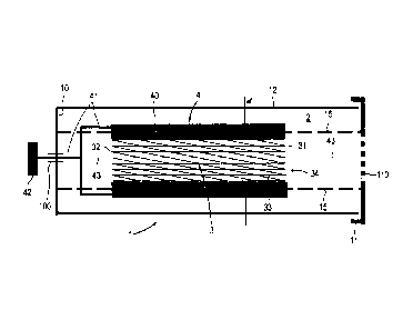
Figure 113
The stent 3 is in a packaging 1 and in the process is
fixed by a support 13 arranged in the packaging 1,
which support first of all comprises a first support
element 131, against which the proximal end 31 butts.
The distal end 32 is held by the second support element
132. The packaging 1 first of all comprises the
container 12 with the base 10 and is sealed by the
cover 11 on the end opposite the base 10. The first
support element 131 extends like a separation wall over
the cross-sectional area of the container 12 and faces
the cover 11, wherein a third support element 133
connects the cover 11 with the first support element
131 in the axial direction. The second support element
132 likewise extends like a separation wall over the
cross-sectional area of the container 12, but it faces
the base 10. There is an inert filling 2 in the
packaging 1 and it protects the surface 35 of the stent
3. The inner faces of the packaging 1 facing the stent
3 are inert.
The preceding treatment of the surface 35 increased the
hydrophilic property thereof. The molecular chemical
CA 02729883 2016-03-29
- 10 -
contaminants on the surface 35 originating from the
atmosphere - mainly hydrocarbons - were significantly
reduced, as a result of which, as a measure of the
hydrophilicity, the contact angle of a water droplet
situated on the surface 35 is reduced.
The chemical contaminants on the surface 35 can
illustratively be reduced by material ablation.
Sputtering as ion bombardment, electric discharge
machining, electrolytic polishing, plasma activation,
laser ablation, mechanically abrasive methods, dry
etching or wet-chemical etching lends itself for this
purpose. Alternatively, the reduction in the chemical
contaminants on the surface 35 is achieved by a
treatment that does not change the topography of the
surface 35. Treatment by means of ultrasound, UV light
or ozone, or a combination treatment formed therefrom,
can be considered for this. An etching medium that does
not corrode the stent material itself is equally
suitable for the treatment, for example an acid
treatment of the surface. 9596-97% sulfuric acid on a
cobalt-chromium alloy has proven its worth.
Figures 2A to 20
This group of figures schematically illustrates the
function of a crimping apparatus 4 arranged in the
packaging 1. At first, the crimping apparatus 4 is
open, and so the jaws 40 thereof assume a dilated
position and thereby encompass the expanded stent 3
situated in the packaging 1 (see figures 2A, 2B). The
stent 3 is pretreated as already explained with
reference to figure 13. The packaging 1 in turn
contains the inert filling 2 and the inner wall of the
packaging is inert. The jaws 40 are seated on a shaft
41, which, in the axial direction, leads outward
through a passage 100 in the base 10 to an actuatable
activator 42. Axes 15, which extend axially between the
base 10 and the cover 11, pass through the container
CA 02729883 2011-01-04
WO 2010/000080 - 11 -
PCT/CH2009/000190
12. A guide mandrel 43 belonging to the crimping
apparatus 4 runs centrally through the container 12,
which mandrel ends within the container 12 in front of
an access 110, which is on the cover 11 and can be
perforated. If the crimping apparatus 4 is closed, the
jaws 40 are narrowed in the radial direction, and so
the stent 3 has a compressed diameter d (see figure
2C).
Figures 3A and 3B
This pair of figures is based on the arrangement as per
figure 2A, wherein the utilized stent 3 is balloon-
expanding and was subjected to a pretreatment in order
to increase the hydrophilicity of the surface 35, as
explained with reference to figure 1B. Once again, an
inert filling in the packaging 1 and an inert property
of the inner wall thereof are assumed. The jaws 40 of
the crimping apparatus 4 are open at first (see figure
3A). The balloon 50 of the catheter 5 arranged on the
shaft 52 has been inserted into the stent lumen 34, tip
55 first, through the access 110, which is in the cover
11 and can be perforated. In the process, the guide
mandrel 43 has penetrated the guide wire lumen 53 in
the shaft 52. The shaft 52 furthermore has the channel-
like dilation lumen 54, by means of which the balloon
50 is brought to expand by being filled up on the
inside - e.g. by means of physiological saline - from
an external source during the operation and thus
dilates the stent 3 from the inside. The stent region
51 of the balloon 50 is in the stent lumen 34, and so
the stent region 51 at least in principle passes
through the entire length 1 of the stent, while the
tapering ends of the balloon 50 protrude from the
proximal end 31 and the distal end 32 of the stent 3.
After actuating the activator 42 by rotating it, e.g.
manually, the crimping apparatus 4 reaches the closed
state, and so the diameter d of the stent 3 is pressed
CA 02729883 2011-01-04
WO 2010/000080 - 12 -
PCT/CH2009/000190
together (see figure 3B). In the case of the now
narrowed stent diameter d and the compressed jaws 40 of
the crimping apparatus 4, the stent region 51 of the
balloon 50 remains in an unchanged axial position
within the stent lumen 34.
Figure 4
As an alternative to the design as per the preceding
figures, where a crimping apparatus 4 is integrated in
the packaging 1, here the packaging 1 now contains a
balloon-expanding stent 3 in the crimped state on the
balloon 50 of a dilation catheter 5. Here, the stent
diameter d is narrowed and the webs 33 are pushed
against one another. The stent region 51 of the balloon
50 once again extends over the length 1 of the stent,
at least in principle. The guide mandrel 43, which
extends from the base 10, has penetrated the guide wire
lumen 53 of the shaft 52. The tip 55 comes to rest near
the base 10. The interior of the packaging 1 is
provided with the inert filling 2 that protects the
surface 35 of the stent 3, which is pretreated as per
the description in respect of figure 1B. Furthermore,
the assumption is made that the inner wall of the
packaging 1 is inert. The dilation catheter 5 including
crimped stent 3 and balloon 50 can be pulled out of the
packaging 1 through the access 110, which is in the
cover 11 and can be perforated.
Figures 5A to 5E
In this sequence of figures, the packaging 1 has an
integrated crimping apparatus 4 and use is made of a
self-expanding stent 3 and a tube catheter 6. The
crimping apparatus 4 once again includes the shaft 41,
which extends to the activator 42 through the passage
100 in the base 10, and the guide mandrel 43 passing
axially through the packaging 1. The packaging 1
contains the inert filling 2 and the packaging inner
wall is inert. The axes 15 again lie within the
ak 02729883 2011-01-04
WO 2010/000080 - 13 -
PCT/CH2009/000190
packaging 1. The surface 35 of the stent 3 has been
pretreated in order to increase the hydrophilicity, as
explained with reference to figure 1B.
Figure 5A (initial situation
The jaws 40 of the crimping apparatus 4 are open; it
follows that the stent 3 is in the uncrimped state and
the inner tubing 66 of the tube catheter 6 has been
pushed through the access 110, which is in the cover 11
and can be perforated, and through the stent lumen 34
to the extent that the tip 65 protrudes from the stent
3 and faces the base 10. The guide mandrel 43 has
penetrated the guide wire lumen 63 of the shaft 62 in
the axial direction. The support tubing 67 and the
outer tubing 68 have likewise been pushed through the
access 110, which can be perforated, but the free ends
thereof are in front of the proximal end 31 of the
stent 3. The stent region 61, which can hold the length
1 of the stent, extends between the free end of the
support tubing 67 and the stop 69 at the tip 65.
Figure 5B (1st continuation steR
The jaws 40 of the crimping apparatus 4 have now been
closed, and so the webs 33 of the stent 3 lie pushed
together and the stent diameter d is narrowed. The
crimping apparatus 4 was actuated by rotating the
activator 42. The tube catheter 6, comprising the tip
65, the inner tubing 66, the support tubing 67, and the
outer tubing 68, remains in the same position. The
stent 3 is cooled in the crimped state in order to
disable the self-expanding property when the
temperature drops below a defined threshold.
Figure 5C (2nd continuation ste=
The jaws 40 of the crimping apparatus 4 are opened,
with the self-expanding stent 3 remaining in the
crimped state with the narrowed stent diameter d and
CA 02729883 2011-01-04
WO 2010/000080 - 14 -
PCT/CH2009/000190
the compacted webs 33 as a result of the prior
temperature drop.
Figure 5D (3rd continuation step)
The stent 3 remaining in the crimped state with the
narrowed stent diameter d allows successive pushing of
the outer tubing 68 onto the stent 3 in the direction
of the distal end 32 from the proximal end 31. The
support tubing 67 and the tip 65 arranged on the inner
tubing 66 remain in the same position. The advance of
the outer tubing 68 also moves the stent 3 in the same
direction, with the stop 69 preventing the further
advance of the stent 3.
Figure 5E (4th continuation step)
The outer tubing 68 has been pushed so far over the
crimped stent 3 that it meets the stop 69 behind the
tip 65 and it follows that it now covers the entire
stent region 61. In this state, the tube catheter 6
with the crimped stent 3 accommodated therein is pulled
out of the packaging 1 through the access 110, which
can be perforated, in order to apply the stent 3, which
has been prepared as detailed above, to the patient at
the predetermined site in the body.
Figure 6
As an alternative to the design as per the preceding
sequence of figures 5A to 5E, where a crimping
apparatus 4 is integrated in the packaging 1, the self-
expanding stent 3 has now been crimped outside of the
packaging 1 and mounted on the tube catheter 6 and
inserted into the packaging 1 such that there is no
need for a crimping apparatus 4 belonging to the
packaging 1. The guide mandrel 43 has been inserted
into the guide wire lumen 63. The shaft 62 with outer
tubing 68, support tubing 67 and inner tubing 66
protrude outward through the access 110, which is in
the cover 11 and can be perforated. The outer tubing 68
CA 02729883 2011-01-04
WO 2010/000080 - 15 -
PCT/CH2009/000190
butts against the stop 69 of the tip 65 and thus
spreads over the entire stent region 61. The free end
of the support tubing 67 is in front of the proximal
end 31 of the stent 3. Further handling is brought
about as in connection with figure 5E.
The assumption is made that the stent 3 utilized in
this case has had the same pretreatment as in all
preceding exemplary embodiments as per figures 13 to 5E
and the packaging 1 contains an inert filling 2 and the
packaging inner wall is inert.