Note: Descriptions are shown in the official language in which they were submitted.
CA 02730362 2016-01-18
Apparatus and Method for Cardiac Tissue Modulation by Topical Application
of Vacuum to Minimize Cell Death and Damage
Related Applications
[0001] The present application claims the benefit of priority of U.S.
Provisional
Application 61/088,558, filed on August 13, 2008 and U.S. Provisional
Application
No. 61/081,997, filed on July 18, 2008.
Field of the Invention
[0002] The present invention relates generally to a method and apparatus for
treating
cardiac tissue, and more particularly, but not exclusively, to modulating
ischemic and
reperfused heart tissue with topical sub-atmospheric pressure to minimize cell
death
and damage.
Background of the Invention
[0003] Myocardial ischemia occurs when a portion of the heart does not receive
sufficient oxygen and energy substrates to meet its demand. This usually
occurs
because of a blockage in the artery due to either atherosclerotic plaque or
thrombus
forination. In a myocardial infarction there is an area of injury where the
cells,
because of lack of blood flow, will die immediately. There is a layer adjacent
where
there is impaired blood flow that is equivalent to the zone of stasis and
there is a
more peripheral unaffected zone. Unfortunately the infarcted heart will
attempt to
increase rate of contracture and overall work to compensate for areas of the
heart that
are not functioning adequately. Consequentially the areas that are in the
"zone of
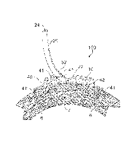
stasis" are called upon to do more work which will increase the energy
requirements
1
CA 02730362 2011-01-10
WO 2010/009294
PCT/US2009/050806
placed upon them and will subsequently result in further progression of death.
If left
untreated, this ischemia will lead to an expanding zone of infarction that may
eventually extend transmurally across the thickness of the ventricle.
[0004] Limiting the degree of infarction resulting from myocardial ischemia is
paramount to improving both short- and long-term outcomes in patients.
Therefore,
in order to salvage this myocardial tissue, timely reperfusion (re-
establishment of
coronary blood flow) of the tissue must take place. The amount of salvageable
tissue
within an ischemic zone is dependent on the timeliness of reperfusion. While
reperfusion halts the ischemic processes by delivering oxygen and nutrients
(including energy substrates), this process also rapidly sets into motion a
series of
events and cascades that exacerbates injury, extending the area of necrosis
beyond
that encountered during ischemia alone. Much of this reperfusion injury
appears to
be inflammatory in nature, but inappropriately directed against host tissues
instead of
foreign substances. Being able to reduce this reperfusion injury allows for
the
salvage of the greatest amount of myocardium.
[0005] Reperfusion injury manifests itself in a number of ways, including
myocardial dysfunction (myocardial stunning), arrhythmias, and a collection of
events that result in lethal reperfusion injury. Currently, there are
effective
pharmacologic therapies to treat reperfusion arrhythmias, and myocardial
stunning
will generally resolve by itself given time, leaving the mediators of lethal
reperfusion
injury as the logical targets in an attempt to preserve ischemic-reperfused,
but viable
tissue.
[0006] There are a large number of potential mediators of lethal reperfusion
injury
including calcium overload, oxygen radicals, changes in osmotic gradients (and
subsequent cell swelling), the mitochondrial permeability transition pore, and
inflammation (itself a complex set of cascades and mediators including
complement
activation, leukocyte infiltration and pro-inflammatory cytokines and
mediators). In
addition, the cardioprotective effects of selective inhibition of any and all
of these
phenomenon, including antioxidants, sodium-hydrogen exchange inhibitors, anti-
inflammatory agents (including adenosine, adhesion molecule antibodies and
complement inhibitors) in animal models of myocardial ischemia-reperfusion are
known. However, very few have demonstrated any degree of clinical success in
2
CA 02730362 2016-01-18
people, likely due to the fact that these therapeutics act selectively at a
single point
within a cascade of events, or on a single facet of a very complex and
multifaceted
process. Thus, though the application of negative (or sub-atmospheric)
pressure
therapy to wounded cutaneous and subcutaneous tissue demonstrates an increased
rate of healing compared to traditional methods (as set forth in US Patent
Nos.
5645081, 5636643, 7198046, and 7216651, as well as US Published Application
Nos. 2003/0225347, 2004/0039391, and 2004/0122434), there remains a need in
the
art for devices and methods for treating myocardial ischemia. In these type
wounds
of cutaneous and subcutaneous wounds the screen/dressing can often be easily
and
non-invasively changed at routine, pre-determined intervals without
significant
disruption to the healing tissues. However, when techniques are used to treat
tissues
or organs in which the overlying skin is intact, the overlying skin must be
surgically
disrupted by the deliberate creation of a wound through the overlying tissue
to
expose the tissue or organ that was originally injured. The overlying,
originally
healthy tissues which were disrupted to expose the injured tissue can be
sutured
closed over top of the injured tissue. This allows for negative pressure
treatment of
the wounded tissues with restoration of the suprawound tissues. Current
commercially available embodiments of negative pressure dressings and cover
are
not biodegradable or bioresorbable. This lack of
biodegradability/bioresorbability
necessitates re-opening of the sutured incision, removal of the dressing and
cover,
placement of a new dressing and cover, and again suturing the incision closed.
This
sequence would have to be repeated until the original wounded tissue is
healed, with
one final re-opening of the incision to remove the dressing and cover. Every
time the
incision is opened to change or remove the dressing and cover, it increases
the risk
that the site will become infected.
Summary of the Invention
[0007] The present invention relates to devices and methods for treating
damaged
heart tissue, such as myocardial infarction in the ischemic or early
reperfusion phase,
by treatment with sub-atmospheric (or negative) pressure. Treatment with the
devices and methods of the present invention may salvage cells in the zone of
stasis
and thereby decrease the size of the infarct. Such treatment would be
especially
efficacious in endstage myocardial disease where bypass or stenting would not
be
3
CA 02730362 2011-01-10
WO 2010/009294
PCT/US2009/050806
possible. The treatment would also be useful as an adjunct to ECMO
(extracorporeal
membrane oxygenation) for resting the heart, following cardiac arrest, in
situations
with left main artery lesions, etc.
[0008] An exemplary negative pressure therapy device of the present invention
may
include a vacuum dressing, e.g., porous material, for placement over the
tissue to be
treated. The vacuum dressing may be bio-incorporable in nature so that a
second
stage for removal would not be required. (As used herein the term "bio-
incorporable" is defined to describe a material that may be left in the
patient
indefinitely and is capable of being remodeled, resorbed, dissolved, and/or
otherwise
assimilated or modified.) The device of the present invention may also include
a bio-
incorporable overlay cover for placement over the vacuum dressing to form a
sealed
enclosure in which sub-atmospheric pressure may be provided and maintained to
the
vacuum dressing and the tissue to be treated. The overlay cover may be
adherent to
the dressing and extend beyond the vacuum dressing to permit attachment of the
overlay cover to surrounding non-damaged heart tissue. The overlay cover may
be
gelatinous in nature to contour to the heart and may be sufficiently pliable
so as not
to interfere with cardiac function. The overlay cover may be secured to the
myocardium with fibrin glue, mini-staples, or sutures.
[0009] In use, the device of the present invention may be placed
thoraeoscopically
over the area of muscle that has infarcted and over the adjacent zone of
stasis. The
device may be placed through a small incision made in the chest wall and
perforated
through the pericardium. The vacuum dressing may be collapsible in structure
such
that it can be rolled up or folded so as to be small enough for insertion
through a
thoracoscope tube. The epicardium may be perforated with a CO2 or similar
laser or
other cutting instrument to expose the underlying ischemic myocardium. The
vacuum dressing may then be placed directly over this ischemic area. The
overlay
cover may also be placed and secured to surrounding heart tissue
endoscopically as
well. A vacuum tube, e.g., a small catheter, may then be introduced so that
the distal
end of the vacuum tube is in gaseous communication with the enclosure under
the
overlay cover to supply sub-atmospheric pressure to the enclosure and the
tissue to
be treated. The other end of the vacuum tube may then be placed in gaseous
communication with a vacuum source to produce sub-atmospheric pressure, and
the
4
CA 02730362 2011-01-10
WO 2010/009294
PCT/US2009/050806
vacuum source may be activated to supply the sub-atmospheric pressure to
effect
negative pressure therapy of the damaged heart tissue. In addition, the sub-
atmospheric pressure may be supplied intermittently at a rate that is matched
to the
heart rate.
[0010] The present invention may also provide delayed treatment of myocardial
infarction where there is already a stable zone of myocardial cell death.
Again
through an endoscope and a small incision in the chest wall, a bio-
incorporable
vacuum dressing may be placed on the area that is infarcted. Again, exposure
of the
myocardium involved and adjacent myocardium may be required and provided with
a CO2 or similar cutting device to perforate the epicardium. The vacuum
dressing
may be modified so that a lattice of myocardial or peripheral muscle cells may
be
incorporated within it. The vacuum dressing may also incorporate a small
catheter
with the ability to reinfuse additional myocardial cells, pleuripotent
progenitor cells,
or peripheral muscle cells at subsequent serial times. In areas where there is
near
complete cell death or there is little or no contraction of the muscle cells
in the
damaged cardiac tissue, new contractile cells could be seeded to replace and
restore
the contractile function of the damaged cardiac tissue. Initially, peripheral
muscle or
peripheral muscle cells grown from culture could be used. These cells have a
finite
life cycle and would be expected to fatigue over time. The myocardium could be
biopsied at the time of the treatment of the initial treatment and myocardial
cells
removed and cultured to create a larger mass of viable of cells. The harvested
myocardial cells could be maintained in culture and used for later periodic
infusion
to develop a myocardial patch that would cover the area of previous
infarction. Also,
progenitor cells could be harvested and immediately infused to the area of
damaged
cardiac tissue, or they could be grown in culture and periodically infused to
the area
of damaged cardiac tissue with the expectation that they would develop into
cardiac
myocytes. Over time the introduced cells would be induced to undergo mitosis
or
self-replication thus increasing the functional mass of the heart. The ability
to
progressively add cells that would be progressively vascularized is a major
step in
regenerative medicine where presently only a sheet of cells can be expected to
survive.
CA 02730362 2011-01-10
WO 2010/009294
PCT/US2009/050806
[0011] More specifically, in one of its aspects the present invention provides
a
method for treating damaged cardiac tissue using sub-atmospheric pressure. The
method comprises placing a porous material in direct or indirect contact with
the
damaged cardiac tissue to provide gaseous communication between one or more
pores of the porous material and the damaged cardiac tissue. The porous
material
may comprise at least one of an electrospun material, a cast material, an open-
cell
foam, or a printed material. Alternatively or additionally, the porous
material may
comprise a bio-incorporable material. The porous material may include, for
example, collagen, chitosan, polycaprolactone, polyglycolic acid, polylactic
acid, and
combinations thereof. In addition, the porous material may be a polyvinyl
alcohol
foam which may be disposed in direct contact with the damaged cardiac tissue.
[0012] The porous material may be sealed in situ over the damaged cardiac
tissue to
provide a region about the damaged cardiac tissue for maintaining sub-
atmospheric
pressure at the damaged cardiac tissue. The porous material may be operably
connected with a vacuum source for producing sub-atmospheric pressure at the
damaged cardiac tissue, and the vacuum source activated to provide sub-
atmospheric
pressure at the damaged cardiac tissue. The sub-atmospheric pressure may be
maintained at the damaged cardiac tissue for a time sufficient to reduce edema
(thus
restoring contractility and compliance), decrease interstitial pressure,
remove
inflammatory mediators, remove inflammatory amplifiers, modulate intracellular
mediators, increase reperfusion and microvascular flow, decrease microvascular
plugging, and/or decrease retention of inflammatory cells within the damaged
cardiac
tissue. Micro and macro deformation of the cardiac tissue being treated would
increase vasculoneogenesis or the formation of new blood vessels in the
ischemic
tissue. This would increase the survivability of the cardiocytes and
ultimately
improve function of the ischemic portion of the heart. In addition, macro and
micro
deformation of small arterioles already existing in the heart would result in
their
physical reorientation into the areas of ischemic tissue, thus increasing
perfusion and
ultimately function.
[0013] For example, the sub-atmospheric pressure may be maintained at about 25-
125 mm Hg below atmospheric pressure. The method may also include locating a
cover, such as a bio-incorporable cover, over damaged cardiac tissue and
sealing the
6
CA 02730362 2011-01-10
WO 2010/009294
PCT/US2009/050806
cover to tissue proximate the damaged cardiac tissue, e.g., to non-damaged
cardiac
tissue, for maintaining sub-atmospheric pressure at the damaged cardiac
tissue. The
cover may be provided in the form of a self-adhesive sheet which may be
located
over the damaged cardiac tissue. In such a case, the step of sealing the cover
may
include adhesively sealing and adhering the self-adhesive sheet to tissue
surrounding
the damaged cardiac tissue to form a seal between the sheet and tissue
surrounding
the damaged cardiac tissue.
[0014] In another of its aspects the present invention provides an apparatus
for
treating damaged cardiac tissue. The apparatus includes a porous material for
treating damaged cardiac tissue having a pore structure configured to permit
gaseous
communication between one or more pores of the porous material and the cardiac
tissue to be treated. The porous material may include at least one of an
electrospun
material, a cast material, and a printed material. Alternatively or
additionally, the
porous material may comprise a bio-incorporable material. In such instances,
it may
also be beneficial for the porous material to be formulated in such a manner
that the
outer edges of the porous material would be resorbed or degraded more quickly
than
the inner portion. The rate of removal (resorption/degradation) of the porous
material could be matched to the rate of formation of new tissue. One way to
control
the rate of degradation or resorption is by varying the number of crosslinks
introduced into the porous material.
[0015] The apparatus may also include a vacuum source for producing sub-
atmospheric pressure; the vacuum source may be disposed in gaseous
communication with the porous material for distributing the sub-atmospheric
pressure to the cardiac tissue. The porous material may have, at least at a
selected
surface of the porous material, pores sufficiently small to prevent the growth
of
tissue therein. In addition, the porous material may have, at least at a
selected
surface of the porous material, a pore size smaller than the size of
fibroblasts and
cardiac cells, and may have a pore size at a location other than the selected
surface
that is larger than that of fibroblasts and cardiac cells. The pore size of
the porous
material may be large enough to allow movement of proteins the size of albumin
therethrough. Also, the porous material may include at least one surface that
is
sealed to prevent the transmission of sub-atmospheric pressure therethrough.
The
7
apparatus may also include a cover, such as a bio-incorporable cover,
configured to cover the
damaged cardiac tissue to maintain sub-atmospheric pressure under the cover at
the damaged
cardiac tissue.
[0016] The bio-incorporable porous material and/or cover may be constructed
from synthetic
materials such as polyglycolic acid, polylactic acid, or poly-o-citrate, or
they can be
constructed of naturally occurring molecules such as collagen, elastin, or
proteoglycans.
Combinations of synthetic molecules, combinations of naturally occurring
molecules, or
combinations of synthetic with naturally occurring molecules can be used to
optimize the
material properties of the porous material and cover.
[0017] An example of a material which may be used to fabricate the porous
material is
polycaprolactone (PCL). In one exemplary formulation, polycaprolactone is
mixed with
sodium chloride (1 part caprolactone to 10 parts sodium chloride) and placed
in a sufficient
volume of chloroform to dissolve the components. The solution is poured into
an
appropriately sized and shaped container and allowed to dry for twelve hours.
The sodium
chloride is then leached out in water.
[0018] A second exemplary cast formulation for the porous material is
chitosan, 1.33%
(weight/volume) in 2% acetic acid. The solution (20 ml) is poured into an
appropriately sized
container and frozen for 2 hours at -70 C, then transferred to a lyophylizer
and vacuum
applied for 24 hours. The freeze dried dressing is then crosslinked with 2.5
to 5%
glutaraldehyde vapor for 12 to 24 hours.
[0018a] In accordance with an aspect of an embodiment, there is provided an
apparatus for
treating damaged cardiac tissue, comprising: a porous bio-incorporable
material for treating
damaged cardiac tissue having a pore structure configured to permit gaseous
communication
between one or more pores of the porous material and the cardiac tissue to be
treated, the
porous material comprising pores sufficiently small at a surface of the porous
material for
placement proximate the damaged cardiac tissue to prevent the ingrowth of
tissue therein and
having a selected surface disposed away from the cardiac tissue to be treated
having a pore
size sufficiently large to promote the formation of granulation tissue
thereat; a bio-
incorporable cover for placement over the damaged cardiac tissue for sealing
engagement
with cardiac tissue proximate the damaged cardiac tissue for maintaining sub-
atmospheric
pressure at the damaged cardiac tissue; and a vacuum source for producing
subatmospheric
pressure disposed in gaseous communication with the porous material for
distributing the
sub-atmospheric pressure to the cardiac tissue to be treated.
8
CA 2730362 2017-07-26
CA 02730362 2016-01-18
[0018b1 In accordance with another aspect of an embodiment, there is provided
an
apparatus for treating damaged cardiac tissue, comprising: a porous bio-
incorporable
material for treating damaged cardiac tissue having a pore structure
configured to
permit gaseous communication between one or more pores of the porous material
and
the cardiac tissue to be treated; a bio-incorporable cover for placement over
the
damaged cardiac tissue for sealing engagement with cardiac tissue proximate
the
damaged cardiac tissue for maintaining sub-atmospheric pressure at the damaged
cardiac tissue; and a vacuum source for producing sub-atmospheric pressure
disposed
in gaseous communication with the porous material for distributing the sub-
atmospheric pressure to the cardiac tissue to be treated.
[0018c] In an embodiment, the porous material comprises at least one of a
porous
sheet and a flexible, sheet-like mesh.
[0018d] In accordance with another aspect of an embodiment, there is provided
a
degradable or resorbable vacuum appliance for treating injured or diseased
tissues in a
body, comprising: a dressing configured to be implanted in the body, the
dressing
having a void structure configured to permit the transmission of sub-
atmospheric
pressure therethrough; and a bio-incorporable cover configured to be implanted
in the
body to cover and enclose the dressing to provide a chamber about the dressing
in
which sub-atmospheric pressure may be maintained.
[0018e] In accordance with another aspect of an embodiment, there is provided
an
apparatus for treating an organ, the apparatus comprising: an air-tight
chamber
configured to surround and contain the organ, and a vacuum source operably
connected
to the chamber for applying and maintaining sub-atmospheric pressure to the
organ.
[0019] Thus, the present invention provides devices and methods for minimizing
the
progression of pathologic processes, minimizing the disruption of
physiological cardiac
integrity, and minimizing the interference with cardiac blood flow and
nutrition and
increasing revascularization of ischemic areas of the heart by vascular
neogenesis and
reorientation of existing vessels. By decreasing cardiac edema and
interstitial pressure
the risk of cardiac cell death and compromise may be minimized. In addition,
the
present invention facilitates the removal of mediators, degradation products,
and toxins
that enhance the inflammatory and pathophysiological response in the damaged
cardiac
tissue.
8a
CA 02730362 2011-01-10
WO 2010/009294
PCT/US2009/050806
Brief Description of the Drawings
[0020] The foregoing summary and the following detailed description of the
preferred embodiments of the present invention will be best understood when
read in
conjunction with the appended drawings, in which:
[0021] Figure 1 schematically illustrates a partial cross-sectional view of an
exemplary configuration of an apparatus of the present invention in situ prior
to the
application of sub-atmospheric pressure;
[0022] Figure 2 schematically illustrates the partial cross-sectional view of
Fig. 1 as
a sub-atmospheric pressure is being applied;
[0023] Figure 3 schematically illustrates the partial cross-sectional view of
Fig. 1
after sub-atmospheric pressure has been applied;
[0024] Figure 4 schematically represents a cross-sectional view of an
exemplary
configuration of the present invention in situ in which the tissues overlying
the heart
have been closed around the tube to create a space capable of maintaining a
vacuum
so no overlay cover is required;
[0025] Figure 5 schematically represents a partial cross-sectional view of the
apparatus of the present invention in situ in which the porous material is
layered with
a smaller pore layer adjacent to the damaged tissue and a layer with larger
pores
above the smaller pore layer;
[0026] Figure 6 schematically represents a view of an exemplary configuration
of a
porous material of the present invention in which only one side of the porous
material is open and not sealed;
[0027] Figure 7 schematically represents a cross-sectional view of an
exemplary
configuration of the present invention in which an overlay cover has been
placed
over the porous material and potential leaks sealed with fibrin glue;
[0028] Figure 8 schematically represents a partial cross-sectional view of an
exemplary configuration of the present invention in which the edges of the
overlay
cover have been turned under;
[0029] Figure 9 schematically represents a cross-sectional view of an
exemplary
configuration of the present invention in which the overlay cover is self
adhesive;
9
CA 02730362 2011-01-10
WO 2010/009294
PCT/US2009/050806
[0030] Figure 10 schematically represents an exemplary configuration of the
cover
of the present invention in which the tube passes through the overlay cover;
[0031] Figure 11 schematically represents a partial cross-sectional view of
the
vacuum tube attaching to the overlay cover;
[0032] Figure 12 schematically represents a kidney, with artery and vein;
[0033] Figure 13 schematically represents an open clamshell or bi-valve
chamber for
application of sub-atmospheric pressure; and
[0034] Figure 14 schematically represents a kidney disposed within the chamber
of
Fig. 13.
Detailed Description of the Invention
[0035] Referring now to the figures, wherein like elements are numbered alike
throughout, the present invention relates to devices and methods that use sub-
atmospheric (or negative) pressure for treating damaged cardiac tissue, where
"damaged" tissue is defined to include tissue that is injured, compromised, or
in any
other way impaired, such as damage due to trauma, disease, infection, surgical
complication, or other pathologic process, for example. More specifically, the
devices and methods of the present invention can effect treatment of
myocardial
infarction.
[0036] An exemplary configuration of a sub-atmospheric cardiac treatment
device
100 of the present invention may include a vacuum source 30 for supplying sub-
atmospheric pressure via a tube 20 to a porous material 10, such as a bio-
incorporable porous material, disposed in direct or indirect contact with the
damaged
cardiac tissue 7, Figs. 1-4. As used here, "indirect contact" is defined to
mean
placement of an intermediate material for transmitting sub-atmospheric
pressure in
contact with both the damaged cardiac tissue 7 and the porous material 10. In
this
regard, the porous material 10 may be structured to deliver and distribute sub-
atmospheric pressure to the damaged cardiac tissue 7. Alternatively, the
porous
material 10 may be comprised of a material that needs to be removed after sub-
atmospheric therapy is given, which could require a second surgery. The
cardiac
treatment device 100 may be applied to a patient by locating a porous material
10 in
contact with the damaged cardiac tissue 7 to provide gaseous communication
CA 02730362 2011-01-10
WO 2010/009294
PCT/US2009/050806
between one or more pores of the porous material 10 and the damaged cardiac
tissue
7. A tube 20 may be connected to the porous material 10 at a distal end 22 of
the
tube 20, and the porous material 10 may be sealed in situ by sutures 8 in the
skin 1
and subcutaneous tissues 2 to provide a region about the damaged cardiac
tissue 7 for
maintaining sub-atmospheric pressure, Fig. 4. The proximal end 24 of the tube
20
may be attached to a vacuum source 30 to operably connect the porous material
10 to
the vacuum source 30 for producing sub-atmospheric pressure at the damaged
cardiac tissue 7 upon activation of the vacuum source 30. Optionally, an
overlay
cover 40, such as a bio-incorporable overlay cover 40, may be located over the
damaged cardiac tissue 7 and sealed proximate the damaged cardiac tissue 7 to
maintain sub-atmospheric pressure at the damaged cardiac tissue 7.
[0037] Turning to Figs. 1-4 in greater detail, an exemplary configuration of a
sub-
atmospheric pressure cardiac treatment device 100 of the present invention is
illustrated in partial cross-section with the porous material 10 in contact
with the
damaged cardiac tissue 7. An overlay cover 40 covers the porous material 10
and
may extend onto healthy cardiac tissue 6 creating an enclosed space 48. An
adhesive
41, such as fibrin glue or other material, may be placed between the overlay
cover 10
and the healthy cardiac tissue 6. The adhesive 41 may also or alternatively be
placed
around the periphery of the overlay cover 10 to prevent leaks, and may also be
placed around a passthrough 52 where the tube exits from the overlay cover 10
to
prevent leaks. Figure 1 depicts the device 100 prior to application of sub-
atmospheric pressure. Figure 2 depicts the device 100 as sub-atmospheric
pressure is
being applied, and the enclosed space 48 decreases in volume as fluid and gas
are
evacuated from the enclosed space 48 and the overlay cover 40 conforms to the
porous material 10. Figure 3 depicts the device 100 after sub-atmospheric
pressure
has been applied, with the overlay cover 40 conforming to the shape of the
porous
material 10.
[0038] Turning to Fig. 4 specifically, an exemplary configuration of a sub-
atmospheric cardiac treatment device 100 of the present invention is
illustrated in
situ in a patient with surrounding tissues shown in partial cross-section. The
tissues
illustrated include the skin 1 and subcutaneous tissue 2, muscle 3, bone 4,
pericardium 5, healthy non-damaged cardiac tissue 6, the damaged cardiac
tissue 7,
11
CA 02730362 2011-01-10
WO 2010/009294
PCT/US2009/050806
and the pleural tissues 12. To provide access to the damaged cardiac tissue 7,
a
portion of the pericardium 5 may be missing due to surgical dissection or
injury. A
porous material 10, such as an open-cell collagen material, may be placed in
the
subcutaneous space in contact (direct or indirect) with the cardiac tissue 7
to be
treated with sub-atmospheric pressure to decrease edema and interstitial
pressure,
oxygen radicals, inflammatory mediators, and other molecules which may
adversely
affect cellular resuscitation or viability within the damaged cardiac tissues
to
improve physiologic function, for example. The distal end 22 of the tube 20
may
connect to the porous material 10 and the tube 20 may exit the body through an
incision. The tube 20 may have one or more fenestrations 23 in that portion of
the
tube 20 in contact with the porous material 10, Fig. 6. The tissues between
the
cardiac tissue 7 up to and including the skin 1 are closed with, for example
sutures 8,
to create an airtight seal capable of maintaining a vacuum. When sub-
atmospheric
pressure is applied, the edges of the incised tissues 1-5 are drawn together
and the
pleural tissues 12 are drawn toward the porous material to help maintain the
vacuum.
The proximal end of the tube 24 may be connected to a vacuum source 30 and the
level of sub-atmospheric pressure controlled by a controller 32. The vacuum
source
30 may include a canister to collect any fluid removed.
[0039] The cover 40 may serve to further confine the region about the damaged
cardiac tissue 7 at which sub-atmospheric pressure is maintained. That is, as
illustrated in Figs. 1-3, 7-9, the cover 40, 50 provides an enclosed
space/region 48,
58 about the damaged cardiac tissue 7 under the cover 40, 50, which can serve
to
isolate the tissues exterior to the cover 40, 50 from exposure to the sub-
atmospheric
pressure applied to the damaged cardiac tissue 7. In contrast, as illustrated
in Fig. 4,
in the absence of an overlay cover, sub-atmospheric pressure delivered to the
porous
material 10 and damaged cardiac tissue 7 may draw the surrounding tissues,
such as
the pericardium 5 and pleural tissues 12, inward towards the tube 20 and
porous
material 10 along the directions of the arrows shown in Fig. 4. In this regard
the
stretched and/or moved tissues, such as pericardium 5 and pleural tissues 12
can help
to confine the applied sub-atmospheric pressure to a region between the
pericardium
and the damaged cardiac tissue 7. In addition the covers 40, 50 may further
protect
the damaged cardiac tissue 7 from exogenous infection and contamination beyond
the protection already afforded by the porous material 10 and sutured skin 1
and
12
CA 02730362 2011-01-10
WO 2010/009294
PCT/US2009/050806
subcutaneous tissue 2. Likewise, the covers 40, 50 may further protect the
damaged
cardiac tissue 7 from the spread of infections from the surrounding tissues
(such as
cardiac abscesses and mediastinitis).
[0040] To assist in maintaining the sub-atmospheric pressure at the damaged
cardiac
tissue 7, a flexible overlay cover 40 (Fig. 7), or a self adhesive flexible
overlay cover
50 (Fig. 9) may be provided over the damaged cardiac tissue 7 to provide a
region
48, 58 about the damaged cardiac tissue 7 where sub-atmospheric pressure may
be
maintained, Figs. 7, 8. Specifically, with reference to Figs. 7 , 8, and 9, an
overlay
cover 40, 50 may be provided over the damaged cardiac tissue 7 and porous
material
by adhering the cover 40, 50 to cardiac tissues proximate the damaged cardiac
tissue 7 to define an enclosed region 48, 58 about the damaged cardiac tissue
7 and
porous material 10. For instance, the cover 40 may be glued to cardiac tissue
using
an adhesive 41, such as a fibrin glue. The adhesive 41 may comprise an auto-
polymerizing glue and/or may desirably include a filler to provide the
adhesive 41
with sufficient bulk to permit the adhesive 41 to conform to the shapes of the
potentially irregular surfaces which the adhesive 41 contacts. The adhesive 41
may
be provided as a separate component or as a portion of the cover 40. For the
flexible
overlay cover 40, an outside edge or border of the flexible overlay cover 40
may
either be rolled away from (or laid flat on) the non-damaged cardiac tissue 6
or rolled
under (or toward) the damaged cardiac tissue 7, Figs. 7, 8. The adhesive 41
may be
placed between the edge of the overlay cover 40 and the healthy cardiac tissue
6 to
promote an airtight seal. The adhesive 41 may also be placed around the tube
20
where it exits through the overlay cover 40. Alternatively, a self-adhesive
flexible
overlay cover 50 may be curled out away from the damaged cardiac tissue 7 so
that
the underside of the cover 50 (that side facing the porous material 10) may
then
contact with the surrounding non-damaged cardiac tissue 6 , Fig. 9.
[0041] In addition to an open-cell collagen material, the porous material 10
may also
include a polyglycolic and/or polylactic acid material, a synthetic polymer, a
flexible
sheet-like mesh, an open-cell polymer foam, a foam section, a porous sheet, a
polyvinyl alcohol foam, a polyethylene and/or polyester material, or other
suitable
materials which may be fabricated by electrospinning, casting, or printing,
for
example. Such materials include a solution of chitosan (1.33% weight/volume in
2%
13
CA 02730362 2011-01-10
WO 2010/009294
PCT/US2009/050806
acetic acid, 20 ml total volume) which may be poured into an appropriately
sized
mold. The solution is then frozen for 2 hours at -70 C, and then transferred
to the
lyophylizer and vacuum applied for 24 hours. The dressing may be cross-linked
by
2.5% ¨ 5% glutaraldehyde vapor for 12 ¨ 24 hours to provide a cast porous
material.
[0042] Additionally, the porous material 10 may be made by casting
polycaprolactone (PCL). Polycaprolactone may be mixed with sodium chloride (1
part caprolactone to 10 parts sodium chloride) and placed in a sufficient
volume of
chloroform to dissolve the components. A desired amount, e.g., 8 ml, of the
solution
may be poured into an appropriately sized and shaped container and allowed to
dry
for twelve hours. The sodium chloride may then be leached out in water for 24
hours.
[0043] The overlay cover 40 may also be bio-incorporable and may consist of an
electrospun mixture of Type I collagen and poly 1,8-octanediol citrate (POC)
(80%:20% weight/weight). The solution concentration may be 15% dissolved in
hexafluoro-2 proponal (HFP) with a total volume of 9.5 ml. The solution may
then
be ejected from a syringe through an 18 gauge needle at a flow rate of 1 ¨ 3
ml/hour.
The voltage may be 25 KV with a working distance of 20 ¨ 25 cm. The film may
then be heat polymerized at 80 C for 48 hours (of 90 C for 96 hours) and cross-
linked in 2.5% ¨ 10% glutaraldehyde vapor for 24 hours.
[0044] It is also possible to use electrospun materials for the porous
material 10 and
cast materials for the overlay cover 40. One example of a formulation and
method
for making an electrospun porous material 10 is a combination of collagen Type
I:chondroitin-6-sulfate (CS): poly 1,8- octanediol citrate (POC) in a ratio of
76%:4%:20%: by weight. Two solvents were utilized for the collagen/CS/POC. The
CS was dissolved in water and the collagen and POC were dissolved in 2,2,2-
trifluorocthanol (TFE). A 20% water/80% TFE solution (volume/volume) solution
was then used. For elcctrospinning, the solution containing the
collagen:CS:POC
mixture was placed in a 3 ml syringe fitted to an 18 Ga needle. A syringe pump
(New Era Pump Systems, Wantaugh, NY) was used to feed the solution into the
needle tip at a rate of 2.0 ml/hr. A voltage of 10-20 kV was provided by a
high
voltage power supply (HV Power Supply, Gamma High Voltage Research, Ormond
Beach. FL) and was applied between the needle (anode) and the grounded
collector
14
CA 02730362 2011-01-10
WO 2010/009294
PCT/US2009/050806
(cathode) with a distance of 15-25 cm. The dressings were then cross-linked
with
glutaraldehyde (Grade II, 25% solution) and heat polymerized (80 C) for 48
hours.
It is also possible to electrospin collagen Type I dressings starting with an
initial
concentration of 80 mg/ml of collagen in 1,1,1,3,3,3-hexafluoro-2-propanol
(HFP),
then use the same electrospinning conditions as the collagen:CS:POC
combination.
[0045] Examples of cast overlay cover formulas include the use of 1,8 poly
(octanediol) citrate (POC) or other combinations of diol citrates, which could
be 1,6
hexanediol or 1,10 decanediol, for example. To make the cast overlay cover 40,
equimolar amounts of anhydrous citric acid and the diol of choice may be
combined
in a round bottom flask. (As an example: 38.4g citric acid and 29.2g
octanediol).
The solution may be heated in an oil bath for 10 min at 165 C until melted,
then
continued to be heated at 140 C for 45min. The polymer may be used in this
form
although unreacted monomers are also present. To remove the unreacted monomer,
equivolume amounts of polymer and 100% acetone may be added to a flask and
shaken until the polymer is completely dissolved, then poured into an
appropriately
shaped mold. The acetone may be evaporated overnight in a chemical hood at
room
temperature. The films may be polymerized at 80 C for 36hr and then 18hr at
110 C.
[0046] Alternatively, to cast overlay covers 40 of chitosan, a solution of 2%
acetic
acid in water may be added to 1% chitosan weight/volume. (For example 40011
acetic acid may be added to 20m1 water, then 200mg chitosan added.) Films may
be
prepared by pouring the mixture directly into the mold and allowing the
solution to
dry overnight. Cast overlay covers 40 of poly L (lactic acid) or poly D,L (co-
glycolic lactic acid) dissolved in chloroform can also be made by pouring the
solution into molds and evaporating the solvent (chloroform) off.
[0047] An additional method for creating porous materials 10 and overlay
covers 40
is to use thermal inkjet printing technologies. Bio-incorporable materials
such as
collagen, elastin, hyaluronic acid, alginates, and polylactic/polyglycolic
acid co-
polymers may be printed. As examples, Type I collagen (Elastin Products Co.,
Owensville, MO) dissolved in 0.05% acetic acid, then diluted to 1 mg/ml in
water
can be printed, as can sodium alginate (Dharma Trading Co., San Raphael, CA)
1 mg/ml in water. A mixture of Type I collagen (2.86 mg/ml in 0.05% acetic
acid)
CA 02730362 2016-01-18
and polylactic/polyglycolic acid (PURACE) America, Blair, NE) (14.29 mg/ml in
tetraglycol (Sigma Aldrich, St. Louis MO)) can also be printed. Hardware from
a
Hewlett Packard 660c printer can be attached to a platform for which the
height can
be adjusted for printing in layers. With minimal changes to the hardware, no
software changes need to be made.
[0048] Turning to Fig. 5, the porous material 10 may comprise layers, with the
layer
112 closest to the damaged cardiac tissue containing pores sufficiently small
at the
interface between the porous material 110 and the damaged cardiac tissue 7 to
prevent the growth of tissue therein, e.g., a pore size smaller than the size
of
fibroblasts and cardiac cells. Otherwise the porous material 110 may stick to
the
damaged cardiac tissue 7 and cause bleeding or trauma, and potentially even
disruption of the ventricular wall when the porous material 110 is removed.
Additionally, growth of tissues into the porous material 110 may result in
eventual
erosion through the ventricular wall or pleural tissues with continual
movement and
rubbing of the porous material 110 against these tissues if the porous
material 110 is
left in the patient. Further, growth of tissues into the porous material 110
may result
in non-contractible scar formation within the porous material or potential
calcification of tissues within the porous material 110 if the porous material
110 is
left within the patient. In addition, the pore size at the interface between
the porous
material 10, 110 and the damaged cardiac tissue 7 may be sufficiently small so
as to
avoid the excessive production of granulation or scar tissue at the damaged
cardiac
tissue 7 which may interfere with the physiologic function of the heart. At
the same
time, the pore size of the porous material 10, 110 may be large enough to
allow
movement of proteins the size of albumin therethrough to permit undesirable
compounds to be removed, such as mediators, degradation products, and toxins.
[0049] The porous material 10, 110 may, however, have a larger pore size
(e.g.,
larger than that of fibroblasts and cardiac cells) interior to the porous
material 10,
110 or at any other location of the porous material 10 that is not in contact
with
cardiac tissue 7. For example, the porous material 110 may comprise a multi-
layer
structure with a non-ingrowth layer 112 having a sufficiently small pore size
to
prevent the growth of tissue therein for placement at the cardiac tissue 7,
and may
16
CA 02730362 2011-01-10
WO 2010/009294
PCT/US2009/050806
have an additional layer 114 of a different material that has a relatively
larger pore
size in contact with the non-ingrowth layer 112.
[0050] Alternatively, as depicted in Fig. 6, the porous material 210 may be
homogeneous in composition and/or morphology. At a location away from the
interface with the damaged cardiac tissue, the porous material 210 may have a
pore
size sufficiently large to promote the formation of granulation tissue at
other tissues
in the spaces surrounding the damaged cardiac tissue, such as promotion of
granulation tissue in areas where cardiac disruption has occurred. In
addition, the
porous material 210 may have a configuration in which one or more sides or
surfaces
212 of the porous material 210 are sealed to prevent the transmission of sub-
atmospheric pressure through such a sealed surface 212, while at the same time
having at least one surface 214 through which sub-atmospheric pressure may be
transmitted. Such a configuration of the porous material 210 can present
preferential
treatment of tissue on one side of the porous material 210 while not treating
tissue on
the other side. For instance, the damaged cardiac tissue could be treated with
the
non-sealed interface on one side 214 of the porous material 210.
[0051] In addition, the porous material 10 may comprise a non-metallic
material so
that an MRI can be performed while the porous material 10 is in situ. The
porous
material 10 may also comprise a material that is sufficiently compliant so
that it does
not interfere with cardiac function. At the same time, the porous material 10
may
comprise a material that is sufficiently firm so that the porous material 10
does not
collapse so much as to create a pull on, or distortion of, the cardiac tissue
6, 7 that
might interfere with cardiac function.
[0052] Turning to Fig. 7, to deliver sub-atmospheric pressure to the porous
material
for distribution to the damaged cardiac tissue 7, a tube 20 may be connected
directly or indirectly in gaseous communication with the porous material 10 at
the
distal end 22 of the tube 20. For example, the distal end 22 of the tube 20
may be
embedded in the porous material 10 or may be placed over the porous material
10.
The distal end 22 of the tube 20 may also include one or more fenestrations 23
to
assist in delivering the sub-atmospheric pressure to the porous material 10
and the
damaged cardiac tissue 7. The tube 20 may extend through an opening in the
skin 1
and subcutaneous tissue 2 which may be secured about the tube 20 with a suture
8 to
17
CA 02730362 2011-01-10
WO 2010/009294
PCT/US2009/050806
assist in providing a seal about the tube 20. The proximal end 24 of the tube
20 may
be operably connected to a vacuum source 30 (e.g., The V.A.C., Model 30015B,
Kinetic Concepts, Inc., San Antonio, TX) to provide sub-atmospheric pressure
that is
transmitted via the tube 20 to the porous material 10 and the damaged cardiac
tissue 7.
[0053] The vacuum source 30 may include a controller 32 to regulate the
production
of sub-atmospheric pressure. For instance, the vacuum source 30 may be
configured
to produce sub-atmospheric pressure continuously or intermittently; e.g., the
vacuum
source 30 may cycle on and off to provide alternating periods of production
and non-
production of sub-atmospheric pressure. The duty cycle between production and
non-
production may be between 1 to 10 (on/off) and 10 to 1 (on/off). In addition,
intermittent sub-atmospheric pressure may be applied by a periodic or cyclical
waveform, such as a sine wave, or may be cycled after initial treatment to
mimic a
more physiologic state, such as the heart rate. The sub-atmospheric pressure
may
also be cycled on-off as-needed as determined by monitoring of the pressure in
the
damaged cardiac tissue 7. In general, the vacuum source 30 may be configured
to
deliver sub-atmospheric pressure between atmospheric pressure and 200 mm Hg
below atmospheric pressure to minimize the chance that the sub-atmospheric
pressure may result in reduction in localized blood flow due to either
constriction of
capillaries and small vessels or due to congestion (hyperemia) within the
damaged
cardiac tissue 7 or otherwise be deleterious to the damaged cardiac tissue 7.
The
application of such a sub-atmospheric pressure can operate to remove edema
from
the damaged cardiac tissue 7, thus preserving cardiac function to increase the
probability of recovery and survival in a more physiologically preserved
state.
[0054] Turning to Fig. 10, sub-atmospheric pressure may be delivered under the
cover 50 by cooperation between the cover 50 and the tube 20. Specifically,
the
flexible overlay cover 40 (or self-adhesive flexible overlay cover 50) may
include a
passthrough 52 through which the distal end 22 of the tube 20 passes to
provide
gaseous communication between the tube 20 and the space under the flexible
overlay
cover 40 over the damaged cardiac tissue.
[0055] In another of its aspects, the present invention also provides a method
for
treating damaged cardiac tissue using sub-atmospheric pressure with, by way of
18
CA 02730362 2011-01-10
WO 2010/009294
PCT/US2009/050806
example, the devices illustrated in Figs. 1-4. In particular, the method may
comprise
locating a porous material 10 proximate the damaged cardiac tissue 7 to
provide
gaseous communication between one or more pores of the porous material 10 and
the
damaged cardiac tissue 7. The porous material 10 may be sealed in situ
proximate
the damaged cardiac tissue 7 to provide a region about the damaged cardiac
tissue 7
for maintaining sub-atmospheric pressure at the damaged cardiac tissue 7. In
this
regard, the muscles 3, and bone 4 may be loosely re-approximated over top of
the
porous material 10 with the tube 20 exiting through the skin 1 and
subcutaneous
tissue 2 and the skin 1 and subcutaneous tissue 2 sutured closed. A further
airtight
dressing may optionally be placed over the suture site to promote an airtight
seal.
The porous material 10 may be operably connected with a vacuum source 30 for
producing sub-atmospheric pressure at the damaged cardiac tissue 7, and the
vacuum
source 30 activated to provide sub-atmospheric pressure at the damaged cardiac
tissue 7. For example, the sub-atmospheric pressure may be maintained at about
25
to 125 mm Hg below atmospheric pressure. The sub-atmospheric pressure may be
maintained at the damaged cardiac tissue 7 for a time sufficient to decrease
edema at
the damaged cardiac tissue 7. In addition, the sub-atmospheric pressure may be
maintained at the damaged cardiac tissue 7 for a time sufficient to prepare
the cardiac
tissue 7 to achieve a stage of healing and diminution of edema and
inflammatory
mediators or amplifiers. The method may be used for at least 2 hours, or can
be used
for many days. At the end of the vacuum treatment, the sutures 8 may be
removed
and the skin 1, subcutaneous tissue 2, muscles 3 and bone 4 re-opened. The
porous
material 10 may then be removed and the skin 1, subcutaneous tissue 2, and/or
muscles 3 re-sutured closed.
[0056] The method may also include locating an overlay cover 40, 50, such as a
bio-
incorporable cover 40, 50, over the damaged cardiac tissue 7 and sealing the
overlay
cover 40, 50 to tissue proximate the damaged cardiac tissue 7 for maintaining
sub-
atmospheric pressure at the damaged cardiac tissue 7. The step of sealing the
overlay
cover 40, 50 to tissue surrounding the damaged cardiac tissue 7 may comprise
adhesively sealing and adhering the overlay cover 40, 50 to tissue surrounding
the
damaged cardiac tissue 7. The overlay cover 50 may be provided in the form of
a
self-adhesive sheet 50 which may be located over the damaged cardiac tissue 7.
In
such a case, the step of sealing the overlay cover 50 may include adhesively
sealing
19
CA 02730362 2011-01-10
WO 2010/009294
PCT/US2009/050806
and adhering the self-adhesive overlay cover 50 to non-damaged cardiac tissue
6
surrounding the damaged cardiac tissue 7 to form a seal between the overlay
cover
50 and the non-damaged cardiac tissue 6 surrounding the damaged cardiac tissue
7.
In addition, the step of operably connecting a vacuum source 30 in gaseous
communication with the porous material 10 may comprise connecting the vacuum
source 30 to the tube 20 which attaches to the vacuum port 42 of the cover 140
Fig. 11.
[0057] In still another aspect of the present invention, in addition to
injured tissues
and organs, the devices and methods may also be used to increase the size and
function of diseased or damaged organs. For example, the size of a partially
functioning kidney may be increased to a size sufficient to return the total
filtering
capacity to normal levels, Figs. 12-14, such as the increase in size of the
remaining
kidney 301 as is observed in patients who only have one functioning kidney
301. In
such a situation, a rigid or semi-rigid bi-valved enclosure 304 with an
opening 305
for the vascular pedicle may be placed around the kidney 301. When the bi-
valved
enclosure 304 is closed, the area where the two halves meet creates an air
tight seal.
The vascular pedicle enters (artery 302) and exits (vein 303) through the
opening
305. Fibrin glue 306 or other biocompatible sealant may be placed around the
artery
302 and vein 303 at the site of the opening 305 to create an airtight seal.
The
enclosure 304 may include a second opening 305 or a nipple 308. A tube 309 may
be inserted through the second opening 305 or attached to the nipple 308. The
tube
309 may exit through the skin, be connected to a collection vessel, and then
connected to a vacuum source. A controlled vacuum of up to 125 mm Hg sub-
atmospheric pressure may be applied either intermittently, with an 'on' time
of up to
five minutes and an 'off' time of up to 10 minutes. Alternatively, the vacuum
may
be applied in a periodic or cyclical manner, such as a sine wave, in which the
absolute value of the lower (closest to atmospheric pressure) values of the
applied
vacuum are less than the diastolic blood pressure to allow blood to flow out
of the
treated organ. The time in which the applied vacuum is greater (in absolute
value)
than the diastolic blood pressure may be up to five minutes, with the time in
which
the applied vacuum is lower (in absolute value) than the diastolic blood
pressure may
be up to ten minutes. The technique is continued until the treated organ has
either
reached the desired level of function or fills the container. As an additional
example,
CA 02730362 2016-01-18
this device and technique may similarly be used on lobes of the liver or for
increasing the size of the pancreas.
Examples
Example 1
[0058] The porcine heart has anatomy similar to that of humans with the main
vasculature consisting of the right and left coronary arteries. The left main
coronary
artery splits into the circumflex coronary artery and the left anterior
descending
(LAD) coronary artery. The LAD runs down along the anterior septum and
perfuses
the anterior portion of the left ventricle with diagonal branches. For these
studies, a
porcine model of ischemia-reperfusion was used that included the temporary
ligation
of 2-3 diagonal branches of the LAD in order to create an ischemic area on the
anterior portion of the heart. These coronary arteries were occluded for 75
minutes
and then reperfused for 3 hours to allow for ischemia/reperfusion injury to
develop.
The negative pressure therapy was applied only during the reperfusion phase of
the
experiments to simulate a clinically relevant treatment window.
[0059] To begin the study, the animals were sedated and transported to the
operating
room. The first 13 animals had the heart exposed through a thoraeotomy, all
subsequent animals had the heart exposed through a sternotomy. The 2-3
diagonal
branches of the LAD were ligated (occluded with suture) in order to create an
ischemic area on the anterior portion of the heart. These coronary arteries
were
occluded for 75 minutes and then reperfused for 3 hours to allow for
reperfusion
injury to develop. The negative pressure therapy was applied only during the
reperfusion phase of the experiments to simulate a clinically relevant
treatment
window. Five control animals were created from the first 13 animals of the
study.
[0060] Following successful completion of control animals to validate the
study
design, the subsequent 5 successful (sternotomy) animals had negative pressure
therapy treatment to the ischemic area of the heart for 3 hours during the
reperfusion
time. For the first 5 successfully treated animals, the vacuum dressing
included use
of a polyvinyl alcohol porous material (Versafoam , KCI, San Antonio TX), cut
to
approximately 1 mm thickness and trimmed to match the ischemic area. The
evacuation tube was either embedded into a slit cut into the porous material
(2
animals), or was sutured to the outer surface of the porous material (3
animals). This
21
CA 02730362 2011-01-10
WO 2010/009294
PCT/US2009/050806
vacuum dressing was then covered with a biologically derived overlay cover.
These
biological coverings included: 1 animal treated with E=Z DERMTm (Non-
perforated
porcine biosynthetic wound dressing, Brennen Medical, St. Paul, MN); 1 animal
treated with bovine pericardium; and 3 animals treated with AlloDerm0 (human
dermis) (LifeCell). The overlay covers were attached to the heart by three
means:
suturing, fibrin glue, and self sealing due to a relatively large 'apron' of
the cover
material around the periphery of the vacuum dressing. The evacuation tube
exited
from under the edge of the 'apron' of the overlay covers. The fibrin glue was
used in
conjunction with suturing and also with spot sealing for the self sealing
application
(at wrinkles, where the evacuation tube exited, etc.). Negative pressure of
125 mm
Hg (i.e., 125 mm Hg below atmospheric) was then applied for 3 hours during the
reperfusion period using The V.A.C., Model 30015B, Kinetic Concepts, Inc., San
Antonio, TX.
[0061] To determine the effects of ischemia/reperfusion, the sutures were re-
tied at
the end of the 3 hour reperfusion period. Blue dye (patent blue, Sigma-Aldrich
Inc,
St. Louis, MO) was injected into the right atrium. This stained the areas of
the heart
that were normally perfused. The left ventricle was dissected free from the
rest of
the heart and weighed (LV in Table). The area of ischemia (non-blue area) was
further dissected from the left ventricle. The blue area of the left ventricle
was then
weighed (Blue in Table). The ischemic area (non-blue tissue) was then stained
with
a dye (2,3,5-triphenyltetrazolium chloride, Sigma-Aldrich Inc., St Louis MO)
which
stains live cells red. The red areas were dissected from the area of ischemia
and were
weighed (Red in Table), leaving areas of pale dead tissue (area of necrosis ¨
AN in
Table), and these pale tissue samples were weighed (Pale in Table). The
combined
Red and Pale areas constitute the area at risk (AAR in Table). The AN/AAR is
the
size of the infarct (percent of tissue that died during the
ischemia/reperfusion time
periods).
[0062] The results for the 5 control animals were:
22
CA 02730362 2011-01-10
WO 2010/009294 PCT/US2009/050806
Pale AAR/LV
Blue Red (AN) LV AAR (1)/0)
AN/AAR (%)
Animal 1 75.6 5.85 2.18 83.63 8.03 9.60 27.15
Animal 2 90.5 10.63 2.44 103.57 13.07 12.62 18.67
Animal 3 85.39 12.16 4.26 101.81 16.42 16.13 25.94
Animal 4 92.45 8.17 3.47 104.09 11.64 11.18 29.81
Animal 5 81.24 9.86 4.34 95.44 14.20 14.88 30.56
Mean 97.71 12.67 12.88 26.43
Std Dev 8.59 3.13 2.66 4.73
__________________________________________________ 5.00 5.00 5.00 5.00
Std Err 3.84 1.40 1.19 2.12
Table 1. Control Animals
[0063] The results for the 5 treated animals were:
AAR/LV
Group Blue Red Pale LV AAR CYO
AN/AAR (%)
Animal 1 73.06 10.31 1.23 84.60 11.54 13.64 10.66
Animal 2 73.2 5.9 0.61 79.71 6.51 8.17 9.37
Animal 3 75 11.15 2.05 88.20 13.20 14.97 15.53
Animal 4 54.1 4.85 0.52 59.47 5.37 9.03 9.68
Animal 5 62.12 8.63 1.42 72.17 10.05 13.93 14.13
Mean 76.83 9.33 11.95 11.87
Std Dev 11.41 3.32 3.11 2.78
__________________________________________________ 5.00 5.00 5.00 5.00
Std Err 5.10 1.48 1.39 1.24
Table 2. -125mm Hg Treated Animals
[0064] Thus, the mean sizes of the infarct (AN/AAR; percent of tissue that
died
during the ischemia/reperfusion time period) for the control and treated
animals
were:
Control 26.43 +/- 2.12 % (mean +/- SEM) (n=5)
Treated 11.87 +/- 1.24 % (mean +/- SEM) (n=5),
with T-test results of P < 0.001 for infarct size and P < 0.625 for area at
risk.
Example 2
[0065] Another experiment was conducted using 50 mm Hg vacuum for treatment
for comparison to original control animals from Example 1 above. The surgical
technique in this experiment was similar to that used for those of Example 1.
These
animals were sedated and prepped for surgery. The heart was exposed through a
23
CA 02730362 2011-01-10
WO 2010/009294
PCT/US2009/050806
midline sternotomy. Branches of the left anterior descending artery were
ligated for
75 minutes. A polyvinyl alcohol vacuum dressing was placed over the ischemic
area
and an AlloDerm0 cover was placed over the vacuum dressing and sealed into
place
with a combination of sutures and fibrin glue. Negative pressure of 50 mm Hg
was
applied for 3 hours. At the end of this time the heart was stained for area of
risk,
removed and then counter stained for area of necrosis. The infarct size
results for
these five, 50 mm Hg negative pressure therapy animals were significantly
smaller
(P<0.001) than for the control animals. The infarct size for the 50 mm Hg
treated
animals was smaller than the infarct size for the 125 mm Hg treated animals,
but was
not significantly smaller.
Group AAR/LV (%) AN/AAR
Control 12.9 1.2 26.4 2.1
50 mm Hg negative 11.8 2.0 9.3 1.8
pressure
125 mm Hg negative 11.9+1.4 11.9+1.2 **
pressure
** p< 0.001 compared to Control animals
[0066] The mean arterial pressure and heart rate of animals in all three
groups
(control, -125 mm Hg, -50 mm Hg) were comparable during the course of these
experiments.
[0067] Fifteen micron neutron-activated microspheres (BioPAL, Inc, Worcester,
MA) were injected into the left atrium at baseline, end of ischemia, 30
minutes into
reperfusion and at 180 minutes of reperfusion (end of the experiment). A
reference
sample of arterial blood was simultaneously drawn from the femoral artery at a
rate
of 7 mL per minute for ninety seconds. Following infarct sizing procedures,
tissue
samples from the non-ischemic (blue tissue), ischemic non-necrotic (red
tissue), and
ischemic necrotic areas (pale tissue) were collected and sent to the
manufacturer for
blood flow analysis (BioPAL, Inc., Worchester, MA). Blood flow was calculated
as
[(FR x CPMT)/CPMR)/ tissue weight in grams, where FR = reference sample flow
rate (7 mL/min), CPMT = counts per minute in tissue samples and CPMR = counts
per minute in the reference blood sample. Blood flow is reported as mUminigram
tissue.
24
CA 02730362 2011-01-10
WO 2010/009294 PCT/US2009/050806
[0068] Analysis of blood flow reveals that both treated groups had similar
baseline
blood flows in all 3 regions. In the normally perfused non-ischemic zone,
blood flow
remained relatively constant throughout the experiment with no significant
group or
time related differences. (Table 3) In the ischemic, non-necrotic (red) and
ischemic,
necrotic zones (pale), ischemia was characterized by an equivalent and nearly
complete loss of blood flow among all three groups. These zones also exhibited
normal reactive hyperemia (30 minutes after reperfusion), and blood flow that
returned approximated baseline flow levels by the end of the 3 hour
reperfusion time.
(Table 4).
Baseline
Control -125 mm Hg -50 mm Hg
Animal blue Red Pale blue Red Pale blue Red Pale
1 0.36 0.328 0.333 0.596 1.1 0.77
2 1.072 0.709 0.716 0.308 0.401 0.448 0.474 0.321 0.551
3 0.378 0.347 0.505 0.392 0.411 0.353 0.531 0.444 0.422
4 0.577 0.729 0.599 0.643 1.32 0.82 0.625 0.629 0.699
0.376 0.495 0.412 0.423 0.687 0.482 0.393 0.57 0.596
Mean 0.603 0.57 0.558 0.4252 0.629 0.487 0.524 0.613 0.608
SD 0.33 0.18 0.13 0.13 0.41 0.20 0.09 0.30 0.13
4 4 4 5 5 5 5 5 5
SEM 0.16 0.09 0.07 0.06 0.18 0.09 0.04 0.13 0.06
During Occlusion
Control -125 mm Hg -50 mm Hg
Animal Blue Red pale blue Red pale blue Red pale
1 0.345 0.065 0.012 0.387 0.056 0.025
2 1.031 0.073 0.0255 0.335 0.064 0.029 0.352 0.008 0.029
3 0.3 0.016 0.022 1.196 0.06 0.051 0.714 0.024 0.041
4 0.428 0.129 0.017 0.454 0.084 0.071 0.494 0.038 0.035
5 0.4 0.024 0.011 0.509 0.054 0.029 0.441 0.037 0.1
Mean 0.540 0.061 0.0189 0.568 0.065 0.038 0.478 0.033 0.046
SD 0.33 0.05 0.01 0.36 0.01 0.02 0.14 0.02 0.03
4 4 4 5 5 5 5 5 5
SEM 0.17 0.03 0.00 0.16 0.01 0.01 0.06 0.01 0.01
CA 02730362 2011-01-10
WO 2010/009294
PCT/US2009/050806
Reperfusion 30 minutes
Control -125 mm Hg -50 mm Hg
Animal blue red pale blue Red pale blue red pale
1 0.379 1.341 1.022 0.441 1.355 2.361
2 1.102 1.522 1.872 0.37 0.559 0.692 0.402 0.628 0.708
3 0.348 0.54 0.286 0.298 0.878 0.6 0.741 1.699
1.626
4 0.439 1.054 1.225 1.439 0.909 1.288 0.603 1.126 1.477
0.496 1.272 1.4 - 0.676 1.866 1.147
Mean 0.596 1.097 1.196 0.622 0.922 0.901 0.573 1.335 1.464
SD 0.34 0.42 0.67 0.55 0.32 0.32 0.15 0.49 0.61
4 4 4 4 4 4 5 5 5
SEM 0.17 0.21 0.33 0.27 0.16 0.16 0.07 0.22 0.27
Reperfusion 180 minutes
Control -125 mm Hg -50 mm Hg
Animal blue red pale blue Red Pale blue red Pale
1 0.404 0.367 0.795 0.467 0.385 0.837
2 1.102 1.522 1.872 0.291 0.365 0.6 0.593 0.186 0.649
3 0.348 0.54 0.286 0.38 0.303 0.515 0.804 0.649 0.699
4 0.439 1.054 1.225 0.513 0.449 0.845 0.912 0.803 0.946
5 0.496 1.272 1.4 0.53 0.477 0.76 0.483 0.471 0.495
Mean 0.596 1.097 1.196 0.424 0.392 0.703 0.652 0.499 0.725
SD 0.34 0.42 0.67 0.10 0.07 0.14 0.20 0.24 0.17
4 4 4 5 5 5 5 5 5
SEM 0.17 0.21 0.33 0.04 0.03 0.06 0.09 0.11 0.08
Table 3. Blood flow (ml/minute/gram tissue) from microsphere analysis
Control -50 mm Hg -125 mm Hg
Blue Red Pale Blue Red Pale Blue Red Pale
0.60 0.57 0.56 0.52 0.61 0.61 0.43 0.63 0.49
Baseline
0.16 0.09 0.07 0.04 0.13 0.06 0.06 0.18 0.09
0.54 0.06 0.02 0.48 0.03 0.05 0.57 0.07 0.04
Occlusion
0.17 0.03t 0.00t 0.06 0.01t 0.01t 0.16 0.01t 0.01
0.60 1.10 1.2 0.57 1.33 1.46 0.62 0.92 0.90
R30
0.17 0.21 0.33 t 0.07
0.22t 0.27*t 0.27 0.16t 0.16
0.41 1.39 0.95 0.65 0.50 0.73 0.42 0.39 0.70
R180
0.04 0.35t 0.16 0.09 0.11 0.08 0.04 0.03 0.06*
Regional myocardial blood flow was determined in 3 regions of the heart: 1)non-
ischemic left
ventricle; 2) ischemic, non-necrotic left ventricle; 3) necrotic left
ventricle.
p<0.05 vs Control within a time period and within tissue area; p <0.05 vs.
Baseline
within group and tissue area.
Table 4. Regional Myocardial blood flow (mL/min/100g tissue)
26
CA 02730362 2011-01-10
WO 2010/009294
PCT/US2009/050806
Example 3
[0069] A subsequent study was performed to examine resorbable vacuum dressings
and overlay covers. One animal was sedated, prepared for surgery as described,
and
the heart exposed through a mid-line sternotomy. Branches of the LAD were
ligated
for 90 minutes. The dressing was prepared by freeze drying. A solution of
chitosan
(1.33% weight/volume in 2% acetic acid, 20 ml total volume) was poured into an
appropriately sized mold. The solution was frozen for 2 hours at -70 C, then
transferred to the lyophylizer for 24 hours. The dressing was cross-linked by
2.5%
glutaraldehyde vapor for 12 hours to provide a porous material. The overlay
cover
was an electrospun mixture of Type I collagen and poly 1,8-octanediol citrate
(POC)
(80%:20% weight/weight). The solution concentration was 15% dissolved in
hexafluoro-20proponal (HFIP) with a total volume of 9.5 ml. The solution was
ejected from a syringe through an 18 gauge needle at a flow rate of 3 ml/hour.
The
voltage was 25 KV with a working distance of 25 cm. The film was then heat
polymerized at 80 C for 48 hours and cross-linked in 2.5% glutaraldehyde vapor
for
24 hours. The overlay cover was able to maintain the vacuum for the duration
of the
experiment. However, the vacuum dressing did not distribute the vacuum equally
throughout the dressing due to collapse and flow of the material under vacuum.
Example 4
[0070] A further study was performed to test variations of the overlay cover.
Three
animals were sedated and the heart exposed through a midline sternotomy. No
infarct was created in this study of materials. The overlay cover was created
similar
to Example 3, but with variations, including changes in voltage, flow rate,
and
concentration of glutaraldehyde vapor for cross-linking. For these animals,
the
porous material vacuum dressing was formed from a solution of 80% Type I
collagen/20% POC, 12% total concentration in 8.5 ml HFIP was used. The flow
rate
was 2 ml/hour, with the fluid ejected through an 18 gauge needle at 35 KV with
a
working distance of 25 cm. The film was heat polymerized at 80 C for 48 hours,
then cross-linked with exposure to 5% glutaraldehyde vapor for 24 hours. The
evacuation tube was sutured to a thin polyvinyl alcohol dressing. The dressing
was
27
CA 02730362 2011-01-10
WO 2010/009294
PCT/US2009/050806
placed over a portion of the left ventricle and tacked in place with 2-4
sutures. The
overlay cover was placed over the dressing and fibrin glue was placed around
the
edges of the overlay cover to insure a vacuum seal. 50 mm Hg was applied
continuously to the dressing. For two animals a small air leak developed after
approximately 2.5 hours, the source of the leak was not identified despite a
diligent
search for the source. The source of the leak could have been at the site of a
wrinkle
in the overlay cover, a tail of the suture material could have punctured a
hole in the
overlay cover, fluid collecting in the pericardial sack could have 'floated' a
small
portion of the cover off the heart tissue, etc. For the third animal, the
negative
pressure was maintained for the duration of the study (4 hours application of
negative
pressure).
Example 5
[0071] Two animals were used to test the dressing. The surgical technique was
similar to that used above. These animals were sedated, prepped for surgery
and the
heart exposed through an midline stemotomy. Branches of the left anterior
descending artery were ligated for 75 minutes. A dressing was made by casting
polycaprolactone (PCL). Polycaprolactone was mixed with sodium chloride (1
part
caprolactone to 10 parts sodium chloride) and placed in a sufficient volume of
chloroform to dissolve the components. 8 ml of the solution was poured into an
appropriately sized and shaped container and allowed to dry for twelve hours.
The
sodium chloride was then leached out in water for 24 hours. The dressing was
cut to
the size of the ischemic area. The evacuation tube was sutured to the dressing
and
the dressing placed over the ischemic area and tacked into place. At the end
of the
75 minutes of ischemia the tissue was reperfused. The dressing was covered
with
AlloDerm0 and fibrin glue was placed around the edges of the AlloDerm0. 50 mm
Hg vacuum was applied for 3 hours. At the end of this time the heart was
stained for
area of risk, removed and then counter stained for area of necrosis as
described for
Examples 1 and 2. For the first animal, the area at risk (ischemic area, AAR)
was
fairly small at 7.9% of the left ventricle (LV). The infarct size (area of
necrosis
divided by area at risk (AN/AAR x 100%) was very small at 2.6% of the area at
risk.
For the second animal, the area at risk was larger at 14.3% (AAR/LV), with an
infarct size (AN/AAR) of 11.52%.
28
CA 02730362 2016-01-18
[0072] These and other advantages of the present invention will be apparent to
those
skilled in the art from the foregoing specification. Accordingly, it will be
recognized
by those skilled in the art that changes or modifications may be made to the
above-
described embodiments without departing from the broad inventive concepts of
the
invention. It should therefore be understood that this invention is not
limited to the
particular embodiments described herein, but is intended to include all
changes and
modifications that are within the scope of the invention as set forth in the
claims.
29