Note: Descriptions are shown in the official language in which they were submitted.
CA 02730544 2016-09-08
ENHANCED IMMUNOASSAY SENSOR
Related Applications and Incorporations By Reference
[ 0001] This application
claims priority of US Provisional Application Serial No.
61/129,688 (entitled "Enhanced Immunoassay Sensor", filed on July 11,
2008, Attorney's docket No. 0089500-008PRO). This application claims priority
as a
continuation-in-part to U.S. Application Serial No. 11/284,097 (entitled
"BIOSENSOR
APPARATUS AND METHODS OF USE," filed November 21, 2005, Attorney's docket
No. 0089500-004US0), which in turn claims priority as a continuation-in-part
to U.S.
Application Serial Nos. 10/105,050 ("DIRECT IMMUNOSENSOR ASSAY," filed March
21, 2002, Attorney's docket No. 0089500-002USO) and 10/830,841 (entitled
"IMMUNOSENSOR," filed April 22, 2004, Attorney's docket No. 0089500-001US1).
[0002] U.S. Application
Serial No. 11/284,097 was published as Pub. No. US
2006/0134713 on June 22, 2006. U.S. Application Serial Nos. 10/105,050 was
published as Pub. No. US 2003/0180814 on September 25, 2003. U.S. Application
Serial No. 10/830,841 was published as Pub. No. US 2004/0203137 on October 14,
2004.
[0003] The following references are identified: (1) EPO 0300628; (2) JP
7227298;
(3) US 4622294; (4) US 2006226008; (5) US 2007131549; (6) W002012885; (7)
W00240058A2; (8) W00207635; (9) W002082078; (10) WO 02082078; (11) WO
03097863; (12) WO 03101427; (13) W004041774; (14) WO 05000902; (15)
W005116654; (16) WO 06035431; (17) W01992003730; (18) W02000062351; (19)
W02004046717; (20) WO 2006046524; (21) WO 2006096619; (22) WO
2006127167; (23) WO 9203730; (24) WO 9203730A1; (25) WO 9820332; (26) WO
96024062; (27) WO 98004743; (28) WO 99010743; and (29) Thomas R. DeCory,
Richard A. Durst, Scott J. Zimmerman, Linda A. Garringer, Gary Paluca, Heleen
H.
DeCory, and Richard A. Montagne. "Development of an lmmunomagnetic Bead-
lmmunoliposome Fluorescence Assay for Rapid Detection of Escherichia coli
0157:H7 in Aqueous Samples and Comparison of the Assay with a Standard
Microbiological Method." App!. Environ Microbiol. 2005,
1
CA 02730544 2016-09-08
71:1856-1864.
Background
[0004] Conventional biomedical sensors, including immunoassays based
systems, have been used to report the presence and/or concentration of a wide
variety
of analytes. Immunoassays are generally classified into two categories:
competition
assays and sandwich assays. In a competition assay, the antigen in the test
sample is
mixed with an antigen-probe complex (commonly referred to as a reporter
complex)
and the mixture then competes for binding to the antibody. In a sandwich
immunoassay, the antigen in the test sample binds to the antibody and then a
second
antibody-probe complex binds to the antigen. In these prior art assay methods,
one or
more washing steps are usually required. The washing steps can introduce
complexity into
the assay procedure and can generate biohazardous liquid waste.
[ 0005] Immunoassays usually provide a user with either a qualitative
result (e.g.,
a "yes/no answer") obtained, most often by a simple visual detection (e.g.,
color
change), or a quantitative result such as a concentration of an antigen. Most
of the
quantitative methods involve expensive pieces of equipment, such as
scintillation
counters (for monitoring radioactivity), spectrophotometers,
spectrofluorimeters (see,
e.g., U.S. Pat. No. 5,156,972), surface plasmon resonance instruments (see,
e.g., U.S.
Pat. No. 5,965,456), and the like. It would therefore be advantageous to
develop an
immunoassay that is both inexpensive and simple enough to use to be suitable
for
home or field use. Such an biosensor would preferably require no
centrifugation,
dilution, pipetting, washing, or timing steps, and would generate minimal
waste.
Summary
[0006] Some embodiments of the disclosure comprise a device for
detecting a
target analyte in a fluid sample, the device comprising: a reaction chamber,
wherein the
reaction chamber comprises internal surfaces, a binding agent and a probe
agent, the
probe agent comprising a binding partner and a vehicle, wherein the binding
partner is
bound to the vehicle, wherein the target analyte in the fluid sample can react
with the
binding agent or the binding partner; a detection chamber; and a fluid
passageway
between the reaction chamber and the detection chamber, wherein the device is
adapted to move the reacted fluid sample from the reaction chamber to the
detection
2
CA 02730544 2011-01-11
WO 2010/004436
PCT/1B2009/006688
chamber through the fluid passageway via capillary action, and wherein the
presence of
the target analyte in the fluid sample at a concentration results in a change
in the
amount of probe agent that moves with the reacted fluid sample to the
detection
chamber, wherein the change is detectable in the detection chamber and
dependent on
at least a threshold of the concentration. The device can further comprise a
fill
chamber, wherein the fill chamber comprises internal surfaces. The internal
surfaces of
the reaction chamber and the fill chamber can comprise internal walls, and/or
the
surfaces of at least one supporting material. The reaction chamber can
comprise an
opening to the atmosphere.
[0 0 0 7 ] The reaction chamber and/or the fill chamber can comprise a
blocking
agent, wherein the blocking agent is capable of preventing non-specific
binding of
proteins or lipidic particles to the internal surfaces of the reaction
chamber. A lipidic
particle can comprise liposomes, vesicles, cellular organelles, and the like.
The
blocking agent can comprise at least one selected from a surfactant and a
blocking
protein. The blocking protein can comprise bovine serum albumin.
[0008] The binding agent molecules and the probe agent molecules can be
bound to different internal surfaces of the reaction chamber.
[0009] The binding agent can comprise at least one magnetic bead. The
device
can comprise a magnetic field to serve to confine the binding agent coated on
magnetic
beads in the reaction chamber.
[13010] The vehicle of a probe agent molecule can comprise at least one
copy of
an activating agent. The vehicle can comprise from about 10 to about 100000
copies of
an activating agent. The activating agent can be encapsulated within a vehicle
which
can comprise at least one lipidic particle. A lipidic particle can comprise
liposomes,
vesicles, cellular organelles, and the like. The activating agent can be
surface bound to
a vehicle which can comprise at least one polymer. The polymer can comprise a
dend rimer.
[0 0 1 1] The binding partner of the probe agent can be adapted to bind to
the
binding agent, or the target analyte which is bound to the binding agent, or
the target
analyte which is not bound to the binding agent.
3
CA 02730544 2016-09-08
[0012] The reaction chamber can comprise an unactivated agent immobilized
in
the reaction chamber, wherein the unactivated agent can bind to an unbound or
unencapsulated activating agent. The unactivated agent can comprise at least
one
magnetic bead, and can be confined within the reaction chamber by a magnetic
field.
[0013] The fill chamber and/or the reaction chamber can comprise a buffer
which
can adjust the pH of a fluid sample. The buffer can comprise a substance
selected
from phosphate, citrate, citraconate, mellitate, tris, pipes, mops, hepes,
phthalate,
innadazole.
[ 0014] The detection chamber can comprise a liberating agent, wherein the
liberating agent can liberate the activating agent from the vehicle. The
liberating agent
can comprise at least one agent selected from a mild detergent, a lytic
peptide, an
enzyme, heating, cooling, ultrasonication and a light source together with a
photochemically activated lysing agent which is incorporated into the vehicle.
The mild
detergent can comprise one detergent selected from n-octyl-B-D-glucopyranoside
or
non-ionic detergents such as for example, tween 20, brij 35 and triton X-100.
The lytic
peptide can comprise one peptide selected from mellitin, and one of a class of
phospholipases, a component of the complement system. The enzyme comprises one
enzyme selected from a protease and trypsin. The liberating agent can comprise
a
physical means, such as, for example, cooling, heating, ultrasonication, or a
combination of physical and chemical means, such as, for example, a
photochemical
reaction initiated by a light source directed into the sensor.
[ 0015] The activating agent can comprise a cofactor for an enzyme. The
detection chamber can comprise an apo-enzyme which can be activated by the
cofactor. The enzyme can comprise glucose oxidase. The cofactor/apo-enzyme
pair
can comprise flavin adenine dinucleotide and apo-glucose oxidase. The enzyme
can
comprise a glucose dehydrogenase. The cofactor/apo-enzyme pair can comprise
pyrolloquinoline quinone (PQQ) and apo-glucose dehydrogenase (GDH). The
detection
chamber can further comprise an enzyme substrate. The enzyme substrate can
comprise an oxidizable substrate. The oxidizable substrate can comprise one
substrate
selected from galactose, maltose, xylose, and acetic acid. The enzyme
substrate can
comprise glucose. The detection chamber can further comprise at least one
mediator.
The mediator can comprise at least one substance selected from
dichlorophenolindophenol, phenazine ethosulphate, ferricyanide, ferrocene and
complexes between transition metals and nitrogen-containing heteroatomic
species.
4
CA 02730544 2011-01-11
WO 2010/004436
PCT/1B2009/006688
[0 0 1 6] The detection chamber can comprise a vent at the distal end. The
vent
can be opened by piercing an outer layer of the device, or by removing a
portion of an
outer layer of the device.
[0 0 1 7] The detection chamber can comprise at least two electrodes for
detecting
an electrochemical reaction in the detection chamber. At least one of the
electrodes
can be formed from an electrically conductive layer. The detection chamber can
comprise a break in the electrically conductive layer that can serve to define
at least
one edge of the electrode in the detection chamber. At least one electrode can
comprise palladium, platinum, gold, iridium, carbon, carbon mixed with binder,
indium
oxide, tin oxide or a mixture thereof. A change in the amount of the probe
agent in the
detection chamber can be detected via an electrochemical reaction in the
detection
chamber.
[0 0 1 8 ] Some embodiments of the disclose comprise a method of detecting
a
target analyte in a fluid sample, comprising: delivering the fluid sample to a
device,
wherein the device comprises a reaction chamber and a detection chamber, the
reaction chamber comprising a binding agent and a probe agent, the probe agent
comprising a binding partner and a vehicle; allowing a reaction to proceed in
the
reaction chamber between the binding agent and the probe agent, wherein the
presence of the target analyte in the fluid sample at a concentration results
in changes
in the amount of probe agent bound in the reaction chamber and in the amount
of
unbound probe agent, wherein the changes are dependent on the concentration of
the
target analyte; moving the reacted sample fluid from the reaction chamber into
the
detection chamber by capillary action such that the unbound probe agent moves
to the
detection chamber; and detecting presence of the probe agent in the detection
chamber
via a detecting agent. The sample can be delivered to the device through a
fill chamber
of the device via a capillary action. The detecting agent can comprise an apo-
enzyme.
The vehicle can comprise at least one copy of an activating agent, wherein one
copy of
the activating agent can activate at least one copy of the detecting agent.
Moving the
sample from the reaction chamber to the detection chamber can comprise opening
a
vent. The detecting can comprise quantifying electrical signals received from
the
detection chamber, wherein the magnitude of the electrical signals can be
dependent
on the concentration of the target analyte in the sample fluid.
CA 02730544 2011-01-11
WO 2010/004436
PCT/1B2009/006688
Brief Description of the Drawings
[0019] Figure IA illustrates an exemplary U-shape biosensor. Part 8 is
where
the reaction and detection can occur;
[0020] Figure 1B illustrates an exemplary tree-shape biosensor. Part 8 is
where
the reaction and detection can occur;
[0021] Figure 2 is a top view of one embodiment of an biosensor disclosed
herein;
[0022] Figure 3 is a cross-sectional view of the biosensor of Figure 2
along line
2-2;
[0023] Figure 4 is a top view of another embodiment of a biosensor
disclosed
herein;
[0024] Figure 5A is a cross-sectional view of the biosensor of Figure 4
along the
line 4A-4A;
[0025] Figure 5B is a cross-sectional view of the biosensor of Figure 4
along the
line 4B-4B;
[0026] Figure 5C is a cross-sectional view of the biosensor of Figure 4
along the
line 4C-4C; and
[0027] Figure 5D is a cross-sectional view of the biosensor of Figure 4
along the
line 4D-4D;
[0028] Figure 6 is a top view of another embodiment of a biosensor
disclosed
herein;
[0029] Figure 7 is a cross-sectional view of the biosensor of Figure 4 or
Figure 6,
illustrating the location of the chemistry.
6
CA 02730544 2016-09-08
Detailed Description
[0030] Various exemplary embodiments are discussed in detail below
including
a preferred embodiment. While specific implementations are discussed, it
should be
understood that this is done for illustration purposes only. A person skilled
in the
relevant art can recognize that the systems, methods and features provided
herein
can be used without parting from the spirit and scope of the invention.
[0031] Glucose dehydrogenase (GDH) is a pyrolloquinoline quinone
(PQQ), which can comprise bacterial enzyme, commercially available in
recombinant
form both with and without its P00 cofactor.
[ 003 2] In an exemplary embodiment, a biosensor can comprise an
electrochemical cell using a potentiostat capable of measuring changes of 1
microampere per minute, it can be estimated that lug/m1 of GDH added to whole
blood can be electrochemically detected using a glucose solution containing
Potassium Ferricyanide previously dried down in the chamber. Potassium
Ferricyanide can act as the mediator for transport of electrons from the
substrate to
the electrode. If, in addition, a second mediator, such as Phenazine
Ethosulphate
(PES), which can make this transfer process more efficient, can also dried
down into
the chamber of the biosensor, as little as 5Onginl GDH can he reliably
detected using
the same potentiostat.
[0033] In an exemplary embodiment, if GDH can be coupled to antibody in a
way that maintains both binding activity of the antibody and the GDH activity,
then
using the electrochemical detection system described above, 50 ng/ml for an
antigen
the same size as GDH, or 500 pM in molar terms, can be detected. For many
antigens, for example, C reactive protein and D dimer, this can be sufficient
to allow
the measurement of the full range of concentrations found in an exemplary
patient
population.
[0034] However, there can be many antigens for which the useful clinical
range can be lower than the foregoing. These include, for example, many
cytokines,
hormones such as Thyroid Stimulating hormone (TSH) and the myocardial
infarction
markers such as Troponin I and Pro BNP. In exemplary embodiments, these can be
present at much lower concentrations, for example, 1-10pM or sub pM ranges.
7
CA 02730544 2011-01-11
WO 2010/004436
PCT/1B2009/006688
[0035] In an exemplary embodiment, a method can be provided which can
allow
rapid detection of antigens at these lower levels. The method can use a format
similar
to U.S. Application Serial No. 11/284,097, but can combine this with the two
addition
properties of the bacterial GDH enzyme. In exemplary embodiments, firstly
there can
be a requirement for activity for the cofactor PQQ, and secondly the inactive
apo-
enzyme and PQQ can recombine under normal pH conditions to form stable active
enzyme.
Mechanism
[0036] As stated above, the present embodiments can be applicable to a
disposal or non-disposable biomedical strip or sensing device which can be
used to
report the presence and/or concentration of a wide variety of analytes via,
such as, for
example, binding reactions. As used herein, a binding reaction can refer to
any
reaction which involves at least two species binding together. It can comprise
a
competitive binding assay, a displacement binding assay, a double-antibody
sandwich
assay, or the like.
[0037] Merely for the purpose of convenience, the mechanism of how such a
biosensor can work is described in terms of a biosensor with two chambers, a
reaction
chamber and a detection chamber, which can be used to test the presence and/or
concentration of a target antigen in a fluid sample. It is understood that
this is done for
illustration purpose only, and is not intended to limit the scope of the
disclosure.
[0038] The reaction chamber of the biosensor can comprise antibodies to
the
target antigen. The antibodies can be immobilized within the reaction chamber.
The
reaction chamber can comprise probe agent molecules which can bind to the
immobilized antibodies and/or the target antigen. The probe agent molecules
which are
not bound to the immobilized antibodies due to the presence of the target
antigen in the
fluid sample can move to the detection chamber with the fluid sample. Each of
the
probe agent molecules can comprise multiple copies of an activating agent. The
detection chamber can comprise detecting agent molecules which can be
activated by
the activating agent. The activated detecting agent can generate a signal
which can be
measured in the detection chamber, and the results can indicate, qualitatively
and/or
quantitatively, the presence and/or concentration of the target antigen in the
fluid
sample. The reaction chamber and detection chamber can be arranged such that
the
8
CA 02730544 2011-01-11
WO 2010/004436
PCT/1B2009/006688
fluid sample can flow from the reaction chamber into the detection chamber in
a
controlled manner.
[0039] A fluid sample can enter the reaction chamber, wherein components
of
the fluid sample can undergo an immunological reaction. For example, one
target
antigen can bind to one immobilized antibody and/or one probe agent molecule.
After
the immunological reaction has taken place in the reaction chamber, at least
some of
the probe agent molecules can be transferred with the reacted fluid sample to
the
detection chamber. The number of the probe agent molecules flowing into the
detection chamber can be dependent on the presence and/or concentration of the
target antigen in the fluid sample. One probe agent molecule can comprise
multiple
copies of an activating agent. If one activating agent molecule can activate
one
detecting agent molecule and generate a unit of signal, then one probe agent
molecule
flowing into the detection chamber can generate multiple units of signal. This
can
increase the sensitivity, and/or accuracy, and/or rate, of the test. The
signal can be
measured and processed to generate a result indicating the presence and/or
concentration of the target antigen.
[0 0 4 0] In an exemplary embodiment, the binding reaction can be between
any
two species that bind to one another. The probe agent can be any agent that
can lead
to the generation of a detectable signal in the detection chamber.
[0 0 4 1] To facilitate an understanding of certain exemplary embodiments,
an
example can be used of the binding agent comprising an antibody, the target
analyte
comprising an antigen which can bind to the binding agent, and the probe agent
comprising an antigen which can bind to the binding agent, but with lower
binding
affinity compared to the target analyte. The antibody can be immobilized by
coated
onto a magnetic bead which is confined in the reaction chamber by a magnetic
field.
The probe agent can comprise an encapsulated enzyme cofactor for
pyrolloquinoline
quinone (PQQ) glucose dehydrogenase (GDH). The cofactor can combine with the
apo
GDH enzyme in the detection chamber, which can lead to the production of an
electrical signal. If an encapsulated particle comprises, for example, 100 or
more PQQ
molecules, each of these can bind to and activate one apo-GDH molecule, then
the
inhibition of a single antibody-PQQ-liposonie binding to the magnetic beads
can lead to
the activation of 100 or more GDH molecules. In this way, as little as 5pM
antigen, for
9
CA 02730544 2011-01-11
WO 2010/004436
PCT/1B2009/006688
example, can be detected, if for example each liposome contains 100 PQQ's, or
500fM
if each contains 1000 PQQ's.
[0 0 4 2 ] However it is to be explicitly understood that (1) the binding
agent, and/or
the target analyte, and/or the probe agent can be, for example, any species
that can
bind to one another, (2) the probe agent can be, for example, any agent which
can lead
to a detectable signal in the detection chamber, wherein the probe agent can
activate
multiple signal generation agent molecules in the detection chamber, and (3)
the signal
detection method can be any suitable method, such as electrochemical and/or
optical
absorption and/or fluorescence.
Biosensor
[0 0 4 3 ] The device can comprise one chamber, wherein the reaction and
the
detection can occur in the same chamber. The device can comprise two chambers,
a
reaction chamber and a detection chamber. The device can comprise more than
two
chambers. Merely by way of example, the device can comprise a fill chamber, in
addition to the reaction chamber and the detection chamber. The device can
comprise
one reaction chamber and two detection chambers, such that more than one
signal can
be detected based on the same or different detection mechanisms in one test.
The
signals can be processed by way of, such as, for example, averaging, to
improve the
accuracy of the result. The signals indicating different parameters can be
detected in
one test. Merely by way of example, different inflammation cytokines related
to
cardiovascular diseases can be measured at the same time in one test, which
can
provide more accurate prediction and/or monitoring of the status of the
disease. If the
device have two or more chambers, any pair of these chambers can be in direct
fluid
communication with each other. As used herein, "direct" indicates that the
pair of
chambers can be in series connection and/or can exchange fluid directly, not
through a
third chamber. Some chambers can be in parallel connection and/or can exchange
fluid through a third chamber. There can be a fluid passageway between a pair
of
chambers through which the fluid sample can flow from one chamber to the
other. The
flow through the fluid passageway can be controlled by a force balance via,
such as, for
example, a capillary action, a pneumatic pressure, an external force, or the
like, or any
combination thereof.
CA 02730544 2011-01-11
WO 2010/004436
PCT/1B2009/006688
[0 0 4 4] The biosensor can have a shape, such as, for example, a generally
"V"
shape, as illustrated in Figure 1A, a "tree'' configuration as illustrated in
Figure 1B, a
rectangular configuration illustrated in Figures 2-7, or the like. In Figure
1A and B, part
8 can be where the reaction chamber and/or detection chamber locate.
[0 0 4 5] Figure 2 is an exemplary illustration of a biosensor. Sensor 20
can
comprise a reaction chamber 22, a detection chamber 28, a sample passageway 38
between the chambers 22 and 28. Vent 30 can locate at the distal end of the
detection
chamber 28. Reaction chamber 22 can comprise an ingress 25 at the proximal end
24
of reaction chamber 22 at edge 37 of sensor 20. Contact area 66 can locate at
an end
of Sensor 20, and can electrically connect the sensor with a meter (not
shown).
Reaction chamber 22 can comprise a vent 26 that can be open to the atmosphere,
thus
allowing air displaced by a fluid sample to escape. A fluid sample can be
drawn into
reaction chamber 22 until it is filled up to the reaction chamber vent 26,
whereupon
filling can stop. The volume of reaction chamber 22 is chosen so as to be at
least equal
to and preferably larger than the volume of detection chamber 28.
[0 0 4 6] Figure 3 is a cross-sectional view of the biosensor of Figure 2
along line
2-2. The middle sheet 36 of sensor 20 has an aperture defining the sidewalls
of
reaction chamber 22 and detection chamber 28. Middle sheet 36 is sandwiched
between one or more additional layers 32, 34, the additional layers 32 and 34
having an
aperture corresponding only to reaction chamber 22. With respect to detection
chamber 28, layers 32 and 34 can define the end walls 60, 62 (i.e., top and
bottom
surfaces) of the chamber. The end walls 60 and 62 of the detection chamber
comprise
electrodes 54 and 52, electrically connectable, via connection means, to a
measuring
circuit. Reaction chamber 22 can comprise immobilized binding agent molecules
44 on
one internal surface, and probe agent molecules 50 on an opposing internal
surface.
Detection chamber 28 can comprise electrodes 54 and 52, reagents coated on at
least
one internal surface, such as, enzyme substrate 64, and vent 30 at the distal
end of the
chamber. The outer layers 42 and 46 extend longitudinal through sensor 20, and
are
not pierced initially. A portion of layer 46 can be removed at 56 to open the
vent 30.
Vent 56 can be opened in a variety of ways, including, for example, by
puncturing an
outer layer of the device, by removing a portion of the outer layer of the
device, and/or
by tearing away a portion of the device.
11
CA 02730544 2011-01-11
WO 2010/004436
PCT/1B2009/006688
[0047] Figure 4 is a top view of another embodiment of biosensor 120.
Sensor
120 can comprise fill chamber 107, reaction chamber 122, and detection chamber
128.
The three chambers are in serial connection in terms of the fluid
communication.
Scratch 106 can locate near the proximal end of detection chamber 128. Vent
130 can
locate at the distal end of detection chamber 128. Electrical contact areas
101, 102
and 103 can electrically connect the sensor to a meter.
[0048] Figure 5A is a cross-sectional view of the biosensor of Figure 4
along the
line 4A-4A. Fill chamber 107 can be formed by removing sections of lower layer
134
and spacer layer 136, but leaving upper layer 132 and sealing layer 142
intact. Sealing
layer 142 can be adhered to the outside face of layer 134 and can serve, with
the sides
of the cut-out sections in layers 134 and 136 and layer 132, to form a
capillary channel
which is capable of drawing sample into it by capillary action.
[0049] Figure 5B is a cross-sectional view of the biosensor of Figure 4
along the
line 4B-4B. Vent hole 130 can be incorporated into detection chamber 128 by
removing
sections of or piercing upper layer 132 (or lower layer 134). Layer 146 can be
laminated to the upper face of the strip to seal off the opening.
Alternatively, if a portion
of lower layer 134 is removed, sealing layer 142 can be pierced/removed to
open vent
hole 130.
[0050] Figure 5C is a cross-sectional view of the biosensor of Figure 4
along the
line 4C-4C. Portions of the electrically conductive film on the upper and
lower layers
132, 134 provides electrodes 152, 154 for performing electrochemical
reactions.
Sealing layer 142 can be adhered to the outside face of layer 134. The portion
of the
bottom surface of layer 134 which is not covered by layer 142 can provide an
electrical
contact area which can electrically connect the sensor to a meter. Reaction
chamber
122 and detection chamber 128 can be formed by removing a portion of spacer
layer
136, but leaving upper 132 and lower layer 134 intact.
[0051] Figure 5D is a cross-sectional view of the biosensor of Figure 4
along the
line 4D-4D. Fill chamber 107 can be formed by removing sections of lower layer
134
and spacer layer 136, but leaving upper layer 132 and sealing layer 142
intact. Sealing
layer 142 can be adhered to the outside face of layer 134. Reaction chamber
122 can
be defined by removing a section of spacer layer 136, but leaving upper layer
132 and
lower layer 134 intact. Contact area 103 can be formed by removing a section
of lower
12
CA 02730544 2016-09-08
layer 134 and spacer layer 136 such that an electrically conductive surface of
upper
layer 132 is exposed.
[0052] Figure 6 is
an exemplary embodiment of a biosensor. The biosensor can
comprise a fill chamber 201, a reaction chamber 202 and a detection chamber
203. The
three chambers are in serial connection in terms of the fluid communication.
Scratch
204 can locate near the proximal end of detection chamber 203. Vent hole 205
can
locate at the distal end of detection chamber 203. The biosensor can comprise
opposing electrodes 206.
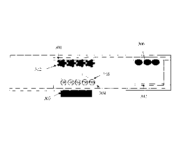
[0053] Figure 7 is
a cross-sectional view of the biosensor of Figure 4 or Figure 6,
illustrating the location of the chemistry. The reaction
chamber can comprise
immobilized binding agent molecules on the upper internal surface and probe
agent
molecules on the bottom internal surface. The top internal surface can be a
top
electrode 301 which can extend into the detection chamber. The bottom internal
surface
can comprise a bottom electrode 304 which can extend into the detection
chamber. The
binding agent can comprise antigen which can be coated on magnetic beads 302.
The
magnetic beads can be confined within the reaction chamber by a magnet 303 at
the
bottom of the sensor. The probe agent can comprise PQQ molecules encapsulated
within liposomes 305, and binding partners. The detection chamber can comprise
apo-
GDH 306 on the top internal surface, and other detection chemistry reagents
307 on the
bottom internal surface.
[0054] A test using
the biosensor can use a fluid sample of less than about 100
milliliters, or less than about 50 milliliters, or less than about 20
milliliters, or less than
about 10 milliliters, or less than about 5 milliliters, or less than about 3
milliliters, or less
than about 2 milliliters, or less than about 1 milliliter, or less than 500
microliters, or less
than about 200 microliters, or less than about 100 microliters, or less than
about 50
microliters, or less than about 10 microliters, or less than about 1
microliter, or less than
about 0.5 microliters, or less than about 0.3 microliters, or less than about
0.1 microliters.
[0055] The
biosensor can comprise at least one reaction chamber. The reaction
chamber can have a proximal end and a distal end. A fluid sample can enter the
reaction chamber from the proximal end, and can exit the reaction chamber
and/or flow
into the detection chamber through the fluid passageway from the distal end.
13
CA 02730544 2011-01-11
WO 2010/004436
PCT/182009/006688
[0 0 5 6] The reaction chamber can comprise at least one wall which can
define the
exterior and/or the interior of the reaction chamber. At least one wall of the
reaction
chamber can comprise a material, such as, for example, polyester, polystyrene,
polycarbonate, polyolefin, polyethylene terephthalate, or a mixture thereof.
At least one
wall of the reaction chamber can comprise a filler, such as, for example,
titanium
dioxide, carbon, silica, glass, and a mixture thereof.
[0 0 5 7] The reaction chamber can comprise an interior with a volume, at
least
part of which can be accessible to the fluid sample. The volume can be less
than about
100 milliliters, or less than about 50 milliliters, or less than about 20
milliliters, or less
than about 10 milliliters, or less than about 5 milliliters, or less than
about 3 milliliters, or
less than about 2 milliliters, or less than about 1 milliliter, or less than
500 microliters, or
less than about 200 microliters, or less than about 100 microliters, or less
than about 50
microliters, or less than about 10 microliters, or less than about 1
microliter, or less than
about 0.5 microliters, or less than about 0.3 microliters, or less than about
0.1
microliters. The interior of the reaction chamber can comprise a cross-
sectional shape
of square, rectangular, circular, oval, triangular, rhomboid, trapezoidal, or
the like. A
cross-section can be perpendicular to the direction of the bulk flow of the
fluid sample
within the reaction chamber. The cross-sections can be uniform in size and/or
shape
along the direction of the bulk flow. The cross-sections can be variable along
the
direction of the bulk flow. Merely by way of example, the cross-sections can
taper
along the direction of the bulk flow.
[00581 The reaction chamber can comprise a capillary distance, h1. The
capillary
distance can refer to the dimension of the reaction chamber and/or its cross
sections
which determines the magnitude of the capillary force to the fluid sample. The
capillary
distance can be the smallest dimension of the interior of the reaction
chamber. The
magnitude of the capillary force can be inversely related to the capillary
distance. The
capillary distance can be less than about less than about 1 centimeter, or
less than
about 5 millimeters, or less than about 2 millimeters, or less than about 1
millimeter, or
less than about 500 micrometers, or less than about 200 micrometers, and less
than
about 100 micrometers, or less than about 50 micrometers. If the biosensor is
used by
a user who and/or with an apparatus which can generate an external force to
transfer
the fluid sample between or among different chambers of the device, the device
and/or
its chambers can comprise a bigger dimension. Merely by way of example, the
14
CA 02730544 2011-01-11
WO 2010/004436
PCT/1B2009/006688
biosensor can comprise a characteristic length less than about 100
centimeters, or less
than about 50 centimeters, or less than about 20 centimeters, or less than
about 10
centimeters, or less than about 5 centimeters, or less than about 1
centimeter. As used
herein, the characteristic length refers to the diameter of the smallest
circle which
encloses an entire cross-sectional surface of the reaction chamber.
[0 0 5 9] The interior of the reaction chamber can comprise at least one
internal
surface. The internal surface(s) can comprise an internal wall/internal walls
which can
define the cross-sectional shape and/or volume of the interior of the reaction
chamber.
The internal wall(s) can comprise, but are not limited to, a solid material, a
fibrous
material, a macroporous material, a powdered material, or the like, or any
combination
thereof. The internal surface(s) can comprise that/those of at least one
independent
support within the reaction chamber. A suitable support can comprise, but are
not
limited to, a solid material, a mesh material, a fibrous material, a porous
material, a
powdered material, or beads of a material, or a mixture thereof. The mesh
material can
comprise, for example, a polymer such as polyolefin, polyester, nylon,
cellulose,
polystyrene, polycarbonate, polysulfone, or a mixture thereof. The fibrous
material can
comprise, for example, a polymer such as polyolefin, polyester, nylon,
cellulose,
polystyrene, polycarbonate, polysulfone, or a mixture thereof. The porous
material can
comprise, for example, a sintered powder, or a macroporous membrane. The
macroporous membrane can comprise, for example, a polymeric material such as
polysulfone, polyvinylidene difluoride, nylon, cellulose acetate,
polymethacrylate,
polyacrylate, or a mixture thereof. The bead material can be selected such
that suitable
support can be provided for an reagent, such as, for example, an binding
agent.
Suitable beads can comprise those marketed as DYNABEADS® by Dynal Biotech
of Oslo, Norway. The beads can comprise, for example, magnetic beads. The
support
can have at least one of the following benefits. Firstly, it can increase the
surface area
where the reagents, such as, for example, the binding agent, the probe agent,
can
attach, and/or where the binding reaction can occur within the reaction
chamber. This
can decrease the reaction time, and/or the chances for an undesirable process
(e.g.,
contamination, clotting, etc) to occur. Secondly, it can increase the
capillary force to
the fluid sample by decreasing the capillary distance of the reaction chamber.
The
reaction chamber can comprise a vent that is open to the atmosphere, thus
allowing air
displaced by the sample to escape. The fluid sample can be drawn into the
reaction
chamber until the reaction chamber is filled up to the reaction chamber vent,
CA 02730544 2011-01-11
WO 2010/004436
PCT/1B2009/006688
whereupon filling can stop. The volume of detection chamber can be chosen so
as to
be roughly equal to and preferably smaller than the volume of the reaction
chamber.
The volume of the detection chamber can be about 100%, or about 95%, or about
90%,
or about 85%, or about 80%, or about 75%, or about 70%, or about 60%, or less
than
about 50% of that of the reaction chamber.
[ 0 0 6 0] The reaction chamber can comprise binding agent and probe agent.
The
relative amounts of the binding agent and the probe agent can be chosen such
that
there is a slight excess of the binding agent over the probe agent. In this
context, a
slight excess can be defined to be such that the excess is small when compared
to the
amount of target analyte to be detected in the fluid sample. For example, the
excess
can comprise less than about 40%, or less than about 30%, or less than about
25%, or
less than about 20%, or less than about 15%, or less than about 10%, or less
than
about 5%, or less than about 3%, or less than about 2%, or less than about 1%
of the
average amount of target analyte expected in the fluid sample. The average
amount of
the target analyte can be estimated, for example, for a population of interest
with and/or
without a target pathological condition.
[0 0 6 1] The binding agent can be immobilized on at least one internal
surface
within the reaction chamber so that the binding agent and the species bound to
them
during the reaction can remain in the reaction chamber and throughout the
test. The
probe agent can be supported on at least one internal surface within the
reaction
chamber. The probe agent molecules which are not bound to the immobilized
binding
agent molecules directly or indirectly can move to the detection chamber with
the fluid
sample. As used herein, "directly" means that a portion of the probe agent
molecule
binds to a portion of the binding agent molecule, e.g., a binding site; while
"indirectly"
means that the probe agent binds to another agent, e.g., the target analyte
which binds
directly or indirectly to the immobilized binding agent.
[0062] The binding agent can be adsorbed or otherwise immobilized onto at
least
one internal surface of the reaction chamber via chemical bonds. The binding
agent
can be coated onto beads which can be confined in the reaction chamber
throughout a
test. Merely by way of example, the beads can be magnetic beads, and the
biosensor
can comprise a magnetic field to confine the magnetic beads with a coating of
binding
agent in the reaction chamber. For example, a magnet below the reaction
chamber can
prevent the transfer of magnetic beads and any species that can be bound to
the beads
16
CA 02730544 2011-01-11
WO 2010/004436
PCT/1B2009/006688
directly or indirectly, such as the binding agent coated on the beads and the
probe
agent molecules bound to the binding agent molecules. As a result, in an
exemplary
embodiment, probe agent molecules not bound to the magnetic beads can be
measured in the detection chamber. The amount and/or concentration of target
analyte
in the fluid sample, for example, can be related to the amount/concentration
of the
probe agent which can be released into the detection chamber.
[0063] The probe agent can comprise a binding partner and a vehicle. The
binding partner can be bound to the surface of the vehicle. The vehicle can
comprise at
least one copy of an activating agent. The binding partner can facilitate a
probe agent
molecule bound to an immobilized binding agent molecule, or to a free target
analyte
which is not bound to an immobilized binding agent molecule, or to a target
analyte
which is bound to an immobilized binding molecule. The at least one copy of
the
activating agent can be surface coated onto or encapsulated within the
vehicle.
[0 0 6 4] The probe agent molecules can be supported on at least one
internal
surface of the reaction chamber. The internal surface of the reaction chamber
and the
method for coating of the probe agent molecules can be chosen such that only
weak
bonds between the probe agent molecules and the internal surface can exist.
This way
the probe agent molecules can be liberated into the sample when the internal
surface is
wet by the sample. The rate of dissolution of the probe agent molecules from
the
internal surface can be chosen such that little dissolution has occurred
during the time
taken for the sample to fill the reaction chamber. In this manner, the probe
agent
molecules can be evenly distributed throughout the area of the reaction
chamber after
filling. The internal surface(s) where the probe agent molecules are supported
can be
the same as, or different from that (those) where the binding agent molecules
are
immobilized.
[0 0 65] In some embodiments, the probe agent molecules can be separate
from
and/or not bound to the binding agent molecules before a fluid sample fills
the reaction
chamber and dissolves the probe agent molecules. In some embodiments, after
dissolved by the fluid sample in the reaction chamber, the probe agent
molecules can
bind to the immobilized agent molecules, but with a lower binding affinity
compared to
the target analyte, to form a competitive assay. The lower binding affinity of
a probe
agent molecule through its binding partner to an immobilized binding agent
molecule
can be due to at least one of the following reasons. Firstly, a probe agent
molecule can
17
CA 02730544 2011-01-11
WO 2010/004436
PCT/1B2009/006688
comprise a larger size compared to the target analyte, because of the vehicle
linked to
the binding partner of the probe agent molecule, and/or the larger size of the
binding
partner itself than the target analyte. Secondly, the binding partner of a
probe agent
molecule can be a chemically or otherwise modified version of the target
analyte such
that the probe agent molecule can bind to the immobilized binding agent
molecule
through the binding partner, but with decreased binding affinity. The interior
binding
kinetics of the probe agent molecules can also result from that it can take
longer for the
probe agent molecules to reach the immobilized binding agent molecules than
the
target analyte in the fluid sample, because it has to be dissolved first by
the fluid
sample, and/or because it can move more slowly in the fluid sample due to its
larger
size than the target analyte. In this manner, the amount/concentration of the
target
analyte in the fluid sample can be positively related to the
amount/concentration of the
probe agent released to the detection chamber where the probe agent can be
measured qualitatively and/or quantitatively. In other embodiments, after
dissolved by
the fluid sample in the reaction chamber, the probe agent molecules can bind
to the
target analyte and form a sandwich assay or a competitive binding assay. A
sandwich
assay can be formed when a probe agent molecule can bind to a target analyte
after
the target analyte is bound an immobilized binding agent molecule. In this
manner, the
amount/concentration of the target analyte in the fluid sample can be
inversely related
to the amount/concentration of the probe agent molecules released to the
detection
chamber where the probe agent molecules can be measured qualitatively and/or
quantitatively. A competitive binding assay can form if a probe agent
molecule, when
dissolved by the fluid sample in the reaction chamber, can bind to a free
target analyte
with a higher binding affinity than it binds to the immobilized binding agent
molecule. A
free target analyte, as used herein, refers to the target analyte which is not
bound to an
immobilized binding agent molecule. In this manner, the amount/concentration
of the
target analyte in the fluid sample can be positively related to the
amount/concentration
of the probe agent released to the detection chamber where the probe agent
molecules
can be measured qualitatively and/or quantitatively.
[0066] In some embodiments, the probe agent molecules can be bound to the
immobilized binding agent molecules when the biosensor is manufactured and/or
before a test of a fluid sample using such a biosensor. The binding partner of
such a
probe agent molecule can comprise a pseudo-analyte, a modified analyte, or the
like.
As used herein, a pseudo analyte can comprise one which can bind to the
immobilized
18
CA 02730544 2011-01-11
WO 2010/004436
PCT/1B2009/006688
binding agent molecule, but not as strongly as the target analyte. Merely by
way of
example, if the target analyte is a human protein, then a suitable pseudo-
analyte can
comprise an animal version of the same protein, such as a dog protein or a pig
protein.
A modified analyte can comprise one chemically or otherwise modified such that
the
binding affinity to the binding agent molecule is decreased. The binding
affinity of the
probe agent molecule to the immobilized binding agent molecule through the
binding
partner can be lower than that of the target analyte. The lower binding
affinity of a
probe agent molecule through its binding partner to an immobilized binding
agent
molecule can be due to at least one of the following reasons. Firstly, a probe
agent can
comprise a larger size compared to the target analyte, because of the vehicle
linked to
the binding partner of the probe molecule, and/or the larger size of the
binding partner
itself. Secondly, the binding partner of a probe agent molecule can be a
chemically or
otherwise modified version or a different version (e.g. an animal version of a
human
analyte) of the target analyte such that the probe molecules can bind to the
immobilized
binding agent molecules through the binding partner, but with decreased
binding affinity.
After dissolved by the fluid sample in the reaction chamber, the probe agent
molecules
can be displaced from the binding agent molecules by the target analyte due to
the
lower binding affinity. In this manner, the amount/concentration of the target
analyte in
the fluid sample can be positively related to the amount/concentration of the
probe
agent molecules displaced and released to the detection chamber where the
probe
agent molecules can be measured qualitatively and/or quantitatively.
[0067] A probe agent molecule can comprise a vehicle. In some embodiments,
the vehicle can comprise at least one labeling molecule, such as, for example,
a
radioisotope, a chromophore, or a fluorophore. The vehicle can comprise at
least about
labeling molecules, or at least about 50 labeling molecules, or at least about
100
labeling molecules, or at least about 200 labeling molecules, or at least
about 500
labeling molecules, or at least about 1,000 labeling molecules, or at least
about 5,000
labeling molecules, or at least about 10,000 labeling molecules, or at least
about
50,000 labeling molecules, or at least about 100,000 labeling molecules. The
labeling
molecule(s) can be coated on the surface of the vehicle, or encapsulated
within the
vehicle. The vehicle can comprise a lipidic particle. The labeling molecule(s)
can be
encapsulated within the lipidic particle. The lipidic particle can comprise
one particle
selected from a liposome, vesicle, cellular organelle, and the like. The
vehicle can
19
CA 02730544 2011-01-11
WO 2010/004436
PCT/1B2009/006688
comprise a polymer. The activating agent molecules can be bound to the surface
of the
polymer. The polymer can comprise a dendrimer, or the like.
[0068] In other embodiments, the vehicle can comprise at least one copy of
an
activating agent. The vehicle can comprise a plurality copies of an activating
agent.
The vehicle can comprise at least about 5 copies, or at least about 10 copies,
or at
least about 50 copies, or at least about 100 copies, or at least about 200
copies, or at
least about 500 copies, or at least about 1,000 copies, or at least about
5,000 copies, or
at least about 10,000 copies, or at least about 50,000 copies, or at least
about 100,000
copies of an activating agent. The activating agent molecules can be coated on
the
surface of the vehicle, or encapsulated within the vehicle. The vehicle can
comprise a
lipidic particle. The activating agent molecules can be encapsulated within
the lipidic
particle. The lipidic particle can comprise one particle selected from a
liposome, vesicle,
cellular organelle, and the like. The vehicle can comprise a polymer. The
activating
agent molec ules can be bound to the surface of the polymer. The polymer can
comprise a dendrimer, or the like.
[ 0 0 6 9] An activating agent molecule can activate a detection agent
molecule in
the detection chamber such that a signal can be generated and detected. Merely
by
way of example, the detection agent can comprise an enzyme, and the activating
agent
can comprise a cofactor which can activate the enzyme. As a more specific
example,
the detection agent can comprise an apo-enzyme, and the activating agent can
comprise the corresponding cofactor. The apo-enzyme and cofactor pair can
comprise
apo-glucose oxidase and flavin adenine dinucleotide. The apo-enzyme and
cofactor
pair can comprise apo-glucose dehydrogenase and PQQ.
[0 0 7 0] The reaction chamber can comprise other agents besides the
binding
agent and the probe agent, such as, for example, a blocking agent, an
unactivated
agent, or any combination thereof.
[0 0 7 1 ] The blocking agent can block non-specific binding of an agent
onto the
immobilized binding agent, the probe agent, and/or the internal surface(s)
within the
reaction chamber. The agent can comprise at least one present in the fluid
sample to
be tested, such as, for example, a protein or a lipidic particle. The lipidic
particle can be
at least one selected from a liposome, a vesicle, a cellular organelle, and
the like. The
blocking agent can comprise at least one agent selected from a blocking
protein and a
CA 02730544 2011-01-11
WO 2010/004436
PCT/1B2009/006688
surfactant. The blocking protein can comprise, for example, bovine serum
protein. A
nonionic surfactant may also be used as such an agent, e.g., TRITON X100
manufactured by Rohm & Haas of Philadelphia, Pa., or TVVEEN manufactured by
ICI
Americas of Wilmington, Del. In some embodiments, the nonionic surfactant
selected
does not denature proteins. The blocking agent can be coated onto any internal
surface(s) of the reaction chamber, comprising where the binding agent
molecules are
immobilized, and/or where the probe agent molecules are supported, and/or
where
neither of the binding agent molecules nor the probe agent molecules are
coated. The
blocking agent and the agents bound to them can be confined in the reaction
chamber
during a test. This can be achieved by any of the methods by which binding
agent
molecules can be immobilized in the reaction chamber. Merely by way of
example, the
binding agent can be absorbed onto a porous internal surface of the reaction
chamber.
[0 0 7 2] The unactivated agent can bind to an unbound or unencapsulated
activating agent. This can prevent an unbound or unencapsulated activating
agent
from moving to the detection chamber and activating a detection agent in a
manner
independent of the presence of the target analyte in the fluid sample, which
can
deteriorate the accuracy and/or validity of a test. As used herein, "unbound"
or
"unencapsulated" means not bound to, encapsulated within, otherwise integral
to the
vehicle when the vehicle (and the probe agent) is within the reaction chamber
and/or
before the vehicle (and the probe agent) moves to the detection chamber with
the
reacted fluid sample. This can result from a vehicle of the probe agent which
can
become leaky, or rupture, or desorb activating agent molecules during
manufacture,
storage, or during a test under certain conditions, such as pH, temperature,
etc. The
unactivated agent can comprise any agent which can bind to the unbound or
unencapsulated activating agent. For example, if the activating agent comprise
a
cofactor which can bind to an apo-enzyme, the unactivated agent can comprise
the
apo-enzyme. The unactivated agent molecules can be immobilized on an internal
surface of the reaction chamber. Therefore, the activating agent molecules
bound to
them can be confined in the reaction chamber during a test. This can reduce
the
amount of the unbound or unencapsulated activating agent molecules that can
move to
the detection chamber in a manner independent of the present and/or the amount
of the
target analyte in a fluid sample. The unactivated agent molecules can be
immobilized
by any of the methods by which binding agent molecules can be immobilized in
the
reaction chamber. Merely by way of example, the unactivated agent molecules
can be
21
CA 02730544 2011-01-11
WO 2010/004436
PCT/1B2009/006688
immobilized within the reaction chamber by binding them to magnetic beads, and
the
magnetic beads can be confined within the reaction chamber by a magnetic
field.
[0073] The reaction chamber can comprise a buffer which can adjust the pH
of
the fluid sample, for example, in the reaction chamber. The buffer can
stabilize as least
one of the reagents in the reaction chamber during manufacture and/or storage.
The
buffer can comprise a substance selected from phosphate, citrate, citraconate,
mellitate,
tris, pipes, mops, hepes, phthalate, imadazole.
[0074] The reaction in the reaction chamber can take from about 0.1
seconds to
about 60 minutes, or from about 1 second to about 30 minutes, or from about 10
seconds to about 25 minutes, or from about 20 seconds to about 20 minutes, or
from
about 30 seconds to about 15 minutes, or from about 1 minute to about 10
minutes, or
from about 2 minutes to about 8 minutes, or from about 3 minutes to about 5
minutes.
[0075] The device can comprise at least one detection chamber. The
detection
chamber can have a proximal end and a distal end. After a fluid sample
finishes the
reaction within the reaction chamber, the reacted fluid sample exiting the
reaction
chamber from the distal end of the reaction chamber can enter the detection
chamber
from its proximal end through the sample passageway. The detection chamber can
be
configured such that it can detect a signal generated in the detection chamber
in a
manner dependent on the presence and/or the amount of the probe agent
molecules
that transfer to the detection chamber.
[0076] The detection chamber can comprise at least one wall which can
define
the exterior and/or the interior of the detection chamber. At least one wall
of the
detection chamber can comprise a filler. The design of the at least one wall
and the
filler of the detection chamber can be similar to those of the reaction
chamber.
[0 0 7 7] The detection chamber can comprise an interior with a volume, at
least
part of which can be accessible to the fluid sample The volume can be less
than about
100 milliliters, or less than about 50 milliliters, or less than about 20
milliliters, or less
than about 10 milliliters, or less than about 5 milliliters, or less than
about 3 milliliters, or
less than about 2 milliliters, or less than about 1 milliliter, or less than
500 microliters, or
less than about 200 microliters, or less than about 100 microliters, or less
than about 50
microliters, or less than about 10 microliters, or less than about 1
microliter, or less than
about 0.5 microliters, or less than about 0.3 microliters, or less than about
0.1
22
CA 02730544 2011-01-11
WO 2010/004436
PCT/1B2009/006688
microliters. The interior of the detection chamber can comprise a cross-
sectional shape
of square, rectangular, circular, oval, triangular, rhomboid, trapezoidal, or
the like. A
cross-section can be perpendicular to the direction of the bulk flow of the
fluid sample
within the detection chamber. The cross-sections can be uniform in size and/or
shape
along the direction of the bulk flow. The cross-sections can be variable along
the
direction of the bulk flow. Merely by way of example, the cross-sections can
taper
along the direction of the bulk flow.
[0 0 7 8 ] The detection chamber can comprise a capillary distance, h2. The
magnitude of the capillary force can be inversely related to the capillary
distance. The
capillary distance can be less than about less than about 1 centimeter, or
less than
about 5 millimeters, or less than about 2 millimeters, or less than about 1
millimeter, or
less than about 500 micrometers, or less than about 200 micrometers, and less
than
about 100 micrometers, or less than about 50 micrometers. The capillary
distance of
the detection chamber h2 can be smaller than that of the reaction chamber, h1.
If the
biosensor is used by a user who and/or with an apparatus which can generate an
external force to transfer the fluid sample between or among different
chambers of the
device, the device and/or its chambers can comprise a bigger dimension. As
used
herein, the external force does not include the force generated by a user to
open the
vent in the detection chamber. Merely by way of example, the device can
comprise a
characteristic length less than about 100 centimeters, or less than about 50
centimeters,
or less than about 20 centimeters, or less than about 10 centimeters, or less
than about
centimeters, or less than about 1 centimeter. As used herein, the
characteristic
length refers to the diameter of the smallest circle which encloses an entire
cross-
sectional surface of the detection chamber.
[0079] The interior of the detection chamber can comprise at least one
internal
surface. The internal surface(s) can comprise an internal wall/internal walls
which can
define the cross-sectional shape and/or volume of the interior of the
detection chamber.
The internal surface(s) can comprise that/those of at least one independent
support
within the detection chamber. The internal wall(s) and/or the independent
support of
the detection chamber can be similar to that/those of the reaction chamber.
[0080] The detection chamber can comprise a vent that can be open to the
atmosphere. The vent can reside at the distal end of the detection chamber. An
exemplary configuration is shown as vent 30 in Figure 2 and vent 130 in Figure
4. The
23
CA 02730544 2011-01-11
WO 2010/004436
PCT/1B2009/006688
vent can be initially closed. This way, when the reaction chamber fills with a
fluid
sample, the sample passageway to the detection chamber can be blocked by a
pneumatic pressure generated by the air trapped within the detection chamber.
This
pneumatic pressure can substantially prevent the fluid sample from filling the
detection
chamber. A small amount of sample can enter the detection chamber during the
time
between when the sample first contacts the sample passageway to the detection
chamber and when the sample contacts the far side of the sample passageway.
When
the sample has wet totally across the sample passageway to the detection
chamber,
filling of the detection chamber can stop. The volume of the reaction chamber
can be
chosen so as to be at least equal to and preferably larger than the volume of
the
detection chamber. By opening the vent in the detection chamber to the
atmosphere,
sample can be transferred to fill the detection chamber. The vent can be
opened by
such as, for example, piercing the device, and/or removing an outer layer,
and/or
tearing a portion of the device (i.e., tearing along a perforation). Merely by
way of
example, the vent can be opened using a needle controlled by a user or a
solenoid in
the meter which is connected to the device. Opening the vent can allow air
displaced
by the sample and trapped within the detection chamber to escape, and
therefore, can
reduce the pneumatic pressure which can prevent the fluid sample from filling
the
detection chamber. The device can be configured such that the capillary force
to the
fluid sample in the detection chamber is higher than that present in the
reaction
chamber. The increased capillary force can be provided by suitably coating the
internal
surfaces of the reaction chamber and of the detection chamber, and/or, by
choosing the
capillary distance for the detection chamber h2 to be smaller than that of the
reaction
chamber hl. This way, the fluid sample can be drawn into the detection chamber
simply by opening the vent, without requiring any other external force
generated by the
user, or by an external device, such as, for example, a pump or a syringe. In
other
embodiments, the filling of the detection chamber by the (reacted) fluid
sample can be
controlled by an external force generated by a user or an external device,
such as, for
example, a pump, a syringe, or any combination thereof. An external force can
also be
supplied as a supplementary force to move the fluid sample from the reaction
chamber
to the detection chamber in addition to the capillary force. As used herein,
the external
force does not include the force generated by a user to open the vent in the
detection
chamber by, for example, piercing.
24
CA 02730544 2011-01-11
WO 2010/004436
PCT/1B2009/006688
[0081] The probe agent molecules can be transferred to the detection
chamber
with the reacted fluid sample. The presence of the probe agent molecules in
the
detection chamber can be detected qualitatively and/or quantitatively by
signals
generated by labeling molecules, such as, for example, radioisotopes,
chromophores,
or fluorophores. At least one wall of the detection chamber can be permeable
to the
signals generated by such labeling molecules. Merely by way of example, at
least one
detection chamber wall can be transparent to a radiation emitted or absorbed
by the
radioisotopes, and the radiation can be indicative of a presence or absence of
the
probe agent molecules in the detection chamber.
[0082] The presence of the probe agent molecules in the detection chamber
can
be detected qualitatively and/or quantitatively by an electrochemical
reaction. In such
embodiments, the detection chamber can comprise an electrochemical cell which
can
comprise at least two opposing electrodes, at least one sensing/working
electrode and
at least one counter/reference electrode. The step of determining the presence
of the
probe agent molecules in the reacted fluid sample can comprise: applying a
potential
between the sensing/working electrode and the counter/reference electrode in
the
electrochemical cell; and measuring a current, wherein the current can be a
qualitative
and/or quantitative indication of the probe agent in the reacted fluid sample
in the
detection chamber, which can be a qualitative and/or quantitative indication
of the
target analyte in the fluid sample.
[0083] The sensing electrode can be sensitive to the amount of reduced
redox
agent in the antioxidant case or oxidized redox agent in the oxidant case. In
the case
of a potentiometric sensor wherein the potential of the sensing electrode is
indicative of
the level of analyte present, at least one other electrode can act as a
reference
electrode to provide a reference potential. In the case of an amperometric
sensor
wherein the sensing electrode current is indicative of the level of analyte in
the sample,
at least one other electrode can act as a counter electrode to complete the
electrical
circuit, and/or a reference electrode. Alternatively, the counter electrode
and the
reference electrode can be two separate electrodes.
[0084] At least one of the electrodes can comprise an electrically
conductive
material, such as, for example, aluminum, copper, nickel, chromium, steel,
stainless
steel, palladium, platinum, gold, iridium, carbon, carbon mixed with binder,
indium oxide,
tin oxide, a conducting polymer, or a mixture thereof. The cathode in the
CA 02730544 2011-01-11
WO 2010/004436
PCT/1B2009/006688
electrochemical cell can comprise an electrically conductive material, such
as, for
example, aluminum, copper, nickel, chromium, steel, stainless steel, platinum,
palladium, carbon, carbon mixed with a binder, indium oxide, tin oxide, mixed
indium/tin
oxides, gold, silver, iridium, a conducting polymer, or the like, or a mixture
thereof. The
conducting polymer can comprise, such as, for example, polypyrrole or
polyacetylene,
or the like, or a combination thereof. The anode in the electrochemical cell
and/or the
electrode(s) which can come into contact with oxidizing substances during
device
manufacture or storage, can comprise at least one electrically conductive
material, such
as, for example, platinum, palladium, carbon, carbon mixed with a binder,
indium oxide,
tin oxide, mixed indium/tin oxides, gold, silver, iridium, a conducting
polymer, or the like,
or a mixture thereof. The conducting polymer can comprise, such as, for
example,
polypyrrole or polyacetylene, or the like, or a combination thereof. Materials
suitable for
use as electrodes can be compatible with the reagents present in the device,
namely,
they do not react chemically with the reagents at the potential of choice,
and/or during
sensor fabrication, and/or storage, and/or usage. The opposing electrodes can
comprise the same conductive material, or different materials.
[0 0 8 5] The sensing/working electrode and the counter/reference electrode
can
reside on at least one internal surface of the detection chamber. The opposing
electrodes can be electrically insulated to each other before the detection
chamber is
filled with the fluid sample. The insulation can be achieved by separating the
two
opposing electrodes with an electrically insulating material, or by creating a
break on an
electrically conductive layer or film. The opposing electrodes reside on the
same
internal surface or different internal surfaces of the detection chamber. The
opposing
electrodes can be separated by about 5 micrometers, or about 10 micrometers,
or
about 15 micrometers, or about 20 micrometers, or about 25 micrometers, or
about 30
micrometers, or about 35 micrometers, or about 40 micrometers, or about 45
micrometers, or about 50 micrometers, or about 75 micrometers, or about 100
micrometers, or about 125 micrometers, or about 150 micrometers, or about 175
micrometers, or about 200 micrometers, or about 250 micrometers, or about 300
micrometers, or about 350 micrometers, or about 400 micrometers, or about 450
micrometers, or about 500 micrometers, or about 600 micrometers, or about 700
micrometers, or about 800 micrometers, or about 900 micrometers, or about 1
millimeter, or greater than about 2 millimeters, or greater than about 3
millimeters, or
greater than about 4 millimeters, or greater than about 5 millimeters. In
terms of the
26
CA 02730544 2011-01-11
WO 2010/004436
PCT/1B2009/006688
relative position, the opposing electrodes can be in a parallel opposing
relationship, or a
side-by-side relationship, or a parallel but offset relationship, or a
coplanar relationship.
The opposing electrodes can be identical or substantially similar in size, or
can be of
different sizes and/or different shapes.
[0086] The device can comprise more than two electrodes. Merely by way of
example, the device can comprise a third electrode which can be a
counter/reference
electrode. The two counter/reference electrodes can be electrically connected.
The
third electrode can form a circuit with the sensing/working electrode which
can detect
the filling of the reaction chamber and/or the detection chamber. Merely by
way of
example, the filling of the reaction chamber detected this way can be used as
a signal
to activate a timing device such that the reaction time can be controlled
and/or a
following step of a test, such as, for example, transferring the reacted fluid
sample to
the detection chamber, can be triggered after a per-determined amount of time.
As
another example, if the device comprises two detection chambers, each
detection
chamber can comprise one counter/reference electrode, and the two detection
chambers can share one sensing/working electrode extending to both detection
chambers. In such embodiments, two electrical signals can be obtained in one
test.
[0 0 8 7 ] Other variations in electrode configuration, spacing, and
construction or
fabrication would be within the scope of the disclosure.
[0088] At least a portion of one of the internal surface of the detection
chamber
can comprise a conductive layer or film which can be electrically connected to
but
extended beyond the electrodes. The extended conductive layer or film can be
used as
a contact area where the device is electrically connected to another device,
such as, for
example, a meter. In certain embodiments, the detection chamber can comprise
two
internal surfaces which comprise conductive films and/or are electrically
connected to
the opposing electrodes, but electrically insulated to each other. As used
herein,
"substantially'' means that at least about 30%, or at least about 40%, or at
least about
50%, or at least about 60%, or at least about 70%, or at least about 80%, or
at least
about 90%, or at least about 95% of either of the two internal surfaces are
coated with
electrically conductive material. Each electrically conductive film can be
continuous or
patterned. For example, the patterned conductive film can form two electrodes
which
are conductive to each other; or an electrode and a contact area, wherein the
contact
27
CA 02730544 2011-01-11
WO 2010/004436
PCT/1B2009/006688
area can electrically connect the electrode to an external device, such as,
for example,
a meter.
[0089] There can be a scratch on at least on of the internal surface of
the
detection chamber. An exemplary illustration can be Scratch 106 in Figure 4.
The
scratch can generate a break in the electrically conductive film within the
detection
chamber. The break can be affected by patterning the conductive film when it
is laid
down or by creating the break during manufacture. Scratch 106 can be affected
by
scratching the film, scraping part of the film away, chemically etching the
conductive
layer or film, laser ablating the conductive layer or film or other methods.
Scratch 106
in the conductive layer or film can serve to, in part, define the active
electrode area of
the detection chamber by electrically isolating the conductive coated in the
detection
chamber from that in the reaction chamber. This can be advantageous as it can
prevent any electric signal that can otherwise flow at the conductive layers
or films in
the reaction chamber from effecting the test results. The scratch can be wide
enough
to reliably break the electrical conduction of the layer where the scratch
reside, but not
so wide as to prevent fluid from crossing it, such as, for example, under
capillary action.
The scratch can be from about 1 micrometer to 10 millimeters, preferably from
about 10
micrometers to about 1 millimeter, and most preferably from about 20
micrometers to
about 200 micrometers. The distance between the scratch and the proximal end
of the
detection chamber can be about 1%, or about 5%, or about 10%, or about 15%, or
about 20%, or about 25%, or about 30%, or about 35%, or about 40%, or about
45%, or
about 50%, or greater than 55% of the distance between the proximal end and
the
distal end of the detection chamber.
[0090] At least one internal surface of the detection chamber can be
coated with
detection agent. The detection agent can be dissolved by and/or diffusible in
the fluid
sample in the detection chamber, and can be coated on any internal surface of
the
detection chamber. In some embodiments, the detection agent cannot be
dissolved by
or diffusible in the fluid sample. Such a detection agent can be coated in the
vicinity of
where the detection can be made. As used herein, "vicinity" means within the
distance
across which the detection agent or the derivative of them can be detected by,
such as,
for example, the electrodes. The derivative of the detection agent means a
species
generated and/or activated by the reaction of the detection agent with a
species carried
to the detection chamber by the probe agent, for example, the activating
agent. The
28
CA 02730544 2011-01-11
WO 2010/004436
PCT/1B2009/006688
detection agent can comprise an enzyme, such as, for example, a glucose
oxidase, a
glucose dehydrogenase. The detection agent can comprise a species which can be
activated by an activating agent surface bound onto or encapsulated within a
probe
agent molecule carried to the detection chamber by the fluid sample. The
detection
agent and activating agent pair can comprise, an apo-enzyme and its cofactor.
Merely
by way of example, the detection agent and activating agent pair can comprise
apo-
glucose oxidase and flavin adenine dinucleotide; the detection agent and
activating
agent pair can comprise apo-glucose dehydrogenase and PQQ. If one copy of the
target analyte present in the fluid sample corresponds to one probe agent
molecule
comprising multiple copies of an activating agent transferred to the detection
chamber,
and one copy of the activating agent can activate one detection agent molecule
to
generate a unit of signal, then one copy of the target analyte can correspond
to multiple
units of signal. This can increase the sensitivity, and/or accuracy, and/or
rate of the test.
[0091] The detection chamber can comprise a liberating agent which can
liberate
the activating agent molecules from the vehicle of a probe agent molecule such
that the
activating agent molecules can react with and activate the detection agent
molecules.
The liberating agent can comprise at least one selected from a mild detergent,
a lytic
peptide, an enzyme, heating, cooling, ultrasonication, a light source together
with a
photochemically activated lysing agent, or the like, or any combination
thereof. The
mild detergent can comprise at least one selected from n-octyl-B-D-
glucopyranoside,
tween 20, brij 35 and triton X-100. The peptide can comprise at least one
selected from
mellitin, and one of a class of phospholipases, a component of the complement
system.
The enzyme can comprise at least one selected from protease and trypsin. The
liberating agent can comprise physical means of releasing the activating agent
molecules from the vehicle. There can comprise heating or cooling,
ultrasonication or a
combination of physical and chemical means, such as, for example, a
photochemical
reaction initiated by a light source directed into the sensor. The liberating
agent can be
dissolved by and/or diffusible in the fluid sample in the detection chamber,
and can be
coated on any internal surface of the detection chamber.
(0092] If the detection agent or its derivative is an enzyme, the
detection
chamber can comprise an enzyme substrate which can react with the detection
agent
or its derivative to produce a detectable signal. An exemplary illustration is
enzyme
substrate 64 in Figure 3. The enzyme substrate can be of sufficient amount
such that
29
CA 02730544 2011-01-11
WO 2010/004436
PCT/1B2009/006688
the rate of reaction of the detection agent or its derivative present with the
enzyme
substrate is determined by the amount of detection agent or its derivative
present in the
detection chamber. The enzyme substrate can comprise an oxidizable substrate.
The
oxidizable substrate can comprise one substrate selected from galactose,
maltose,
xylose, and acetic acid. For instance, if the detection agent or its
derivative is glucose
oxidase or glucose dehydrogenase, the enzyme substrate can comprise glucose.
[0093] The
detection chamber can comprise at least one mediator which can
react with the detection agent or its derivative to produce a detectable
signal. The
mediator can be of sufficient amount such that the rate of reaction of the
detection
agent or its derivative present with the enzyme substrate is determined by the
amount
of detection agent or its derivative present in the detection chamber. In an
embodiment
wherein an electrochemical detection system is used, ferricyanide can be a
suitable
mediator. Other
suitable mediators can comprise one selected from
dichlorophenolindophenol, and complexes between transition metals and nitrogen-
containing heteroatomic species. The detection chamber can comprise two or
more
mediators which can increase the rate of a detection reaction in certain
embodiments.
[0094] The
detection chamber can comprise a buffer which can adjust the pH of
the fluid sample, for example, in the detection chamber. The buffer can
stabilize as
least one of the reagents in the detection chamber during manufacture and/or
storage.
The buffer can comprise a substance selected from phosphate, citrate,
citraconate,
mellitate, tris, pipes, mops, hepes, phthalate, imadazole.
[00951 The
enzyme substrate, and/or the mediator, and/or the buffer reagents
can be present in sufficient quantity/quantities such that the rate of
reaction of the
detection agent or its derivative with the enzyme substrate is limited by the
amount of
the detection agent or its derivative present in the detection chamber.
[0096] There
can be a sample passageway between a pair of chambers through
which the fluid sample can flow from one chamber to the other. For example,
there can
be a sample passageway 38 between the reaction chamber and the detection
chamber,
as illustrated in Figure 2. The flow through the fluid passageway can be
controlled by a
force balance via, such as, for example, a capillary action, a pneumatic
pressure, an
external force, or the like, or any combination thereof. The fluid passageway
can
comprise an open passageway. The fluid passageway can comprise an entity. The
CA 02730544 2011-01-11
WO 2010/004436
PCT/1B2009/006688
entity can be semipermeable and/or can allow the passage of certain species
and block
the others based on, for example, size, charge, osmolarity, or the like, or
any
combination thereof. Merely by way of example, such an entity can provide a
mechanism to confined certain species within the reaction chamber. The sample
passageway can comprise a cross-sectional shape of square, rectangular,
circular, oval,
triangular, rhomboid, trapezoidal, or the like. A cross-section can be
perpendicular to
the direction of the bulk flow of the fluid sample from the reaction chamber
to the
detection chamber. The cross-sections can be uniform in size and/or shape
along the
direction of the bulk flow. The cross-sections can be variable along the
direction of the
bulk flow. Merely by way of example, the cross-sections can taper along the
direction
of the bulk flow. The fluid passageway can comprise a capillary distance. The
capillary
distance can be between those of the two chambers it connects. It can allow
for a fluid
sample to flow from one chamber to the other through the fluid passageway via
capillary action alone, i.e., in the absence of an external force. As used
herein, an
external force does not include the force generated by a user to open the vent
in the
detection chamber by, for example, piercing. For example, in some embodiments,
the
fluid sample can transfer from one chamber to the other upon opening the vent
at the
distal end of the detection chamber by, for example, piercing. The capillary
force can
be manipulated by, for example, the coating on at least one of the internal
surface of
the fill chamber. If the biosensor is configured to use an external force
generated by a
user or an apparatus, such as, for example, a pump, syringe, or the like, the
capillary
distance of the fluid passageway can be larger or smaller than either of those
of the
chambers it connects.
[ 0 0 97 ] The device can comprise a fill chamber. An exemplary
illustration can be
Fill Chamber 107 in Figure 4. The fill chamber can comprise at least one
internal
surface. The fill chamber can comprise at least one wall which can define the
exterior
and/or the interior of the reaction chamber. The fill chamber can comprise an
interior
with a volume, at least part of which can be accessible to the fluid sample.
The volume
can be less than about 100 milliliters, or less than about 50 milliliters, or
less than about
20 milliliters, or less than about 10 milliliters, or less than about 5
milliliters, or less than
about 3 milliliters, or less than about 2 milliliters, or less than about 1
milliliter, or less
than 500 microliters, or less than about 200 microliters, or less than about
100
microliters, or less than about 50 microliters, or less than about 10
microliters, or less
31
CA 02730544 2011-01-11
WO 2010/004436
PCT/1B2009/006688
than about 1 microliter, or less than about 0.5 microliters, or less than
about 0.3
microliters, or less than about 0.1 microliters.
[0098] The interior of the fill chamber can comprise a cross-sectional
shape of
square, rectangular, circular, oval, triangular, rhomboid, trapezoidal, or the
like. A
cross-section can be perpendicular to the direction of the bulk flow of the
fluid sample
within the fill chamber. The cross-sections can be uniform in size and/or
shape along
the direction of the bulk flow. The cross-sections can be variable along the
direction of
the bulk flow. Merely by way of example, the cross-sections can taper along
the
direction of the bulk flow.
[ 0 0 9 9] The fill chamber can comprise a capillary distance. A fluid
sample can be
drawn into the fill chamber. The capillary distance can be larger than that of
the
reaction chamber. It can allow for a fluid sample to flow from the fill
chamber to the
reaction chamber via capillary action alone, i.e., in the absence of an
external force. As
used herein, an external force does not include the force generated by a user
to open
the vent in the detection chamber by, for example, piercing. For example, in
some
embodiments, the fluid sample can transfer from one chamber to the other upon
opening the vent at the distal end of the detection chamber by, for example,
piercing.
The capillary force can be manipulated by, for example, the coating on at
least one of
the internal surface of the fill chamber. If the biosensor is configured to
use an external
force generated by a user or an apparatus, such as, for example, a pump,
syringe, or
the like, the capillary distance of the fill chamber can be smaller than that
of the reaction
chamber.
[00100] At least one internal surface of the fill chamber can be coated
with a
blocking agent. The blocking agent in the fill chamber can be similar to that
in the
reaction chamber. At least one internal surface of the fill chamber can be
coated with a
buffer. The buffer can be similar to that in the reaction chamber and/or in
the detection
chamber.
[00101] The biosensor can comprise at least one sealing layer which can
prevent
leakage of fluid and/or electrical signal. Exemplary materials for a sealing
layer can
comprise, for example, plastics (e.g. PET, PETG, polyimide, polycarbonate,
and/or
polystyrene), silicon, ceramic, glass, and any combination thereof. A sealing
layer can
comprise, or be formed substantially of, an adhesive. If the detection chamber
32
CA 02730544 2011-01-11
WO 2010/004436
PCT/1B2009/006688
comprises an electrochemical cell, a sealing layer positioned adjacent to at
least one of
the electrodes and/or the electrically conductive layers or films. A sealing
layer can be
positioned adjacent to at least one of the first and second electrically
conductive layers
or films. The sealing layer can be positioned over the vent hole at the distal
end of the
detection chamber to provide a cover for the pre-formed vent hole. In some
embodiments, a porting of the sealing layer can be removing by, for example,
piercing,
to create the vent hole where trapped air can escape from a chamber of the
biosensor.
A sealing layer can be configured such that a portion of the conductive layer
or film can
be exposed to form a contact area where the biosensor can be electrically
connected to
a meter. Such configuration can comprise that the sealing layer can have a
smaller
area than that of the conductive layer or film, and/or that the sealing layer
can be
positioned so that it does not cover the entire conductive layer or film.
Method of manufacture
[0 0 1 02] Merely for the purpose of convenience, methods of manufacturing
a
biosensor as described herein are described in terms of several exemplary
embodiments. However, it is understood that it is for illustration purpose
only, and is
not intended to limit the scope of the disclosure.
[00103] The biosensor 20, illustrated in Figures 2 and 3, comprises a
detection
chamber 28 comprising an electrochemical cell and a reaction chamber 22
containing
immobilized binding agent molecules and probe agent molecules. The detection
chamber 28 and reaction chamber 22 can be prepared by forming an aperture
extending through a sheet of electrically resistive spacer material 36. The
aperture can
be shaped such that it defines a sidewall of both the reaction chamber 22 and
detection
chamber 28, as well as a sample passageway 38 between chambers 22, 28. By
extending the aperture from a proximal end 24 of reaction chamber 22 through
to an
edge 37 of sensor 20, a sample ingress 25 can be formed. In one embodiment,
the
thickness of sheet 36 can define the height of the reaction chamber 22 and
detection
chamber 28, and the chambers can have an equal height. According to this
embodiment the capillary force in the detection chamber can be greater than
that in the
reaction chamber. This can be achieved by modifying the surfaces of the
reaction
chamber and/or detection chamber or by adding filling materials, such as those
herein
disclosed, to the detection chamber.
33
CA 02730544 2011-01-11
WO 2010/004436
PCT/1B2009/006688
[00104] In another embodiment, the height of reaction chamber 22 can be
greater
than that of detection chamber 28. A reaction chamber 22 of greater height
than
detection chamber 28 can be prepared, for example, by layering multiple inner
sheets
32, 34, 36 and/or outer sealing sheets 42, 46 together. For example, in Figure
3 the
middle sheet 36 of sensor 20 has an aperture defining the sidewalls of
reaction
chamber 22 and detection chamber 28 as described above. Middle sheet 36 can
then
be sandwiched between one or more additional layers 32, 34, the additional
layers 32
and 34 having an aperture corresponding only to reaction chamber 22. With
respect to
detection chamber 28, layers 32 and 34 define the end walls 60, 62 (i.e., top
and
bottom surfaces) of the chamber. In this embodiment, the end walls 60 and 62
of the
detection chamber comprise electrodes 54 and 52, electrically connectable, via
connection means, to a measuring circuit. The electrodes are described in more
detail
below.
[00105] In one aspect, the electrodes 52 and 54 can be placed in electrical
connection with a meter (not shown) through the connection end 66. The
connection
end can allow a meter (not shown) to electrically communicate with the
electrodes 52
and 54 in the detection chamber 28 via electrically conductive tracks (not
shown). The
meter in connection with the connection area 66 can apply a potential between
the
electrodes 52 and 54 in the detection chamber 28 and detecting the electrical
signals
generated during an electrochemical reaction.
[00106] The biosensor 120, illustrated in Figures 4, 5 and 7, or the
biosensor
illustrated in Figure 6, comprises three chambers. The sensor can include a
fill
chamber 107 in addition to a reaction chamber 122 and a detection chamber 128.
Sensor 120 can be formed from multiple layers as described above, including
for
example, a sealing layer 142, a lower layer 134, a spacer layer 136, and an
upper layer
132. In one aspect, each layer can comprise an insulating material, while
upper and
lower layers 132, 134 additionally include an electrically conductive film as
discussed in
more detail below. By removing portions of the layers at different points in
the sensor,
a fill chamber 107, reaction chamber 122, and a detection chamber 128 are
formed. In
addition, exposing portions of the electrically conductive film on the upper
and lower
layers 132, 134 provides electrodes 152, 154 for performing electrochemical
reactions
and provides electrical contact areas 101, 102, 103 for electrically
connecting the
sensor to a meter.
34
CA 02730544 2011-01-11
WO 2010/004436
PCT/1B2009/006688
[0 0 1 0 7] Contact area 101, for electrically contacting a lower layer 134
carrying the
lower conductive film, can be formed by extending lower layer 134 out past the
end of a
spacer layer 136 and the upper layer 132. Contact area 102 can be formed by
removing sections of layers 134 and 136 to expose a section of upper layer
132.
Contact area 103 can be similarly formed by removing a section of lower layer
134 and
spacer layer 136 as shown in Figure 5D (cross-section D-D' in Figure 4).
[00108] Fill chamber 107 can be formed by removing sections of lower layer
134
and spacer layer 136, but leaving upper layer 132 and sealing layer 142
intact. Sealing
layer 142 can be adhered to the outside face of layer 134 and can serve, with
the sides
of the cut-out sections in layers 134 and 136 and layer 132, to form a
capillary channel
which is capable of drawing sample into it by capillary action. This channel
is illustrated
in Figure 5A (cross-section A-A' in Figure 4).
[00109] Reaction chamber 122 can be formed by removing a section of the
spacer
layer 136 but leaving layers 134 and 132 intact. This can form a capillary
space where
the height of the capillary spacer is smaller than the height of the filling
chamber 122.
This can allow to draw liquid from the filling chamber 122 into the reaction
chamber 128
by capillary action. The small height of the reaction chamber can allow for
relatively
rapid mixing of components in the reaction chamber. In one aspect, reaction
chamber
122 can open at the lateral edge(s) of the strip to allow air to vent while
liquid fills the
reaction chamber.
[00110] Detection chamber 128 can be formed in a similar fashion to the
reaction
chamber 122 by removing a section of the spacer layer 136 while leaving the
layers
134 and 132 intact. Initially, the detection chamber 128 can open to the
reaction
chamber 122 at one end but has no other opening.
[00111] Vent hole 130 can be incorporated into the detection chamber 128 by
removing sections of or piercing upper layer 132 (or lower layer 134). A layer
146
shown in Figure 5B (cross-section B-B' in Figure 4) can be laminated to the
upper face
of the strip to seal off the opening. Alternatively, if a portion of lower
layer 134 is
removed, sealing layer 142 can be pierced/removed to open vent hole 130.
[00112] In one aspect, the electrically conductive film defining electrodes
52, 152,
54, 154 can be adhered to a surface the inimunosensor by means of an adhesive.
Suitable adhesives can comprise, for example, heat activated adhesives,
pressure
CA 02730544 2011-01-11
WO 2010/004436
PCT/1B2009/006688
sensitive adhesives, heat cured adhesives, chemically cured adhesives, hot
melt
adhesives, hot flow adhesives, and the like. In an alternative aspect, the
electrically
conductive film can be prepared by coating (e.g., by sputter coating or screen
printing)
a sheet of electrically resistive material with a suitable electrically
conductive material,
for example, platinum, palladium, carbon, indium oxide; tin oxide, mixed
indium/tin
oxides, gold, silver, iridium, mixtures thereof, and the like. Materials
suitable for use as
the electrodes can be compatible with the reagents present in the sensor 20,
120.
Suitable electrically resistive materials include, for example, polyesters,
polystyrenes,
polycarbonates, polyolefins, mixtures thereof, and the like.
[00113] Scratch 106 in Figure 4 denotes a break in the electrically
conductive film
defining upper electrode 154 on upper layer 132. The break can be affected by
patterning the conductive film when it is laid down or by creating the break
during
manufacture. Scratch 106 can be affected by scratching the film, scraping part
of the
film away, chemically etching the film, laser ablating the film or other
methods as
commonly known.
[0 0 1 1 4] Methods for making numerous types of liposome with different
functionalities and small molecule inclusions are well known in the
literature. For
exmaple, see Liposomes: A Practical Approach (Second Edition, Editors: VA
Torchilin
and V Weissig, Oxford University 2003). Alternative antibody can be added
indirectly.
For example, a biotinlyated lipid such as Biotin DHPE (N-(biotinoyI)-1,2-
dihexadecanoyl-sn-glycero-3-phosphoethanolamine), can be incorporated into the
Liposome, then streptavidin or avidin added and then finally a biotinlyated
antibody. In
addition, methods are known for construction of polymers with small molecule
attachment. For example, see "In Vitro Targeting of Synthesized Antibody-
Conjugated
Dendrimer Nanoparticles" by Thomas et al. (Biomacromolecules, 5(6): 2269-2274,
November 8, 2004).
[0 0 1 1 5] Reagents for use in the blosensor, e.g., immobilized
antibody/antigen,
probe-linked antigen/antibody, buffer, mediator, enzyme substrate, and the
like, can be
supported on the walls the reaction chamber 22, 122 or on an independent
support
contained within chambers, within a matrix, or can be self supporting. If the
reagents
are to be supported on the internal chamber walls or the electrodes, the
chemicals can
be applied by use of printing techniques, e.g., ink jet printing, screen
printing,
lithography, and the like. In an alternative embodiment, a solution containing
the
36
CA 02730544 2011-01-11
WO 2010/004436
PCT/1B2009/006688
reagent can be applied to an internal surface within a chamber and allowed to
dry. A
reagent, such as, for example, an immobilized binding agent and/or a probe
agent can
be dried onto an independent support materials, which can then be placed into
the
reaction chamber. Alternatively, either the immobilized binding agent or the
probe
agent can be incorporated onto an independent support material and the other
component can be supported on one of the internal wall within the reaction
chamber.
The internal walls of the reaction chamber can be porous, with the immobilized
binding
agent and/or the probe agent incorporated therein. This can be accomplished
by, for
example, using a macroporous membrane to form the internal wall(s) of the
reaction
chamber and compressing the membrane around the reaction chamber to prevent
leakage of sample out of the desired area. The immobilized binding agent
and/or the
probe agent can be supported on beads. Such beads can comprise a polymeric
material, e.g., agarose, polystyrene, polymethacrylate,
polymethylmethacrylate,
optionally encasing a magnetic material (such as, for example, gamma Fe203 and
Fe304). The bead material can be selected such that suitable support can be
provided
for the species to be attached. A magnet can be included in such a biosensor
to hold
the magnetic beads in the reaction chamber and to stop them from moving to the
detection chamber. For example, the immobilized binding site can be positioned
on
magnetic beads within the reaction chamber.
Method of use
[00116] Merely for the purpose of convenience, methods of using a biosensor
described herein are described in terms of the embodiments illustrated in
Figures 4-7.
It is understood it is for illustration purpose only, and is not intended to
limit the scope of
the disclosure.
[0 0 1 1 7 ] In use, a user can first introduce a fluid sample into the
fill chamber and/or
the reaction chamber. The sample can be drawn into the fill chamber and/or the
reaction chamber under the influence of capillary or wicking action. The
sample can be
drawn into the fill chamber and/or the reaction chamber by an external force
generated
by a device such as, for example, a syringe, and/or a pump, and/or the user.
The
reaction chamber can comprise a vent that is open to the atmosphere, thus
allowing air
displaced by the sample to escape. Or the filling of the fill chamber and/or
the reaction
chamber by a fluid sample can displace air to the detection chamber. The
volume of
37
CA 02730544 2011-01-11
WO 2010/004436
PCT/1B2009/006688
reaction chamber 122 can be chosen so as to be at least equal to and
preferably larger
than the volume of the detection chamber 128.
[00118] Entry of a biological sample, such as whole blood containing a
target
analyte, for example, an antigen, into the reaction chamber, can disperse
magnetic
beads from one internal surface and probe agent molecules from the other
internal
surface. The magnetic beads can be coated with the binding agent molecules.
The
binding agent can comprise an antigen. The probe agent can comprise a binding
partner and a vehicle. The binding partner can be an antibody which can bind
to the
target antigen in the blood, and can bind to the immobilized binding agent,
but with
lower binding affinity. The vehicle can comprise, for example, PQQ
encapsulated
within a liposome, or PQQ surface bound to a polymer, such as a dendrimer.
Each
liposome can encapsulate multiple copies of PQQ. The presence of the target
analyte
in the sample can interfere the binding of the probe agent molecules to the
binding
agent molecules coated on a magnetic bead in a dose dependant manner.
C00119} After a given time, for example, about two minutes, a venthole can
be
punched, which can allow the transfer of reacted fluid sample by capillary
action to the
detection chamber. The detection chamber can comprise the reagents for the
electrochemical measurement of the enzyme activity. The detection chamber can
be
sufficiently filled, namely, that sufficient sample is transferred to the
detection chamber
such that the presence of probe agent can be detected and analyzed by the
detection
method employed.
[00120] Magnets below the reaction chamber can prevent the transfer of
magnetic
beads and the probe agent molecules bound to the beads through the binding
agent
molecules. As a result, in this exemplary embodiment, the more target analyte
in the
fluid sample, the fewer probe agent molecules can bind to the immobilized
binding
agent molecules, and the more probe agent molecules can move to the detection
chamber to be detected in the detection chamber.
[00121] The detection chamber can comprise apo-GDH and a liberating agent
which can release the PQQ from the vehicle, and allow the interaction with the
apo-
GDH. If one vehicle contains, for example, 100 or more PQQ molecules, and each
of
these can bind and activate one apo-GDH, then the inhibition of a single
antibody-PQQ-
liposome probe agent binding to the magnetic beads can lead to the activation
of 100 or
38
CA 02730544 2016-09-08
more GDH molecules. In this way, as little as 5pM antigen, for example, can be
detected, if for example each liposome contains 100 PQQ's, or 500f M if each
contains 1000 PQQ's.
[00122] The skilled
artisan will recognize the applicability of various configurations
and features from different embodiments described herein. Similarly, the
various
configurations and features discussed above, as well as other known
equivalents for
each configuration or feature, can be mixed and matched by one of ordinary
skill in
this art to perform methods in accordance with principles described herein. It
is to be
understood that examples described are for illustration purposes only, and are
not
limiting as to the scope of the invention.
39