Note: Descriptions are shown in the official language in which they were submitted.
CA 02730899 2011-01-14
WO 2010/017085 PCT/US2009/052190
VASCULAR DEVICE WITH VALVE FOR APPROXIMATING VESSEL WALL
This application claims priority to provisional application No. 60/967,227,
filed
August 31, 2007, the entire contents of which are incorporated herein by
reference.
BACKGROUND
Technical Field
This application relates to a vascular device and more particularly to a
vascular
device for approximating the vessel wall and placing a valve for treating
venous valve
insufficiency.
Background of Related Art
Veins in the body transport blood to the heart and arteries carry blood away
from
the heart. The veins have one-way valve structures in the form of leaflets
disposed
annularly along the inside wall of the vein which open to permit blood flow
toward the
heart and close to prevent back flow. That is, when blood flows through the
vein, the
pressure forces the valve leaflets apart as they flex in the direction of
blood flow and
move towards the inside wall of the vessel, creating an opening therebetween
for blood
flow. The leaflets, however, do not normally bend in the opposite direction
and therefore
return to a closed position to prevent blood flow in the opposite, i.e.
retrograde, direction
after the pressure is relieved. The leaflet structures, when functioning
properly, extend
radially inwardly toward one another such that the tips contact each other to
block
backflow of blood.
In the condition of venous valve insufficiency, the valve leaflets do not
function
properly as they thicken and lose flexibility, resulting in their inability to
extend
sufficiently radially inwardly to enable their tips to come into sufficient
contact with each
other to prevent retrograde blood flow. The retrograde blood flow causes the
buildup of
hydrostatic pressure on the residual valves and the weight of the blood
dilates the wall of
the vessel. Such retrograde blood flow, commonly referred to as reflux, leads
to swelling
and varicose veins, causing great discomfort and pain to the patient. Such
retrograde
blood flow, if left untreated can also cause venous stasis ulcers of the skin
and
subcutaneous tissue. There are generally two types of venous valve
insufficiency:
CA 02730899 2011-01-14
WO 2010/017085 PCT/US2009/052190
primary and secondary. Primary venous valve insufficiency is typically a
condition from
birth, where the vein is simply too large in relation to the leaflets so that
the leaflets
cannot come into adequate contact to prevent backflow. More common is
secondary
venous valve insufficiency which is caused by clots which gel and scar,
thereby changing
the configuration of the leaflets, i.e. thickening the leaflets creating a
"stub-like"
configuration. Venous valve insufficiency can occur in the superficial venous
system,
such as the saphenous veins in the leg, or in the deep venous system, such as
the femoral
and popliteal veins extending along the back of the knee to the groin.
A common method of treatment of venous valve insufficiency is placement of an
elastic stocking around the patient's leg to apply external pressure to the
vein, forcing the
walls radially inwardly to force the leaflets into apposition. Although
sometimes
successful, the tight stocking is quite uncomfortable, especially in warm
weather, as the
stocking must be constantly worn to keep the leaflets in apposition. The
elastic stocking
also affects the patient's physical appearance, thereby potentially having an
adverse
psychological affect. This physical and/or psychological discomfort sometimes
results in
the patient remove the stocking, thereby preventing adequate treatment.
Another method of treatment has been developed to avoid the discomfort of the
stocking. This method involves major surgery requiring the implantation of a
cuff
internally of the body, directly around the vein. This surgery requires a
large incision,
resulting in a long patient recovery time, scarring and carries the risks,
e.g. anesthesia,
inherent with surgery.
Another invasive method of surgery involves selective repairing of the valve
leaflets, referred to as valvuloplasty. In one method, sutures are utilized to
bring the free
edges of the valve cusp into contact. This procedure is complicated and has
the same
disadvantages of the major surgery described above.
Commonly assigned U.S. patents 6,695,878 and 6,527,800, the entire contents of
which are incorporated herein by reference as noted above, disclose an
advantageous
method and device to minimally invasively treat venous valve insufficiency
without
requiring an outer stocking or internal cuff. Such device avoids the physical
and
psychological discomfort of an external stocking as well as avoids the risk,
complexity
and expense of surgically implanted cuffs. The device is advantageously
inserted
2
CA 02730899 2011-01-14
WO 2010/017085 PCT/US2009/052190
minimally invasively, i.e. intravascularly, and functions to effectively bring
the valve
leaflets into apposition. This device first expands against the vessel wall to
grasp the
wall, and then contracts to bring the vessel wall radially inwardly so the
leaflets can be
pulled closer together to a functional position.
The vascular device of commonly assigned U.S. Patent No. 6,676,698, the entire
contents of which is incorporated by reference, utilizes the device of these
foregoing
applications for bringing the vessel wall radially inwardly to correct the
dilation of the
wall, but rather than rely on the patient's existing valve leaflets which may
be scarred or
non-functional, contains a replacement valve as a substitute for the patient's
leaflets.
Thus, advantageously, venous valve insufficiency can be treated minimally
invasively by
bringing the vessel wall inwardly and replacing the patient's valve.
It would be beneficial to provide additional retention structure for such
devices.
Additionally, it would be beneficial to minimize the insertion dimension of
the device,
thereby reducing the incision size to maximize the advantages of minimally
invasive
surgery.
SUMMARY OF THE INVENTION
The present application provides a vascular device comprising a body having a
proximal portion and a distal portion and movable from a collapsed insertion
position to
an expanded position having a larger cross-sectional dimension. The body
includes a
plurality of struts. At least two elongated struts extend distally from the
body and a
plurality of vessel engaging members extend outwardly from the body for
engaging the
internal wall of a vessel. A valve is movable between a collapsed delivery
position and an
expanded placement position, wherein at least a portion of the valve extends
distally from
the body in the delivery and placement positions and the elongated struts
engage a distal
portion of the valve in the placement position of the valve to retain the
valve.
Preferably, the vessel engaging members pull the internal wall of the vessel
radially inwardly.
In one embodiment, the body is composed of shape memory material and the
expanded position substantially corresponds to a memorized position of the
body, and the
body is further expanded to a second expanded position by an expandable
member, and
subsequently the body returns toward its memorized position.
3
CA 02730899 2011-01-14
WO 2010/017085 PCT/US2009/052190
Preferably, in the collapsed position, the elongated struts extend external to
at
least a proximal portion of the valve. In one embodiment, in the collapsed
position, a
distal end of elongated struts extends over a distal edge of the valve. In
another
embodiment, the valve includes at least two openings and in the placement
position, a
portion of the elongated struts extends through the openings in the valve.
In one embodiment, upon exposure of the valve from a delivery sheath, a
portion
of the valve is pulled proximally toward the body.
The device may include an expandable ring positioned within the valve, wherein
in the insertion position the ring is in a collapsed position.
The present application also provides a vascular device comprising a body
having
a proximal portion and a distal portion, a valve extending distally from the
body, and a
retraction member connected to the valve. The body and valve are movable
between a
collapsed insertion position within a delivery sheath to an expanded placement
position
exposed from the delivery sheath, the body and valve having a larger cross-
sectional
dimension in the placement position. Upon exposure from the sheath, the
retraction
member applies a retraction force to the valve to pull the valve in a proximal
direction.
In one embodiment, the retraction member comprises at least one shape memory
wire.
In a preferred embodiment, the device includes at least two elongated members
extending distally of the body, wherein the elongated members are preferably
external to
the valve as the valve is pulled proximally by the retraction member. In one
embodiment, the elongated members engage a distal portion of the valve to
retain the
valve. . In one embodiment, at least one of the elongated members is attached
to the
valve. In another embodiment, the valve has a plurality of openings, wherein
upon
retraction of the valve, the elongated members extend through the openings in
the valve
The device can further include an expandable ring contained within the valve
wherein the expandable ring applies a radial force to the valve to force the
valve against a
vessel wall.
In one embodiment, a distal portion of the elongated members extend radially
inwardly toward a longitudinal axis of the valve.
4
CA 02730899 2011-01-14
WO 2010/017085 PCT/US2009/052190
In one embodiment, the body is composed of a shape memory material and is
expandable first to a memorized condition in response to exposure to body
temperature
and subsequently expanded to the expanded position by an expandable member.
The present invention also provides a method for treating venous valve
insufficiency comprising:
insetting into a target vessel adjacent the region of the removed portion of
valve
leaflets a delivery device and a vascular device in an insertion position
within the
delivery device, the vascular device including a body, a plurality of vessel
engaging
members extending from the body, a valve, and a retraction member connected to
the
valve, wherein in the insertion position the body, retraction member and valve
are in a
reduced transverse dimensional configuration with at least a portion of the
valve
positioned distal of the body; and
exposing the vascular device from the delivery device, wherein the exposure
enables the retraction member to move the valve in a proximal direction and
the exposure
enables the body to expand to enable the vessel engaging members to engage the
internal
wall of the vessel.
Preferably, in the insertion position, at least a portion of the retraction
member is
positioned proximal of the valve. Preferably, in the insertion position the
vascular device
has a first overall length and after the retraction member self coils, the
vascular device
has a second overall length less than the first overall length.
BRIEF DESCRIPTION OF THE DRAWINGS
Preferred embodiment(s) of the present disclosure are described herein with
reference to the drawings wherein:
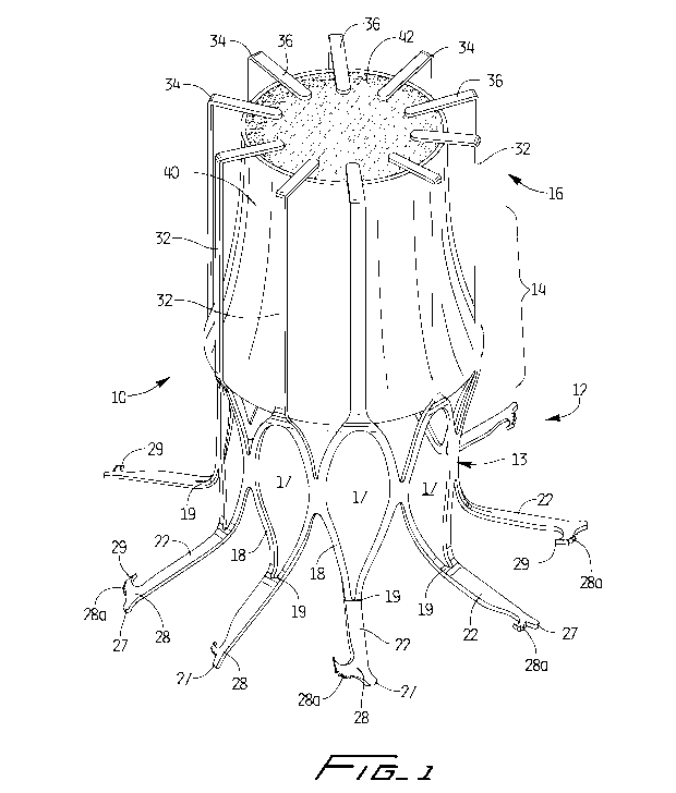
Figure 1 is a perspective view of a first embodiment of the vascular device of
the
present invention shown in the expanded configuration;
Figure 2 is a side view of the vascular device of Figure 1;
Figures 3A-3C are transverse cross-sectional views of the vascular device of
Figure 1 showing its interaction with the vessel wall during delivery and
placement;
Figure 4 is a perspective view of an alternate embodiment of the vascular
device
of the present invention shown in the expanded configuration;
CA 02730899 2011-01-14
WO 2010/017085 PCT/US2009/052190
Figure 5 is a side view of the vascular device of Figure 4 with the valve
partially
collapsed;
Figure 6 is a perspective view of another alternate embodiment of the vascular
device of the present invention shown in the collapsed position within a
delivery sheath;
Figure 7 is a perspective view similar to Figure 6 showing the vascular device
partially deployed from the delivery sheath;
Figure 8 is a perspective view similar to Figure 7 showing the vascular device
further deployed from the delivery sheath;
Figure 9 is a perspective view similar to Figure 8 showing the valve of the
vascular device pulled proximally;
Figure 10 is a perspective view of the vascular device of Figure 6 shown fully
deployed from the sheath and in the expanded configuration;
Figure 11 is a perspective view of another alternate embodiment of the
vascular
device of the present invention shown in the collapsed position;
Figure 12 is a perspective view of the vascular device of Figure 11 shown
fully
deployed from the sheath and in the expanded configuration;
Figure 13 is a perspective view of yet another alternate embodiment of the
vascular device of the present invention shown in the collapsed position; and
Figure 14 is a perspective view of another alternate embodiment of the
vascular
device shown in the expanded configuration.
DETAILED DESCRIPTION OF PREFERRED EMBODIMENTS
Referring now in detail to the drawings where like reference numerals identify
similar or like components throughout the several views, the device is
designated
generally by reference numeral 10 and is expanded to engage the internal wall
of the
vessel and contracted to pull the vessel walls radially inwardly in the manner
disclosed in
U.S. Patent No. 6,676,698, the entire contents of which is incorporated herein
by
reference.
Figures 1 and 2 illustrate vascular device 10 of a first embodiment of the
present
invention in the expanded configuration. Vascular device 10 is preferably
composed of a
shape memory material, such as a nickel-titanium alloy, e.g. Nitinol, so that
in its
6
CA 02730899 2011-01-14
WO 2010/017085 PCT/US2009/052190
memorized configuration it assumes the shape shown in Figure 1. This shape
memory
material characteristically exhibits rigidity in the austenitic state and more
flexibility in
the martensitic state. To facilitate passage from the delivery catheter, in
one embodiment,
the shape memory device is maintained in a collapsed configuration inside a
delivery
sheath where it is cooled by a saline solution to maintain the device below
its transition
temperature. The cold saline maintains the temperature dependent device in a
relatively
softer condition as it is in the martensitic state within the sheath. This
facilitates the exit
of device 10 from the sheath as frictional contact between the device and the
inner wall of
the sheath would otherwise occur if the device was maintained in a rigid, i.e.
austenitic,
condition. When the device 10 is released from the sheath to the target site,
it is warmed
by body temperature, thereby transitioning in response to this change in
temperature to an
austenitic expanded condition.
Device 10 is preferably formed from a tubular member, preferably by laser
cutting. Device 10 includes a proximal portion 12, an intermediate portion 14
and a
distal portion 16. In the expanded condition, at the proximal portion 12, the
body 13 of
device 10 includes struts 18 forming curved somewhat diamond shaped cells
forming
curved substantially diamond shaped openings 17. The proximal end regions 19
of the
cells 18 extend into struts 22 bent outwardly from the plane of the remainder
of the cell,
in a direction away from the longitudinal axis of the vascular device 10. This
better
enables the vessel engaging members, described below, to engage the vessel
walls.
Although shown bent at close to a 90 degree angle, other angles are
contemplated. For
clarity, not all the identical parts in the views are labeled.
A vessel engaging member 28 is formed at the end of each strut 22. The member
28 is in the form of a hook having a penetrating tip 29 to pierce the vessel
wall. That is,
the struts 22 terminate in hooks 28. Hooks 28 include a series of teeth 28a to
engage the
vessel wall to provide additional retention to prevent movement of the device
10. A heel
27 extends past the hook to function to limit penetration of the strut
portions through the
vessel wall. This hook configuration is described in detail in commonly
assigned co-
pending patent Publication No. 2007/012368 (serial no. 11/801,547, filed July
30, 2006),
the entire contents of which are incorporated herein by reference.
7
CA 02730899 2011-01-14
WO 2010/017085 PCT/US2009/052190
The sharp penetrating tips 29 penetrate the vessel wall in a radial direction
and
hold the vessel against axial movement with respect to the device 10, and
restrict radial
movement with respect to the device 10, thereby together securely retaining
(grasping)
the vessel wall for radial inward movement described below.
It should be understood that fewer or greater number of vessel engaging
members
as well as different engaging structures can be provided as long as they
achieve the vessel
retaining function described herein.
When the vascular device 10 expands, struts 22 are moved to a shape memorized
orientation bent outwardly at an angle, preferably about 90 degrees, with
respect to the
longitudinal axis of the device 10 with regions 19 bending out of the plane to
increase the
distance the members can extend from the center to the vessel wall. In the
collapsed
delivery position, struts 22 preferably lie substantially parallel to the
struts 32 extending
distally from the cells 18 and to the longitudinal axis of the device, with
the hooks 28 also
lying in the same plane to reduce the profile of the device for insertion.
That is, in the
delivery position, the struts 32 extend substantially parallel to the
longitudinal axis of
device 10 in the same way the struts 22 and hooks 28 are substantially
parallel. Once
released from the delivery sheath, they move to their memorized position shown
in
Figure 1.
The intermediate portion 14 and distal portion 16 of the device 10 include the
strut portions (elongated members) 32 extending longitudinally, alongside
(external to)
the valve 40. Struts 32 then bend inwardly at region 34, at for example a
ninety degree
angle, although other angles are contemplated, including more radiused bent
regions, so
strut portions 36 lie over a distal portion 42 of the valve 40 to secure the
valve 40 to the
device 10. The expanded placement position is shown in Figure 1, with the
struts 32
assuming their shape memory bent position.
An alternate embodiment of the vascular device is illustrated in Figures 4 and
5
and designated generally by reference numeral 100. Device 100 has a proximal
portion
112 identical to the proximal portion 12 of Figure 1, with identical parts
labeled in the
'100 series. Device 100 differs from device 100 in the intermediate portion
114 and
distal portion 116. These portions 114, 116 include two strut portions
(elongated
members) 132 extending longitudinally from the cell struts 118, alongside an
external
8
CA 02730899 2011-01-14
WO 2010/017085 PCT/US2009/052190
surface of the valve 140. Preferably there are two strut portions 132, spaced
approximately 180 degrees apart, however, a different spacing and a different
number of
strut portions 132 could be provided. The strut portions 132 terminate in ends
134
extending inwardly at region 133, at for example a ninety degree angle,
although other
angles are contemplated, including more radiused bent regions, so that strut
portions 134
lie over a distal edge 142 of the valve 140 to secure the valve 140 to. the
device 100.
Thus, elongated strut members 134 form a retaining hook to retain the valve.
The strut
portions can be fixed to the valve, enabling the valve to move between open
and closed
positions while still being retained. The strut portions 132 in one embodiment
can flex to
accommodate collapsing of the valve, as shown for example in Figure 5. The
expanded
placement position of device 100 is shown in Figures 4 and 5 with the struts
132
assuming their shape memory bent position. Note that the struts 122 (and 22)
can be bent
at angles other than those shown. Struts 122 lie substantially parallel to the
longitudinal
axis of the device in the collapsed delivery position.
There are several different methods of insertion of the vascular devices of
the
present invention for treating venous valve insufficiency of the popliteal or
saphenous
vein. These are disclosed in detail in the 6,676,698 patent, showing for
example
placement into the popliteal vein in the patient's leg and advanced to a
region adjacent
the leaflets to deploy the vascular device upstream of the leaflets. The
delivery catheter is
explained in the 6,676,698 patent as delivered in an antegrade fashion, with
the tip
extending downstream of the leaflets to deploy the device just upstream
(defined in
reference to the direction of blood flow) of the leaflets.
The vascular devices can be inserted through the right jugular vein, where the
device will be advanced through the superior and inferior vena cava, past the
iliac vein,
through the femoral vein and into the popliteal vein through leaflets in a
retrograde
fashion, i.e. opposite the direction of blood flow. The delivery catheter thus
extends
through the leaflet region just upstream of the leaflets. It alternatively can
be placed in
the right femoral vein, where it will be advanced in a retrograde manner to
the popliteal
vein. In the contralateral approach, it is inserted through the left femoral
vein where it
will be advanced around the iliac vein and through the left femoral vein into
the popliteal
vein.
9
CA 02730899 2011-01-14
WO 2010/017085 PCT/US2009/052190
In use in one method, the catheter or delivery sheath is inserted over a
conventional guidewire (not shown) so the distal tip of the catheter shaft
extends past, i.e.
downstream, of the valve leaflets L extending annularly from vessel wall. As
can be
appreciated, since there is a gap between the valve leaflets, the valve cannot
function
properly because the leaflets cannot properly close to prevent backflow. Also,
due to the
malfunctioning of the valve, the vessel wall becomes dilated as the weight and
pressure
of the backflow blood pushes out the vessel wall.
Once the position of the sheath is confirmed by venography, intravascular
ultrasound, or other means, the sheath is withdrawn with respect to catheter
tip to expose
the device 10 (100) so it is warmed by the body temperature and transitions to
its
austenitic phase and the first memorized expanded configuration of Figure 1.
Note that
device 100 (and devices 200, 300, 400 and 500 described below) can be inserted
in the
same ways as device 10 and therefore reference to insertion of device 10 also
contemplates inserting device 100.
Next, as described in the 6,676,698 patent, the balloon member on the catheter
shaft which is positioned within device is inflated via introduction of fluid
through an
inflation lumen to further expand the device 10 to a second expanded
configuration.
That is, the device is expanded to a larger diameter than the diameter in its
memorized
configuration of Figure 1 so that vessel engaging members 28 will engage the
vessel wall
with the sharp tips 29 penetrating the vessel wall to firmly grasp and secure
it. This
securement restricts both radial and axial movement of the vessel to enhance
retention by
the device 10.
After retention of the vessel wall, the balloon is deflated (and the catheter
removed), resulting in the device 10 contracting from the second expanded
configuration
towards its memorized configuration. Preferably, the device 10 will return to
substantially the same diameter as the first (memorized) expanded
configuration. As
contracted, the device 10, due to the engagement of the vessel engaging
members with
the internal wall of the vessel, pulls the vessel wall radially inwardly. As
can be
appreciated, the vessel wall is no longer dilated and sufficiently
approximated for proper
functioning of the replacement valve as the device 10 remains inside the
vessel.
CA 02730899 2011-01-14
WO 2010/017085 PCT/US2009/052190
The changing diameters of the vascular device 10 can also be appreciated by
reference to the transverse cross-sectional views of Figure 3A-3C which show
vascular
device 10' with hooks 28' different than hooks 28 of Figure 1. (Vascular
device 100
would also have changing diameters like device 10). The delivery device has
been
removed for clarity. More specifically, Figure 3A corresponds to the initial
position of
the vascular device 10' wherein the device 10' has been delivered to the
target vessel, and
has expanded to the first expanded (memorized) configuration but the vessel
engaging
members have not penetrated the vessel wall. It should be appreciated that in
this
configuration, the vessel engaging members may or may not be in contact with
the vessel
wall, but in either case, do not fully penetrate and secure the vessel to the
same extent as
in the second position. As shown, by way of example, the unhealthy dilated
vessel can
have an internal diameter D1 of approximately 14mm. The balloon is not shown
in
Figure 3A for clarity.
Figure 3B corresponds to the position of the vascular device wherein the
balloon
has been inflated to radially expand the device 10' to a second expanded
position to
enable the vessel engaging members to penetrate and retain (secure) the vessel
wall. In
this configuration, the vessel wall is further expanded for example, to a
diameter D2 of
about 16mm, as the device is expanded to a diameter of about 16mm, with the
hooks
extending an additional 2mm so the device is expanded to 20mm.
Figure 3C corresponds to the position of the vascular device 10' wherein the
balloon has been deflated and the device contracted to bring the vessel wall
radially
inwardly. The internal vessel wall diameter will preferably be about 12mm. The
diameter of the vascular device 10' preferably returns to the same diameter as
in Figure
3A, e.g. about 12mm. As can be seen the device 10' abuts the vessel wall V.
Note these dimensions are provided by way of example as other dimensions are
also contemplated.
The vascular device 10 (and 100) can also be placed downstream (with respect
to
the direction of blood flow) of the valve leaflets. The delivery catheter is
inserted in the
same antegrade manner as described above, except it is advanced sufficiently
past the
valve leaflets L to enable downstream delivery of the device 10 (100). The
device 10
11
CA 02730899 2011-01-14
WO 2010/017085 PCT/US2009/052190
(100) would grasp the vessel wall downstream of the valve leaflets to pull the
vessel wall
radially inwardly to bring the leaflets into apposition.
Turning now to Figures 6-10, an alternative embodiment of the vascular device
is
illustrated and configured for low profile insertion. With reference first to
Figure 10,
vascular device 200 has a proximal portion 212, intermediate portion 214 and
distal
portion 216. These portions 214, 216 include two elongated struts (or strut
portions) 232
extending longitudinally from the proximal portion 212, alongside (external
of) the valve
240, preferably substantially parallel to the longitudinal axis of the valve
240. Preferably,
two elongated strut members 232 are provided, spaced approximately 180 degrees
apart;
however, a different spacing and a different number of struts 232 could be
provided. The
vessel engaging members (or hooks) 228 are illustratively identical to the
vessel engaging
members 28 of Figure 1 and for brevity their description is not repeated
herein. As with
hooks 28, the hooks 228 are preferably provided in alternating small and
larger size to
enable nesting to reduce the profile for insertion.
As shown in Figure 10 illustrating the expanded condition of the vascular
device
200, the struts 232 extend longitudinally along and external to the valve 240
and bend
inwardly at region 234, preferably at an angle of about 90 degrees but other
angles are
also contemplated, to form radial portion 233. Radial portion 233, extending
substantially transversely to the longitudinal axis of the valve 240, then
bends inwardly at
region 236 forming angled portion 237 terminating in blunt tip 238. The angle
shown is
an obtuse angle but other angles are also contemplated. As shown, the valve
240
includes two spaced apart openings 241 to accommodate passage of the radial
portion
233 of the struts 234 to secure the valve to the struts 232. Additional
openings can be
provided to accommodate additional struts if provided. The engagement with the
openings 241 fixes the struts to the valve 240, enabling the valve to move
between open
and closed positions while still being retained. The expanded placement
position of
device 200 is shown in Figure 10 with the struts 232 assuming their shape
memory bent
position. Note that the struts 232 can be configured to bend at angles other
than those
shown.
Device body 202 is substantially identical to that of the embodiment of Figure
4,
having for example, curved diamond-like closed cells 217 formed by struts 218.
Adjacent
12
CA 02730899 2011-01-14
WO 2010/017085 PCT/US2009/052190
cells 217 are joined at region 219. For clarity, not all identical parts are
labeled. The
proximal end regions 215 of the cells 217 extend into struts 216 bent
outwardly from the
plane of the remainder of the cell, in a direction away from the longitudinal
axis of the
vascular device 200, and terminating in vessel engaging hooks 228.
With reference to Figure 8, wires 250 extend from a distal end of body 202.
That
is, the wires extend distally from the distal end of the cells 217. The wires
extend distally
within the delivery sheath D, and are attached to a proximal end of the valve
240. The
wires 250 are preferably made from shape memory material with a shape
memorized
expanded position to pull the valve in a proximal direction upon delivery as
discussed in
detail below. The wires can also form a ring shaped configuration in the
placement
configuration, with its rim positioned transverse to a longitudinal axis of
the valve 240, to
apply a radial force against the valve 240 to help retain it in the lumen. In
an alternate
embodiment, a separate ring (not shown) is provided which would be in an
elongated
position for delivery to the vessel and then expands to a ring shape in the
placement
position. Although two wires are shown, it is also contemplated that a
different number
of wires could be provided.
The vascular device 200 is configured to minimize the insertion profile. To
achieve this, the components of the device are aligned along a longitudinal
axis during
delivery. More specifically, as shown in Figure 6, the struts of the proximal
portion 212
of the device 200 are substantially parallel in the collapsed position, with
the vessel
engaging members (hooks) 228 staggered so smaller hooks 228a nest within
larger hooks
228b. Distal of the ring 250 in the delivery position is the valve 240 in the
collapsed
position. Wires 250 are in a collapsed coiled condition, positioned between
the distal end
of the closed cell region of the body 202 and the proximal end of the valve
240, attached
at one end to the body 202 and the other end to the valve 240.
In use, the device 200 is inserted within sheath D in the position shown in
Figure
6. Once at the desired location, pusher P is advanced distally within delivery
sheath D
against the end of the device 200 in the direction of the arrow of Figure 7,
advancing the
device 200 distally from the sheath, with the valve 240 being the first
component
exposed. Figure 8 illustrates the valve 240, elongated struts 232 and wires
250 exposed
from the sheath just before the wires recoil. That is, once the wires 250 are
exposed, they
13
CA 02730899 2011-01-14
WO 2010/017085 PCT/US2009/052190
self coil, pulling the valve 240 in a proximal direction within the space
between the
expanded struts 232. Thus, as shown the overall length of the device 200 is
now less in
the expanded placement position than in the delivery position due to the
proximal
movement of the valve 240. In one embodiment, the wires 250 returns to a ring
shape to
apply a radial force against the valve 240.
As the valve 240 is pulled proximally, radial portions 233 of struts 232 pass
through the respective openings 241 in the valve 240 thereby securing the
valve to the
struts 232. Figure 9 illustrates the valve pulled proximally by the coiling of
the wires 250.
Further advancement of the device 200 exposes the body 202, allowing the
struts 218 and
216 to expand to the placement configuration of Figure 10, forming cells 217
with the
hook regions extending at an angle to the struts 218 in a similar manner as
the
embodiment of Figure 4.
An alternate embodiment is illustrated in Figures 11 and 12. The vascular
device
is similar to the vascular device 300 of Figure 6 except for the position of
the shape
memory wires 350 and elongated strut portions 332 during delivery and the
cooperation
of the end of the strut portions 332 and the valve 340. Body portion 302 of
device 300 is
identical to body portion 202 of device 200, with identical parts labeled in
the `300 series,
and therefore its structure, e.g. cells 317, struts 318 and 316 and vessel
engaging
members 328 and function, for brevity are not discussed herein.
Turning to the differences of vascular device 300 from device 200, the wires
350
are contained within the valve 340 during delivery. Also, the longitudinally
extending
struts 332 extend to a distal end 343 of the valve 340 during delivery. In
this version,
upon delivery, the valve 340 is not pulled proximally as in the embodiment of
Figure 6.
Upon exposure from the sheath S, the valve 340 expands and the angled portions
337
extend from radial or transverse portions 333 of struts 332 are already
positioned distal of
the valve to prevent distal movement thereof. Moreover, as shown, the valve
340 is
retained by the angled portions 337 of the struts 332, forming hook-like
members
extending over the distal edge (rim) of the valve 340 to retain the valve.
Transverse
portions 333 extend radially inwardly toward the longitudinal axis of the
valve, at an
angle of about 90 degrees, although other angles are clearly contemplated.
Bent (angled)
portions 337 extend at an obtuse angle to transverse portion 333, although
other angles
14
CA 02730899 2011-01-14
WO 2010/017085 PCT/US2009/052190
are contemplated. The distal edge 342 of the valve 340 is thus captured by the
bent strut
configuration of Figure 12. As can be appreciated, struts 332 are unattached
to the valve,
but rely on the angles to retain the valve.
In the alternate embodiment of Figure 13, in the insertion position, the wires
450
of vascular device 400 extend between a distal end of the body 402 and a
proximal end of
the valve 440 as in Figure 6, however, the elongated strut portions 432 are
positioned at
the distal end of the valve 440. Upon delivery, the valve would expand within
the
confines of the bent portions of the struts 432 in the same manner as in
Figure 11. Except
for the positioning of the wires 450 for delivery, the vascular device 400 is
identical to
the device 330 and therefore for brevity will not be discussed in further
detail herein.
Figure 14 illustrates an alternate embodiment of the vascular device,
designated
generally by reference numeral 500. Vascular device 500 is identical to device
200 of
Figure 6 except for the shape of the cells formed by the struts in the
proximal region. As
shown, the cells 517 are diamond shaped with struts 518 forming the boundary
of the cell
being substantially straight walls. The device engages the valve 540 in the
same manner
as valve 240 and is delivered and moves to its expanded operative position in
the same
manner as the device of Figures 6-10. For convenience, device 500 is labeled
with
reference numerals in the `500 series identical to the parts labeled with the
`200 series in
device 200 and for brevity these parts and their function are not repeated
herein.
As an alternative to shape memory, a stainless steel or polymeric vascular
device
could be utilized. Such device would be expanded by a balloon below its
elastic limit,
thus enabling the device to return to its smaller configuration after the
balloon is deflated.
The vascular device could also be in the form of a braided structure which can
be
expanded to engage the vessel wall by squeezing or compressing its end(s), and
then
releasing it to enable it to return to its more elongated position of reduced
diameter to
approximate the vessel wall.
The vascular device is inserted in the vessel to expand and contract as
described
above, bringing the dilated vessel wall radially inwardly and leaving the
replacement
valve inside the vessel attached to the implanted vascular device. This
replacement valve
can be utilized as a total replacement wherein the patient's valve leaflets
are removed, or
can be placed upstream or downstream of the patient's leaflets, leaving the
CA 02730899 2011-01-14
WO 2010/017085 PCT/US2009/052190
nonfunctioning leaflets in place. The valves can be attached at the proximal
end, distal
end, or intermediate the proximal and distal ends of the vascular devices.
Valve 40 is conically shaped as shown and is secured to the struts of the
vascular
device 10 and device 100 by various techniques. Other types of valves can be
utilized
such as those described in the 6,676,698 patent.
A reinforcement ring could also optionally be provided. The valve can be multi-
layered, with an outer layer composed of one material and an inner layer
composed of
another material.
The device can be repositioned by a grasper as described in the `698 patent.
The
valve can be attached to the vascular device by sewing, molding or other
techniques. The
valves can be composed of a variety of materials such as PET, PTFE,
polycarbonate
polyurethane, swine intestinal submucosa, collagen and other biomaterials. The
valve
and /or the vascular device surface can optionally be coated with anti-
platelet or anti-
thrombin/anti-clotting materials, 2b/2a coating, receptors, heparin coating,
endothelial
cell coating, etc.
While the above description contains many specifics, those specifics should
not
be construed, as limitations on the scope of the disclosure, but merely as
exemplifications
of preferred embodiments thereof. For example, instead of a balloon to expand
the
device to its second expanded diameter/condition, a mechanical means such as
an
expandable wire frame can be utilized. Also, instead of moving the sheath to
expose the
vascular device, the catheter can be advanced with respect to the sheath or
both the
catheter and sheath can move relative to each other in opposite directions.
Those skilled
in the art will envision many other possible variations that are within the
scope and spirit
of the disclosure as defined by the claims appended hereto.
16