Note: Descriptions are shown in the official language in which they were submitted.
CA 02731082 2016-02-10
N
TISSUE SCAFFOLD COMPRISING AN ACELLULAR TISSUE MATRIX AND
SODIUM ACETATE
[0001]
BACKGROUND
[0002] Reduced pressure, or vacuum-assisted, therapies can
be effective
for improving wound healing due to a variety of different causes and at a
number of
different anatomical locations. Typically, reduced pressure therapies include
a
porous material that is placed at a wound site, which aids in the distribution
of the
reduced pressure. Typical porous materials are sized to fit the wound, and may
be
periodically replaced with smaller pieces of the porous material as the wound
begins
to heal and becomes smaller. Typically, a membrane or drape is placed over the
porous material to provide an airtight seal at the wound area, and a negative
pressure is applied to the porous material to provide a reduced pressure at
the
wound site.
SUMMARY
[0003] According to certain embodiments, a method of
processing an
acellular tissue matrix for preparing a tissue scaffold is provided. In
certain
embodiments, a method of preparing a tissue scaffold is provided, comprising
adding
an acellular tissue matrix to a first aqueous solution of sodium acetate;
incubating
the first aqueous sodium acetate solution containing the acellular tissue
matrix;
removing the incubated acellular tissue matrix from the first aqueous sodium
acetate
solution; treating the incubated acellular tissue matrix with a second aqueous
solution of sodium acetate to form a suspension; homogenizing the suspension
to
1
CA 02731082 2011-01-14
WO 2010/019753 PCT/US2009/053667
- form a slurry; cooling the slurry; casting the slurry in a casting
container; and
lyophilizing the slurry. In certain embodiments, a tissue scaffold comprising
an
acellular tissue matrix and sodium acetate is provided. In certain
embodiments, a
wound treatment device comprising a reduced pressure source and a tissue
scaffold
is provided. In certain embodiments, a tissue scaffold, comprising an
acellular tissue
matrix that has been processed to have a porosity of between 75% and 90% is
provided.
DESCRIPTION OF THE DRAWINGS
[0004] Fig. 1 illustrates a wound treatment device, which provides
reduced
- pressure therapy, according to certain exemplary embodiments.
[0005] Fig. 2 illustrates a method of treating a cartilage defect
using a
tissue scaffold, according to certain embodiments.
[0006] Figs. 3A-3D are graphs showing the strut spacing for tissue
scaffolds, as described in Example 1.
[0007] Fig. 4 is a graph showing the permeability versus composition
for
tissue scaffolds, as described in Example 2.
[0008] Fig. 5 is a graph showing the strut spacing for a tissue
scaffold
produced according to certain exemplary embodiments, as described in Example
3.
[0009] Figs. 6A-6B are photomicrographs of chondrocytes cultured on
tissue scaffolds, as described in Example 4.
_
[0010] Figs. 7A-7B are photomicrographs of chondrocytes cultured on
tissue scaffolds, as described in Example 4
2
CA 02731082 2011-01-14
WO 2010/019753 PCT/US2009/053667
DESCRIPTION OF CERTAIN EXEMPLARY EMBODIMENTS
[0011] Reference will now be made in detail to the certain exemplary
embodiments according to the present disclosure, certain examples of which are
illustrated in the accompanying drawing.
[0012] The present disclosure pertains to a method of processing an
acelluar tissue matrix for preparing a tissue scaffold. In some embodiments,
the
tissue scaffold of the present disclosure may be used as part of a wound
treatment
device that provides reduced pressure therapy. In some embodiments, the
physical
properties of certain tissue scaffolds such as porosity, strut density, and
permeability,
may be controlled or altered by adjusting the concentrations, components, and
temperatures at which the scaffolds are produced.
[0013] In this application, the use of the singular includes the
plural unless
specifically stated otherwise. In this application, the use of "or" means
"and/or"
unless stated otherwise. Furthermore, the use of the term "including", as well
as
other forms, such as "includes" and "included", is not limiting. Also, terms
such as
. "element" or "component" encompass both elements and components comprising
one unit and elements and components that comprise more than one subunit,
unless
specifically stated otherwise. Also the use of the term "portion" may include
part of a
moiety or the entire moiety.
[0014] The term "acellular tissue matrix," as used herein, refers
generally
to any tissue matrix that is substantially free of cells and other antigenic
material. In
various embodiments, acellular tissue matrices derived from human or xenogenic
sources may be used to produce the scaffolds. Skin, parts of skin (e.g.,
dermis), and
other tissues such as blood vessels, heart valves, fascia, nerve, or other
collagen
3
CA 02731082 2011-01-14
WO 2010/019753
PCT/US2009/053667
containing-organ or tissue may be used to create an acellular matrices to
produce
tissues scaffolds within the scope of the present disclosure.
[0015] In certain embodiments, the term "permeability" refers
generally to
the movement of fluid through a porous medium. In certain embodiments, the
specific permeability values of particular tissue scaffolds are calculated by
Darcy's
Law:
Q=1=,u
[0016] k= _____
AP=A
[0017] where Q equals the total discharge (units of volume per time,
e.g.,
m2/s), (A) is the cross-sectional area to flow, AP is the pressure drop across
the
system, p is the dynamic viscosity (in SI units e.g. kg/(ms) or Pa's), and (I)
is the
length over which the pressure drop is taking place over.
[0018] The term "reduced pressure," as used herein, generally refers
to a
pressure less than the ambient pressure at a tissue site that is being
subjected to
treatment. In most cases, this reduced pressure will be less than the
atmospheric
pressure at which the patient is located. Alternatively, the reduced pressure
may be
less than a hydrostatic pressure of tissue at the tissue site. Reduced
pressure may
initially generate fluid flow in the area of the tissue site and/or a fluid
conduit in
communication with the tissue site, for example, as shown in Fig. 1. As the
hydrostatic pressure around the tissue site approaches the desired reduced
- pressure, the flow may subside, and the reduced pressure is then maintained.
In
some embodiments, small amounts of gas can be introduced at intervals to
facilitate
fluid movement if required. Unless otherwise indicated, values of pressure
stated
herein are gage pressures.
4
CA 02731082 2016-02-10
[0019] The term "fluid" as used herein generally refers to a gas or liquid,
but may also include any other flowable material, including but not limited to
gels,
colloids, and foams.
. [0020] The section headings used herein are for organizational purposes
only and are not to be construed as limiting the subject matter described.
[0021] In various embodiments, devices of the present disclosure can be
used for treatment at numerous different anatomical sites. According to
various
embodiments, tissue scaffolds can be used in a wide array of applications.
Certain
exemplary applications include, but are not limited to, absorptive dressing,
dermal
regeneration (for example, for treatments of all types of ulcers and burns),
nerve
regeneration, cartilage regeneration, connective tissue regeneration or repair
(for
example, tendon/ligament sleeve), bone regeneration, periodontal applications,
wound/foam lining, integrated bandage dressing, substrate/base for skin
grafts,
vascular regeneration, cosmetic surgery, cosmetic injectable gel, metal and/or
polymer implant coating (for example, to increase implant integration and
biocompatibility), and replacement of lost tissue (e.g., after trauma, breast
reduction,
mastectomy, lumpectomy, parotidectomy, or excision of tumors).
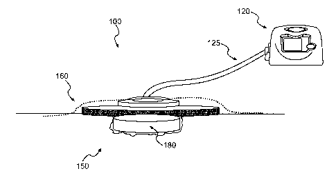
[0022] Fig. 1 illustrates a wound treatment device 100, including a reduced
pressure source 120, according to certain exemplary embodiments. In various
embodiments, a variety of reduced pressure therapy devices can be used. For
example, suitable reduced pressure therapy devices include V.A.C.0 therapy
devices produced by Kinetic Concepts, Inc. (San Antonio, Texas). Such reduced
CA 02731082 2011-01-14
WO 2010/019753 PCT/US2009/053667
pressure therapy devices can include a vacuum pump that can be fluidly
connected
to the wound site 150, via a fluid conduit 125 or other fluid connection. Such
devices
may also include a flexible sheet 160 to cover the wound site 150 and at least
partially seal the wound to allow reduced pressure therapy to be provided at
the
wound site. In addition, such systems may include a tissue scaffold 180, that
is
placed at the wound site and facilitates wound closure, healing, tissue
regeneration
or repair, prevents or treats infection, and/or has other beneficial effects.
In certain
embodiments, the tissue scaffold 180 assists in distributing fluid flow or
negative
pressure across a site to be treated.
[0023] In some embodiments, the flexible sheet 160 will include a
flexible
polymeric material. In various embodiments, any suitable polymeric material
can be
selected. In various embodiments, the material does not cause significant
irritation,
immune response, or heightened risk of infection. In various embodiments, the
specific material generally should be of sufficient thickness and
impermeability to
allow reduced pressure therapy at a wound site under the sheet 160.
[0024] In some embodiments, the device 100 will include an adhesive.
As
used here, and throughout the disclosure, adhesive will be understood to refer
to any
substance that causes the surfaces of two objects to be attached to one
another. In
various embodiments, suitable adhesives can include a variety of different
cements,
glues, resins, or other materials that can facilitate attachment of the
flexible sheet
160 to tissue. In some embodiments, the adhesive can include a pressure-
sensitive
acrylic adhesive. In various embodiments, the adhesives can be applied
directly to
the structures to be joined, or the adhesives may be applied on tape, or with
other
supporting substrate materials.
6
CA 02731082 2011-01-14
WO 2010/019753 PCT/US2009/053667
[0025] In some embodiments, the adhesive can be applied to a surface
of
the flexible sheet 160 to attach the sheet to skin or other tissue. In some
embodiments, the adhesive will be applied to the surface of the sheet and
packaged
and/or distributed with the sheet 160. In some embodiments, the adhesive is
applied
to a surface of the sheet 160 and covered by a non-adhesive material that can
be
removed to expose the adhesive for use. In certain embodiments, the adhesive
can
be supplied as a separate component (e.g., in a container or on a tape) that
is
applied to the sheet 160 to attach the sheet 160 to tissue. In some
embodiments,
the adhesive can be applied to a patient's skin or other tissue, and the sheet
can be
applied to the adhesive.
[0026] In various embodiments, tissue scaffold 180 can include a
variety of
suitable materials. For example, a number of different tissue scaffolds will
be
- compatible for use with the above-noted V.A.C.0 treatment systems. In some
embodiments, the tissue scaffold may comprise a processed acellular tissue
matrix.
In some embodiments, the acellular tissue matrix may be derived from human
skin
or from a xenogenic source. In various embodiments, other tissues such as
blood
vessels, heart valves, fascia, nerve, connective tissue, or other collagen-
containing
organs or tissues may be used to create a specific acellular matrix within the
scope
of the present disclosure. In some embodiments, the acellular tissue matrix is
an
acellular dermal matrix. In various embodiments, the acellular dermal matrix
is
produced from human dermis or pig dermis. In some embodiments, the methods
disclosed herein utilize a dehydrated acellular tissue matrix tissue, such as
_ ALLODERMQ which is commercially available from LifeCell Corporation,
Branchburg, New Jersey. In some embodiments, the methods disclosed herein
7
CA 02731082 2016-02-10
utilize an acellular tissue matrix tissue, such as STRATTICETm, which is
commercially available from LifeCell Corporation, Branchburg, New Jersey.
[0027] In various embodiments, acellular tissue matrices can be produced
using a variety of different tissue processing techniques. For example,
certain
exemplary tissue processing techniques for producing acellular tissue matrices
are
described in U.S. Patent No. 5,336,616 and 5,364,756, both to Livesey et al.,
in U.S.
Patent No. 6,933,326 to Schiff et al.
In some embodiments, acellular tissue matrices made from non-human
animals can be treated to remove various antigens, or produced from animals
genetically modified to lack certain antigens. For example, certain exemplary
methods of processing tissues to produce acellular matrices with reduced
amounts
of or lacking alpha-1,3-galactose moieties, are described in Hui, X. et al.,
"A Porcine-
Derived Acellular Dermal Scaffold that Supports Soft Tissue Regeneration:
Removal
of Terminal Galactose-a-(1,3)-Galactose and Retention of Matrix Structure,"
Tissue
Engineering, Vol. 15, 1-13 (2009).
[0028] Fig. 2 illustrates use of a tissue scaffold to treat a cartilage
defect,
according to certain embodiments. As shown, a scaffold 180 is used to treat a
cartilage defect in a long bone (e.g., femur or humerus). In various
embodiments, a
scaffold 180 can be used to treat an articular surface or cartilage 510 of any
joint. In
various embodiments, the tissue scaffold 180 is placed in a defect or excised
area of
an articular surface or cartilage 510, and a negative pressure is applied to
the tissue
scaffold 180 through a fluid conduit 125, as described above. In some
embodiments, a second material 190 is applied over the tissue scaffold 180,
and the
second material 190 acts as a manifold to distribute pressure to tissue
scaffold 180.
8
CA 02731082 2011-01-14
WO 2010/019753 PCT/US2009/053667
. In some embodiments, the fluid conduit is in fluid communication with the
tissue
scaffold 180 without a second material 180.
[0029] In some embodiments, the tissue scaffold can be used as a
primary
treatment method or in connection with another procedure or treatment. For
example, in various embodiments, cartilage repair or regeneration can be
performed
using a technique known in the art as microfracture. As shown in Fig. 2,
during a
microfracture procedure, a surgeon creates small fractures or openings 515 in
bone
adjacent to an articular defect. In various instances, the fractures or
openings 515
can allow chondrocytes or other cells that can differentiate into chondrocytes
to
migrate to the articular defect from adjacent bone, bone marrow space, or
cartilage.
In various instances, these cells can, in turn, help repair or regenerate
cartilage.
[0030] In some embodiments, after the fractures or openings 515 are
produced in the bone 500, the tissue scaffold 180 is placed over the
microfracture
site, and negative pressure is applied to the scaffold. In some embodiments,
the
tissue scaffold acts as a manifold to distribute negative pressure over the
site to be
treated. In some embodiments, the tissue scaffold provides a substrate to
support
tissue growth, repair, and/or regeneration. In some embodiments, negative
pressure
is applied to draw cells, growth factors, and/or other biologic elements into
the tissue
scaffold 180 from the bone 500.
[0031] In some embodiments, a method of processing an acellular
tissue
matrix to produce a tissue scaffold is provided. In some embodiments, the
acellular
- tissue matrix comprises collagen, elastin, and vascular channels. In some
embodiments, the acellular tissue matrix is ALLODERMO. In some embodiments,
the acellular tissue matrix is STRATTICETm.
9
CA 02731082 2011-01-14
WO 2010/019753 PCT/US2009/053667
[0032] In some embodiments, a method for producing a tissue scaffold
is
provided. In some embodiments, a method comprises adding an acellular tissue
matrix to a first aqueous solution of sodium acetate; incubating the first
aqueous
sodium acetate solution containing the acellular tissue matrix; removing the
incubated acellular tissue matrix from the first aqueous sodium acetate
solution;
treating the incubated acellular tissue matrix with a second aqueous solution
of
sodium acetate to form a suspension; homogenizing the suspension to form a
slurry;
cooling the slurry; casting the slurry in a casting container; and
lyophilizing the frozen
slurry.
[0033] In some embodiments, the acellular tissue matrix added to the
first
aqueous sodium acetate solution is in dehydrated form. In some embodiments,
the
acellular tissue matrix is cut into small pieces, e.g., cubes, after removal
from the first
aqueous sodium acetate solution prior to homogenization. In some embodiments,
the incubating step takes place at about 4 C for more than 12 hours.
[0034] In some embodiments, the homogenizing step and the cooling
step
- are repeated at least three times. In some embodiments, the homogenizing
step is
accomplished by a homogenizing Dremmel probe. In some embodiments, the
cooling step is accomplished at about 0 C for about 1.5 minutes or longer. In
some
embodiments, the casted slurry is frozen at about -70 C or less for about 2
hours or
longer. In some embodiments, the casted slurry is frozen at about -200 C or
less.
[0035] In some embodiments, the desired shape and height of the
resulting tissue scaffold is determined by the shape and height of the casting
container. In some embodiments, the first aqueous sodium acetate solution has
a
pH of about 3.4, or between about 3.4 and 7.0, or between about 3.4 and 5Ø
In
some embodiments, the first aqueous solution achieves a final concentration of
CA 02731082 2011-01-14
WO 2010/019753 PCT/US2009/053667
about 0.1% w/v to about 15% w/v of acellular tissue matrix. In some
embodiments,
the porosity of the resulting tissue scaffold is from about 75% to about 90%.
In some
embodiments, the strut density of the resulting tissue scaffold is from about
0.13
g/cm3 to about 0.24 g/cm3. In some embodiments, mechanical strength, porosity,
hydration and fluid conductance are controlled by freezing rate, freezing
temperature, and the composition of the casting container. In some
embodiments,
the acellular tissue matrix comprises collagen, elastin, and vascular
channels. In
some embodiments, the acellular tissue matrix is ALLODERM . In some
embodiments, the acellular tissue matrix comprises collagen, elastin, and
vascular
- channels. In some embodiments, the acellular tissue matrix is STRATTICETm.
[0036] In certain embodiments, the tissue scaffold has a desired
permeability. For example, the permeability may be selected to allow adequate
manifolding or distribution of pressure or flow applied to a wound or therapy
site
across the site. In certain embodiments, the permeability is controlled by
controlling
the porosity of the tissue scaffold. In certain embodiments, the permeability
is at
least 1x10-11 m2.
[0037] In some embodiments, sodium bicarbonate is further added to
either one or both of the first or second aqueous sodium acetate solutions,
and/or to
the slurry. In some embodiments, sodium bicarbonate can be added to the
solution
just before or during casting. The amount of sodium acetate can be selected to
cause foaming of the solution and/or slurry. In certain embodiments, the
amount of
foaming is selected to control the porosity of all or a portion of a tissue
scaffold. In
certain embodiments, the slurry is frozen soon after adding sodium acetate to
create
a desired porosity in the tissue scaffold. In some embodiments, the tissue
scaffold
can have a porosity that varies across its thickness.
11
CA 02731082 2011-01-14
WO 2010/019753 PCT/US2009/053667
[0038] In some embodiments, the tissue scaffold comprises an acellular
tissue matrix and sodium acetate. In some embodiments, the tissue scaffold
further
comprises sodium bicarbonate.
- [0039] In certain instances, it may be desirable for tissue scaffolds to
be
resorbed by the body rather than persist for extended periods. In certain
instances,
tissue scaffolds persist for extended periods, e.g., several months or longer.
In
certain instances, extended periods provide continued tissue regrowth,
remodeling,
and regeneration. In certain instances, with some negative pressure wound
treatment systems, a material placed over a wound bed is generally replaced
periodically (e.g., every few days). In certain instances, replacement of the
materials
can be painful or damaging to the wound site, especially if granulation tissue
has
grown into the material. In some embodiments, the tissue scaffolds are
bioresorbable. In some embodiments, the tissue scaffolds can be placed in a
wound
site or implanted, and will be resorbed by the body such that the devices are
not
removed or replaced.
[0040] The following examples demonstrate certain exemplary
embodiments of the disclosure. It should be appreciated by those of skill in
the art
that the techniques disclosed in the examples herein may be modified to
achieve
similar results.
Preparation of a Tissue Scaffold
[0041] Aseptically prepared ALLODERM is cut into strips (approximately
2-3mm wide) and weighed dry. The desired weight is immersed in the appropriate
volume of 20mM sodium acetate to achieve a final concentration of about 0.1%
to
about 15.0% w/v (weight of dried material to volume of solution). The strips
are then
removed from solution, diced into small cubes (approximately 2x2 mm) using a
12
CA 02731082 2011-01-14
WO 2010/019753 PCT/US2009/053667
scalpel, and immersed in a second 20mM aqueous sodium acetate solution to
achieve the desired % w/v. The suspension is then homogenized at full speed
using
a Dremmel type probe tip for 1 minute, followed by cooling on ice for about 1
minute
or longer. The suspension is cooled sufficiently to prevent heating of the
suspension
to near the melting point of collagen within the suspension during subsequent
homongenization, thereby preventing thermal damage to the collagen. The
homogenization and cooling steps are repeated three (3) times. The homogenizer
tip is then rinsed, and the slurry is then poured into the desired casting
containers for
the right shape and height. The containers are then covered and placed in a
freezer
at -70 C for more than 2 hours to ensure complete freezing. As described
below,
samples may be frozen at other temperatures to achieve faster freezing, e.g.,
on
liquid nitrogen at about -200 C. The samples are removed from the freezer and
placed in a freeze drier. The scaffolds are then removed from the freeze drier
upon
completion (e.g., about 24 hours or when the temperature of the vessel reaches
ambient temperature) of the lyophilization process, and are stored in a
dessicator or
under vacuum. In some embodiments, sodium bicarbonate can be added to the
sample before or after any of the homogenization steps, or just before or
during
casting.
- [0042] In various embodiments, freezing rate, freezing temperature, the
addition of sodium bicarbonate and the material compositions may all be
modified.
In certain embodiments, the modifications control the final composition,
mechanical
strength, hydration, and/or fluid conductance of the resulting tissue
scaffold.
Example 1: Effect of Scaffold Thickness on Strut Spacing
[0043] The effect of material thickness on scaffold structure was
evaluated.
Aseptically prepared ALLODERM was cut into strips (approximately 2-3mm wide)
13
CA 02731082 2011-01-14
WO 2010/019753 PCT/US2009/053667
and weighed dry. The strips were immersed in the appropriate volume of 20 mM
. sodium acetate to achieve a final concentration of 5.0% w/v (weight of dried
material
to volume of solution) and pH of about 3.4. The samples were incubated
overnight
at 4 C. After incubation overnight, the sample pH rose to about 7.0, and the
pH was
adjusted back to about 3.4 before further processing. Samples were homogenized
three times using a Dremmel type probe to produce a slurry. Samples were
cooled
on ice for 1 minute between each homogenization step. The samples were casted
in
six-well culture plates. The wells in which samples were casted had a
cylindrical
structure with a 35 mm diameter. Samples were cooled at -70 C for four hours
and
were then freeze-dried to produce tissue scaffolds. Scaffolds were produced
using
slurry volumes of 1 ml, 2 ml, 4 ml, and 6 ml. Figs. 3A-3D are graphs showing
the
strut spacing for the tissue scaffolds. Fig. 3A represents data for a 1 ml
sample, Fig.
3B is date for a 2 ml sample, Fig. 3C is data for a 4 ml sample, and Fig. 3D
is data
for a 6 ml sample.
[0044] As shown in Figs. 3A-3D, thicker samples resulted in a larger
pore
size (larger strut spacing) and wider variation pore size. Table 1 provides
data
including the average pore size, standard deviation, median pore size, and
minimum
and maximum pore sizes for samples having varying thicknesses. As shown in
Table 1, the standard deviation of pore size, range of pore sizes (difference
between
minimum and maximum pore sizes) and average pore size all increase with
increasing material thickness.
14
CA 02731082 2011-01-14
WO 2010/019753 PCT/US2009/053667
Table 1
Final Volume of Na0Ac for Scaffolds Produced with 5% w/v
Slurry (material to casting solution)
1 ml 2m1 4 ml 6 ml
Average Strut 89 pm 108 pm 133 pm 212 pm
Spacing
Standard 29 pm 41 pm 51 pm 67 pm
Deviation of
Strut Spacing
Median Strut 83 pm 101 pm 97 pm 202 pm
Spacing
Maximum Strut 167 pm 211 pm 260 pm 356 pm
Spacing
Minimum Strut 43 pm 33 pm 35 pm 96 pm
Spacing
Example 2: Effect of Scaffold Composition on Water-Binding Capacity, Porosity,
and
Permeability:
[0045] In certain embodiments, the ability of tissue scaffolds to
bind water
can be important for scaffolds remaining hydrated and being effective in
supporting
tissue repair or regeneration. Sample permeability and water-binding capacity
were
studied as a function of sample composition (i.e., variation in w/v%).
Scaffolds were
produced by casting and lyophilizing slurries having varying compositions.
[0046] Aseptically prepared ALLODERMO was cut into strips
(approximately 2-3mm wide) and weighed dry. The desired weight of the strips
was
immersed in the appropriate volume of 20 mM sodium acetate to achieve a final
concentrations of 0.1% w/v, 0.5% w/v, 1.0% w/v, 3.0% w/v, 5% w/v, and 8% w/v
CA 02731082 2011-01-14
WO 2010/019753 PCT/US2009/053667
(each being material weight to sodium acetate solution volume). Each sample pH
was about 3.4. Samples were incubated overnight at 4 C. After incubation, the
sample pHs rose to about 7.0, and the pHs were adjusted back to about 3.4
before
further processing. Samples were homogenized three times using a Dremmel type
probe to produce a slurry. Samples were cooled on ice for 1 minute between
each
homogenization step. Samples were cooled at -70 C for four hours and were then
freeze-dried to produce tissue scaffolds.
[0047] Table 2 provides data for sample water-binding capacity as a
function of sample composition, and Fig. 4 is a graph showing the permeability
versus composition for tissue scaffolds. As shown in Table 2 and Fig. 4, as
the
sample w/v% increased, the sample water-binding capacity increased and sample
permeability decreased.
Table 2
Soaking Capacity of Exemplary Tissue Scaffolds
Dry Soaking Thickness Length (% Breadth (% Volume (%
AlloDerm/NaOac Capcity ( /0 change) change) change)
change)
(w/v) (g/cm3)
0.1% n/a -79.2 8.5 5.6 -76.1
0.5% 0.53 -42.9 1.5 1.6 -41.0
1.0% 0.32 3.8 2.3 3.4 9.8
3.0% 0.56 3.6 2.4 2.6 8.8
5.0% 0.68 7.6 6.7 4.8 20.3
8.0% 0.80 9.1 0.7 0.8 10.8
[0048] Table 3
provides data on sample strut density and porosity as a
function of sample composition. The sample porosity followed a relatively
normal
16
CA 02731082 2011-01-14
WO 2010/019753 PCT/US2009/053667
distribution, with sample strut spacing and porosity increasing as sample
composition varies from 0.1% to about 5.0%, and decreasing with further
increase in
sample composition.
Table 3
Porosity and Strut Density of Tissue Scaffolds
Dry AlloDerm/Na0Ac (w/v) Strut Density (g/cm3) % Porosity
0.1% 0.132 89.0
0.5% 0.075 84.6
1.0% 0.154 89.9
4.0% 0.264 88.7
5.0% 0.423 91.0
8.0% 0.486 89.2
10.0% 0.395 84.1
15.0% 0.234 79.7
Example 3: Effect of Freezing Rate/Temperature on Scaffold Structure:
[0049] In some embodiments, it may be desirable to decrease average
strut spacing and/or control variation in sample porosity. In some
embodiments,
these features can be controlled by controlling the temperature at which the
sample
is cooled and/or the cooling rate.
[0050] Aseptically prepared ALLODERM was cut into strips
(approximately 2-3mm wide) and weighed dry. The strips were immersed in the
appropriate volume of 20 mM sodium acetate to achieve a final concentration of
5.0% w/v (weight of dried material to volume of solution) and pH of 3.4.
Samples
were incubated for about 48 hours at 4 C. After incubation, the sample pH rose
to
17
CA 02731082 2011-01-14
WO 2010/019753
PCT/US2009/053667
about 7.0, and the pH was adjusted back to about 3.4 before further
processing.
Samples were homogenized three times using a Dremmel type probe to produce a
slurry. Samples were cooled on ice for 1 minute between each homogenization
step. The slurry was poured into copper wells. The wells in which samples were
casted had a cylindrical structure with a 35 mm diameter. The samples were
flash
frozen at -200 C by placing copper wells filled with slurry in liquid nitrogen
and then
immediately freeze-drying to produce tissue scaffolds.
[0051] Fig. 5 shows the strut spacing for tissues produced in this
manner,
- and Table 4 provides data including the average pore size, standard
deviation,
median pore size, and minimum and maximum pore sizes for samples having
varying thicknesses. As shown, the sample had less variation in pore size and
smaller average pore size than samples cooled at -70 C, as shown in Figs. 3A-
3D.
Table 4: Foam Strut Spacing for Samples Cooled at -200 C
Foam Strut Spacing (microns)
Average 9.5
Standard Deviation 2.2
Median 9.7
Maximum 14.2
Minimum 4.2
Example 4: Culture of Isolated Chondrocvtes on Tissue Scaffolds
[0052] In certain instances, when cultured in typical media, (see
Eyrich, D.
et a., "Long-term stable fibrin gels for cartilage engineering", Biomaterials,
28(1):55-
65.(2007)), chondrocytes are known to dedifferentiate into fibroblasts.
Therefore, in
certain embodiments, tissue scaffolds used for regeneration of cartilage
support a
18
CA 02731082 2011-01-14
WO 2010/019753 PCT/US2009/053667
continued chondrocyte phenotype when implanted at a treatment site. Certain
tissue
scaffolds were tested to determine if they support growth of chondrocytes
without
causing differentiation to fibroblasts. One set of scaffolds was produced
using
human acellular dermal matrices (ALLODERM10), and a second set was produced
using porcine acellular dermal matrices (STRATTICE11"). The material was cut
into
strips (approximately 2-3mm wide) and weighed dry. The strips were immersed in
the appropriate volume of 20 mM sodium acetate to achieve a final
concentration of
5.0-8.0% w/v (weight of dried material to volume of solution) and were stored
for
about 48 hours at 4 C. Samples were homogenized three times using a Dremmel
type probe to produce a slurry. Samples were cooled on ice for 1 minute
between
each homogenization step. The samples were casted in six-well culture plates.
The
wells in which samples were casted had a cylindrical structure with a 35 mm
diameter. Samples were cooled at -70 C for four hours and were then freeze-
dried
to produce tissue scaffolds. The scaffolds were seeded with primary sheep
articular
chondrocytes isolated via an overnight digestion in collagenase, according to
standard prototocols, The cells were cultured for 14 or 21 days in 10% Fetal
Bovine
Serum in DMEM at 37 C and 5% Atmospheric CO2 with 100% humidity
[0053] Figs. 6A-6B are photomicrographs of chondrocytes cultured on
tissue scaffolds produced with ALLODERM , and Figs. 7A-7B are
photomicrographs of chondrocytes cultured on tissue scaffolds produced with
STRATTICErm. Both human and porcine tissue scaffolds supported chondrocyte
growth and infiltration, and grossly appeared to maintain chondrocyte
phenotypes.
[0054] While systems and methods have been described with reference
to
- tissue growth and healing in human patients, it should be recognized that
these
systems and methods for applying reduced pressure tissue treatment can be used
in
19
CA 02731082 2011-01-14
WO 2010/019753 PCT/US2009/053667
any living organism in which it is desired to promote tissue growth or
healing.
Similarly, the systems and methods may be applied to any tissue, including
without
limitation bone tissue, adipose tissue, muscle tissue, neural tissue, dermal
tissue,
vascular tissue, connective tissue, cartilage, tendons, or ligaments. While
the healing
of soft tissue may be an exemplary focus of applying reduced pressure tissue
treatment as described herein, the application of reduced pressure tissue
treatment,
especially to tissues located beneath a patient's skin, may also be used to
generate
- tissue growth in tissues that are not diseased, defective, or damaged. For
example,
it may be desired to use the percutaneous implantation techniques to apply
reduced
pressure tissue treatment to grow additional tissue at a tissue site that can
then be
harvested. The harvested tissue may be transplanted to another tissue site to
replace diseased or damaged tissue, or alternatively the harvested tissue may
be
transplanted to another patient.
[0055] Other embodiments will be apparent to those skilled in the art
from
consideration of the specification and practice of the devices and methods
disclosed
herein.