Note: Descriptions are shown in the official language in which they were submitted.
CA 02732011 2011-01-25
Description
Title of Invention: METHOD FOR MEASUREMENT OF PHYSIOLOGICALLY ACTIVE
SUBSTANCE DERIVED FROM ORGANISM AND MEASUREMENT APPARATUS
Technical Field
[0001] The present invention relates to a measurement method
and a measurement apparatus for detecting a physiologically active
substance derived from an organism, which has a property of gelating
by a reaction with LAL, such as endotoxin or (3-D-glucan, in a sample
containing the physiologically active substance or for measuring
the concentration of the physiologically active substance.
Background Art
[0002] Endotoxin is a lipopolysaccharide present in a cell wall
of a Gram-negative bacterium and is the most typical pyrogen. If
a transfusion, a medicine for injection, or blood contaminated with
the endotoxin is introduced into the human body, the endotoxin may
induce severe side effects such as fever and shock. Therefore, it
has been required that the above-mentioned medicine or the like
be kept so as not to be contaminated with endotoxin.
[0003] By the way, a limulus amoebocyte lysate (hereinafter,
also referred to as "LAL") contains serine proteases which are
activated by endotoxin. In the case where LAL reacts with endotoxin,
a coagulogen present in LAL is hydrolyzed into a coagulin by an
enzyme cascade of the serine proteases activated depending on the
1
CA 02732011 2011-01-25
amount of endotoxin, and the coagulin is associated with one another
to form an insoluble gel. By using the characteristic of LAL,
endotoxin can be detected with a high sensitivity.
[0004] Meanwhile, P-D-glucan is a polysaccharide which
constitutes a cell membrane characteristic of a fungus. Measurement
of 03-D-glucan is effective, for example, for screening of infectious
diseases due to a variety of fungi including not only fungi which
are frequently observed in general clinical practices, such as
Candida, Spergillus, and Cryptococcus, but also rare fungi.
[0005] In measurement of 3-D-glucan, by using the
characteristic of a limulus amoebocyte lysate to coagulate
(coagulate to form a gel) by (3-D-glucan, (3-D-glucan can be detected
with a high sensitivity.
[0006] Various methods have been proposed as a method for
detection or concentration measurement of a physiologically active
substance derived from an organism (hereinafter, also referred to
as a predetermined physiologically active substance) which can be
detected by a limulus amoebocyte lysate, such as endotoxin or
(3-D-glucan. One of the methods is a semi-quantitative gelation
method involving: leaving a mixture obtained by mixing a sample
to be used for detection or concentration measurement of the
predetermined physiologically active substance (hereinafter, also
simply referred to as "measurement of the predetermined
physiologically active substance") with LAL to stand still;
inverting the container after a lapse of a predetermined time period;
2
CA 02732011 2011-01-25
and judging whether the sample has gotten gelation or not based
on the presence or absence of dipping of the sample to examine whether
or not the sample contains endotoxin at a certain concentration
or higher. As other examples of the method, there are also given
a turbidimetric method involving analyzing a sample by measuring
the time course of the turbidity of the sample caused by gel formation
by a reaction between LAL and the predetermined physiologically
active substance, a colorimetric method using a synthetic substrate
which is hydrolyzed by an enzyme cascade to develop a color, and
the like.
[0007] In the case where the predetermined physiologically
active substance is measured by the above-mentioned turbidimetric
method, a mixture of a measurement sample and LAL is produced in
a dry-heat-sterilized glass measurement cell. Then, gelation of
the mixture is optically measured from the outside. However, the
turbidimetric method may require a very long period of time of for
gelation of LAL particularly in a sample containing the predetermined
physiologically active substance at a low concentration. To solve
the problem, a method which can measure the predetermined
physiologically active substance in a short period of time has been
required. As examples of the method, there has been proposed a laser
light scattering particle counting method or a stirring
turbidimetric method capable of forming fine gel-particles by
stirring a mixture of a measurement sample and LAL using a magnetic
stirring bar, for example, and determining the presence of the
3
CA 02732011 2011-01-25
predetermined physiologically active substance in the sample in
a short period of time based on the intensity of laser light scattered
by the fine gel-particles or based on the intensity of light
transmitted through the mixture.
[0008] The above-mentioned various methods have been developed
to reduce a detection time period or measurement time period of
the predetermined physiologically active substance or to improve
measurement sensitivity. However, all the methods have both
advantages and disadvantages, and it has been desired to further
improve the methods in terms of reduction in measurement time period,
increase in the sensitivity, and elimination of interfering
substances.
Citation List
Patent Literature
[0009] [PTL 1] JP 2004-061314 A
[PTL 2] JP 10-293129 A
[PTL 3] WO 2008/038329 Al
Summary of Invention
Technical Problem
[0010] The present invention has been made in view of the
above-mentioned problems, and an object of the present invention
is to provide a measurement method which can detect a physiologically
active substance derived from an organism or can reduce the
4
CA 02732011 2011-01-25
measurement time period in measurement of the concentration, and
a measurement apparatus using the method.
Solution to Problem
[00111 In the present invention, the inventors of the present
invention have found that the measurement time period can be reduced
by directly detecting coagulins themselves (coagulin monomers),
which are final products of protease cascade, and extremely small
associated products obtained by associating the coagulins (coagulin
aggregates). The greatest characteristic of the method is detection
of the concentration of the predetermined physiologically active
substance or measurement of the concentration based on the increase
rate of scattered light generated by irradiating a mixture of a
sample for measurement of the predetermined physiologically active
substance and LAL with light to cause the collision with particles
by the mixture and detected in a light receiving element.
[0012] That is, the present invention is based on a novel finding
which has resulted in intensive study by the inventor, that is,
the finding that in the case where scattered light is generated
by irradiating a mixture of the predetermined physiologically active
substance and LAL with light to cause the collision with particles
in the mixture, the increase rate of the scattered light detected
by the light receiving element depends on the concentration of the
predetermined physiologically active substance.
[00131 The present invention is based on the turbidimetric
method using no special reagent as used in the colorimetric method,
CA 02732011 2011-01-25
but as is the case with the colorimetric method, differentiation
is applied to judgment by detecting scattered light of hydrophobic
coagulin monomers changed from water-soluble protein coagulogens
and oligomers obtained by aggregating some of the monomers, which
are generated at an extremely early time of the gelation reaction
of LAL with the predetermined physiologically active substance.
[0014] More specifically, the present invention is a method
of measuring a physiologically active substance derived from an
organism, which is used for detecting the physiologically active
substance derived from an organism in a sample or measuring a
concentration of the physiologically active substance, by reacting
the physiologically active substance in the sample with LAL which
is a limulus amoebocyte lysate, including:
emitting light into a mixture of the sample and the LAL and
obtaining intensity of scattered light generated from the mixture
by the incident light, after mixing of the sample and the LAL; and
detecting the physiologically active substance in the sample
or measuring the concentration of the physiologically active
substance based on an increase rate of the scattered light intensity.
[0015] According to this method, it is possible to perform
detection and concentration measurement of endotoxin or(3-D-glucan
by obtaining an increase rate of scattered light intensity in the
case of emitting light into a mixture obtained by mixing endotoxin
or (3-D-glucan and LAL. Therefore, as is the case with the
turbidimetric method, it is not necessary to wait for physical
6
CA 02732011 2011-01-25
quantities to be obtained to exceed a predetermined threshold value,
and detection or concentration measurement of the predetermined
physiologically active substance can be performed at earlier time.
[0016] Further, in the present invention, the detecting the
physiologically active substance in the sample or the measuring
the concentration of the physiologically active substance may be
performed based on the increase rate before a predetermined acute
change in the increase rate of the scattered light intensity.
[0017] Here, in the present invention, generation of coagulin
monomers and oligomers obtained by aggregating several monomers,
which is caused at an extremely early time of the gelation reaction
of LAL with the predetermined physiologically active substance,
is detected by scattered light. As for the detection, the scattered
light is considered to be mainly based on Rayleigh scattering because
the scattered particles are very small and each have a size smaller
than the wavelength of the incident light. In addition, as the
particles grow thereafter, the scattered light is changed into one
mainly based on Mie scattering.
[0018] Thus, when the particle system at the early time in a
small region based on Rayleigh scattering is switched to that based
on Mie scattering as the particles grow, a point where the increase
rate of the scattered light is drastically changed is observed.
[0019] In response, in the present invention, the increase rate
before a predetermined acute change in the increase rate of the
scattered light intensity, i.e., the increase rate of the scattered
7
CA 02732011 2011-01-25
light mainly based on Rayleigh scattering is detected. Therefore,
it is possible to more accurately obtain the increase rate of scattered
light from coagulin monomers and oligomers obtained by aggregating
several monomers, generated at an extremely early time of the gelation
reaction of LAL with the predetermined physiologically active
substance. As a result, more accurate detection and concentration
measurement of the predetermined physiologically active substance
can be performed.
[0020] That is, in the present invention, weak scattered light
from particles with an extremely small sizes is detected as described
above, and hence power density of incident light is desirably as
high as possible. Moreover, it has been newly found that detection
can be favorably performed when the power density is 50 mW/mm2 or
more. Therefore, in the present invention, the power density of
the light emitted into the mixture is desirably 50 mW/mm2 or more.
The output density may be adjusted by the power of the light source
or by concentrating the incident light to more small diameter.
[00211 Further, in the present invention, a wavelength of the
light entering the mixture may be 300 nm or more and 800 nm or less.
That is, it has been found that the intensity of the scattered light
based on Rayleigh scattering depends on the wavelength of the incident
light, and a shorter wavelength is more advantageous for detection.
On the other hand, an extremely short wavelength may negatively
affect the functions of LAL and may cause a disadvantage in that
a material such as an optical device must be changed into one suitable
8
CA 02732011 2011-01-25
for such short wavelength. Under such circumstances, the use of
a wavelength having the above-mentioned range can allow favorable
measurement.
[0022] Further, in the present invention, there maybe performed
sampling and comparing of a plurality of scattered light intensities
obtained in a predetermined period and determining of a minimum
value of the intensities or a mode value of a histogram as scattered
light intensity in the period, in obtaining the increase rate of
the scattered light intensity.
[0023] As described above, in the present invention, the
increase rate of the intensity of scattered light from particles
having extremely small diameters, generated at an extremely early
time of the gelation reaction of LAL with the predetermined
physiologically active substance is measured. On the other hand,
the mixture may contain contaminants such as undissolved reagents,
remaining fine particles with a size of a micrometer level in
production of the reagents, and small air bubbles generated by
stirring of the sample. The number of the scattered light beams
from such contaminants is small, but the scattered light is very
strong. Therefore, weak signals scattered from coagulin monomers
and small coagulin aggregates cannot be measured in some cases because
the weak signals are overwhelmed by the scattered light from the
contaminants.
[0024] On the other hand, in the present invention, a filter
is used to determine, as a scattered light intensity in a predetermined
9
CA 02732011 2011-01-25
period, a minimum value or a mode value of a histogram of values
obtained by sampling and comparing a plurality of scattered light
intensities obtained in the period. When the minimum value of the
scattered light intensities sampled in the predetermined period
or the mode value of the histogram is selected, the effect of the
scattered light from the contaminants can be removed because the
frequency itself of generation of strong scattered light from the
contaminants is low.
[0025] Further, in the present invention, the mixture may be
stirred in obtaining the scattered light intensity.
[0026] In the case where the mixture is left to stand without
stirring, the sample finally gets gelation as is the case with the
turbidimetric method to cause an increase in scattered light, but
it maybe difficult to detect the increase in scattered light generated
from coagulin monomers and small coagulin aggregates at the early
time of the reaction. Stirring of the mixture enables efficiently
performing uniformization of the reaction, promotion of the reaction,
and rapid conversion of generated coagulin monomers into oligomers.
Moreover, stirring can suppress lowering of measurement accuracy
due to an unwilling increase in the scattered light intensity, caused
by stagnation of the undissolved reagents, remaining fine particles
with a size of a micrometer level in production of the reagents,
and small air bubbles in the mixture in a scattering region.
[0027] Further, in the present invention, a rate of stirring
of the mixture may be 300 rpm or more and 3000 rpm or less.,
CA 02732011 2011-01-25
[00281 Here, when the above-mentioned stirring rate is too small,
the whole sample cannot be stirred. On the other hand, when the
stirring rate is too large, measurement may be negatively affected
because air bubbles may be easily mixed in a sample, or the process
of aggregation of coagulin monomers may be inhibited. Therefore,
when the stirring rate of the mixture is adjusted to the
above-mentioned range, it is possible to successfully suppress
mixing of fine particles with a size of a micrometer level and air
bubbles and to avoid inhibition of the process of coagulin monomer
aggregation.
[0029] Further, in the present invention, the physiologically
active substance derived from an organism may be endotoxin or
(3-D-glucan.
[0030] In such case, detection or concentration measurement
of endotoxin which is the most typical pyrogen can be carried out
more accurately, and it is possible to suppress entryof a transfusion,
a medicine for injection, or blood contaminated with endotoxin into
the human body to induce side effects. Similarly, detection or
concentration measurement of P-D-glucan can be carried out more
accurately, and it is possible to carry out more accurate screening
of infectious diseases due to a variety of fungi including not only
fungi which are frequently observed in general clinical practices,
such as Candida, Spergillus, and Cryptococcus, but also rare fungi.
[0031] Further, the present invention may be an apparatus for
measuring a physiologically active substance derived from an
11
CA 02732011 2011-01-25
organism, which is used for detecting the physiologically active
substance derived from an organism in a sample or measuring a
concentration of the physiologically active substance by reacting
the physiologically active substance in the sample with LAL which
is a limulus amoebocyte lysate, including:
mixture retaining means which retains a mixture of the sample
and the LAL so that light is capable of entering thereinto, and
progresses the reaction between the physiologically active substance
and the LAL;
light emitting means which emits light into the mixture in
the mixture retaining means;
light receiving means which receives scattered light generated
from the mixture by the incident light and converts the light into
an electrical signal; and
derivation means which derives the concentration of the
physiologically active substance in the sample based on an increase
rate of intensity of the scattered light obtained from the electrical
signal converted by the light receiving means.
[00321 According to the measurement apparatus, it is possible
to detect the predetermined physiologically active substance such
as endotoxin or P-D-glucan or to measure the concentration of the
substance in a shorter period of time.
[00331 Further, the derivation means may derive the
concentration of the physiologically active substance in the sample
based on the increase rate after mixing of the sample and the LAL
12
CA 02732011 2011-01-25
by the mixture retaining means and before a predetermined acute
change in the increase rate. In this manner, the increase rate of
scattered light from coagulin monomers and oligomers obtained by
aggregating several monomers can be obtained more accurately, and
it is possible to more accurately detect the predetermined
physiologically active substance and to more accurately measure
the concentration of the substance.
[0034] Further, in such case, the power density of the light
emitted by the light emitting means may be 50 mW/mm2 or more. In
addition, the wavelength of the light emitted by the light emitting
means may be 300 nm or more and 800 nm or less. In this manner,
more efficient and successful measurement can be realized.
[0035] Further, in the present invention, there may be further
provided a minimum value filter which outputs a minimum value of
values obtained by sampling and comparing a plurality of electrical
signals converted by the light receiving means in a predetermined
period or a mode filter which outputs a mode value of a histogram.
In this manner, the effect of scattered light from a variety of
contaminants in the mixture can be removed, and more accurate
detection or concentration measurement of the predetermined
physiologically active substance can be performed.
[0036] Further, the mixture retaining means may have stirring
means which stirs the mixture. In such case, the rate of stirring
of the mixture by the stirring means is desirably 300 rpm or more
and 3000 rpm or less. In this manner, it is possible to suppress
13
CA 02732011 2011-01-25
lowering of measurement accuracy due to an unwilling increase in
the scattered light intensity caused by stagnation of the
contaminants in a scattering region and to avoid inhibition of the
process of aggregation of the coagulin monomers.
[0037] The physiologically active substance derived from an
organism may be endotoxin or (3-D-glucan.
[0038] It should be noted that the above-mentioned means for
solving the problems of the present invention may be combined to
a maximum extent.
Advantageous Effects of Invention
[0039] According to the present invention, it is possible to
reduce the measurement time period in detection or concentration
measurement of a physiologically active substance derived from an
organism such as endotoxin or (3-D-glucan by using a reaction between
the physiologically active substance and LAL.
Brief Description of the Drawings
[0040] [FIG. 1] A diagram illustrating a schematic
configuration of a measurement system for a predetermined
physiologically active substance in Examples of the present
invention.
[FIG. 2] A graph showing temporal changes in intensities of scattered
light from mixtures obtained in Examples 1 and 2 of the present
invention.
14
CA 02732011 2011-01-25
[FIG. 3] A double logarithmic graph obtained by plotting a
relationship between a concentration of endotoxin and an increase
rate of initial scattered light intensity in Example 5 of the present
invention.
[FIGS. 4] Graphs showing temporal changes in scattered light
intensity and a relationship between a concentration of endotoxin
and an increase rate of initial scattered light intensity in Example
6 of the present invention.
[FIG. 5] A double logarithmic graph obtained by plotting a
relationship between a concentration of (3-D-glucan and an increase
rate of initial scattered light intensity in Example 7 of the present
invention.
[FIG. 6] A schematic diagram illustrating a gelation process of
LAL by endotoxin or (3-D-glucan and a method of detecting them.
Description of Embodiments
[0041] The process of forming a gel by a reaction between LAL
and endotoxin has been studied well. That is, as illustrated in
FIG. 6 , when endotoxin is bound to a serine protease, i . e . , factor
C in LAL, the factor C is activated to become activated factor C.
The activated factor C hydrolyzes and activates another serine
protease, i . e . , factor B in LAL, and then the factor B is activated
to become activated factor B. The activated factor B immediately
hydrolyzes a precursor of clotting enzyme in LAL to form clotting
enzyme, and further the clotting enzyme hydrolyzes a coagulogen
CA 02732011 2011-01-25
in LAL to generate coagulin. Thus, the generated coagulin are then
associated with each other to further form an insoluble gel, and
the whole LAL is involved in the formation to turn into a gel.
[0042] In addition, similarly, when P-D-glucan is bound to
factor Gin LAL, the factor G is activated to become activated factor
G. The activated factor G hydrolyzes a precursor of clotting enzyme
in LAL to produce clotting enzyme. As a result, as is the case with
the reaction between endotoxin and LAL, coagulin are generated,
and the generated coagulin are associated with each other to further
generate an insoluble gel.
[0043] The series of reactions as described above are similar
to the process of forming a fibrin gel via serine proteases such
as Christmas factor or thrombin present in mammals. Such enzyme
cascade reactions have a very strong amplification effect because
even a very small amount of an activation factor activates the
subsequent cascade in a chain reaction. Therefore, according to
a method of measuring a predetermined physiologically active
substance using LAL, it is possible to detect a very small amount
(sub-pg/mL order) of the predetermined physiologically active
substance.
[0044] Examples of a measurement method which can quantify the
predetermined physiologically active substance include the
turbidimetric method and the laser light scattering particle
counting method, as described above. As illustrated in FIG. 1, in
such measurement methods, measurement can be performed with a high
16
CA 02732011 2011-01-25
sensitivity by detecting association products of coagulin formed
by the enzyme cascade reactions in LAL as the turbidity of a sample
in the former method or as fine gel-particles formed in the system
in the latter method.
[00451 In particular, in the laser light scattering particle
counting method,fine gel-particles formed in the system are directly
measured, and hence the method is more sensitive than the
turbidimetric method. In addition, gel formation can be detected
in a short period of time compared with the turbidimetric method
because in general, a sample containing LAL and an analyte is forcibly
stirred.
[00461 In addition, another method of measuring endotoxin
further includes a colorimetric method. As illustrated in FIG. 6,
the method does not measure the turbidity of a sample caused by
a coagulin gel although the method is based on the enzyme cascade
reactions in LAL. The method utilizes such as synthetic substrate
that is hydrolyzed by clotting enzyme to develop a color, and is
performed by measuring absorbance changes caused by a reaction
between an analyte and LAL containing the synthetic substrate. In
the colorimetric method, the concentration of a chromogenic
substance formed in the system is measured, and hence a lower
concentration predetermined physiologically active substance can
be measured in a shorter period of time compared with the turbidimetric
method or laser light scattering particle counting method, in both
of which gel formation in a sample is measured.
17
CA 02732011 2011-01-25
[0047] The turbidimetric method is evaluated to be convenient
in actual use because a special reagent is not required unlike the
colorimetric method and the concentration range of the predetermined
physiologically active substance able to be measured is wide.
However, the turbidimetric method has a problem in that it takes
a very longperiod of time to measure a low concentration predetermined
physiologically active substance. This is because the
turbidimetric method does not focus on the amount of the generated
coagulins themselves which are final products of the protease cascade
but focus on the process of a decrease in light transmittance due
to a gel formed by associating the coagulins.
[0048] That is, it is necessary to wait for formation of a gel
in order to detect the predetermined physiologically active
substance by the turbidimetric method because gelation is not caused
until the concentration of coagulins reaches a certain level.
Therefore, in the case where the concentration of the physiologically
active substance is high, the measurement time period is reduced
because a sufficient concentration of coagulins are rapidly
generated to initiate gelation, while in the case where the
concentration of the physiologically active substance is low, it
takes a long period of time to reach the coagulin concentration
required for gelation, resulting in increasing the measurement time
period.
[0049] In addition, the laser light scattering particle
counting method has been developed by improving the turbidimetric
18
CA 02732011 2011-01-25
method in that a sample is stirred and in that particles are detected
by laser instead of gelation, and the laser light scattering particle
counting method can drastically reduce the measurement time period
compared with the turbidimetric method. However, the observed gel
particles are relatively large (several micrometers or more), and
the reduction degree of the measurement time period is lower than
that of the colorimetric method. The turbidimetric method and the
laser light scattering particle counting method are common in that
the time when a physical quantity exceeds a certain threshold value
is recorded as a reaction starting point (hereinafter, the method
is referred to as threshold method for convenience) although the
physical quantities observed in both the methods are different.
[0050] On the other hand, the above-mentioned colorimetric
method detects color development of a stained metabolite of a
synthetic substrate corresponding to a final product of the protease
cascade, and hence the progression degree of color development in
a predetermined time period (increase rate=differential) may be
detected. Therefore, it is not necessary to wait for the occurrence
of gelation, and the measurement time period can be reduced
(hereinafter, the method is called differential method). However,
the method has problems such as the need of a special reagent and
the narrow range of concentration able to be measured.
[0051] In the present invention, the following method has been
completed to solve the disadvantages in the above-mentioned various
methods. That is, light from a light source is focused onto, and
19
CA 02732011 2011-01-25
irradiated to, the mixture of the predetermined physiologically
active substance and LAL to cause the collision with coagulins
themselves which are final products of the protease cascade (coagulin
monomers) and extremely small associated products obtained by
associating the monomers (coagulin aggregates), thereby generating
scattered light. Then, the increase rate of scattered light detected
by a light receiving element is calculated to measure the
concentration of the predetermined physiologically active substance,
which highly correlates with the increase rate.
[0052] As described above, the present invention is based on
the turbidimetric method which detects the LAL gelation itself,
and hence it is possible to detect a low concentration predetermined
physiologically active substance using a usual LAL reagent in a
short period of time without using a special reagent. Moreover,
in the process of the concentration measurement, the differential
method which detects the increase rate of the scattered light
intensity in a predetermined time period is employed. Therefore,
the measurement time period can be reduced because it is not necessary
to wait for the occurrence of gelation as is the case with the
colorimetric method.
[0053] <Focusing of incident light>
Laser or high-intensity LED is used as the light source of
the present invention, and the light is focused by a lens and
irradiated to the mixture. Thus, the light energy of the incident
light can be concentrated to the irradiated part, and hence it is
CA 02732011 2011-01-25
possible to generate and detect scattered light with sufficient
intensity from extremely fine particles such as coagulin monomers
and small coagulin aggregates.
[0054] On the other hand, in the case where wide parallel light
such as a laser pointer is emitted into the sample, the light energy
cannot be concentrated and irradiated to one point in the sample,
and hence scattered light with sufficient intensity cannot be
obtained from extremely fine particles such as coagulin monomers
and small coagulin aggregates. Such method is within a range of
the conventional turbidimetric method because laser is merely
substituted for the light source.
[0055] <Stirring of sample>
Further, in the present invention, the sample is stirred by
a stir bar incorporated into a measurement container, and stirring
of the sample enables efficiently performing uniformization of the
reaction, promotion of the reaction, and rapid conversion of
generated coagulin monomers into oligomers. In the case where the
sample is left to stand without stirring, it may be difficult to
accurately detect an increase in scattered light from coagulin
monomers and small coagulin aggregates at the early time of the
reaction although an increase in scattered light is observed because
the sample finally gets gelation as is the case with the turbidimetric
method.
[0056] <Removal of noise (filtering) >
Further, the sample contains undissolved reagents, remaining
21
CA 02732011 2011-01-25
fine particles with a size of a micrometer level in production of
the reagents, and small air bubbles generated by stirring of the
sample. Weak signals scattered from coagulin monomers and small
coagulin aggregates cannot be measured without further treatments
because, although the number of the contaminants is small, the
contaminants generate scattered light which is very strong, and
hence the weak signals are overwhelmed by the scattered light of
the contaminants.
[0057] In the present invention, a plurality of scattered light
intensities obtained in a predetermined period are sampled and
compared, and the effects of contaminants are eliminated by using
a filter which outputs the minimum value of the values or the mode
value of a histogram as the scattered light intensity in the period,
to thereby obtain weak scattered light of a target substance.
[0058] <Combination of the present invention and threshold
method>
As described above, the present invention focuses attention
on that the rate of the temporal change in the obtained weak scattered
light generated from an measuring target becomes larger as the
concentration of endotoxin used increases, and the rate becomes
smaller as the concentration decreases. Therefore, even in the case
of a sample containing a low concentration of endotoxin, the
concentration can be quantitatively measured in a short period of
time without waiting for appearance of aggregated fine particles
or gelation. In the present invention, this is achieved by the effect
22
CA 02732011 2011-01-25
of utilizing differential method as is the case with the colorimetric
method.
[0059] However, in the case where the concentration of the
predetermined physiologically active substance is very high,
coagulogen polymers with a size of a micrometer level may be formed
by associating target substances such as coagulogen monomers and
coagulogen oligomers before the increase rate of weak scattered
light from the substances is sufficiently observed. In such case,
the increase rate of weak scattered light generated from the measuring
target may not be calculated. Theref ore, in such case, the threshold
method for calculating the concentration at the time when the
scattered light intensity exceeds a certain level may be employed.
It is possible to simultaneously measure a wide range of
concentrations by combining the advantages of the differential
method and threshold method depending on different cases as described
above.
[0060] Hereinafter, best modes for carrying out this invention
are described illustratively in detail. However, the present
invention is not limited to the following modes.
[0061] FIG. 1 illustrates a schematic configuration of the
measurement system 1 for the predetermined physiologically active
substance in this mode. Laser or high-intensity LED is used as a
light source 2 used in the measurement system 1. Light emitted from
the light source 2 is focused by the optical system for incident
light 3 and the focused light enters the sample cell 4. The sample
23
CA 02732011 2011-01-25
cell 4 retains a mixture of a sample which requires measurement
of the predetermined physiologically active substance and an LAL
reagent. The light which has entered the sample cell 4 is scattered
by particles (measuring targets such as coagulogen monomers and
coagulogen oligomers) in the mixture.
[0062] The optical system for outgoing light 5 is arranged
laterally to the incident light axis in the sample cell 4. Further,
a light receiving element 6 which receives scattered light that
has been scattered by particles in the mixture in the sample cell
4 and has been focused by the optical system for outgoing light
5, and which converts the light into an electrical signal is arranged
on an extension of the optical axis of the optical system for outgoing
light 5. The light receiving element 6 is electrically connected
to an amplifier circuit 7 which amplifies the electrical signal
obtained by photoelectric conversion in the light receiving element
6. The measurement system is further equipped with a filter 8 for
removing noises from electrical signals amplified by the amplifier
circuit 7, a calculation apparatus 9 which calculates the increase
rate of scattered light based on the electrical signals after removal
of the noises and further derives the concentration of the
predetermined physiologically active substance, and a display 10
which displays the results.
[0063] In the measurement system 1, the measuring target is
small, and hence the scattered light from the measuring target is
considered to be generated by Rayleigh scattering. In such case,
24
CA 02732011 2011-01-25
the scattered light intensity ks is represented by the following
expression.
[0064] [Math. 1]
2ir6 m2_ 1)2
C`5
s 3 nl2+2 A4
[0065] Here, n represents the number of particles, d represents
a particle size, m represents a reflection coefficient, and X
represents a wavelength of incident light. Therefore, measurement
can be more advantageously performed when the wavelength in the
light source 2 is shorter. However, LAL contains a high concentration
of proteins, and hence an extremely short wavelength of light is
impractical because the light has harmful effects on the function
of LAL and requires a special material for optically transmitting
such short wavelength of light.
[0066] Therefore, the wavelength of the incident light is not
required to be particularly limited but is desirably within a range
of 250 nm to 1200 nm. The wavelength is more desirably within a
range of 300 nm to 800 nm. If the wavelength of the incident light
is within the range of 300 nm to 800 nm, scattered light can be
obtained quite efficiently. Moreover, the light has no effects on
the function of LAL and enables use of optical parts made of a general
material. Further, in the case where the light source is focused
onto the sample, scattered light from fine particles (measuring
targets such as coagulogen monomers and coagulogen oligomers) in
the mixture is required to have sufficient intensity. Therefore,
CA 02732011 2011-01-25
the beam width (beam diameter) of light entering the sample cell
4 is preferably 3 mm or less, more preferably 1 mm or less. If the
power density of the incident light of 50 mW/mm2 or more can be achieved
using a general light source which generates incident light with
a wavelength in a range of 250 nm to 1200 nm, scattered light having
sufficient intensity can be obtained.
[00671 Next, the light receiving element 6 which receives the
scattered light is required to detect weak scattered light with
low noise. Therefore, as the light receiving element 6, there are
given a photodiode, a phototransistor, and an array including many
of them, and a photomultiplier.
[00681 Further, in addition to the above-mentioned elements,
a line sensor or an area sensor using charge-coupled device (CCD)
or complementary metal oxide semiconductor (C-MOS) may be used.
The intensity of the scattered light obtained by the light receiving
element 6 is extremely weak compared with the intensity of scattered
light obtained from fine particles with a size of a micrometer level,
and hence, in general, it is necessary to amplify the electrical
signal using at least one amplifier circuit 7 using a resistor or
an operational amplifier.
[00691 Next, as the filter 8 for removing the effects of fine
particles which contaminate a sample or a reagent, there are given:
1) a minimum value filter which outputs the minimum value of values
obtained by sampling and comparing some scattered light intensities
in a restricted time period (in predetermined period) or a mode
26
CA 02732011 2011-01-25
filter which outputs the mode value of a histogram; 2) a frequency
filter for removing scattered light from contaminants, which is
generated infrequently compared with the scattered light from a
target substance, in an electronic circuit; and 3) a digital filter
which obtains temporal potential changes and removes contaminants
digitally. When at least one of the filters is used, it is possible
to eliminate the effects of the contaminants and to obtain weak
scattered light of the objective substance.
[0070] Further, the weak scattered light from the measuring
target, which has been passed through the filter 8, may have a large
value at the time of measurement starting because of the amplifier
apparatus 6 in the previous stage. Therefore, the value may be
removed as a baseline, and the resultant value may be used for an
analysis after appropriately amplifying the value.
[0071] Next, means for calculating the concentration of the
predetermined physiologically active substance based on the
intensity of the weak scattered light obtained from the measuring
target in the calculation apparatus 9 is shown below. That is,
dilution series of the predetermined physiologically active
substances in known concentrations are prepared, and 1) the increase
rate of scattered light intensity, which is obtained as a slope
when the time and scattered light intensity are plotted on the
horizontal axis and the vertical axis, respectively (differential
method) , and 2) the time when a difference obtained by subtracting
the initial scattered light intensity from the scattered light
27
CA 02732011 2011-01-25
intensity at each time exceeds a predetermined threshold value
(threshold method) are calculated for the respective samples. Then,
a relational expression (calibration curve) of the concentration
of the predetermined physiologically active substance and the
resultant values is calculated, and a value obtained for a sample
containing an unknown concentration of the predetermined
physiologically active substance is applied to any one of or both
of the calibration curve obtained by the differential method and
the calibration curve obtained by the threshold method. Asa result,
the concentration of the predetermined physiologically active
substance can be measured.
[0072] In addition, in order to efficiently generate coagulin
monomers and small coagulin aggregates, the sample is desirably
stirred. Therefore, in this mode, the sample cell 4 is equipped
with a magnetic stir bar (stir bar) 11 which rotates by being applied
with electromagnetic force from outside for stirring the mixture
as a sample, and the measurement system 1 outside the sample cell
4 is equipped with a magnetic stirrer 12. The stirrers enable
adjustment of whether or not to perform stirring, and the stirring
rate.
[0073] Here, if the stirring rate is too low, the whole sample
cannot be stirred. On the other hand, if the rate is too high, the
measurement may be adversely affected because air bubbles may be
easily mixed in the sample, or the process of aggregation of the
coagulin monomers is inhibited. Therefore, the stirring rate is
28
CA 02732011 2011-01-25
preferably in a range of 100 rpm to 5000 rpm, more preferably in
a range of 300 rpm to 3000 rpm. In the case where the stirring rate
is 2000 rpm or more, suppression of the aggregation may be observed,
while in the case where the stirring rate is 500 rmp or less, the
coagulin aggregation may be observed only on the lower side of the
sample because the sample is insufficiently stirred. Moreover, the
stirring has an effect of preventing fine particles larger than
the measuring targets such as coagulin monomers and small coagulin
aggregates (which are air bubbles, contaminants contained in the
sample from the beginning, or the like) from remaining on the beam
of the light source, and hence the effects of variation in the data
or apparent increase in the scattered light can be suppressed.
[0074] <Production Example 1>
A stainless stir bar ((pl mm, length 5 mm) was placed in a glass
container (outer diameter cp7 mm, length 50 mm, hereinafter,
abbreviated as cuvette), and the opening section of the cuvette
was covered with aluminum foil. Some of the covered cuvettes were
collectively further covered with the aluminum foil and subjected
to heat treatment at 250 C for 3 hours to sterilize the glass
containers (dry-heat sterilization). According to this procedure,
endotoxin adhering to the containers were thermally decomposed and
inactivated.
[0075] <Example 1>
An illumination optical system capable of focusing and
irradiating laser beam (diameter of input port into sample is 0.2
29
CA 02732011 2011-01-25
mm) was produced using a semiconductor laser (power 10 mW, wavelength
655 nm) in appropriate combination of lenses. In this case, the
power density of incident light is about 80 mW/mm2. A sample
containing 0.01 EU/mL endotoxin was mixed with LAL (Limulus ES-II
Single Test Wako: manufactured by Wako Pure Chemical Industries,
Ltd.), and then the mixture was charged in the cuvettes produced
in Production Example 1. The cuvettes were set in a holder part
capable of rotating the stainless stir bar 11 in the sample by the
magnetic stirrer 12 as illustrated in FIG. 1 to stir the mixture.
In this example, the cuvettes were used as the sample cells 4.
[0076] Stirring of the sample was performed at 1000 rpm. It
should be noted that the holder part was heat-retained at 37 C to
progress the gelation reaction of LAL. The sample containing the
LAL reagent and endotoxin was irradiated with incident light from
the above-mentioned light source 2, and laterally scattered light
generated from the coagulin monomers and small coagulin aggregates
generated in the sample was received by the light receiving element
6 placed in the direction of 90 degrees with respect to the light
axis of the light source. Aphotodiode was used as the light receiving
element 6 of the laterally scattered light.
[0077] The received scattered light components include, in
addition to weak scattered light generated from the coagulin monomers
and small coagulin aggregates, strong scattered light generated
from fine particles in the sample (such as undissolved reagents,
fine particles contained in the reagents from the beginning, and
CA 02732011 2011-01-25
small air bubbles) . Therefore, the scattered light generated from
the contaminants was removed by utilizing a minimum value filter
as the filter 8, and time-series changes in the scattered light
were recorded. The minimum value filter was used to measure the
minimum value of 25 data items, which are obtained by repeating
a process including appropriately amplifying the light potential
received by the light receiving element 6 in the amplifier circuit
7 and performing analog-digital conversion (10 bits) , 25 times every
20 milliseconds. Here, the 20 milliseconds correspond to the
predetermined period in this example.
[0078] <Example 2>
An optical system (diameter of input port into sample is 3.0
mm) capable of irradiating laser light as parallel light like a
laser pointer was produced using a semiconductor laser (power 10
mW, wavelength 655 nm) in appropriate combination of lenses. The
differences from Example 1 are only the conditions of the light
source optical system, and as for the other conditions, the same
treatment as in Example 1 was performed to record time-series changes
in the scattered light. It should be noted that, in this case, the
power density of incident light is about 0.35 mW/mm2.
[0079] FIG. 2 shows the results of Examples 1 and 2. In FIG.
2, the horizontal axis represents the time (min) , and the vertical
axis represents the scattered light intensity obtained from the
power of the photodiode. In Example 1, as shown in the part A
surrounded by the ellipse in the figure, there is a phase where
31
CA 02732011 2011-01-25
the scattered light increases form the early time of the reaction,
and then the phase is changed to the phase B with a larger slope
via the folding point. On the other hand, in Example 2, the phase
A is little observed, and the subsequent phase B is mainly observed.
The measurement conditions including the concentration of endotoxin
used in Examples 1 and 2 are the same except for the measurement
conditions of the light source 2. Therefore, in Example 2, it is
considered that changes progressing in the sample cannot be detected
because sufficient scattered light cannot be obtained from the fine
particles. It should be noted that the folding point in the curve
of Example 1 corresponds to the point when the predetermined acute
change occurs.
[00801 As described above, in order to obtain scattered light
from detection targets such as coagulin monomers and small coagulin
aggregates, it is necessary to ensure sufficient power density by
sufficiently focusing the light source 2 such as high-power laser
onto a sample and irradiating the sample with the light.
[00811 <Example 3>
Under the same conditions as in Example 1 but without stirring
of a sample,time -serieschangesin the scattered light were recorded.
As a result, even in the case where stirring is not performed, coagulin
monomers and small coagulin aggregates were found to increase over
time. However, there was a lot of variation in the obtained data.
Moreover, in the case where particles larger than target fine
particles were present on the beam of the light source 2, the particles
32
CA 02732011 2011-01-25
stay at the same position for a long period of time, and hence very
strong apparent scattered light was observed in some cases even
when a minimum value filtering treatment or the like was performed
for the data. In such case, it was difficult to accurately evaluate
a phenomenon where weak scattered light increases. On the other
hand, in the case where the sample was stirred, evaluation was able
to be performed accurately because the large particles did not stay
on the beam and were rapidly deviated.
[0082] <Example 4>
A treatment was performed under the same conditions as in
Example 1 using the light source 2 and optical system for incident
light 3 used in Example 1 except that the scattered light filtering
treatment was performed using a mean filter, and time-series changes
in the scattered light were recorded. The mean filter was used to
output a mean value of 25 data items, which are obtained by repeating
a process including appropriately amplifying the light potential
received by the light receiving element 6 in the amplifier circuit
7 and performing analog-digital conversion (10 bits), 25 times every
20 milliseconds.
[0083] As a result, the resultant data was greatly affected
by the scattered light of the particles larger than the target fine
particles, and it was difficult to accurately evaluate the phenomenon
where weak scattered light was increasing. As the particles larger
than the target fine particles, such as coagulin monomers and small
coagulin aggregates, there are given small air bubbles and
33
CA 02732011 2011-01-25
undissolved reagents mixed into the mixture, and cell fragments
which are not removed in a process of producing the reagent. The
particles appear differently from mixture to mixture, and hence
it is considered that the particles cannot be removed appropriately
as long as the mean-value method is employed.
[0084] Next, in order to test the general filter performances,
the performances of the following filters 8 were examined using
a simulated sample of an measuring target, i.e., a mixture of a
neutral fat (0.0002% Intralipid) and polystyrene latex particles
(cp1 m, percentage by weight 0.0025%) . The filters 8 include: 1)
a median filter which outputs the 13th value of 25 data items arranged
in descending order, the 25 data items being obtained by repeating
a process including appropriately amplifying a light potential
received by the light receiving element 6 in the amplifier circuit
land performing analog-digital conversion (10 bits), 25 times every
20 milliseconds; 2) a mean filter which outputs the mean value of
the 25 data items; 3) a minimum value filter which outputs the minimum
value of the 25 data items; and 4) a maximum value filter which
outputs the maximum value of the 25 data items. Values were output
every 6 seconds through the filters, and mean values (the numeric
values are voltage values after amplification in the amplifier
circuit 7) and standard deviations of the numeric values obtained
by the treatments of the filters were calculated based on 50 data
items in total obtained in 5 minutes, and the results shown in Table
1 were obtained.
34
CA 02732011 2011-01-25
[0085] [Table 1]
Type of filter Median Mean Minimum value Maximum value
filter filter filter filter
Mean of output
2.64 2.80 2.17 4.78
(voltage)
Standard deviation 0.15 0.17 0.06 0.32
[0086] The results were obtained for the simulated sample, but
it is obvious that the minimum value filter is most suitable for
measuring small scattered light of a target substance because of
the following reasons. 1) The mean value is lower than the results
of the other filters, but this is because the filter mainly outputs
scattered light of the target substance with lower intensity. 2)
The standard deviation is smaller than the results of the other
filters, which indicates that the filter is hardly affected by the
scattered light of larger fine particles contaminating the sample.
On the other hand, the maximum value filter outputs a large mean
value and a large standard deviation because the filter does not
output the scattered light of the target substance but mainly outputs
signals of fine particles contaminating the sample. Meanwhile, the
mean filter outputs an intermediate value of the value of the minimum
value filter and the value of the maximum value filter because the
filter outputs a mean value of both weak scattered light of the
target substance and scattered light of fine particles contaminating
the sample. In addition, the median filter provides the results
similar to those of the mean filter and does not demonstrate superior
performance.
CA 02732011 2011-01-25
[0087] It should be noted that, in the case where the number
of sampled data items is small, the above-mentioned minimum value
filter is considered to be effective. However, in the case of
performing continuous sampling, a histogram can be produced because
the number of data items is large. In such case, the mode filter
which outputs the most frequent value (a site where a peak is present)
in the histogram may be used to eliminate the effect of the scattered
light of larger fine particles contaminating the sample.
[0088] In addition, as a modified example of the minimum value
filter, a filter which outputs a smaller value of a specific order
of the sampled data and a mean value thereof may be employed. For
example, a filter which outputs a mean value of 5 data items of
6 smaller values (excluding the smallest value) of 25 data items
obtained and sorted is effectively used.
[0089] <Example 5>
Endotoxin dilution series were prepared at a variety of
concentrations by the method shown in Example 1 to examine a
relationship between the concentration of endotoxin and the increase
rate of initial scattered light generated from coagulin monomers
and small coagulin aggregates. As a result, it was found that, the
increase rate became lower as the concentration of endotoxin became
lower, and the increase rate became higher as the concentration
became higher. When the relationship between the concentration of
endotoxin and the increase rate of the initial scattered light was
plotted in a double logarithmic graph, a linear relation was obtained
36
CA 02732011 2011-01-25
as shown in FIG. 3. The horizontal axis represents the concentration
of endotoxin (EU/mL), and the vertical axis represents the increase
rate of initial small scattered light (the slope of the rise curve
of initial scattered light).
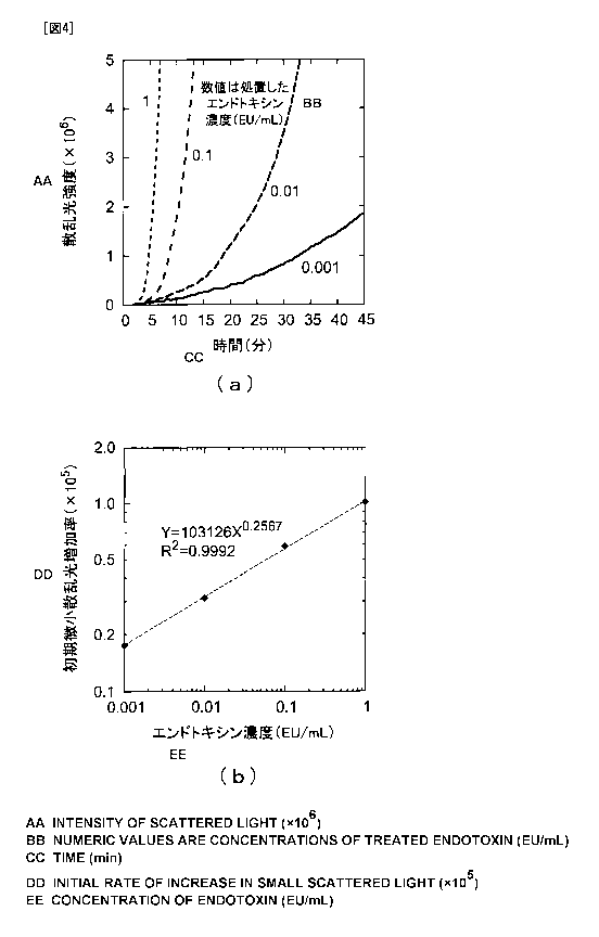
[0090] <Example 6>
An apparatus was produced in the same way as in Example 1 except
that a CCD area sensor was used instead of the photodiode used as
the scattered light receiving element 6, to thereby, similarly to
Example 5, examine a relationship between the concentration of
endotoxin and the increase rate of initial scattered light. FIG.
4 (b) shows the results. In FIG. 4 (b) , the horizontal axis represents
the concentration of endotoxin (EU/mL), and the vertical axis
represents the increase rate of initial small scattered light (the
slope of the rise curve of initial scattered light).
FIG. 4 (a) further shows the temporal changes in the intensity
of scattered light. In FIG. 4(a), the horizontal axis represents
the time (min) , and the vertical axis represents the scattered light
intensity obtained based on the output of the CCD area sensor. The
graph also shows that, also in the case of using the CCD area sensor,
the initial scattered light increases linearly like the phase A
in FIG. 2 and further changes to the phase of a larger increase
rate (the phase shown by the ellipse B in FIG. 2) via the folding
point.
[0091] <Example 7>
An examination was performed in the same way as in Example
37
CA 02732011 2011-01-25
1 using (3-D-glucan instead of endotoxin. P-Glucan Test Wako
(manufactured by Wako Pure Chemical Industries, Ltd.) was used as
the LAL reagent. 3-D-glucan dilution series were prepared at a
variety of concentrations to examine a relationship between the
concentration of (3-D-glucan and the increase rate of initial
scattered light generated from coagulin monomers and small coagulin
aggregates. As the result , it was found that the increase rate became
lower as the concentration of (3-D-glucan became lower, and the
increase rate became higher as the concentration became higher.
When the relationship between the concentration of (3-D-glucan and
the increase rate of the initial scattered light was plotted in
a double logarithmic graph, a linear relation was obtained as shown
in FIG. 5. In FIG. 5, the horizontal axis represents the
concentration of (3-D-glucan (pg/mL) , and the vertical axis represents
the increase rate of initial small scattered light (the slope of
the rise curve of initial scattered light).
[0092] Here, a measurement apparatus including the entire of
the measurement system 1 for the predetermined physiologically
active substance as shown in FIG. 1 may be configurated. In such
case, the above-mentioned measurement can be automatically performed
only byintroducing a mixture of a sample containing the predetermined
physiologically active substance and LAL into the sample cell 4
and providing direction of measurement starting. Moreover, in the
calculation apparatus 9, the concentration of the predetermined
physiologically active substance may be calculated based on the
38
CA 02732011 2011-01-25
calibration curves obtained in FIGS. 3, 4, and 5 and the increase
rate obtained from scattered light from the mixture, and the results
may be automatically displayed by the display 10.
[0093] In such case, the sample cell 4 corresponds to mixture
retaining means, the light source 2 and optical system for incident
light 3 correspond to light emitting means, the optical system for
outgoing light 5 and light receiving element 6 correspond to light
receiving means, and the calculation apparatus 9 corresponds to
derivation means. In addition, the stir bar 11 and the magnetic
stirrer 12 correspond to stirring apparatuses.
[0094] It should be noted that the above-mentioned Examples
according to the present invention have the following merits: 1)
a general limulus reagent used in the turbidimetric method can be
used without further treatments; 2) the configuration of the
measurement system (measurement apparatus) can be simplified, and
multi -channelization (8 tol6ch) can be relatively easily performed;
and 4) measurement can be completed in almost the same time period
as that in the case of the colorimetric method using a special reagent.
Reference Signs List
[0095] 1 measurement system
2 light source
3 optical system for incident light
4 sample cell
optical system for outgoing light
6 light receiving element
39
CA 02732011 2011-01-25
7 amplifier circuit
8 filter for removing noises
9 calculation apparatus
display
11 stir bar
12 magnetic stirrer