Note: Descriptions are shown in the official language in which they were submitted.
CA 02732313 2011-01-27
WO 2010/014707 PCT/US2009/052101
METHOD AND APPARATUS FOR STRAIGHTENING AND FLATTENING
THE SIDE WALL OF A BODY LUMEN OR BODY CAVITY SO AS TO
PROVIDE THREE DIMENSIONAL EXPOSURE OF A LESION OR
ABNORMALITY WITHIN THE BODY LUMEN OR BODY CAVITY,
AND/OR FOR STABILIZING AN INSTRUMENT RELATIVE TO THE
SAME
Inventors
Jeffrey Milsom
Howard Riina
John Fredrick Cornhill
Robert Andrews
Edward Dickinson
Reference To Pending Prior Patent Application
This patent application claims benefit of pending
prior U.S. Provisional Patent Application Serial No.
61/137,361, filed 07/30/2008 by Jeffrey Milsom et al.
for METHOD AND APPARATUS FOR STRAIGHTENING AND
FLATTENING THE SIDE WALL OF A BODY LUMEN OR BODY
CAVITY SO AS TO PROVIDE THREE DIMENSIONAL EXPOSURE OF
CA 02732313 2011-01-27
WO 2010/014707 PCT/US2009/052101
- 2 -
A LESION OR ABNORMALITY WITHIN THE BODY LUMEN OR BODY
CAVITY, AND/OR FOR STABILIZING AN INSTRUMENT RELATIVE
TO THE SAME (Attorney's Docket No. CORN-0311 PROV),
which patent application is hereby incorporated herein
by reference.
Field Of The Invention
This invention relates to surgical methods and
apparatus in general, and more particularly to
surgical methods and apparatus for straightening and
flattening the side wall of a body lumen or body
cavity so as to provide three dimensional exposure of
a lesion or abnormality within the body lumen or body
cavity, and/or for stabilizing an instrument relative
to the same.
Background Of The Invention
The human body comprises many different lumens
and cavities. By way of example but not limitation,
the human body comprises lumens such as the
gastrointestinal (GI) tract, blood vessels, lymph
CA 02732313 2011-01-27
WO 2010/014707 PCT/US2009/052101
- 3 -
nodes, the ureter, etc. By way of further example but
not limitation, the human body comprises cavities such
as the abdomen, the chest, the nasal sinuses, the
bladder, etc.
In many cases, it may be desirable to
endoscopically examine and/or treat a disease process
or abnormality within or on the side wall of a body
lumen and/or body cavity. By way of example but not
limitation, it may be desirable to examine the lumen
or side wall of the gastrointestinal tract for lesions
and, if a lesion is found, to biopsy, remove, and/or
treat the lesion.
The endoscopic examination and/or treatment of
the side wall of a body lumen and/or body cavity can
be complicated by the geometry of the side wall of the
body lumen or body cavity. By way of example but not
limitation, the intestine is an elongated organ having
an inner lumen characterized by frequent turns and
side walls characterized by numerous folds. It can be
difficult to examine and/or treat a lesion formed on
CA 02732313 2011-01-27
WO 2010/014707 PCT/US2009/052101
- 4 -
the side wall of the intestine due to this varying
side wall geometry.
It would be advantageous to provide an endoscopic
device capable of straightening and flattening the
side wall of a body lumen or body cavity so as to
better present the side wall tissue for examination
and/or treatment during an endoscopic procedure.
It would also be advantageous to provide an
endoscopic device capable of steadying, or maintaining
in a fixed position, the tip(s) or working end(s) of
an instrument (or instruments) inserted into a body
space.
Summary Of The Invention
The present invention comprises the provision and
use of a novel endoscopic device capable of
straightening and flattening the side wall of a body
lumen or body cavity so as to better expose or present
the side wall tissue in all dimensions for examination
and/or treatment during an endoscopic procedure.
CA 02732313 2011-01-27
WO 2010/014707 PCT/US2009/052101
- 5 -
The present invention also comprises the
provision and use of a novel endoscopic device capable
of steadying or stabilizing the tip or working end of
an instrument or several instruments.
In one preferred form of the present invention,
there is provided apparatus for straightening and
flattening a side wall of a body lumen or body cavity
so as to provide three dimensional exposure of a
lesion or abnormality within the body lumen or body
cavity, the apparatus comprising:
a deployable hoop expander comprising a
longitudinally-extending structure defining a volume;
the deployable hoop expander being configured so
as to be transitionable between (i) a reduced cross-
sectional configuration, and (ii) an expanded cross-
sectional configuration, whereby the deployable hoop
expander can be configured in its reduced cross-
sectional configuration for easy insertion into a body
lumen, and it can thereafter be re-configured into its
expanded cross-sectional configuration so as to
engage, expose, straighten and flatten the side wall
CA 02732313 2011-01-27
WO 2010/014707 PCT/US2009/052101
- 6 -
of the body lumen, whereby to provide three
dimensional exposure of the lesion or abnormality
within the body lumen or body cavity.
In one preferred form of the present invention,
the deployable hoop expander comprises an open lattice
configuration allowing access to the lumen or side
wall of the body lumen or body cavity.
And in another preferred form of the present
invention, the deployable hoop expander is configured
so as to allow an instrument (or instruments) to dock
with the deployable hoop expander, whereby to permit
the deployable hoop expander to steady or stabilize
the tip (s) or working end (s) of the instrument(s).
In another preferred form of the present
invention, there is provided a method for
straightening and flattening a side wall of a body
lumen or body cavity so as to provide three
dimensional exposure of a lesion or abnormality within
the body lumen or body cavity, the method comprising:
providing a deployable hoop expander;
CA 02732313 2011-01-27
WO 2010/014707 PCT/US2009/052101
- 7 -
the deployable hoop expander comprising a
longitudinally-extending structure defining a volume;
and
the deployable hoop expander being
configured so as to be transitionable between (i) a
reduced cross-sectional configuration, and (ii) an
expanded cross-sectional configuration;
configuring the deployable hoop expander in its
reduced cross-sectional configuration;
advancing the deployable hoop expander into a
body lumen; and
re-configuring the deployable hoop expander into
its expanded cross-sectional configuration so as to
engage, expose, straighten and flatten the side wall
of the body lumen, whereby to provide three
dimensional exposure of the lesion or abnormality
within the body lumen or body cavity.
In one preferred form of the present invention,
the deployable hoop expander comprises an open lattice
configuration allowing access to the side wall of the
body lumen or body cavity.
CA 02732313 2011-01-27
WO 2010/014707 PCT/US2009/052101
- 8 -
And in one preferred form of the present
invention, the deployable hoop expander is configured
so as to allow an instrument (or instruments) to dock
with the deployable hoop expander, whereby to permit
the deployable hoop expander to steady or stabilize
the tip (s) or working end (s) of the instrument(s).
Brief Description Of The Drawings
These and other objects and features of the
present invention will be more fully disclosed or
rendered obvious by the following detailed description
of the preferred embodiments of the invention, which
is to be considered together with the accompanying
drawings wherein like numbers refer to like parts and
further wherein:
Figs. 1-3 are schematic views showing a novel
endoscopic device formed in accordance with the
present invention, wherein the novel endoscopic device
is being deployed in a body lumen so as to straighten
and flatten the side wall of the body lumen so as to
provide three dimensional exposure of a lesion or
CA 02732313 2011-01-27
WO 2010/014707 PCT/US2009/052101
- 9 -
abnormality within the body lumen or body cavity
and/or to provide stability to the working end of an
instrument;
Figs. 4-12 are schematic views showing various
aspects of a novel endoscopic device comprising a
deployable hoop expander formed out of spring material
and constrained by an outer net;
Figs. 13 and 14 are schematic views showing
various aspects of a novel endoscopic device
comprising a deployable hoop expander formed out of
spring material and constrained by an outer sleeve;
Figs. 15-17 are schematic views showing various
aspects of a novel endoscopic device comprising a
deployable hoop expander erected with control wires;
Figs. 18-20 are schematic views showing the use
of a grasping tool for repositioning or removing the
deployable hoop expander; and
Figs. 21 and 22 are schematic views showing
delivery of the deployable hoop expander vis-a-vis an
endoscope or other device.
CA 02732313 2011-01-27
WO 2010/014707 PCT/US2009/052101
- 10 -
Detailed Description Of The Present Invention
The Novel Endoscopic (Or Surgical) Device In General
The present invention comprises the provision and
use of a novel endoscopic (or surgical) device capable
of straightening and flattening the side wall of a
body lumen or body cavity so as to better present the
side wall tissue for examination and/or treatment
during an endoscopic or surgical procedure and/or to
stabilize an instrument relative to the same.
More particularly, the present invention
comprises the provision and use of a novel
endoscopic/surgical device for facilitating the
alignment and presentation of the side wall of a body
lumen or body cavity during an endoscopic or other
surgical procedure, and/or for stabilizing an
instrument relative to the same. In this respect, the
term "endoscopic procedure" is intended to mean
substantially any minimally-invasive procedure,
diagnostic or surgical, for accessing the inside of a
vessel or organ within the body for purposes of
CA 02732313 2011-01-27
WO 2010/014707 PCT/US2009/052101
- 11 -
viewing, biopsying and/or treating the tissue
(including removing a lesion), and the term surgical
procedure is intended to mean substantially any
medical operative procedure performed on the body.
The novel endoscopic/surgical device of the present
invention (hereinafter sometimes referred to, for
convenience, as simply an endoscopic device) is
adapted to straighten and flatten bends and/or curves
and/or folds in the side wall of the vessel or organ,
and can provide a platform for the performance of
numerous procedures within the vessel or organ,
including the possible docking and/or fixation of an
endoscope and/or other surgical instruments within the
vessel or organ.
In accordance with the present invention, and
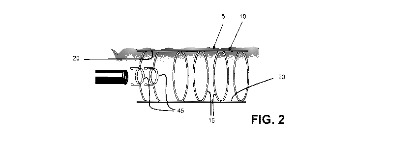
looking now at Figs. 1-3, there is shown an endoscopic
device 5 capable of straightening and flattening the
side wall of a body lumen or body cavity so as to
better present the side wall tissue for examination
and/or treatment during an endoscopic procedure,
and/or for stabilizing an instrument relative to the
CA 02732313 2011-01-27
WO 2010/014707 PCT/US2009/052101
- 12 -
same. More particularly, endoscopic device 5
generally comprises a deployable hoop expander 10
which generally comprises a plurality of parallel
rings 15 connected together by a plurality of struts
20. Deployable hoop expander 10 is configured so as
to be transitionable between (i) a reduced cross-
sectional configuration (Fig. 1) wherein the planes of
the parallel rings 15 are disposed at an acute angle
(when seen in side view) to the longitudinal axis of
deployable hoop expander 10, and (ii) an expanded
cross-sectional configuration (Fig. 2) wherein the
planes of the parallel rings 15 are disposed
perpendicular (when seen in side view) to the
longitudinal axis of deployable hoop expander 10. As
a result of this construction, deployable hoop
expander 10 can be configured in its reduced cross-
sectional configuration for easy insertion into a body
lumen (e.g., a vascular lumen, an organ lumen, etc.)
(Fig. 1), and it can thereafter be re-configured
20 into its expanded cross-sectional configuration so as
to engage, expose, straighten and flatten the side
CA 02732313 2011-01-27
WO 2010/014707 PCT/US2009/052101
- 13 -
wall 30 of body lumen 25 (Fig. 2). This engagement,
straightening and flattening of side wall 30 of body
lumen 25 better presents the side wall tissue for
examination and/or treatment during an endoscopic
procedure, e.g., such as one utilizing an endoscopic
instrument 35 (Fig. 3). By way of example but not
limitation, endoscopic instrument 35 may comprise an
endoscope carrying an extendable biopsy device 40
therein. Preferably deployable hoop expander 10 has
an open lattice configuration allowing access to the
side wall of the body lumen or body cavity. In one
preferred form of the invention, deployable hoop
expander 10 comprises a polygonal structure.
As will hereinafter be discussed in further
detail, deployable hoop expander 10 is preferably also
configured so as to allow an instrument (e.g.,
endoscopic instrument 35) to dock with deployable hoop
expander 10, whereby to permit the deployable hoop
expander to steady or stabilize the tip(s) or working
end(s) of the instrument (or instruments).
CA 02732313 2011-01-27
WO 2010/014707 PCT/US2009/052101
- 14 -
Parallel rings 15 can have a circular
configuration (in which case deployable hoop expander
will have a cylindrical configuration when
expanded), an oval configuration (in which case
5 deployable hoop expander 10 will have an ovoid shape
when expanded), or other shapes which may be
determined by the organ or cavity into which
deployable hoop expander 10 is inserted, etc.
Furthermore, parallel rings 15 can have diameters
10 which vary along the length of deployable hoop
expander 10, such that the deployable hoop expander
can form a particular non-cylindrical or non-ovoid
geometry when it is in its expanded cross-sectional
configuration. By way of example but not limitation,
parallel rings 15 can have various diameters such that
the deployable hoop expander expands into a spherical
configuration.
Additionally, deployable hoop expander 10 can be
closed at its distal end, and/or partially closed at
its proximal end, and/or partially closed at its
proximal and distal ends, if desired.
CA 02732313 2011-01-27
WO 2010/014707 PCT/US2009/052101
- 15 -
Preferably, the proximal end of deployable hoop
expander 10 comprises one or more guides 45 for
receiving the distal end of endoscopic instrument 35
relative to deployable hoop expander 10. Among other
things, guides 45 can be configured so as to allow
endoscopic instruments 35 to dock with deployable hoop
expander 10, so that guides 45 can act to steady or
stabilize endoscopic instruments 35 relative to
deployable hoop expander 10 during an endoscopic
procedure. In this respect it will be appreciated
that providing a stable support platform for
endoscopic instrument 35 can greatly facilitate
stabilizing and aligning an instrument relative to the
anatomy when conducting tissue inspection, biopsy
and/or removal. This stable support platform can also
enable and/or facilitate more extensive surgical
procedures such as full thickness bowel resection
(wall excision or segmental resection) and/or repair
during intestinal procedures, peritoneal exploration
(including natural orifice trans-endoscopic surgical
procedures, which are sometimes referred to as NOTES),
CA 02732313 2011-01-27
WO 2010/014707 PCT/US2009/052101
- 16 -
treatment of obstructions, and/or other complex
endoscopic surgical procedures.
The expansible nature of deployable hoop expander
is such that it may be "sprung open" (i.e.,
5 transitioned from its reduced cross-sectional
configuration to its expanded cross-sectional
configuration) by a variety of means. Among other
things, and as will hereinafter be discussed in
further detail, deployable hoop expander 10 can be
10 formed out of a spring material (e.g., a shape memory
alloy such as Nitinol, a hardened stainless steel
wire, a flexible plastic such as a self-expanding
polymer, etc.) and constrained by an outer net; or
deployable hoop expander 10 can be formed out of a
spring material and constrained by an outer sheath; or
deployable hoop expander 10 can be manipulated between
its reduced cross-sectional configuration and its
expanded cross-sectional configuration by manipulating
control wires connected to struts 20; or by pinching
one end of the device with a specialized tool, thereby
elongating and narrowing the device, etc.
CA 02732313 2011-01-27
WO 2010/014707 PCT/US2009/052101
- 17 -
Deployable Hoop Expander Formed Out Of Spring
Material And Constrained By An Outer Net
More particularly, and looking now at Figs. 4 and
5, there is shown a configuration in which parallel
rings 15 of deployable hoop expander 10 are formed out
of a spring material (e.g., a shape memory alloy such
as Nitinol, a hardened stainless steel wire, a
flexible plastic such as a self-expanding polymer,
etc.) and constrained by an outer net 50. In one
preferred form of the invention, deployable hoop
expander 10 is mounted over endoscopic instrument 35,
with deployable hoop expander 10 being maintained in
its reduced cross-sectional configuration using outer
net 50. Alternatively, deployable hoop expander 10
may be maintained in its reduced cross-sectional
configuration via outer net 50 and the entire assembly
delivered through a lumen of endoscopic instrument 35;
or deployable hoop expander 10 may be inserted
alongside of, or entirely separately from, endoscopic
instrument 35. In any case, in use, after deployable
CA 02732313 2011-01-27
WO 2010/014707 PCT/US2009/052101
- 18 -
hoop expander 10 has been delivered to the therapeutic
site, the net is withdrawn by pulling the net pull
wires 55 (Figs. 6 and 7), thereby exposing and
releasing parallel rings 15. Upon net removal,
parallel rings 15 automatically expand (Fig. 8) so as
to engage the side wall of the body lumen and thereby
expose, straighten and flatten the side wall tissue.
At the conclusion of the procedure, deployable hoop
expander 10 may be returned to its reduced cross-
sectional configuration and removed from the body
lumen, or it may under certain circumstances remain in
place.
In another form of the present invention, and
looking now at Figs. 9-11, net 50 may be loosened so
as to release the restrained deployable hoop expander
10, thereby allowing parallel rings 15 to expand, but
the net is still kept in place around the expanded
deployable hoop expander (Fig. 9). As a result, by
forming net 50 with a closed distal end, the net can
sit as a "trap" about the therapeutic site so as to
catch and retain anything which may be removed or
CA 02732313 2011-01-27
WO 2010/014707 PCT/US2009/052101
- 19 -
dislodged from the side wall of the body lumen during
a procedure. Thereafter, at the conclusion of the
procedure, deployable hoop expander 10 can be returned
to its reduced cross-sectional configuration and
removed from the body lumen, and then net 50 can be
withdrawn from the body lumen (Figs. 10 and 11),
carrying with it any tissue or debris removed from the
side wall of the body lumen and entrapped by the net.
In this form of the invention, it may be desirable to
form net 50 with a distal end reservoir 58 (Fig. 12)
for storing excised tissue (e.g., biopsy specimens) or
dislodged debris.
Deployable Hoop Expander Formed Out Of Spring
Material And Constrained By An Outer Sleeve
In another form of the invention, and looking now
at Figs. 13 and 14, there is provided a configuration
in which deployable hoop expander 10 is formed out of
a spring material (e.g., a shape memory alloy such as
Nitinol, a hardened stainless steel wire, a flexible
plastic such as a self-expanding polymer, etc.) and
CA 02732313 2011-01-27
WO 2010/014707 PCT/US2009/052101
- 20 -
constrained by an outer sleeve 60. In one preferred
form of the invention, deployable hoop expander 10 is
mounted over endoscopic instrument 35, with deployable
hoop expander 10 being maintained in its reduced
cross-sectional configuration using outer sleeve 60.
Alternatively, deployable hoop expander 10 may be
maintained in its reduced cross-sectional
configuration via outer sleeve 60 and the entire
assembly delivered through a lumen of endoscopic
instrument 35. Or deployable hoop expander 10 may be
inserted alongside of, or entirely separately from,
endoscopic instrument 35. In any case, in use, after
deployable hoop expander 10 has been delivered to the
therapeutic site, the outer sleeve is removed, thereby
releasing parallel rings 15. With outer sleeve 60
removed, parallel rings 15 automatically expand so as
to engage the side wall of the body lumen and
straighten and flatten the same.
Deployable Hoop Expander Erected With Control Wires
CA 02732313 2011-01-27
WO 2010/014707 PCT/US2009/052101
- 21 -
Looking next at Figs. 15-17, there is shown a
configuration in which deployable hoop expander 10 can
be manipulated by control wires 65, 70 connected to
struts 20 so as to expand parallel rings 15. In one
preferred form of the invention, deployable hoop
expander 10 is mounted over an endoscopic instrument
(not shown in Figs. 15-17), with the deployable hoop
expander being maintained in its reduced cross-
sectional configuration. Alternatively, deployable
hoop expander 10 could be delivered through a lumen of
an endoscopic instrument, again with the deployable
hoop expander being maintained in its reduced cross-
sectional configuration. Or deployable hoop expander
10 may be inserted alongside of, or entirely
separately from, an endoscopic instrument. In any
case, in use, after deployable hoop expander 10 has
been delivered to the therapeutic site, the deployable
hoop expander is erected by pulling distally on
control wire 65. This action erects the deployable
hoop expander so that it assumes its expanded cross-
sectional configuration (see Figs. 15-17).
CA 02732313 2011-01-27
WO 2010/014707 PCT/US2009/052101
- 22 -
Significantly, in this form of the invention,
deployable hoop expander 10 does not need to be formed
out of a resilient material, since the deployable hoop
expander is erected by pulling on control wire 65, and
is not erected by the resilient nature of the
deployable hoop expander itself.
Additionally, deployable hoop expander 10 can be
collapsed by releasing the previously-pulled control
wire 65. Deployable hoop expander 10 can then be
moved by pulling on both control wires 65, 70. Once
deployable hoop expander has been properly re-
positioned, it can be expanded again simply by pulling
on control wire 65.
It should be appreciated that, in the
configurations shown in Figs. 15-17, struts 20 are
preferably connected to parallel rings 15 with a hinge
arrangement which allows the struts to pivot relative
to parallel rings 15. This hinge could be a pinned
hinge, a living hinge, etc.
Forming The Deployable Hoop Expander
CA 02732313 2011-01-27
WO 2010/014707 PCT/US2009/052101
- 23 -
With A Helical Construction
In another form of the invention, deployable hoop
expander 10 may be formed with a helical construction.
More particularly, in this form of the invention, the
plurality of parallel rings 15 may be replaced by a
helix, a double helix, another form of spiral, or
another collapsible/expandable polygonal structure
defining a volume. Where deployable hoop expander 10
is formed out of a helix, double helix or another form
of spiral, struts 20 may or may not be provided.
Regardless of the particular construction chosen
for the deployable hoop expander, preferably at least
a portion of the deployable hoop expander has an open
configuration (e.g., an open lattice configuration)
allowing access to the side wall of the body lumen or
body cavity.
Use Of Grasping Tool For Re-positioning
Or Removing The Deployable Hoop Expander
Looking next at Figs. 18-20, deployable hoop
expander 10 may also be retracted (e.g., for re-
CA 02732313 2011-01-27
WO 2010/014707 PCT/US2009/052101
- 24 -
positioning or complete removal) with a specialized
grasping tool 75. More particularly, specialized
grasping tool 75 comprises a hook 80 which can grasp
the end-most parallel ring 15 of the deployable hoop
expander so as to cause the entire device to elongate
and narrow, thereby making it re-positionable or
removable.
Delivery Of The Deployable Hoop Expander
Vis-a-vis An Endoscope Or Other Device
As noted above, deployable hoop expander 10 is
designed such that it can be delivered over an
endoscope or other instrument, or through the working
channel of an endoscope or other instrument, or it can
be delivered alongside an endoscope or other
instrument, or entirely outside of or separate from an
endoscope or other instrument. See, for example,
Figs. 21 and 22, which show deployable hoop expander
10 disposed in a channel 85 disposed parallel to
endoscopic instrument 35. Channel 85 may be affixed
CA 02732313 2011-01-27
WO 2010/014707 PCT/US2009/052101
- 25 -
to endoscopic instrument 35 in various ways, e.g.,
with an optional snap-on ring 90.
Maintaining The Deployable Hoop Expander In The
Body At The Conclusion Of An Endoscopic Procedure
It is anticipated that, in most cases, deployable
hoop expander 10 will be removed from the patient at
the conclusion of the endoscopic procedure. However,
in some cases it may be desirable to provide support
to the vessel or organ for some period of time after
the conclusion of the endoscopic procedure. In this
case, deployable hoop expander 10 may be left in the
body lumen at the conclusion of the procedure and
thereafter, when support is no longer required, the
deployable hoop expander can be removed from the body.
Optionally, deployable hoop expander 10 can be formed
out of a biodegradable/absorbable material. In this
case, the device can be left in the body at the
conclusion of the endoscopic procedure, whereupon it
will thereafter biodegrade or be absorbed by the body.
CA 02732313 2011-01-27
WO 2010/014707 PCT/US2009/052101
- 26 -
Applications
The novel endoscopic/surgical device of the
present invention can be used in substantially any
endoscopic or surgical procedure to facilitate the
alignment and presentation of tissue during an
endoscopic procedure or to fix, dock, or stabilize the
working end of an endoscope or other instrument during
such a procedure.
The present invention is believed to have widest
applications with respect to the gastrointestinal (GI)
tract, which is generally characterized by frequent
turns and which has a side wall characterized by
numerous folds. However, the methods and apparatus of
the present invention may also be used inside other
body cavities (e.g., the cranium, thorax, abdomen,
pelvis, nasal sinuses, chest, bladder, etc.) and other
tubular viscera (e.g., the esophagus, stomach,
duodenum, vagina, ureter, fallopian tubes, urethra,
blood vessels, bronchi, etc.).
Thus, for example, the novel endoscopic device of
the present invention can be used in the performance
CA 02732313 2011-01-27
WO 2010/014707 PCT/US2009/052101
- 27 -
of certain specialized endoscopic procedures including
Natural Orifice Trans-Endoscopic Surgery (NOTES)
procedures, as well as other complex endoscopic
procedures which could involve endoscopic surgery.
Modifications
While the present invention has been described in
terms of certain exemplary preferred embodiments, it
will be readily understood and appreciated by one of
ordinary skill in the art that it is not so limited,
and that many additions, deletions and modifications
may be made to the preferred embodiments discussed
above while remaining within the scope of the present
invention.