Note: Descriptions are shown in the official language in which they were submitted.
CA 02732787 2011-02-01
WO 2010/017537 PCT/US2009/053237
-1-
APPARATUS AND METHODS FOR ACCESSING AND REMOVING MATERIAL
FROM BODY LUMENS
FIELD OF THE INVENTION
The present invention relates generally to apparatus for treating obstructive
material, e.g., thrombus, stenosis, and/or other obstructions within a body
lumen of a
patient, e.g., within a tubular graft, aorto-venous fistula, blood vessel, and
the like. More
particularly, the present invention relates to apparatus for removing or
otherwise capturing
thrombus or other obstructive material within a body lumen, and/or for
dilating a body
lumen, and to methods for making and using such apparatus.
BACKGROUND
Flow within a blood vessel or other body lumen within a patient's vasculature
may
become constricted or ultimately interrupted for a variety of reasons. For
example, a
vessel may gradually narrow due to inflammation and/or cell proliferation. In
addition,
thrombus may form due to such narrowing or other flow problems within a
vessel.
For example, an aorto-venous graft may be implanted in an arm of a patient
experiencing kidney failure, e.g., to facilitate dialysis treatment. Such
grafts may be a
fistula formed directly in the patient's body, e.g., through tissue between an
adjacent artery
and vein or other vessels, may be a xenograft implanted between two vessels,
or may be a
synthetic graft. Such grafts only have a limited life cycle due to
inflammation, thrombus
formation, and the like. Once such a graft becomes sufficiently occluded or
otherwise
deteriorates, a new graft must be implanted at a new location for subsequent
treatment.
Accordingly, apparatus and methods for removing material from aorto-venous
grafts, blood vessels, or other body lumens and/or otherwise treating body
lumens would
be useful.
SUMMARY
The present invention is directed to apparatus for treating a body lumen of a
patient, e.g., a tubular graft, aorto-venous fistula, blood vessel, and the
like. More
particularly, the present invention is directed to apparatus for removing or
otherwise
capturing thrombus or other obstructive material within a body lumen, and/or
for dilating a
body lumen, and to methods for making and using such apparatus.
CA 02732787 2011-02-01
WO 2010/017537 PCT/US2009/053237
-2-
In accordance with one embodiment, a system is provided for removing
obstructive material from a body lumen that includes an elongate tubular
member
including a proximal end, a distal end sized for introduction into a body
lumen, a lumen
extending between the proximal and distal ends, and an elongate treatment
member
insertable through the lumen and including an expandable treatment element
selectively
expandable for treating one or more regions within a body lumen. For example,
the
treatment element may be used to direct obstructive material into the tubular
member
lumen.
In an exemplary embodiment, the tubular member may include an annular
expandable member on the distal end that is expandable from a collapsed
configuration to
an expanded configuration wherein the expandable member adopts a tapered shape
that
flares outwardly away from the distal end to define an inlet that is larger
than and
communicates with the lumen, the expandable member sized to engage a wall of a
body
lumen within which the expandable member is expanded.
For example, the expandable member may include an outer annular membrane
surrounding an inner annular membrane, the outer and inner membranes being
attached
together and to the tubular member distal end such that the outer and inner
membranes
together define a substantially enclosed interior. The tubular member may
include an
inflation lumen communicating with the interior such that, when inflation
media is
delivered via the inflation lumen into the interior, the expandable member is
expanded to
the expanded configuration.
In an exemplary embodiment, the outer membrane may have a greater axial length
than the inner member such that proximal ends of the outer and inner membranes
are
spaced apart from one another along the distal end of the tubular member. For
example,
the proximal end of the outer membrane may be attached to the distal end of
the tubular
member at a first location and the proximal end of the inner membrane may be
attached to
the distal end of the tubular member at a second location distal to the first
location.
In addition or alternatively, the inner membrane may be more resistant to
bending
or compression than the outer membrane. For example, the inner membrane may
include
a reinforcement layer to increase the resistance of the inner membrane to
bending or
compression.
Optionally, the tubular member may include an access port on the proximal end
providing access to the lumen for removing material therefrom.
CA 02732787 2011-02-01
WO 2010/017537 PCT/US2009/053237
-3-
In another option, the system may include one or more additional components.
For
example, a dilator may be disposed within the lumen of the tubular member such
that a
tapered distal tip extends beyond the expandable member in the collapsed
configuration to
provide a substantially smooth transition during introduction of the tubular
member into a
body lumen. In an exemplary embodiment, the dilator may include a recess
adjacent the
distal tip for receiving a distal end of the expandable member in the
collapsed
configuration. The dilator may be removable proximally from the tubular member
lumen
after the tubular member is introduced into a body lumen such that the
treatment device
may be inserted into the tubular member lumen.
Optionally, the tubular member may include an expandable section including at
least the distal end that is expandable from a relaxed size to an enlarged
size to
accommodate receiving material larger than the relaxed state directed into the
lumen by
the treatment device. For example, the tubular member may be expandable along
the
entire length between the proximal and distal ends.
In an exemplary embodiment, the expandable section of the tubular member may
include an elastic layer coupled to an inelastic layer including one or more
slots therein to
accommodate the inelastic layer expanding, the elastic layer biasing the
expandable
section to return towards the relaxed size. For example, the elastic layer may
include an
elastic sleeve surrounding the inelastic layer. In addition or alternatively,
the inelastic
layer may include a tubular body and wherein the one or more slots comprise
one or more
longitudinal slots extending along the length of the tubular body.
Alternatively, the
inelastic layer may include a tubular body and the one or more slots may
include a
plurality of longitudinal slots spaced apart axially and circumferentially
from one another
along the length of the tubular body.
In any of these embodiments, the treatment element of the treatment device may
include a first non-compliant balloon mounted on a distal end of the treatment
device. In
addition or alternatively, a second compliant balloon may be mounted on
treatment device,
e.g., over the first balloon.
In addition or alternatively, the treatment device comprises a traction member
at
least partially covering the treatment element. For example, the traction
member may be
movable between a first position wherein the traction member is disposed
adjacent the
treatment element and a second position wherein the traction member at least
partially
CA 02732787 2011-02-01
WO 2010/017537 PCT/US2009/053237
-4-
covers the treatment element. In an exemplary embodiment, the traction member
may
include an expandable mesh.
In accordance with another embodiment, an apparatus is provided for providing
access to a body lumen that includes an elongate tubular member comprising a
proximal
end, a distal end sized for introduction into a body lumen, and a lumen
extending between
the proximal and distal ends; and an annular expandable member on the distal
end that is
expandable from a collapsed configuration to an expanded configuration wherein
the
expandable member adopts a tapered shape that flares outwardly away from the
distal end
to define an inlet that is larger than and communicates with the lumen, the
expandable
member sized to engage a wall of a body lumen within which the expandable
member is
expanded.
In accordance with yet another embodiment, an apparatus is provided for
removing
obstructive material from a body lumen and for dilating the body lumen. For
example, the
apparatus may include an outer tubular member having a proximal end, an
expandable
distal end, and a first lumen extending between the proximal and distal ends.
The
apparatus may also include an inner member comprising a proximal end and a
distal end
having a tapered profile, wherein the inner member is configured for being
slidably
disposed within the first lumen of the outer member such that the inner member
proximal
end extends proximally from the outer member proximal end and the inner member
distal
end extends distally from the outer member distal end. Optionally, the inner
member may
have an undercut configured for engaging the outer member distal end when the
outer
member distal end is in an unexpanded configuration. In addition or
alternatively, the
apparatus may include an elongate member configured for being slidably
disposed within
the first lumen of the outer member. The elongate member may include two
concentric
balloons on a distal end thereof, wherein one of the balloons is more
compliant than the
other balloon. For example, the less compliant balloon may be configured for
dilating the
body lumen.
In one embodiment, the distal end of the outer member may include an inner
membrane, an outer membrane, and a cavity between the inner and outer
membranes.
Optionally, the inner membrane may include a reinforcement layer that allows
radial
expansion and resists bending and/or compression in an axial direction. The
inner
membrane may have a shorter length than the outer membrane or they may have
similar
CA 02732787 2011-02-01
WO 2010/017537 PCT/US2009/053237
-5-
lengths. The outer member may further include a second lumen extending between
the
outer member proximal and distal ends and communicating with the cavity.
The outer member expandable distal end may have an expanded configuration
wherein a distal opening of the outer member has a first diameter, and the
distal end tapers
from the first diameter proximally towards a second diameter, wherein the
first diameter is
larger than the second diameter. For example, the first diameter may be
adjustable such
that the distal opening may be substantially equal to an inner diameter of a
body lumen
within which the expandable distal end is expanded.
In accordance with still another embodiment, an apparatus is provided for
treating
a body lumen. The apparatus includes an tubular sheath having a proximal end,
an
expandable distal end having an expanded configuration and an unexpanded
configuration,
a shaft extending between the proximal and distal ends, and a first lumen
extending
between the proximal end and a distal opening in the distal end, wherein, when
the distal
end is in the expanded configuration, the distal end tapers from a first
diameter at the distal
opening to a second diameter of the shaft, wherein the first diameter is
larger than the
second diameter.
Optionally, the apparatus may also include a dilator slidably disposed within
the
first lumen of the sheath. The dilator includes a proximal end adjacent the
sheath
proximal end, a distal end extending from the sheath distal end and having a
tapered distal
tip, and an annular region adjacent the distal tip for receiving the sheath
distal end when
the sheath distal end is in the unexpanded configuration, e.g., to provide a
substantially
smooth transition between the distal tip and the sheath.
In accordance with yet another embodiment, an apparatus for treating a body
lumen is provided that includes a tubular member including a proximal end, an
expandable
distal end, a first lumen extending between the proximal and distal ends, and
a second
inflation lumen extending between the proximal and distal ends, wherein the
distal end, in
an expanded configuration, has a tapered profile that tapers from a first
diameter at a distal
opening proximally towards a second diameter, wherein the first diameter is
greater than
the second diameter.
In one embodiment, the tubular member distal end may include an inner
membrane, an outer membrane, and a cavity between the inner membrane and the
outer
membrane, and the second lumen may communicate between the cavity and an
inflation
port on the tubular member proximal end. Optionally, the inner membrane may
include a
CA 02732787 2011-02-01
WO 2010/017537 PCT/US2009/053237
-6-
reinforcement layer. The reinforcement layer may be formed of a material that
is resistant
to bending and/or compression in an axial direction relative to the tubular
member. In
addition or alternatively, the outer membrane may have a length that is
greater than a
length of the inner membrane or the lengths may be substantially the same. For
example,
when the tubular member distal end is in the expanded configuration, the inner
membrane
may have a concave configuration and the outer membrane may have a convex
configuration.
Optionally, the apparatus may also include an inner member slidably disposed
within the first lumen of the tubular member. The inner member includes a
proximal end
adjacent the tubular member proximal end, and a distal end extending from the
tubular
member distal end and having a tapered distal tip. The inner member distal end
may
include an annular undercut having an opening facing towards the inner member
proximal
end, wherein the tubular member distal end, in an unexpanded configuration,
may fit
within the opening.
In addition or alternatively, the apparatus may further include an elongate
member
configured for being slidably disposed within the tubular member first lumen.
The
elongate member may include one or more expandable members on a distal end
thereof,
e.g., a single balloon or concentric balloons, e.g., where the outer balloon
is more
compliant than the inner balloon.
In accordance with another embodiment, a method for treating a body lumen is
provided that includes introducing a flow restoration apparatus into the body
lumen,
wherein the flow restoration apparatus comprises an outer member and an inner
member
disposed within a lumen of the outer member; expanding a distal end of the
outer member
from an unexpanded profile to a tapered, expanded profile, e.g., such that the
outer
member distal end engages or otherwise contacts an inner wall of the body
lumen; and
withdrawing the inner member from the outer member, thereby leaving the outer
member
in the expanded configuration disposed within the body lumen.
A procedure may then be performed via the outer member, e.g., to remove or
otherwise treat obstructive material within the body lumen. For example, an
elongate
treatment member may be introduced through the outer member and into the body
lumen
and manipulated to capture or remove material within the body lumen. In one
embodiment, a distal end of the treatment member may be advanced from the
expanded
distal end of the outer member and beyond material to be removed. A first
expandable
CA 02732787 2011-02-01
WO 2010/017537 PCT/US2009/053237
-7-
member on the treatment member distal end may be expanded, and the treatment
member
may be withdrawn towards the outer member to direct material in the body lumen
into the
outer member expanded distal end, e.g., by pulling the expanded first
expandable member
entirely into the outer member. Optionally, an additional obstruction within a
portion of
the body lumen may be located, and a second expandable member on the treatment
member distal end may be expanded to dilate the portion of the body lumen.
Other aspects and features of the present invention will become apparent from
consideration of the following description taken in conjunction with the
accompanying
drawings.
BRIEF DESCRIPTION OF THE DRAWINGS
It will be appreciated that the exemplary apparatus shown in the drawings are
not
necessarily drawn to scale, with emphasis instead being placed on illustrating
the various
aspects and features of the illustrated embodiments.
FIG. IA is a cross-sectional view of a first exemplary embodiment of a flow
restoration apparatus or system, including an outer sheath and a dilator,
disposed within a
body lumen.
FIG. I B is a detail of a distal portion of the apparatus shown in FIG. IA.
FIG. 2A is a cross-sectional view of the apparatus of FIG. IA, showing an
expandable member on the sheath in an expanded configuration within the body
lumen.
FIG. 2B is a detail of a wall of the expandable member shown in FIG. 2A.
FIG. 3 is a cross-sectional view of the apparatus of FIGS. IA and 2A with the
expandable member in the expanded configuration and the dilator removed.
FIG. 4 is a cross-sectional view of a patient's body showing an exemplary
dialysis
graft extending between a vein and an artery, and including obstruction
material within the
graft lumen.
FIGS. 5-10 are a cross-sectional views of the dialysis graft of FIG. 4,
showing an
exemplary method of removing obstructive material from the graft lumen.
FIGS. 11-12 are cross-sectional views of the dialysis graft of FIG. 4, showing
a
method for dilating an occluded region of the graft.
FIG. 13 is a perspective view of an alternative embodiment of a sheath that
may be
included in the flow restoration apparatus and methods of FIGS. 1A-12.
CA 02732787 2011-02-01
WO 2010/017537 PCT/US2009/053237
-8-
FIG. 14A is a side view of an alternative embodiment of a balloon catheter
that
may be used in the methods shown in FIGS. 5-12.
FIGS. 14B-14D are side views of another alternative embodiment of the balloon
catheter shown in FIG. 14A.
FIGS. 15A and 15B are cross-sectional views of a second exemplary embodiment
of a flow restoration apparatus including an outer sheath and a guidewire.
FIG. 16A is a cross-sectional view of a third embodiment of a flow restoration
apparatus including an outer sheath carrying an expandable member and having
an
expandable wall, and a balloon catheter.
FIG. 16B is a cross-section of the sheath of FIG. 16A taken along line 16B-
16B.
FIG. 16C is a detail of an alternative embodiment of the sheath of FIG. 16A,
including slots for providing an expandable wall.
DETAILED DESCRIPTION OF THE EXEMPLARY EMBODIMENTS
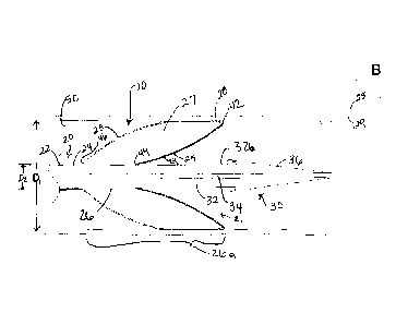
Turning to the drawings, FIGS. IA-3 show a flow restoration apparatus 10 for
providing access and/or treating a body lumen 50, e.g., for removing thrombus,
objects,
debris, and/or obstructive material from within the body lumen 50, and/or for
dilating
region of the body lumen 50, e.g., a blood vessel, aorto-venous fistula,
tubular graft, and
the like. Generally, the apparatus 10 includes an outer sheath or other
tubular member 20,
and, optionally, a dilator or other inner member 30, which together with one
or more
treatment apparatus (not shown), described further below, may provide a system
for
removing obstructive material and/or otherwise treating occluded regions
within body
lumens in a patient's body. In addition, such a system may include one or more
additional
components, e.g., one or more guidewires, syringes or other sources of
inflation media
and/or vacuum, and the like, such as guidewire 64 and syringe 18, as shown in
FIG. 7 and
described further below.
With additional reference to FIGS. 6-9, the sheath 20 is an elongate tubular
body
or shaft 22 including a proximal end 22a, a distal end 26, and a lumen 24
(best seen in
FIGS. IA-3) extending therebetween. The distal end 26 may be expandable, i.e.,
movable
between a collapsed configuration (as shown in FIGS. IA and 1B) and an
expanded
configuration (as shown in FIGS. 2A and 3), e.g., in which the expandable
distal end 26
has a tapered profile or transition. In the embodiment shown, the distal end
26 includes a
CA 02732787 2011-02-01
WO 2010/017537 PCT/US2009/053237
-9-
balloon or other expandable member 26a attached to and/or extending from the
distal end
26 of the shaft 22 to provide an expandable transition, as described further
below.
Optionally, a handle or hub 13 may be coupled to or otherwise provided on the
proximal end 22a of the sheath 20, e. g., for manipulating the sheath 20
and/or the entire
apparatus 10. The handle 13 may have an ergonomic shape, e.g., to facilitate
holding
and/or manipulating the handle 13. The handle 13 may include one or more
ports, e.g.,
side port 14, for coupling one or more fluid sources to the apparatus 10, such
as a source
of inflation media, a source of vacuum, and/or a source of diagnostic and/or
therapeutic
agents (not shown). The side port 14 may include one or more connectors (not
shown) to
facilitate coupling one or more sources of fluid to the side port 14, e.g., a
Luer lock
connector, and/or one or more seals, e.g., a hemostatic seal, to prevent fluid
from leaking
from the side port 64. In addition, the handle 13 may include a port 15
communicating
with the lumen 24, e.g., for receiving the dilator 30, a guidewire or other
device (not
shown) into and/or through the lumen 24. The port 15 may include one or more
seals,
e.g., a hemostatic seal (not shown), to accommodate passage of the dilator 30
or other
device therethrough without risk of substantial risk of leakage of blood or
other body
fluids from the lumen 24.
The sheath 20 may have a substantially uniform construction along its length,
or
alternatively, the construction may be varied. For example, a proximal portion
of the
sheath 20 may be substantially rigid or semi-rigid to facilitate advancement
of the
apparatus 10 by pushing or otherwise manipulating the proximal end 22a. In
addition or
alternatively, a distal portion of the sheath 20 may be flexible, e.g., to
facilitate bending
and/or advancement through tortuous anatomy without substantial risk of
kinking or
buckling. In exemplary embodiments, the sheath 20 may be formed from materials
such
as metal, plastic, e.g., PEEK, Grilamed L25, and the like, or composite
materials. The
sheath 20 may have a length between about five and one hundred thirty
centimeters (5-130
cm) and an outer diameter between about 1.6 to 2.0 millimeters, and the lumen
24 may
have a diameter between about 1.4 and 1.8 millimeters.
Returning to FIGS. IA-3, the dilator 30 may be removably disposed within the
lumen 24 of the sheath 20, and generally includes a shaft 32 including a
proximal end 32a
(not shown, see FIG. 6), a distal end 32b terminating in a tapered distal tip
36, and an
accessory lumen 34 extending the proximal and distal ends 32a, 32b. The distal
tip 36
may provide a substantially atraumatic transition for facilitating
introduction of the
CA 02732787 2011-02-01
WO 2010/017537 PCT/US2009/053237
-10-
apparatus 10 through skin, vessel walls, and other tissues and/or advancement
within a
body lumen. The tapered distal tip 36 may also provide a smooth transition
between a
guidewire and the outer sheath 20. The shaft 32 of the dilator 30, although
flexible to
accommodate bending, may be more rigid than the shaft 22 of the sheath 20 such
that the
dilator 30 may provide columnar support to the sheath 20 as it is advanced
during
introduction or subsequent manipulation.
As best seen in FIGS. IA and 1B, the distal tip 36 of the dilator 30 extends
distally
from the distal end 26 of the sheath 20. The distal tip 36 of the dilator 30
may include an
annular undercut or other region 38 at the proximal end of the distal tip 36,
thereby
defining an opening or recess 37 that faces towards the proximal end 32a of
the dilator 30.
When the dilator 30 is disposed within the lumen 24 of the sheath 20 and the
expandable
member 26a is in its collapsed configuration, a distal end 28 of the
expandable member
26a may be received in the recess 37, as shown in FIGS. IA and lB. In this
manner, the
region 38 may provide a substantially smooth transition between the dilator 30
and the
sheath 20 for facilitating passage of the apparatus 10 through tissue. The
region 38 may
also retain the expandable member 26a against the dilator 30, e.g., to prevent
proximal
migration of the expandable member 26a during advancement, which may otherwise
cause
the expandable member 26a to bunch up or compress axially.
In the embodiment shown in FIGS. IA-3, the expandable member 26a includes an
outer annular membrane or surface 23 and an inner annular membrane or surface
25
together defining a substantially enclosed interior or region 27 therebetween.
A lumen
(not shown) may extend from the inflation port 14 on the handle 13 through the
proximal
end 22a (not shown, see FIGS. 6-9) of the sheath 20 to the distal end 26 of
the sheath 20,
e.g., along a wall thereof, that communicates with the interior 27. As shown
in FIGS. 6-9,
a source of inflation media or vacuum, e.g., a syringe 18, may be coupled to
the inflation
port 14, such that the interior 27 of the expandable member 26a may be in
fluid
communication with the source 18a to allow inflation media, e.g., saline,
water, and the
like, to be delivered into and evacuated from the interior 27 of the
expandable member
26a.
The outer and inner membranes 23, 25 may be formed from a single sheet or
multiple sheets of material that may be bonded or otherwise attached together
into annular
sleeves, e.g., by bonding with adhesive, sonic welding, heat fusing, and the
like, to
substantially seal and/or enclose the interior 27. Alternatively, the outer
and inner
CA 02732787 2011-02-01
WO 2010/017537 PCT/US2009/053237
-11-
membranes 23, 25 may be formed as sleeves, e.g., by extrusion, injection
molding, and the
like. In the embodiment best seen in FIG. 1B, the outer and inner membranes
23, 25 may
be formed as separate sleeves of material whose proximal and distal edges are
attached
together and/or to the distal end 26 of the sheath 20 to define the expandable
member 26a.
The membranes 23, 25 may be formed from substantially inelastic or
noncompliant
materials, e.g., such that the expandable member 26a expands to a
predetermined size
and/or shape upon inflation.
For example, the outer and inner membranes 23, 25 may be attached together at
a
distal bond location 42 (best seen in FIG. 1 B) and attached to the distal end
26 of the shaft
22 at proximal bond locations 44, 46, respectively (best seen in FIGS. IA and
2). For
example distal edges of the outer and inner membranes 23, 25 may be lapped or
butted
together. The proximal edge of the outer membrane 23 may be attached to an
outer
surface of the shaft 22, while the proximal edge of the inner membrane 25 may
be attached
to an outer surface of the shaft 22, to an inner surface of the first lumen
24, or butted to the
distal end 26.
As shown in FIGS. IA and 2A, the proximal bond locations 44, 46 may be spaced
apart axially from one another, although alternatively, the proximal edges of
the outer and
inner membranes 23, 25 may be attached to the distal end 26 at the same or
similar axial
locations, e.g., with the outer membrane 23 attached over the inner membrane
25. In the
collapsed configuration, the outer and inner membranes 23, 25 may extend
substantially
axially with the outer membrane 23 substantially surrounding the inner
membrane 25.
As shown in FIGS. IA and 2, the outer membrane proximal bond location 46 is
further from the distal end 28 of the expandable member 26a than the inner
membrane
proximal bond location 44. This may promote a desired shape for the expandable
member
26a in the expanded configuration, e.g., providing a substantially continuous
tapered
internal diameter communicating with the lumen 24 of the sheath 20, as shown
in FIG.
2A. For example, the tapered expanded profile may be due to the outer membrane
23
having a longer chord length between its proximal and distal bonding locations
46, 42 than
that of the inner membrane 25. Upon inflation, the greater chord length of the
outer
membrane 23 may generate greater tension in the outer membrane 23 than in the
inner
membrane 25 with its shorter chord length. To balance these tensions, the
expandable
member 26a of the sheath 20 may expand radially outwardly in a curved or
conical shape
to reduce the tension in the outer membrane 23. Thus, when the expandable
member 26a
CA 02732787 2011-02-01
WO 2010/017537 PCT/US2009/053237
-12-
is expanded to the expanded configuration, the diameter of the inner membrane
25 (the
inner diameter of the expandable member 26a) may taper from a relatively large
distal
diameter Di at distal opening 21 to a relatively small proximal diameter, D2.
The material
and size of the expandable member 26a may be selected such that the relatively
large
distal diameter Di is substantially equal to or greater the inner diameter of
a body lumen
50 within which the apparatus 10 is introduced, as explained further below.
The expandable member 26a of the sheath 20 may be expanded by introducing
inflation media into the interior 27 defined by the outer and inner membranes
23, 25. FIG.
2A shows the apparatus 10 after the expandable member 26a has been fully
inflated.
During the balloon inflation, the distal end 28 of the expandable member 26a
may flare
radially outwardly away from a central longitudinal axis of the sheath 20 and
become free
from the recess 38 in the dilator 30. The inflation pressure may exert a
tension on the
outer membrane 23 that causes it to expand away from the longitudinal axis of
the sheath
20. Tension may also be exerted on the distal bond location 42 and proximal
bond
locations 44, 46. The proximal bond locations 44, 46 do not undergo a
substantial shape
change in response to the applied tension because they are bonded to the
sheath shaft 22,
which does not allow radial expansion. The distal bond location 42 is pulled
by the
tension on the outer membrane 23 in a radially outward direction as the
expandable
member 26a expands to the expanded configuration.
To further enhance the expandable member 26a adopting a tapered shape in the
expanded configuration, different materials may be provided for the outer and
inner
membranes 23, 25. For example, the inner membrane 25 may be more rigid than
the outer
membrane 23, e.g., by providing a different relatively stiff material, a
similar but greater
thickness material, and the like for the inner membrane 25. Optionally, as
shown in FIG.
2B, the inner membrane 25 may include a reinforcement layer 29 to create or
enhance
adoption of the tapered shape. For example, strips of material (not shown),
e.g., hard
plastic such as nylon or PEEK, or metal, such as stainless steel or Nitinol,
may be bonded,
embedded, or otherwise attached to the inner membrane 25 such that the strips
extend
substantially axially at least partially between the proximal and distal bond
locations 44,
42. Alternatively, a braid or other mesh (not shown) may be embedded within or
attached
to an inner or outer surface of the inner membrane 25. The reinforcement layer
29 may be
substantially uniform or different along the axial length of the inner
membrane 25. For
example, the reinforcement layer 29 may increase the rigidity of the inner
membrane 25
CA 02732787 2011-02-01
WO 2010/017537 PCT/US2009/053237
-13-
adjacent the proximal bond 44 relative to the region adjacent the distal bond
42, if desired,
e.g. to enhance the inner membrane 25 expanded into a bell shape during
expansion of the
expandable member 26a.
In addition or alternatively, the material of the reinforcement layer 29 may
allow
radial expansion, while resisting bending or compression of the inner membrane
25 axially
relative to the longitudinal axis of the sheath 20. For example, the
combination of the
reinforcement layer 29 and the proximal bond locations 44, 46 may cause the
expandable
member 26a to adopt a pointed oval or almond shape in cross-section in the
expanded
configuration, as shown in FIGS. 2A and 3. For example, the inner membrane 25
may
have define a bell shape, e.g., having a concave cross-section, or may have a
substantially
straight conical shape when the inner membrane 25 flares or tapers. The
tapered shape of
the expandable member 26a may be advantageous for removing debris from a body
lumen
50 because of the resulting relatively large diameter inlet 21 that may funnel
material into
a relatively small lumen 24 for removal from the body lumen, as described
further below.
Once the expandable member 26a is expanded and the distal end 28 leaves the
recess 37 of the dilator 30, the dilator 30 is free to be withdrawn proximally
from the
sheath 20. The expanded sheath 20 with the dilator 30 removed is shown in FIG.
3.
Thrombus and other unwanted materials within a body lumen 50 within which the
sheath
is deployed may then be swept into the lumen 24 of the sheath 20 via the
tapered inlet
20 21, as described in more detail below.
For example, turning to FIGS. 4-10, the apparatus 10 may be used to provide
access and/or removing thrombus or other material within or adjacent a
dialysis or other
tubular graft. Although the method of using the apparatus 10 is shown and
described
below as taking place within a tubular graft, the apparatus 10 is not
restricted to use within
such a graft and may be used other body lumens within a patient's body, such
as an aorto-
venous fistula, xenograft, blood vessel, and the like.
An exemplary graft lumen 50 is shown in FIG. 4, which extends between an
artery
51 and a vein 53, e.g., within a patient's arm or other location within a
patient's body.
Thus, the graft lumen 50 may connect to the arterial blood flow through an
arterial
anastomosis 52, and to the venous blood flow through a venous anastomosis 54.
As is
common in dialysis graft failures, FIG. 4 shows that the venous anastomosis 54
is
narrowed due to the presence of a stenosis 56, which may be caused by
inflammation and
cell proliferation (also known as neointimal hyperplasia). A second stenosis
57 is shown
CA 02732787 2011-02-01
WO 2010/017537 PCT/US2009/053237
-14-
in the mid-graft area. Additionally, a thrombus 58 is shown formed at the
arterial
anastomosis 52, e.g., due to the slowed blood flow through the graft lumen 50
as a result
of the stenoses 56, 57 present.
Initially, as shown in FIG. 5, a needle 62 may be inserted through the
patient's skin
and into the graft lumen 50. A guidewire 64 may then be advanced through the
needle 62
and into the graft lumen 50 for some distance, e.g., to provide mechanical
stability during
subsequent instrument introduction.
Next, as shown in FIG. 6, the apparatus 10 may be introduced over the
guidewire
64 and into the graft lumen 50. The apparatus 10 may be provided from the
manufacturer
with the dilator 30 loaded into the sheath 20, as shown in FIGS. IA and 6.
Alternatively,
the dilator 30 may be loaded into the sheath 20, e.g., by inserting the distal
tip36 into the
port 15 immediately before the procedure. In this alternative, the dilator 30
may be
advanced such that the distal tip 36 passes beyond the distal end 28 of the
expandable
member 26a, and the dilator 30 may be withdrawn to capture the distal end 28
in the
recess 37. In another alternative, the annular region 38 and recess 37 may be
omitted from
the dilator 30 and the dilator 30 may simply pass through and extend beyond
the
expandable member 26a and distal end 26 of the sheath 20.
With the apparatus 10 assembled, the guidewire 64 may be backloaded through
the
distal tip 36 of the dilator 30 into the accessory lumen 34 and out the
proximal hub 39.
The apparatus 10 may then be advanced over the guidewire 64 through the skin
and any
intervening tissue into the graft lumen 50 with the expandable member 26a in
the
collapsed configuration until the expandable member 26a is received completely
in the
graft lumen 50, as shown in FIG. 6. Optionally, the dilator 30 and/or
expandable member
26a may include one or more markers (not shown), e.g., one or more radiopaque
bands or
other markers, such that external imaging, e.g., fluoroscopy, x-ray imaging,
ultrasound,
and the like may be used to position the expandable member 26a to a desired
position
within the graft lumen 50.
Turning to FIG. 7, with the sheath 20 in a desired position within the graft
lumen
50, the distal end 26 of the sheath 20 may be expanded. With the distal end 28
of the
expandable member 26a removed from the region 37, the dilator 30 may be
removed from
the graft lumen 50 and sheath 20. Alternatively, if the dilator 30 does not
include the
annular region 38 and recess 37, the dilator 30 may be removed before the
expandable
member 26a is expanded.
CA 02732787 2011-02-01
WO 2010/017537 PCT/US2009/053237
- 15-
As shown in FIG. 7, a syringe 18 may be coupled to the inflation port 14 to
expand
the distal end 26 of the sheath 20 via an inflation lumen (not shown) within
the wall of the
sheath 20 that allows fluid communication with the interior 27 (shown in FIGS.
IA-3) of
the expandable member 26a.
The expandable distal end 26a of the sheath 20 may provide several advantages
over existing non-expanding sheaths. First, because the expanded diameter Di
of the
distal end 26 is substantially equal to or greater than the inner diameter of
the graft lumen
50, the expanded expandable member 26a may form a seal with the graft lumen
50,
thereby preventing blood flow and lowering the chances of embolization of
thrombus or
other particles to the rest of the body during the procedure. Second, the
gradual tapered
internal diameter of the expandable member 26a may facilitate removal of
material from
the graft lumen 50 by providing a funnel or gradual, smooth taper. Third, the
expandable
member 26a may substantially stabilize the sheath 20 within the graft lumen 50
by the
traction between the expandable member 26a and the wall of the graft lumen 50,
e.g., to
prevent undesired migration of the sheath 20 during the procedure.
Next, as shown in FIG. 8, with the expandable member 26a of the sheath 20
expanded and the dilator 30 removed, one or more treatment devices may be
introduced
via the sheath 20 into the graft lumen 50, e.g., to perform one or more
diagnostic and/or
therapeutic procedures.
As shown in FIG. 8, a balloon catheter 82 may be inserted over the guidewire
64
and through the lumen 24 of the sheath 20, into the graft lumen 50, e.g., to a
position on
the far side of the material forming stenosis 57 and thrombus 58. The catheter
82 may
include a guidewire lumen (not shown) extending between a proximal hub or
handle 90 on
a proximal end 82a of the catheter 82 and a distal end 82b of the catheter 82,
i.e., for use
as an over-the-wire system (shown). Alternatively, the guidewire lumen in the
catheter 82
may extend from the distal end 82b to an intermediate location (not shown),
e.g., for use
as a rapid-exchange system.
In one embodiment, the balloon catheter 82 may be a low-pressure embolectomy
catheter, e.g., including a compliant balloon on the distal end 82b, e.g., as
shown in FIG.
14A. However, in the exemplary embodiment shown in FIG. 8, the balloon
catheter 82
may include multiple balloons or expandable members to provide a multiple
purpose
device. For example, as shown, two concentric balloons 84, 86 may be provided
on the
distal end 82b of the balloon catheter 82. A first, non-compliant, high
pressure balloon 84
CA 02732787 2011-02-01
WO 2010/017537 PCT/US2009/053237
-16-
may be bonded or otherwise attached to the shaft of the catheter 82. The non-
compliant
balloon 84 may be in independent fluid communication with a first inflation
port 88 on the
hub 90, e.g., to which a syringe or other source of inflation media 18a may be
coupled. A
second, compliant, low-pressure balloon 86 may be bonded concentrically over
the non-
compliant balloon 84. The compliant balloon 86 may be in independent fluid
communication with a second inflation port 92 on the catheter hub 90, e.g., to
which a
syringe or other source of inflation l8b may be coupled.
Turning to FIG. 9, with the catheter 82 in place with the distal end 82b
within the
artery 51 or otherwise beyond the thrombus 58, the compliant balloon 86 may be
expanded. The fluid and obstructive material (e.g., stenosis 57 and thrombus
58) within
the graft lumen 50 may thus be substantially isolated between the sheath 20
and the
compliant balloon 86, e.g., substantially reducing the chance of material
being embolized
into the artery 51, vein 53 and/or elsewhere in the body.
As shown in FIG. 10, the balloon catheter 82 may then be retracted proximally
towards the sheath 20, pulling thrombus or other obstructive material with it.
Optionally,
the balloon catheter 82 may be pulled completely into and through the sheath
20, e.g., to
substantially reduce the risk of the material becoming lodged in or otherwise
occluding the
sheath lumen 24.
All of the unwanted material may not be removed in a single pass of the
compliant
balloon 86. For example, as shown in FIG. 10, a portion of the mid-graft
stenosis 57 may
remain attached to the lumen wall. For this reason, multiple passes may be
completed,
e.g., collapsing the compliant balloon 86, advancing the balloon catheter 82,
expanding the
compliant balloon 86, and pulling the balloon catheter 86 one or more
additional times, as
desired. Optionally, any stenosis, thrombus, or other debris (e.g., stenosis
56) that was not
reachable due to the orientation of the sheath 20 towards the artery 51 may be
removed by
removing the apparatus 10 and introducing the apparatus 10 (or another new
apparatus
with a dilator placed within a sheath, not shown) in the opposite direction
within the graft
lumen 50.
Some obstructions such as the mid-graft stenosis 57, shown in FIG. 10, may not
completely removed by the compliant balloon 86. To address any stenosis that
does not
respond to balloon embolectomy, a high pressure dilation of the stenosis may
be
performed, as discussed in further detail with reference to FIGS. 11-12. The
apparatus 10
CA 02732787 2011-02-01
WO 2010/017537 PCT/US2009/053237
-17-
along with the dual-balloon catheter 82 may offer an improved method of
performing
dilation because of the dual balloon construction.
First, as shown in FIG. 11, the compliant balloon 86 may be inflated with a
contrast solution and moved axially within the lumen 50. When the balloon 86
encounters
the stenosis 57, it deforms to adopt the shape of the stenosis 57, as shown,
which may be
visible via fluoroscopy or other external imaging. In addition, when the
compliant balloon
86 encounters the stenosis 57 during retraction, a greater resistance may be
felt than when
moving the balloon 86 in an unobstructed vessel, giving the user tactile
feedback as well
as or instead of the external imaging. Thus, using visual and/or tactile
feedback, the
compliant balloon 86 may be accurately positioned over the stenosis 57, as
shown in FIG.
11.
Turning to FIG. 12, with the balloon catheter 82 in the position identified
using the
compliant balloon 86, the non-compliant balloon 84 may then be inflated to
dilate the
stenosis 57. The compliant balloon 86 may deflated substantially
simultaneously with
inflation of the non-compliant balloon 84 or immediately before inflation.
Thus, the
stenosis 57 may be dilated, as shown, leaving a substantially unobstructed
graft lumen 50.
One of the advantages of this dilation procedure over conventional dilation
procedures is that that the stenosis 57 may easily and directly be located
using the same
catheter 82 that will be used for dilation, thereby eliminating catheter
exchanges otherwise
needed to replace the balloons. Another advantage is that no contrast solution
is released
into the bloodstream of the patient during this dilation procedure. Many
patients have
problems tolerating contrast solutions, especially those patients with
compromised kidney
function, and therefore may benefit from the dilation procedure described
above.
In further alternatives, other devices may be introduced using the apparatus
10,
e.g., to perform a procedure within the graft lumen 50 or at other locations
within a
patient's body where the sheath 20 is deployed. Exemplary apparatus and
methods that
may be used are disclosed in co-pending applications, Serial Nos. 61/099,171,
filed
September 22, 2008, 61/143,603, filed January 9, 2009, 61/152,227, filed
February 12,
2009, 12/480,664, filed June 8, 2009, 12/497,135, filed July 2, 2009, and in
International
Publication No. WO 2009/ 076482.
During removal of stenosis or thrombus, e.g., using any of the apparatus and
methods described herein, it may be desirable to unclog or prevent clogging of
the sheath.
To facilitate this, it may be desirable to provide an access port into the
proximal end of the
CA 02732787 2011-02-01
WO 2010/017537 PCT/US2009/053237
-18-
sheath. Turning to FIG. 13, a sheath 20' is shown, which may be similar to
that shown in
FIGS. IA-3 and described elsewhere herein, which may include a door 102 in its
handle
13.' The door 102 may be opened by a user, e.g., at any time during a
procedure, so that
thrombus or other material 104 drawn into the sheath 20' may be manually
removed from
the handle 13,' e.g., if the sheath 20 becomes clogged. Alternatively, other
types of
repeatably openable and closeable access ports or structures may be provided
instead of
the door 102, e.g., a breach or other slidable mechanism (not shown), which
may provided
on the handle 13' or elsewhere on the sheath 20.' In a further alternative,
the handle 13'
may have a compartment (not shown) into which material (e.g., thrombus 104)
may be
pushed, e.g., when the material is withdrawn proximally through the sheath
20,' e.g., using
the balloon 86 of the balloon catheter 82 or other device described herein.
Turning to FIGS. 14B-14D, another embodiment of a treatment device is shown
that may be included with the apparatus 10 to provide a system for treating a
body lumen.
In this embodiment, a balloon catheter 82' is shown that may be introduced
through the
sheath 20 (not shown, see FIGS. IA-3) to remove thrombus or other material
from a graft
or other body lumen. For example, FIG. 14A shows a balloon catheter 82
including a
compliant balloon 86 having a smooth outer surface, which may limit the
ability of the
balloon 86 to remove materials that are adherent to a wall of a body lumen.
To increase traction, as shown in FIGS. 14B-14D, a balloon catheter 82' may be
provided that includes a compliant, low pressure balloon 86' and a traction
sheath 112.
The balloon 86' may be provided on a distal end 82b' of the catheter 82,'
similar to other
embodiments herein, and the traction sheath 112 may be received concentrically
over the
catheter 82' and/or over the balloon 86.' In the embodiment shown, the
traction sheath
112 includes a tubular body carrying an expandable mesh 114 on its distal end.
The
traction sheath 112 may be movable axially relative to the catheter 82,' e.g.,
such that the
expandable mesh 114 may selectively surround at least a portion of the balloon
86.'
Initially, during a procedure, the expandable mesh 114 may be provided
adjacent to the
balloon 86,' i.e., without covering any portion of the balloon 86.'
If the balloon 86' alone does not remove sufficient thrombus or other material
from
a body lumen, as desired, the traction of the balloon 86' may be increased by
advancing
the traction sheath 112 from a first position proximal to the balloon 86,' as
shown in FIG.
14C, to a second position wherein the mesh 114 is disposed at least partially
over the
balloon 86,' as shown in Figure 14D. The balloon catheter 82' may include an
actuator on
CA 02732787 2011-02-01
WO 2010/017537 PCT/US2009/053237
-19-
its proximal end (not shown) for directing the traction sheath 112 between the
first and
second positions. Alternatively, the actuator may allow the traction sheath
112 to be
directed to multiple positions, e.g., to cover the balloon 86' with as much of
the
expandable mesh 114 as desired. In a further alternative, the traction sheath
112 may be
attached or fixed to the catheter 82' such that the expandable mesh 114 covers
a
predetermined portion of the balloon 86.'
The mesh 114 may provide a rough surface that may be pushed into or otherwise
enhance engagement with material to be removed from the body lumen using the
inflated
balloon 86.' Furthermore, the amount of tension that may be applied to the
traction sheath
112 may be higher than that of the balloon 86' alone, because the traction
sheath 112 is
constructed of thicker and/or stronger materials than the compliant balloon
86.' The mesh
114 on the traction sheath 112 may be constructed of a variety of materials,
including a
hollow braid of metal or polymer strands, a polymer or metal tube cut with a
plurality of
apertures to form a stent-like structure, or a series of longitudinal struts
composed of metal
or plastic that remain substantially parallel to each other during their
initial advancement
over the balloon 86.'
Turning to FIGS. 15A and 15B, a sheath 20 is shown that includes a lumen 24
and
an expandable member 26a, similar to the other embodiments herein. As will be
appreciated, the lumen 24 of the sheath 20 must accommodate a guidewire, a
treatment
device, such as balloon catheter 82 (not shown), as well as material being
captured or
removed using the treatment device. Given that these devices occupy space
within the
lumen 24, the sheath 20 may be limited in how large the particles are that the
sheath is
able to receive. FIGS. 15A and 15B show an alternative embodiment of a
guidewire 164
that may be used in conjunction with the sheath 20 and/or apparatus 10 (not
shown),
which may be any of the embodiments described herein. Thus, the guidewire 164
may be
included as part of a system including the apparatus 10 and/or any of the
treatment devices
described herein or in the references identified elsewhere herein.
In one embodiment for maximizing the ability of the sheath 20 to remove large
particles, the space that the guidewire occupies within the lumen 24 of the
sheath 20 may
be minimized. Relatively larger guidewires (e.g., approximately 0.035 inches
in diameter)
are commonly used due to their high levels of support for devices tracked over
them. FIG.
15A shows an alternative embodiment of a guidewire 164 over which a sheath 20
has been
advanced. The guidewire 164 includes an inner portion or core 168 over which
an outer
CA 02732787 2011-02-01
WO 2010/017537 PCT/US2009/053237
-20-
portion or sleeve 166 is slidably disposed. With the sleeve 166 positioned
over the core
168, the guidewire 164 may have properties similar to other guidewire, e.g.,
allowing the
guidewire 164 to be introduced easily through a needle or other instrument
into a graft
lumen 50 or other body lumen, e.g., similar to the process shown in FIG. 5.
Once the
guidewire 164 is positioned sufficiently into the graft lumen 50, an apparatus
10 (not
shown) including sheath 20 may be advanced over the guidewire and into the
graft lumen
50, similar to the embodiments shown in FIGS. 6 and 7. If desired, the
guidewire 164 may
be manipulated further if not already positioned, e.g., such that the distal
end of the
guidewire 164 extends beyond thrombus or other material to be removed or
treated, e.g.,
as shown in FIG. 8.
Once the sheath 20 and guidewire 164 are in place, the sleeve 166 of the
guidewire
164 may be removed, leaving behind the relatively small diameter core 168 of
the
guidewire 164, as shown in FIG. 15B. This smaller guidewire 168 occupies less
space
inside the lumen 24 of the sheath 20 and, thereby allows larger particles to
be drawn into
the lumen 24, e.g., using an embolectomy balloon catheter or other treatment
device, such
as those described elsewhere herein. In this embodiment, the treatment device
may have a
relatively small accessory lumen to slidably accommodate the core 168 of the
guidewire
164 therein, and therefore the shaft of the treatment device may also have a
relatively
small outer diameter compared to a device that is advanced over a larger
guidewire. Thus,
the smaller guidewire 164 and consequent treatment device shafts may leave
more room
within the lumen 24 to remove larger particles from of the body lumen.
Turning to FIGS. 16A-16C, another embodiment of a sheath 120 is shown that
may increase the ability of the sheath 20 to remove relatively large
particles. Generally,
the sheath 120 may include features similar to the other embodiments described
herein,
and may be incorporated into the apparatus 10 shown in FIGS. IA-3 or any other
apparatus or system. For example, the sheath 120 includes a shaft 122 carrying
an
expandable member 126a, which may be constructed and used similar to the
previous
embodiments.
Unlike the previous embodiments, at least a portion of the shaft 122 of the
sheath
120 may be radially expandable to accommodate a relatively large particle 116
passing
therethrough, as shown in FIG. 16A. In this example, an embolectomy balloon 86
is being
used to pull the particle 116 through the sheath lumen 24 and out of a body
lumen and a
patient's body.
CA 02732787 2011-02-01
WO 2010/017537 PCT/US2009/053237
-21-
As shown in more detail in FIG. 16B, the shaft 122 of the sheath 120 may
include
an inner layer 152 constructed from one or more substantially inelastic
material, e.g., for
providing desired structural rigidity to the sheath 120. One or more slots 154
may be
provided in the inner layer 152, e.g., extending along the entire length of
the sheath 120 or
only along a distal portion of the sheath 120, for example, if the sheath 120
include a
relatively large diameter proximal portion. The shaft 122 also includes an
outer shaft layer
156, which may be constructed from one or more elastic materials that provide
a fluid-
tight outer skin for the shaft 122, e.g., to prevent fluid from passing
through the slot(s) 154
in the inner layer 152. If a radially-outward force is applied, e.g., the
slot(s) 154 may open
and the outer layer 156 may elastically deform to allow radial expansion of
the shaft 122.
When the radially-outward force is removed, the outer layer 156 may
resiliently return
inwardly to the original size, closing the slot(s) 154.
As shown, the outer layer 156 may be an enclosed sleeve surrounding the inner
layer 152. The outer layer 156 may be attached to the inner layer 152, e.g.,
to an outer
surface of the inner layer 152, for example, by interference fit, by bonding
with adhesive,
sonic welding, heat fusing, and the like. Alternatively, the elastic layer 156
may be
bonded to the inside diameter of the inelastic layer 152 (not shown), which
may provide a
harder and/or more lubricious outer surface for the sheath 120, if desired. In
an alternative
embodiment, the elastic layer may be bonded to an inside surface of the
slotted layer.
During use, a treatment device, such as balloon catheter 82 shown in FIG. 16A
may be used to draw thrombus or other material 116 into the lumen 124 of the
sheath 120,
similar to the previous embodiments. If the material 116 is larger than the
diameter of the
lumen 124 with the outer layer 156 in its relaxed or relatively low potential
energy state,
then pulling the material 116 into the lumen 124 may cause the outer layer 156
to stretch,
thereby opening the slot(s) 154 and increasing the diameter of the inner layer
152 and
shaft 122. As the material 116 passes along the expandable portion of the
shaft 122, the
shaft 122 may resiliently expand and contract back towards the relaxed state,
as can be
seen in FIG. 16A.
Turning to FIG. 16C, another embodiment of a sheath 120' is shown (with an
expandable member not shown merely for convenience) that includes an
alternative
radially expandable shaft 122' Instead of one or more elongated slots along
the entire
length of expandable portion of the shaft 122,' a plurality of discrete length
slots 158' may
be provided in a relatively inelastic layer 152,' e.g., similar to a slotted-
tube stent. An
CA 02732787 2011-02-01
WO 2010/017537 PCT/US2009/053237
-22-
elastic layer (not shown) may again be provided on the outside or inside of
the inelastic
layer 152.' The slots 158' may be formed in the wall of the inelastic layer
152,' e.g., by
cutting the slots 158' into a tube, e.g., by laser cutting, mechanical
cutting, or by cutting
the slots in a sheet and rolling and attaching longitudinal edges of the sheet
(not shown).
The material of the inelastic layer 152' may be sufficiently flexible to
accommodate
deformation of the inelastic layer 152,' e.g., such that radial expansion of
the shaft 122'
may occur similarly to the opening of a stent as oversized material is pulled
through the
lumen 124.' The elastic layer may resiliently bias the inelastic layer 152'
and
consequently the shaft 122' to return inwardly towards the relaxed or smaller
diameter.
It will be appreciated that elements or components shown with any embodiment
herein are exemplary for the specific embodiment and may be used on or in
combination
with other embodiments disclosed herein.
While the invention is susceptible to various modifications, and alternative
forms,
specific examples thereof have been shown in the drawings and are herein
described in
detail. It should be understood, however, that the invention is not to be
limited to the
particular forms or methods disclosed, but to the contrary, the invention is
to cover all
modifications, equivalents and alternatives falling within the scope of the
appended
claims.