Note: Descriptions are shown in the official language in which they were submitted.
CA 02732879 2011-02-02
WO 2010/017295 PCT/US2009/052850
SYSTEM AND METHOD FOR MANAGING A PATIENT
CROSS REFERENCE TO RELATED CASES
[0001] The present application claims priority to U.S. Provisional Application
61/086,254, which was filed on August 5, 2008, and U.S. Provisional
Application
61/224,621, which was filed on July 10, 2009, each entitled System (apparatus
and
method) to guide clinical hemodynamic management of patients requiring
anesthetic
care, perioperative care and critical care using cardiac ultrasound. The
present
application also claims priority to U.S. Provisional Application 61/140,767,
which was
filed on December 24, 2008 and entitled Peripheral Ultrasound system
(apparatus and
method) for automated and uninterrupted data acquisition. The disclosures of
each of the
aforementioned applications are hereby incorporated by reference herein in
their
entireties.
FIELD OF THE INVENTION
[0002] The present disclosure relates to patient management. More
particularly, the
present disclosure relates to monitoring, responding to, and reporting on
patient
conditions. Even more particularly, the patient conditions can relate to
circulatory
function or hemodynamic status.
BACKGROUND
[0003] Proper circulatory function is essential to sustain and prolong life.
From a more
practical standpoint, circulatory function can be a factor affecting health
care costs
resulting from complications, hospital readmissions, and mortality. According
to some
professionals, ensuring the adequacy of circulatory function is one of the
most important
clinical goals of healthcare providers for anesthetic, perioperative, or
critical care
procedures. Currently, the American Society of Anesthesiology (ASA) endorses
the use
of the EKG monitor, systemic blood pressure (BP), pulse oximeter, and urine
output
(UO), known as the conventional parameters, as the basic standard of care for
assessing
1
CA 02732879 2011-02-02
WO 2010/017295 PCT/US2009/052850
circulatory function. However, these conventional parameters may not always
provide
suitable information for managing circulatory function.
[0004] Using conventional parameters may be clinically acceptable for patients
with
normal cardiovascular function. However, conventional parameters often provide
incomplete information for patients with cardiovascular risk factors and/or
comorbidities.
For example, in surgical and critical care settings, managing the circulatory
function of a
congestive heart failure (CHF) patient with conventional parameters can lead a
practitioner to deliver inappropriate amounts of intravenous (IV) fluid and/or
maintain an
inappropriate level of blood pressure leading to volume overload of the
circulatory
system of the patient. As a result of the incomplete information, many
patients currently
undergoing surgical procedures and/or requiring critical care medicine may not
receive
optimal hemodynamic management. This can lead to cardiovascular complications,
hospital readmission, and/or mortality. This result is both detrimental to the
health of the
patient and costly to the health care system.
[0005] This weakness in the standard of care is exacerbated by the fact that
CHF, with
normal or reduced contractile function, is the leading admission diagnosis for
medicine
and cardiology services in the United States. Further adding to the problem is
that
diastolic dysfunction, often the underlying cause of CHF, is common among the
baby
boomer population. For individuals over 65, 53.8% suffer from some degree of
diastolic
dysfunction. (40.7% mild and 13.1 % moderate or severe). The number of
individuals
over 65 has been projected to increase by 50% from 2000 to 2020 and as a
result, the
baby boomer population is recognized as a driving force for healthcare
services.
[0006] Conventional circulatory function parameters may provide incomplete
information for patients with cardiovascular risk factors and/or
comorbidities. CHF is an
example of one of those conditions and is also a common condition among the
baby
boomer population and the population as a whole. The health related and
economic costs
associated with complications, readmissions, and mortality rates need to be
addressed.
Accordingly, there is a need for a more capable system for managing the
hemodynamics
of patients.
2
CA 02732879 2011-02-02
WO 2010/017295 PCT/US2009/052850
SUMMARY
[0007] In one embodiment, a system for assisting a provider in managing a
patient may
include a patient interface adapted to obtain ultrasound information about the
patient. The
system may also include a provider interface adapted to facilitate
communication
between the system and the provider. The system may include a controller in
communication with the patient interface and the provider interface, the
controller
including a clinical management module adapted to receive the ultrasound
information
and to recommend a clinical management strategy based upon the ultrasound
information.
[0008] In another embodiment, a method of presenting a clinical management
strategy
for a patient may include obtaining ultrasound information regarding a
condition of the
patient from an ultrasound probe, communicating the ultrasound information to
a
controller in communication with the ultrasound probe, employing the
controller to
develop from the ultrasound information a determinant reflecting the condition
of the
patient, and providing on an output device in communication with the
controller a clinical
management strategy based on the determinant.
[0009] In another embodiment, a method of developing a cardiovascular
determinant of a
patient, may include receiving ultrasound information from a patient
interface, the patient
interface being adapted to obtain ultrasound information related to
cardiovascular
function status of the patient, processing the ultrasound information to
determine the
cardiovascular function status of the patient, and sending the status to a
clinical
management module for the development of a clinical strategy.
[0010] In another embodiment, a method of suggesting a clinical management
strategy
may include comparing a first order data point to a plurality of categories,
where the first
order data point is associated with ultrasound information, assigning a
category from the
plurality of categories to the first order data point based on which category
of the
plurality of categories, the first order data point falls, selecting a
recommended
intervening measure based on the assigned category, and presenting the
recommended
intervening measure on a display.
[0011] In another embodiment, a method of managing a patient may include
positioning
ultrasound probes on a patient, the ultrasound probes being in communication
with a
3
CA 02732879 2011-02-02
WO 2010/017295 PCT/US2009/052850
controller, using an input device to instruct the controller to obtain
cardiovascular
function information from the patient via the ultrasound probes, reviewing a
suggested
clinical management strategy, the strategy including a recommended intervening
measure
and being based on the cardiovascular function information, deciding whether
to conduct
the recommended intervening measure, a different intervening measure, or no
intervening
measure.
[0012] In another embodiment, a method of monitoring a patient may include
monitoring
a patient via ultrasound and generating information from the ultrasound. The
method may
also include, based upon the information, recording a clinical finding and
recommending
and recording an intervening measure, displaying a list of clinical findings
including the
clinical finding and related clinical findings, prompting a user to select
from the list of
clinical findings, displaying a list of intervening measures including the
intervening
measure and related intervening measures, prompting the user to select from
the list of
intervening measures, compiling a report including the selected clinical
finding and the
selected intervening measure.
BRIEF DESCRIPTION OF THE FIGURES
[0013] FIG. 1 shows a system for managing a patient according to certain
embodiments.
[0014] FIG. 2 is a schematic cross-sectional view of a probe according to
certain
embodiments.
[0015] FIG. 3 is a schematic view of an external imaging plane mechanism.
[0016] FIG. 4 is a schematic view of an internal imaging plane mechanism.
[0017] FIG. 5 is a side view of a probe according to certain embodiments.
[0018] FIG. 6 is a top view of a probe positioned on a patient according to
certain
embodiments.
[0019] FIG. 7 is a front view of a connecting pad according to certain
embodiments.
[0020] FIG. 8 is an isometric view of one embodiment of a connecting pad.
[0021] FIGS. 9 & 10 are each front views of a display according to certain
embodiments.
[0022] FIG. 11 is a schematic view of a controller according to certain
embodiments.
[0023] FIG. 12 is an exemplary 2D black and white ultrasound image display
according
to certain embodiments.
4
CA 02732879 2011-02-02
WO 2010/017295 PCT/US2009/052850
[0024] FIG. 13 is an exemplary color Doppler image display according to
certain
embodiments.
[0025] FIG. 14 is and exemplary spectral Doppler image display according to
certain
embodiment.
[0026] FIG. 15 is a chart showing categories for statuses of several
cardiovascular
determinants according to certain embodiments.
[0027] FIGS. 16-27 are each charts reflecting clinical management strategy
processes
according to one or more embodiments.
[0028] FIG. 28 is an exemplary report input screen for use in preparing a
report.
[0029] FIG. 29 is an exemplary report.
[0030] FIG. 30 is an exemplary list of an international classification of
diseases for use in
preparing a DRG report.
[0031] FIG. 31 is an exemplary DRG report.
[0032] FIG. 32 is an exemplary professional billing report.
[0033] FIGS. 33-36 are each charts reflecting steps taken to obtain patient
information
according to certain embodiments.
[0034] FIG. 37 is a chart showing steps taken by a hemodynamic management
system to
assist in managing a patient according to certain embodiments.
[0035] FIG. 38 is a chart showing a method of presenting a clinical management
strategy
for a patient.
[0036] FIG. 39 is a chart showing a method of developing a cardiovascular
determinant
of a patient.
[0037] FIG. 40 is a chart showing a method of suggesting a clinical management
strategy.
[0038] FIG. 41 is a chart showing a method of managing a patient.
[0039] FIG. 42 is a chart showing a method of monitoring a patient.
DETAILED DESCRIPTION
[0040] The present disclosure relates to a hemodynamic management system. The
system
can be an ultrasound based system capable of non-invasive monitoring of
circulatory
function including cardiac output and filling pressures. The system can be
used for live
CA 02732879 2011-02-02
WO 2010/017295 PCT/US2009/052850
monitoring of patients in a clinical setting. The system can also be used for
patients
undergoing anesthetic, perioperative, critical care, or other procedures and
can assist in
developing clinical management strategies. The live monitoring may allow
providers in
this setting to obtain circulatory function information previously limited to
a diagnostic
ultrasound setting. Access to this information in these procedural settings
may allow
providers to actively manage patients' circulatory function during a
procedure. Moreover,
the hemodynamic management may be more suitable than that which was available
with
the conventional parameters described above.
[00411 Referring now to FIG. 1, a system is shown including a patient
interface 100, a
controller 102, a provider interface 104, an auxiliary device interface 106,
and a network
interface 108. The system can preferably be a hemodynamic management system
where
the patient interface 100 includes one or more probes 110, the controller 102
is a
hemodynamic controller, and the provider interface 104 is an input and/or
output device
or system. The hemodynamic management system can allow the controller 102 to
access
circulatory information relating to a patient through the patient interface
100 and the
provider interface 104 can be used to facilitate the activities of the
controller 102 and to
receive output information from the controller 102. In a preferred embodiment,
the
auxiliary device interface 106 may function to interface with devices related
to
conventional parameters such as an EKG or a blood pressure monitor, but other
devices
may also be connected through the auxiliary device interface 106. The network
interface
108 can function, preferably, for use in remote supervision or quality
assessment, but
may be adapted for other types of network communication and data transmission.
100421 The patient interface 100 can include one or more probes 110 adapted to
be
positioned on a patient and adapted to obtain information about a patient.
Preferably, the
probes 110 can be adapted to obtain circulatory function information about a
patient. The
probes 110 can be in the form of a transducer adapted to alternate between
sending and
receiving signals. For example, in a preferred embodiment the probes 110 can
be
ultrasonic transducers adapted to intermittently or continuously produce and
detect
ultrasonic waves.
[00431 The probes 110 can be positioned on a patient in a suitable location
related to the
information desired to be collected by any given probe 110. In a preferred
embodiment,
6
CA 02732879 2011-02-02
WO 2010/017295 PCT/US2009/052850
the probes 110 can be adapted to gather information relating to the
hemodynamic status
of a patient. In this embodiment, the probes 110 can be positioned in suitable
locations
for gathering information about the heart and may be referred to herein as
cardiac probes
110. Accordingly, the probes 110 can be placed in one of several available
windows. A
window can be defined as a transducer location from where the heart can be
imaged
using ultrasound-based imaging and the windows can be external or internal to
the
patient's body. In a preferred embodiment, four external cardiac probes 110A-D
can be
provided and can be positioned in the transthoracic parasternal window, the
transthoracic
apical window, the sub-costal window, and the suprasternal notch window,
respectively.
[0044] The transthoracic parasternal window can be defined as being located on
the left
side of the sternum between the 3"' and 4h rib. The transthoracic apical
window can be
defined as being located on the chest between the 5`h and 6`h left ribs
posterior and lateral
to the nipple line. The sub-costal window can be defined as being located
under the right
costal ridge and directed toward the left shoulder. The suprasternal notch
window can be
defined as being located at the suprasternal notch.
[0045] Preferably, an internal cardiac probe 110E can also be provided in the
mid-
esophageal window and thus can be positioned midway down the esophagus. In the
preferred embodiment, a sixth probe 110F can be included in the form of an
external non-
cardiac probe 110. The sixth probe 110F can be adapted to image superficial
non-cardiac
structures outside the chest.
[0046] Additional or fewer probes 110 can be provided. The probes 110 can all
be of the
same type or they may differ and combinations of probe type or style can be
included.
Preferably the probes 110 can all be ultrasonic transducers. Alternatively,
some of the
probes 110 may include pressure, electrical signal, or temperature sensors in
lieu of
ultrasonic transducers and other probe types can be provided.
[0047] Referring to FIG. 2, in a preferred embodiment, the four external
cardiac probes
1 I0A-D are ultrasonic transducers. The probes 1 I OA-D can have a relatively
low profile
with a height 111 of between approximately 1 cm to approximately 10 cm.
Preferably,
the height 111 is between approximately 2 cm to approximately 8 cm. The probes
110A-
D can have a surface contact area of approximately 1 cm to 3 cm by
approximately 3 cm
7
CA 02732879 2011-02-02
WO 2010/017295 PCT/US2009/052850
to 8 cm, or approximately 3 to 24 cm2. Preferably, the contact area is
approximately 2 cm
by approximately 5 cm, or approximately 10 cm2.
[0048] In a preferred embodiment, the internal cardiac probe 110E is also an
ultrasonic
transducer. The probe 11 OE can be approximately 1 cm to 2 cm by approximately
2.5 cm
to 3.5 cm, or approximately 2.5 to 7 cm2. Preferably, the internal cardiac
probe 110E is
approximately 1.5 cm by 3 cm, or approximately 4.5 cm2.
[0049] In a preferred embodiment, the external non-cardiac probe 110F can also
be an
ultrasonic transducer with a higher frequency than the cardiac probes 110A-E
and thus
adapted for imaging more superficial structures. For example, the external non-
cardiac
probe 110F may be used to identify superficial vascular structures outside the
chest. As
used herein, superficial can be understood to mean less than approximately 12
cm under
the skin or preferably less than 10 cm under the skin. The probe 110F can be
used when
inserting a central line or a peripheral venous or arterial catheter.
Alternatively or
additionally, the probe 110F can be used for identifying large nerve bundles
of the neck
or an upper or lower extremity when performing a peripheral nerve blockade for
surgical
or post-operative pain control. The external non-cardiac probe 110F can have a
height of
between approximately 1 cm to approximately 12 cm. Preferably, the height is
between
approximately 2 cm and 8 cm. The external non-cardiac probe 110F can have a
surface
contact area of approximately I to 3 cm by approximately 8 to 10 cm, or
approximately 8
to 30 cm2. Preferably, the external non-cardiac probe 1 l OF has a contact
area of 2 cm by
8 to 10 cm, or 16 to20cm2.
[0050] In a preferred embodiment, each of the external or internal probes 110
can be
adapted for obtaining information suitable for two-dimensional imaging, three-
dimensional imaging, B-mode, M-mode, color Doppler, and spectral Doppler
output. The
probes 110 can be built with piezo-electric crystals 113 adapted to emit
ultrasonic signals.
The probes 110 can include a suitable crystal array. For example, the cardiac
probes 110
can be constructed with a phased array of crystals or a matrix of a phased
array of
crystals. The phased array of crystals may provide for a two dimensional pie-
shaped
cross-sectional image. The matrix may provide for a three dimensional image.
The probes
110 adapted to image more superficial elements can be constructed with a
linear array of
crystals allowing for higher frequency imaging and may provide for a
rectangular image.
8
CA 02732879 2011-02-02
WO 2010/017295 PCT/US2009/052850
Other arrangements of crystals such as, for example, a circular array can be
used and are
within the scope of the disclosure. Moreover, mechanical transducers could be
used in
lieu of or in addition to the piezo-electric crystal type transducers
described. In other
embodiments the probes 110 can be adapted to obtain other information such as
temperature, pressure, moisture, EKG signals, electrical signals, or other
information
indicative of patient condition. Accordingly, the probes 110 can take the form
of a
thermometer or a pressure transducer or sensor. The probes 110 can monitor
other
conditions and can take the form of other suitable devices adapted to detect
and/or
measure a condition.
[0051] Referring generally to FIGS. 3 and 4, the probes 110 can include a
variable probe
view. In a preferred embodiment, the probe view can be adjusted with an
imaging plane
mechanism 112 allowing each probe 110 of the system to acquire optimal quality
images
with minimal or no intervention by the provider. The mechanism 112 can be
adapted to
allow for adjustment of the imaging plane of the probe 110 by providing a
rotation angle
adjustment and an elevation angle adjustment. In some embodiments, this
mechanism
112 may be external and thus the imaging plane may be manually adjustable
through
physical adjustment of knobs, pins, levers, or other mechanical adjustment
features. In
other embodiments, the mechanism 112 may be internal and the imaging plane may
be
adjustable automatically by the controller 102 or manually through provider
interaction
with the controller 102.
[0052] In another embodiment, the patient interface 100 can include a housing
114
enclosing the probe 110 and the probe 110 can be adjustable within the housing
114. In
this embodiment, the variable imaging plane mechanism 112 results from the
interaction
of the probe 110 with the housing 114. For example, the probe 110 can be
rotatably
positioned within the housing 114 about an axis substantially orthogonal to
the patient
body surface. The housing 114 may include an upper half and a lower half
slidably
connected about a circular perimeter allowing the upper half to rotate
relative to the lower
half. The probe 110 may be connected to the upper half allowing for the
rotation of the
probe 110 via rotation of the upper half relative to the lower half. The probe
110 can
alternatively or additionally be pivotal about an axis substantially parallel
to the patient
body surface. The probe 110 may be positioned on a pivot rod extending from
the
9
CA 02732879 2011-02-02
WO 2010/017295 PCT/US2009/052850
housing 114 where the pivot rod is pivotally connected to the housing 114. The
pivot rod
may include a pivot knob for adjusting the pivotal position of the pivot rod
thereby
adjusting the pivotal position of the probe 110. In other embodiments, the
probe 110 can
be slidably positioned within the housing 114 allowing the probe 110 to
translate in one
or more directions parallel to the patient body surface. The probe 110 can be
adapted to
move in a direction relative to the housing 114 allowing for adjustability of
the signal
being emitted and/or received from the probe 110.
[0053] As shown in FIG. 3, an exemplary external imaging plane mechanism 112
is
shown. As shown, the probe 110 may include a connecting pad 116, a housing 114
allowing for rotation of the transducer in a plane substantially parallel to
the patient
surface, and a lateral side bar 118 for pivoting the transducer in elevation.
The external
imaging mechanism 112 may be adjusted automatically with a series of
controlled
actuators and/or the system may be adjusted manually. In FIG. 4, an exemplary
internal
imaging plane mechanism 112 is shown. The mechanism 112 includes a rotation
pulley
120 and cable 122 for rotating the transducer in a plane substantially
parallel to the
patient surface and a elevation pulley 124 and cable 126 for pivoting the
transducer
relative to the patient surface. As with the external mechanism 112, the
internal
mechanism 112 may be adjusted automatically and/or manually.
[0054] Referring to FIGS. 5-8, the probes 110 of the patient interface 100 can
be
positioned on a patient and connected to the patient with a securing system.
The securing
system can include a connecting pad 116 and the probe 110 can be affixed to
the
connecting pad 116. Alternatively, the connecting pad 116 can be omitted and
the probe
110 can be adhered or externally secured directly to the body surface.
Additionally, the
securing system can include a probe detection device 128 adapted to trigger
activation
and calibration of an attached probe 110. As shown, the probe 110 can be
connected to
the controller 102 with a lead 115.
[0055] Referring particularly to FIG. 8, the connecting pad 116 can be an
elastomeric
material such as rubber or foam rubber. Preferably, the connecting pad 116 can
be a latex
free elastomeric material. The connecting pad 116 can include a single layer
or multiple
layers. The connecting pad 116 can include an aperture 130 for receiving a
distal end of
the probe 110. The aperture 130 can extend fully through the connecting pad
116 or can
CA 02732879 2011-02-02
WO 2010/017295 PCT/US2009/052850
extend partially through the pad 116 as shown. Where the aperture 130 extends
fully
through the connecting pad 116, a distal end of the probe 110 can be placed in
direct
contact with the patient body surface through the aperture 130. Preferably,
the contact
between the probe 110 and the body surface is free of air voids. In some
embodiments, an
ultrasonic gel 131 can be provided between the probe 110 and the patient body
surface as
shown in FIG. 2. Where the aperture 130 extends partially through the
connecting pad
116, the portion of the pad 116 between the probe 110 and the body surface can
be solid
or a liquid ultrasonic gel type material. Preferably, the portion of the pad
116 between the
probe 110 and the body surface is free of voids or air pockets.
[00561 The probe detection device 128 can be integrated into the connecting
pad 116.
The device 128 may be adapted to sense that a probe 110 is connected to the
pad 116 and
may further be adapted to trigger activation and calibration of the probe 110.
The probe
detection device 128 can be in electrical and/or data communication with the
controller
102 and can thus signal the controller 102 when a probe 110 is present. This
communication may be facilitated through contact with the probe 110. That is,
the device
128 may not be in communication with the controller 102 unless or until the
probe 110 is
attached to the connecting pad. Alternatively or additionally, the device 128
may be in
direct communication with the controller 102 via a wired or wireless
connection. In a
preferred embodiment, the probe detection device 128 can be an electronic chip
embedded in the connecting pad 116. The chip can include a contact or other
sensing
mechanism, such as a pressure sensor, for sensing the attachment of a probe
110 to the
connecting pad 116. Upon attachment of a probe 110, the chip may be configured
to
signal the controller 102 to activate and calibrate the attached probe 110. In
some
embodiments, the connecting pads 116 may be adapted for use at a particular
position or
window. In these embodiments, the chip of the probe detection device 128 may
be
designed, configured, or otherwise adapted to indicate its position to the
controller 102
such that the attached probe 110 can be activated and calibrated for a
particular position
on the patient.
[00571 The connecting pad 116 can be secured to the patient with a securing
system.
Preferably, the securing system is an adhesive and more preferably is a
biocompatible
adhesive. Alternatively or additionally, the connecting pad 116 can be
connected to the
11
CA 02732879 2011-02-02
WO 2010/017295 PCT/US2009/052850
patient with an external system in the form of a superimposed layer of
adhesive material.
For example an oversized piece of tape can be positioned over the probe 110
and the
connecting pad 116 to secure the assembly to the patient. The superimposed
adhesive
material could alternatively include a central aperture for receiving the
probe 110 so as to
secure the connecting pad 116 to the body surface without covering the probe
110. The
superimposed adhesive material can include a slit or slot through the portion
of the
material around the aperture to allow the material to be positioned around the
lead 115
extending from the probe 110 and allowing the material to be easily removed
and
replaced. In yet another alternative, the external system can be one or more
bands, belts,
or straps positioned to secure the probe 110 and/or connecting pad 116 to the
patient's
body surface. The external system can extend around the patient's body and be
drawn
tight or connect to a supporting table in the form of a tie-down. The external
system can
extend across the surface of the probe 110 and/or connecting pad 116 or it can
be secured
to the probe 110 and/or connecting pad 116 via a hook, a loop, a button, a
hook and loop
system, or some other securing mechanism. The external system can connect to
itself
with any or a combination of any of the above listed connections.
[0058] The patient interface 100 can be in data communication with the
controller 102
via a lead 115, in the case of a wired connection, or the patient interface
100 can be in
wireless data communication with the controller 102. Where a wired connection
is
provided, the connection can include power flowing to the patient interface
100 from the
controller 102 or the patient interface 100 can includes its own power source.
Where
wireless communication is provided, the patient interface 100 can include its
own power
source. The power source, in either a wired or wireless condition, can include
probe
specific batteries, or an overall patient interface battery connected to all
of the probes
110.
[0059] The probe or probes 110 can be the same or similar to the probe
described in U.S.
Provisional Patent Application No. 61/140,767 filed on December 24, 2008
entitled
Peripheral Ultrasound system (apparatus and method) for automated and
uninterrupted
data acquisition. The probe or probes 110 can alternatively be the same or
similar to the
device described in U.S. Patent No. 5,598,845 to Chandraratna et al. The probe
or probes
110 can alternatively be the same or similar to the device described in U.S.
Patent No.
12
CA 02732879 2011-02-02
WO 2010/017295 PCT/US2009/052850
6,261,231 to Damphousse. The probe or probes 110 may alternatively include
features
and combinations of any or all of the above disclosures.
[0060] Referring now to FIGS. 9-10, a provider interface 104 is shown. The
provider
interface 104 can include one or more provider output devices and one or more
provider
input devices. Regarding the provider output devices, a display 132 in the
form of a
cathode-ray tube (CRT), liquid crystal display (LCD), Plasma based display, or
another
type of display 132 can be provided. The provider output device can also
include a printer
and can include a speaker for transmitting sound type output in the form of
tones or
verbal output.
[0061] In a preferred embodiment, the display 132 may be large enough to
present clear
ultrasound images and image acquisition sequencing. For example, the display
132 may
be adapted to present four digital loops at the same time as shown in FIG. 10.
More or
fewer loops can also be provided. The display 132 may also be adapted for
displaying an
EKG signal or a blood pressure value. In one embodiment, the display 132 can
show a
value for continuous left-sided cardiac output. For example, the display 132
may read 5
Liters/min. Additionally, consideration can be given to the workspace of the
provider and
as such, the display 132 can be similar in size to a monitor display on an EKG
or a blood
pressure monitor. Other output type devices may be provided.
[0062] Regarding the input devices, a keyboard, mouse, or joystick can be
provided.
Additionally, a touchpad can be included or a microphone for receiving an
audio type
input can be provided. In a preferred embodiment, the display 132 output
device can
double as an input device via a touch screen for receiving input information
from the
provider. Alternatively or additionally, the display 132 may include buttons
or switches
as shown in FIGS. 9 and 10. Other input devices can also be used.
[0063] Referring to FIG. 11, the auxiliary device interface 106 can include
one or more
ports on the controller 102 for connection of the auxiliary devices. The ports
can be any
suitable plug-type socket on the controller 102 for receiving a lead from an
auxiliary
device. Alternatively, the auxiliary device interface 106 can be a wireless
based interface
for receiving input information from an auxiliary device.
[0064] Still referring to FIG. 11, the network interface 108 can include one
or more jacks
on the controller 102 for connection to a network. This jack can be any
suitable
13
CA 02732879 2011-02-02
WO 2010/017295 PCT/US2009/052850
connection socket on the controller 102 for receiving a network cable for
connection to a
near by network jack. For example, an Ethernet connection jack, USB port, or
phone jack
may be provided. Other suitable connection systems can be provided. The
network
interface 108 can also include a wireless based interface for communicating
with a
wireless network.
[0065] Referring still to FIG. 11, a controller 102 is shown. The controller
102 can
include a computer adapted to connect and control several interfaces.
Alternatively, the
controller 102 can be more particularly constructed for a particular process
or purpose.
The controller 102 can be in the form of a field programmable gate array, a
mixed signal
micro controller 102, an integrated circuit, a printed circuit board, or the
controller 102
can be created in a virtual product development platform such as LabVIEW or
the like.
Accordingly, the controller 102 can include any combination of hardware and
software
and can be adapted for a particular purpose.
[0066] Processes and analyses performed by the controller 102 can be performed
by
modules including hardware, software, or some combination of hardware and
software.
In a preferred embodiment, the controller 102 includes a patient interface
module 134, an
analysis module 136, and a provider interface module 138. The provider
interface module
138 may further include a clinical management module 140, an electronic
reporting
module 142, and a Diagnosis Related Group (DRG) reporting module 144. Other
modules can be included and can be adapted for receiving, sending,
interpreting, or
analyzing data and any combination of processes can also be included in any
given
module.
[0067] The controller 102 can include hardware and/or software to interact
with and
control any or all of the several included modules and/or interfaces.
Moreover, any
combination of the software, hardware, and/or modules is within the scope of
the present
disclosure. Accordingly, complete or partial overlap of the functionality of
the modules
should be understood to exist in certain circumstances.
[0068] The controller 102 can include a patient interface module 134 adapted
to control
the patient interface 100. More particularly, the patient interface module 134
can be
adapted to drive the probes 110. In a preferred embodiment, the patient
interface module
134 may include an image generating module 146. The image generating module
146 can
14
CA 02732879 2011-02-02
WO 2010/017295 PCT/US2009/052850
be adapted to control ultrasonic transducers and can be adapted to generate,
transmit, and
receive ultrasonic waves via the transducers. Accordingly, the image
generating module
146 can perform beamforming, array beamforming, and all signal processing
functions.
The image generating module 146 can produce two-dimensional and three-
dimensional
imaging as well as B-mode, M-mode, color Doppler, and spectral Doppler data
points. In
the case of alternative or additional types of probes 110, the patient
interface 100 can be
adapted to initiate suitable probe signals and/or receive probe data.
[0069] In addition, the patient interface module 134 can control the
adjustment of the
probe view. That is, where the probe 110 is adjustable relative to its
position on the
patient, the patient interface module 134 can control actuation devices for
rotating,
pivoting, translating, or otherwise adjusting the position and probe view
obtained by the
probe 110. Alternatively or additionally, the adjustment of the probes 110 may
be
manually performed with knobs or other physical adjustment devices.
[0070] The patient interface module 134 can be adapted to periodically or
continuously
collect data via the probes 110 of the patient interface 100. In a preferred
embodiment,
the patient interface module 134 can automatically acquire ultrasound-
generated data
points at a selected time interval. For example, the patient interface module
134 can be
set by the provider to obtain cardiovascular information about a patient every
minute,
every two minutes, every 10 minutes, or at any time interval selected by a
provider.
[0071] The patient interface module 134 can also be adapted to control the
manner in
which the probes 110 collect the data. That is, the patient interface module
134 can select
from one or more modes for any given probe 110 to use when collecting
information. For
example, a first mode of data collection may include a two-dimensional (2D)
black and
white image of the moving heart muscle and valves, as shown in FIG. 12. In
this mode,
one or more heart beat cycles may be acquired for each 2D image cross-section.
The
heart beat cycles can be shown on the display 132 in a video loop format
called a 2D clip
such that the heart looks to be beating continuously. A second mode of data
collection
may include color Doppler imaging. This mode may also include a region of
interest
(ROI) box superimposed on a 2D ultrasound image. The ROI box may be defined by
the
provider by clicking and dragging a mouse to form a box. Other known methods
of
selecting a box may be used and other shapes other than a box may also be
used. Within
CA 02732879 2011-02-02
WO 2010/017295 PCT/US2009/052850
the ROI, the velocity and direction of the blood flow during a cardiac cycle
may be
shown using a range of shades of blue and red colors. The blue and red colors
may reflect
the direction of flow toward or away from the probe 110. (i.e., red being
toward the probe
110 and blue being away from the probe 110.) In FIG. 13, the blood flow is
toward the
probe and would appear on a color display in red. Similar to the first mode,
this mode
may also be shown on the display 132 in a video loop format. A third mode of
data
collection may include spectral Doppler tracings. Similar to the second mode,
this third
mode may also use a ROI defined by the provider. The spectral Doppler may
measure
and display the direction and velocity of the blood flow within the ROI as
shown in FIG.
14. The spectral Doppler mode allows calculation of clinically useful volumes,
flows, and
pressures using the measured velocities.
[0072] After imaging and acquisition, all ultrasound-generated data may be
recorded and
stored in a memory of the controller 102. Alternatively or additionally, the
data may be
directly communicated to the analysis module 136 for further processing. The
memory of
the controller 102 may be a digital memory of a hard drive where a computer
system is
provided as the controller 102. Other memory types can be used. The ultrasound-
generated information can allow for determination of the assessment of
ventricular
contractility, valvular structure and function, cardiac output and filling
pressures.
[0073] The controller 102 can also include an analysis module 136. The
analysis module
136 can be adapted for use with a specific type of probe 110 or it maybe a
more general
module adaptable for use with several, and/or differing types, of probes 110.
The analysis
module 136 can use information received from the probes 110 and can process
that
information into additional data or results.
[0074] In a preferred embodiment, the analysis module 136 can be adapted for
use with
ultrasonic transducer type probes 110. The analysis module 136 can include one
or more
algorithms configured for analyzing the circulatory function information
obtained by the
transducers and for developing cardiovascular determinants. These algorithms
may
include interpretive processes or more calculated processes depending on the
information
received and the determinants being developed. As discussed above, the
information
received may be provided in one of at least three forms including: a) 2D or 3D
black and
white images b) Color Doppler images, and c) Spectral Doppler tracings. The
16
CA 02732879 2011-02-02
WO 2010/017295 PCT/US2009/052850
determinants being developed and used for monitoring patients can include:
contractile
function, valvular function, cardiac output, and filling pressures.
[0075] These determinants can be developed by the analysis module 136 through
interpretation of one or more types of ultrasound-generated images and/or
calculations
based on ultrasound data. In some cases, for example the cardiac output, the
development
of the determinant may be a substantially calculated process. However, in
other cases, for
example the contractile function, the development of these determinants may be
a
substantially interpretive process. For example, determining whether the
contractile
function is normal requires knowledge of how a normal contracting heart
appears.
Accordingly, this interpretive process may include comparing a captured image
clip to
image clips with known values or categorizations. Image recognition software
may be
employed for comparing the captured clip to a series of stored clips. A
correlation
algorithm for making the comparison may be based on previously defined visual
assessment pattern correlations, where the visual assessment was performed by
clinical
diagnostic experts in cardiac ultrasound imaging and the clinically adequate
and relevant
correlation is made possible by evaluating and computing a large number of
cases and
images. Alternatively or additionally, where the provider is viewing the
display 132, the
provider may interpret the image or may compare the image to the database of
images.
Accordingly, the provider may develop the determinants separate from and/or in
addition
to the system.
[0076] In one embodiment, the correlation algorithm may include analyzing a
captured
image clip with an image recognition module 148 and may further include
comparing the
result to a series of stored image clips in a database. Each of the stored
image clips in the
database may be assigned to a category based on previous clinical studies as
discussed
above. A rating may be given to the comparison of the captured image clip to a
respective
stored image clip for each comparison made. The captured clip maybe compared
to all of
the stored clips and a category may be assigned to the captured image clip
consistent with
those image clips to which the comparison had the highest ratings.
Alternatively or
additionally, a trend of a likeness to a given category of stored clips may be
recognized
and a category may be assigned accordingly. In either case, the captured image
clip may
be categorized consistent with the stored image clip or clips that it most
closely
17
CA 02732879 2011-02-02
WO 2010/017295 PCT/US2009/052850
resembles. Other algorithms may be followed to correlate a captured image clip
with a
category of clips in a database and these other algorithms are within the
scope of the
present disclosure.
[0077] Regarding the contractile function, the analysis module 136 can develop
both
right and left contractile function information by analyzing a 2D and/or 3D
captured
image clip provided by the patient interface 100. The captured image clip can
be
compared to image clips in a contractile function image clip database and a
category may
be assigned to the captured image clip as shown in FIG. 15. Accordingly, the
correlation
algorithm may be used to categorize the acquired 2D image clip into a a)
hyperdynamic,
b) normal, c) moderately reduced, or d)severely reduced ventricular
contractile function
pattern.
[0078] Regarding the valvular function, the analysis module 136 can provide an
assessment of the presence and severity of mitral, aortic, and tricuspid valve
regurgitation
by analyzing color Doppler images. A color Doppler image clip of these valves
can be
captured by the patient interface 100. The analysis module 136 can compare the
image to
image clips in respective mitral, aortic, and tricuspid image clip databases.
A category
can be assigned to the captured image clip for each valve. Accordingly, the
correlation
algorithm can be used to categorize the valvular function of each valve as
shown in FIG.
15. For the mitral valve, the algorithm may categorize the captured image clip
into a a)
mild, b) moderate, or c) severe mitral regurgitation pattern. For the aortic
valve, the
algorigthm may categorize the captured image clip into a a) mild, b) moderate,
or c)
severe aortic regurgitation pattern. For the tricuspid valve, the algorithm
may categorize
the captured image clip into a a) mild, b) moderate, or c) severe tricuspid
regurgitation
pattern.
[0079] Regarding the cardiac output and filling pressures, the analysis module
136 can
utilize spectral Doppler tracings to determine these and other related values.
For example,
spectral Doppler can be used by the analysis module 136 to provide a basic
assessment of
the left ventricular diastolic function, the left ventricular filling
pressure, the systolic
pulmonary artery pressure, the presence and severity of aortic stenosis, and
the cardiac
output.
18
CA 02732879 2011-02-02
WO 2010/017295 PCT/US2009/052850
[0080] Regarding diastolic function, a spectral Doppler tracing relating to
the mitral
inflow (i.e., the mitral inflow tracing) can be used to obtain an image clip
with the patient
interface 100. The captured clip can be compared to stored clips in a
diastolic dysfunction
image clip database and a category can be assigned to the captured image clip
as shown
in FIG. 15. Accordingly, the captured image clip can be categorized into a a)
mild, b)
moderate, or c) severe diastolic dysfunction pattern.
[0081] Regarding the left ventricular filling pressure, a general filling
pressure
determinant can be developed using a spectral Doppler tracing relating to the
pulmonary
venous flow. A captured image can be obtained of the spectral Doppler tracing
using the
patient interface 100, a comparison can be made to a database of filling
pressure image
clips, and a category can be assigned to the captured clip as shown in FIG.
15.
Accordingly, the captured clip can be categorized into a a) normal or b)
elevated left
ventricle filling pressure pattern. Alternatively or additionally, the filling
pressure can be
estimated by calculating the ratio between two spectral Doppler direct
measurements.
The peak velocity of the E wave of the mitral inflow and of the e' mitral
annulus wave of
the tissue Doppler may be directly measured using spectral Doppler. The ratio
of the E
wave velocity to the e' mitral annulus wave velocity can provide a numerical
estimate of
the left ventricular filling pressure. Once calculated, the filling pressure
can be
numerically compared to known normal pressures. For example, approximately 5-
15 mm
Hg may be considered normal and values above or below this range may be deemed
high
or low respectively.
[0082] Regarding the systolic pulmonary artery pressure, a spectral Doppler
tracing of
the velocity of the red cells of the systolic tricuspid regurgitation jet may
be obtained by
the patient interface 100. A direct measurement of the peak velocity may
provide a
clinically relevant estimation of the systolic pulmonary artery pressure using
the
simplified Bernoulli equation. The nonnal range of the systolic pulmonary
artery pressure
may be less than 30 mm Hg.
[0083] Regarding mitral and aortic stenosis, direct measurements may be made
of
spectral Doppler tracings to develop these determinants. For mitral stenosis,
the mean
gradient of pressure may be directly measured from the spectral Doppler
tracing of the
mitral inflow and the severity of mitral stenosis may thus be defined as
either a) mild
19
CA 02732879 2011-02-02
WO 2010/017295 PCT/US2009/052850
(mean gradient of 5 mm Hg), b) moderate (>5 and <15 mm Hg), or c) severe (> 15
mm
Hg.) For aortic stenosis, the peak velocities may be directly measured from
the spectral
Doppler tracing of the red cells in the left ventricular outflow tract (LVOT)
and at the
aortic valve. The ratio of the peak velocities of the red cells in the LVOT to
those at the
aortic valve may define the severity of aortic stenosis as either a) mild if
the ratio is 1:2,
b) moderate if the ratio 1:3, or c) severe if the ratio is 1:4.
[0084] Regarding the cardiac output, two direct measurements may lead to the
development of this determinant. The profile of the spectral Doppler tracing
obtained
from the LVOT during systole may be used to determine the average distance red
cells
travel during this event. That is, the area under the spectral Doppler
tracing, or the
integral of the tracing, may provide this average distance. Additionally, the
diameter of
the LVOT may be directly measured allowing for the geometric calculation of
LVOT
area. With those two data points, the average distance of red cell travel and
LVOT area,
the patient stroke volume and therefore the cardiac output can be calculated.
A normal
cardiac output may be from 5 to 6 L/min.
[0085] The controller 102 can also include a provider interface module 138 for
receiving
instructions from the provider and for displaying patient interface 100 or
analysis data.
The provider interface module 138 can include software and/or hardware
suitable for
receiving and interpreting information from several input devices such as a
mouse,
keyboard, touch screen, joystick, or other input devices. In the case of audio
input, the
provider interface may include a voice recognition software for interpreting
provider
commands. The provider interface module 138 can include a display module 150
including software and/or hardware for displaying graphs, images, text,
charts, or other
displays for review and/or interpretation by a provider or other user. Other
software
and/or hardware can be provided for other output types such as printing. In a
preferred
embodiment, the display module 150 can include software and/or hardware for a
series of
menus accessible by the provider for producing reports, medical record data,
billing
information, and other output types.
[0086] In a preferred embodiment, the display module 150 can be adapted for
producing
image displays adapted to display anatomy scanned by the probes 110. That is,
the
display module 150 can be adapted to show the data obtained from the several
modes of
CA 02732879 2011-02-02
WO 2010/017295 PCT/US2009/052850
operation of the probes 110. In a preferred embodiment, the probes 110 produce
ultrasound data and the ultrasound-generated data may be displayed on the
monitor as
standard ultrasound images. As shown in FIGS. 10 and 12, the 2D cross-section
images
may be black and white moving clips of the heart beating. The images may be
looped
video clips giving the end-user the appearance of a continuous heart beating.
As shown in
FIG. 13, the color Doppler images may be 2D cross-section images with a ROI
color box
superimposed on a valvular structure and showing the direction and velocity of
the blood
flow based on the shade and color displayed. This image may also be a looped
video clip
showing the heart beating. As shown in FIG. 14, the spectral Doppler tracings
may be
still images displaying a graphical representation of the variation of the
measured red
cells velocities over time, usually one cardiac cycle. In another embodiment,
the 2D
images may be displayed as 3D images and provide the equivalent information on
ventricular contractility and valvular structure and function.
[0087] The controller 102 can include a clinical management module 140. The
clinical
management module 140 can be adapted to receive data from the analysis module
136
and/or the provider interface module 138 and present suggested clinical
strategies to the
provider. The clinical management module 140 can be based upon knowledge and
studies
conducted regarding suitable clinical management of patients. For example, the
clinical
management module 140 can include suggested clinical strategies relating to a
particular
system of the human body, such as the nervous system, digestive system, or
circulatory
system. The clinical management module 140 can alternatively or additionally
include
suggested clinical strategies relating to particular organs or conditions.
Strategies relating
to other aspects of patients requiring clinical management can be included and
the
clinical management module 140 can be directed to one or more of these aspects
of
patient management. Accordingly, the clinical management module 140 can be
adapted
to provide a menu or other selection screen allowing for the focusing of the
device for a
particular clinical management.
10088] In a preferred embodiment, the clinical management module 140 can be
directed
toward managing the anesthesia or hemodynamic status of a patient. Preferably,
the
clinical management module 140 can be adapted for use while the patient
undergoes an
anesthetic, perioperative, or critical care procedure. Accordingly, the
clinical
21
CA 02732879 2011-02-02
WO 2010/017295 PCT/US2009/052850
management module 140 can be adapted for use with the analysis module 136 and
patient
interface 100 described above. The clinical management module 140 can receive
ultrasound or other data from the analysis module 136 and provide a suitable
clinical
management strategy. Alternatively or additionally, the data can be provided
by the
provider upon interpretation of the ultrasound generated images and/or data.
[0089] In the preferred embodiment, the clinical management module 140 may use
the
cardiac output and the left ventricular filling pressures as first order data
points to manage
a patient's hemodynamic status. Additionally, the clinical management module
140 may
use the valvular function and the biventricular contractile function as second
order data
points to manage a patient's hemodynamic status. The clinical management
module 140
can assess the primary and/or secondary order data points and suggest a
suitable clinical
strategy. The clinical strategy may suggest the adjustment of one or more
cardiovascular
determinants. In particular, the strategy may suggest the adjustment of
cardiovascular
control determinants such as the preload, the afterload, the heart rate, and
the ventricular
contractility. The clinical strategy can be followed by the provider or the
provider may
choose not to follow the strategy.
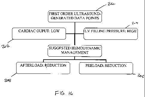
[0090] As shown in FIG. 16-25, the clinical management module 140 can include
one or
more algorithms to be followed based upon the input information provided.
Referring to
FIG. 16, in clinical cases where the first order data points 200 indicate a
low cardiac
output 202 and high filling pressure 204, the clinical management module 140
may
suggest that the provider reduce the preload 206 and reduce the afterload 208
(Strategy
1). Referring to FIG. 17, where the first order data points 200 indicate a low
cardiac
output 202 and filling pressure 204 within normal limits, the module may
suggest that the
provider reduce the afterload 208 and maintain the current preload 206
(Strategy 2). In
FIG. 18, the first order data points 200 indicate a low cardiac output 202 and
low filling
pressure 204 and the strategy suggests that the provider increase the preload
206
(Strategy 3). In FIG. 19, the first order data points 200 indicate a normal
cardiac output
202 and high filling pressure 204 and the strategy suggests that the preload
206 be
reduced and that the systemic blood pressure be maintained if within normal
limits
(Strategy 4). The strategy may also suggest that the afterload 208 be reduced
if the
systemic blood pressure is high (Strategy 4). Referring to FIG. 20, where the
first order
22
CA 02732879 2011-02-02
WO 2010/017295 PCT/US2009/052850
data points 200 indicate a normal cardiac output 202 and normal filling
pressures 204, the
strategy may be to maintain the current preload 206 and afterload 208
conditions
(Strategy 5). As shown in FIG. 21, in clinical cases where the cardiac output
202 remains
low despite optimal preload 206 and afterload 208 management and the second
order
ultrasound-generated data points 210 indicate a reduced contractile function
212, the
strategy may be made to use inotropic support 214 (Strategy 6).
[0091] Referring now to FIG. 22, where the second order data points 210
indicate mitral
valve regurgitation 216, the strategy may be to reduce the afterload 208 and
maintain a
faster heart rate 220 and higher preload 206 (Strategy 7). Where mitral valve
stenosis 218
is indicated, the strategy may be to reduce the preload 206 and maintain a
slower heart
rate 220 (Strategy 7). Referring to FIG. 23, where the second order data
points 210
indicate aortic valve regurgitation 222, the strategy may include reducing the
afterload
208 and maintaining a faster heart rate 220 and higher preload 206 (Strategy
8). As
shown in Fig 24, in clinical cases where the second order data points 210
indicate aortic
valve stenosis 224 with high filling pressures 204, the strategy may suggest
to reduce the
preload 206 and maintain a slower heart rate 220 (Strategy 9). As shown in Fig
25, where
the second order data points 210 indicate aortic valve stenosis 224 with
normal filling
pressures, the strategy may be to maintain a slower heart rate 220 and the
module may
also include an indication that afterload 208 reduction is safe (Strategy 10).
[0092] Referring now to FIGS. 26 and 27, clinical management strategies are
shown with
additional detail. Moreover, these strategies are shown to interface with a
conventional
parameter such as systolic blood pressure 226. With reference to FIG. 26,
where the first
order data points 200 indicate that the cardiac output 202 is low the clinical
management
module 140 can then look to the additional first order data point, filling
pressure 204, to
determine which of two branches to follow for determining a clinical strategy.
Where the
filling pressure 204 is high, three additional branches are based upon
systolic blood
pressure 226. For a systolic blood pressure (BP) 226 greater than 120 mm Hg,
the clinical
strategy may suggest reducing the afterload by 15% and limiting intravenous
fluid (IV) as
required to keep the vein opened (KVO). For a systolic BP 226 of 90 to 120 mm
Hg, the
clinical strategy may suggest reducing the afterload by 10% and limiting the
IV preload
to KVO. For a systolic BP 226 less than 90 mm Hg, the clinical strategy may
suggest
23
CA 02732879 2011-02-02
WO 2010/017295 PCT/US2009/052850
limiting the IV preload to KVO and to consider inotropic support. Similarly,
where the
filling pressures are normal, three additional branches also based on systolic
BP 226 are
shown. Where systolic BP 226 is greater than 120 mm Hg the clinical management
strategy may be to reduce the afterload by 15% and maintain basal IV fluid
intake. For a
systolic BP 226 of 90 to 120 mm Hg, the clinical strategy may suggest to
reduce the
afterload by 10% and maintain basal IV fluid intake. Where systolic BP 226 is
less than
90 min Hg, the clinical strategy may suggest limiting the afterload reduction.
A normal
ejection fraction (EF) may be considered to be from 55% to 70% and in this
case if the
EF is greater than 40% the strategy may suggest that the provider consider an
IV bolus of
250 ml. If the EF is less than 40%, the strategy may suggest that the provider
consider
inotropic support and if there is no increase or minimal increase in Stroke
volume (SV),
the strategy may further suggest that the provider consider an IV bolus of 100
ml.
[0093] A similar strategy to that shown in FIG. 26, is shown in FIG. 27 where
the cardiac
output 202 is normal. Here, the strategy differs from that shown in FIG. 26,
in the normal
filling pressure 204 branch. That is, in the normal filling pressure 204
branch, where the
systolic BP 226 is greater than 120 mm Hg, the strategy suggests an afterload
reduction
of 10% in lieu of 15%. Also, for a systolic BP 226 of 90 to 120 mm Hg, the
strategy
suggests maintaining the afterload and the basal IV intake levels in lieu of
reducing the
afterload by 10% with maintained basal IV intake levels.
[0094] It is noted that the present disclosure is not to be limited to the
specific
percentages of reductions or increases shown and described. The reductions and
increases
in cardiovascular control determinants have been provided here as examples and
do not
reflect an exhaustive list of the available adjustments in the cardiovascular
determinants.
For example, the afterload reductions shown include reductions of 10% and 15%.
The
afterload reduction may range from approximately 0% to approximately 50% and
preferably ranges from approximately 10% to approximately 20%. Additionally,
in cases
of sepsis or systemic infection, the afterload may be maintained or increased.
[0095] Additionally, the exemplary strategies shown are not an exhaustive
list. For
example, FIGS. 26 and 27 are based solely on cardiac output 202, filling
pressure 204,
and systolic BP 226. Other strategies can be included and can be based on any
combination of cardiovascular determinants. The strategies can be further
based on
24
CA 02732879 2011-02-02
WO 2010/017295 PCT/US2009/052850
clinical experience and testing shown to bring cardiovascular functions closer
to normal
ranges.
[0096] The controller 102 can include an electronic reporting module 142. The
electronic
reporting module 142 can be adapted to facilitate the development of a report
145 for
record keeping or other purposes. The report 145 compiled by the electronic
reporting
module 142 can include the clinical findings relating to patient condition and
can also
include the intervention measures taken to adjust, stabilize, or otherwise
change the
patient's condition. The electronic reporting module 142 can be adapted to
prompt the
provider with one or more report input screens 143 allowing the provider to
select,
confirm, modify, or otherwise tailor the report 145 and can also compile the
report based
on this input from the provider. The electronic reporting module 142 can be
accessible
via one or more of the input devices of the provider interface 104. That is, a
menu button
on the display 132 can be available for activating the electronic reporting
module 142 and
the menu button can be selected via a mouse, a touch screen, or any other
input device.
Other suitable activation elements and methods can be included such as a tab
selection, a
drop down box, and the like.
[0097] In a preferred embodiment, the electronic reporting module 142 can be
adapted to
compile an electronic and/or printed medical report. Preferably, the report
145 can
include information relating to the hemodynamic management of a patient.
Accordingly,
as shown, for example in FIG. 28, the electronic reporting module 142 can
prompt the
provider with one or more report input screens 143. The screens 143 can prompt
the
provider for input relating to one or more of the clinical findings obtained
by the analysis
module 136 and/or intervention measures taken by the provider. The findings on
any
particular screen or screens 143 can include, the cardiac output, the filling
pressures, the
valvular structure and function, and the contractile function. Additionally,
the screens can
include intervention measures such as adjustments in the afterload, preload,
heart rate,
and contractility. Other findings or intervention measures can be included on
the screens.
[0098] As shown, in FIG. 28 for example, the report input screen 143 can be
directed to
the left-sided cardiac output. The screen may list a series of options
suitable for the
particular finding or intervention measure being addressed. Each of the
options may
include a short descriptive sentence representing a more detailed description
of a clinical
CA 02732879 2011-02-02
WO 2010/017295 PCT/US2009/052850
finding or an intervention measure. The selection of a report item can be in
the form of
radio buttons as shown or the selection can be check boxes, highlights, or
other known
selection types. The module 142 can be configured to allow only one selection
or it can
allow multiple selections for any given report item.
[0099] For each finding or intervention, the electronic reporting module 142
can make an
initial selection for reporting based on information from the analysis module
136. That is,
for example, if the analysis module 136 found that the LVOT was mildly
decreased, the
reporting module 142 can make an initial selection for confirmation or
modification by
the provider. If the provider has information indicating that the LVOT was
something
other than mildly decreased, the provider can select the appropriate finding.
In the case of
intervention measures, for example, if the clinical management module 140
suggested a
preload reduction, the reporting module 142 may make an initial selection of
preload
reduction. However, if the actual intervention measure taken was not to adjust
the
preload, the provider can change the selection to, for example, maintain
preload. In some
embodiments, the module 142 can omit the initial selection and allow the
provider to
select the appropriate finding or intervention. It is noted, that the report
input screens 143
can be directed to clinical findings or intervention measures not obtained or
suggested,
respectively, by the system. In these cases, the initial selection may be
omitted. Where a
common finding or intervention measure is known, the system can be configured
to select
the common finding or measure as a default for further review by the provider.
[00100] Upon selection or verification of the appropriate finding or
intervention
measure, the provider can be prompted to continue. Alternatively, the
selection or
verification can automatically cause the module to continue. The provider can
be
prompted with additional displays as required to select, verify, or otherwise
obtain all of
the necessary information for the report 145. Once complete, the electronic
reporting
module 142 can compile a suitable report 145. For example, as shown in FIG.
29, the
report 145 can include the detailed descriptions of each of the clinical
findings or
intervention measures taken and can also include a summary of the procedures.
[00101] The compiled report 145 can be in electronic form in a database report
format, a word processing format, or another format. The report 145 can be
saved,
printed, or otherwise stored as a record. The report 145 can be formatted to
comply with
26
CA 02732879 2011-02-02
WO 2010/017295 PCT/US2009/052850
the medical record bylaws of a particular healthcare facility or series of
facilities. In
addition, the report 145 may be electronically coded according to Hospital
Language
(HL) protocol and sent out as a patient electronic medical record in a
compatible format.
[00102] The controller 102 can include a DRG module 144. Many healthcare
system revenues are determined by the Diagnosis Related Group (DRG) billing
codes
resulting from a patient's visit to their facilities. Each DRG code can be
associated with
a specific fee for which the hospital can be reimbursed relating to a specific
rendered
healthcare service. Most DRG codes have two formats: a basic DRG and a DRG
with
complications and comorbidities (CCs). DRG codes associated with clearly
documented
CCs are typically reimbursed at a higher rate than those without CCs (i.e., a
basic DRG).
In the event that CCs are adequately identified and documented, reimbursement
at the
higher, DRG with CCs, rate is possible. In addition, identification of CCs at
the time of
admission of the patient to the healthcare facility allows for the
documentation of cardiac
comorbidities as Present On Admission (POA), as opposed to a post-operative
complication diagnosis. This may reduce the likelihood of lower reimbursement
that is
now tied to the pay-for-performance Medicare and other insurance carrier
programs. The
device described herein allows identification of cardiovascular complications
and
comorbidities and as such may allow for early identification of conditions and
thus a
higher rate of reimbursement.
[00103] The DRG module 144 may allow for the documentation of identified CCs.
When activated by the healthcare provider, the DRG module 144 may display a
list of
International Classification Diseases (ICD) codes describing cardiovascular
CCs capable
of being identified by the device. This list may be displayed on the display
132 as
described above and as shown, by way of example, in FIG. 30. By selecting the
most
appropriate diagnosis (ICD codes) identified by the device, the end-user may
generate a
series of billing codes that may be used by the healthcare facility to
document the CCs.
The billing codes may be documented in a separate report called the DRG
optimization
report 147 as exemplified in FIG. 31. The report 147 may be printed on paper
or written
in an electronic document. The report 147 may be added to the patient paper or
electronic
medical record. The report 147 may also be sent by paper and or electronically
to the
healthcare facility billing and coding department as a separate document from
the
27
CA 02732879 2011-02-02
WO 2010/017295 PCT/US2009/052850
medical record. This report 147 may improve the capture of reimbursement for
CCs by
the healthcare facility billers and coders for optimization of the patient's
final DRG code
submitted to the insurance company for the services rendered. The billing
codes
generated may also be used in a separate document called a professional
billing claim 149
as shown, by way of example, in FIG. 32. This document may allow for the
healthcare
provider to be paid for the professional services rendered with use of the
device
according to the Current Procedural Terminology (CPT) code fee schedule.
[00104] Referring now to FIGS. 33-36, the system methodology may be described.
The system can function to acquire data from patients for use in managing the
patient's
condition and may further be used as a reporting tool. Using the patient
interface 100, the
system may be adapted to obtain patient information relevant to a particular
procedure or
condition. The system can be further adapted to analyze and/or display that
information.
In addition, the system can suggest a suitable clinical strategy for managing
the condition
of the patient.
[00105] In a preferred embodiment, the probes 110 of the preferred patient
interface 100 described, can be used to obtain cardiovascular function
information from a
patient. The probes 110 may obtain information based upon their position on
the patient.
That is, certain positions can represent a cardiovascular window as described
above and
can lend themselves toward collection of particular items of cardiovascular
information.
Accordingly, in a preferred embodiment, each probe 110 may have a particular
set of data
collection allocated to it based on the particular window it is positioned in.
However,
depending on patient anatomy and other factors, a probe 110 in any given
position may
not be able to access the information typically available from its respective
position. In
these cases, other positions can be used to compile the most complete set of
data
available.
[00106] More particularly, in a preferred embodiment, the basic sequence of
data
acquisition may occur through the use of two probes 110. That is, in some
embodiments,
two probes 110 may be able to collect all of the cardiovascular function
information by
allocating some of the information to a first probe 110 and the remaining
information to
the second probe 110. In other embodiments, two probes 110 may not be
sufficient due to
obstructions or other intervening causes. In still other embodiments,
additional probes
28
CA 02732879 2011-02-02
WO 2010/017295 PCT/US2009/052850
110 may be used to get additional information by viewing particular structures
from
additional views. In some embodiments, a single probe 110 may be sufficient.
In other
embodiments, any number of probes 110 maybe used.
[00107] Referring to FIG. 33, in a preferred embodiment, a first probe 110 can
be
secured on a patient's chest at the parasternal window 300. This probe 110
maybe set by
the patient interface module 134 to a first mode for a 2D black and white
image. The
patient interface module 134 can adjust the probe 110 to acquire a parasternal
long-axis
2D imaging cross-section 302 of the heart for one or more heart beats. This
black and
white 2D image clip can show the left ventricular heart muscle contracting and
the mitral
and aortic valves open and close. From the same 2D cross-section, for example,
without
adjusting the view of the probe 110, the mode of the first probe 110 can be
changed to a
second mode and a color Doppler ROI box may be superimposed on the aortic 304
and
mitral 306 valves 2D live image. A clip of the data may be acquired for one or
more heart
beats. The color Doppler allows the assessment of the valves functionality by
revealing
the blood flow through the valves. Still using the first probe 110, additional
data may be
acquired by adjusting the probe 110 from the parasternal long-axis 2D imaging
cross-
section 302 to a parasternal short-axis 2D imaging cross-section 308 for one
or more
heart beats. This short-axis probe view 308 can allow for the assessment of
the left
ventricular contractile function and volume status.
[00108] Referring to FIG. 34, in a preferred embodiment, a second probe 110
can
be secured on the patient's chest at the apical window. This second probe 110
can be set
by the patient interface module 134 to a first mode for a 2D black and white
image. The
patient interface module 134 can adjust the second probe 110 to acquire an
apical four-
chamber 2D imaging cross-section 312 for one or more heart beats. This 2D clip
can
evaluate the right and left ventricular contractile function, as well as the
mitral and
tricuspid valve. This additional 2D clip allows for the three-dimensional
heart structure to
be assessed by a series of two-dimensional cross-sections by relying on view
from
several angles. The probe 110 can be set to a second mode for a color Doppler
image of
the mitral 313 and triscuspid valve 315. From the same 2D cross-section, for
example,
without adjusting the view of the probe 110, the mode of the first probe 110
can be
changed to the third mode and a pulsed-wave spectral Doppler ROI box may be
29
CA 02732879 2011-02-02
WO 2010/017295 PCT/US2009/052850
superimposed on the open mitral valve 314 to measure the velocity of the red
cells
coming into the heart during diastole. The data may be acquired and displayed
on a
spectral graph showing velocity over time. The same pulsed-wave spectral
Doppler ROI
box, for example, without changing the size of the ROT box, may be
superimposed on the
right upper pulmonary vein 316. The velocity/time spectral graph of the
pulmonary
venous flow may then be acquired. The pulsed-wave spectral Doppler ROT box may
also
be superimposed on the septal or lateral side of the mitral valve annulus 318
to measure
the tissue Doppler velocities of the left ventricle. Those three spectral
Doppler
measurements may then be used to assess the left ventricular diastolic
function and filling
pressure. Also, a continuous wave Doppler sampling of the tricuspid
regurgitation jet 319
peak velocity may be made to estimate the right ventricular/pulmonary artery
pulmonary
pressure.
[001091 In a preferred embodiment, the patient interface module 134 can set
the
second probe 110 back to mode 1 and adjust the second probe 110 to acquire a
2D cross-
section called an apical long-axis 320 for one or more heart beats. From the
same apical
long-axis 2D cross-section, patient interface module 134 can set the second
probe 110 to
the 3`d mode and a pulsed-wave spectral Doppler sampling area may be
superimposed on
the left ventricular outflow tract (LVOT) 322 to measure the velocity of the
red cells
being ejected out of the left heart over a cardiac cycle (left-sided cardiac
output).
Additionally, a continuous-wave spectral Doppler may be directed in the same
longitudinal axis to measure the velocity of the red cells at the level of the
aortic valve
324. This additional velocity allows the evaluation and quantification of
aortic valve
stenosis.
1001101 As mentioned, in some embodiments, the information gathered from the
first and second probes 110 may be insufficient due to obstructed views or
other
intervening causes or additional views may be desired. Referring to FIG. 35,
in some
embodiments, a third probe 110 can be secured on the patient's upper abdomen
under the
right costal ridge in the sub-costal window. The patient interface module 134
can set the
third probe 110 to a first mode for a 2D black and white image. The patient
interface
module 134 can adjust the third probe 110 to acquire a sub-costal four chamber
2D
imaging cross-section 326 for one or more heart beats. This 2D clip may
evaluate the
CA 02732879 2011-02-02
WO 2010/017295 PCT/US2009/052850
right and left ventricular contractile function, the size of the inferior vena
cava as well as
the mitral and tricuspid valve. From the same 2D cross-section, the patient
interface
module 134 can set the third probe 110 to a second mode and a color Doppler
region of
interest (ROI) box may be superimposed on the mitral valve 328 and the
tricuspid valve
329. A clip of the data may be acquired for one or more heart beats. The color
Doppler
can allow the assessment of the mitral and tricuspid valve functionality. In
the present
embodiment, and still using the third probe 110, the patient interface module
134 can set
the third probe 110 to a first mode. The third probe 110 can be adjusted for a
sub-costal
right ventricular inflow-outflow 2D imaging cross-section 331, which may be
acquired
for one or more heart beats. This allows the evaluation of the right heart
structures and
function. From the same 2D cross-section, the patient interface module 134 can
set the
third probe 110 to a third mode and a pulsed-wave spectral Doppler sampling
area may
be superimposed on the right ventricular outflow tract (RVOT) 332 to measure
the
velocity of the red cells being ejected out of the right heart over a cardiac
cycle (right-
sided cardiac output). Still using the third probe 110, a sub-costal LV short-
axis 2D
imaging cross-section 330 may be acquired for one or more heart beats. This
allows the
assessment of the left ventricular contractile function and volume status.
[00111] When the ultrasound-generated data points from the second probe 110
regarding the left heart cardiac output are inadequate or when additional
views are
desired, the user may rely on a fourth probe 110 to acquire a continuous-wave
spectral
Doppler tracing signal of either the ascending aorta or the distal aortic arch
or the
descending aorta.
[00112] When the ultrasound-generated data points from the first, second,
third, or
fourth probes 110 are inadequate or as an additional available set of data, a
fifth probe
110 can be used. Referring to FIG. 36, the fifth probe 110 may be positioned
in the mid-
esophageal window and may acquire ultrasound-generated data points from behind
the
heart (inside the body). The fifth probe 110 may acquire a mid-esophageal four
chamber
2D imaging cross-section 334 for one or more heart beats. This 2D clip
evaluates the
right and left ventricular contractile function, as well as the mitral and
tricuspid valves.
From the same 2D cross-section, a color Doppler region of interest (ROI) box
may be
superimposed on the mitral 336 and tricuspid 338 valves 2D live image. A clip
of the
31
CA 02732879 2011-02-02
WO 2010/017295 PCT/US2009/052850
data may also be acquired for one or more heart beats. The color Doppler
allows the
assessment of the mitral and tricuspid valve functionality. From the same 2D
cross-
section, a pulsed-wave spectral Doppler sampling area may be superimposed on
the
opened mitral valve 340 to measure the velocity of the red cells coming into
the heart
during diastole. The data may be acquired and displayed on a spectral graph
showing
velocity over time. Then, the same pulsed-wave spectral Doppler sampling area
may be
superimposed on the left upper pulmonary vein 342. The velocity/time spectral
graph of
the pulmonary venous flow may then be acquired. The pulsed-wave sampling
Doppler
may then be superimposed on the septal or lateral side of the mitral valve
annulus 344
and may measure the tissue Doppler velocities of the left ventricle. Those
three spectral
Doppler measurements may be used to assess the left ventricular diastolic
function and
filling pressure. A continuous wave Doppler sampling of the tricuspid
regurgitation jet
339 peak velocity may be made to estimate the right ventricular /pulmonary
artery
pulmonary pressure.
[001131 The method resulting from the use of the described device may be
referred
to as Echocardiography-Guided Anesthesia Management (EGAM) and/or
Echocardiography-Guided Hemodynamic Management (EGHEM). EGAM/EGHEM may
automatically acquire ultrasound-generated real-time data points like cardiac
output and
filling pressures to assess, manage, modify and optimize the patient cardiac
preload,
afterload, heart rate and contractility. Two clinical case studies were
conducted as
described below.
CLINICAL EXAMPLE 1
Step 1: Patient selection
[001141 Male patient, 81 year old, scheduled for a left hip pinning for a
fracture
repair. He weighs 89 Kg and is 178 cm tall. His BSA is 2.1 m2. The patient has
long-
standing hypertension, and has a history of transmural myocardial infarction
(MI) 4 years
prior. The patient has a limited functional capacity of approximately 5 METs
with
32
CA 02732879 2011-02-02
WO 2010/017295 PCT/US2009/052850
symptoms of shortness of breath (SOB), occasional chest pain stable for last
two years,
and hip pain. His medication includes an ACEI and a beta-blocker.
Step 2: Baseline Pre-operative assessment
[001151 The device and methods previously described in this document were
applied to this patient. This process was performed at bedside before
anesthesia was
provided. The process was pain free and took a few minutes to complete. Below
is the
summary of the information provided by the device:
Baseline vital signs:
a. blood pressure (BP) = 160/85 mmHg,
b. heart rate (HR) = 82 bpm, regular,
c. Sp02 = 92% room air.
Primary EGAM/EGEM findings:
a) Reduced cardiac output: LVOT diameter is 2cm, LVOT VTI=12 cm. CO: 3.1
L/min, CI=1.5L/min/m2
b) LV Filling pressures are elevated based on a pseudonormal LV filling
pattern, a
pulmonary venous flow diastolic dominant and an E/e' ratio of 25.
Secondary EGAM/EGHEM findings;
a) Mitral valve: mild regurgitation.
b) Aortic valve: sclerosis without significant stenosis.
c) LV contractile function: moderately reduced with a visually estimated
ejection
fraction (EF) at 30%.
33
CA 02732879 2011-02-02
WO 2010/017295 PCT/US2009/052850
Step 3: Management Strategies
[00116] The patient presents a low cardiac output, high filling pressure, high
systemic blood pressure, reduced LV contractile function and mild mitral
regurgitation.
The suggested EGAM/EGHEM strategy based on FIG. 26 recommendation is to reduce
the afterload and blood pressure by 15% and limit all IV intakes only to keep
the vein
open. A general anesthetic is planned with IV induction agents and maintenance
done
with an inhalational agent. If required, the basal IV intake needs are 65
ml/hour. The
EGAM/EGHEM data will be controlled 5 minutes after induction.
34
CA 02732879 2011-02-02
WO 2010/017295 PCT/US2009/052850
Step 4: Ongoing intra-operative assessment
The following table summarizes the intra-operative findings and interventions
Timeline Cardiac Filling Blood LV Interventions
output pressure pressure contractility
Baseline 3.1 L/min High 160/85 EF=30% Limit preload
E/e'=25 Reduce to
systolic BP to
136
min post- 3.5L/min High 132/78 No change Limit preload
induction E/e'=20 Reduce BP to
112
Control #1 3.8L/min Normal 108/72 Mild increase Maintain
E/e'=13 basal needs
min later Reduce BP to
98
Control # 2 4.2L/min Normal 96/68 No change Maintain
15 min later E/e'=12 basal needs
Reduce BP to
Control # 3 4.4Umin Normal 84/62 EF=35% Give IV bolus
7 min later E/e'=10 100 ml
Limit afterload
reduction
Control # 4 4.5L/min Normal 92/64 No change Maintain
5 min later E/e'=14 basal needs
Maintain
afterload
Control # 5 4.3L/min Normal 96/68 No change Maintain
15 min later E/e'=12 basal needs
Maintain
afterload
Control # 6 3.8L/min Normal 145/72 No change Maintain
In recovery E/e'=14 basal needs
room Reduce BP to
low 90's
CA 02732879 2011-02-02
WO 2010/017295 PCT/US2009/052850
Follow-up events
[00117] The case lasted for about 1 hour. The patient received a total of 250
ml of
IV fluid. The urine output during the procedure was 150ml. The blood loss was
estimated
at 150m1. The Sp02 on room air in recovery room as well as post-op day 1 was
98%. The
patient remained comfortable. The post-operative course included an increase
of blood
pressure medication and the addition of a low dose diuretic, as well as a
reduced salt and
fluid intake. The target systolic BP was in the 90's. The discharge weight was
83 kg, the
CO was 4.3L/min, BP=96/72. The patient tolerated those changes well and
reported no
orthostatic hypotension, no stroke, and no changes of renal function. He was
still alive
and doing well at 30 days post-op and did not require readmission during the
same period
and had no new cardiac events.
[00118] The device effectively identified that the patient was in a non
compensated
state of congestive heart failure with reduced cardiac output and ventricular
contractility.
The clinical strategy used to address those issues was significantly different
than what the
standard pre-operative evaluation was dictating because the supplemental
information
provided by the device suggested a completely opposite strategy. By using the
invention,
the health care provider had access to more accurate information, was able to
provide
better care to the patient and reduce the risk of post-operative
cardiovascular
complications.
Case Study 2
Step 1: Patient selection
[00119] Female patient, 82 year old, scheduled for elective, right
hemicolectomy.
She weights 79 Kg and is 160 cm tall. Her BSA is 1.9 m2. Patient has medically
treated
hypertension with a hydrochlorothiazide. She stopped smoking two year ago but
has a 20
pack-years history. She is complaining of a progressive shortness of breath
and reduction
of her functional capacity over the last year, currently estimated at 6 or 7
METs. She has
no chest pain or palpitations.
Step 2: The baseline pre-op assessment
[00120] The device and methods previously described in this document were
applied to this patient. This process was performed at bedside before
anesthesia was
36
CA 02732879 2011-02-02
WO 2010/017295 PCT/US2009/052850
provided. The process was pain free and took a few minutes to complete. Below
is the
summary of the information provided by the device:
Baseline vital signs.
a.blood pressure (BP) = 168/92 mmHg,
b. heart rate (HR) 70 bpm, regular,
c. Sp02 = 90% room air.
Primary EGAM/EGHEM findings:
a) Normal cardiac output: LVOT diameter is 2cm, LVOT VTI=22 cm. CO:
4.8L/min, CI=2.5L/min/m2
b) LV Filling pressures are elevated based on a restrictive filling pattern, a
pulmonary venous flow diastolic dominant and an E/e' ratio of 35.
Secondary EGAM/EGHEM findings:
a) Mitral valve: mild to moderate regurgitation.
b) Aortic valve: sclerosis with mild stenosis.
c) LV contractile function is normal with a visually estimated EF at 60%
Step 3: Management strategies
[00121] The patient presents a normal cardiac output, high filling pressure,
high
systemic blood pressure, a normal LV contractile function, mild to moderate
mitral
regurgitation and mild aortic stenosis. The suggested EGAM/EGHEM strategy
based on
FIG. 27 is to reduce the afterload and blood pressure by 15% and limit all IV
intakes only
to keep the vein open. A general anesthetic is planned with IV induction
agents and
maintenance done with total intravenous anesthetics agents. If required, the
basal IV
intake needs are 60 ml/hour. The EGAM/EGHEM data will be controlled 5 minutes
after
induction.
37
CA 02732879 2011-02-02
WO 2010/017295 PCT/US2009/052850
Step 4: Ongoing intra-operative assessment
The following table summarizes the intra-operative findings and interventions
Timeline Cardiac Filling Blood LV Interventions
output pressure pressure contractility
Baseline 4.8 Umin High 162/92 EF=60% Limit preload
E/e'=35 Reduce to
systolic BP to
145
min post- 5.1Umin High 141172 No change Limit preload
induction E/e'=30 Reduce BP to
120
Control #1 5.5Umin High 128/67 No change Limit preload
E/e'=26 Reduce BP to
min later 110
Control # 2 5.3LImin High 105/59 No change Limit preload
15 min later E/e'=24 Reduce BP to
Control # 3 5.4Umin High 92/55 No change Limit preload
15 min later E/e'=22 Maintain
afterload
Control # 4 5.2Umin Normal 96/58 No change Maintain
15 min later E/e'=14 basal needs
Maintain
afterload
Control # 5 5.3Umin Normal 98/64 No change Maintain
15 min later E/e'=12 basal needs
Maintain
afterload
Control # 6 4.8Lmin Normal 78/48 No change Give IV bolus
15 min later E/e'=10 of 250ml
Maintain
afterload
Control # 7 5.1Umin Normal 105/74 No change Maintain
In recovery E/e'=14 basal needs
room Reduce BP to
90's
38
CA 02732879 2011-02-02
WO 2010/017295 PCT/US2009/052850
Follow-up events
[00122] The case lasted for about 2 hours. The patient received a total of
300m1 of
IV fluid. The urine output during the procedure was 450ml. The blood loss was
estimated
at 250ml. The Sp02 on room air in recovery room was 97%. The patient remained
comfortable. The post-operative course included an increase of his existing
blood
pressure medication and the addition of an ACEI, as well as low sodium diet.
The target
systolic BP was in the 90's. The discharge weight was 72 kg, the CO was
5.2L/min,
BP=100/68. The patient tolerated those changes well and reported no
orthostatic
hypotension, no stroke, and no changes of renal function. She was still alive
and doing
well at 30 days post-op and did not require readmission during the same period
and no
new cardiac events.
[00123] The device effectively identified that the patient was in a non
compensated
state of congestive heart failure with normal cardiac output and ventricular
contractility
but very high filling pressures. The clinical strategy used to address those
issues was
significantly different than what the standard pre-operative evaluation was
dictating
because the supplemental information provided by the device suggested a
completely
opposite strategy. By using the invention, the health care provider had access
to more
accurate information, was able to provide better care to the patient and
reduce the risk of
post-operative cardiovascular complications.
[00124] As shown and described regarding FIGS. 37-42, the system may perform
several methods. The steps included in any of the described methods may be
completed
in any order and any or all of the steps may be included.
[00125] Referring to FIG. 37, a method of is shown including at box 400,
Generate
ultrasound data point, at box 402, Interpret ultrasound data points provided
by each of the
probes 110, at box 404, Rely on a system of first order and second order data
points to
suggest an optimal clinical strategy, at box 406, Output the suggested
strategy to a
display wherein the strategy includes modification (increase, reduce or
maintain) of one
or more cardiovascular determinants such as preload, afterload, heart rate,
and ventricular
contractility, at box 408, Display a list of possible clinical findings, at
box 410, Prompt
39
CA 02732879 2011-02-02
WO 2010/017295 PCT/US2009/052850
end-user to select from a list, at box 412, Receive input from end-user, and
at box 414,
Generate a Final Report.
[001261 In addition, the method may include at box 416, Prompt user with a
list of
ICD codes for selection based on output from system analysis, at box 418,
Receive input
from end-user regarding ICD codes, at box 420, Prepare DRG optimization
report, and at
box 422, prepare a professional billing claim.
1001271 Referring to FIG. 38, a method is shown including, at box 424,
obtaining
ultrasound information regarding a condition of the patient from an ultrasound
probe, at
box 426, communicating the ultrasound information to a controller in
communication
with the ultrasound probe, at box 428, employing the controller to develop a
determinant
from the ultrasound information reflecting the condition of the patient, and
at box 430,
providing on an output device in communication with the controller a clinical
management strategy based on the determinant.
[001281 Referring to FIG. 39, a method is shown including, at box 432,
receiving
ultrasound information from a patient interface, the patient interface being
adapted to
obtain ultrasound information related to cardiovascular function status of the
patient, at
box 434, processing the ultrasound information to determine the cardiovascular
function
status of the patient, and at box 436, sending the status to a clinical
management module
for the development of a clinical strategy.
[001291 Referring to FIG. 40, a method is shown including, at box 438,
comparing
a first order data point to a plurality of categories, wherein the first order
data point is
associated with ultrasound information, at box 440, assigning a category from
the
plurality of categories to the first order data point based on which category
of the
plurality of categories, the first order data point falls, at box 442,
selecting a
recommended intervening measure based on the assigned category, and at box
444,
presenting the recommended intervening measure on a display.
[001301 Referring to FIG. 41, a method is shown including, at box 446,
positioning
ultrasound probes on a patient, the ultrasound probes being in communication
with a
controller, at box 448, using an input device to instruct the controller to
obtain
cardiovascular function information from the patient via the ultrasound
probes, at box
450, reviewing a suggested clinical management strategy, the strategy
including a
CA 02732879 2011-02-02
WO 2010/017295 PCT/US2009/052850
recommended intervening measure and being based upon the ultrasound
information, and
at box 452, deciding whether to conduct the recommended intervening measure, a
different intervening measure, or no intervening measure.
[00131] Referring to FIG. 42, a method is shown including, at box 454,
monitoring
a patient via ultrasound and generating information with the ultrasound and
based upon
the information, recording a clinical finding and recommending and recording
an
intervening measure, at box 456, displaying a list of clinical findings
including the
clinical finding and related clinical findings, at box 458, prompting a user
to select from
the list of clinical findings, at box 460, displaying a list of intervening
measures including
the intervening measure and related intervening measures, at box 462,
prompting the user
to select from the list of intervening measures, and at box 464, compiling a
report
including the selected clinical finding and the selected intervening measure.
[00132] While the term provider has been used throughout the specification, it
is to
be understood that this is not limited to a licensed medical doctor,
physicians assistant,
nurse practitioner, and the like. Instead, provider can by any user of the
system.
Preferably, the provider is someone working under the guidance of a licensed
practitioner
and who understands cardiovascular function so as to provide suitable input to
the
system.
[00133] Additionally, while the phrase black and white has been used with
reference to certain ultrasound images, it is to be understood that black and
white means a
non-color image. That is, an image that does not accurately depict the colors
of the
displayed elements, but rather displays similar but varying tones of several
elements to
make them distinguishable from one another. For example, black and white,
sepia,
orange, or green colors may be included within the black and white
description.
[00134] Additionally, the categories of cardiovascular determinants are not to
be
limited to those categories disclose. More or less precise categories could be
used and the
image clip databases and categories can be adjusted accordingly. For example,
with
respect to contractile function, rather than using hyperdynamic, normal,
moderately
reduced, and severely reduced as categories, the categories could instead be
normal and
abnormal. The contractile function image clip database can be adjusted to
include normal
41
CA 02732879 2011-02-02
WO 2010/017295 PCT/US2009/052850
clips and abnormal clips and to include only two categories in lieu of four.
This holds
true for all of the image clip databases and the associated categories.
[001351 Although the present invention has been described with a certain
degree of
particularity, it is understood the disclosure has been made by way of
example, and
changes in detail or structure may be made without departing from the spirit
of the
invention as defined in the appended claims.
42