Note: Descriptions are shown in the official language in which they were submitted.
CA 02733119 2011-01-07
1
RECOMBINANT EXPRESSION VECTOR FOR ANIMAL CELL
FIELD OF THE INVENTION
The present invention relates to a recombinant expression vector for an animal
cell, to a
cell line transformed by the vector, and to a method for preparing a target
protein using the
same.
BACKGROUD ART
In general, animal cell culture is a preferred technique in the industry for
overexpressing the target protein. Because proteins with industrial value are
mostly human or
animal derived proteins, and specific protein modification mechanisms
(glycosylation,
phosphorylation, amidation) are carried out easily in animal cells. The animal
cells currently
used in industry are CHO (Chinese Hamster Ovary), BHK (Baby Hamster Kidney)
and
myeloma cells, where the target protein is expressed by transfecting
expression vector into
the cells, similar to the microorganism based expression system.
However, animal cells have a disadvantage of showing low level of transfected
foreign
gene expression compared to an expression in the microorganism. The system
widely used
in the industry to overcome this disadvantage is the foreign gene
amplification system, which
uses dihydrofolate reductase (DHFR) gene and its gene activation inhibitor,
methotrexate
NM. This system is based on the phenomenon of the DHFR gene required for the
survival
and the foreign gene located close by being amplified together. In detail, the
gene coding for
the target protein and gene coding for the selective marker, DHFR protein,
that are inserted
in the same region of the chromosomal DNA are amplified simultaneously when
the
concentration of MIX is increased artifidally.
It has been previously reported that the gene located near the DHFR gene in
the
expression vector is amplified simultaneously when treated with MIX (Kaufman
et al. Mol Cell
Biol. Jul; 5(7):1750-9(1985)). There is a report of high level of a foreign
gene being co-
expressed in the animal cell when it is inserted in the vicinity of the DHFR
gene in the
CA 02733119 2013-01-07
2
expression vector (Alt et al. Cold Spring Hart) Symp Quant Bio/. 42 Pt 2:649-
57(1.978); US
Patent No. 4656134).
Gene amplification is generally a very rare phenomenon, but there are
indications
that acquiring gene amplified cells could be achieved through selecting rmlls
that are resistant
to the serially increased MD( concentration. It takes about 3-4 weeks for the
MD( resistant
colonies to form, and several multiple steps of amplification process to
achieve industrially
significant levels of amplification using MTX concentrations ranging from 50
nM to 500 mM.
However, during the process of inducing gene amplification using MTX
treatment,
problems such as reduction in cell growth rate and in productivity may occur.
For instance,
there is a report indicating a decrease in the level of the recombinant
protein expression, rather
than an increase, despite the increase in MIX concentration (Kaufman et al.
Mal Cell Biol.
;5(7):1750-9(1985)). Similarly, there is a rase report of significantly
increasing the gene
amplification effect of MTX by mutating the DHFR gene control factor sequenm
in the
expression vector (Bai et al. Aonghua Yi Xue Za Zhi. Feb 25;83 (4):333-
7(2003)).
Throughout this application, various patents and publications are refererkrd
and
citations are provided in parentheses.
DETAILED DESCRIPTION OF THE INVENTION
The present inventors have performed intensive research to solve the problems
when
expressing protein using animal cell transformed with DHFR gene containing
vector, such as
the low level of expression and decrease in cell growth rate and in
productivity by high
concentrations of methotrexate. As a result, the present inventors developed a
remmbinant
expression vector for acquiring large amounts of foreign protein by using a
recombinant vector
containing human derived DHFR promoter sequence operatively connected to the
mouse
derived DHFR promor. This process can effectively amplify the foreign gene at
a lower
concentration of methotrexate, and thus completed the present invention.
CA 02733119 2011-01-07
int
3
Accordingly, it is an object of the present invention to provide a reoombinant
vector for
a dhfr- animal cell.
It is another object of this invention to provide a dhfr- animal cell line
transfected by the
vector.
It is still another object of this invention to provide a method for preparing
the protein
using the transfected dhfr- animal cell line.
Other objects and advantages of the present invention will become apparent
from the
detailed description to follow and together with the appended claims and
drawings.
According to an aspect of this invention, the present invention provides (a) a
dihydrofolate reductase (DHFR) promoter comprising nucleotide sequence listed
in SEQ ID
NO: 1 or SEQ ID NO: 2; and (b) a recombinant vector for a dhfr- animal cell
comprising DHFR-
coding nucleotide sequence operatively linked to the promoter.
The present inventors have performed intensive rcscarch to solve the problems
when
expressing protein using animal cell transformed with DHFR gene containing
vector, such as
the low level of expression and decrease in cell growth rate and in
productivity by high
concentrations of methotrexate. As a result, the present inventors developed a
recombinant
expression vector for acquiring large amounts of foreign protein by using a
recombinant vector
containing human derived DHFR promoter sequence operatively oonnected to the
mouse
derived DHFR promoter. This process can effectively amplify the foreign gene
at a lower
concentration of methotrexate.
The term "DHFR (Dihydrofolate reductase)" used herein refers to an enzyme that
reduces dihydrofolic acid to tetrahydrofolic add, which is a key enzyme for
nudeic add
synthesis and an essential enzyme for cell growth.
The present invention relates to an expression vector for producing large
amounts of
foreign protein in a dhfr- animal cell under low conmntration of DHFR
inhibitor, particularly
under a low concentration of methotrexate (NTD).
As used herein, "dhfr- animal cell" refers to a transformed animal cell
without or almost
any DHFR enzyme activity in the cell by lack of normal DHFR expression. This
invention is
CA 02733119 2011-01-07
, .
4
directed to using gene amplification principle of the gene including DHFR
gene, to use as host
cell character and host cell selection. That is, dhfr animal cell is
transformed by dhfr gene
containing vector, and then the transformed cell is treated with DHFR
inhibitor. The cells
amplified with high numbers of dhfr containing vectors are selected.
Therefore, the vector
amplification is achieved.
As used herein, "MD( (methotrexate)" refers to a DHFR inhibitor, which
inhibits the
reduction of folic add to dihydrofolate (FH2) and then to tetrahydrofolate (FI-
14).
According to a preferred embodiment, the DHFR promoter is mouse derived, and
the
DHFR-coding nucleotide is human derived.
The DFHR promoter used in this invention is a partial sequence from the
promoter of
mouse dhfr gene which has the promoter activity suitable to the purpose of the
present
invention. The SEQ ID NO: 1 and SEQ ID NO: 2 promoters used in the present
invention
showed relatively low promoter activity compared to the conventional animal
cell expression
vectors. This leads to a reduction in dhfr gene expression operatively
connected to the
promoter, and the cells with high level of amplification of vectors containing
dhfr gene at low
concentration of MIX are selected. As a result, the vector amplification is
achieved, and the
expression of the foreign protein of purpose is increased at the same time.
According to a preferred embodiment, the promoter sequence used in this
invention
consists of the nucleotide sequence listed in SEQ ID NO: 1 and SEQ ID NO: 2.
The DHFR-coding nucleotide sequence in the recombinant expression vector is
operatively linked to the promoter. The term "operatively linked" used herein
refers to
functional connection between nucleic acid expression regulation sequence
(e.g., promoter
sequence) and the other nucleic acid sequence, through the regulation sequence
that controls
the transcription and/or translation of the other nucleic add sequence.
The DHFR-coding nucleotide sequence used in this invention is preferably a
human
derived DHFR gene, more preferably human DHFR gene CDC (coding sequence,
nucleotide
sequence numbers 493-1056) sequence as described in GenBank accession number
NM_000791 may be used as the DHRF-coding nucleotide sequence.
The recombinant expression vector of the present invention is used in a dhfr
animal cell.
Acoording to the preferred embodiment of the present invention, the animal
cell is yeast
CA 02733119 2011-01-07
(Saccharornyces cerevislae), insect cell or mammalian animal cell, more
preferably, a
mammalian animal cell, still more preferably, CHO (Chinese hamster ovary) cell
line, W138,
BHK, COS-7, 293, HepG2, 3T3, RIN, MDCK cell line or BHK (Baby Hamster Kidney)
cell line,
most preferably, CHO cell line. Since the safety and effectiveness of DHFR-
defident CHO cell
5 has been verified and approved by FDA, the cell line is widely used in
producing recombinant
protein for dinical use.
Aocording to a preferred embodiment, the expression vector includes an
additional
nucleotide sequence of a foreign gene.
The foreign gene coding for the target protein to be expressed indude any gene
sequences. For instance, the foreign gene indudes the nucleotide sequence
which encodes
hormones, hormone analogues, enzymes, enzyme inhibitors, signal transduction
proteins or its
partial regions, single chain antibodies, binding proteins or its binding
domains, antigens,
adhesion proteins, structure proteins, regulatory proteins, toxin proteins,
cytokines, various
regulators, blood clotting factors or vaccine proteins. In detail, the foreign
gene amplified and
expressed by the vector comprises nucleotide sequences of insulin, IGF-
1(insulin-like growth
factor 1), growth hormone, BMP (bone morphogenetic protein), TGF (transforming
growth
factor), erythropoietin, G-CSFs (granulocyte-colony stimulating factors), GM-
CSFs
(granulocyte/macrophage-colony stimulating factors), interferon-a, interferon-
13, interferon-y,
interleukin-1 a and 13, interieukin-3, interieukin-4, interleukin-6,
interleukin-2, EGFs (epidermal
growth factors), calcitonin, ACTH (adrenocorticotropic hormone), TNF (tumor
necrosis factor),
TNFR(tumor necrosis factor receptor), IDS(iduronate-2-sulfatase), atobisban,
buserelin,
cetrorelix, deslorelin, desmopressin, dynorphin A (1-13), elcatonin,
eleidosin, eptifibatide,
GHRH-II (growth hormone releasing hormone-II), gonadorelin, goserelin,
histrelin, leuprorelin,
lypressin, octreotide, oxytocin, pitressin, secretin, sincalide, teriipressin,
thymopentin,
thymosine al, triptorelin, bivalirudin, carbetocin, cydosporin, exedine,
lanreotide, LHRH
(luteinizing hormone-releasing hormone), nafarelin, parathyroid hormone,
pramlintide, T-20
enfuvirtide, thymalfasin or Ziconotide.
According to the preferred embodiment, the upstream nucleotide sequence of the
foreign gene is connected by a promoter sequence that can function in a
eukaryotic cell. The
promoter sequence that can function in the eukaryotic cell is SV40 promoter
(SV40 late
CA 02733119 2011-01-07
promoter and SV40 early promoter), tk promoter of HSV (herpes simplex virus),
adenovirus 2
major late promoter (Pmni), adenovirus 2 early promoter (PmE2), p19 promoter
of AAV (human
parvo virus-associated virus), Epstein-Barr virus (EBV) promoter, Rous Sarcoma
virus (RSV)
promoter, Vaccinia virus 7.5K promoter, mouse metallothionein promoter, MT
promoter, MMTV
LIR promoter, HIV LTR promoter, 13-actin promoter, EF1 a-promoter, human IL-2
gene
promoter, human INF gene promoter, human IL-4 gene promoter, human
lyrnphotoxin
promoter, human GM-CSF gene promoter and human hemoglobin, human muscle
creatine or
human methalotionein derived promoter, but is not limited thereto.
The expression vector of the present invention contains a polyadenylation
sequence as
the transcription termination sequence, e.g., bovine growth hormone terminator
(Gimmi, E. R.,
et al., Nucleic Acids Res. 17:6983-6998(1989)), SV40 derived polyadenylation
sequence (Schek,
N, et al., Mol. Cell Biol. 12:5386-5393(1992)), polyA site of HIV-1 (Klasens,
B. I. F., et al.,
Nudeic Acids Res. 26:1870-1876(1998)), polyA site of P-globin (Gil, A., et al,
Cell 49:399-
406(1987)), polyA site of HSV TK (Cole, C. N. and T. P. Stacy, Md. Cell Biol.
5:2104-
2113(1985)) or polyA site of polyomavirus (Batt, D. B and G. G. Carmichael,
Md. Cell Biol.
15:4783-4790(1995), but is not limited thereto.
In addition, the expression vector of present invention may contain an
antibiotic
resistance gene that is known to those of skill in the art as a selective
marker gene, e.g.,
ampicillin, gentamycin, carbenicillin, chbramphenicol, streptomycin,
kanamycin, Genetidn
(G418), neomycin or tetracycline.
In a more preferably embodiment, the animal cell expression vector for
producing high
level of foreign protein is a vector with the gene map depicted in Fig. 3,
most preferably, the
vector is pJK-dhfr-1 (KCTC 11299BP) or pJK-dhfr-2 (KCTC 11300BP).
In one aspect, the present invention provides a dhfr animal cell line
transformed by the
dhfr- vector for the animal
The method for transforming animal cell with dhfr- vector indudes
microinjection
method (Capecchi, M.R., Cell, 22:479(1980)), calcium phosphate precipitation
method
(Graham, F.L. et al., Virology, 52:456(1973)), electroporation (Neumann, E. et
al., EMBO
1:841(1982)), liposome-mediated transformation method (Wong, T.K. et al.,
Gene,
CA 02733119 2011-01-07
7
10:87(1980)), DEAE-dextran treatment method (Gopal, Mot Cell Blot, 5:1188-
1190(1985)),
and gene bombardment (Yang et al., Proc Nat/. Acad. Sci, 87:9568-9572(1990)).
In another aspect of this invention, there is provided a method for preparing
foreign
protein, which comprises (a) a step for culturing the cell line supplemented
with dihydrofolate
reductase inhibitor to produce large amounts of foreign protein; and (b) a
step for purifying
the foreign protein from the cmll culture medium.
The DHFR inhibitor includes, but not limited to, aminoptrein and methotrexate
(MX).
More preferably, the DHFR inhibitor is methotrexate (MM.
The Is4TX used in gene amplification is expensive. Even if the amount used for
in vitro
experiments in labs may not be an important factor, when used in large
quantities, it could be
an important factor to consider. In addition, it takes more than 6 months for
the cells to adjust
gradually up to 1 pM of MIX, and cells may show adverse side effect of
decreased growth rate
when high concentration of MIX is supplemented in the culture medium.
Therefore, there has been ongoing research to reduce the concentration of
lv11X
supplemented in the culture medium. The concentration of MT)( conventionally
used for gene
amplification is 0.05-5 mM. The cell line used in the method for preparation
is a transformed
cell line which can amplify the gene at low concentrations of tviDC
Preferably, the
concentration of MIX supplemented in the present invention is 0.001-10 pM,
more preferably,
0.003-1 pM, and most preferably 0.005-0.32 pM.
According to the method for preparation, (mIls may be cultured in any of the
conventional animal cell culture mediums, e.g., Eagle's MEM (Eagle's minimum
essential
medium, Eagle, H. Science 130:432(1959)), a-MEM (Stanner, C.P. et al., Nat.
New Biol.
230:52(1971)), Isoove's MEM (Iscove, N. et al., J. Exp. Med. 147:923(1978)),
199 medium
(Morgan et al., Proc Soc Exp. Bia Med,, 73:1(1950)), CMRL 1066, RPMI 1640
(Moore et al.,
J. Amer. Med. Assoc 199:519(1967)), F12 (Ham, Proc Nat/. Acad. Sci. USA
53:288(1965)),
F10 (Ham, R.G. Evp. Cell Res. 29:515(1963)), DMEM (Dulbecco's modification of
Eagle's
medium, Dulbecco, R. et al., Virology 8:396(1959)), complex medium of DMEM and
F12
(Barnes, D. et al., Anal. Biochem, 102:255(1980)), Way-mouth's MB752/1
(Waymouth, C. J.
Nat/. Catrxr Inst. 22:1003(1959)), McCoy's 5A (McCoy, T.A., et al., Proc Sac
Exp. Biol. Med
CA 02733119 2013-01-07
8
100:115(1959)) and MCDB series (Ham, R.G. et at., In Vitro 14:11(1978)). The
medium is
described in details in R. Ian Freshney, allure of Animal Cells, A Manual of
Basic Technique,
Alan R. Liss, Inc., New York.
In the cell culturing step, the foreign protein expressed by the host cell is
secreted into
the culture medium. A large amount of the target protein can be obtained by
purifying this
secreted protein. The purification step in the present invention may indude
the conventional
purification methods know to those skilled in the art, e.g., solubility
fractionation by
ammonium sulfate or PEG, ultrafiltration to fractionation by molecular weight,
fractionation by
various chromatography methods (manufactured to separated based on size,
charge,
hydrophobicity or affinity), or combination of the above mentioned
purification methods.
The features and advantages of the present invention will be summarized as
follows:
(i) The present invention provides recombinant vector for a dhfr animal cell
comprising
DHFR promoter with reduced promoter activity.
(ii) The vector of the present invention ensures an effective selection of a
cell line clone
with DHFR gene and foreign gene amplified under low concentrations of
methotrexate
compared to existing animal ll expression vector.
(iii) The present invention has advantageous effects on cost reduction by
using reduoad
concentration of methotrexate and in the aspect of cell growth rate and
productivity.
BRIEF DESCRIPTION OF THE DRAWINGS
Figs. 1a-1b are DFHR basic promoter sequenoa A and sequence B used in the
present
invention.
Fig. 2 is the graph comparing the promoter activities through the expression
level of
luciferase.
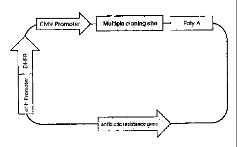
Fig. 3a is a diagram showing the gene map of the recombinant expression vector
for an
animal cell. Fig. 3b is a detailed diagram of the recombinant expression
vector for the animal
cell, p3K-DHFR-1. Fig. 3C is a diagram showing the gene map of pMS expression
vector used
to construct the recombinant expression vector for the animal cell in the
present invention. In
Fig. 3a, DHFR: coding nucleotide sequence of the human derived DHFR; dhfr
Promoter: SEQ
CA 02733119 2011-01-07
9
ID NO: 1 or SEQ ID NO: 2 derived from mouse.
Fig. 4 is a diagram showing the structure of the pJK-DFHR-2 expression vector
of the
present invention.
Fig. 5 is a diagram showing the structure of the pJK-DFHR-0r2 expression
vector of the
present invention.
Figs. 6a-6b are diagrams showing the structure of the pJKIg and pJKIg-RSV HK
expression vectors of the present invention.
Fig. 7 is an ELISA analysis result showing the expression level of human INFR
in the
cell line transformed with pJK-DHFR-1 vector.
Fig. 8 is an ELISA analysis result showing the expression level of human IDS
in the cell
line transformed with DHFR-Or2 vector.
Fig. 9 is an ELISA analysis result showing the expression level of RSV
antibody in the
cell line transformed with pJKIg vector.
Fig. 10 is an ELISA analysis result showing the expression level of GS051
antibody in
the cell line transformed with pJKIg vector.
Fig. 11-a is an ELISA analysis result showing the expression level of GS071
antibody in
the cell line transformed with pJKIg vector.
Fig. 11-b is an ELISA analysis result showing the expression level of GS071
antibody in
the cell line prepared by amplification with 20 nM MIX, and then subcloning.
Fig. 11-c is an ELISA analysis result showing the expression level of GS071
antibody in
the cell line prepared by amplification with 80 nM Is4Dc and then subcloning.
The present invention will now be described in further detail by examples. It
would be
obvious to those skilled in the art that these examples are intended to be
more concretely
illustrative and the scope of the present invention as set forth in the
appended daims is not
limited to or by the examples.
EXAMPLES
Example 1: Cloning of mouse derived DHFR promoter, SV40 early promoter and
SV40 virus early promoter and enhancer
CA 02733119 2011-01-07
The polymerase chain reaction (PCR) was performed as follows.
First, mouse genomic DNA was isolated using DNA extraction kit (Intion, Korea)
to
obtain the DHFR promoter region which is included in the 5'-end sequence of
DFHR gene and
has strong TATA sequence and basic promoter activity. The mouse derived DHFR
promoter
5 was
amplified by PCR using 200 ng of isolated mouse DNA, 50 pmol of P1 and P2
primers, 0.5
nnM of dNTP and Softmax DNA polymerase (Intron, Korea). The PCR cycle was 29
cycles of
denaturation at 1 min at 95 C, 40 sec at 50 C and 40 sec at 72 C followed by
10 min at 72 C
The DNA fragments of early promoter and promoter/enhancer (enhancer
operatively
linked to the promoter) were amplified by PCR using 10 ng of pcDNA3 vector
(Invitrogen,
10 USA) as the
template and either P3, P4 and P3, P5 Omer sets, respectively. The PCR method
(temperature, time and cycle) was similar as the method described above for
amplifying
mouse derived DHFR promoter. The base sequensw of each primers and the size of
the DNA
fragments obtained by PCR are shown in Table 1.
Table 1.
Primer Base sequence DNA
fragment size
PI 5'-TCGAAGCTTGATGGCAGCGGGGATAA-3 118 bp
P2 5'-GGGCTCGAGTAAGCA1T -3' 118 bp
P3 5'-GATAAGCITCGAAAAAGGATATACAA-3' 243 bp
P4 5'-CAACTCGAGCATCTCAA1TAGTCAGC-3' 243 bp
P3 5'-GATAAGC1TCGAAAAAGGATATACAA-3' 340 bp
P5 5.-CCACTCGAGCCAGGCAGGCAGAAGTA-3' 340 bp
P6 5'-CCCAAAATATGGGGA1TGGCAAGAAC-3' 1462 bp
P7 5'-GGGGGATCCGACATGATAAGATACAT-3' 1462 bp
P8 5'-GGGGGA1TCACTAGAGCA1T ACAGCTCAGGGCTGC-3' 165 bp
P9 5'-CCAATCCCCATA I 11 FGGGACACGGC-3' 165 bp
P8 5'-GGGGGATCCACTAGAGCA1T ACAGCTCAGGGCTGC-3' 1605 bp
P7 5'-GGGGGATCCGACATGATAAGATACAT-3' 1605 bp
P 10 5'-GGGGGATCCACAGCTCAGGCTGCGAT-3' 1583 bp
P7 5'-GGGGGATCCGACATGATAAGATACAT-3' 1583 bp
Pll 5'-TGCATCTAGATA1TCTATAGTGTCAC-3' 3I6bp
PI2 5'-CCCCAGCTGG1TC1 11 CCGCCTCAGAA-3' 316 bp
Each of the DNA fragment amplified by PCR was digested with restriction
enzymes
CA 02733119 2011-01-07
11
Hind/H and Xho/, purified by GeneClean III Turbo Kit (BIO 101, USA) then
subcloned into
pGL2-Basic vectors (Promega, USA) which were digested with the same
restriction enzymes,
to construct pGL2-DHFR vector, pGL2-SV40 promoter vector and pGL2-SV40
promoter/enhancer vector, respectively. The pGL2-Basic vector is a vector
encoding the
luciferase gene.
Example 2: Comparing the promoter activity by measuring the expression level
of
luciferase gene transcriptionally regulated by pGL2-DHFR, SV40 promoter and
SV40 promoter /enhancer
1) Gene transfecton
COS7 cells (ATCC, USA) were plated in DMEM (Dulbecco's Modified Eagle Medium;
GIBCO BRL, USA) supplemented with 10% fetal bovine serum and subcultured in a
37 C, 5%
CO2 incubator. The cells were plated at a density of 1 x 10⁶cells/m1 in a 100
mm culture plate
and incubated overnight at 37 C, before washing 3 times with OPTI-MEM
(osteogenic media
I; GIBCO BRL, USA) solution. Meanwhile, 5 pg of pGL2-Basic, pGL2-DHFR, pG1.2-
SV4OP
(Promoter) and pGL2-SV4O P/E (Promoter/Enhancer) prepared were each diluted in
500 pl of
OPTI-MEM I. Twenty five pl of lipofectamine (GIBCO BRL, USA) was also diluted
in 500 pl of
OPTI-MEM I. The expression vector and the diluted lipofectamine solution was
mixed in a 15-
ml tube and incubate at room temperature for 15 min or longer to allow DNA-
lipofectamine
complex to form. Each of the DNA-lipofectamine complexes was mixed with 5 ml
of OPTI-
MEM I then added homogeneously onto fresh rinsed COS7 cells. The cells were
incubated for
48 hrs in a 37 C, 5% CO2 incubator.
2) CompanSon of the lucitrase expression levels
The level of luciferase expressed in each vector was analyzed by comparing the
activities of the promoters inserted in the vector. After incubating the cells
for 48 hrs after the
transfection, the cells were washed with 5 ml of PBS. One ml of PBS was added
and the cells
were collected using a scraper. The cells were centrifugation at 9000 rpm for
5 min at 4 C and
the supernatant was discarded. To lysis the cells, 50 pl of 250 mM Tris (pH
7.8)/1 mM DTT
(Dithiothreitol) solution was added, and then submerged in the liquid nitrogen
for 1 min before
CA 02733119 2011-01-07
12
retuming to 1 min incubation at 37 C. This procedure was repeated for three
times. Then the
cell free supernatant were collected after centifugation for 15 min at 13,000
rpm at 4 C and
stored at -20 C.
The luciferase activity was measured by aliquoting 350 pl of solution A (25 mM
glycylglydne (pH 7.8), 0.2 M ATP, 1 M MgSO4, H20) in a 5 ml (12 x 75 mm) tube
then adding
100 pl each of solution B (25 mM glycylglydne (pH 7.8) and D-Iuciferin (5
mg/16.5 ml H20).
The tube was inserted in the luminometer for analysis. To measure the
luciferase activity, 40 pl
of sample solution was added in the solution A and the luciferase activity was
measured for 30
sec at 25 C. As a result, the newly selected DHFR basic promoter showed a
prominent
decrease of 2,300-fold and 3,800-fold lower promoter activities compared to
the existing SV40
promoter or SV40 promoter/enhancer (Table 2 and Fig. 2).
Table 2.
Vector Used Luciferase activity
Cell only 1,412
pGL2-Basic 2,457
pGL2-DHFR promoter 6,346
pGL2-SV40 promoter 14,713,514
pGL2-SV40 promoter/enhancer 24,355,978
Example 3: Construction of pJK-DHFR-1 vector
Human genomic DNA was isolated from human blood using DNA extraction kit
(Intron,
Korea) to clone the DHFR gene. DHFR gene was amplified by PCR using the
purified human
genomic DNA as a template.
The polymerase chain reaction (PCR) was performed as follows. First, DHFR gene
was
amplified by PCR using 200 ng of isolated human genomic DNA as the template,
50 pmol of
P6 and P7 primers, 0.5 mM of afTP and Softmax DNA polymerase (Intron, Korea).
The PCR
cycle was 29 cycles of denaturation at 1 min at 95 C, 40 sec at 55 C and 40
sec at 72 C
followed by 10 min at 72 C, resulting in amplification of 1462 bp DHFR gene.
Mouse DFHR
basic promoter was amplified by PCR using pGL2-DFHR as the template and P8 and
P9 primer
pair, following the PCR method (temperature, time and cycle) similar as
described above.
CA 02733119 2011-01-07
13
The 3'-region of amplified DHFR basic promoter and the 5'-region of DHFR gene
both
has conserved 19 bp base sequence region. This conserved region was PCR
amplified using
P8 and P7 primers, resulting in a 1605 bp DNA fragment, where the basic
promoter region
and DFHR gene region were connected. The base sequence of each primer and
their DNA
fragment size amplified by PCR are shown in Table 1.
The DNA fragment of the DHFR promoter and the DHFR gene amplified by PCR was
digested with restriction enzyme, BamH/and the pMS vector (Aprogen, Korea) was
digested
with Bg1// enzyme. The DHFR promoter and gene were then inserted into the
vector to
construct pJK-DHFR-1 (Fig. 3a and 3b). The pJK-DHFR-1 vector was deposited at
the gene
bank of Korea Research Institute of Bioscience and Biotechnology on March 11,
2008 (deposit
No: KCTC 11299BP).
Example 4: Construction of pJK-DHFR-2 vector
The pJK-DHFR-2 vector was constructed by shortening the DHFR promoter region
of
pJK-DHFR-1 vector. PCR amplification was performed by the method described in
Example 3.
The DFHR promoter and DHFR gene were amplified by PCR using plK-DFHR-1 vector
as the
template and using P10 and P7 primer pair. The base sequence of each primer
and their DNA
fragment size amplified by PCR are shown in Table 1.
The PCR amplified DNA fragment of the DHFR promoter and the DHFR gene were
digested with restriction enzyme, BamH/and the pJK-DHFR-1 vector were digested
with Bg1//
enzyme. The DHFR promoter and gene were inserted into the vector to construct
pJK-DHFR-2
(Fig. 4). The pJK-DHFR-2 vector was deposited at the gene bank of Korea
Research Institute
of Bioscience and Biotechnology on March 11, 2008 (deposit No: KCTC 11300BP).
Example 5: Construction of pJK-DHFR-0r2 vector
Following is the method for constructing pJK-DHFR-0r2 vector, which has the
DHFR
gene in a reverse direction compared to pJK-DHFR-1 vector. As described in
Example 3, the
DNA fragment of the DHFR promoter and the DHFR gene amplified by PCR was
digested with
restriction enzyme, BamH/and the pJK-DHFR-1 vector was digested with Bg1//
enzyme. The
DHFR promoter and the gene were inserted into the vector, and then screened
for the vector
CA 02733119 2011-01-07
14
that has DHFR gene cloned in the reverse direction. This vector is referred to
as pJK-DHFR-0r2.
(Fig. 5).
Example 6. Construction of recombinant antibody vector using p.11K1g vector
and
pJKIg vector
The pJKIg vector for doning the gene for antibody heavy chain and the light
chain were
constructed using pJK-DHFR-1 vector.
First, the Hind/htBamH/ fragment of the pJK-DHFR-1 vector was removed and then
ligated by treating with Klenow enzyme (Roche, Switzerland). The Xhol-Apa/
fragment was
removed from the vector and re-ligated. The vector was prepared by cutting
with Bsm/ and
treating with Klenow enzyme.
In another pJK-DHFR-1 vector, BamH/-Xho/ region in the multiple doning site
was
removed, and self-ligated using Klenow enzyme and ligase. To remove the Apa/
site on the
multiple cloning site, the vector was digested with Xba/and Pvu//restriction
enzymes. A 316
bp fragment PCR product of the Xba/and Pvu//region was PCR amplified using P11
and P12
primer pairs and re-inserted. The pJK-DHFR-1 vector inserted with Xba/ and
Pvu// fragment
were digested with Nru/-Pvu// restriction enzymes to generate a 1075 bp
fragment. This
fragment was inserted into the above mentioned vector which was digested with
Bsm/ to
generate pJKIg vector (Fig. 6a). A pJKIg-RSV HK vector, which has the heavy
chain and the
light chain of RSV (respiratory syncytial virus) antibody in the pJKIg vector,
was constructed as
follows. The variable and constant region of immunoglobulin heavy chain in
pGEM T/RSV
HvHc vector, which is a pGEM T vector (Promega, USA) inserted with a variable
and constant
region of the antibody heavy chain that interacts with RSV, was digested with
EcoRT-Not/
enzyme, and then inserted and ligated into pJKIg vector using the same
restriction enzyme
sites. Similar to the method described above, the constant and variable region
of the
immunoglobulin light chain in pGEM T/RSV KvKc vector was digested with
Hin/ThXba/enzyme,
and then inserted into pJKIg vector to construct pJKIg-RSV HK vector (Fig.
6b).
Example 7. Establishment of a cell line producing recombinant protein and
antibody to confirm the effectiveness of p3K-DHFR and pilag vectors
CA 02733119 2013-01-07
To use the piK-DHFR vector system, p3K-DHFR-1 and p3K-DHFR-0r2 vectors were
digested with EcoR/ and Xba/ restriction enzymes and inserted with cDNA
encoding human
derived TNF-R (tumor necrosis factor-receptor) and IDS (iduronate-2-sulfatase)
enzyme. The
RIKIg-GS051 H/K vector was constructed by cutting the heavy chain region in
pJK[g-RSV HK
5 vector with EcoR/and Apa/restriction enzymes and inserting cDNA encoding
the heavy chain
of GS051 antibody, and cutting the light chain region with Hind/H and Bsi W/
restriction
enzymes and repladng with cDNA encoding the light chain region of GS051
antibody. Also,
following the method described above, pJK[g-GS071 H/K vector expressing GS071
antibody
was constructed.
10 The vectors expressing the target protein or expressing antibody were
each transfected
into DHFR gene function deficient CHO DG44 (Columbia University, USA) cells
and the cell line
was primary selected using antibiotics, G418 (Giboo GRL, USA). The MIX
concentration in the
selected cell line culture medium was gradually increased to 20, 80, 320 and
1000 nM, the
highly productive cell line was selected according to the expression level of
target protein or
15 antibody in each clone. The expression level was analyzed by plating
each of the selected cell
line in a 6-well plate at a density of 5 x 10⁵ cells and incubating for 3
days. The medium was
collected for ELISA (Enzyme-Linked Immunosorbent Assay) analysis. The purified
protein with
a known concentration was used as a standard.
As the result shown in Fig. 7, a highly expressive cell line was selected at
80 nM
concentration of NTDC In Hg. 9, a highly expressive cell line was selected at
320 nM and 1 pM
concentration of IvIDC However, the cell line selected at 1 pM concentration
of MD( showed
slow cell growth. In Fig. 11, clones 3-5-6 that were amplified and selected at
20 nM and 80
nM of MIX were subdoned and selected for highly expressive cell line. By using
a weak DHFR
promote and gene, the present inventors provided evidence that the target
protein and
antibody is highly expressed at 0.005-0.32 pM of low concentrations of MTX
compared to the
existing MD( concentrations used for expressing the target protein and the
antibody.
Having described a preferred embodiment of the present invention, it is to be
understood that variants and modifications thereof may become apparent to
those skilled
in this art.