Note: Descriptions are shown in the official language in which they were submitted.
CA 02733789 2011-02-10
WO 2010/027651 PCT/US2009/054155
STENT GRAFT HAVING EXTENDED LANDING AREA
AND METHOD FOR USING THE SAME
FIELD OF THE INVENTION
The present invention relates to medical devices and associated methods for
treating various target sites and, in particular, to medical devices
configured for use in
arcuate lumens and associated methods for delivering such medical devices.
BACKGROUND OF THE INVENTION
An aortic aneurysm is a weak area in the aorta wall, which may be caused, for
example, by arteriosclerosis. As blood flows through the aorta, the weak area
of the
vessel wall thins over time and expands like a balloon. Most commonly, aortic
aneurysms occur in the portion of the vessel below the renal artery origins.
Eventually, an untreated aortic aneurysm will burst if the vessel wall gets
too thin.
Such rupturing of an aortic aneurysm frequently leads to death. As such, once
an
aneurysm reaches about 5 cm in diameter, it is usually considered necessary to
treat to
prevent rupture (below 5 cm, the risk of the aneurysm rupturing is considered
lower than
the risk of conventional heart surgery in patients with normal surgical
risks).
Aneurysms, including aortic aneurysms, may be treated with surgery. The
surgical procedure for treating an aortic aneurysm involves replacing the
affected portion
of the aorta with a synthetic graft, usually comprising a tube made out of an
elastic
material with properties very similar to that of a normal, healthy aorta.
However,
surgical treatment is complex and may pose additional risks to the patient,
especially the
elderly.
More recently, instead of performing surgery to repair an aortic aneurysm,
vascular surgeons have installed an endovascular stent graft delivered to the
site of the
aneurysm using elongated catheters. An endovascular stent graft is a tube
composed of
Mood impervious fabric supported by a metal mesh called a stent. It can be
used for a
variety of conditions involving the blood vessels, but most commonly is used
to reinforce
aneurysms. Typically, the surgeon will make a small incision in the patient's
groin area
and then insert into the vasculature a delivery catheter containing a
collapsed, self-
expanding or balloon-expandable stent graft. The delivery catheter is advanced
to a
-1-
CA 02733789 2011-02-10
WO 2010/027651 PCT/US2009/054155
location bridging the aneurysm, at which point the stent graft is delivered
out from the
delivery catheter and expanded to approximately the normal diameter of the
aorta at that
location. Over time, the stent graft becomes endothelialized and the space
between the
outer wall of the stent graft and the aneurysm ultimately fills with clotted
blood, which
prevents the aneurysm from growing further.
Depending on where the location of the aneurysm is within a vessel relative to
other branch vessels, different design variations of the stent graft may be
needed. For
example, in treating an aortic aneurysm in the area of the renal arteries, the
stent graft
should be placed so as not to exclude blood flow through the renal arteries.
Moreover,
the stent graft should be anchored within the lumen, such as by promoting
endothelialization or fixation with the lumen, in order to reduce the
incidence of
migration. Enhanced fixation of the stent graft to the arterial wall may also
reduce the
occurrence of endoleaks or blood flowing around the stent, which may prevent
further
weakening of the arterial wall at the site of the aneurysm.
Providing for adequate fixation of a stent graft in the area of the aortic
arch can be
challenging due to the various arteries that branch from the aorta in that
region. The stent
graft must provide adequate contact force against the vessel walls to prevent
migration
and endoleaks, but must not restrict blood flow to the branching arteries.
Therefore, there is a need for a stent graft that is capable of being deployed
in a
lumen having an arcuate portion, such as in the vicinity of the aortic arch.
The stent graft
should easily be deliverable and should be capable of being adequately
anchored within
the lumen.
SUMMARY OF THE INVENTION
Embodiments of the present invention provide a medical device, such as, for
example, a stent graft, for treating a target site within the body. For
example, one
embodiment provides a medical device for treating a target site within a lumen
having an
arcuate portion and at least one branch lumen extending therefrom. The medical
device
includes a first tubular portion comprising a proximal and distal end, and a
second tubular
portion comprising a proximal and distal end. The first and second tubular
portions each
may include at least one layer of a metallic material, such as a shape memory
alloy, that
is configured to be heat set to an expanded heat set configuration, and in
some cases may
-2-
CA 02733789 2011-02-10
WO 2010/027651 PCT/US2009/054155
include multiple layers. The first and second tubular portions may each be
configured to
be constrained to a smaller diameter than the respective expanded heat set
configuration,
for example, for delivery within a catheter, and may return to the respective
expanded
heat set configuration when deployed from the catheter.
The medical device further includes a linking portion that may be, for
example, a
filament, a fiber, a wire, a cord, a cable, a braid, a fabric, and/or a beam,
that couples the
first and second tubular portions. At least part of the linking portion may
have a preset,
memorized arcuate configuration that is configured to conform to at least a
portion of the
arcuate portion of the lumen. The linking portion may be resilient and/or
adjustable in
length. An opening may be defined between the distal end of the first tubular
portion and
the proximal end of the second tubular portion and be configured to align with
the at least
one branch lumen and facilitate fluid flow between the at least one branch
lumen and the
arcuate portion of the lumen. According to one aspect, a location of each of
the linking
portion and the opening within the arcuate portion of the lumen may be
rotationally
dependent on a location of the at least one branch lumen.
The first tubular portion may be, for example, a stent graft configured to be
positioned downstream of the arcuate portion of the lumen, such as within a
descending
thoracic aorta. The second tubular portion may be configured to anchor the
medical
device upstream of the arcuate portion of the lumen, such as within an
ascending thoracic
aorta. The linking portion may be configured to be positioned within the
arcuate portion
of the lumen, such as within an aortic arch, such that the opening defined
between the
first and second tubular portions is configured to align with at least one
branch lumen
extending from the arcuate portion of the lumen. In some embodiments, the
first tubular
portion, said second tubular portion, and linking portion can be integrally
formed from a
common material, such as a braided metallic material.
In another embodiment, a medical device for treating an aneurysm within an
aortic arch is provided. The medical device includes a first tubular portion
configured to
be positioned within a descending thoracic aorta and a second tubular portion
configured
to be positioned within an ascending thoracic aorta. The medical device
further includes
a linking portion coupling the first and second tubular portions and an
opening defined
between the first and second tubular portions. The linking portion is
configured to be
-3-
CA 02733789 2011-02-10
WO 2010/027651 PCT/US2009/054155
positioned within, and conform at least partially to, the aortic arch. The
opening is
configured to align with at least one artery extending from the aortic arch.
In yet another embodiment, a method of delivering a medical device to a target
site within a lumen having an arcuate portion and at least one branch lumen
extending
therefrom is provided. The method includes providing a medical device that has
a first
tubular portion comprising a proximal and distal end, a second tubular portion
comprising a proximal and distal end, and a linking portion coupling the first
and second
tubular portions. The medical device also includes an opening defined between
the distal
end of the first tubular portion and the proximal end of the second tubular
portion. The
first and second tubular portions and the linking portion can be constrained
from
respective expanded configurations to a smaller diameter for delivery within a
catheter,
for example, by respectively axially elongating the tubular and linking
portions. The
medical device can be delivered, for example, over a guidewire, to the target
site, where
the device can be deployed from the catheter such that the first and second
tubular
portions respectively assume their expanded configurations, the linking
portion conforms
to the arcuate portion of the lumen, and the opening aligns with the at least
one branch
lumen and facilitates fluid flow between the at least one branch lumen and the
arcuate
portion of the lumen.
In some embodiments, the first and second tubular portions respectively self.
expand and return to their expanded configurations when deployed from the
catheter. In
other embodiments, the first and second tubular portions may be axially
compressed so as
to urge the first and second tubular portions to return to the respective
expanded
configurations. In some embodiments, the medical device can be deployed from
the
catheter such that the second tubular portion is disposed within the ascending
thoracic
aorta of the body, the first tubular portion is disposed within the descending
thoracic
aorta of the body, and the linking portion is disposed within, and conforms
generally to,
at least a portion of the shape of the aortic arch. In addition, the
deployment of the
medical device may be rotationally dependent on a location of each of the
linking portion
and the opening within the arcuate portion of the lumen with respect to a
location of the
at least one branch lumen.
-4-
CA 02733789 2011-02-10
WO 2010/027651 PCT/US2009/054155
BRIEF DESCRIPTION OF THE DRAWINGS
Having thus described the invention in general terms, reference will now be
made
to the accompanying drawings, which are not necessarily drawn to scale, and
wherein:
Fig. 1 is a perspective view of a medical device configured in accordance with
an
exemplary embodiment;
Fig. 2 is a perspective view of the medical device of Fig. 1 in a constrained
configuration;
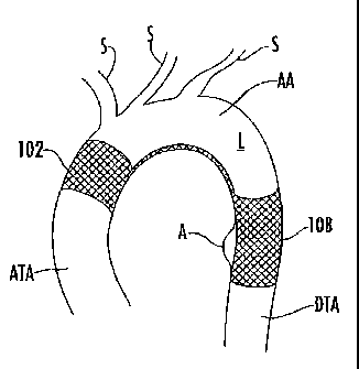
Fig. 3 is a cross-sectional view of the medical device of Fig. 1 deployed at a
target
site around an aortic arch;
Figs. 4-6 are perspective views demonstrating a process for fabricating a
medical
device in accordance with an exemplary embodiment;
Fig. 7 is a side view of the medical device of Fig. 1 disposed within the bore
of a
delivery catheter;
Fig. 8 is a perspective view of a medical device configured in accordance with
another exemplary embodiment;
Fig. 9 is a cross-sectional view of the medical device of Fig. 8 taken along
line 9-
9 of Fig. 8;
Figs. 10 and 11 are perspective views of medical devices configured in
accordance with still other exemplary embodiments;
Fig. 12 is an exploded perspective view of the medical device of Fig. 10;
Figs. 13A-B are perspective views demonstrating a process for fabricating a
medical device according to an additional embodiment;
Fig. 14 is a perspective view of a medical device according to another
embodiment of the present invention; and
Figs. 15-17 are perspective views of medical devices having an adjustable
linking
portion according to various embodiments of the present invention.
DETAILED DESCRIPTION OF THE INVENTION
The present invention now will be described more fully hereinafter with
reference
to the accompanying drawings, in which preferred embodiments of the invention
are
shown. This invention may, however, be embodied in many different forms and
should
-5-
CA 02733789 2011-02-10
WO 2010/027651 PCT/US2009/054155
not be construed as limited to the embodiments set forth herein; rather, these
embodiments are provided so that this disclosure will be thorough and
complete, and will
fully convey the scope of the invention to those skilled in the art. Like
numbers refer to
like elements throughout.
Embodiments of the present invention provide a medical device for use in
treating
a target site within the body, such as excluding or occluding various vascular
abnomialities, which may include, for example, excluding an aneurysm. The
device may
also be used as a graft for lining a lumen of a vessel. It is understood that
the use of the
term "target site" is not meant to be limiting, as the device may be
configured to treat any
target site, such as an abnormality, a vessel, an organ, an opening, a
chamber, a channel,
a hole, a cavity, or the like, located anywhere in the body. For example, the
abnormality
could be any abnormality that affects the shape or the function of the native
lumen, such
as an aneurysm, a lesion, a vessel dissection, flow abnormality or a tumor.
Furthermore,
the term "lumen" is also not meant to be limiting, as the abnormality may
reside in a
variety of locations within the vasculature, such as a vessel, an artery, a
vein, a
passageway, an organ, a cavity, or the like.
As used herein the term "proximal" shall mean closest to the operator (less
into
the body) and "distal" shall mean furthest from the operator (further into the
body). In
positioning of the medical device from a downstream access point, distal is
more
upstream and proximal is more downstream.
As explained in further detail below, embodiments of the present invention
provide medical devices for treating various target sites. The medical devices
may
include tubular portions separated and coupled by an arcuate or flexible
linking portion.
The arcuate or flexible linking portion may allow the medical device to
conform to an
arcuate target site. Moreover, the arcuate or flexible linking portion may
enable the
tubular portions to be disposed on opposing sides of an arcuate portion of a
target site,
which may improve the fixation of the medical device at such an arcuate target
site.
Furthermore, the medical device may include an opening configured to align
with one or
more branch lumens extending from the arcuate portion in order to reduce
blockage of
the one or more branch lumens.
-6-
CA 02733789 2011-02-10
WO 2010/027651 PCT/US2009/054155
Referring to Fig. 1, therein is shown a medical device 100 configured in
accordance with an exemplary embodiment. The medical device 100 includes a
first
tubular portion, such as a stent graft 108, which has a proximal 112 end and a
distal end
110. The medical device 100 also includes a second tubular portion, such as an
anchoring
structure 102, having a proximal end 106 and a distal end 104. Each of the
first and
second tubular portions may be generally cylindrical, but could be various
shapes
depending on the configuration of the lumen in which the tubular portions are
to be
positioned. An arcuate/linking portion 114 couples the stent graft 108 and
anchoring
structure 102, and an opening 116 extending between the proximal end 106 of
the
anchoring structure 102 and the distal end 110 of the stent graft 108.
Referring to Figs. 1 and 2, one or both of the stent graft 108 and anchoring
structure 102 may include at least one layer of a metallic material, the layer
being in the
form of, for example, a sheet or a woven, knitted, or braided tubular metallic
fabric. The
fabric can be composed of multiple metallic strands. Although the term
"strand" is
discussed herein, "strand" is not meant to be limiting, as it is understood
the fabric may
comprise one or more wires, cords, fibers, yams, filaments, cables, threads,
or the like,
such that such terms may be used interchangeably. The stent graft 108 and
anchoring
structure 102 may be a variety of occlusive materials capable of at least
partially
inhibiting blood flow therethrough in order to facilitate the formation of
thrombus and
epithelialization around the device. Moreover, the stent graft 108 and
anchoring structure
102 could be a self-expandable or balloon-expandable material, such as
stainless steel or
etched stents. For example, the stent graft 108 and anchoring structure 102
could be
independently expanded via respective balloons. One could mount the anchoring
structure 102 over the balloon of a balloon catheter and the balloon after
inflating and
expanding the anchoring structure could be deflated and retracted into the
stent graft and
used to expand the stent graft. Alternatively the anchoring structure and the
stent graft
could be mounted on a catheter having two spaced apart balloons where by each
of the
anchoring and the stent graft portions are mounted respectively onto the
distal and
proximal balloons such as by crimping.
In some embodiments, one or both of the stent graft 108 and anchoring
structure 102 may include multiple layers of metallic material. The layers may
have
-7-
CA 02733789 2011-02-10
WO 2010/027651 PCT/US2009/054155
different porosity or opening sizes. In addition, one layer may be for
structural support
and a second layer may inhibit blood flow through the layer. The layer(s) of a
metallic
material can be configured to be heat set to an expanded heat set
configuration. For
example, in one embodiment, one or both of the stent graft 108 and anchoring
structure 102 can be composed at least partially of a shape memory material in
order to
provide for being heat set in an expanded configuration and to retain the
expanded shape
at or below body temperature. The stent graft 108 and anchoring structure 102
can then
be configured to be constrained to a smaller diameter than their respective
expanded heat
set configurations for delivery through a catheter to a target location within
the body. For
example, the stent graft 108 and anchoring structure 102 may be braided
tubular
structures that have expanded configurations in which the outer diameters of
the stent
graft and anchoring structure are approximately the same as the inner diameter
of the
aorta, and which can be reduced to smaller diameters for delivery within a
catheter, such
as by axially elongating the stent graft and anchoring structure. The linking
portion may
be configured to pass through a catheter without necessarily needing any axial
elongation.
In one embodiment, the stent graft 108, anchoring structure 102, and/or
linking
portion 114 are formed from a shape memory alloy, such as Nitinol. It is also
understood
that the stent graft 108, anchoring structure 102, and/or linking portion 114
may comprise
various materials other than Nitinol that have highly elastic properties, such
as spring
stainless steel, and alloys such as Elgiloy , flastelloy , CoCrNi alloys
(e.g., trade name
Phynox), MP35NO, or CoCrMo alloys.
According to one embodiment, each layer of the device may comprise 36-144
wire strands ranging in diameter from about 0.0005 to 0.010 in. formed of a
shape
memory alloy that are braided so as to define fenestrations with an area of
about 0.00015
to 0.0015 sq. in., which are sufficiently small so as to slow the blood flow
through the
wall of the device and to facilitate thrombus formation thereon. Inner and
outer braided
layers may have pitch angles that are about equal to obtain desirable collapse
and
expansion characteristics, such as maintaining a uniform overall length. The
stiffness of
the device may be increased or decreased by altering the wire strand size, the
shield
angle, the pick rate, and the number of wire strand carriers or the heat
treatment process.
-8-
CA 02733789 2011-02-10
WO 2010/027651 PCT/US2009/054155
Thus, the stent graft 108 can also be configured to facilitate thrombosis, for
example, by at least partially inhibiting blood flow therethrough in order to
facilitate the
formation of thrombus and epithelialization around the stent graft. In
particular, the braid
of a metallic fabric may be chosen to have a predetermined pick and pitch to
define
openings or fenestrations so as to vary the impedance of blood flow
therethrough. For
instance, the formation of thrombus may result from substantially precluding
or impeding
flow, or functionally, that blood flow may occur for a short time, e.g., about
3-60 minutes
through the metallic fabric, but that the body's clotting mechanism or protein
or other
body deposits on the braided wire strands results in occlusion or flow
stoppage after this
initial time period. For instance, occlusion may be clinically represented by
injecting a
contrast media into the upstream lumen of the stent graft 108 and if no
contrast media
flows through the wall of the stent graft after a predetermined period of time
as viewed
by fluoroscopy, then the position and occlusion of the stent graft is
adequate. Moreover,
occlusion of the target site could be assessed using various ultrasound echo
doppler
modalities. Although the stent graft 108 has been described as having one or
more layers
of occlusive material, it is understood that the anchoring structure 102
and/or linking
portion 114 may also or alternatively include one or more layers of occlusive
material to
facilitate thrombosis or the wires may be coated with a thrombus promoting
substance.
According to one embodiment, the stent graft 108 could be configured to be
positioned within a lumen having an aneurysm. For instance, the stent graft
108 could be
positioned within a lumen having an aneurysm A in the descending thoracic
aorta (DTA).
In addition or alternatively, the anchoring structure 102 could comprise
occlusive
material and be configured to exclude an aneurysm in the ascending thoracic
aorta (A TA).
The linking portion 114 may have a preset, memorized arcuate configuration or
be flexible enough to easily conform to the curvature of an arcuate portion of
a lumen. In
some embodiments, the arcuate configuration may conform to at least a portion
of an
arcuate portion of the lumen of a vessel, this aspect making such embodiments
well-
suited for deployment within a lumen having an arcuate portion, such as, for
example, the
aortic arch (AA). For example, referring to Fig. 3, the stent graft 108 can be
configured to
be positioned downstream of the arcuate portion of the lumen L (e.g., in the
DTA), the
anchoring structure 102 can be configured to anchor the medical device
upstream of the
-9-
CA 02733789 2011-02-10
WO 2010/027651 PCT/US2009/054155
arcuate portion of the lumen (e.g., in the ATA), and the linking portion 114
can be
configured to be positioned within the arcuate portion of the lumen (e.g., in
the AA). In
this way, the opening 116 defined between the stent graft 102 and the
anchoring
structure 108 may be configured to align with at least one side or branch
lumen S
extending from the arcuate portion of the lumen L. Thus, the linking portion
114 may be
configured to conform to the arcuate portion of the lumen opposite the branch
lumens S,
while the opening 116 is configured to facilitate fluid flow between the
arcuate portion
and the branch lumens. Thus, the location of the linking portion 114 and the
opening 116
may be rotationally dependent on one another.
The linking portion 114 can include a filament, a fiber, a wire, a cord, a
cable, a
braid, a fabric, and/or a beam. The linking portion 114 can be composed at
least partially
of a shape memory material and may be heat set in the arcuate configuration.
Alternatively, the linking portion 114 can be formed (e.g., molded or cold-
worked) so as
to have an arcuate shape and highly elastic properties so as to pass through a
catheter and
retain its arcuate shape upon exiting the catheter. Moreover, the stent graft
102 and the
anchoring structure 108 may be a different material than that of the linking
portion 114.
For instance, the stent graft 102 and the anchoring structure 108 could be a
metal
naaterial, while the linking portion 114 could be fabricated of a polymeric
material.
The curvature of the linking portion 114 and/or orientation of the stent graft
108
to the anchoring structure 102 with respect to one another may vary depending
on the
particular arcuate lumen being treated or a particular patient. In many cases,
the linking
portion 114 may be resilient, either due to the material used to form the
linking portion,
the geometry of the linking portion, or both. For example, the reduced cross-
section of
the linking portion 114 (e.g., relative to that of the stent graft and
anchoring
structure 108, 102) may make an otherwise straight linking portion
sufficiently flexible to
conform to an arcuate portion of a lumen.
The size and configuration of the opening 116 may depend on the particular
linking portion 114 employed. In addition, the size and configuration of the
opening 116
chosen may depend on the number and location of branch lumens to be aligned
with the
opening. For example, a linking portion 114 comprising a thin or small
diameter wire or
band would provide a large opening 116 (see e.g., FIG. 10), whereas an opening
defined
40-
CA 02733789 2011-02-10
WO 2010/027651 PCT/US2009/054155
in a braided fabric may be much smaller (see e.g., FIGS. 1 and 8).
Furthermore, in one
embodiment, the linking portion 114 may include a "loose" fabric that is less
densely
braided than the stent graft 108 and anchoring structure 102, such that the
blood may
readily flow through the larger openings 116 defined in the loose fabric.
Therefore, at
least one opening 116 may be defined in the linking portion 114 and may be
located at
one or more locations in the linking portion (including up to about the entire
circumference of the linking portion).
Still referring to Figs. 1 and 2, the stent graft 108, anchoring structure
102, and
linking portion 114 can be integrally formed from a common material. For
example,
referring now to Figs. 4-6, a single braided metallic (e.g., Nitin.ol) tube
220 can be formed
by partially cross cutting the tube 220 along sections 222, 223 (e.g., by
cutting the wire
strands) in order to form a medical device 200 having a linking portion 214
and
opening 216 between a first tubular portion 208 and a second tubular portion
202. The
linking portion 214 may be formed by axial elongation to reduce its diameter
and
constraining the linking portion during a heat setting operation to memorize
the
constrained diameter. During the same heat treatment process, the stent graft
108 and
anchoring structure 102 may be heat set in their expanded diameters to
memorize their
shape. The braided tube 220 may also be heat formed so as to have an arcuate
region in
the portion that will become the linking portion 214, or can be processed
after forming
the linking portion 214 such that the linking portion assumes an arcuate
configuration, for
example, by being forced (e.g., via forces F in Fig. 6) into an arcuate shape
and then heat
set.
Figs. 13A-B and 14 illustrate a medical device 200 according to another
embodiment of the present invention. The device 200 may be formed from a
single
length of tubular braided shape memory alloy capable of being heat treated to
have a
shape transformation temperature below body temperature (e.g., 20-37 C).
Openings
216 can be formed in the tube 220 by pushing a cone shaped probe 230 into the
side wall
at one location but typically two axial aligned locations along the outer
surface to form
the openings 206, 210 as shown in Fig. 13B. Once the wires have been displaced
by the
conical probes 230 sufficiently to establish the desired opening diameter, the
braided
portion between the two openings 206, 210 can be axially elongated to form the
linking
-11-
CA 02733789 2011-02-10
WO 2010/027651 PCT/US2009/054155
portion 214 and opening 216. The process of forming the device 200 will result
in loose
wires that need to be manually realigned by axial tension from either wire
ends while
holding the device in the desired final device shape. Once the wires are
aligned as
desired, the device may be heat set to memorize the desired final shape. The
final device
shape as shown in Fig. 14 has formed openings 206, 210 at an angle less than
perpendicular to the central axis of the tubular portions 202, 208.
Referring to Figs. 8 and 9, therein is shown a medical device 400 with the
stent
graft 408 and anchoring structure 402 disposed coaxially with one another
according to
one embodiment of the present invention. The opening 416 extends between the
stent
graft 402 and anchoring structure 408 and may be defined, in cross section, by
a curved
(e.g., circular) sector having an angle a between 0 and 360 degrees. For
example, the
opening 416 may have an angle a in the range of about 45 to about 225 degrees.
The
medical device 400 shown in Figs. 8 and 9 could be resilient structure and
thereby
conform to an arcuate .vessel or heat set in the arcuate configuration as
described above.
Referring to Figs. 10-12, therein are shown medical devices 500, 600
configured
in accordance with further exemplary embodiments. The devices 500, 600 include
first
tubular portions 508, 608 and second tubular portions 502, 5-02. A linking
portion 517,
617 couples the corresponding first and second tubular portions 508, 502, 608,
602. As
indicated above, the linking portion 517, 617 can be include a filament, a
fiber, a wire, a
cord, a cable, a braid, a fabric, and/or a beam. In either case, the thickness
of the linking
portion 517, 617 can be sufficiently small as compared to its length so as to
be relatively
flexible. The flexibility of the linking portion 517, 617 may, in some cases,
facilitate
either delivery of the device 500, 600 to a target site in the body or the
ability of the
device to conform to the target site once delivered. The device 500 may be
fabricated by
separately forming the first 508 and second 502 tubular portions, forming the
linking
portion 517, and coupling the beam to the first and second tubular portions
(e.g., with
adhesives or laser welding).
In a further embodiment the medical device 600 may be fabricated by laser
cutting or acid etching a pattern into a shape memory tube to form the first
and second
tubular portions 602, 608 (see Fig. 11) by removing most of the circumference
of a
central portion between the tubular portions to leave a remainder linking
portion 614 and
-12-
CA 02733789 2011-02-10
WO 2010/027651 PCT/US2009/054155
corresponding opening 616. Alternatively, the tubular portions 602, 608 may be
= fabricated as individual components and connected to a separate linking
portion 617
similar to that shown in Fig. 12. The tubular portions 602, 608 may be
manually
expanded to the desired diameter and/or curved to an arcuate preset shape and
along with
the linking portion 617, heat set in an oven while constrained to the desired
final shape to
memorize the desired final device shape. The tubular portions 602, 608 may be
radially
compressed in diameter or elongated for delivery through a catheter to a
treatment site
within the body. The device may self expand to the memorized shape upon
exiting the
catheter. Catheter based delivery devices for self expanding stents may be an
appropriate
means for delivery of the medical device 600. It should be noted that the
devices 100,
200, 400, 500, 600 may be sized larger than the vessel diameter by 10-30% to
ensure that
the device exhibits an anchoring force against the vessel wall. The devices,
therefore,
may not achieve 100% of their preset shape when exiting a catheter restraint
due to vessel
resistance to expansion.
Referring again to Fig. 1, in some embodiments, the linking portion 114 may be
adjustable in length. For example, the linking portion 114 may include a
compressed
braid that can be selectively decompressed (and, in some cases, re-compressed)
to an
extent that is adjustable. Alternatively, the linking portion 114 can include
a series of
everting links.
Fig. 15 illustrates one exemplary embodiment for facilitating the length
adjustment of the linking portion 114. In particular, Fig. 15 illustrates that
the anchoring
structure 102 and stent graft 108 may include respective threaded portions
118, 120
configured to engage one another. The anchoring structure 102 may include a
threaded
connector 122 that is configured to engage a threaded end 124 on a distal
delivery device
126. The stent graft 108 may also include a threaded connector 128 that is
configured to
engage a threaded end 130 on a proximal delivery device 132. The distal
delivery device
126 is deliverable through an internal sheath 134 and catheter 136. Thus, both
the distal
delivery device 126 and internal sheath 134 are configured to be axially
displaced
through the threaded connector 128 and proximal delivery device 132. The
length of the
linking portion 114 may be adjusted by threading the threaded portions 118,
120 with
respect to one another, which may occur before delivery of the device based on
an image
-13-
CA 02733789 2011-02-10
WO 2010/027651 PCT/US2009/054155
of the target site (e.g., using fluoroscopy), or the device could be removed
prior to being
fully deployed and the length of the linking portion adjusted. When the
threaded ends
124, 130 are engaged with respective threaded connectors 122, 128, rotation of
the distal
126 and proximal 132 delivery devices results in adjustment of the length of
the linking
portion 114 as the threaded portions 118, 120 are rotated with respect to one
another.
Fig. 16 illustrates another embodiment wherein the length of the linking
portion
114 may be adjusted using a locking member 134. More specifically, the locking
member 134 may be configured to engage respective free ends 136, 138 of the
anchoring
structure 102 and stent graft 108. Thus, once the free ends 136, 138 have been
axially
displaced with respect to one another to achieve a desired length of the
linking portion
114, the locking member 134 may engage the free ends together to prevent any
further
axial displacement. The locking member 134 may include a pair of hook-shaped
members 140 that are configured to engage the free ends 136, 138 and may
include
various materials, such as a metallic material. The locking member 134 may be
configured to self expand upon release from a delivery catheter 142, which may
be
facilitated by a pusher shaft 144, wherein both the delivery catheter and
pusher shaft are
capable of being axially displaced within the free ends 136, 138 to a desired
location
prior to release of the locking member. The end of the delivery catheter 142
may include
a material capable of resisting puncture by the locking member 134 and
facilitate axial
displacement of the locking member out of the delivery catheter. For example,
the distal
end of the delivery catheter 142 may be a metallic material or reinforced
sleeve, while the
remaining portion of the delivery device may be a flexible, polymeric
material. The
locking member 134 may be radially constrained for delivery within the
delivery catheter
142, and the pusher shaft 144 may be used to push the locking member out of
the
delivery catheter.
Another embodiment of a medical device having an adjustable linking portion
114 is illustrated in Fig. 17. In this particular embodiment, the anchoring
structure 102
includes a clamp 150 and a single wire 152 extending proximally therefrom.
Similarly,
the stent graft 108 includes a clamp 154 and a pair of wires 156 extending
distally
therefrom, wherein the pair of wires are configured to extend over the wire
152. Once a
desired length of the linking portion 114 is obtained by axially displacing
the wire 152
-14-
CA 02733789 2016-01-12
= and the pair of wires 156 with respect to one another, the wires 152, 156
may be crimped
together with a clamp 158 or using any other suitable techniques for securing
the wires
together, such as with a set screw.
Referring to Figs. 1-3 and 7, in order to deliver the medical device 100 to a
target
site within a lumen having an arcuate portion, such as an aortic arch, the
stent graft 108
and the anchoring structure 102 can be constrained from respective expanded
configurations (shown in Fig. 1) to a smaller diameter (shown in Fig. 2). For
example,
where the stent graft 108 and the anchoring structure 1.02 are formed of a
braided metallic
fabric, each of the stent graft and the anchoring structure may have a first
diameter and
may be capable of being collapsed to a second, smaller diameter by being
axially
elongated.
The constrained device 100 can then be positioned in a delivery catheter 340,
which is a catheter that defines an axial bore 341. In this way, the device
100 is
maintained in the constrained configuration during delivery by the wall
defining the
bore 341 of the catheter 340. The catheter 340 and device 100 can then be
advanced, for
example, over a guidewire, until disposed at the target site (in this case the
aortic arch
area), where the device 100 can be deployed from the catheter. Once the device
100 has
been deployed completely out of the catheter 340, the stent graft 108 and
anchoring
structure 102 may assume the expanded shape (to the extent permitted by the
surrounding
vasculature, e.g., the ascending and descending thoracic aorta, respectively)
and the
linking portion may conform to the arcuate portion of the lumen (e.g., the
aortic arch).
Further examples of the procedures by which a medical device configured in
accordance
with exemplary embodiments can be delivered are provided in U.S. Patent Appl.
Publ.
No. 2006/0253184 filed May 4, 2005.
In some embodiments, the stent graft 108 and anchoring structure 102 may self-
expand upon being deployed from the catheter 340 as the constraining forces of
the
catheter are removed. In other embodiments, the stein graft 108 and anchoring
structure 102 may be physically urged into or toward the expanded shape, say,
by
inflating a balloon located within the stent graft and anchoring structure, or
by axially
compressing the tube following deployment from the catheter 340.
-15-
CA 02733789 2011-02-10
WO 2010/027651 PCT/US2009/054155
The location of the medical device 100 may be rotationally dependent on the
location of one or more branch lumens extending from the arcuate lumen, such
as the AA.
Thus, the linking portion 114 may be positioned opposite the openings of the
branch
lumens, while the opening 116 may be configured to align with the openings of
the
branch lumens in order to facilitate fluid flow therethrough. In order to aid
in the
alignment of the medical device within the lumen, the medical device may also
comprise
one or more radiopaque markers to indicate angular orientation of the device
such that the
linking portion 114 is located along the inside smallest radius of the arcuate
lumen (see
e.g., Fig. 3). For example, radiopaque markers could line the linking portion
or the
openings of the tubular portions adjacent the ostia of the branch lumens,
and/or the braid
itself could include one or more radiopaque strands so that the medical device
is properly
positioned and does not block any branch lumens. Radiopaque markers may also
facilitate location of the anchoring portion 102 and the stent graft 108
relative to desired
target locations. It is further contemplated that the expanded diameter
portions of the
anchoring portion 102 and the stent graft 108 may be heat set to incorporate a
corrugated
portion or a sinusoidal wave pattern in the outer surface to increase radial
strength as
described in pending U.S. Appl. No. 12/181,639, entitled Medical Device
including
Corrugated Braid and Associated Method. The anchoring portion 102 and/or the
stent
graft 108 may additionally comprise hooks for engaging the lumen to ensure the
device
does not migrate.
Embodiments of the present invention may provide several advantages. For
example, the medical device is capable of conforming to a variety of arcuate
portions
within a vessel and is, thus, adaptable for a variety of target sites and
patients. The
medical device may include a heat set or resilient linking portion that
facilitates such
adaptability. The linking portion may include an opening that is configured to
align with
one or more branch vessels extending from the arcuate portion such that the
opening
reduces blockage in the arcuate portion, such as in the aortic arch. The
medical device
may also include a stent graft configured to facilitate occlusion at a target
site, such as at
an aneurysm. Moreover, the medical device may include an anchoring structure
in order
to facilitate fixation within the vessel and reduce the incidence of
migration. Therefore,
the medical device is capable of treating target sites within a vessel that
may be otherwise
-16-
CA 02733789 2011-02-10
WO 2010/027651 PCT/US2009/054155
difficult to anchor therein or susceptible to blockage of branch vessels when
a
conventional stent graft is employed.
Many modifications and other embodiments of the invention will come to mind to
one skilled in the art to which this invention pertains having the benefit of
the teachings
presented in the foregoing descriptions and the associated drawings.
Therefore, it is to be
understood that the invention is not to be limited to the specific embodiments
disclosed
and that modifications and other embodiments are intended to be included
within the
scope of the appended claims. Although specific terms are employed herein,
they are
used in a generic and descriptive sense only and not for purposes of
limitation.
-17-