Note: Descriptions are shown in the official language in which they were submitted.
CA 02733792 2011-02-10
WO 2010/025131 PCT/US2009/054882
BONE MINERAL DENSITY RATIOS AS A PREDICTOR
OF OSTEOARTHRITIS
CROSS REFERENCE TO RELATED APPLICATIONS
This application claims priority to provisional patent application 61/092,246,
filed August
27, 2008, which is herein incorporated by reference in its entirety.
GOVERNMENT SUPPORT
This invention was made with government support under grant number ROl
AR051361-
01A1 awarded by the National Institutes of Health. The government has certain
rights in the
invention.
FIELD OF THE INVENTION
The present invention relates to systems, compositions, and methods for using
bone
mineral density ratios as a predictor of osteoarthritis. In particular, the
present invention relates
to comparing ratios of bone mineral density involving bones that are
periarticular to determine a
risk assessment for features of osteoarthritis.
BACKGROUND OF THE INVENTION
Osteoarthritis (OA, also known as degenerative arthritis, degenerative joint
disease), is
the most common form of arthritis, affecting at least 10% of the population
over the age of 65,
and at present there is little available in the treatment of this condition,
notwithstanding NSAIDs
and total joint replacements. Disability from OA is one of the leading causes
of disability in the
elderly. Unfortunately, the pathophysiology of this disease has not been
clarified to date.
The diagnosis of osteoarthritis (OA) is primarily based on history and
physical
examination. Usually, the clinical features that a patient exhibits
specifically the symptoms he
complains of and the signs noted on examination are sufficient to make the
diagnosis of OA.
To date, the most common means of confirming a diagnosis of OA is by obtaining
plain
radiographs of the affected joint; however, it is well-established that
radiographs are notoriously
insensitive to the detection of OA. Particularly because few effective
treatments are available to
treat this condition, identification of a measure that could predict the
development of OA would
1
CA 02733792 2011-02-10
WO 2010/025131 PCT/US2009/054882
be very useful. Additional methods are needed to assess early signs of
osteoarthritis and to
identify those who are at high risk of developing OA.
SUMMARY OF THE INVENTION
The present invention relates to systems, compositions, and methods for using
bone
mineral density ratios as a predictor of osteoarthritis. In particular, the
present invention relates
to comparing ratios of bone mineral density involving bones that are
periarticular to determine a
risk assessment for features of osteoarthritis.
Embodiments of the present invention provide inexpensive, non-invasive systems
and
methods for screening, diagnosing and monitoring the progression of
osteoarthritis. For
example, some embodiments of the present invention provide research and
clinical systems and
methods for utilizing BMD ratios for identifying subjects at risk of
developing osteoarthritis and
having progressive osteoarthritis. In some embodiments, the present invention
provides systems
and methods for screening compounds useful in the treatment or prevention of
osteoarthritis.
Accordingly, in some embodiments, the present invention provides a method of
determining a risk of osteoarthritis in a subject, comprising: determining one
or more ratios of
bone mineral density in a region of a joint bone (e.g., knee bone) of the
subject selected from, for
example, medial bone mineral density: lateral bone mineral density, medial
proximal bone
mineral density: medial bone mineral density, or proximal medial bone mineral
density: distal
medial bone mineral density; and identifying subjects at risk of developing
osteoarthritis when
the ratio of bone mineral density is increased relative to the level in
control subjects (e.g.,
subjects that do not have osteoarthritis, data from the same subject at an
earlier time period, etc.).
In some embodiments, the bone mineral density is determined using dual X-ray
absorptiometry
(DXA). In some embodiments, a medial proximal bone mineral density: medial
bone mineral
density greater than 1.32 is indicative of subjects at risk of developing
osteoarthritis.
In further embodiments, the present invention provides a method of monitoring
progression of osteoarthritis in a subject diagnosed with osteoarthritis,
comprising: determining
one or more ratios of bone mineral density at an initial time point in a
region of a joint bone (e.g.,
knee bone) of the subject selected from, for example, medial bone mineral
density: lateral bone
mineral density, medial proximal bone mineral density: medial bone mineral
density, or proximal
medial bone mineral density: distal medial bone mineral density; determining a
second one or
2
CA 02733792 2011-02-10
WO 2010/025131 PCT/US2009/054882
more initial ratios of bone mineral density at a later time point in a region
of a joint bone of the
subject selected from, for example, medial bone mineral density: lateral bone
mineral density,
medial proximal bone mineral density: medial bone mineral density, or proximal
medial bone
mineral density: distal medial bone mineral density; and identifying subjects
as having a
progression of osteoarthritis when the ratio of bone mineral density is
increased at the later time
point relative to the initial time point. In some embodiments, a medial
proximal bone mineral
density: medial bone mineral density greater than 1.32 is indicative of
subjects at risk of having
progression of osteoarthritis. In some embodiments, the bone mineral density
is determined
using dual X-ray absorptiometry (DXA). In some embodiments, the later time
point is
approximately one year after the initial time point. In some embodiments, the
method further
comprises the step of determining a further one or more initial ratios of bone
mineral density at
later time points in a region of a joint bone of the subject selected from,
for example, medial
bone mineral density: lateral bone mineral density, medial proximal bone
mineral density:
medial bone mineral density, or proximal medial bone mineral density: distal
medial bone
mineral density. In some embodiments, the later time points are spaced
approximately one year
apart. In some embodiments, the method further comprises the step of
administering a test
compound or other intervention to the subject.
Additional embodiments of the present invention provide a system, comprising:
an
imaging device; and computer hardware and software configured to calculate
bone mineral
density in a region of a joint bone of a subject selected from, for example,
medial bone mineral
density: lateral bone mineral density, medial proximal bone mineral density:
medial bone
mineral density, or proximal medial bone mineral density: distal medial bone
mineral density;
and a user interface configured to display the bone mineral density ratios. In
some embodiments,
the imaging device is a DXA device. In some embodiments, the imaging device
determines the
region of a joint bone. In some embodiments, the computer hardware maintains a
database of
bone mineral density ratio. In some embodiments, the bone mineral density
ratios in the
database are tagged with subject identification tags and time stamp tags.
Additional embodiments are described herein.
DESCRIPTION OF THE FIGURES
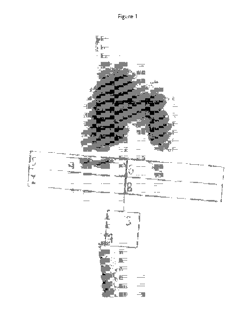
Figure 1 shows a DXA image of the knee.
3
CA 02733792 2011-02-10
WO 2010/025131 PCT/US2009/054882
Figure 2 shows a DXA image of the knee with labels identifying the medial and
lateral
zones (both proximal, distal and total).
Figure 3 shows a trabecular MRR sequence.
Figure 4 shows bone volume fraction vs. bone mineral density (BMD).
DEFINITIONS
As used herein, the term "substantially" refers to greater than 75% (e.g.,
greater than
80%, 85%, 90%, 95%, 98%, or 99%).
As used herein, the term "subject" refers to any animal (e.g., a mammal),
including, but
not limited to, humans, non-human primates, rodents, and the like, which is to
be the recipient of
a particular treatment. Typically, the terms "subject" and "patient" are used
interchangeably
herein in reference to a human subject.
As used herein, the term "subject suspected of having osteoarthritis in a
joint" refers to a
subject that presents one or more symptoms or risk factors indicative of
osteoarthritis (e.g., pain
on walking, family history, etc.) or is being screened for osteoarthritis
(e.g., during a routine
physical).
As used herein, the term "a subject diagnosed with osteoarthritis in a joint"
refers to a
subject that has been diagnosed with osteoarthritis based on one or more
diagnostic assays (e.g.,
MRI of the joint, x-ray, physical examination, etc.)
DETAILED DESCRIPTION OF THE INVENTION
The present invention relates to systems, compositions, and methods for using
bone
mineral density ratios as a predictor of osteoarthritis. In particular, the
present invention relates
to comparing ratios of bone mineral density involving bones that are
periarticular to determine a
risk assessment for features of osteoarthritis.
Animal models of OA show that increases in thickness of the subchondral plate
occur
early in, or even antedate, the development of cartilage loss (Radin et al., J
Orthop Res
1984;2:221-34; Carlson et al., J Orthop Res 1994;12:331-9). Studies in mice,
rabbits and dogs
(Benske et al., Acta Orthop Scand 1988;59:536-41; Newberry et al., J Orthop
Res 1997;15:450-
5; Burr DB, J Rheumatol Suppl 2004;70:77-80) indicate that thickening and
remodeling of the
subchondral plate is closely linked to cartilage destruction. The observation
that lower systemic
4
CA 02733792 2011-02-10
WO 2010/025131 PCT/US2009/054882
bone mineral density (BMD) is strongly associated with knee OA progression
indicates that
lower mineral content is also detrimental (Zhang et al., J Rheumatol
2000;27:1032-7).
According to Wolff's Law, trabecular size and orientation reflect internal
patterns of
tensile and compressive stress (Wolff, Clin Orthop Relat Res 1988:2-11). The
fact that these
forces increase with proximity to the articular surface indicates that the
ability of the trabecular
network to absorb loads is inversely related to intra-trabecular spacing, size
and connectivity. In
other words, multiple small trabecular compartments may be better able to
attenuate loads than a
smaller number of large compartments. Furthermore, it appears that the overall
pattern of
trabecular orientation contributes to pressure kinetics by influencing the
directionality of intra-
osseous fluid flow (Nauman et al., Ann Biomed Eng 1999;27:517-24).
Elevated peri-articular BMD as measured by DXA reflects an increase in the
amount of
mineralized bone in that region (Pastoureau et al., Osteoarthritis Cartilage
1999;7:466-73). At a
trabecular level this could result from an increase in thickness and volume of
the individual
trabeculae, and/or spatial compression or collapse of a number of trabeculae
into a smaller area.
Both are expected to impair the biomechanical properties of the bone. Thus,
elevated tibial peri-
articular BMD indicates a liability for development or progression of knee OA.
The medial:lateral (M:L) tibial BMD ratio has construct validity as an
indicator of knee
OA. It correlates with knee OA severity (Akamatsu et al., Clin Orthop 1997:207-
14; Wada et al.,
Rheumatology (Oxford) 2001;40:499-505) and with compartment-specific
radiologic features
including joint space narrowing (JSN), osteophytes and sclerosis (Lo et al.,
Osteoarthritis
Cartilage 2006;14:984-90). The M:L BMD ratio is more sensitive because it
retains an
association with radiographic knee even among knees that do not exhibit
radiographic sclerosis
(Akamatsu et al., supra). Furthermore, the M:L BMD ratio is associated with
subchondral
pathologies such as bone marrow lesions, which are themselves associated with
OA progression
(Akamatsu et al., supra; Lo et al., Arthritis Rheum 2005;52:2814-21; Carbone
et al., Arthritis
Rheum 2004;50:3516-25; Felson et al., Ann Intern Med 2001;134:541-9; Felson et
al., Ann
Intern Med 2003;139:330-6; Hunter et al., Arthritis Rheum 2006;54:1529-35;
Pessis et al.,
Osteoarthritis Cartilage 2003;11:361-9; Sowers et al., Osteoarthritis
Cartilage 2003;11:387-93).
Analyses of tibial DXA in various settings demonstrated tibial subchondral BMD
to be
associated with radiographic joint space loss and malalignment, cartilage
damage on MRI (Lo et
5
CA 02733792 2011-02-10
WO 2010/025131 PCT/US2009/054882
al., Arthritis Rheum 2006;56:S125), and with risk for progression of
functional decline (Smith et
al., Arthritis & Rheumatism 2008;58:5424).
There is also evidence that tibial subchondral BMD predicts risk for
subsequent
longitudinal progression of knee OA. The predictivity of a single unadjusted
measure (i.e. not the
ratio) of medial tibial subchondral BMD for subsequent 1-year loss of joint
space width
measured among 56 patients with knee OA was found to be strongly correlated
with the 1-year
change in minimum joint space width (r= -0.43, p=0.02). After adjustment for
age, sex, body
mass index, and baseline joint space width, BMD of the subchondral bone
remained predictive of
change in joint space width ((3 = -4.6, p=0.02).
Conversely, the tibial subchondral BMD appears to be responsive to
improvements in
mechanical loading (Akamatsu et al., Clin Orthop 1997:207-14; Katsuragawa et
al., Int Orthop
1999;23:164-7), a feature not seen in any other OA measure. One investigation
evaluated 23
knees with medial compartment OA following high tibial osteotomy (Akamatsu et
al., supra).
They reported that the medialaateral BMD ratio decreased sharply in all 23
knees within 1 year
after the procedure. Another studied the effect of a valgus knee brace for
medial compartment
knee OA. After 3 months the lateral:medial subchondral BMD ratio in the braced
knees
increased (i.e. improved) from an average of 0.69 0.12 to 0.71 0.13, and in
unbraced knees
from an average of 0.76 0.10 to 0.77 0.10 (Katsuragawa et al., supra).
In some embodiments, the present invention provides research, screening,
diagnostic, and
prognostic methods and systems for determining and utilizing BMD ratios.
1. BMD Ratios
Embodiments of the present invention utilize BMD ratios in research and
clinical
applications. In some embodiments, the present invention utilizes Dual X-ray
Absorptiometry
(DXA) or other imaging systems to measure Bone Mineral Density (BMD). DXA is a
means of
measuring BMD that utilizes technology where two X-ray beams with differing
energy levels are
aimed at the patient's bones (See e.g., US Patents 7,415,146, 6,217,214,
6,029,078, 5,785,041,
5,748,705, 5,687,211; each of which is herein incorporated by reference). When
soft tissue
absorption is subtracted out, the BMD can be determined from the absorption of
each beam by
bone. Dual energy X-ray absorptiometry (DXA) is the most widely used and most
thoroughly
studied bone density measurement technology. Common applications of DXA
measurements
6
CA 02733792 2011-02-10
WO 2010/025131 PCT/US2009/054882
include assessment of osteoporosis. Systems for performing DXA are
commercially available,
for example, from GE Medical Systems (Waukesha, WI) and Hologic (Bedford, MA).
Experiments conducted during the course of development of embodiments of the
present
invention demonstrated that the strength of relationships of different regions-
of-interest (figure
2) with OA characteristics varies, indicating that these reflect differing
biological processes
(Smith et al., Arthritis & Rheumatism 2008;58:5424).
Embodiments of the present application demonstrate the use of BMD ratios in
predicting
early joint OA, including early structural changes identified by MRI.
Experiments conducted
during the development of embodiments of the present invention resulted in the
development of
ratios of BMD that find use in predicting the risk of developing OA,
monitoring the progression
of OA, and monitoring the effectiveness of OA treatments (e.g., known and
experimental
treatments).
In some embodiments, the ratio is the ratio of proximal or closer to the
surface bone
BMD (e.g., substantially or completely subchondral plate) to distal or deeper
bone BMD (e.g.,
substantially or completely trabecular bone). In other embodiment, the ratio
is the ratio of
proximal BMD to total BMD. In some embodiments, the ratio of medial to lateral
BMD is then
calculated (e.g., proximal medial BMD to proximal lateral and distal medial to
distal lateral
BMD).
In still further embodiments, the BMD ratio is, for example, medial BMD:
lateral BMD,
medial proximal BMD: medial total BMD, and proximal medial BMD: distal medial
BMD.
In some embodiments, one or more (e.g., 2 or more, 3 or more, etc.) ratios may
be
utilized in combination. In some embodiments, different ratios that are
indicative of different
risk factors are utilized in combination.
II. Therapeutic Methods
In some embodiments, the present invention provides methods of screening,
diagnosing
and monitoring osteoarthritis in a joint. In some embodiments, the present
invention provides
methods of diagnosing osteoarthritis in a joint. In some embodiments, the
present invention
provides methods of identifying individuals at risk of developing
osteoarthritis in a joint.
The present invention is not limited to a particular cut off for determining
the risk of OA.
In some embodiments, a threshold of the ratios is indicative of an increased
risk of developing
7
CA 02733792 2011-02-10
WO 2010/025131 PCT/US2009/054882
OA, although other ratios may also find use. For example, in some embodiments,
those with a
medial proximal:distal ratio of > 1.3 have more pain with walking, difficulty
with walking and
slower walk time and those with a proximal medial:lateral BMD ratio of > 1.4
are associated
with medial tibio-femoral articular cartilage damage, with advanced
radiographic OA, and with
various malalignment (a known risk factor for medial tibiofemoral OA
progression).
Accordingly, in some embodiments, the present invention provides methods of
comparing ratios of BMD in a joint in order to determine risk of OA. In some
embodiments, the
regions to be compared are determined by an operator. In other embodiments,
determination of
the regions is automated (e.g., using software associated with the DXA or
other X-ray
equipment).
In some embodiments, the ratio is used to identify those at high risk for OA,
to monitor
progression of OA over time (e.g., measured multiple times per year, once per
year, or every 2 or
more years). In other embodiments, the ratio is used to monitor therapies over
time (e.g., non-
steroid anti-inflammatory medication or other OA treatment). In still further
embodiments, ratios
are used (e.g., in clinical studies) to assess new or candidate OA therapies.
The methods of embodiments of the present invention find use in assessing OA
in any
number of joints (e.g., knee (e.g., tibial plateau), hip, hand, finger, foot,
femur and vertebrae).
The methods of embodiments of the present invention are exemplified using the
knee. However,
the present invention is not intended to be limited to the assessment of OA in
the knee.
III. Systems
In some embodiments, a system is provided comprising imaging devices and
appropriate
software (e.g., software for data collection, data analysis, imaging device
control, user interfaces,
etc.). In some embodiments, data analysis software is incorporated into the
imaging device (e.g.,
on a computer processor attached to the imaging device).
In some embodiments, the system provides an image of the joint to be analyzed
and the
user (e.g., clinician) uses computer software to identify the regions for
calculating BMD ratios.
In other embodiments, the computer software identifies the regions of
interest. In some
embodiments, the computer software identifies the regions and the user refines
or revises the
regions. In some embodiments, the computer software refines the regions of
interest over time
based on user refinement and adaptive learning algorithms.
8
CA 02733792 2011-02-10
WO 2010/025131 PCT/US2009/054882
In some embodiments, a database of past patient data is used to refine regions
of interest
and diagnostic and/or prognostic assessments. In some embodiments, the
computer software and
computer hardware store region of interest information for a specific subject
so that identical
regions can be compared over time. In some embodiments, the computer software
and hardware
utilize a registration algorithm to confirm alignment and positioning of the
joint of interest in the
DXA machine.
In some embodiments, data analysis software provides information in a format
that is
useful for a clinician without further analysis. For example, in some
embodiments, one or more
BMD ratio are provided. In some embodiments, a representation (e.g.,
graphical) of the change
in BMD ratios of a given subject over time are provided. In some embodiments,
the data
analysis software provides a quantitative (e.g., probability) or qualitative
assessment of the risk
of developing osteoarthritis or the risk of progression of existing
osteoarthritis based on the
BMD ratios or the change in ratios over time.
IV. Drug Screening Methods
In some embodiments, the present invention provides methods of screening
candidate
osteoarthritis compounds. In some embodiments, compounds are administered to a
subject
diagnosed with osteoarthritis and the progression of disease is monitored over
time (e.g., in
comparison to a subject not diagnosed with or having symptoms of
osteoarthritis).
The test compounds of the present invention can be obtained using any of the
numerous
approaches in combinatorial library methods known in the art, including
biological libraries;
peptoid libraries (libraries of molecules having the functionalities of
peptides, but with a novel,
non-peptide backbone, which are resistant to enzymatic degradation but which
nevertheless
remain bioactive; see, e.g., Zuckennann et at., J. Med. Chem. 37: 2678-85
[1994]); spatially
addressable parallel solid phase or solution phase libraries; synthetic
library methods requiring
deconvolution; the 'one-bead one-compound' library method; and synthetic
library methods using
affinity chromatography selection. The biological library and peptoid library
approaches are
preferred for use with peptide libraries, while the other four approaches are
applicable to peptide,
non-peptide oligomer or small molecule libraries of compounds (Lam (1997)
Anticancer Drug
Des. 12:145).
9
CA 02733792 2011-02-10
WO 2010/025131 PCT/US2009/054882
Examples of methods for the synthesis of molecular libraries can be found in
the art, for
example in: DeWitt et at., Proc. Natl. Acad. Sci. U.S.A. 90:6909 [1993]; Erb
et at., Proc. Nad.
Acad. Sci. USA 91:11422 [1994]; Zuckermann et at., J. Med. Chem. 37:2678
[1994]; Cho et at.,
Science 261:1303 [1993]; Carrell et al., Angew. Chem. Int. Ed. Engl. 33.2059
[1994]; Carell et
at., Angew. Chem. Int. Ed. Engl. 33:2061 [1994]; and Gallop et at., J. Med.
Chem. 37:1233
[1994].
Libraries of compounds may be presented in solution (e.g., Houghten,
Biotechniques
13:412-421 [1992]), or on beads (Lam, Nature 354:82-84 [1991]), chips (Fodor,
Nature 364:555-
556 [1993]), bacteria or spores (U.S. Pat. No. 5,223,409; herein incorporated
by reference),
plasmids (Cull et at., Proc. Nad. Acad. Sci. USA 89:18651869 [1992]) or on
phage (Scott and
Smith, Science 249:386-390 [1990]; Devlin Science 249:404-406 [1990]; Cwirla
et at., Proc.
Natl. Acad. Sci. 87:6378-6382 [1990]; Felici, J. Mol. Biol. 222:301 [1991]).
EXPERIMENTAL
The following examples are provided in order to demonstrate and further
illustrate certain
preferred embodiments and aspects of the present invention and are not to be
construed as
limiting the scope thereof.
Example 1
Overall 50.2% of participants were female, 72.3% were White, 25.5% were Black,
1.1%
were Hispanic, and the mean age was 66.1 years. All participants had MRIs,
knee DXA scans,
and blood samples drawn per protocol. At baseline, there were 14 participants
without a
trabecular sequence scan or completed MRI. There were 11 (1.8%) participant
withdrawals from
the ancillary and/or parent study. Measurement of tibial subchondral BMD in
the 6 regions of
interest was performed on 425 participant knee DXA scans.
This analysis was based on the first 226 enrollees, a sample with mean age
65.3 years
(s.d. 9.0), 46.5% were male, 73.5% were White, and mean body mass index (BMI)
was 29.9 kg
M-2 (s.d. 5.1). The computation of tibial subchondral BMD and the ratio
measures was based on
the regions of interest illustrated in figure 2. Absolute medial BMD was
derived from box 1; the
medialaateral ratio from boxes 1 and 2; and the medial:medial ratio from boxes
3 and 5.
CA 02733792 2011-02-10
WO 2010/025131 PCT/US2009/054882
The medial: lateral tibial subchondral ratio in the overall group was 1.13
(s.d. 0.15). Knees with
greater pain, characterized by WOMAC pain subscale score >7 had a mean
medialaateral BMD
ratio of 1.17 (s.d. 0.16) compared to the rest of the sample whose mean value
was 1.13 (s.d.
0.15), a difference of -0.04. Mean medial: lateral BMD ratio values (s.d.)
according to presence
and severity of radiographic joint space narrowing was as follows: no joint
space narrowing 1.08
(0.13); mild/moderate joint space narrowing (grades 1 and 2) 1.17 (0.19);
severe joint space
narrowing (grades 3) 1.30 (0.14).
There were highly significant relationships of tibial subchondral BMD with
structural
features - alignment and joint space narrowing (Tables 1 and 2). Absolute
medial tibial
subchondral BMD is strongly correlated with femoral neck BMD.
Table 1. Tibial Subchondral BMD and Knee Alignment
Absolute BMD BMD Ratios
coefficient; p fl-co efficient; p
Medial (g/cm2) Medial: Lateral Medial: Medial
arcs 0.11; p=0.01 0.09; p=0.003 0.02; p=0.04
normal (ref.) - - -
algus 0.04; p=0.3 -0.04; p=0.1 0.009; p=0.2
Table 2. Tibial Subchondral BMD and Joint Space Narrowing
Absolute BMD BMD Ratios
fl-co p fl-co efficient; p
JSN Grade Medial (g/cm2) Medial: Lateral Medial: Medial
0 (ref.) - - -
1 or 2 0.05; p=0.1 0.09; p<0.0001 0.02; p=0.002
3 0.21; p<0.0001 0.21; P<0.0001 0.05; p<0.0001
Table 3. Tibial Subchondral BMD and Demographics
Absolute BMD BMD Ratios
fl-co p fl-co efficient; p
Medial (g/cm2) Medial: Lateral IIvIedial:Medial
11
CA 02733792 2011-02-10
WO 2010/025131 PCT/US2009/054882
ge -0.006; p=0.0001 0.001; p=0.4 0.0007; p=0.009
Gender 0.18; p<0.0001 0.05; p=0.02 0.004; p=0.4
Race -0.01; p=0.7 0.06; p=0.01 0.006; p=0.3
BMI 0.02; p<0.0001 0.002; p=0.4 -0.0009; p=0.05
Femoral BMD 0.88; p<0.0001 0.05; p=0.4 -0.04; p=0.01
There were also relationships with other covariates (Table 3). There is a
negative
correlation between medial tibial subchondral BMD and age, but that
association is in the
opposite direction for the medial:medial ratio, possibly on a mechanistic
basis. Adjustment of
these relationships for structural covariates made little difference to the
correlations or level of
significance.
It was investigated whether tibial subchondral BMD predicts longitudinal
progression in
functional ability and pain among individuals with symptomatic knee OA. The
sample was
drawn from participants in a trial of vitamin D for knee OA had complete
function assessments
and WOMAC questionnaire reports both at baseline and at their one-year follow-
up visit.
Walking ability was assessed using a timed 20-meter walk test. Walking pain
and difficulty
were assessed using the two pertinent WOMAC questions. Worsening on the timed
walk test
was defined as an increase in walk time between baseline and the one-year
assessment and
worsening on the WOMAC questions as an increase in reported severity. DXA
scans of both
knees were obtained at baseline using a GE-Lunar scanner. Subchondral BMD in
regions of
interest as depicted in figure 2 were calculated. Different combinations of
tibial subchondral
BMD ratio measures including medial: lateral (box 1 versus box 2 in figure 2)
and within medial
compartment (box 3 versus box 1 in figure 2) were calculated. Logistic
regression with case-
based tertiles of BMD ratios as predictors, and worsening of walk time,
walking pain, and
walking difficulty as outcomes was performed. Analyses were adjusted for age,
sex, BMI, and
radiographic OA severity (Kellgren-Lawrence grade). These analyses were
repeated with the
Kellgren-Lawrence grades as a covariate.
The eligible participants (N=89) had a mean age of 64.2 years (+8.7), and mean
BMI of
30.3 Kg m-2 (+5.3) and 67.4% were female. Those with a greater within medial
compartment
BMD ratio were significantly more likely to report worse pain or exhibit
deterioration in walk
12
CA 02733792 2011-02-10
WO 2010/025131 PCT/US2009/054882
time at follow-up (Table 4). However, no such relationship was found with the
medial to lateral
ratio.
The observation that the within medial, but not medial to lateral, tibial
subchondral BMD
ratio was associated with worsening pain and function indicates that these
have differing
biological relevance. The influence of the within medial BMD ratio was also
independent of
radiographic OA severity, indicating that this DXA measure is more predictive
of patient
outcomes than radiography.
Table 4. Within medial compartment tibial subchondral BMD* and knee OA
progression
Textile Range Odds Ratio (95% CI)
Worsening of Pain during
Walking
1 1.03-1.32 1/38 (2.6%) Referent -
2 1.32-1.40 6/24 (25.0%) 12.7 1.4-118
3 1.40-1.70 10/27 (37.0%) 26.9 3.0 - 243
Worsening of Difficulty Walking
1 1.03-1.32 2/38 (5.3%) Referent -
2 1.32-1.40 6/24 (25.0%) 4.9 0.8 - 29
3 1.40-1.70 6/27 (22.2%) 4.9 0.8-28.9
Worsening of 20-m Walk Time
1 1.03-1.32 17/38 (44.7%) Referent -
2 1.32-1.40 18/24 (75.0%) 4.1 1.3- 13.1
3 1.40-1.70 18/27 (66.7%) 2.7 0.9-8.1
* Within medial compartment tibial subchondral BMD ratio defined as ROI box 3
vs. box 1
(figure 2)
Experiments were conducted to test whether BMD measures of the superficial
zone of
subchondral bone (0-1 cm), which includes the subchondral plate, have stronger
association with
radiographic features of knee OA than those of deeper bone (1-2 cm). This
study used the same
sample of participants as described above. All participants had baseline
posterio-anterior
13
CA 02733792 2011-02-10
WO 2010/025131 PCT/US2009/054882
semiflexed weight-bearing knee radiographs, knee DXA scans and 1.5 Tesla MRIs
taken of their
study knee. The knee radiographs were scored for OA severity according to the
Kellgren and
Lawrence system. Cartilage damage was assessed on the MRIs using the
semiquantitative
Boston Leeds Osteoarthritis Knee Score (BLOKS), which was developed for this
purpose
(Hunter et al., Osteoarthritis & Cartilage; 13:241).
Knee DXA scans were used to calculate BMD ratios between medial and lateral,
superficial and deep, regions of interest as depicted in figure 2. X-rays were
scored for Kellgren
and Lawrence grade (0-4) and anatomic alignment. To convert the anatomic
alignment to
mechanical axis (MA), in women 3.5 degrees were subtracted from the anatomic
alignment and
in men 6.4 degrees, as recommended (Kraus et al., Arthritis Rheum 2005;52:1730-
5). A logistic
regression with case-based tertiles of BMD ratios as predictors and moderate-
severe cartilage
loss in the medial tibiofemoral compartment as the outcome was performed.
These tertiles of
medial to lateral BMD were also used to predict radiographic OA severity
(Kellgren and
Lawrence grade 3 or 4) and biomechanical alignment. The analyses was repeated
with the
superficial medial to lateral BMD (0-1 cm depth) and the deep medial to
lateral BMD (1-2 cm
depth) as predictors. This analysis found strong associations of the
superficial medial to lateral
BMD ratio with ipsi-compartmental cartilage damage (by MRI), Kellgren and
Lawrence
radiographic severity grade, and biomechanical axis (Table 5). Those with the
highest
superficial medial to lateral BMD ratio all had Kellgren and Lawrence
radiographic severity
grade 3 or 4 and had varus malalignment. However, the deep medial to lateral
BMD ratio
showed similar associations with cartilage damage (by MRI), Kellgren and
Lawrence
radiographic severity grade, and biomechanical axis, albeit less strongly.
These indicate that
biologically relevant bone changes in knee OA extend beyond the superficial
subchondral
region.
Table 6. Vitamin D Status and Longitudinal Change in Tibial BMD in Knee OA
Vitamin D Group Hith vs. Low
M:L BMD change Proportional
Low High OR (95% CI)
groups OR (95% CI)
Increase 0.045 to 0.158 12/40 (30%) 8/40(20%) 0.6 (0.2 - 1.6) 0.4 (0.2 - 0.9)
14
CA 02733792 2011-02-10
WO 2010/025131 PCT/US2009/054882
-0.038 to
Stable 0.041 23/40 (57.5%) 17/40 (42.5%) Referent
Decrease -0.177 to -0.03 5/40(12.5%) 15/40(37.5%) 4.2(l.4-13.1)
Using the study sample described above the relationship between vitamin D
status and
change in knee BMD was analyzed. 25-hydroxyvitamin D levels were obtained at
baseline using
a commercial HPLC/Mass spectrometry Assay. Knee DXA scans were obtained at
baseline and
at one year follow-up. These were used to calculate the medial to lateral
tibial BMD ratio with a
region of interest depth of 2cm. Those in the highest quartile of change in
medial to lateral tibial
BMD ratio were defined as increased, those in the middle two quartiles as
stable, and those in
the lowest quartile as decreased. The median vitamin D level was used to
dichotomize those with
a high vs. low vitamin D level. To focus on medial compartment disease, knees
with moderate-
severe cartilage thickness loss in the lateral tibiofemoral compartment were
excluded.
Logistic regression analyses were performed using increase in medial to
lateral tibial
BMD ratio as the outcome and baseline vitamin D as the predictor and repeated
using decrease
in medial to lateral tibial BMD ratio as the outcome. Table 6 presents the
results of these
analyses.
Table 5. Proximal & Distal Tibial Subchondral BMD Ratios as Predictors of OA
Structural
Features
Medial tibiofemoral
X-Ray: KL 3 or 4 Varus malalignment
cartilage damage (MRI)
OR OR OR
Prevalence Prevalence Prevalence
95% CI 95% CI 95% CI
Tertile 1 11/68 - 20/68 - 20/68 -
36.3 10.4 16.8
PROXIMA Tertile 2 14/16 13/16 14/16
7.2-182 3.2-29 3.5-81
L M:L BMD
20.7
Tertile 3 12/15 15/15 co 15/15 co
5.1 - 86
DISTAL Tertile 1 12/59 - 20/68 - 19/68 -
CA 02733792 2011-02-10
WO 2010/025131 PCT/US2009/054882
M:L BMD 7.3 9.6 6.3
Tertile 2 13/20 13/16 15/16
2.4-22 3.2-29 2.0-20
5.9 28.9 6.3
Tertile 3 12/20 15/15 15/15
2.0-18 5.6-148 2.0-20
Sufficient change occurred in medial to lateral tibial BMD ratios to detect a
difference
over one year. Furthermore, baseline vitamin D level predicted change in
medial to lateral tibial
BMD ratio, such that those with a high level were less likely to have an
increase in BMD ratio
and more likely to have a decrease (Table 6). Since greater medial to lateral
tibial BMD ratios
are associated with greater OA severity, this indicates a protective
relationship between vitamin
D and knee OA.
At baseline, the incidence cohort had 3,284 participants with 6,472 knees
available for
analysis (96 knees had missing information on radiographic change or
symptoms). The
breakdown of radiographic changes of OA and frequent knee pain among this
sample at baseline
was as follows:
= Normal knees (no radiographic OA and no knee pain) 2,489 (38%)
= Radiographic knee OA only (radiographic changes but no knee pain 2,870 (44%)
= Knees pain but no radiographic OA 785 (12%)
= Symptomatic knee OA (knee pain plus radiographic changes) 328 (5%)
For the 12-month follow-up exam, symptom data was available for 6153 knees
space.
Among the knees classified at baseline as "normal", 12% now have chronic knee
pain. Among
the knees which at baseline had only radiographic changes, 14% now have
chronic pain. This
makes them classifiable as having developed incident symptomatic knee OA.
For the 24-month follow-up exam, symptom information was available for the
first half
of the cohort (3312 knees). Among the subset that had radiographic changes
only at baseline, the
prevalence of chronic knee pain is approximately 16% (i.e. have symptomatic
knee OA).
4Qimaging has developed a fully automated, atlas-based segmentation and
analysis
system to segment and analyze cartilage and bone features, and other anatomic
regions from
knee MR image data (Clinical Image Processing and Analysis System - CiPAS).
The objective
of this analysis was to compare the repeatability and reproducibility of their
automated system
against an expert radiologist.
16
CA 02733792 2011-02-10
WO 2010/025131 PCT/US2009/054882
The atlas for the automated system was created by manually tracing five
subjects' 3D
DESS images from the OAI public use dataset and selecting the best performing
atlas for further
refinement. The repeatability study used 30 randomly selected 3D DESS images
from the OAI
public use dataset. Of these, 10 were randomly selected to create 40 de-
identified images for
manual and automated segmentation. The automated segmentations were performed
five times
with varying initial parameters. The final measurements were obtained by
trimming the highest
and lowest values and averaging the three remaining measurements.
The reproducibility test used 38 de-identified image sets from 19 subjects who
participated in a scan-rescan reproducibility test for the OAI pilot study.
These were segmented
both semi-manually and automatically. The automated measurements were
generated with a
trimmed average of five segmentations using varying initial parameters.
Quantitative measurements of the central medial and lateral tibial and femoral
cartilage
included volume, articulating surface area, subchondral bone surface area,
average thickness and
standard deviation of average thickness, as well as the bone parameters that
we proposed to
analyze in this competing revision (see table 7). Those values ranged from
1.7% to 5.37% RMS
CV for the automated approach and 3.9 to 7.8% RMS CV for the expert edited
approach.
The RMS CV includes error from re-positioning and reacquiring the image as
well as
measurement error. This data set contained a mix of healthy and significantly
arthritic subjects
and the largest source of variation was from the abnormal subjects.
Table 7. Reproducibility (root mean square coefficient of variation) of CiPAS
measurements
proposed for use in this competing revision compared to manual segmentation by
an expert
Quantitative Measurements of Bone Shape and Signal Parameters 4Qi Expert
Medial Tibia Subchondral Bone Surface Area 2.95% 5.17%
Medial Tibia Subchondral Bone Surface Area 2.46% 5.69%
Medial Tibia Cartilage-bone Contrast 9.43% 17.6%
Lateral Tibia Cartilage-bone Contrast 9.14% 12.4%
Medial Tibia Bone Curvature 31.46% 24.4%
Lateral Tibia Bone Curvature 46.5% 55.4%
17
CA 02733792 2011-02-10
WO 2010/025131 PCT/US2009/054882
The automated atlas based MR image analysis system used in this study to
segment the knee into
bones and cartilage, and divide the joint in regions and in sub-segments
provided repeatable and
highly reproducible signal intensity measurements in the medial and lateral
weight bearing
regions of the knee. These automated tools provide a realistic opportunity to
characterize the
behavior of structural and compositional changes in cartilage and non-
cartilage tissues in OA by
analyzing larger populations such as the OAI or other longitudinal datasets.
Example 2
Association of Absolute And Relative Tibial Subchondral BMD Measures With
Individual
Characteristics And Structural Features Of Knee Osteoarthritis
The absence of a biomarker for detection and monitoring of knee osteoarthritis
(OA) is a
fundamental obstacle to the development of structure-modifying interventions.
This Example
describes the use of tibial subchondral dual x-ray absorptiometry (tsDXA)
finds use to generate
reproducible measures of knee bone mineral density (BMD). DXA involves low
radiation, is
easy to operate, relatively inexpensive and widely available.
This was a cross-sectional analysis of right knee of 226 enrollees into the
Osteoarthritis
Initiative (OAI) Bone Ancillary Study, who received standardized semiflexed
knee radiography
and tsDXA. Medial JSN (0 - 2) and osteophytosis (0-1) was scored on parent
study (OAI)
baseline images. A goniometer was used to evaluate static alignment on OA1
baseline physical
exam. Normal alignment was 0 degrees, valgus was <0 and varus was >0. Knee and
femoral
neck DXAs were obtained at either the OAI 30 or 36 month follow-up visit.
tsBMD was
computed from the tibial subchondral bone: absolute medial tibial BMD; medial
tibial: lateral
tibial ratio; medial proximal tibial: medial tibial ratio.
The mean age was 65.3 years (s.d. 9.0), 46.5% were male, 73.5% White, mean BMI
was
29.9 kg m 2 (s.d. 5.1); 25.7% had varus deformity, 38.9% had radiographic
medial tibiofemoral
JSN of grade I or 2 and 84.5% had osteophytosis. The mean (s.d.) values for
the tsBMD
measures in the sample were: absolute medial 1.16 (0.21); medialaateral ratio
1.13 (s.d. 0.15);
medial:medial ratio 1.14 (s.d. 0.04). The associations of the tsBMD measures
with structural
features of OA and participant characteristics are presented in the Table 8.
All tsBMD measures were positively associated with the highest grade of medial
JSN, a
hallmark of knee OA. Further. all tsBMD measures were also associated with
varus alignment.
18
CA 02733792 2011-02-10
WO 2010/025131 PCT/US2009/054882
Higher absolute medial BMD was associated with younger age, male sex, greater
BMI, and
systemic BMD. Higher medial:lateral BMD ratio was associated with male sex and
white race.
Higher medial:medial BMD ratio was associated with older age, lower BMI, and
lower systemic
BMD.
Each measure of tsBMD is associated with medial JSN and with varus
malalignment,
indicating that these are meaningful measures of knee OA. However, each is
also associated with
a different established risk factor of knee OA in the expected direction,
absolute medial BMD
with BMI, medial:lateral ratio with White race, and medial:medial ratio with
age, indicating that
each measure might reflect a different process occurring in medial
tibiofemoral knee OA.
Absolute medial BMD and the medial:medial ratio are associated with systemic
BMD in
opposite directions.
Table 8
Absolute BMD BMD Ratios
Medial BMD (g/cm2) Medial:Lateral Medial:Medial
Beta p-value Beta p-value Beta p-value
Medial JSN grade 0 --- --- --- --- --- ---
(ref)
Medial JSN grade 1 0.05 0.1 0.09 <0.0001 0.02 0.002
Medial JSN grade 2 0.21 <0.0001 0.21 <0.0001 0.05 <0.0001
Varus 0.11 0.01 0.09 0.003 0.02 0.04
Normal (ref) --- --- --- ---
Valgus 0.04 0.3 -0.04 0.1 0.009 0.2
Age -0.006 <0.0001 0.001 0.4 0.0007 0.009
Sex (male) 0.18 <0.0001 0.05 0.02 0.004 0.4
Race (white) -0.01 0.7 0.06 0.01 0.006 0.3
BMI 0.02 <0.0001 0.002 0.4 -0.0009 0.05
Femoral Neck 0.88 <0.0001 0.05 0.4 -0.04 0.01
BMD
19
CA 02733792 2011-02-10
WO 2010/025131 PCT/US2009/054882
Example 3
Increased Medial Tibial Bone Mineral Density (BMD) is Associated with
Deterioration in
Walking Ability and Pain in Individuals with Knee Osteoarthritis (KOA)
This Example describes a cross-sectional evaluation of baseline data for
evaluation of
baseline knee BMD data with longitudinal change of functional status.
Participants enrolled in
an ongoing randomized controlled clinical trial of vitamin D for KOA who had
data from both
baseline and 1 year follow-up visits and were age 45 and older at time of
enrollment and had at
least 1 knee with symptomatic radiographic tibio-femoral KOA (K/L grade > 2)
were eligible for
participation. Each participant was assigned a study knee based on K/L grade
and pain
symptoms Baseline and 1-Year Visits. 20 meter timed walk test (2 trials),
timed chair stand test
(2 trials of 5 chair stands), WOMAC pain and function questions (Likert) and
bilateral knee
DXA scans with a GE-Lunar scanner were performed.
Knee BMD has been assessed in multiple ways - one being evaluation of the
medialaateral BMD Ratio (M:L BMD Ratio). Most of the loading within the knee
passes
through the medial compartment with weight bearing. The preponderance of OA
occurs in the
medial tibio-femoral compartment.
The following ratios were determined:
Overall Medial:Lateral BMD Ratio (M:L)
Proximal M:L BMD Ratio
Distal M:L BMD Ratio
Medial BMD Ratios
PM:DM: Ratio of proximal M-BMD to distal M-BMD
PM:TM: Ratio of proximal M-BMD to overall M-BMD
Cross-sectional evaluations
Study knee baseline BMD ratios associations with baseline physical function
Longitudinal evaluations
Study knee baseline BMD ratios associations with change in physical function
over one year
Logistic Regression
CA 02733792 2011-02-10
WO 2010/025131 PCT/US2009/054882
Independent variable:
Baseline case-based tertiles of:
Overall M:L BMD Ratio
Proximal M:L BMD Ratio
Distal M:L BMD Ratio
Proximal Medial: Total Medial BMD Ratio (PM:TM)
Proximal Medial: Distal Medial BMD Ratio (PM:DM)
Cross sectional analyses:
Functional Outcomes (Dependent variable)
Walk time (dichotomized at the median)
Chair stand time (dichotomized at the median)
WOMAC Function
Sum of WOMAC function questions (dichotomized at the median)
Individual WOMAC function question 6 evaluating walking (dichotomized as score
of >2)
Longitudinal analyses:
Functional outcomes (Dependent variable)
Worsening of walk time (any increase in time)
Worsening of chair stand time (any increase in time)
Worsening total WOMAC function sum (any increase in total score)
Worsening of score on individual WOMAC function question 6 evaluating walking
(any increase
in individual scores)
P-value for trends
Median BMD Ratio values were used for each case-based tertile group.
Results: Baseline Characteristics
N=89
Mean age: 64.2 ( 8.7)
Mean BMI: 30.3 ( 5.3)
67.4% female
Cross-sectional Analyses
21
CA 02733792 2011-02-10
WO 2010/025131 PCT/US2009/054882
Table 9
Medial:Lateral BMD Ratios (M:L)
Baseline 20 Meter Walk
Odds Ratio
Time > 16 seconds
Tertile 1 (0.74 -
14/23 (60.9%) Referent
1.05)
0.44
-
Case-based M:L BMD Tertile 2 (1.05 15/37 (40.5%) (95% Cl 0.15-
Groups 1.24) 1.27)
0.60
Tertile 3 (1.24 -
14/29 (48.3%) (95% Cl 0.20 -
1.72)
1.82)
p for trend = 0.47
Baseline Chair Stand
Odds Ratio
Time > 19 seconds
Tertile 1 (0.74 -
16/27 (59.3%) Referent
1.07)
0.79
-
Case-based M:L BMD Tertile 2 (1.07 16/30 (53.3%) (95% Cl 0.27-
Groups 1.22) 2.25)
0.69
Tertile 3 (1.22 -
16/32 (50.0%) (95% Cl 0.24 -
1.72)
1.93)
p for trend = 0.48
Table 10
Medial:Lateral BMD Ratios (M:L)
22
CA 02733792 2011-02-10
WO 2010/025131 PCT/US2009/054882
Baseline Total WOMAC
Odds Ratio
function score > 22
Tertile 1 (0.74 - 1.07) 15/28 (53.6%) Referent
0.62
Case-based M:L BMD Tertile 2 (1.07 - 1.26) 15/36 (41.7%)
(95% Cl 0.23- 1.67)
Groups
1.3
Tertile 3 (1.26 - 1.72) 15/25 (60.0%)
(95% Cl 0.44 - 3.87)
p for trend = 0.66
Baseline Difficulty
Odds Ratio
Walking Score of > 2
Tertile 1 (0.74 - 1.07) 9/28 (32.1%) Referent
0.51
Case-based M:L BMD Tertile 2 (1.07 - 1.29) 10/38 (26.3%)
(95% Cl 0.26- 2.20)
Groups
1.62
Tertile 3 (1.29 - 1.72) 10/23 (43.5%)
(95% Cl 0.52 - 5.10)
p for trend = 0.43
Table 11
Proximal Medial:Distal Medial BMD Ratios (PM:DM)
Baseline Walk Time >
Odds Ratio
16 seconds
Tertile 1 (1.03 - 1.27) 14/20 (70.0%) Referent
0.27
Case-based M-BMD Tertile 2 (1.27 - 1.39) 15/39 (38.5%)
(95% Cl 0.08- 0.85)
Groups (PM:DM)
0.38
Tertile 3 (1.39 - 1.70) 14/30 (46.7%)
(95% Cl 0.11- 1.24)
23
CA 02733792 2011-02-10
WO 2010/025131 PCT/US2009/054882
p for trend = 0.17
Baseline Chair Stand
Odds Ratio
Time > 19 seconds
Tertile 1 (1.03 - 1.27) 16/23 (69.6%) Referent
0.39
Case-based M-BMD Tertile 2 (1.27 - 1.38) 16/34 (47.1%)
(95% Cl 0.13- 1.19)
Groups (PM:DM)
0.44
Tertile 3 (1.38 - 1.70) 16/32 (50.0%)
(95% Cl 0.14- 1.35)
p for trend = 0.19
Table 12
Medial:Lateral BMD Ratios (M:L)
Worsening of 20-meter
Odds Ratio
Walk Time(Objective)
Tertile 1 (0.74 - 1.09) 17/32 (53.1%) Referent
1.59
Case-based M:L BMD Tertile 2 (1.09 - 1.24) 18/28 (64.3%)
(95% Cl 0.56 - 4.49)
Groups
Tertile 3 (1.24 - 1.72) 18/29 (62.1%) 1.44
(95% Cl 0.52 - 4.01)
p for trend = 0.47
Worsening of Chair
Odds Ratio
Stand Time (Objective)
Tertile 1 (0.74 - 1.09) 7/31 (22.6%) Referent
Case-based M:L BMD 1.83
Tertile 2 (1.09 - 1.20) 8/23 (34.8%)
Groups (95% Cl 0.55 - 6.09)
Tertile 3 (1.20 - 1.72) 8/35 (22.9%) 1.02
24
CA 02733792 2011-02-10
WO 2010/025131 PCT/US2009/054882
(95% Cl 0.32 - 3.22)
p for trend = 0.94
Table 13
Medial:Lateral BMD Ratios (M:L)
Worsening of Total
WOMAC Function Odds Ratio
Score (Subjective)
Tertile 1 (0.74 - 1.09) 9/31 (29.0%) Referent
1.63
Case-based M:L BMD Tertile 2 (1.09 - 1.21) 10/25 (40.0%)
(95% Cl 0.53 - 4.97)
Groups
1.06
Tertile 3 (1.21 - 1.72) 10/33 (30.3%)
(95% Cl 0.36 - 3.11)
p for trend = 0.96
Worsening of Difficulty
Odds Ratio
Walking (Subjective)
Tertile 1 (0.74 - 1.10) 4/33 (12.1%) Referent
1.73
Case-based M:L BMD Tertile 2 (1.10 - 1.23) 5/26 (19.2%)
(95%CI0.41-7.21)
Groups
1.45
Tertile 3 (1.23 - 1.72) 5/30 (16.7%)
(95% Cl 0.35 - 6.00)
p for trend = 0.62
Table 14
Proximal Medial:Distal Medial BMD Ratios (PM:DM)
Worsening of 20-meter Adjusted Odds Ratio
CA 02733792 2011-02-10
WO 2010/025131 PCT/US2009/054882
Walk Time (Objective)
Tertile 1 (1.03 - 1.32) 17/38 (44.7%) Referent
3.71
Case-based M-BMD Tertile 2 (1.32 - 1.40) 18/24 (75.0%)
(95% CI 1.20 - 11.40)
Groups (PM:DM)
2.47
Tertile 3 (1.40 - 1.70) 18/27 (66.7%)
(95% CI 0.89 - 6.88)
p for trend = 0.08
Worsening of Chair
Adjusted Odds Ratio
Stand Time (Objective)
Tertile 1 (1.03 - 1.25) 7/18 (38.9%) Referent
0.35
Case-based M-BMD Tertile 2 (1.25 - 1.40) 8/44 (18.2%)
(95% CI 0. 10 - 1. 18)
Groups (PM:DM)
0.66
Tertile 3 (1.40 - 1.70) 8/27 (29.6%)
(95% CI 0.19 - 2.33)
p for trend = 0.70
Table 15
Proximal Medial:Distal Medial BMD Ratios (PM:DM)
Worsening of 20-meter
Adjusted Odds Ratio
Walk Time (Objective)
Tertile 1 (1.03 - 1.32) 17/38 (44.7%) Referent
3.71
Case-based M-BMD Tertile 2 (1.32 - 1.40) 18/24 (75.0%)
(95% CI 1.20 - 11.40)
Groups (PM:DM)
2.47
Tertile 3 (1.40 - 1.70) 18/27 (66.7%)
(95% CI 0.89 - 6.88)
p for trend = 0.08
26
CA 02733792 2011-02-10
WO 2010/025131 PCT/US2009/054882
Worsening of Chair Stand
Adjusted Odds Ratio
Time (Objective)
Tertile 1 (1.03 - 1.25) 7/18 (38.9%) Referent
Tertile 2 (1.25 - 0.35
Case-based M-BMD 8/44 (18.2%)
1.40) (95% Cl 0.10 - 1.18)
Groups (PM:DM)
Tertile 3 (1.40 - 0.66
8/27 (29.6%)
1.70) (95% Cl 0.19 - 2.33)
p for trend = 0.70
Table 16
Proximal Medial:Distal Medial BMD Ratios (PM:DM)
Worsening of Total
WOMAC Function Odds Ratio
Score(Subjective)
Tertile 1 (1.03 - 1.32) 9/42 (21.4%) Referent
2.82
Case-based M-BMD Tertile 2 (1.32 - 1.40) 10/23 (43.5%)
(95% Cl 0.93 - 8.52)
Groups (PM:DM)
2.62
Tertile 3 (1.40 - 1.70) 10/24 (41.7%)
(95% Cl 0.88 - 7.84)
p for trend = 0.08
Worsening of
Difficulty Walking Odds Ratio
(Subjective)
Tertile 1 (1.03 - 1.35) 4/49 (8.2%) Referent
Case-based M-BMD
Groups (PM:DM) Tertile 2 (1.35 - 1.41) 5/21 (23.8%) 3.52
(95% Cl 0.84-
27
CA 02733792 2011-02-10
WO 2010/025131 PCT/US2009/054882
14.74)
4.02
Tertile 3 (1.41 - 1.70) 5/19 (26.3%) (95% CI 0.95 -
17.04)
p for trend = 0.05
Table 17
Proximal Medial:Total Medial BMD Ratios (PM:TM)
Worsening of 20-
meter Walk Time Odds Ratio
(Objective)
Tertile 1 (1.01 - 1.13) 17/37 (45.9%) Referent
Case-based M-BMD Tertile 2 (1.13 - 1.16) 18/25 (72.0%) 3.03
Groups (PM:TM) (95% CI 1.02 - 8.97)
Tertile 3 (1.16 - 1.24) 18/27 (66.7%) 2.35
(95% CI 0.84 - 6.58)
p for trend = 0.09
Worsening of Chair
Stand Time (Objective) Odds Ratio
Tertile 1 (1.01 - 1.11) 7/19 (36.8%) Referent
Case-based M-BMD Tertile 2 (1.11 - 1.16) 8/44 (18.2%) 0.38
Groups (PM:TM) (95% CI 0.11 - 1.27)
Tertile 3 (1.16 - 1.24) 8/26 (30.8%) 0.76
(95% CI 0.22 - 2.66)
p for trend = 0.72
Table 18
Pain with Walking: Medial BMD Ratios
28
CA 02733792 2011-02-10
WO 2010/025131 PCT/US2009/054882
I.. Y1
p for trend U.006
5.8. 9
1 2 -4 -7
E: for trrs 3d W 0.004
No cross-sectional BMD Ratios were associated with any functional assessments.
Increased
medial BMD ratios were associated with deterioration in walking ability and
pain over one year
as evidenced by slower walk times, worsening composite WOMAC function scores,
worsening
reported difficulty walking (WOMAC question) and worsening reported pain while
walking
(WOMAC question). Increased M:L BMD Ratios were not associated with
longitudinal
functional decline over one year.
Example 4
Increased Medial Tibial Bone Mineral Density (BMD) is Associated with
Deterioration in
Walking Ability and Pain in Individuals with Knee Osteoarthritis (KOA)
KOA is a major cause of pain and functional limitation in the community, but
little is
known about factors that predicate clinical progression. However, it is
evident that processes in
periarticular bone play an important role in KOA progression. Quantitative
techniques to
measure tibial periarticular BMD show strong cross-sectional relationships
with clinical and
pathological features of KOA, yet the potential of tibial BMD to predict
longitudinal progression
has not been tested.
29
CA 02733792 2011-02-10
WO 2010/025131 PCT/US2009/054882
Methods
The sample was drawn from participants in a trial of vitamin D for KOA who had
complete function assessments and WOMAC questionnaire reports at both baseline
and 1-year
visits. 89 eligible participants had a mean age of 64.2 ( 8.7), BMI of 30.3 (
5.3); 67.4% were
female. Walking ability was assessed using a timed 20-meter walk test. Pain
and difficulty
walking were assessed using the 2 pertinent questions from the WOMAC
questionnaire.
Worsening on the walk test was defined as any increase in walk time from
baseline to 1-year,
and on the WOMAC questions as any increase in reported severity.
DXA scans of both knees were obtained at baseline using a GE-Lunar scanner.
Medialaateral tibial BMD (M:L BMD) was calculated using a region of interest
(ROI) depth of
2cm, and computed M-BMD ratios in two ways: (1) Ratio of proximal M-BMD to
distal M-
BMD (PM:DM); and (2) Ratio of proximal M-BMD to total M-BMD (PM:TM). Logistic
regression was performed with case-based tertiles of study knee BMD ratios as
predictors, and
worsening of walk time, walking pain, and walking difficulty as outcomes.
Analyses were
adjusted for age, sex, BMI, and Kellgren-Lawrence (K/L) grade. These analyses
were repeated
with K/L grades as predictors.
Results
Results are shown in Table 19.
Table 19
Worsening of Pain during
Adjusted Odds Ratio
Walking (Subjective)
Tertile 1
Case-based 1.03 - 1.39 5/59 (8.5%) Referent
()
M-BMD
12.87
Groups Tertile 2
6/12 (50.0%) (95% CI 2.79 -
(PM:DM) ( 1.39 - 1.42 )
59.48)
CA 02733792 2011-02-10
WO 2010/025131 PCT/US2009/054882
7.32
Tertile 3
1.42 - 1.70 6/18 (33.3%) (95% CI 1.48 -
()
36.19)
p for trend < 0.005
Worsening of Difficulty
Adjusted Odds Ratio
Walking (Subjective)
Tertile 1
( 1.03 - 1.35 4/49 (8.2%) Referent
)
Case-based 2.87
Tertile 2
M-BMD 1.35 - 1.41 5/21 (23.8%) (95% CI 0.63 -
( )
Groups 13.09)
(PM:DM) 3.31
Tertile 3
5/19 (26.3%) (95% CI 0.66 -
( 1.41 - 1.70) 16.65)
p for trend = 0.12
Worsening of 20-meter
Adjusted Odds Ratio
Walk Time (Objective)
Tertile 1
Case-based 1.03 - 1.32) 17/38 (44.7%) Referent
(
4.13
M-BMD Tertile 2
Groups 1.32 - 1.40) 18/24 (75.0%) (95% CI 1.30 -
(
(PM:DM) 13.14)
Tertile 3 2.74
18/27 (66.7%)
(1.40-1.70) (95%CI0.92-8.10)
p for trend = 0.06
Individuals with higher M-BMD ratios (PM:DM) were significantly more likely to
report
worse pain at follow-up. Deterioration in walk time and walking difficulty
followed the same
pattern but were not significant (Table 19). Similar associations were found
for PM:TM ratios,
but not for M:L BMD ratios. K/L grade was unrelated to any of these measures.
31
CA 02733792 2011-02-10
WO 2010/025131 PCT/US2009/054882
Increased M-BMD ratios are strongly predictive of clinical progression of KOA
as
gauged by deterioration in walking ability and pain, and are more predictive
than radiographs.
Example 5
Baseline Vitamin D Status is Predictive of Longitudinal Change in Tibial BMD
in
Knee Osteoarthritis
With its lack of effective treatment and high prevalence, the public health
impact of OA
is substantial. Peri-articular bone in OA can be evaluated with the
medialaateral tibial BMD
ratio (M:L BMD) obtained from dual x-ray absorptiometry (DXA). Higher M:L BMD
is
associated with medial OA features on MRI and x-ray.
Methods:
This is a longitudinal study of participants in a randomized controlled trial
(RCT) of
vitamin D for symptomatic knee OA. The parent study is ongoing so
investigators are still
blinded to treatment allocation. Baseline vitamin D levels (ng/mL) were
measured. DXA and
1.5 T MRIs of the study knee were obtained at baseline and at 1 year follow-
up.
The M:L BMD with a region of interest (ROI) depth of 2cm were calculated from
knee
DXAs. The PROXIMAL M:L BMD measuring the proximal 1 cm of the aforementioned
ROI
and the DISTAL M:L BMD the distal lcm were also measured. Those in the highest
quartile of
change in M:L BMD over 1 year were defined as increase in M:L BMD, the middle
two as stable
M.L BMD, and the lowest as decrease in M.L BMD. The median vitamin D level
defined high v.
low vitamin D status. To focus on medial disease, those with lateral cartilage
damage on MRI
were excluded.
Logistic regression was performed with increase in M.L BMD as the outcome and
baseline vitamin D as the predictor. Decrease in M.=L BMD as the outcome was
also
investigated. An ordinal logistic regression with increase, stable, and
decrease in M:L BMD as
the outcome was also performed. All analyses were repeated evaluating the
PROXIMAL M:L
BMD and the DISTAL M:L BMD.
Results:
32
CA 02733792 2011-02-10
WO 2010/025131 PCT/US2009/054882
Prevalence of
change in M:L BMD
Low Vit High
Proportional
D Vit D Odds Ratio
Odds Ratio
Group Group
Increase in M.L
12/40 8/40 0.6
BMD
(0.045 - 0.158) (30%) (20%) (0.2 -1.6)
Change in Stable M:L BMD 23/40 17/40 0.4
Referent
M:L BMD (-0.038 - 0.041) (57.5%) (42.5%) (0.2-0.9)
Decrease in M.L
5/40 15/40 4.2
BMD
(-0.177- (-0.038)) (12.5%) (37.5%) (1.4-13.1)
Increase in M.L
11/40 9/40 0.8
BMD
(0.081 - 0.286) (27.5%) (22.5%) (0.3-2.1)
Change in
Stable M:L BMD 18/40 22/40 1.0
PROXIMAL Referent
M:L BMD (-0.006 - 0.082) (45.0%) (55.0%) (0.4-2.3)
Decrease in M.L
11/40 9/40 0.8
BMD
(-0.122- (-0.006)) (27.5%) (22.5%) (0.3-2.1)
Increase in M.L
15/40 6/40 0.3
BMD
Change in (0.007 - 0.080) (37.5%) (15.0%) (0.1-0.9)
0.4
DISTAL Stable M:L BMD 18/40 22/40
M:L BMD (-0.048 - 0.007) (45.0%) (55.0%) Referent (0.2-0.9)
Decrease in M.L 7/40 12/40 2.0
BMD (17.5%) (30.0%) (0.7-5.8)
33
CA 02733792 2011-02-10
WO 2010/025131 PCT/US2009/054882
(-0.27- (-0.048))
Participants (N=80) (age 65.7 (+8.6), BMI 30.0 ( 5.0), 63.8% female) had a
mean
vitamin D level of 31.5 ng/mL (+13.3). In those with symptomatic knee OA, a
high baseline
vitamin D level was associated with a lower odds of increase in M:L BMD and
higher odds of
decrease in M:L BMD over 1 year. Sufficient change occurred in M:L BMD to
detect a
difference over 1 year. Results were similar when evaluating the DISTAL M:L
BMD but not
PROXIMAL M:L BMD. Vitamin D status beneficially influences local changes in
bone in knee
OA, even in bone somewhat distal from the joint. M:L BMD is useful as a simple
inexpensive
outcome measure of bone in OA that changes over 1 year.
Example 6
Higher Subchondral Bone Volume is Associated with Higher DXA Bone Mineral
Density
and Knee OA Severity.
There is growing evidence that the subchondral bone changes are pathologic in
OA.
Radiologic imaging allows for visualization of bone in vivo in humans.
Apparent bone mineral
density (BMD) as measured by Dual X-ray Absoptiometry (DXA) can assess the
amount of
mineralization within a region of interest (ROI) while MRI is able to measure
bone volume
fraction (BVF). The relationship of tibial BMD (tBMD) with MRI measured BVF
was
compared.
Methods
50 participants of the Osteoarthritis Initiative Bone Ancillary Study who had
knee DXAs
and knee trabecular bone MRIs obtained at the same visit were included in this
study. DXAs
were obtained using a customized protocol on GE Lunar Discover Bone
Densitometry scanners.
Medial proximal tibial BMD (tBMD) including 1 cm depth of subchondral bone was
measured.
MRIs were obtained at 3T with lmm slice thickness, in-plane spatial resolution
of 0.2 mm X 0.2
mm, with a 12 cm imaging field-of-view, 512 X 512 matrix, 72 slice coverage
with TE 4.92
msec (fat-water in-phase), TR 20 msec, flip angle 500, phase right/left,
interpolation to 1024 X
34
CA 02733792 2011-02-10
WO 2010/025131 PCT/US2009/054882
1024, and no partial Fourier. MRIs were analyzed utilizing proprietary
software that measured
BVF in the medial proximal tibia (tBVF). Results were evaluated for a
correlation between
medial tBMD and tBVF. Scatter plots of tBMD v. tBVF stratified by JSN and ran
ANOVAs of
tBMD and tBVF by JSN were then created.
Results
The mean age was 67.2 (9.5), BMI 28.1 (4.1), and 50% were male. 31 had JSN of
0, 14
with JSN of 1, and 5 with JSN of 2. The correlation between the medial tBMD
and tBVF was r
= 0.64, p <0.0001. The tBMD by JSN were 1.17 g/cm2, and 1.60 for JSN 0, 1, and
2
respectively, p = 0.0003. The tBVF by JSN were 0.15, 0.22, and 0.29 for JSN 0,
1, and 2
respectively, p = 0.0042. Figure 5 summarizes the results of the present
example.
In those with symptomatic knee OA, a high baseline vitamin D level was
associated with
a lower odds of increase in M:L BMD and higher odds of decrease in M:L BMD
over 1 year.
Sufficient change occurred in M:L BMD to detect a difference over 1 year.
Results were similar
when evaluating the DISTAL M:L BMD but not PROXIMAL M:L BMD. Vitamin D status
seems to beneficially influence local changes in bone in knee OA, even in bone
somewhat distal
from the joint.
Various modifications and variations of the described method and system of the
invention
will be apparent to those skilled in the art without departing from the scope
and spirit of the
invention. Although the invention has been described in connection with
specific preferred
embodiments, it should be understood that the invention as claimed should not
be unduly limited
to such specific embodiments. Indeed, various modifications of the described
modes for
carrying out the invention that are obvious to those skilled in the relevant
fields are intended to
be within the scope of the following claims.