Note: Descriptions are shown in the official language in which they were submitted.
CA 02734171 2011-02-14
WO 2010/009074 PCT/US2009/050460
1
METHOD FOR PREDICTING AND DETECTING TUMOR METASTASIS
CROSS-REFERENCE TO RELATED APPLICATIONS
[0001] This patent application claims the benefit of U.S. Provisional Patent
Application No.
61/080,508, filed July 14, 2008, and U.S. Provisional Patent Application No.
61/161,568, filed
March 19, 2009, which are each incorporated by reference.
SEQUENCE LISTING
[00021 Incorporated by reference in its entirety herein is a nucleotide/amino
acid sequence
listing submitted concurrently herewith.
BACKGROUND OF THE INVENTION
[0003] Detecting cancer prior to metastasis greatly increases the efficacy of
treatment and the
chances of a subject's long-term survival. Although biomarkers have been
reported as useful in
identifying aggressive tumor types and predicting prognosis (He, Hum. Pathos.,
35: 1196-209
(2004); and Brouwers, Ann. N.Y Acad. Sci., 1073: 541-56 (2006)), each
biomarker is specific for
a particular type of cancer. In addition, due to a lack of reliability,
several markers typically are
required to determine the prognosis and course of therapy.
[00041 There exists a desire in the art for a universal biomarker that can
determine the
prognosis for a number of different cancers.
BRIEF SUMMARY OF THE INVENTION
[0005] The invention provides a method of determining the prognosis of cancer
in a subject.
The method comprises (a) obtaining a sample from the subject, (b) analyzing
the sample for an
expression level of a carboxypeptidase E (CPE) splice variant that lacks the N
terminus (CPE-
AN), and (c) correlating the expression level of CPE-AN in the sample with the
prognosis of
cancer in the subject.
[0006] The invention provides a method of diagnosing cancer in a subject, the
method
comprising (a) obtaining a sample from the subject, (b) analyzing the sample
for an expression
CA 02734171 2011-02-14
WO 2010/009074 PCT/US2009/050460
2
level of a carboxypeptidase E (CPE) splice variant that lacks the N terminus
(CPE-AN), and (c)
correlating the expression level of CPE-AN in the sample to a diagnosis of
cancer in the subject.
[0007] The invention further provides a method of determining the stage of
cancer in a
subject. The method comprises (a) obtaining a sample from a tumor, (b)
analyzing the tumor
sample for an expression level of CPE-AN (e.g., RNA or protein), and (c)
correlating the
expression level of CPE-AN in the sample with the stage of cancer in the
subject.
[0008] The invention also provides a method of treating a cancer in a subject
by
administering an effective amount of an inhibitor of CPE-AN.
[0009] The invention additionally provides a composition comprising an
inhibitor of CPE-
AN and a pharmaceutically acceptable carrier. In particular, the invention
provides a nucleic
acid comprising a nucleic acid sequence selected from the group consisting of
SEQ ID NO: 25,
SEQ ID NO: 26, and SEQ ID NO: 27.
[0010] The invention provides a kit for detecting mRNA expression of CPE-AN
comprising
one or more primers that detect CPE-AN mRNA.
BRIEF DESCRIPTION OF THE SEVERAL VIEWS OF THE DRAWING(S)
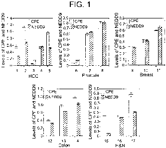
[0011] Figure 1 is a bar graph depicting expression levels of hCPE-AN and
NEDD9 (Neural
precursor cell expressed, developmentally down-regulated gene 9) for HCC,
prostate, breast,
colon, and head and neck (H&N) cancer cell lines, corrected for actin levels
and expressed as
mean SEM in arbitrary units (n=3 separate experiments). The HCC cell lines
represented are
H2P (1), H2M (2), MHCC97L (3), MHCC97H (4), and MHCCLM3 (5). The prostate
cancer
cell lines represented are LNCAP (6), PC-3 (7), and DU145 (8). The breast
cancer cell lines
represented are MCF-7 (9), T47D (10), and MDA-MB-231 (11). The colon cell
lines
represented are SW480 (12), HT-116 (13), and HT-29 (14). The H&N cancer cell
lines
represented are TU167 (15), TU159 (16), and MDA-1986 (17). * indicates highly
metastatic or
aggressive cell lines.
[0012] Figure 2 is a bar graph depicting the fold increase of proliferation or
invasion in
MHCCLM3 cells transfected with CPE-AN or empty vector (EV). The data
demonstrate
increased proliferation (1.92 0.05 fold, SEM, n=3, p<0.0001) and invasion
(2.72 0.15 fold,
SEM, n=5, p=0.0013) in cells transfected with CPE-AN versus EV.
CA 02734171 2011-02-14
WO 2010/009074 PCT/US2009/050460
3
[00131 Figure 3A is a bar graph depicting the CPE expression level (as a
percent of control).
Data is from Western blots of CPE-AN performed on highly metastatic tumor cell
lines from
breast (MDA-MB-231), prostate (DU145), head and neck (MDA1986), colon (HT-29),
and liver
(MHCCLM3) cancers transfected with si-scr (control) or si-CPE-AN (which
suppresses CPE-AN
and CPE mRNA expression). In particular, the bar graph shows the percent of -
40 kD CPE-AN
in si-CPE-AN treated cells relative to si-scr treated cells (made equal to
100%). Mean values
SEM (n=3) are shown.
[0014] Figure 3B is a bar graph depicting the number of colonies with >50
cells in si-scr
treated cells and si-CPE-AN treated cells. Mean values SEM (n=3) are shown.
[00151 Figure 3C is a bar graph depictingthe percent invasion of si-CPE-AN
treated cells
relative to the si-scr treated cells (made equal to 100%). Mean values SEM
(n=3) are shown.
[0016] Figure 4A is a bar graph depicting the ratio of hCPE-AN mRNA levels in
tumor (T)
versus surrounding non-tumor tissue (N) in HCC clinical samples for HCC
patients that (i) were
disease-free (Non-Recurrence; n=49) or (ii) had a recurrence of either
intrahepatic or
extrahepatic metastases one year after surgical resection (Recurrence; n=50).
Mean + SEM
(p<0.001) are shown.
[0017] Figure 4B is a bar graph depicting the ratio of hCPE-AN protein levels
in tumor (T)
versus surrounding non-tumor tissue (N) in HCC clinical samples for HCC
patients that (i) were
disease-free (Non-Recurrence; n--34) or (ii) had a recurrence of either
intrahepatic or
extrahepatic metastases one year after surgical resection (Recurrence; n=46).
The intensity of
the CPE-AN band from the Western blots was quantified by densitometry and
expressed in
arbitrary units after correction for the actin level in the sample. Mean + SEM
(p<0.001) are
shown.
[00181 Figure 5A is a growth curve for wild-type Neuro2A cells transfected
with empty
vector (EV) and clones stably expressing CPE (clones 3, 6, and 17). Each value
represents
means of replicates of 3 SEM. Experiments were repeated four times.
[00191 Figure 5B is a diagram showing mouse wild-type (WT) and mouse CPE-AN
mRNA
and protein.
[0020] Figure 6 is a diagram showing human WT and human CPE-AN mRNA and
protein.
CA 02734171 2011-02-14
WO 2010/009074 PCT/US2009/050460
4
[0021] Figure 7A-E are bar graphs depicting fold differences in expression of
hCPE-AN
mRNA in tumor cell lines relative to primary tumor cells with lowest hCPE-AN
mRNA
expression (first gray bar in each graph) made equal to 1. Highly metastatic
cell lines: white
bars, low metastatic cell lines: gray bars. The tumor cell lines represented
are HCC (A), prostate
(B), breast (C), colon (D), and head and neck (E).
DETAILED DESCRIPTION OF THE INVENTION
[0022] The inventors identified a splice variant isoform of the prohormone
processing
enzyme, carboxypeptidase E (CPE), which promotes growth and metastasis of
several types of
human epithelial-derived tumor cells. The splice variant isoform of CPE (CPE-
AN) lacks the N-
terminus (see Fig. 5B). In humans, the CPE-AN polypeptide comprises the amino
acid sequence
of SEQ ID NO: 2 and is encoded by the nucleic acid sequence of SEQ ID NO: 1.
In mice, the
CPE-AN polypeptide comprises the amino acid sequence of SEQ ID NO: 4 and is
encoded by the
nucleic acid sequence of SEQ ID NO: 3.
[0023] The invention provides a method of determining the prognosis of cancer
in a subject.
The invention provides a method of determining the prognosis of cancer in a
subject. The
method comprises (a) obtaining a sample from the subject, (b) analyzing the
sample for an
expression level of CPE-AN, and (c) correlating the expression level of CPE-AN
in the sample
with the prognosis of cancer in the subject.
[0024] The invention further provides a method of diagnosing cancer in a
subject. The
method comprises (a) obtaining a sample from the subject, (b) analyzing the
sample for an
expression level of CPE-AN (e.g., RNA or protein), and (c) correlating the
expression level of
CPE-AN in the sample with a diagnosis of cancer in the subject.
[0025] The invention provides a method of determining the stage of cancer in a
subject. The
method comprises (a) obtaining a sample from a tumor, (b) analyzing the tumor
sample for an
expression level of CPE-AN (e.g., RNA or protein), and (c) correlating the
expression level of
CPE-AN in the sample with the stage of cancer in the subject.
[0026] The sample to be analyzed can be any suitable tissue or fluid obtained
from the
subject. For example, the tissue can be tumor tissue, tissue adjacent to
and/or surrounding the
CA 02734171 2011-02-14
WO 2010/009074 PCT/US2009/050460
tumor, tissue from a location that is not adjacent to a primary tumor but that
is suspected of
harboring metastasized tumor, or blood.
[0027] The sample can be obtained by any suitable method. For example, sample
tissue can
be obtained via surgery, biopsy, resected tissue specimen, or arterial or
venous blood withdrawal.
[0028] Preferably, the inventive methods further comprise the step of
obtaining a sample
from surrounding non-tumor tissue (N) for the purpose of comparison. In
particular, the methods
comprise (a) obtaining a sample from a tumor (T) and a sample from surrounding
non-tumor
tissue (N), (b) analyzing the tumor (T) sample for an expression level of CPE-
AN (e.g., RNA or
protein) relative to an expression level of CPE-AN in the surrounding non-
tumor tissue sample
(N), and (c) correlating the expression level of CPE-AN in tumor/non-tumor
(TIN) with the
prognosis and/or stage of cancer in the subject.
[0029] The subject can be any mammal (e.g., mouse, rat, rabbit, hamster,
guinea pig, cat,
dog, pig, goat, cow, horse, primate, or human). Preferably, the subject is a
human of any age and
sex.
[0030] Without wishing to be bound by any particular theory, it is believed
that CPE-AN
promotes growth and metastasis of a variety of human cancer cells by up-
regulating the
expression of the metastasis gene, NEDD9 (see, e.g., Kim, Cell, 125: 1269-
81(2006)).
Additionally, it is believed that CPE-LiN activates gene expression by
epigenetic mechanisms by
interacting with histone deacetylase and transcription factor SATB I. In this
regard, CPE-AN can
serve as a biomarker to reliably predict future metastasis of a variety of
cancers based on the
level of CPE-AN in the resected primary tumor.
[0031] Examples of cancers that can be detected utilizing the inventive method
include
nerve, adrenal, thyroid, liver (such as hepatocellular carcinoma (HCC)),
prostate, lung, colon,
breast, head and neck, skin, pancreatic, ovarian, cervical, paraganglioma,
pheochromocytoma,
melanoma, esophagus, cervical, brain, and stomach cancer. The inventive method
is particularly
useful in detecting thyroid, paraganglioma, liver, prostate, colon, breast,
and head and neck
cancers.
[0032] The expression level of CPE-zN can be determined by detecting and,
optionally,
quantifying the levels of mRNA and/or protein of CPE-AN (referred to herein as
"biomarker" or
"biomarkers") in the sample.
CA 02734171 2011-02-14
WO 2010/009074 PCT/US2009/050460
6
[0033] Methods for detecting and quantifying such biomarkers are well within
the art. In
particular, suitable techniques for determining the presence and level of
expression of the
biomarkers in cells are within the skill in the art. According to one such
method, total cellular
RNA can be purified from cells by homogenization in the presence of nucleic
acid extraction
buffer, followed by centrifugation. Nucleic acids are precipitated, and DNA is
removed by
treatment with DNase and precipitation. The RNA molecules are then separated
by gel
electrophoresis on agarose gels according to standard techniques, and
transferred to
nitrocellulose filters by, e.g., the so-called "Northern" blotting technique.
The RNA is then
immobilized on the filters by heating. Detection and quantification of
specific RNA is
accomplished using appropriately labeled DNA or RNA probes complementary to
the RNA in
question. See, for example, Molecular Cloning: A Laboratory Manual, J.
Sambrook et al., eds.,
2nd edition, Cold Spring Harbor Laboratory Press, 1989, Chapter 7, the entire
disclosure of
which is incorporated herein by reference.
[0034] Methods for the preparation of labeled DNA and RNA probes, and the
conditions for
hybridization thereof to target nucleotide sequences, are described in
Molecular Cloning: A
Laboratory Manual, J. Sambrook et al., eds., 2nd edition, Cold Spring Harbor
Laboratory Press,
1989, Chapters 10 and 11, the entire disclosures of which are incorporated
herein by reference.
For example, the nucleic acid probe can be labeled with, e.g., a radionuclide
such as 3H, 32P, 33P,
14C, or 35S; a heavy metal; or a ligand capable of functioning as a specific
binding pair member
for a labeled ligand (e.g., biotin, avidin, or an antibody), a fluorescent
molecule, a
chemiluminescent molecule, an enzyme, or the like.
[0035] Probes can be labeled to high specific activity by either the nick
translation method of
Rigby et al., J. Mol. Biol., 113: 237-251 (1977), or by the random priming
method of Fienberg,
Anal. Biochem., 132: 6-13 (1983), the entire disclosures of which are herein
incorporated by
reference. The latter can be a method for synthesizing 32P-labeled probes of
high specific
activity from RNA templates. For example, by replacing preexisting nucleotides
with highly
radioactive nucleotides according to the nick translation method, it is
possible to prepare 32P-
labeled nucleic acid probes with a specific activity well in excess of 108
cpmlmicrogram.
Autoradiographic detection of hybridization then can be performed by exposing
hybridized
filters to photographic film. Densitometric scanning of the photographic films
exposed by the
CA 02734171 2011-02-14
WO 2010/009074 PCT/US2009/050460
7
hybridized filters provides an accurate measurement of biomarker levels. Using
another
approach, biomarker levels can be quantified by computerized imaging systems,
such as the
Molecular Dynamics 400-B 2D Phosphorimager (Amersham Biosciences, Piscataway,
N.J.,
USA).
[0036] Where radionuclide labeling of DNA or RNA probes is not practical, the
random-
primer method can be used to incorporate an analogue, for example, the dTTP
analogue 5-(N-(N-
biotinyl-epsilon-aminocaproyl)-3-aminoallyl)deoxyuridine triphosphate, into
the probe molecule.
The biotinylated probe oligonucleotide can be detected by reaction with biotin-
binding proteins,
such as avidin, streptavidin, and antibodies (e.g., anti-biotin antibodies)
coupled to fluorescent
dyes or enzymes that produce color reactions.
[0037] In addition to Northern and other RNA blotting hybridization
techniques, determining
the levels of RNA transcript can be accomplished using the technique of in
situ hybridization.
This technique requires fewer cells than the Northern blotting technique, and
involves depositing
whole cells onto a microscope cover slip and probing the nucleic acid content
of the cell with a
solution containing radioactive or otherwise labeled nucleic acid (e.g., cDNA
or RNA) probes.
This technique is particularly well-suited for analyzing tissue biopsy samples
from subjects. The
practice of the in situ hybridization technique is described in more detail in
U.S. Patent
5,427,916, the entire disclosure of which is incorporated herein by reference.
The inventive
method encompasses automated quantification of CPE-4N (e.g., in formalin-fixed
slides).
[0038] The relative number of RNA transcripts in cells also can be determined
by reverse
transcription of RNA transcripts, followed by amplification of the reverse-
transcribed transcripts
by polymerase chain reaction (RT-PCR). The levels of RNA transcripts can be
quantified in
comparison with an internal standard, for example, the level of mRNA from a
standard gene
present in the same sample. Suitable genes for use as an internal standard
include, for example,
myosin or glyceraldehyde-3 -phosphate dehydrogenase (G3PDH). The methods for
quantitative
RT-PCR and variations thereof are within the skill in the art.
[0039] Any suitable primers can be used for the quantitative RT-PCR.
Preferably, the
primers are specific to CPE- N and do not amplify wild-type CPE. It is within
the skill in the art
to generate primers specific to CPE-AN (see Figs. 5B and 6 for a comparison of
wild-type CPE
and CPE-AN). Primers can be of any suitable length, but preferably are between
9 and 70 (e.g.,
CA 02734171 2011-02-14
WO 2010/009074 PCT/US2009/050460
8
10, 11, 12, 13, 14, 15, 16, 17, 18, 19, 20, 21, 22, 23, 24, 25, 26, 27, 28,
29, 30, 31, 32, 33, 34, 35,
36, 37, 38, 39, 40, 41, 42, 43, 44, 45, 46, 47, 48, 49, 50, 51, 52, 53, 54,
55, 56, 57, 58, 59, 60, 61,
62, 63, 64, 65, 66, 67, 68, 69, as well as ranges of the values described
herein) nucleotides.
[0040] In one embodiment, the invention provides a pair of primers specific to
human CPE-
AN, such as fwd: 5'-ATGGCCGGGCATGAGGCGGC-3' (SEQ ID NO: 5) and rev: 5'-
GCTGCGCCCCACCGTGTAAA-3' (SEQ ID NO: 6). The primers also can have greater or
fewer nucleotides. In particular, a maximum length primer pair specific to
human CPE-AN is
fwd: 5'-GAGCGCAGCGATGGCCGGGCATGAGGCGGCGCCGGCGGC-3' (SEQ ID NO: 7)
and rev: 5'-GGCCCTCGAAGCTGCGCCCCACCGTGTAAATCCTGCTGAT-3' (SEQ ID NO:
8), and a minimal length primer pair specific to human CPE-AN is fwd: 5'-
CGGGCATGA-3'
(SEQ ID NO: 9) and rev: 5'-CCCCACCGT-3' (SEQ ID NO: 10). Primer pairs of
intermediate
lengths (e.g., between the minimal and maximum length primer pairs) also are
encompassed by
the invention.
[0041] In another embodiment, the invention provides a pair of primers
specific to mouse
CPE-AN, such as fwd: 5'- GACAAAA.GAGGCCAGCAAGA-3' (SEQ ID NO: 17) and rev: 5'-
CAGGTTCACCCGGCTCAT-3' (SEQ ID NO: 18). The primers also can have greater or
fewer
nucleotides. In particular, a maximum length primer pair specific to mouse CPE-
AN is fwd: 5'-
CAGACAAAAGAGGCCAGCAAGAGGACGGCA-3' (SEQ ID NO: 19) and rev: 5'-
ATTCAGGTTCACCCGGCTCATGGACCCCG-3' (SEQ ID NO: 20), and a minimal length
primer pair specific to mouse CPE-AN is fwd: 5'-AGGCCAGCAA-3' (SEQ ID NO: 21)
and rev:
5'-GTTCACCCGG-3' (SEQ ID NO: 22). However, primer pairs of intermediate
lengths (e.g.,
between the minimal and maximum length primer pairs) also are encompassed by
the invention.
[0042] A tissue microarray can be utilized to detect biomarker expression. In
the tissue
microarray technique, a hollow needle is used to remove tissue cores as small
as 0.6 mm in
diameter from regions of interest in paraffin-embedded tissues such as
clinical biopsies or tumor
samples. These tissue cores are then inserted in a recipient paraffin block in
a precisely spaced,
array pattern. Sections from this block are cut using a microtome, mounted on
a microscope
slide and then analyzed by any method of standard histological analysis. Each
microarray block
can be cut into 100 - 500 sections, which can be subjected to independent
tests. Tests commonly
CA 02734171 2011-02-14
WO 2010/009074 PCT/US2009/050460
9
employed in tissue microarray include immunohistochemistry and fluorescent in
situ
hybridization.
[0043] In some instances, it may be desirable to use microchip technology to
detect
biomarker expression. The microchip can be fabricated by techniques known in
the art. For
example, probe oligonucleotides of an appropriate length, e.g., 40
nucleotides, are 5'-amine
modified at position C6 and printed using commercially available microarray
systems, e.g., the
GENEMACHINE OmniGrid 100 Microarrayer and Amersham CODELINK activated slides.
Labeled cDNA oligomer corresponding to the target RNAs is prepared by reverse
transcribing
the target RNA with labeled primer. Following first strand synthesis, the
RNA/DNA hybrids are
denatured to degrade the RNA templates. The labeled target cDNAs thus prepared
are then
hybridized to the microarray chip under hybridizing conditions, e.g., 6 times
SSPE/30%
formamide at 25 C for 18 hours, followed by washing in 0.75 times TNT at 37
C for 40
minutes. At positions on the array, where the immobilized probe DNA recognizes
a
complementary target cDNA in the sample, hybridization occurs. The labeled
target cDNA
marks the exact position on the array where binding occurs, thereby allowing
automatic detection
and quantification. The output consists of a list of hybridization events,
which indicate the
relative abundance of specific eDNA sequences, and therefore the relative
abundance of the
corresponding complementary biomarker, in the subject sample. According to one
embodiment,
the labeled cDNA oligomer is a biotin-labeled cDNA prepared from a biotin-
labeled primer.
The microarray is then processed by direct detection of the biotin-containing
transcripts using,
e.g., Streptavidin-Alexa647 conjugate, and scanned utilizing conventional
scanning methods.
Image intensities of each spot on the array are proportional to the abundance
of the
corresponding biomarker in the subject sample.
[0044] The use of the array has one or more advantages for mRNA expression
detection.
First, the global expression of several hundred genes can be identified in a
single sample at one
time. Second, through careful design of the oligonucleotide probes, the
expression of both
mature and precursor molecules can be identified. Third, in comparison with
Northern blot
analysis, the chip requires a small amount of RNA and provides reproducible
results using 2.5 [.g
of total RNA.
CA 02734171 2011-02-14
WO 2010/009074 PCT/US2009/050460
[0045] Protein in a sample can be detected using a variety of methods, such as
protein
immunostaining, immunoprecipitation, protein microarray, radio-immunoassay,
and Western
blot, all of which are well known in the art. Immunostaining is a general term
in biochemistry
that applies to any use of an antibody-based method to detect a specific
protein in a sample.
Similarly, immunoprecipitation is the technique of precipitating an antigen
out of solution using
an antibody specific to that antigen. This process can be used to enrich a
given protein to some
degree of purity. A Western blot is a method by which protein may be detected
in a given
sample of tissue homogenate or extract. It uses gel electrophoresis to
separate denatured proteins
by mass. The proteins are then transferred out of the gel and onto a membrane
(typically
nitrocellulose), where they are "probed" using antibodies specific to the
protein. As a result,
researchers can examine the amount of protein in a given sample and compare
levels between
several groups.
[0046] The expression level of CPE-AN can be correlated to a prognosis by
comparing the
biomarker expression level in the sample to biomarker expression in
surrounding non-tumor
tissue or to a standard. The standard with which the sample is compared can be
a normalized
standard and/or can be a sample taken at an earlier time from the same
subject. That is, the
sample can be compared to a sample taken from the same subject prior to
treatment or the
subject after treatment has commenced (i.e., the subject at an earlier time).
In this way, the
efficacy of treatment also can be determined.
[0047] The prognosis of the cancer in a subject can be determined in the
inventive method.
The cancer can be from a primary tumor and/or a metastatic lesion. In this
regard, the prognosis
can be that the cancer in the subject is or is not likely to metastasize or
already has metastasized.
The prognosis can be that the cancer in the subject is or is not a metastatic
lesion. The prognosis
also can include combinations of the above.
[0048] The diagnosis of cancer in a subject can be determined in the inventive
method.
Cancer cells are circulating in the blood even before a tumor is formed. After
the tumor is
formed, the tumor continually sheds cancer cells, which circulate in the
blood. The expression
level of CPE-AN in the sample can be used to determine whether a subject has
cancer. For
example, if the expression level of CPE-AN in a sample is >2 (e.g., 2.5, 3,
3.5, 4, 4.5, 5, 6, 7, 8,
9, 10, 20, 30, 40, 50, or greater) times than that of a control sample (e.g.,
a sample from a subject
CA 02734171 2011-02-14
WO 2010/009074 PCT/US2009/050460
11
without cancer), the diagnosis is that the subject has cancer. Additionally,
the expression level of
CPE-AN in a sample (e.g., blood sample) can be used to diagnose a suspected
cancer as benign
or malignant. Alternatively, the expression level of CPE-AN in a sample (e.g.,
blood sample)
can be used to diagnose a suspected cancer as metastatic.
[0049] The stage of a cancer in the subject can be determined in the inventive
method. For
instance, the stage may be that the cancer is benign (non-malignant, non-
metastatic) or
metastatic. Even if a clinician diagnoses a cancer as benign based on the
pathology of the
primary tumor and the absence of visible metastases, a patient with increased
expression of CPE-
AN mRNA in the tumor has an increased risk of recurrence and future metastases
(e.g., within 2,
3, 4, 5, 6, 7, 8, 9, or 10 years from resection of the primary tumor) based on
the expression level
of CPE-AN mRNA in the tumor. A patient with an increased expression of CPE-AN
mRNA
should be closely monitored for recurrence and metastases.
[0050] In one embodiment, the prognosis and/or stage of cancer is based on the
ratio of CPE-
AN mRNA in tumor (T) versus non-tumor (NT) tissue. In particular, subjects
with CPE-AN
(e.g.,, mRNA or protein) TINT ratios of <2 are much less likely than subjects
with CPE-AN
TINT ratios of >2 (e.g., 2.5, 3, 3.5, 4, 4.5, 5, 6, 7, 8, 9, 10, 20, 30, 40,
50, or greater) to have
metastatic cancer or a recurrence of cancer (e.g., metastatic cancer).
[0051] In another embodiment, the prognosis, diagnosis, and/or stage of cancer
is based on
the copy number of CPE-AN mRNA in tumor tissue. The copy number can be
determined by
any suitable method (e.g., quantitative RT-PCR).
[00521 When the cancer is paraganglioma (PGL), CPE-AN mRNA copy numbers in
tumor
tissue of about 200,000 or less (e.g., 150,000 or less, 100,000 or less, or
50,000 or less) correlate
to a prognosis that the tumor is benign. Patients in this group have a low
risk of recurrence or
metastasis (e.g., within 2, 3, 4, 5, 6, 7, 8, 9, or 10 years from resection of
the primary tumor). In
contrast, CPE-AN mRNA copy numbers in tumor tissue of about 1 million or
greater (e.g., 2
million or greater, 3 million or greater, 4 million or greater, 5 million or
greater, 6 million or
greater, 7 million or greater, 8 million or greater, 9 million or greater, 10
million or greater, 15
million or greater, or 20 million or greater) correlate with a prognosis that
the tumor is
metastatic.
CA 02734171 2011-02-14
WO 2010/009074 PCT/US2009/050460
12
[00531 When the cancer is differentiated thyroid carcinoma (DTC), CPE-AN mRNA
copy
numbers in tumor tissue of about 200,000 or less (e.g., 150,000 or less,
100,000 or less, or
50,000 or less) correlate to a prognosis that the tumor is benign. CPE-AN mRNA
copy numbers
in tumor tissue of about 200,000 to about 600,000 (e.g., 250,000, 300,000,
350,000, 400,000,
450,000, 500,000, 550,000 or ranges of any of the values described herein)
correlate to a low risk
of recurrence or metastasis (e.g., within 2, 3, 4, 5, 6, 7, 8, 9, or 10 years
from resection of the
primary tumor). CPE-AN mRNA copy numbers in tumor tissue of about 600,000 to
about 1
million (e.g., 650,000, 700,000, 750,00, 800,000, 850,000, 900,000, 950,000,
or ranges of any of
the values described herein) correlate to an increased risk of recurrence or
metastasis (e.g.,
within 2, 3, 4, 5, 6, 7, 8, 9, or 10 years from resection of the primary
tumor). CPE-AN mRNA
copy numbers in tumor tissue of about 1 million or greater (e.g., 2 million or
greater, 3 million or
greater, 4 million or greater, 5 million or greater, 6 million or greater, 7
million or greater, 8
million or greater, 9 million or greater, 10 million or greater, or 15 million
or greater) correlate
with a prognosis that the tumor is metastatic.
[0054] The invention also provides a kit to measure CPE-AN mRNA and protein
(e.g., from
tissue biopsies and resected primary tumor tissues) for diagnostic or assay
purposes. For
example, the kit can comprise one or more primer pairs that detect CPE-AN mRNA
levels and/or
one or more probes that detect CPE-AN protein levels. Preferably, the primers
and probes can
differentiate between CPE-AN and wild-type CPE. The kits can be used to
determine metastasis
in a subject, to predict future recurrence/metastasis, and/or to monitor tumor
progression in a
subject (e.g., to determine efficacy of a cancer treatment).
[0055] The invention further provides a method of treatment for the subject
that is
accordance with the determined prognosis. The treatment can be any suitable
treatment.
Suitable treatments include chemotherapy, radiation, surgery, suppression of
CPE-AN, NEDD9
inhibition, and combinations thereof. Methods of chemotherapy, radiation, and
surgical
intervention are well within the art and can be determined on a case-by-case
basis depending on
the location, type, and stage of the cancer.
[0056] In one embodiment, the treatment includes suppression of CPE-AN. In
this regard, an
effective amount of an inhibitor of CPE-AN is administered to the subject.
Desirably, the
inhibitor prevents metastasis or slows the progression of metastasis (e.g., by
at least 5%, 10%,
CA 02734171 2011-02-14
WO 2010/009074 PCT/US2009/050460
13
15%, 20%, 25%, 30%, 35%, 40%, 50%, 55%, 60%, 65%, 70%, 75%, 80%, 85%, 90%,
95%, or
100%).
[0057] The inhibitor can be administered at any time following a prognosis
determination.
The inhibitor can be administered alone or in combination with other
treatments. For instance,
the inhibitor can be administered prior to surgical resection of a tumor. The
inhibitor also can be
administered following surgical resection of a tumor. One skilled in the art
can readily
determine an effective amount of the inhibitor composition to be administered
to a given subject,
by taking into account factors such as the size and weight of the subject, the
extent of disease
penetration, the age, health, and sex of the subject, the route of
administration, and whether the
administration is regional or systemic.
[0058] One skilled in the art also can readily determine an appropriate dosage
regimen for
administering a composition that alters biomarker levels or gene expression to
a given subject.
For example, the composition can be administered to the subject once (e.g. as
a single injection
or deposition). Alternatively, the composition can be administered multiple
times on any
suitable schedule, e.g., once or twice daily, monthly, bimonthly, or
biannually. The
administration of the treatment to a subject can be for a period ranging from
days, weeks,
months, or years. In certain embodiments, the treatment continues throughout
the life of the
subject. Where a dosage regimen comprises multiple administrations, it is
understood that the
effective amount of the composition administered to the subject can comprise
the total amount of
composition administered over the entire dosage regimen.
[0059] The inhibitor can be any suitable entity that suppresses/inhibits
expression or
transcriptional activity of CPE-AN. For example, the inhibitor can comprise a
nucleic acid that
is complementary to DNA or RNA (i.e., mRNA or tRNA) of CPE-AN that binds to
and inhibits
expression of CPE-AN. Alternatively, the treatment can include the
administration of a NEDD9
inhibitor comprising a nucleic acid that is complementary to the NEDD9 DNA or
RNA (i.e.,
mRNA or tRNA).
[0060] In this regard, the invention further provides a composition comprising
an inhibitor of
CPE-AN and/or a NEDD9 inhibitor and a pharmaceutically acceptable carrier.
[0061] Suitable compositions for inhibiting the expression of genes, such as
the gene
encoding CPE-AN and/or NEDD9, include double-stranded RNA (such as short- or
small-
CA 02734171 2011-02-14
WO 2010/009074 PCT/US2009/050460
14
interfering RNA or "siRNA"), antisense nucleic acids, and enzymatic RNA
molecules such as
ribozymes. These components can be targeted to a given biomarker gene product
and can
destroy or induce the destruction of the target biomarker gene product.
[0062] For example, expression of a given gene can be inhibited by inducing
RNA
interference of the gene with an isolated double-stranded RNA ("dsRNA")
molecule which has
at least 90%, for example, at least 95%, at least 98%, at least 99%, or 100%,
sequence homology
with at least a portion of the gene product. In a preferred embodiment, the
dsRNA molecule is a
"short or small interfering RNA" or "siRNA" (e.g., shRNA).
[0063] siRNA useful in the inventive methods comprise short double-stranded
RNA from
about 17 nucleotides to about 29 nucleotides in length, and preferably from
about 19 to about 25
nucleotides in length. The siRNA comprise a sense RNA strand and a
complementary antisense
RNA strand annealed together by standard Watson-Crick base-pairing
interactions (hereinafter
"base-paired"). The sense strand comprises a nucleic acid sequence which is
substantially
identical to a nucleic acid sequence contained within the target gene product.
[0064] As used herein, an siRNA "substantially identical" to a target sequence
contained
within the target nucleic sequence is a nucleic acid sequence that is
identical to the target
sequence or differs from the target sequence by at most one or two
nucleotides. The sense and
antisense strands of the siRNA can comprise two complementary, single-stranded
RNA
molecules, or can comprise a single molecule in which two complementary
portions are base-
paired and are covalently linked by a single-stranded "hairpin" area (shRNA).
[0065] The siRNA also can be altered RNA that differs from naturally-occurring
RNA by the
addition, deletion, substitution, and/or alteration of one or more
nucleotides. Such alterations
can include the addition of non-nucleotide material, such as to the end(s) of
the siRNA or to one
or more internal nucleotides of the siRNA, or modifications that make the
siRNA resistant to
nuclease digestion, or the substitution of one or more nucleotides in the
siRNA with
deoxyribonucleotides.
[0066] One or both strands of the siRNA also can comprise a 3' overhang. As
used herein, a
"3' overhang" refers to at least one unpaired nucleotide extending from the 3'-
end of a duplexed
RNA strand. Thus, in one embodiment, the siRNA comprises at least one 3'
overhang of from 1
to about 6 nucleotides (which includes ribonucleotides or
deoxyribonucleotides) in length,
CA 02734171 2011-02-14
WO 2010/009074 PCT/US2009/050460
preferably from 1 to about 5 nucleotides in length, more preferably from 1 to
about 4 nucleotides
in length, and most preferably from about 2 to about 4 nucleotides in length.
In a preferred
embodiment, the 3' overhang is present on both strands of the siRNA, and is 2
nucleotides in
length. For example, each strand of the siRNA can comprise 3' overhangs of
dithymidylic acid
("TT") or diuridylic acid ("uu").
[0067] The siRNA can be produced chemically or biologically, or can be
expressed from a
recombinant plasmid or viral vector (e.g., lentiviral, adenoviral, or
retroviral vector), as described
above for the isolated gene product. Exemplary methods for producing and
testing dsRNA or
siRNA molecules are described in U.S. Patent Application Publication No.
2002/0173478 and
U.S. Patent 7,148,342, the entire disclosures of which are incorporated herein
by reference.
Examples of shRNA include SEQ ID NOs: 25-27.
[0068] Expression of a given gene also can be inhibited by an antisense
nucleic acid. As
used herein, an "antisense nucleic acid" refers to a nucleic acid molecule
that binds to target
RNA by means of RNA-RNA or RNA-DNA or RNA-peptide nucleic acid interactions,
which
alter the activity of the target RNA. Antisense nucleic acids suitable for use
in the inventive
methods are single-stranded nucleic acids (e.g., RNA, DNA, RNA-DNA chimeras,
and peptide-
nucleic acids (PNA)) that generally comprise a nucleic acid sequence
complementary to a
contiguous nucleic acid sequence in a gene product. Preferably, the antisense
nucleic acid
comprises a nucleic acid sequence that is 50-100% complementary, more
preferably 75-100%
complementary, and most preferably 95-100% complementary, to a contiguous
nucleic acid
sequence in a gene product.
[0069] Antisense nucleic acids can also contain modifications to the nucleic
acid backbone
or to the sugar and base moieties (or their equivalent) to enhance target
specificity, nuclease
resistance, delivery, or other properties related to efficacy of the molecule.
Such modifications
include cholesterol moieties, duplex intercalators such as acridine, or the
inclusion of one or
more nuclease-resistant groups.
[0070] Antisense nucleic acids can be produced chemically or biologically, or
can be
expressed from a recombinant plasmid or viral vector, as described above for
the isolated gene
products. Exemplary methods for producing and testing are within the skill in
the art, as
CA 02734171 2011-02-14
WO 2010/009074 PCT/US2009/050460
16
disclosed in, for example, Stein, Science, 261: 1004 (1993), and U.S. Patent
5,849,902, the entire
disclosures of which are incorporated herein by reference.
[0071] Expression of a given gene also can be inhibited by an enzymatic
nucleic acid. As
used herein, an "enzymatic nucleic acid" refers to a nucleic acid comprising a
substrate binding
region that has complementarity to a contiguous nucleic acid sequence of a
gene product, and
which is able to specifically cleave the gene product. Preferably, the
enzymatic nucleic acid
substrate binding region is 50-100% complementary, more preferably 75-100%
complementary,
and most preferably 95-100% complementary, to a contiguous nucleic acid
sequence in a
biomarker gene product. The enzymatic nucleic acids also can comprise
modifications at the
base, sugar, and/or phosphate groups. An exemplary enzymatic nucleic acid for
use in the
inventive methods is a ribozyme.
[0072] The enzymatic nucleic acids can be produced chemically or biologically,
or can be
expressed from a recombinant plasmid or viral vector, as described above for
the isolated gene
products. Exemplary methods for producing and testing dsRNA or siRNA molecules
are
described in Werner, Nucl. Acids Res., 23: 2092-96 (1995); Hammann, Antisense
and Nucleic
Acid Drug Dev., 9: 25-31 (1999); and U.S. Patent 4,987,071, the entire
disclosures of which are
incorporated herein by reference.
[0073] The inventive compositions can be administered to a subject by any
means suitable
for directly or indirectly delivering these compositions to the subject (e.g.,
the lungs, stomach,
and/or blood vessels of the subject). For example, the compositions can be
administered by
methods suitable to transfect cells of the subject with these compositions.
Preferably, the cells
are transfected with a plasmid or viral vector comprising sequences encoding
at least one
biomarker gene product or biomarker gene expression inhibiting product.
[0074] Transfection methods for eukaryotic cells are well known in the art,
and include, e.g.,
direct injection of the nucleic acid into the nucleus or pronucleus of a cell,
electroporation,
liposome transfer or transfer mediated by lipophilic materials, receptor-
mediated nucleic acid
delivery, bioballistic or particle acceleration, calcium phosphate
precipitation, and transfection
mediated by viral vectors.
[0075] For example, cells can be transfected with a liposomal transfer
composition, e.g.,
DOTAP (N-[ 1-(2, 3 -dio leoyloxy)propyl] -N,N,N-trimethyl-ammonium
methylsulfate,
CA 02734171 2011-02-14
WO 2010/009074 PCT/US2009/050460
17
Boehringer-Mannheim) or an equivalent, such as LIPOFECTINTM Reagent
(Invitrogen
Corporation). The amount of nucleic acid used is not critical to the practice
of the invention;
acceptable results may be achieved with 0.1-100 micrograms of nucleic acid/105
cells. For
example, a ratio of about 0.5 micrograms of plasmid vector in 3 micrograms of
DOTAP per 105
cells can be used.
[0076] The composition also can be administered to a subject by any suitable
enteral or
parenteral administration route. Suitable enteral administration routes
include, e.g., oral or
intranasal delivery. Suitable parenteral administration routes include, e.g.,
intravascular
administration (e.g., intravenous bolus injection, intravenous infusion, intra-
arterial bolus
injection, intra-arterial infusion, and catheter instillation into the
vasculature); subcutaneous
injection or deposition, including subcutaneous infusion (such as by osmotic
pumps); direct
application to the tissue of interest (i.e., lung, liver tissue, etc.), for
example by a catheter or other
placement device (e.g., an implant comprising a porous, non-porous, or
gelatinous material);
intramuscular injection; and inhalation.
[0077] The composition can be administered to the subject either as naked RNA,
in
combination with a delivery reagent, or as a nucleic acid (e.g., a recombinant
plasmid or viral
vector) comprising sequences that express the biomarker gene product or
expression inhibiting
composition. Suitable delivery reagents include, e.g., the Mirus Transit TKO
lipophilic reagent,
LIPOFECTINTM Reagent (Invitrogen Corporation), LIPOFECTAMINETM (Invitrogen
Corporation), CELLFECTIN (Invitrogen Corporation), polycations (e.g.,
polylysine), and
liposomes.
[0078] Recombinant plasmids and viral vectors comprising sequences that
express the
biomarker or biomarker gene expression inhibiting compositions, and techniques
for delivering
such plasmids and vectors to a tissue, are discussed above.
[0079] In a preferred embodiment, liposomes are used to deliver a gene
expression-inhibiting
composition (or nucleic acids comprising sequences encoding them) to a
subject. Liposomes can
also increase the blood half-life of the gene products or nucleic acids.
[0080] Liposomes suitable for use in the invention can be formed from standard
vesicle-
forming lipids, which generally include neutral or negatively charged
phospholipids and a sterol,
such as cholesterol. The selection of lipids is generally guided by
consideration of factors such
CA 02734171 2011-02-14
WO 2010/009074 PCT/US2009/050460
18
as the desired liposome size and half-life of the liposomes in the blood
stream. A variety of
methods are known for preparing liposomes, for example, as described in Szoka,
Ann. Rev.
Biophys. Bioeng., 9: 467 (1980); and U.S. Patents 4,235,871, 4,501,728,
4,837,028, and
5,019,369, the entire disclosures of which are incorporated herein by
reference.
[0081] The liposomes can comprise a ligand molecule that targets the liposome
to lungs (i.e.,
small airways and/or large airways). Ligands which bind to receptors prevalent
in the lungs,
such as monoclonal antibodies that bind small airway epithelial cells, are
preferred.
[0082] The composition of the invention typically includes a pharmaceutically
acceptable
carrier. The pharmaceutically acceptable carrier can be any suitable
pharmaceutically acceptable
carrier, such as one or more compatible solid or liquid fillers, diluents,
other excipients, or
encapsulating substances which are suitable for administration into a human or
veterinary
patient. The pharmaceutically acceptable carrier can be an organic or
inorganic ingredient,
natural or synthetic, with which the active ingredient is combined to
facilitate the application of
the active ingredient. The pharmaceutically acceptable carrier desirably is co-
mingled with one
or more of the active components, and with each other, in a manner so as not
to substantially
impair the desired pharmaceutical efficacy of the active components.
Pharmaceutically
acceptable carriers desirably are capable of administration to a patient
without the production of
undesirable physiological effects such as nausea, dizziness, rash, or gastric
upset. It is, for
example, desirable for the pharmaceutically acceptable carrier not to be
immunogenic when
administered to a human patient for therapeutic purposes.
[0083] The pharmaceutical composition optionally can contain suitable
buffering agents,
including, for example, acetic acid in a salt, citric acid in a salt, boric
acid in a salt, and
phosphoric acid in a salt. The pharmaceutical composition also optionally can
contain suitable
preservatives, such as benzalkonium chloride, chlorobutanol, parabens, and
thimerosal.
[0084] The pharmaceutical composition conveniently can be presented in unit
dosage form
and can be prepared by any of the methods well known in the art of pharmacy.
Such methods
include the step of bringing the active agent into association with a carrier
that constitutes one or
more accessory ingredients. In general, the composition is prepared by
uniformly and intimately
bringing the active component(s) into association with a liquid carrier, a
finely divided solid
carrier, or both, and then, if necessary, shaping the product.
CA 02734171 2011-02-14
WO 2010/009074 PCT/US2009/050460
19
[0085] A composition suitable for parenteral administration conveniently
comprises a sterile
aqueous preparation of the inventive composition, which is preferably isotonic
with the blood of
the recipient. This aqueous preparation can be formulated according to known
methods using
suitable dispersing or wetting agents and suspending agents. The sterile
injectable preparation
also can be a sterile injectable solution or suspension in a non-toxic
parenterally-acceptable
diluent or solvent, for example, as a solution in 1,3-butane diol. Among the
acceptable vehicles
and solvents that can be employed are water, Ringer's solution, and isotonic
sodium chloride
solution. In addition, sterile, fixed oils are conventionally employed as a
solvent or suspending
medium. For this purpose any bland fixed oil can be employed, including
synthetic mono-or di-
glycerides. In addition, fatty acids such as oleic acid can be used in the
preparation of
injectables. Carrier formulations suitable for oral, subcutaneous,
intravenous, intramuscular, etc.
administrations can be found in Remington's Pharmaceutical Sciences, Mack
Publishing Co.,
Easton, PA, which is incorporated herein by reference thereto.
[0086] The composition of the invention can be in the form of a time-released,
delayed
release, or sustained release delivery system. The inventive composition can
be used in
conjunction with other therapeutic agents or therapies. Such an approach can
avoid repeated
administrations of the inventive composition, thereby increasing convenience
to the subject and
the physician, and may be particularly suitable for certain compositions of
the invention.
[0087] Many types of release delivery systems are available and known to those
of ordinary
skill in the art. They include polymer base systems such as poly(lactide-
glycolide),
copolyoxalates, polycaprolactones, polyesteramides, polyorthoesters,
polyhydroxybutyric acid,
and polyanhydrides. Microcapsules of the foregoing polymers containing drugs
are described in,
for example, U.S. Patent 5,075,109. Delivery systems also include non-polymer
systems that are
lipids including sterols such as cholesterol, cholesterol esters, and fatty
acids or neutral fats such
as mono-, di-, and tri-glycerides; hydrogel release systems; sylastic systems;
peptide based
systems; wax coatings; compressed tablets using conventional binders and
excipients; partially
fused implants; and the like. Specific examples include, but are not limited
to: (a) erosional
systems in which the active component is contained in a form within a matrix
such as those
described in U.S. Patents 4,452,775, 4,667,014, 4,748,034, and 5,239,660 and
(b) diffusional
systems in which an active component permeates at a controlled rate from a
polymer such as
CA 02734171 2011-02-14
WO 2010/009074 PCT/US2009/050460
described in U.S. Patents 3,832,253 and 3,854,480. In addition, pump-based
hardware delivery
systems can be used, some of which are adapted for implantation.
[0088] The following examples further illustrate the invention but, of course,
should not be
construed as in any way limiting its scope.
EXAMPLE 1
[00891 This example demonstrates the identification of the CPE splice variant
isoform that is
a biomarker for cancer metastasis.
[0090] Cell Lines. Three lines of human oral squamous cell carcinoma Tu167,
Tu159 and
MDA1986 (Myers et al., Clin. Cancer Res., 8: 293-298 (2002)) established from
freshly resected
human tumors were obtained from the laboratory of Dr. Gary L. Clayman, The
University of
Texas M.D. Anderson Cancer Center. Human HCC cell lines MHCC97L, MHCC97H, and
MHCCLM3 (Li et al. , World J. Gastroenterol., 7: 630-636 (2001)) were obtained
from Liver
Cancer Institute, Fudan University (Shanghai, China). H2P and H2M (Hu et al.,
Oncogene, 23:
298-302 (2004)) were obtained from Dr. X. Y. Guan from the Department of
Clinical Oncology,
University of Hong Kong. Human prostate adenocarcinoma cell lines (PC3, LNCaP,
and
DU145), human colon cancer cell lines (HT1 16, HT29, and SW480), human breast
cancer cell
lines (MDA-MB-23 1, T47D, and MCF-7), and Neuro2A cells were obtained from
ATCC
(Manassas,VA, USA).
[00911 Patient Samples. HCC samples used for Western blotting were obtained
with
informed consent from 80 patients undergoing hepatectomy for HCC from 2002 to
2005 in the
Department of Surgery at the University of Hong Kong (Hong Kong, China). Forty-
six of the
patients developed recurrence or extrahepatic metastasis within 6 months of
surgery, while the
other 34 remained disease-free during that time. HCC samples used for RT-PCR
and
inmmunohistochemistry were obtained with informed consent from 99 patients who
underwent
surgical resection for HCC from 2001 to 2005 in the Department of Surgery in
the University of
Hong Kong. Colon cancer samples used for RT-PCR were obtained from 68 patients
who
underwent surgical resection in 2006 in the Department of Surgery at the
University of Hong
Kong. Tissue specimens for tissue microarray (TMA) were obtained from 31
patients who
underwent surgical operation for colon cancer between 1999 and 2005 at the
University of Hong
CA 02734171 2011-02-14
WO 2010/009074 PCT/US2009/050460
21
Kong and subsequently developed extra-colonic metastases to liver. Matched
pairs of primary
and metastatic colon cancer samples were obtained for the TMA.
[0092] For prediction of metastasis, CPE-AN mRNA from resected primary tumor
(T) and
surrounding normal tissue (N) from a set of 40 HCC patients, which showed
recurrence or were
disease-free, were determined by quantitative RT-PCR (qRT-PCR). A threshold
TIN value of 2
was established, above which indicated tumor recurrence within a year.
Thereafter 99 and 80
different HCC patients were used in a blinded study as the test groups to
measure CPE-AN
mRNA by qRT-PCR and protein by Western blot, respectively, to predict those
patients who
would remain disease-free and those patients who would show a recurrence
within 1 year (for
qRT-PCR) or 6 months (for Western blots) after surgery based on their TIN
ratio. A blinded
clinical study also was conducted in 68 patients with colon cancer to
determine which patients
would remain disease-free or would exhibit metastasis/recurrence based on
their CPE-AN
mRNA TIN ratios in the resected tumor and surrounding tissues.
[0093] Growth and Proliferation of Neuro2A Clones. Neuro2A cells were stably
transfected with the expression vector, pcDNA3.1/CPE, and selected with 800
j.g/ml G418.
Individual colonies were picked and screened for the overexpression of CPE
from which three
clones (clones 3, 6, and 17) were selected (N2A/CPE cells). Neuro2A cells
transfected with the
pcDNA3.1 empty vector were batch selected with 800 pg/ml G418 and used as
control wild-type
(WT) Neuro2A cells (WT cells). Cells were plated in 10 cm plates at a density
of 6 x 10⁵
cells/dish in replicates of 3. Each 10 cm dish contained 6 coverslips.
Proliferation was assessed
over a period of 4 days with a MTT assay, as described in McGirr et al.,
Endocrinology, 146:
4514 (2005). Briefly, one coverslip was removed from each dish every day and
placed into a 6-
well plate containing 500 gl media. Twenty p.l of 2.5 g/ml thiazolyl blue was
added to each
well, and cells were incubated for 4 hours at 37 C. Media were removed, and
metabolized MTT
was dissolved in 200 l of acidified isopropanol. Absorbance for duplicate 90
i samples was
read at 590 nm in a plate reader.
[0094] Bioinformatics. A non-redundant nucleotide sequence database search was
carried
out with human and mouse CPE nucleotide sequence as queries (NM_00 1873.2 and
NM013494.3, respectively). Potential spliced variants (Genbank accession
number AK090962
and 13Y270449) were screened based on difference in nucleotide sequence
between the query
CA 02734171 2011-02-14
WO 2010/009074 PCT/US2009/050460
22
and the subject sequences. Specific primers at the splice junctions were
designed to amplify
these variants by PCR in Neuro2A cells and MHCC97 cells for mouse and human
splice
variants, respectively.
[0095] Semi-Quantitative PCR of WT-CPE and CPE-AN Transcripts in Neuro2A
Clones and HCC Cells. RNA was extracted from Neuro2A clones, MHCC97L and
MHCC97H, using the RNEASYTM Mini Kit (Qiagen, CA, USA). First strand cDNA was
synthesized with 1 gg of total RNA from MHCC97L and MHCC97H cells using
Transcriptor
First strand eDNA synthesis kit (Roche Applied Science, Germany). Semi-
quantitative
polymerase chain reaction was performed to quantify CPE-AN transcripts using
TAKARATM
Taq polymerase (TaKaRa Bio Inc., Shiga, Japan). 18S RNA was used as a
housekeeping gene
for normalization. Primer sequences specific for human AN-splice variant CPE-
AN RNA were
fwd: 5'-ATGGCCGGGCATGAGGCGGC-3' (SEQ ID NO: 5) and rev:
5'-GCTGCGCCCCACCGTGTAAA-3' (SEQ ID NO: 6). Primer sequences specific for mouse
AN-splice variant CPE-AN RNA in Neuro2A cells were fwd: 5'-
GACAAAAGAGGCCAGCAAGA-3' (SEQ ID NO. 17) and rev:
5'-CAGGTTCACCCGGCTCAT-3' (SEQ ID NO: 18) and for mouse WT CPE RNA were fwd:
5-TGCTGCTGGCGCTGTGT-3' (SEQ ID NO: 21) and rev: 5'-CAGGTTCACCCGGCTCAT-3'
(SEQ ID NO, 22). The primers for mouse WT CPE are specific for WT and do not
prime the
CPE-AN transcript. Primer sequences for amplifying 18S RNA were fwd:
5'-CTCTTAGCTGAGTGTCCCGC-3' (SEQ ID NO: 23) and rev:
5'-CTGATCGTCTTCGAACCTCC-3' (SEQ ID NO: 24). 0.25 g of eDNA from MHCC97L
and MHCC97H cells were used for every reaction. PCR cycling was at 94 C for
15 seconds,
annealing at 65 C for 30 seconds, extension at 72 C for 30 seconds and a
final extension at 72
C for 10 minutes. Same conditions were optimized and used for all 3 sets of
primers. 16 l of
each sample were removed every 5 cycles from 24 to 35 cycles in each reaction
to amplify CPE-
AN and 18S fragments. Amplified PCR products were separated on 1.5% agarose
gels with Tris-
borate EDTA buffer and stained with ethidium bromide. Gels were captured as
digital images
and the corresponding bands quantified by densitometry (ImageJ, NIH).
[0096] Verification of the Specificity of CPE-AN Specific Primers. To verify
the
specificity of the CPE-AN primers, the ability of the CPE-AN primers to prime
and amplify WT
CA 02734171 2011-02-14
WO 2010/009074 PCT/US2009/050460
23
CPE transcript obtained from a tissue enriched in CPE was assayed. Normal
human adrenal
medulla where CPE is expressed in abundance was utilized. First strand eDNA
was synthesized
from 100 ng of total RNA from this tissue using the Transcriptor First strand
eDNA synthesis kit
(Roche Applied Science, Germany). The semi-quantitative polymerase chain
reaction was
performed with TAKARATM Taq polymerase (TaKaRa Bio Inc., Shiga, Japan) with 35
PCR
cycles at 94 C for 15 seconds, annealing at 65 C for 30 seconds, extension
at 72 C for 30
seconds, and a final extension at 72 C for 10 minutes. The PCR was carried
out with both
generic CPE primers (fwd: 5'-CCATCTCCGTGGAAGGAATA-3' (SEQ ID NO: 11) and rev:
5'-CCTGGAGCTGAGGCTGTAAG-3' (SEQ ID NO: 12)) and CPE-AN specific primers (fwd:
5'-ATGGCCGGGCATGAGGCGGC-3' (SEQ ID NO: 5), rev:
5'-GCTGCGCCCCACCGTGTAAA-3' (SEQ ID NO: 6)). The correctly sized product was
amplified using the generic primers, but no product was amplified with the CPE-
AN specific
primers, which indicated that the CPE-AN primers are specific for CPE-AN cDNA.
[00971 Verification of Lack of CPE WT in HCC Cells and Human HCC Tumors. Since
primers that specifically amplified the human WT CPE mRNA were not identified,
an alternative
method was used to determine if MHCC97H cells contain WT CPE. A standard curve
was
generated, so that the amount of template in an unknown sample in terms of
copy number could
be determined. A complete clone of hCPE eDNA was excised from its plasmid and
purified, and
its concentration was determined spectophotometrically. Serial dilutions of
the eDNA were
made and used as templates for qRT-PCR. The PCR was carried out in triplicate
for each sample
from eight different concentrations. Generic CPE primers were used, and the
crossing point was
determined from the qRT-PCR program, averaged for each point, plotted as a
function of the
starting template concentration, and expressed as template copy number. The
mRNA copy
numbers in the MHCC97H cells or HCC tumor tissue were compared using the set
of generic
primers (fwd: 5'-CCATCTCCGTGGAAGGAATA-3' (SEQ ID NO: 11) and rev:
5'-CCTGGAGCTGAGGCTGTAAG-3'(SEQ ID NO: 12)) that amplifies both WT and CPE-AN
eDNA and using primers specific for hCPE-AN (fwd: 5'-ATGGCCGGGCATGAGGCGGC-3'
(SEQ ID NO: 5) and rev: 5'-GCTGCGCCCCACCGTGTAAA-3'(SEQ ID NO: 6)). Conditions
for the qRT-PCR for CPE using both sets of primers were as follows: initial
denaturation for 3
minutes at 95 C, followed by 45 cycles of 15 seconds at 95 C, 15 seconds at
62 C, and 5
CA 02734171 2011-02-14
WO 2010/009074 PCT/US2009/050460
24
seconds at 72 C. The PCR reaction was followed by a melting curve program (65
C-95 C)
with a heating rate of 0.1 C per second, a continuous fluorescence
measurement, and a cooling
program at 40 C. Negative controls consisting of no-template (water) reaction
mixtures were
run with all reactions. PCR products also were run on agarose gels to confirm
the formation of a
single product of the predicted size. The copy numbers in the HCC cells and
the human tumor
samples using either the generic primers or the CPE-AN specific primers were
identical,
indicating that MHCC97H cells and HCC tumors lacked WT CPE. Additionally,
Western blots
of MHCC97H cells and human HCC samples using AtT-20 cells as a positive
control showed no
WT CPE band.
[0098] Microarray Hybridization and Data Analysis of Neuro2A Cells. Neuro2A
clonal
cells expressing CPE and the WT cells transfected with vector alone were used
for microarray
studies. All GeneChips were processed at the London Regional Genomics Centre
(Robarts
Research Institute, London, Ontario, Canada). RNA was extracted from clone 17,
and the WT
cells and the quality of the RNA was assessed using the Agilent 2100
Bioanalyzer (Agilent
Technologies Inc., Palo Alto, CA, USA) and the RNA 6000 Nano kit (Caliper Life
Sciences,
Mountain View, CA, USA). All procedures, including cRNA synthesis, labeling,
and
hybridization to Affymetrix Mouse Genome 2.0 GeneChips, were performed as
described in the
Affymetrix Technical Analysis Manual (Affymetrix, Santa Clara, CA, USA).
GeneChips were
scanned with the Affymetrix GeneChip Scanner 3000 (Affymetrix, Santa Clara,
CA, USA).
Probe signal intensities for genes were generated using GCOS 1.4 (Affymetrix
Inc., Santa Clara,
CA, USA) using default values for the statistical expression algorithm
parameters and a target
signal of 150 for all probe sets and a normalization value of 1. Gene level
data was generated
using the RMA preprocessor in GeneSpring GX 7.3.1 (Agilent Technologies Inc.,
Palo Alto, CA,
USA). Data from 4 different microarrays were transformed (measurements less
than 0.01 set to
0.01) and normalized per chip to the 50th percentile and per gene to the WT
Neuro2A cells.
Genes were grouped according to function in development and considered
significantly changed
using Venn analysis to screen at least 2-fold changes, followed by a one-way
ANOVA with ap
value cutoff of 0.05.
[0099] Western blot for CPE-AN and NEDD9 in Cell Lines and Clinical Specimens.
Proteins from clinical specimens were prepared using urea buffer (8 M urea, 10
mM Tris, pH 7).
CA 02734171 2011-02-14
WO 2010/009074 PCT/US2009/050460
Briefly, frozen tissue blocks were homogenized, and cells were placed on ice
for 15 minutes and
then centrifuged at 13,000 x g for 5 minutes at 4 C. The protein supernatant
was collected, and
its concentration was determined. Proteins from human cancer cell lines were
prepared using cell
lysis buffer (Cell Signaling Technology, Beverly, MA, USA) or, for Neuro2A
cells, with M-per
mammalian protein extraction reagent (Pierce, Rockford, IL, USA) supplemented
with Complete
Inhibitor Cocktail (Roche, Indianapolis, IN, USA) to prevent protein
degradation. The cell
lysate was collected and centrifuged at 15,000 x g for 10 minutes at 4 C. The
protein
concentrations from supernatants of the cell lysates were determined. Twenty
p.g of protein were
denatured, run on 4-20% or 12% SDS-PAGE gels, and transferred onto
nitrocellulose membrane
or PVDF membrane (Millipore, Billerica, MA, USA) using the standard protocol.
After
blocking with 5% nonfat milk at room temperature for 1 hour, CPE-AN on the
membrane was
detected using a CPE monoclonal antibody directed against amino acid residues
49-200 of the
human WT CPE sequence (R&D Systems, Inc., Minneapolis, MN) at 1:4000 dilution.
NEDD9
was detected with mouse anti-human HEF1 generated using the N-terminal 82-398
residues of
the NEDD9 protein (clone 14A11 at 1:1000 dilution, Rockland Immunochemicals,
Gilbertsville,
PA, USA) and rabbit polyclonal C-terminal antibody from Professor Mirimoto
(Japan) (Sasaki et
al., Stroke, 36: 2457 (2005)). Following primary antibody binding, the
membrane was incubated
with horseradish peroxidase-conjugated anti-mouse or rabbit antibody
(Amersham) and then
visualized by enhanced chemiluminescence plus according to the manufacturer's
protocol. The
intensity of the bands was quantified by densitometry and expressed as
arbitrary unites (AU).
The expression of CPE-AN and NEDD9 levels of each cell line was corrected for
their actin level
and expressed as the mean SEM of AU from three separate experiments.
[001001 Lentiviral Based CPE-AN Suppression in Tumor Cell Lines. Lentiviral
based
shRNAs against human CPE, which also suppress CPE-AN mRNA expression
(CCGGCCAGTACCTATGCAACGAATACTCGAGTATTCGTTGCATAGGTACTGGTTTTT
G (SEQ ID NO: 25);
CCGGCTCCAGGCTATCTGGCAATAACTCGAGTTATTGCCAGATAGCCTGGAGTTTTT
G (SEQ ID NO: 26); and
CCGGGATAGGATAGTGTACGTGAATCTCGAGATTCACGTACACTATCCTATCTTTTT
G (SEQ ID NO: 27)) and scramble control were obtained from DFCI-Broad RNAi
Consortium
CA 02734171 2011-02-14
WO 2010/009074 PCT/US2009/050460
26
in a pLKO.puro vector. VSV.G-pseudotyped lentiviral particles were generated
by calcium
phosphate cotransfection of 293T cells, and viral supernatants were collected
after 48 hours.
Lentiviral supernatants were used to transduce (i) MHCCLM3, (ii) HT29, (iii)
MDA-MB-231,
(iv) DU145, and (v) MDA1986 cells. At 2 days post-transduction, cells were
selected by
puromycin at a concentration of 2 ug/ml.
[00101] Immunofluorescence of CPE-AN in HCC Tumor Cells. MHCCLM3 cells
transfected with either si-scrambled or si-CPE-AN (which down-regulates CPE-AN
mRNA
expression) were cultured on chamber slides, permeabilized with 0.1 % Triton X-
100, and fixed
with 4% paraformaldehyde in PBS. The cells were incubated with monoclonal
antibodies
against CPE (1:100) (R&D Systems, Inc., Minneapolis, MN, USA). The secondary
antibody was
TRITC-conjugated goat anti-mouse IgG (Molecular Probes). The slide was
subsequently
stained with fluorescein phalloidin (Molecular Probes) in 1% BSA (dilution
factor, 1:50) at 37 C
for 1 hour and counterstained by DAPI (AppliChem GmbH). All images were
visualized by
confocal microscopy and photographs were taken at 600x magnification.
[00102] Colony Formation Assay. Growth analysis of cells was performed by the
colony
formation assay described Ng et al., Cancer Res., 60: 6581-6584 (2000). Eighty
percent
confluent cells were trypsinized, and single-cell suspensions were obtained.
Four hundred viable
cells were seeded per well in 6-well plates. Ten days later, cells were fixed
with 70% ethanol
and stained with 10% (v/v) Giemsa (MERCK, Damstadt, Germany). Colonies
consisting of
more than 50 cells were counted. Each experiment was done in triplicate, and
the mean values
SEM were determined.
[00103] Matrigel Invasion Assay. Invasion assay was carried out as described
in Lee et al.,
Cancer Res., 66: 9948-9956 (2006). Conditioned medium from cells transfected
with si-
scramble or si-CPE-AN was placed in the lower chambers as chemo-attractants.
After 22 hours
in culture, the cells were removed from the upper surface of the filter by
scraping with a cotton
swab. The cells that invaded through the Matrigel and were adherent to the
bottom of the
membrane were stained with crystal violet solution. The cell-associated dye
was eluted with
10% acetic acid, and its absorbance at 595 nm determined. Each experiment was
done in
triplicate, and the mean values SEM were determined.
CA 02734171 2011-02-14
WO 2010/009074 PCT/US2009/050460
27
[00104] Generation of Luciferase-Expressing Cells. For luciferase labeling of
MHCCLM3
cells, lentiviral vector containing the sequence of the firefly luciferase
gene was constructed and
transfected into the cells (see, e.g., Lee et al., Cancer Res., 67: 8800
(2007)). Stable transfectants
were generated from a pool of>20 positive clones, which were selected
byblasticidin at a
concentration of 2 [.g/ml.
[00105] Bioluminescent Imaging of Live Animals Bearing Tumors. Animal care and
euthanasia were conducted with full approval by the Committee on the Use of
Live Animals in
Teaching and Research of the University of Hong Kong. Approximately 1 x 10⁶
MHCCLM3
cells stably expressing firefly luciferase were transfected with either si-
scramble or si-CPE-AN
and injected subcutaneously into the right flank of four-week-old male BALB/c-
nulnu mice with
a30-gauge hypodermic needle (see, e.g., Fu et al., Proc. Natl. Acad. Sci.
U.S.A., 88: 9345
(1991)). The mice were imaged on day 0 and day 30 after cell inoculation. Mice
were
anesthetized with ketamine-xylazine mix (4:1). Imaging was done using an
Xenogen IVISTM
100 cooled CCD camera (Caliper Life Sciences, Hopkinton, MA, USA). The mice
were injected
with 200 pL of 15 mg/ml D-luciferin i.p. for 15 minutes before imaging, after
which they were
placed in a light-tight chamber. A gray-scale reference image was obtained
followed by the
acquisition of a bioluminescent image. The acquisition time ranged from 3
seconds to 1 minute.
[00106] Metastatic Orthotopic Nude Mouse Model. Approximately 1 x 10⁶ MHCCLM3
cells (in 0.2 ml culture medium) transfected with either si-scramble or si-CPE-
AN were injected
subcutaneously into the right flank of nude mice, which were then observed
daily for signs of
tumor development. Once the subcutaneous tumor reached 1 to 1.5 cm in
diameter, the tumor
was removed and cut into about 1 to 2 mm cubes, which were implanted into the
left liver lobe of
the nude mice (see, e.g., Livak et al., Methods, 25: 402 (2001)). The mice
were imaged on day 0
and day 35 after tumor inoculation. Mice were anesthetized with ketamine-
xylazine mix (4:1).
Imaging was performed using a Xenogen IVIS 100 cooled CCD camera (Xenogen) and
metastasis to the lung and intestines was tracked. After imaging, metastasis
to these tissues was
confirmed by inspection and imaging of the dissected tissues.
[00107] Histopathology. To confirm that metastasis to the lungs occurred, the
animal was
autopsied as soon as the original signal was recorded. Lungs were examined and
imaged with
the Xenogen camera to confirm the bioluminescence of this tissue and then
fixed by
CA 02734171 2011-02-14
WO 2010/009074 PCT/US2009/050460
28
intrabranchial perfusion of 10% neutralized formalin solution. Paraffin-
embedded sections (4
gm) were cut and stained with H&E.
[001081 Quantitative RT-PCR of CPE-AN in Cell Lines and Clinical Specimens.
RNA
was extracted from both cancer cells (as described above) and each patient's
tumor and
surrounding non-tumor tissue using trizol (Invitrogen, CA, USA). Complementary
DNA
amplified from 0.2 gg mRNA in the tissues was subjected to real-time
quantitative PCR for
CPE-AN expression using a Fast SYBRTM Green Master Mix PCR kit (Applied
Biosystems,
Foster City, CA, USA) under the following cycling conditions: 95 C for 20
seconds, followed
by 40 cycles of 95 C for 1 second, 60 C for 20 seconds. Reactions were
performed using an
ABI PRISM 7900 Sequence Detector (Applied Biosystems). Fluorescence signals
were
analyzed using SDS 1.9.1 software (Applied Biosystems). 18S was used as the
endogenous
normalization control. Primer sequences for CPE-ANRNA were fwd:
5'-ATGGCCGGGCATGAGGCGGC-3' (SEQ ID NO: 5), rev:
5'-GCTGCGCCCCACCGTGTAAA-3' (SEQ ID NO: 6); 18S-fwd:
5'-CTCTTAGCTGAGTGTCCCGC3' (SEQ ID NO: 23); and 18S-rev:
5'-CTGATCGTCTTCGAACCTCC-3' (SEQ ID NO: 24). All PCRs were performed in
duplicate and were averaged to obtain the data point for each specimen. The
relative amount of
CPE-AN mRNA was normalized to an internal control, 18S, and relative to a
calibrator (see, e.g.,
Livak et al., Methods, 25: 402 (2001)): 2-MCT, where AACT = [CT(CPE) -
CT(I8S)]test -
[CT(CPE) - CT(18S)]calibrator. The threshold value (CT) was defined as the
fractional cycle
number at which the amount of amplified target reached a fixed threshold. The
CT value
correlated with the input target mRNA levels, and a lower CT value indicated a
higher starting
copy number. One of the samples was designated as the calibrator to compare
the relative
amount of target in different samples and used to adjust for the plate-to-
plate variation in
amplification efficiency. The relative expression level of CPE of each patient
was evaluated as
the relative fold change in log 2 scale.
[001091 Construction of Tissue Microarray (TMA). Tissue microarrays were
constructed
with 0.6 mm diameter cores using a MTA-1 tissue arrayer (Beecher Instruments,
Sun Prairie,
WI) (see, e.g., Kononen, Nat. Med., 4: 844-847 (1998)). The final array
contained 31 pairs of
primary and matched metastatic colon to liver cancer samples. Five gm sections
were cut and
CA 02734171 2011-02-14
WO 2010/009074 PCT/US2009/050460
29
immunostained as described below. Regions of interest were selected from
hematoxylin and
eosin stained sections after review by two pathologists.
[001101 Immunostaining and Quantification of CPE in Human Tissue Sections. HCC
tumor tissue and surrounding non-tumor tissue were formalin-fixed and paraffin-
embedded.
Four gm sections were cut, dewaxed in xylene and graded alcohols, hydrated,
and washed in
PBS. After pretreatment in a microwave oven (12 minutes in sodium citrate
buffer (pH 6)), the
endogenous peroxidase was inhibited by 0.3% H202 for 30 min, and the sections
were incubated
with 10% normal goat serum for 30 minutes. Mouse monoclonal anti-
carboxypeptiase E (1:100)
(R&D Systems, Inc., Minneapolis, MN) was applied overnight in a humidity
chamber at 4 C. A
standard avidin-biotin peroxidase technique (DAKO, Carpinteria, CA) was
applied. Briefly,
biotinylated goat anti-mouse iminunoglobulin and avidin-biotinperoxidase
complex were
applied for 30 minutes each with 15-minute washes in PBS. The reaction was
finally developed
with the Dako Liquid DAB+ Substrate Chromogen System (Dako, Glostrup,
Denmark). Slides
were imaged on a SCANSCOPETM CS imager (Aperio, Vista, CA, USA), generating
0.43
gm/pixel whole slide images. These images were compiled and analyzed using the
SPECTRUMTM software (Aperio, Vista, CA, USA) with a pixel count algorithm
(see, e.g.,
Brennan et al., Clin. Cancer Res., 14: 2681 (2008)). Quantified expression of
tumor tissue minus
adjacent normal tissue was compared for patients with and without recurrence.
[001111 Results. In order to investigate the possible role of CPE, wild-type
(WT) CPE cDNA
was stably transfected into a clone of the mouse neuroblastoma cell line,
Neuro2A, with low
CPE expression. The isolated clones proliferated faster than Neuro2A cells
transfected with
empty vector (see Fig. 5A).
[00112] This result prompted an exhaustive non-redundant nucleotide sequence
database
search that uncovered a splice variant isoform of CPE that lacks the N-
terminus (CPE-AN) (see
Fig. 5B). PCR identified two different CPE transcripts expressed in the stably
transfected
Neuro2A clones: WT and CPE-AN (see Fig. 5B). This finding suggested that CPE-
AN is
responsible for the enhanced proliferation of Neuro2A cells.
[00113] To identify genes up-regulated in the CPE-transfected Neuro2A clones
that promote
proliferation, gene microarray analysis of a Neuro2A clone stably
overexpressing CPE versus
WT Neuro2A cells transfected with empty vector was performed. The analysis
showed, among
CA 02734171 2011-02-14
WO 2010/009074 PCT/US2009/050460
changes in other mRNAs, a 6-fold higher expression of NEDD9 in the CPE-
transfected Neuro2A
clone. NEDD9 has been shown to be expressed during embryonic development, and
expression
of NEDD9 is down-regulated in adult mice (see, e.g., Kumar et al., Biochem.
Biophys. Res.
Commun., 185: 1155 (1992); and Aquino et al., Gene Expression Patterns, 8: 217
(2008)).
NEDD9 has been implicated in cancer development (see, e.g., Merrill et al.,
Dev. Dyn., 231: 564
(2004); and O'Neill et al., Cancer. Res., 67: 8975 (2007)) and recently was
identified as a
metastasis promoting gene in melanomas (see, e.g., Kim et al., Cell, 125: 1269-
81 (2006)).
NEDD9 promoted growth and enhanced invasion in vitro and metastasis in viva of
normal and
transformed melanocytes by interacting with focal adhesion kinase (FAK) (see,
e.g., Kim, Cell,
125: 1269-81 (2006); and McLean et al., Nat. Rev. Cancer, 5: 505 (2005)).
NEDD9 is highly
expressed in human melanomas and governs the metastatic potential of these
tumors.
[00114] These findings led to the hypothesis that CPE-AN promotes growth and
metastasis of
tumor cells by up-regulating NEDD9 gene expression. This hypothesis was tested
on human
HCC cells. Semi-quantitative RT-PCR showed that high metastatic MHCC97H cells
had
elevated levels of CPE-AN mRNA compared to low metastatic cells (MHCC97L) (see
Fig. 6).
Moreover, WT CPE mRNA or protein was not expressed in these epithelial-derived
MHCC97
cells, unlike neuroendocrine tumors, which express both WT and CPE-AN.
Quantitative RT-
PCR showed that CPE-AN mRNA was 8.5-fold higher in MHCC97H versus MHCC97L
cells
(see Fig. 7).
[00115] The translation product derived from the human CPE-AN splice variant
transcript (see
Fig. 6) in HCC cells has an apparent molecular mass of --.40 kD. Other highly
metastatic human
tumor cell lines of epithelial origin derived from HCC, colon, breast,
prostate, and head and neck
tumors also had elevated expression of CPE-AN mRNA (see Fig. 7) and the -40 kD
CPE-AN
protein, as well as NEDD9 protein, compared to matched tumor lines with low
metastatic
potential (see Fig. 1). The forms of NEDD9 that increased with metastatic
potential in these
cells were primarily a 70 kD N-terminal domain that contains the FAK binding
domain involved
in metastasis (see, e.g., O'Neill et al., Mol. Cell Biol., 15: 5094 (2001))
and a 35 kD C-terminal
cleavage product.
[00116] To verify that CPE-AN regulates NEDD9 gene expression, low metastatic
MHCC97L
cells were transfected with the CPE-AN cDNA. A concomitant increase in CPE-AN
and NEDD9
CA 02734171 2011-02-14
WO 2010/009074 PCT/US2009/050460
31
protein was observed. Furthermore, MHCC97L cells transfected with CPE-AN
showed
increased proliferation and invasion compared to cells transfected with empty
vector, thereby
demonstrating a role of CPE-AN in growth promotion and invasion of tumor cells
(see Fig. 2).
[00117] Conversely, siRNA-mediated down-regulation of CPE-AN in the highly
metastatic
MHCCLM3 cells resulted in a decrease in NEDD9 expression in each of the 3
clones stably
transfected with 3 different si-RNA sequences. Immunofluorescence microscopy
of MHCCLM3
cells transduced with scrambled-si (si-scr) RNA revealed immunoreactive CPE-AN
primarily in
the nucleus versus cytoplasm. In MHCCLM3 cells down-regulated in expression of
CPE-AN by
si-CPE-AN, CPE-AN immunofluorescence was barely detectable. These results
indicate that
CPE-AN that lacks a signal peptide is expressed in the cytoplasm and can be
translocated into the
nucleus to modulate gene expression.
[00118] To demonstrate that CPE-AN mediates growth and cell invasion in
multiple types of
human tumors, highly metastatic cell lines from breast (MDA-MB-23), prostate
(DU145), head
and neck (MDA 1986), colon (HT29), and liver (MHCC97M3) were down-regulated in
CPE-AN
expression using si-RNA (SEQ ID NO: 26) (see Fig. 3A). Suppression of CPE-AN
expression in
these tumor cell lines led to 56-85% inhibition of growth (Fig. 3B) and 70-85%
inhibition of
invasion (see Fig. 3B).
[00119] In addition to the in vitro assays, in vivo animal studies were
performed in two
models. Nude mice were subcutaneously injected with MHCCLM3 cells transduced
with either
si-CPE-AN (SEQ ID NO: 26) or si-scr (see, e.g., Lee et al., Clin. Cancer Res.,
11: 8458 (2005)).
Thirty days after cell inoculation, control mice injected with the si-scr
MHCCLM3 cells had
liver tumors with 16.2-fold higher intensity (which reflects increased volume)
when compared to
mice injected with si-CPE-AN cells.
[00120] Using a metastatic orthotopic nude mouse model (see, e.g., Lee et al.,
Cancer Res.,
67: 8800 (2007)), MHCCLM3 cells transduced with either si-scr or si-CPE-AN
were injected
subcutaneously into the right flank of the mice. When the subcutaneous tumor
was -1.5 cm in
diameter, the tumor was removed, cut into 1- 2 mm cubes, and implanted into
the liver of nude
mice (see, e.g., Lee et al., Cancer Res., 67: 8800 (2007)). Thirty-five days
post-implantation,
mice with the si-scr MHCCLM3-derived tumors showed 13.9-fold higher intensity
(reflecting
increased volume) and developed intrahepatic metastasis and extrahepatic
metastasis to lung and
CA 02734171 2011-02-14
WO 2010/009074 PCT/US2009/050460
32
intestine (see, e.g., Li et al., Clin. Cancer Res., 12: 7140 (2006)), while
mice inoculated with si-
CPE-AN MHCCLM3-derived tumors had smaller tumors and failed to demonstrate
metastasis.
[00121] To determine if CPE-AN is a useful marker for predicting future
recurrence and
metastasis, quantitative RT-PCR and Western blot analysis were performed on
primary tumors
from HCC and colon cancer patients to measure CPE-AN mRNA and protein levels,
respectively. RT-PCR verified that only CPE-AN mRNA, and not WT CPE mRNA, was
expressed in primary HCC tumors. CPE-AN was quantified by qRT-PCR in the
primary tumor
(T) versus surrounding non-tumor (N) tissue, and its ratio determined. Forty-
four of 49 (89.8%)
HCC patients, who were disease-free one year after surgery, had CPE-AN mRNA
T/N ratios of
<_2. In contrast, 46 of 50 (92%) of patients with extra- or intra-hepatic
metastasis/recurrence had
a T/N ratio >2 (see Fig. 4A and Table 1).
Table 1. Clinical significance of CPE in HCC tissues.
Clinicopathological CPE Expression
Variables T/NT < 2 T/NT > 2 p value
TNM (UICC) classification
Early Stage (I-II) 39 26 13
<0.002*+
Late Stage (1I1-IV) 57 20 37
Recurrence in the first year
Yes 50 4 46
<0.001*
No 49 44 5
* statistically significant; + incomplete data
[00122] The survival analysis of disease-free survival of 99 HCC patients
showed shorter
survival times (p<O.0001) when CPE-AN mRNA TIN were >2 (high) in the primary
tumor
compared to patients with TIN <_2 (low). The mean survival was 17.44 months
and 82.75
months for the high group versus the low group.
CA 02734171 2011-02-14
WO 2010/009074 PCT/US2009/050460
33
[00123] Similar results were obtained for 68 colon cancer patients. Twenty-
nine of 31
(93.5%) patients that were disease-free had a CPE-AN mRNA T/N ratio of <_2,
whereas 32 of 37
(86.5%) patients that developed lymph node or distant metastasis within a year
after surgery had
a CPE-AN mRNA T/N ratio of>2 (see Table 2).
Table 2. Clinical significance of CPE in colon cancer.
Clinicopathological CPE Expression
n
Variables TINT < 2 T/NT > 2 p value
TNM (UICC) classification
Early Stage (I-1I) 29 28 1
<0.00t*
Late Stage (III-N) 39 6 33
Lymph node and distant metastases
Yes 37 5 32
<0.001*
No 31 29 2
* statistically significant
[00124] Additionally, Western blots showed higher CPE-AN levels in patients
with recurrence
compared to the disease-free group. Quantitative analysis of the T/N ratios of
the CPE-AN band
from Western blots from 80 patients revealed that 28 of 34 (82.3%) patients
with primary HCC
who were disease-free after surgery had tumor CPE-AN levels with T/N 52 (see
Fig. 4B).
However, 35 of 46 (76%) patients who developed intra-hepatic recurrence or
extra-hepatic
metastasis within 6 months had primary tumor CPE-AN levels of TIN >2.
[00125] Immunohistochemistry (IHC) of CPE-AN in HCC tumors from patients
revealed
immunostaining primarily in the nuclei of tumor cells in patients who
subsequently developed
recurrence that was absent in the cell nuclei of patients who remained disease-
free. Staining was
primarily in the cytoplasm of tumor cells in patients who remained disease-
free. The intensity of
immunostaining as determined by image analysis (0.402 0.032 SEM versus 0.279
0.036
SEM) was statistically different (p<0.02) between the groups.
CA 02734171 2011-02-14
WO 2010/009074 PCT/US2009/050460
34
[001261 Analysis of primary colon cancer cells that had metastasized to the
liver also revealed
increased numbers of CPE-AN positive cells in the metastatic tissue, and,
similar to the HCC
cells, staining was observed primarily in the nuclei of these metastatic colon
cells.
[00127] These retrospective studies demonstrate that measurement of CPE-AN
mRNA and
protein levels in resected tumors is a powerful prognostic tool for predicting
recurrence/metastasis in cancer (e.g., HCC and colon cancer) patients. In
particular,
measurement of CPE-AN mRNA levels in resected primary tumors versus
surrounding non-
tumor tissues from HCC and colon cancer patients by qRT-PCR has proven to be a
very reliable
tool for predicting future metastasis/recurrence with high prognostic
significance (p<0.0001).
[001281 The data resulting from these in vitro and in vivo assays demonstrate
that CPE-AN
functions to govern growth, invasion, and metastasis in multiple types of
cancer cells, including
liver, prostate, breast, colon, and head and neck cancers, by up-regulating
the metastasis gene
NEDD9, which has been shown to promote growth and metastasis of melanoma
cells. The
.results from CPE-AN analysis of clinical resected primary and metastatic
tumors from colon
cancer and HCC patients demonstrate that CPE-AN can serve as a biomarker for
metastasis and a
reliable predictor of impending metastasis within 6 months of diagnosis of HCC
based on CPE-
AN levels in the resected primary HCC tumor.
[00129] Since CPE-AN is translocated into the nucleus and has a domain
homologous to
histone deacetylase interacting proteins (human CPE-AN amino acids 111 to 196
with a
consensus of 60%), CPE-AN could mediate regulation of expression of NEDD9 by
an epigenetic
mechanism, such as modification of histone acetylation.
EXAMPLE 2
[00130] This example demonstrates that the expression level of CPE-AN is
correlated with
metastasis.
[00131] Paragangliomas (PGLs) are catecholamine-producing neuroendocrine
tumors that
derive from sympathetic tissue in adrenal (also known as pheochromocytomas)
and extra-adrenal
locations or from parasympathetic tissue of the head and neck. Despite
improved diagnostic
techniques there is generally a 3-year delay between the initial symptoms and
final diagnosis of
PGL. Advances in genetic testing have led to the recognition of the high
prevalence of PGLs in
CA 02734171 2011-02-14
WO 2010/009074 PCT/US2009/050460
certain familial syndromes. The accumulation of evidence now indicates that
the hereditary
basis of PGLs accounts for 24% of patients with the tumor, with no obvious
initial evidence of a
syndrome or family history. Germline mutations in five genes have been
associated with
familial syndromes: the von Hippel-Lindau gene (VHL), which causes von Hippel-
Lindau
syndrome; the RET gene leading to multiple endocrine neoplasia type 2 (MEN2);
the
neurofibromatosis type 1 gene (NFl), which is associated with von
Recklinghausen's disease;
and the genes encoding the B and D subunits of mitochondrial succinate
dehydrogenase (SDHB
and SDHD), which are associated with familial PGLs and pheochromocytomas.
Pheochromocytomas are not always present and usually are not the first
clinical manifestation of
syndromes due to mutations of VHL, RET, and NF1 genes. Pheochromocytomas in
these three
syndromes usually are associated with other benign or malignant neoplasms.
[001321 Prevalence of metastasis is much higher for patients with specific
mutations such as
those causing some form of PGL (e.g., SDHB). SDHB/SDHD patients develop
pheochromocytomas, head and neck tumors, and abdominal PGLs. There are three
genes
involved in the pathogenesis of familial PGL syndrome described to date: those
encoding the B,
C and D subunits of mitochondrial complex II enzyme succinate dehydrogenase
(SDHB, SDHC,
and SDHD). SDHD/C-associated tumors are predominantly benign; however, SDHB
mutations
predispose to malignant PGL with poor prognosis. Up to 70% of abdominal and
thoracic PGLs
in patients carrying a SDHB mutation were reported to develop into metastatic
disease.
Currently, there is no marker available that would either predict malignant
behavior or diagnose
malignancy of these tumors. Furthermore, the diagnosis of SDHB-related PGL may
be delayed
by lack of typical symptoms and signs of catecholamine excess.
[001331 To determine the copy numbers of CPE-AN mRNA in resected tumors from
patients,
mRNA was extracted from frozen resected tumor tissues from patients using the
SV Total RNA
Isolation System (Promega, USA) according to manufacture's instructions. 0.2
gg of total
mRNA was converted to eDNA using the First Strand cDNA Synthesis Kit for RT-
PCR (Roche
Applied Sciences, Germany). 0.25 g of the first strand eDNA was used to
determine the CPE-
AN mRNA copy number in the samples by absolute quantification obtained from a
standard
curve generated for every assay. The standard curve was generated using
defined concentrations
of full-length CPE-WT eDNA cloned in pcDNA3.1His vector (Invitrogen, USA). The
CPE-WT
CA 02734171 2011-02-14
WO 2010/009074 PCT/US2009/050460
36
cDNA sequence was cleaved from pcDNA3.1 CPE-WT cDNA by XhoI and BamHI
restriction
enzymes, and the digest run on a 1.5% agarose gel. The CPE-WT cDNA, which runs
at about
1500 bp, was cut from the gel, and the DNA extracted. The cDNA concentrations
were
determined spectrophotometrically, and the microgram value was converted to
copy number
using standard methods (e.g., a software program which converts weight to copy
number, such as
that described in U.S. Provisional Patent Application 61/161,568).
[00134] 1 tg of cDNA was serially diluted (1:102, 1:103, 1:104, 1:105, 1:106,
and 1:107) to
generate the standard curve using the real time PCR setting with generic
primers (SEQ ID NOs:
11 and 12) as crossing point values. Each point on the standard curve was
averaged from
triplicate determinations. Exact mRNA copy numbers of the patient samples were
determined by
running the cDNA sample in triplicates and averaged. The average crossing
points of the sample
were read from the standard curve generated by the real time PCR, and the copy
number was
determined.
[00135] Based on the mRNA copy numbers, the metastatic state of 9 tumors was
assigned in a
blinded analysis (see Table 3).
Table 3. Determination of CPE-AN mRNA copy number in SDHB/D tumors.
Sample Number State* Genotype Copy Number Follow-up
S55 Benign SDHD 167,550
S85 Benign SDHD 187,809
S73 Benign SDHB 200,000
S82 Benign SDHB 200,714
S31 Benign SDHB 11,894,562 Metastasis
S18 Metastatic SDHB 5,583,686
S22 Metastatic SDHB 10,937,462
S95-A-1 Metastatic SDHB 6,181,873
M20 Metastatic SDHB 11,057,100
* At the time of surgery
[00136] Four of the SDHB patients diagnosed with metastatic tumors based on
the pathology
of the tumor and lymph node invasion had copy numbers within the range of 5-11
million copies.
CA 02734171 2011-02-14
WO 2010/009074 PCT/US2009/050460
37
[00137] In the group of three SDHB patients diagnosed with benign tumors, two
had copy
numbers of about 200,000. Interestingly, one of these patients had a copy
number of almost 12
million. This patient was recalled, and it was found that he had developed
recurrence and
metastasis.
[00138] The two SDHD patients with benign tumors showed CPE-AN mRNA copy
numbers
of about 180,000.
[00139] Patients with CPE-AN mRNA copy numbers of 180,000 - 200,000 that had
tumors
diagnosed as benign showed no recurrence between 2-5 years after surgery.
[00140] These analyses demonstrate the accuracy (100%) of this method of
assaying CPE-AN
biomarker in diagnosing and predicting future metastasis in SDHB/SDHD
patients.
1001411 To further test the method, a group of MEN2 patients were examined in
a blinded
study. Most MEN2 patients develop bilateral adrenal tumors. Extra-adrenal
localization and
malignant disease are very rare in this group of patients. Five MEN2 patients
were diagnosed
with benign tumors at the time of surgery (see Table 4). Of those, three had
CPE-AN mRNA
copy numbers in the 170,000-200,000 range consistent with the numbers found in
SDHDIB with
benign tumors. However, two patients had copy numbers of 6.7-14.8 million. The
patients were
recalled, and one was found to have developed metastasis. The other patient
showed some
symptoms of recurrence and is under surveillance.
Table 4. Determination of CPE-AN mRNA copy number in MEN2 tumors.
Sample Number State* Genotype Copy Number Follow-up
M05 Benign MEN 2 14,825,680 Symptoms of
recurrence
M06 Benign MEN 2 6,750,151 Metastatic
M12 Benign MEN 2 194,801
M13 Benign MEN 2 190,139
M15 Benign MEN 2 168,984
* At the time of surgery
[00142] A group of eight patients with PGL that had no hereditary basis for
the disease also
were studied. This group has been termed "sporadic" cases. Of the eight cases
diagnosed as
CA 02734171 2011-02-14
WO 2010/009074 PCT/US2009/050460
38
having a benign tumor at time of surgery, four of them had CPE-AN mRNA copy
numbers in the
130,000-185,000 range and have not shown any recurrence 2-5 years post surgery
(Table 5).
[001431 In contrast, there were four patients that had CPE-AN mRNA copy
numbers of 4.3-
7.2 million. One patient has since developed thyroid cancer, and another
showed capsular and
vascular invasion, indicating that the primary tumors of these patients were
not typical benign
tumors. Another patient had an unusually large (8 cm) adrenal tumor, while one
had surgery
only a year ago, and it is too early to know if he will show recurrence later.
Nevertheless, the
high CPE-AN mRNA copy number in these patients certainly warrants close follow-
up.
Table 5. Determination of CPE-AN mRNA copy number in sporadic PGL tumors.
Sample Number State* Genotype Copy Number Follow-up
S39 Benign SPORADIC 175,080
S45 Benign SPORADIC 6,181,873 Thyroid cancer
S48 Benign SPORADIC 184,761
S49 Benign SPORADIC 181,713
S67 Benign SPORADIC 135,270
S71 Benign SPORADIC 6,451,057 Large tumor
(8 cm)
S75 Benign SPORADIC 4,357,402
S76 Benign SPORADIC 7,228,701 Capsular and
vascular invasion
* At the time of surgery
[00144] These results from 22 patients in different categories (SDHB/D, MEN2,
and sporadic)
clearly demonstrate that the measurement of CPE-AN m RNA copy numbers can be
used as a
prognostic tool in diagnosing and predicting recurrence/metastasis of PGLs.
Tumors with CPE-
AN mRNA copy numbers of less than 200,000 clearly are benign. In contrast,
tumors with CPE-
AN mRNA copy numbers of 1-12 million or greater are malignant, or patients
with these tumors
have a high probability of showing recurrence and future metastasis.
CA 02734171 2011-02-14
WO 2010/009074 PCT/US2009/050460
39
EXAMPLE 3
[00145] This example demonstrates that the expression level of CPE-AN is
correlated with
metastasis.
[00146] Differentiated thyroid carcinoma (DTC) is a malignancy of epithelial
origin that is on
the rise in the U.S. The vast majority of patients who have low risk disease
do not develop
distant metastases and have excellent survival. However, the minority of
patients who have
high-risk disease develop distant metastases and experience reduced survival.
1001471 Some prognostic indicators are helpful in predicting which patients
are more likely to
develop distant metastases. Examples of prognostic indicators that can be
assessed at the time of
thyroidectomy are age at diagnosis, tumor size, and soft tissue invasion.
Other prognostic
indicators include BRAF mutations, cyclin D expression, galectin-3 expression,
p53 expression,
tumor vascularity, post-operative serum thyroglobulin levels, and the presence
of variant types of
differentiated thyroid cancer such as the tall cell variant. However, the
performance of these
prognostic indicators is imperfect. Distant metastases clearly portend a worse
prognosis, but the
presence of such disease frequently is not discovered until imaging studies
are performed at
various follow-up visits after the initial thyroidectomy.
[001481 An area of particular interest with respect to thyroid cancer
prognosis is potential
gender differences. Differentiated thyroid cancer has a higher incidence and
prevalence in
females than males. There is disagreement as to whether there is truly a
different biologic
behavior of thyroid carcinoma between genders, or whether thyroid cancer is
simply detected at
a later, less treatment-responsive stage in males. Certainly the role of
estrogen as a modulator of
apoptosis in thyroid cancer cells has been investigated, but although the
findings from such
studies could potentially explain the increased incidence of thyroid cancer in
women, the
findings do not provide on explanation for the better prognosis of thyroid
cancer in females.
[001491 There is, therefore, a need for an accurate prognostic indicator that
can be readily
assessed at the time of initial surgery.
[00150] The use of CPE-AN mRNA copy number as biomarker for metastasis for DTC
was
evaluated in a blinded study. The CPE-AN mRNA copy numbers from resected DTC
from
different categories (papillary carcinoma and hurthle cell carcinoma) were
determined using the
methods described in Example 2 (see Table 6).
CA 02734171 2011-02-14
WO 2010/009074 PCT/US2009/050460
Table 6. Determination of CPE-AN mRNA copy number in DTC tumors.
Sample Number State* Description Copy Number Follow-up
12-T Metastasis Papillary 11,097,674 Died from the
Carcinoma cancer
39-T Metastasis Papillary 14,279,070
Carcinoma
Surrounding non
39N Normal 3,260
tumor tissue
219-T-2 No Metastasis Papillary 563,118
Carcinoma
Surrounding non
219-N-1 Normal 162,888
tumor tissue
296T-1 No Metastasis Papillary 235,311 No recurrence
Carcinoma after 2 yrs
Surrounding non
296N-2 Normal 6,530
tumor tissue
189T No Metastasis Papillary 6,114
Carcinoma
Surrounding non
189-N-1 Normal 7,950
tumor tissue
333T No Metastasis Papillary 7,025
Carcinoma
650T-1 No Metastasis Hurthle cell 223,347
carcinoma
246-1-T No Metastasis Hurthle cell 602,598
carcinoma
* At the time of surgery
CA 02734171 2011-02-14
WO 2010/009074 PCT/US2009/050460
41
[00151] CPE-AN mRNA copy numbers of metastatic tumors from two patients were
11
million and 14 million (see Table 6). CPE-AN mRNA copy numbers in tumors that
did not show
metastasis ranged from 6,000-600,000. Where there was patient follow up data
available, the
patient with 11 million copies died of the cancer, whereas a patient with
235,000 copies did not
show recurrence in 2 years.
[00152] From the eight patient samples for DTC, it can be concluded that CPE-
AN mRNA
copy numbers of 250,000 or less fall into a very low risk group with values
typically found in
normal tissue and benign tumors. This is similar to PGLs (see Example 2).
[00153] In contrast, CPE-AN mRNA copy numbers of greater than 1 million
indicates a
metastatic tumor. Patients having non-malignant tumors with CPE-AN mRNA copy
numbers
between 400,000-1 million (e.g., 500,000, 600,000, 700,000, 800,000, 900,000,
or ranges of the
values described herein) could potentially develop recurrence or metastasis
and should be
carefully monitored.
[00154] All references, including publications, patent applications, and
patents, cited herein
are hereby incorporated by reference to the same extent as if each reference
were individually
and specifically indicated to be incorporated by reference and were set forth
in its entirety herein.
[00155] The use of the terms "a" and "an" and "the" and similar referents in
the context of
describing the invention (especially in the context of the following claims)
are to be construed to
cover both the singular and the plural, unless otherwise indicated herein or
clearly contradicted
by context. The terms "comprising," "having," "including," and "containing"
are to be
construed as open-ended terms (i.e., meaning "including, but not limited to,")
unless otherwise
noted. Recitation of ranges of values herein are merely intended to serve as a
shorthand method
of referring individually to each separate value falling within the range,
unless otherwise
indicated herein, and each separate value is incorporated into the
specification as if it were
individually recited herein. All methods described herein can be performed in
any suitable order
unless otherwise indicated herein or otherwise clearly contradicted by
context. The use of any
and all examples, or exemplary language (e.g., "such as") provided herein, is
intended merely to
better illuminate the invention and does not pose a limitation on the scope of
the invention unless
CA 02734171 2011-02-14
WO 2010/009074 PCT/US2009/050460
42
otherwise claimed. No language in the specification should be construed as
indicating any non-
claimed element as essential to the practice of the invention.
[001561 Preferred embodiments of this invention are described herein,
including the best
mode known to the inventors for carrying out the invention. Variations of
those preferred
embodiments may become apparent to those of ordinary skill in the art upon
reading the
foregoing description. The inventors expect skilled artisans to employ such
variations as
appropriate, and the inventors intend for the invention to be practiced
otherwise than as
specifically described herein. Accordingly, this invention includes all
modifications and
equivalents of the subject matter recited in the claims appended hereto as
permitted by applicable
law. Moreover, any combination of the above-described elements in all possible
variations
thereof is encompassed by the invention unless otherwise indicated herein or
otherwise clearly
contradicted by context.