Note: Descriptions are shown in the official language in which they were submitted.
CA 02734734 2011-02-18
WO 2010/020036 PCT/CA2009/001143
Siderophore-Mediated Iron Uptake in Bacterial Infection
Field of Invention
[0001] The present invention relates to iron uptake pathways in
infectious bacteria,
and in particular, to methods of utilizing siderophore-mediated iron uptake
pathways to
inhibit such bacteria.
Background of the Invention
[0002] With few exceptions, iron is an essential nutrient for all
microbes. Under
physiological conditions, iron persists predominantly in insoluble ferric
(Fe3+) hydroxides
and is typically complexed to proteins for transport and storage through
animal fluids.
Intracellular iron is borne by ferritins, a phylogenetically ubiquitous class
of globular iron
storage proteins, and by heme associated proteins, while serum iron is bound
by
glycoproteins, principally transferrin. Enhanced iron sequestration, known as
hypoferremia,
is a facet of the innate immune response that further restricts iron
availability to invading
pathogens. This arises from endocytosis of ferrated glycoproteins, an increase
in hepatically
localized ferritin, and restriction of iron release into the extracellular
milieu by the
reticuloendothelial system. Owing to its low solubility and stringent
sequestration, free iron
in human tissues is estimated to be around 10-18 M, well below the threshold
required to
sustain microbial life, making iron acquisition a major challenge faced by
agents of systemic
infection.
[0003] Numerous bacteria, fungi, and plants overcome iron limitation by
secreting
siderophores: low molecular weight, high affinity ferrichelators. In mammalian
sera, these
may compete with transferrin for host iron. Ferrated siderophores are
recognized by cognate
cell surface receptor proteins and transported through the cytosolic membrane
via ATP-
binding cassette (ABC) transporters. Siderophore mediated iron uptake makes a
significant
contribution to the pathogenesis of many Gram-positive and Gram-negative
bacterial
pathogens, including Yersinia pestis, Burkholderia cepacia, Pseudomonas
aeruginosa,
septicemic Escherichia coli and Staphylococcus aureus.
CA 02734734 2011-02-18
WO 2010/020036 PCT/CA2009/001143
[0004] Staphylococcus aureus (S. aureus) is a commensal organism as well
as a
pathogen of several mammalian species, including humans and cattle. S. aureus
isolates that
caused infection in cows, horses, goats, sheep and camel have been reported.
Isolates of
zoonotic S. aureus in which infection has passed from humans to other animals
and vice
versa have also been reported.
[0005] S. aureus is a colonist of human mucosa] and epidermal surfaces,
and a
frequent opportunistic pathogen of surgical wounds and implanted medical
devices. S.
aureus expresses a myriad array of virulence factors, including adhesins,
proteases, lysins,
and superantigens, many of which act to improve iron availability through
processes such as
erythrolysis. Systemic dissemination through blood and soft tissues is
characterized by rapid
bacterial proliferation and tissue destruction, manifesting in syndromes
including septicemia,
endocarditis, and necrotizing pneumonia. Coordinated expression of a broad
swath of
staphylococcal virulence factors takes its cue from iron restriction, a
phenomenon mediated
by the ferric uptake regulator, Fur. This DNA binding protein recognizes Fe2+
as a repressive
cofactor. Plunging levels of soluble iron lead to its dissociation from
cognate Fur boxes in
operator regions of the iron responsive regulon and derepression of
transcription.
[0006] S. aureus is a prevalent human pathogen that causes a wide range
of infections
ranging from minor skin lesions, impetigo and food poisoning to more serious
diseases such
as sepsis, endocarditis, osteomyelitis, pneumonia, bacteremia, and toxic shock
syndrome.
Initially, penicillin could be used to treat even the worst S. aureus
infections. However, the
emergence of penicillin-resistant strains of S. aureus has reduced the
effectiveness of
penicillin in treating S. aureus infections and most strains of S. aureus
encountered in
hospital infections today do not respond to penicillin.
[0007] Methicillins, introduced in the 1960s, largely overcame the
problem of
penicillin resistance in S. aureus. However, methicillin resistance has
emerged in S. aureus,
along with resistance to many other antibiotics effective against this
organism, including
vancomycin, aminoglycosides, tetracycline, chloramphenicol, macrolides and
lincosamides.
In fact, methicillin-resistant strains of S. aureus generally are multiply
drug resistant.
Methicillian-resistant S. aureus (MRSA) has become one of the most important
nosocomial
pathogens worldwide and poses serious infection control problems. Drug
resistance of S.
aureus infections poses significant treatment difficulties, which are likely
to get much worse
unless new therapeutic agents are developed.
- 2 -
CA 02734734 2011-02-18
WO 2010/020036
PCT/CA2009/001143
[0008] Accordingly, it would be desirable to develop novel methods of
treating S.
aureus infection based on a more thorough understanding of the essential iron-
uptake
pathways in this organism.
Summary of the Invention
[0009] The genes and proteins involved in the siderophore-mediated iron
uptake of S.
aureus have now been determined, and are useful for the provision of novel
methods to treat
aureus infection.
[0010] Accordingly, in one aspect of the invention, a method of
inhibiting S. aureus is
provided comprising inhibiting staphyloferrin-mediated iron uptake.
[0011] In another aspect of the invention, methods of making
staphyloferrins,
including staphyloferrin A and staphyloferrin B, are provided comprising
incubating Sbn/Sbt
polypeptides with staphyloferrin-producing substrates to yield functional
staphyloferrin.
[0012] These and other aspects of the invention are described in the
detailed
description that follows by reference to the following drawings.
Brief Description of the Drawings
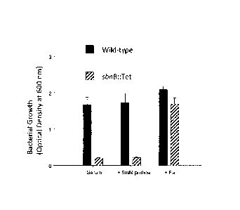
[0013] Figure 1 graphically illustrates impaired growth of S. aureus sbn
operon
deletion strain in serum, and the inset graphically illustrates growth of the
deletion strain in
serum supplemented with FeCl3;
[0014] Figure 2 is a bar graph comparing siderophore production in wild-
type S.
aureus (black bars) with an S. aureus sbn operon deletion strain (grey bars)
in the presence
and absence of iron;
[0015] Figure 3 is a schematic representation of the sbt-hts region of
the S. aureus
chromosome including locus numbers from the sequenced chromosome of strain
Newman;
[0016] Figure 4 provides an intergenic region between sbtA and sbtBCD
coding
regions (A) and an intergenic region between sbtD and htsABC coding regions
(B)
identifying putative Fur box sequences, start codons for the sbtA, sbtB and
htsA genes
(boldface) and Shine-Dalgarno sequences (S.D.);
- 3 -
CA 02734734 2011-02-18
WO 2010/020036
PCT/CA2009/001143
[0017] Figure 5 is a bar graph showing that transcription of the sbt-hts
locus, namely
sbtA (white bars), sbtB (grey bars), and htsA (black bars), is regulated by
iron and Fur;
[0018] Figure 6 graphically compares the growth of wild-type S. aureus
(*), an S.
aureus sin operon deletion strain (AsbtABCD::Tet) (A) and an S. aureus tandem
sbn/sbt
locus mutant (AsbnABCDEFGHLTet AsbtABCD::Kan) (0) in serum, and the inset
graphically illustrates growth of the tandem deletion strain in serum
supplemented with
FeCl3,
[0019] Figure 7 is a bar graph comparing siderophore production in wild-
type S.
aureus (black bars) with an S. aureus sbt operon deletion strain (grey bars)
and an S. aureus
tandem sbn/sbt locus mutant (white bars) in the presence and absence of iron;
[0020] Figure 8 graphically illustrates the effect of ABC transporter
gene
inactivations on the growth of S. aureus wild-type (0), sirA::Km (J), AhtsABC
(A), and
tandem sirA/htsABC (0);
[0021] Figure 9 graphically illustrates the growth in serum of S. aureus
wild-type
(*), AsbnABCDEFGHL:Tet (El), AsbtABCD::Km (A) and tandem Asbnl Asbt (0)
transformed with empty cloning vector (pL150) (A), a plasmid carrying sbtABCD
from S.
aureus (pEV90)(B) and a plasmid carrying shtABCD from S. epidernfidis (pEV95)
(C);
[0022] Figure 10 illustrates the absorption spectra of purified 1sdE,
HtsA and SirA
proteins in the absence of heme (A) and on exposure to heme (B);
[0023] Figure 11 is a bar graph comparing bacterial growth of S. aureus
wild-type
and an sbnB mutant in the absence and presence of proline and iron;
[0024] Figure 12 is a bar graph comparing bacterial growth of S. aureus
wild-type,
an sifith mutant and an sbnB mutant including a plasmid carrying sbnB in the
absence and
presence diaminopropionic acid (DAPA), Fe and Sb;
[0025] Figure 13 illustrates a physical map of the S. aureus sir-sbn
genetic locus
identifying type A (angle lines), type B (solid) and type C (vertical lines)
NIS synthetases and
a decarboxylase (dotted), and the structure of staphyloferrin B(B);
-4-
CA 02734734 2011-02-18
WO 2010/020036 PCT/CA2009/001143
[0026] Figure 14
graphically illustrates bacterial growth of iron-starved S. aureus
Asbnl Asbt mutant in the presence of increasing amounts of staphyloferrin B
(SB) synthesized
in vitro reaching levels comparable to growth in the presence of iron (A); and
graphically
compares bacterial growth of a AsfaAsbn mutant in the presence and absence of
iron, and in
the presence of staphyloferrin B with no iron (B):
[0027] Figure 15
provides bar graphs illustrating substrate specificity of Sbn enzymes
for the carboxylic acid substrates, citrate (A), citryl-Dap (B), citryl-Dae
(C) and a-KG (D);
[0028] Figure 16
illustrates a proposed scheme for the biosynthesis of staphyloferrin
B;
[0029] Figure 17 illustrates the structure of staphyloferrin A;
[0030] Figure 18
provides bar graphs illustrating growth of wildtype S. aureus (A)
and a SirA mutant (B), and, and no growth of Hts (C) and HtsSir mutants (D) in
the presence
of staphyloferrin A synthesized in vitro;
[0031] Figure 19
illustrates a proposed scheme for the biosynthesis of staphyloferrin
A; and
[0032] Figure 20
provides schematics of the HtsA crystal structure identifying the
staphyloferrin A binding region (box 4) (A) including residues within the
binding region (B),
and a schematic of the SirA crystal structure identifying the stapyloferrin B
binding region
(box) (C).
Detailed Description of Embodiments
[0033] A method
of inhibiting S. aureus is provided comprising inhibiting
siderophore-mediated iron uptake, for example, staphyloferrin-mediated iron
uptake.
[0034]
Siderophore-mediated iron uptake encompasses siderophore biosynthesis
and/or siderophore uptake or transport. Two paths of siderophore biosynthesis
and uptake
have now been identified. In one path, the siderophore, staphyloferrin A, is
produced by Sbt
polypeptides and transported for cellular uptake by Hts polypeptides. In
another path, the
siderophore, staphyloferrin B, is produced by Sbn polypeptides and transported
for cellular
uptake by Sir polypeptides
- 5 -
CA 02734734 2011-02-18
WO 2010/020036 PCT/CA2009/001143
[0035] The terms "staphyloferrin A" and "staphyloferrin B" refer to high
affinity cc-
hydroxycarboxylate iron-chelating compounds or siderophores of S. aureus which
bind
iron, generally in the form of ferric (Fe3+ ) ions. The structure of
staphyloferrin A is provided
in Fig. 17 while the structure of staphyloferrin B is provided in Fig. 16.
[0036] Staphyloferrin A is produced by Sbt polypeptides expressed from a
gene
cluster referred to herein as the "sbt" or "sfna" gene cluster, and
transported into the cell by
HtsABC polypeptides expressed from hts genes.
[0037] The "sbt" or "sfa" gene cluster refers to a group of S. aureus
genes, namely,
sbtA, sbtB, sbtC, and sbtD that have been isolated from a common chromosomal
locus and
respectively encode polypeptides SbtA, SbtB, SbtC and SbtD which are involved
in the
biosynthesis of staphyloferrin A. In one embodiment, SbtB and SbtD are NIS
(NRPS-
independent siderophore) synthetases, SbtC is an amino acid racemase and SbtA
is a
membrane embedded siderophore efflux protein. Exemplary nucleotide sequences
of sbt
genes and corresponding encoded Sbt polypeptides may be found in GenBank
accession
number AP009351 or RefSeq accession number NC 009641 at loci NWMN_2079,
NWMN_2080, NWMN_2081 and NWMN_2082. Exemplary amino acid sequences of Sbt
polypeptides may also be found at GenBank Protein ID accession numbers
BAF68351,
BAF68352, BAF68353 and BAF68354.
[0038] It has been determined that staphyloferrin A may be synthesized
outside of its
native environment, for example, recombinantly in bacteria in which it is not
endogenously
expressed, as well as under cell-free conditions. Thus, in one embodiment, sbt
genes may be
transfected into selected bacterial cells, using established technology, and
incubated in media
including Sbt substrates, e.g. (e.g. citrate, D-ornithine) and cofactors (ATP
and Mg2+) for
expression to yield functional staphyloferrin A. In another embodiment, Sbt
polypeptides,
including SbtA, SbtB, SbtC and SbtD, may be incubated in a cell-free
environment under
conditions designed to emulate the basic functional biochemistry of the cell,
for example,
including a carboxylate substrate such as citric acid and D-ornithine to yield
functional
staphyloferrin A.. In a further embodiment, Sbt sy-nthetases, such as SbtB and
SbtD alone,
may be incubated in the presence of a carboxylate substrate such as citric
acid and D-
ornithine to yield functional staphyloferrin A. If D-ornithine is substituted
with L-ornithine,
then SbtC may be added to convert to the D-racemate. In this regard, the sbt
genes or Sbt
- 6 -
CA 02734734 2011-02-18
WO 2010/020036 PCT/CA2009/001143
peptides may be derived from any one of S. aureus, S. epidermidis, S.
haemolyticus and S.
saprophyticus sources, or derived from a combination of these sources.
[0039] The HtsABC transporter is encoded by an "hts operon" comprising a
group of
bacterial genes including htsA, htsB, and htsC that share a common promoter.
This operon
encodes a protein system that functions to transport ferrated siderophore,
namely
staphyloferrin A, into S. aureus cells, also known as an ABC transporter. The
promoter
element, which is upstream of the htsA coding region, is iron-regulated
through the Fur
protein. The htsA gene encodes a heme or siderophore binding protein (HtsA),
while htsB
and htsC encode transmembrane components (HtsB/C) of the ABC-transporter.
Exemplary
nucleotide sequences of hts genes and corresponding encoded Hts polypeptides
may be found
in GenBank accession number AP009351 or RefSeq accession number NC_009641 at
loci
NWMN 2076, NWMN 2077, and NWMN 2078. Exemplary amino acid sequences of Hts
polypeptides may also be found at GenBank Protein ID accession numbers
BAF68348,
BAF68349, and BAF68350. The hts-encoded siderophore transport system interacts
with a
FhuC ATPase as will be described.
[0040] The siderophore, staphyloferrin B, also previously referred to as
staphylobactin, an a-hydroxycarboxylate siderophore comprised of L-2,3-
diaminopropionic
acid, citric acid, ethylenediamine and a-ketoglutaric acid, is produced by a
gene cluster
referred to as the "sbn" gene cluster. Ferrated staphyloferrin B is
transported into the
organism via a SirABC transporter system.
[0041] The "sbn" gene cluster refers to a group of S. aureus genes,
namely, sbnA,
sbnB, sbnC, sbnD, sbnE, sbnF, sbnG, sbnH, and sbnl that share a common
promoter. The
promoter element, which is upstream of the sbnA coding region, is iron-
regulated.
Exemplary nucleotide sequences of sbn genes may be found at Genbank accession
no.
AY251022. The sbn genes respectively encode the polypeptides "SbnA", "SbnB",
"SbnC",
"SbnD", "SbnE", "SbnF", "SbnG", "SbnH", and "Sbnl" which are involved in the
synthesis of
staphyloferrin B. Sequence information for these polypeptides may be found in
published
PCT application, WO 06/043182. Accordingly, in one embodiment, sbnA encodes a
cysteine
synthase, sbnB encodes an omithine cyclodeaminase, sbnC encodes a biosynthesis
protein,
sbnD encodes an efflux protein, sbnE encodes a siderophore biosynthesis
protein, sbnF
encodes a siderophore biosynthesis protein, sbnG encodes an aldolase protein,
and sbnH
encodes an amino acid decarboxylase.
- 7 -
CA 02734734 2011-02-18
WO 2010/020036 PCT/CA2009/001143
[0042] It has been determined that staphyloferrin B may be synthesized
outside of its
native environment, for example, rccombinantly in bacteria in which it is not
endogenously
expressed, as well as under cell-free conditions. Thus, in one embodiment, sbn
genes may be
transfected into selected bacterial cells, using established technology, and
incubated in media
including Sbn substrates, (e.g. citrate, L-2,3-diaminopropionic acid, alpha-
ketoglutarate,
ATP, Mg2) for expression to yield functional staphyloferrin B. In another
embodiment, Sbn
polypeptides, including SbnA, SbnB, SbnC, SbnD, SbnE, SbnF, SbnG, SbnH and
SbnI, may
be incubated with suitable Sbn substrates in a cell free environment under
conditions
designed to emulate the basic functional biochemistry of the cell to yield
functional
staphyloferrin. In a further embodiment, Sbn synthetases, such as SbnC, SbnE
and SbnF, and
decarboxylases, such as SbnH, alone may be incubated in the presence of
substrates such as:
L-2,3-diaminopropionic acid, citric acid, and a-ketoglutaric acid, to yield
functional
staphyloferrin B.
[0043] The SirABC transporter is encoded by a "sirABC operon" comprising
a group
of genes including sirA, sirB, and sirC that share a common promoter. This
operon encodes a
protein system that functions to transport ferrated siderophore into S. aureus
cells, also
known as an ABC transporter. Exemplary nucleotide and polypeptide sequences of
sirABC
operon, and the Sir proteins it encodes, may be found at GenBank Accession No.
AY251022
and GenBank Accession No. AF079518. The sirA gene encodes an extracellular
protein
(SirA), while sirB and sirC encode transmembrane domains (SirB/SirC) of the
ABC-
transporter. The term "SirABC iron-siderophore transport system" refers the
SirABC
transporter that is comprised of SirA, SirB, SirC, and FhuC polypeptides.
[0044] FhuC is a polypeptide of the "ferric hydroxamate uptake system" or
"Thu
system". The fhu system is encoded by five genes: fhuC, fhuB, and fhuG, and
fhuDI and
fhuD2. .fhuC, ,fhuB, and fhuG are present in an operon (f1mCBG operon) and
encode
components of an ATP-binding cassette (ABC) transporter. fhuC encodes an
ATPase that
interacts with both sir and hts encoded siderophore transport systems. fhuD1
and fhuD2 are
separately encoded and encode lipoproteins that bind ferric hydroxamate
complexes with
high affinity. Exemplary nucleotide and amino acid sequences for the fhuCBG
operon may be
found in GenBank, Accession Nos. AF251216, AAF98153, AAF98154, and AAF98155;
for
fhuD/, Accession No. AF325854 and A AK92085; and for fhuD2 AF325855 and
AAK92086.
- 8 -
CA 02734734 2011-02-18
WO 2010/020036
PCT/CA2009/001143
The terms "FhuC", "FhuB", "FhuG", "FhuD1", and "FhuD2" refer to the proteins
encoded by
fhuC,JhuB, jhuG, fhuD 1 and fhilD2, respectively.
[0045] In accordance with an aspect of the invention, S. aureus may be
inhibited by
inhibiting staphyloferrin-mediated iron uptake, i.e. staphyloferrin A-mediated
iron uptake and
staphyloferrin B-mediated iron uptake. The term "inhibited" as used herein
with respect to
inhibition of S. aureus refers to at least partial growth inhibition, and
includes complete
growth inhibition, of S. aureus. The term "inhibiting" as used herein with
respect to
staphyloferrin A and B iron uptake refers to at least partial inhibition,
including complete
inhibition, of iron uptake by S. aureus, and includes inhibition of
siderophore synthesis,
secretion of siderophore and cell uptake of siderophore.
[0046] Inhibition of staphyloferrin-mediated iron uptake may be achieved
by
inhibiting the expression or function of at least one of the Sbt polypeptides
required for the
production of staphyloferrin A, e.g. at least one of SbtA, SbtB, SbtC and
SbtD, for example,
SbtB and SbtD, and inhibiting the expression or function of at least one of
the Sbn
polypeptides required for the production of staphyloferrin B, e.g. at least
one of SbnA, SbnB,
SbnC, SbnD, SbnE, SbnF, SbnG, SbnH, and Sbnl, for example, SbnC, SbnE, SbnF
and
SbnH. At the nucleic acid level, Sbt/Sbn expression may be blocked using well-
established
methods in the art including, for example, antisense and RNA interference
technologies
(siRNA, shRNA and microRNA) to prevent siderophore synthesis. At the protein
level, the
function of one or more of the Sbt polypeptides and one or more of the Sbn
polypeptides may
be inhibited to prevent synthesis of each siderophore.
[0047] The term "antisense oligonucleotide" as used herein means a
nucleotide
sequence that is complementary to a target sbt/sbn nucleic acid sequence. The
term
"oligonucleotide refers to an oligomer or polymer of nucleotide or nucleoside
monomers
consisting of naturally occurring bases, sugars, and intersugar (backbone)
linkages. The term
also includes modified or substituted oligomers comprising non-naturally
occurring
monomers or portions thereof, which function similarly. Such modified or
substituted
oligonucleotides may be preferred over naturally occurring forms because of
properties such
as enhanced cellular uptake, or increased stability in the presence of
nucleases. The antisense
oligonucleotides of the present invention may be ribonucleic or
deoxyribonucleic acids and
may contain naturally occurring bases including adenine, guanine, cytosine,
thymidine and
uracil. The oligonucleotides may also contain modified bases such as xanthine,
hypoxanthine
- 9 -
CA 02734734 2011-02-18
WO 2010/020036 PCT/CA2009/001143
and 2-aminoadenine. Other antisense oligonucleotides of the invention may
contain modified
phosphorous, oxygen heteroatoms in the phosphate backbone, short chain alkyl
or cycloalkyl
intersugar images or short chain heteroatomic or heterocyclic intersugar
linkages. For
example, the antisense oligonucleotides may contain phosphorothioates,
phosphotriesters,
methyl phosphonates, and phophorodithioates. The antisense oligonucleotides of
the
invention may also comprise nucleotide analogs that may be better suited as
therapeutic or
experimental reagents. An example of an oligonucleotide analogue is a peptide
nucleic acid
(PNA) in which the deoxribose (or ribose) phosphate backbone in the DNA (or
RNA), is
replaced with a polymide backbone which is similar to that found in peptides.
Suitable
antisense oligonucleotides will be at least 5 nucleotides in length, and
preferably at least
about 15 nuceotides long, and will be sufficient to prevent transcription of a
target gene to
yield functional protein.
[0048] In another embodiment, RNA interference technologies (such as
siRNA,
shRNA and microRNA) may be applied to prevent expression of Sbt/Sbn
polypeptides.
Application of nucleic acid fragments such as siRNA fragments that correspond
with regions
of a target sbt/sbn gene, at least to the extent required to bind thereto, may
be used to block
expression resulting in inhibition of siderophore production. Such blocking
occurs when the
siRNA fragments bind to the target gene thereby preventing translation of the
gene to yield
functional Sbt/Sbn polypeptides. Suitable siRNAs are of a length suitable to
inhibit
expression of a target gene, e.g. at least about 10-15 nucleotides in length,
and comprise
sufficient complementarity to the target gene to hybridize thereto under
desired conditions,
e.g. in a cell. The antisense and RNA oligonucleotides may be introduced into
tissues or cells
using techniques in the art including vectors (retroviral vectors, adenoviral
vectors and DNA
virus vectors) or physical techniques such as microinjection.
[0049] Antisense and RNA oligonucleotides may be constructed using
chemical
synthesis and enzymatic ligation reactions using procedures known in the art
based on
sequence information provided. The oligonucleotides may be chemically
synthesized using
naturally occurring nucleotides or variously modified nucleotides designed to
increase the
biological stability of the molecules or to increase the physical stability of
the duplex formed
with mRNA or the native gene, e.g. phosphorothioate derivatives and acridine
substituted
nucleotides. Alternatively, the oligonucleotides may be produced biologically
using
recombination technology as is well-established in the art.
-10-
CA 02734734 2011-02-18
WO 2010/020036
PCT/CA2009/001143
[0050] Inhibition of Sbt/Sbn polypeptides may be achieved using one or
more
compounds that interfere with the function of the polypeptide. Thus, given
knowledge of the
function of a target Sbt/Sbn polypeptide, suitable inhibitory compounds may be
identified or
developed. For example, the polypeptides SbnC, SbnE and SbnF have been
identified as NIS
synthetases necessary for staphyloferrin B synthesis. Thus, a compound useful
to inhibit
such a synthetase would be suitable to inhibit staphyloferrin B synthesis.
Inhibition of the
decarboxylase activity of SbnH will also inhibit staphyloferrin B synthesis.
In this regard,
substrate analogs may be useful to block synthetase and/or decarboxylase
activity.
[0051] Alternatively, Sbt/Sbn polypeptides may be inhibited by limiting
access to one
or more substrates required for staphyloferrin biosynthesis. In this regard,
the enzymes that
produce the substrates necessary for staphyloferrin synthesis may be
inhibited, for example,
inhibition of SbnA or SbnB would inhibit the synthesis of the substrate,
diaminopropionic
acid, that is required for staphyloferrin B synthesis.
[0052] In another embodiment, inhibition of staphyloferrin-mediated iron
uptake may
be achieved by inhibiting the Hts- and Sir-mediated transport of ferrated
staphyloferrin into
the cell. In this regard, the expression or function of at least one of the
Hts polypeptides, e.g.
HtsA, HtsB and HtsC, required for the transport of ferrated staphyloferrin A
into the cell, and
the expression or function of at least one of the Sir polypeptides, e.g. SirA,
SirB and SirC,
required for the transport of ferrated staphyloferrin B into the cell may be
inhibited.
Alternatively, expression of FhuC may be inhibited to inhibit the transport of
both
staphyloferrin A and staphyloferrin B into the cell.
[0053] As one of skill in the art will appreciate, staphyloferrin-
mediated iron uptake
may be inhibited by inhibiting the synthesis of staphyloferrin A and B (as set
out above), the
transport of staphyloferrin A and B into the cell (as set out above), as well
as inhibiting the
synthesis of staphyloferrin A combined with inhibiting the transport of
staphyloferrin B into
the cell, or inhibiting the transport of staphyloferrin A into the cell
combined with inhibiting
the synthesis of staphyloferrin B.
[0054] In order to identify agents that modulate staphyloferrin-mediated
iron uptake,
screening assays may be developed to screen for agents that modulate the iron-
transport
activity of the Sir or Hts polypeptides. For example, appropriate
concentrations of test agents
for modulating the iron-transport activity of the Hts proteins may be
determined by any
- 11 -
CA 02734734 2011-02-18
WO 2010/020036
PCT/CA2009/001143
method known to one skilled in the art. In one embodiment, the screening assay
may include
whole S. aureus cells expressing wild type Hts and Sbt polypeptides. The
ability of a
compound to alter the iron transport activity of the Hts and/or Sbt
polypeptides can be
detected by analysis of the cells. For example, antagonists of iron-transport
can by detected
by scoring for alterations in growth or differentiation (phenotype) of the
cell in iron-replete
media. The growth of wild- type S. aureus strains in the presence of test
agent(s) may be
compared with the growth of SbtA, SbtB, SbtC, SbtD, HtsA, HtsB or HtsC
deficient S.
aureus strains. Each culture may be treated with a test agent from a library
of compounds or
natural extracts, and monitored for the effect that the particular agent has
on the growth on
the wild-type and the Hts-deficient strain. Bacterial growth may be monitored
using a Klett
meter. In this way, compounds that specifically interfere with the
HtsABC/sbtABCD iron
siderophore transport system can be identified.
[0055] As another example, S. aureus cells may be cultured and treated
with test
agents and then screened for the presence of iron in the cell using atomic
absorption
spectroscopy techniques. Alternatively, inhibition of the iron transport
activity may be
measured by using radioactively labeled iron. Compounds that interfere with
the HtsABC
iron siderophore transport system will result in a lowered uptake of the
radioactively labeled
iron. A control assay can also be performed to provide a baseline for
comparison. In the
control assay, the uptake of radioactively labeled iron in a S. aureus cell
may be quantitated
in the absence of the test compound. Examples of radioactively labeled iron
may include 59Fe
or 55Fe.
[0056] Antagonists that interfere with the expression of a nucleic acid
or protein
involved in siderophore-mediated iron uptake may also be identified. To
identify such
antagonists, S. aureus cells may be treated with a compound(s) of interest,
and then assayed
for the effect of the compound(s) on nucleic acid expression or protein
production in respect
of nucleic acids and corresponding encoded proteins involved in siderophore-
mediated iron
uptake. For example, total RNA can be isolated from S. aureus cells cultured
in the presence
or absence of test agents, using any suitable technique such as the single-
step guanidinium-
thiocyanate-phenol-chloroform method. The expression of nucleic acids such as
sir, sbn, hts,
sbt or Jhu nucleic acids may then be assayed by any appropriate method such as
Northern blot
analysis, the polymerase chain reaction (PCR), reverse transcription in
combination with the
polymerase chain reaction (RT-PCR), and reverse transcription in combination
with the
- 12 -
CA 02734734 2011-02-18
WO 2010/020036 PCT/CA2009/001143
ligase chain reaction (RT-LCR). Levels of mRNA encoding Sir, Sbn, Hts, Sbt or
Fhu
polypeptides may also be assayed, for example, using the RT-PCR method to
determine the
effect of a selected test agent in comparison to a control sample. The
expression of Sir, Sbn,
Hts, Stb or Fhu polypeptides may also be quantitated following the treatment
of S. aureus
cells with a test agent using antibody-based methods such as immunoassays. Any
suitable
immunoassay can be used, including, without limitation, competitive and non-
competitive
assay systems using techniques such as western blots, radioimmunoassays, ELISA
(enzyme
linked immunosorbent assay), "sandwich" immunoassays, immunoprecipitation
assays,
precipitin reactions, gel diffusion precipitin reactions, immunodiffusion
assays, agglutination
assays, complement-fixation assays, immunoradiometric assays, fluorescent
immunoassays
and protein A immunoassays. For example, SbtA, SbtB, SbtC, SbtD, HtsA, HtsB or
HtsC
polypeptides may be detected in a sample obtained from S. aureus cells treated
with a test
agent, by means of a two-step sandwich assay. In the first step, a capture
reagent (e.g., either
a SbtA, SbtB, SbtC, SbtD, HtsA, HtsB or HtsC antibody) is used to capture the
specific
polypeptide. The capture reagent can optionally be immobilized on a solid
phase. In the
second step, a directly or indirectly labeled detection reagent is used to
detect the captured
marker. In one embodiment, the detection reagent is an antibody. The amount of
SbtA, SbtB,
SbtC, SbtD, HtsA, HtsB or HtsC polypeptide present in S. aureus cells treated
with a test
agent can be calculated by reference to the amount present in untreated S.
aureus cells to
determine the effect of the test agent on polypeptide expression.
[0057] Siderophore-mediated iron uptake by S. aureus may also be
inhibited by
interfering with the siderophore binding region within the Hts and Sir
polypeptides, for
example, the siderophore binding region within HtsA and SirA. As set out in
the examples
that follow, the binding region within these polypeptides has been identified
and serves as a
target region for inhibiting the interaction of Hts/Sir transport systems with
ferrated
siderophore for uptake. Thus, based on this determination of the binding
region, antagonists
can readily be designed to block Hts/Sir siderophore binding, and may include
siderophore
mimeties, immunological antagonists and the like.
[0058] Embodiments of the invention are described by reference to the
following
specific examples which are not to be construed as limiting.
- 13 -
CA 02734734 2011-02-18
WO 2010/020036
PCT/CA2009/001143
Examples
Example 1: Materials and Methods for Examples 2 to 10
[0059] Bacterial strains, plasmids, and growth media. Bacterial strains
and plasmids
used in this study are described in Table 1. Bacteria were cultured at 37 C,
unless otherwise
indicated.
TABLE 1. Bacterial strains, plasmids, and oligonucleotides used in this study
Bacterial strains, Description' Source or
plasmids, and reference
oligonucleotides
Bacteria
E. coil
DH5a 080d/acZAM15 recAl endA I gyrA96 thi-1 hsdRI7(rK- mK+)supE44
Promega
re/Al deoR A(lacZYA-argh)U169
ER2566 F k7huA2 [Ion] ompTlacZ::T7 genel gal sulA I I New England
A(mcrC-mrr)114::ISIO R(mcr-73::miniTn/0)2 R(2gb-210::Tn/0)1 Biolabs
(Tets) endA I [dcm]
RP523 tin-1 leuB6 thi-1 lacYl tonA2I supE44 F X hemB (Li etal.,
1988)
S. aureus
RN4220 rK- mi('; accepts foreign DNA (Kreiswirth et al.,
1983)
RN6390 Prophage-cured wild type strain (Peng et al., 1988)
Newman Wild type clinical isolate (Duthie and
Lorenz, 1952)
H1324 RN6390 AsbnABCDEFGHL:Tet; TetR This study
H1331 Newman AsbnABCDEFGHLTet; TetR This study
H1661 RN6390 AsbtABCD::Km; KmR This study
H1665 Newman AsbtABCD::Km; KmR This study
H1649 RN6390 AsbnABCDEFGIII::Tet AsbtABCD::Km; TetR Km' This
study
H1666 Newman AsbnABCDEFGHL:Tet AsbtABCD::Km; TetR Km' This study
H306 RN6390 sirA::Km; Km" (Dale etal.,
2004b)
H803 Newman sirA::Km; Km" (Dale etal.,
2004b)
H1448 RN6390 AhtsABC::Tet: TetR This study
H1262 Newman AhtsABC::Tet; TetR This study
HI480 RN6390 sirA::Km AhtsABC::Tet; TetR KniR This study
H1497 Newman sirA::Km AhtsABC::Tet; TctR Km? This study
H706 Newmanfur:Km; Km" (Dale etal.,
2004b)
S. epidermidis
846-1 Plasmid-cured type strain W. Kloos
1457-M10 Biofilm deficient (icd) mutant; EmR (Dobinsky etal.,
2002)
S. saprophyticus Clinical type strain (Kuroda et al.,
ATCC 15305 2005)
- 14 -
CA 02734734 2011-02-18
WO 2010/020036 PCT/CA2009/001143
Plasmids
pAUL-A Temperature-sensitive S. aureus suicide vector; EmR LcR
(Chakraborty et
al., 1992)
pALC2073 E coli1S. aureus shuttle vector; AmR CmR (Bateman c/ al.,
2001)
pBAD30-IsdE pBAD30 derivative encoding the IsdE protein; ApR (Muryoi et
al.,
2008)
pBC SK(+) E. coli cloning vector; CmR Stratagene
pDG780 BluescriptKS' derivative that carries a kanamycin resistance
(Guerout-Fleury et
cassette; APR al., 1995)
pDG1513 pMTL22 derivative that carries a tetracycline resistance
cassette; (Guerout-Fleury et
APR al., 1995)
pET28a(+) Vector for overexpression of His-tagged proteins using the T7
Novagen
bacteriophage promoter; KmR
pEV55 pL150 derivative containing htsABC from S. aureus; CmR
'this study
pEV83 pAUL-A derivative containing htsABC:Tet; EmR TetR This
study
pEV90 pLI50 derivative containing sbtABCD from S. aureus; CmR
This study
pEV93 pL150 derivative containing htsABC from S. epidermidis; CmR
"[his study
pEV95 pLI50 derivative containing sbtABCD from S. epidermidis; CmR
This study
pEV96 pLI50 derivative containing sbtABCD from S. saprophyticus; CmR
This study
pEV98 pE1'28a(+) derivative encoding soluble portion of protein
SirA; This study
KmR
pEV99 pET28a(+) derivative encoding the soluble portion of protein
fitsA; This study
KmR
pFB10 pAUL-A derivative containing AsbnABCDEFGHL:Tet; EmR TetR
This study
pFB24 pXEN-1 derivative containing Psbtk This study
pFB25 pXEN-1 derivative containing PsbtBCD This study
pFB26 pXEN-1 derivative containing PhtsABC This study
pFB50 p1,150 derivative with repB frameshift mutation, containing
This study
AsbtABCD::Km; CmR KmR
pFB54 pALC2073 tetO/R- derivative containing a transcriptional
fusion of This study
S. aureus fhuC and sirABC operons
pFB56 pALC2073 tetO/R- derivative containing the S. aureus fhuC gene
This study
01355 pALC2073 tetO/R- derivative containing a transcriptional
fusion of This study
S. aureus fliuC and sirABC operons, where jhuC has a 3' end
deletion
pLI50 E. coliIS. aureus shuttle vector; APR CmR (Lee and landolo,
1986)
psirABC pBC SK(+) derivative containing sirABC (Dale et al.,
2004b)
pUC 19 E. colt cloning vector; APR (Yanisch-Perron et
al., 1985)
Oligonucleotidesb
Purpose Sequence
Cloning of GTATAGATTGTATTTAATAAGTTAATGTAATCC (forward)
sbtAsbtBCD TGCAAACGATATGTAGTATAACTTGTCAAC (reverse)
from S. aureus
Cloning of ATATGAATTCTTGAGCATGACGCTCAAGTGC (forward,
sbtAsbtBCD EcoRI)
from ATATCCCGGGGAGACGGTGCGTTGAGTTAAAGG (reverse,
S. epidermidis SmaI)
Cloning of TGAGCTCTGCGATTACATTGGAGGCTG (forward, Sad)
htsABC from S. TGCCCGGGGTTAGTTATTTCATTCTTCG (reverse, Smal)
aureus
Cloning of CAGTTCTAGACCTTGTTCAGAACTTCGATATG (forward,
htsABC from Xbal)
- 15 -
CA 02734734 2016-07-19
S. epidermidis CAGTGAGCTCCAGGCTCTATAACTAAAAAATACG (reverse,
Sad)
Cloning of fhuC TTGATAGCATGCCATGACAAATCGAGCTATCC (forward,
from S. aureus SphI)
TTGATACTGCAGTTAAGAATAAGCTCTGCGACA (reverse,
PstI)
Cloning of sbtA TTGCGCGAATTCCATAAAACTTACACCCGCATTC (forward,
promoter from S. EcoRI)
aureus TTGCGCGGATCCCATAATTCACCTCTATGAAATA (reverse,
Baml II)
Cloning of sirA AACATATGACAAC ITCAATTAAACATGCAATG (forward,
(soluble Ndel)
component) from AAGAATTCCTCCTTAATTATTTTGATTGTTTTTC (reverse,
S. aureus EcoRI)
Cloning of htsA AAGCTAGCACTATTTCGGTAAAAGATGAAAATG (forward,
(soluble NheI)
component) from AAGGATCCCATTTACTTCCACCTTACTITTGTTC (reverse,
S. aureus BamHI)
RT-PCR: sbtA CCTCTAATGCAATGCCATATTTA (forward)
ACAATGAATCACCTATCGTGACA (reverse)
RT-PCR: sbtB AGTCTATCATGCGCCAACAAC (forward)
AACCTGTCGCCATAATCAATAA (reverse)
RT-PCR: htsA TTTAAATCCAGAGCGTATGATCA (forward)
CAGAAGAAATTAAGCCACGAGAT (reverse)
RT-PCR: gyrB ATAATTATGGTGCTGGGCAAAT (forward)
AACCAGCTAATGCTTCATCGATA (reverse)
'Abbreviations: Apr', CmR, EmR, Km, LcR, and TetR, resistance to ampicillin,
chloramphenicol, erythromycin,
kanamycin, lincomycin, and tetracycline, respectively; ATCC, American Type
Culture Collection.
bRestriction sites for cloning of PCR products are underlined.
[0060] Antibiotics were used at the following concentrations: ampicillin
(100
lag/mL) and erythromycin (300 pg/mL) for E. coli selection; chloramphenicol (5
gg/mL),
tetracycline (10 itg/mL), kanamycin (50 Ltg/mL), neomycin (50 pz/mL), and
erythromycin (3
mg/mL) for S. aureus selection. For molecular-genetic manipulations, bacteria
were grown in
Luria-Bertani (for E. coil) or tryptic soy broth (for S. aureus). Iron
restricted media were
either i) the chemically-defined Tris-minimal succinate medium containing 0.1
p.M
ethylenediamine-di-o-hydroxyphenylacetic acid (LGC Promochem), ii) RPMI broth
(Gibco
BRL) containing 1% w/v casamino acids (Difco), or iii) a 60:40 ratio of
complement-
inactivated horse serum (Sigma-Aldrich) to TMS broth. All solutions and media
were made
with water purified through a Milli Q water purification system (Millipore).
[0061] Recombinant DNA methodology. Plasmid DNA was isolated from bacteria
using Qiaprep mini-spin kits (Qiagen), as directed. For plasmid isolation from
S. aureus,
cells were incubated for 30 min at 37 C in P1 buffer containing 50 mg/mL
lysostaphin
(Roche Diagnostics) prior to addition of lysis buffer P2. Restriction
endonucleases, T4 DNA
- 16-
CA 02734734 2011-02-18
WO 2010/020036 PCT/CA2009/001143
ligase, Klenow fragment, and PwoI polymerase were obtained from Roche
Diagnostics, and
oligonucleotides were purchased from Integrated DNA Technologies.
[0062] sbn operon deletion. The sbnABCDEFGHLTet knockout allele consisted
of
the tetracycline resistance cassette, excised from plasmid pDG1513 with
restriction enzymes
Sspl and Noel and blunted with Klenow enzyme, flanked by DNA sequences
homologous to
regions upstream of sbnA and immediately downstream of sbnl. The knockout
allele was
cloned to the temperature sensitive E. coli1S. aureus shuttle vector pAUL-A,
and
subsequently passaged through S. aureus RN4220 prior to transduction into S.
aureus
RN6390. Recombinant RN6390 was cultured at 30 C to mid-log phase before the
incubation
temperature was shifted to 42 C and bacteria were incubated a further 16 hours
before being
plated onto TSA containing tetracycline. Colonies were screened for
sensitivity to
erythromycin, indicating a loss of pAUL-A backbone DNA following integration
of the
knockout allele into the chromosome via homologous recombination on either
side of the
tetracycline resistance cassette. The sbn::tet deletion was mobilized to other
S. aureus
backgrounds by transduction using phage 80oc.
[0063] sbt locus deletion. The sbtABCD::Kan knockout allele consisted of
the
kanamycin resistance cassette, excised from plasmid pDG780 flanked by DNA
sequences
homologous to regions downstream of shtA and shtD, and cloned into the E.
coli1S. aureus
shuttle vector pLI50. A S. aureus suicide vector was generated from this
plasmid by
introduction of a frameshift mutation into repB (encoding the Gram-positive
replicase
protein) following Nsil digestion and Klenow fragment fill-in. This plasmid,
incapable of
unassisted replication in S. aureus, was introduced into S. aureus strain
RN4220 carrying
unmodified pLI50, enabling replication through complementation in trans with
wild type
RepB. The construct was then transduced to S. aureus strain RN6390 and
recombinant
bacteria were plated onto TSA containing kanamycin and neomycin. Colonies were
screened
for sensitivity to chloramphenicol, indicating a loss of vector DNA following
homologous
recombination on either side of the kanamycin resistance gene. The sbt::kan
deletion was
mobilized to other S. aureus backgrounds by transduction using phage 80a.
[0064] htsABC::Tet deletion. The htsABC::Tet knockout allele targeted the
5' and 3'
noncoding regions around htsABC. The 5' arm was PCR amplified and cloned Sad
to BamHI
to plasmid pUC19, followed by PCR amplified 3' arm cloned BamHI to Xbal. A
tetracycline
resistance cassette was excised from plasmid pDG1513 with restriction enzymes
BamHI and
- 17-
CA 02734734 2016-07-19
Bg111 and cloned into the arms at the Smal site. The knockout allele was
excised and cloned
to shuttle vector pAUL-A, Sad I to XbaI. Passaging to S. aureus and selection
for
chromosomal integration was performed as described for the sbn operon
mutation.
[0065] Complementation vectors. The S. aureus sbtAsbtBCD::Km mutant was
complemented using plasmid pEV90. Additionally, complementation was performed
using
sbtAsbtBCD from S. epidermidis 846-1 and S. saprophyticus ATCC 15305. These
loci were
PCR amplified and cloned to pLI50 between restriction sites EcoRI and SmaI (S.
epidermidis) or BamHI and SmaI (S. saprophyticus) . htsABC::Tet mutants were
complemented using plasmids pEV55 and pEV93. his operons was PCR amplified
from S.
aureus Newman (pEV55) or S. epidermidis 846-1 (pEV93) and cloned to pLI50
between
restriction sites Sac! and SmaI or Sac! and Xbal, respectively. Vectors were
constructed in E.
coli DH5a, electroporated into S. aureus strain RN4220, and transduced to S.
aureus RN6390
and Newman strain lines using phage 80a.
[0066] Growth curves. Bacteria were cultured for 12 hours in TMS broth then
12
hours in TMS broth containing 100 ttIV1 2,2' dipyridyl (Sigma). Cells were
washed twice in
saline, and diluted 1:100 into 60% horse serum/40% TMS broth. For iron replete
media, 50
tiM FeCl3 was included. Cultures were grown under constant medium amplitude
shaking in a
Bioscreen C machine (Growth Curves, USA). Optical density was measured at 600
nm every
30 min.
[0067] Supernatant preparations and plate bioassays. S. aureus strains were
grown
with aeration in TMS broth containing 0.1 RIVI EDDHA for 40 h at 37 C. Cells
were
removed by centrifugation and supernatants were lyophilized. Dried supernatant
was
extracted with methanol (half the original supernatant volume), passed through
WhatmanTM
no. 1 filter paper to remove insoluble material, and rotary evaporated.
Material was
solubilized in water to 5% of the original supernatant volume. The ability of
supernatant
concentrates to promote the iron-restricted growth of S. aureus was assessed
using
siderophore plate bioassays, performed as previously described (Sebulsky et
al., 2000) with
modifications. Briefly, S. aureus strains were incorporated into TMS agar (1 x
104 cells/m1)
containing 7.5 ttM EDDHA. Concentrates (10 L) were added to sterile paper
discs which
were then placed onto the plates. Growth promotion was quantified by measuring
the radius
of growth around the disc after 36 hours at 37 C.
- 18 -
CA 02734734 2011-02-18
WO 2010/020036 PCT/CA2009/001143
Example 2: S. aureus sbn mutants still produce siderophore
[0068] Strains of S. aureus containing complete sbn operon deletions
(i.e. deleted for
all of sbnA through sbnI genes) were constructed. In S. aureus Newman
background, the sbn
deletion strain was called 111331 whereas in RN6390 background, it was called
H1324.
When cultured in scrum at 37 C, H1331 demonstrated markedly impaired growth
during the
first 15 hours of incubation compared to wildtype Newman, before eventually
growing to an
equivalent cell density as Newman by approximately 30 hours (Fig. 1). Analysis
of the spent
culture supernatant, from the 35 h timepoint, for iron chelating activity
using the chrome
azurol S assay (Schwyn and Neilands, 1987) revealed comparable siderophore
activity
between Newman and 111331 (Fig. 2). Supplementation of serum growth media with
iron
obviated the growth defect of H1331 (Fig. 1, inset), and suppressed
siderophore production in
both Newman and H1331 (Fig. 2). Finally, concentrated supernatant from both S.
aureus
Newman and H1331, both cultured in iron-restricted TMS media, promoted growth
of S.
aureus (Newman and RN6390 responded equivalently) in siderophore plate
bioassays (Table
2). To ensure that this was not due to a strain-specific phenomenon, all
experiments
described above were repeated using instead the RN6390 genetic background
(i.e. RN6390
vs. H1324). Equivalent results were obtained (data not shown).
TABLE 2. S. aureus supernatants promote growth of wildtype S. aureus Newman
Concentrated supernatant Growth promotion of strain Newman'
Newman 12.17 0.29b
H1331 (Newman Asbn) 8.17 0.58
H1665 (Newman Asbt) 11.83 0.29
H1666 (Newman Asbn Asbt) 0
aGrowth promotion of supernatants on strain RN6390 was equivalent to that
observed for strain Newman; bGrowth
promotion is measured as the diameter of growth around disc.
[0069] Taken together, these findings indicate that S. aureus synthesizes
at least two
siderophores, one of which does not require any of the products of the sbn
operon for
synthesis. The production of this additional siderophore compensates for the
absence of the
sbn-derived siderophore (staphylobactin/staphyloferrin B), but only after
prolonged
incubation in serum.
- 19-
CA 02734734 2011-02-18
WO 2010/020036 PCT/CA2009/001143
Example 3: A second iron-regulated siderophore biosynthetic locus in S. aureus
is also
conserved among coagulase-negative staphylococci
[0070] Since S. aureus sbn deletion mutants still produced siderophore,
other genetic
loci in S. aureus whose products would be capable of synthesizing a
siderophore were
determined. Examination of the available S. aureus genome sequences identified
a four-gene
locus with potential to encode siderophore biosynthetic enzymes; in strain
Newman, these
open reading frames are identified as NWMN_2079-NWMN_2082 (Fig. 3). This locus
was
identified as sbt, for siderophore biosynthesis two. The sbt locus resides on
the genome
immediately upstream of the htsABC operon (NWMN2078-2076), encoding components
of
an ABC transporter that was previously proposed to transport heme into the
staphylococcal
cell. sbtA is divergently transcribed from what is likely a polycistronic
message comprised of
sbtB-sbtD.
[0071] In contrast to the sbn operon which, among the staphylococci, is
only present
in S. aureus species, the sht locus is conserved in coagulase-negative
staphylococci, at least
where genomic information is available. As shown in Table 3, predicted Sbt
products share
significant similarity with predicted protein products from S. epidermidis, S.
haemolyticus
and S. saprophyticus indicating the functional similarity among these
Staphylococcal Sbt
products.
TABLE 3. The sht locus is found in S. aureus as well as CoNS
S. aureus Percent identity; total similarity
protein S. epidermidis S. haemolyticus S. saprophyticus
SbtA 59;71 54;64 53;64
SbtB 71;84 64;80 60;75
SbtC 64;74 58;71 59;72
SbtD 62;76 56;70 58;74
[0072] The intergenic region between divergently oriented sbtA and sbtB
genes, and
the region immediately upstream of the htsABC operon contain 19-bp sequences
(see Figs. 3
and 4) that are highly similar to consensus Fur box sequences, suggesting that
the sbtA, sbtB,
and hts transcripts are iron-regulated via the activity of the ferric uptake
regulator (Fur)
repressor protein. Consistent with the observation that siderophore is only
made by S. aureus
- 20 -
CA 02734734 2011-02-18
WO 2010/020036 PCT/CA2009/001143
during iron restriction (Fig. 2), and similar to the regulation observed for
the sbn operon, both
the sbtA and sbtB transcripts were up-regulated in an iron and Fur-dependent
manner (Fig. 5).
The htsABC operon was shown to be regulated in a similar fashion (Fig. 5).
Example 4: Deletion of the sbt locus in wildtype S. aureus backgrounds yields
no
growth-deficient phenotype, but does have an additive affect on growth when
combined
with sbn locus deletion
[0073] As described above, the sbt genes are expressed under conditions
of iron
limitation, and two of the genes (sbtA and sbtC) encode products with
similarity to
siderophore biosynthesis enzymes. It was, thus, hypothesized that deletion of
the locus
would lead to a drop in siderophore production, and consequently this would
result in a strain
deficient in its ability to grow in iron-restricted growth media (e.g. serum),
mimicking what
was observed for strains harboring sbn operon deletions (see Fig. 1). Strain
H1665, which is
strain Newman carrying a deletion of the sbtABCD gene cluster, was created.
Surprisingly, it
was found that, in contrast to strain H1331 (sbn locus deletion), there was no
discernible
growth attenuation in strain H1665 in serum (Fig. 6), in spite of the fact
that endpoint
analysis of culture supernatants identified a significant decrease in
siderophore output
compared with wildtype (Fig. 7). Concentrated culture supernatant from H1665
was able to
readily promote the growth of iron-restricted wildtype S. aureus strains
(Table 2), indicating
the presence of at least one siderophore molecule (likely encoded by products
of at least the
sbn genes).
[0074] To address whether Sbn-mediated siderophore activity was able to
compensate
for the loss of Sbt-mediated siderophore activity, both deletions were
combined into one
strain, H1666. Compared with either strain H1331 (Asbn) or H1665 (Asbt),
strain H1666
(Asbn Asbt) demonstrated severe attenuation of growth in serum (Fig. 6).
Growth of H1666
could, however, be resuscitated to wildtype levels if the growth media were
replete with iron
(Fig. 6, inset), indicating that the growth deficiency is due solely to the
inability to scavenge
available iron. Endpoint analysis of iron-restricted supernatants of the H1666
revealed that
siderophore activity was reduced to background levels (Fig. 7). Importantly,
concentrated
culture supernatant from iron-restricted H1666 did not contain any molecule
capable of
promoting the iron-restricted growth of wildtype S. aureus (Table 2).
-21-
CA 02734734 2016-07-19
[0075] Taken together, these data strongly suggest that all siderophore
production by
S. aureus is mediated by the sbn and sbt loci in the production of independent
siderophores.
To rule out potential strain specific effects, all deletions were
reconstructed in the RN6390
background, and similar growth and siderophore activity trends were observed
(data not
shown).
Example 5: The sirABC and htsABC operons encode ABC transporters associated
with
transport of the sbn-derived siderophore and the sbt-derived siderophore,
respectively
[0076] The transport of staphylobactin, produced by enzymes encoded within
the sbn
operon, is mediated by the SirABC ABC transporter which is encoded from an
operon
divergently transcribed from the sbn operon.
[0077] The relationship between the htsABC operon and the sbt locus (see
Fig. 3),
was then determined, despite the previous suggestion that HtsABC was involved
in heme
transport. Initially, it was noted that the growth kinetics, in serum, of H803
(sirA::Km) were
very similar to those observed for H1331 (sbn) (Fig. 8). Similar to the lack
of a growth
defect in serum for H1665 (Asbt), there was no growth defect for the htsABC
deletion strain,
H1262 (Fig. 8). Also similar to the results observed for mutants containing
the double
siderophore biosynthetic loci deletions (H1666 (Asbn Asbt)), inactivation of
sirA and htsABC
in the same strain (H1497) lead to drastic growth attenuation in serum (Fig.
8). More direct
evidence for the role of HtsABC in transport of siderophore derived from the
function of sbt
gene products was obtained through siderophore plate bioassays. Spent culture
supernatants
from each of H1331, H1665 and H1666 grown under iron-restricted conditions
were
concentrated and provided to wildtype, sirA::Km, AhtsABC and sirA::Km AhtsABC
strains.
As shown in Table 4, material produced by wildtype cells could promote growth
of all strains
except the double sirA::KmAhtsABC mutant, whereas material derived from H1331
(Asbn)
could only promote growth of wildtype and sirA::Km cells, and material derived
from H1665
(Asbt) could only promote growth of wildtype and htsABC::erm cells. Last,
concentrated
spent culture supernatant from H1666 was unable to promote growth of any S.
aureus strain,
including wildtype S. aureus (see also Table 2). This indicates that the sbn
and sbt genes
produce distinct material or siderophore with a non-overlapping specificity
for their
respective SirABC and HtsABC transport system.
- 22 -
CA 02734734 2011-02-18
WO 2010/020036 PCT/CA2009/001143
TABLE 4.
Concentrated Strain
supernatant
Newman H803 H1262 H1497
(Newman (Newman (Newman
AsirA) AhtsABC) AsirA
AhtsABC)
WT 12.17 + 0.290 8.33 + 0.29 13.17 0.29
0.00
Asbt 11.83 0.29 0.00 13.83 0.29
0.00
Asbn Asbt 0.00 0.00 0.00 0.00
Asbt pEV90 11.83 0.29 10.17 + 0.29 10.83 0.29
0.00
Asbn Asbt pEV90 11.33 + 0.29 12.17 + 0.29 0.00 0.00
Asbi pEV95 12.33 0.29 10.17 0.29 12.17 0.29 0.00
Asbn Asbt pEV95 10.67 0.29 10.83 0.29 0.00 0.00
'Growth promotion is measured as the diameter of growth around disc. See
Materials and Methods for assay details.
Example 6: sbtABCD and htsABC from S. epidermidis complement S. aureus mutants
[0078] As stated above, in contrast to sirABC and sbnA-I which, among the
staphylococci are only found in S. aureus, genome sequences from coagulase-
negative
staphylococci (CoNS) contain homologs of sbtABCD (see Table 4). To determine
whether
CoNS sbt homologs are functionally analogous to those in S. aureus, it was
determined
whether the S. epidermidis sbt genes would complement the S. aureus sbt growth
defect and,
secondly, whether a product would be made that could be transported through S.
aureus
HtsABC. As shown in Fig. 9, growth in serum of the siderophore-deficient S.
aureus mutant
H1666 (i.e. Asbn Asbt) was capable of being equivalently augmented when
containing the sbt
locus either from S. aureus or S. epidermidis. Interestingly, the multicopy
sbt genes were
incapable of promoting faster growth of H1331 (i.e. Asbn). Evidence that the
sbt genes from
S. epidermidis synthesize an identical (or highly similar) molecule as that
produced by sbt
genes from S. aureus is derived from the result that sbt genes in trans,
whether from S. aureus
or S. epidermidis, in H1666 (i.e. Asbn Asbt) produced culture supernatant
capable of
promoting growth of wildtype S. aureus and S. aureus AsirA, but not AhtsABC
mutants of S.
aureus.
- 23 -
CA 02734734 2011-02-18
WO 2010/020036
PCT/CA2009/001143
Example 7: Lack of evidence for heme binding or transport by S. aureus Hts
[0079] A previous report indicated that mariner transposon insertion into
hts lead to a
strain that preferentially took iron from transferrin, as opposed to heme,
leading to the
suggestion that htsABC encoded an ABC transporter specific for heme. However,
evidence is
provided herein that S. aureus HtsABC is involved in transport of a
siderophore molecule
that is made by the products of the sbt locus, a locus that is transcribed
from the chromosome
directly upstream of htsABC. In this example, direct evidence for heme
transport by HtsABC
or, at the very least, heme binding by the solute binding protein, HtsA, was
tested.
[0080] In liquid growth assays, no attenuation of the htsABC deletion
mutant, H1665,
when grown on heme as a sole iron source (data not shown) was observed.
Moreover, as
shown in Fig. 10, when assayed for heme binding functionality, little to no
heme would
associate with purified HtsA in comparison to IsdE, a lipoprotein proven to
associate with
heme. This indicates that, in contrast to what was previously thought, Hts is
involved in the
transport of siderophore-bound iron, as opposed to heme.
Example 8: Several S. aureus Sbn mutants demonstrate an iron-restricted growth
defect
[0081] A nine-gene operon called sbn for siderophore biosynthesis, has
been
identified. In order to determine if sbn products play a role in siderophore
production, Asbn
S. aureus mutants, including sbnA, sbnB, sbnC, sbnD, sbnE, sbnF and sbnH have
been
generated. Growth assays with each of these mutants has shown that none of
these mutants
make staphylobactin and all of these mutants grow poorly in iron-restricted
media such as in
human or animal sera. Therefore, at least sbnA, sbnB, sbnC, sbnD, sbnE, sbnF,
sbnH or their
respective encoded protein products are candidate targets for inhibitors of
staphylobactin
biosynthesis. Thus, sbn nucleic acids, corresponding encoded Sbn protein
products or
corresponding S. aureus Sbn mutants can be used in screening methods to
identify inhibitors
of the staphylobactin biosynthetic pathway. Furthermore, components of
reactions catalyzed
by Sbn enzymes, as well as the Sbn enzymes themselves, may be used to develop
biochemical assays for use in high-throughput screening for inhibitors of Sbn
enzymes.
Example 9: Biochemical assays using SbnA and SbnB
[0082] As mentioned in Example 8 several 4sbn S. aureus mutants produce
an iron-
restricted growth defect phenotype, in for example human or animal sera. Using
SbnA and
- 24 -
CA 02734734 2011-02-18
WO 2010/020036 PCT/CA2009/001143
SbnB mutants in growth culture assays, compounds were added to the growth
media and
tested for the ability to overcome the iron-restricted phenotype. Figure 11
shows that proline
is unable to overcome the iron-restricted growth defect of the SbnB mutant
indicating that
proline is not a product of an SbnB mediated enzymatic reaction that is used
in
staphylobactin biosynthesis. The iron-restricted growth defect of S. aureus
SbnA and SbnB
mutants (due to the lack of production of staphylobactin) can be overcome by
addition of 2,3-
diamonpropionate to the growth media (Figure 12; data not shown for the SbnA
mutant).
This data shows that the modified amino acid 2,3-diaminopropionate is a
component of
staphylobactin.
[00831 Without wishing to be limited by theory, SbnB, a putative
ornithine
cyclodeaminase (OCD) is believed to liberate NH3 as it converts and cyclizes
ornithine to
proline. SbnA, a predicted 0-acetyl-L-serine sulfhydrylase may use the NH3 in
a reaction that
converts 0-acetyl serine to 2,3-diaminopropionate (DAPA) in an NAD -dependent
manner.
[0084] SbnA and SbnB have been overexpressed and purified and an HPLC-
based
assay comprising these two proteins has been developed. This HPLC-based assay
has been
used to show that the product DAPA is produced when SbnA and SbnB are put
together with
0-acetyl-serine, ornithine, and NAD+ as reaction substrates. DAPA is not made
when either
enzyme is used in isolation of the other. These results confirm the SbnA and
SbnB-dependent
consumption of ornithine and 0-acetyl-L-serine and the concomitant appearance
of proline
and 2,3-diaminopropionate. Furthermore, the ability to monitor production of
DAPA in this
HPLC-based assay indicates that this assay can be used for high-throughput
screening for
inhibitors that target SbnA or SbnB enzymes.
Example 10: FhuC is an ATPase for both Sir and Hts siderophore transporters
[0085] An FhuC mutant, strain H1071, was assayed to determine whether its
iron-
restricted growth defect could be overcome in the presence of either one of
the siderophores,
staphyloferrin A and staphyloferrin B, or in the presence of both
siderophores. More
specifically, the ability of supernatant concentrates of cell cultures
producing one or both of
the siderophores to promote the iron-restricted growth of the FhuC S. aureus
mutant was
assessed using siderophore plate bioassays.
- 25 -
CA 02734734 2011-02-18
WO 2010/020036
PCT/CA2009/001143
[0086] The FhuC mutant showed no growth in the plate bioassay when using
either
siderophore alone or using both together. This data indicates that FhuC is the
ATPase that
energizes transport of both S. aureus siderophores.
Example 11 ¨ Biosynthesis of Staphyloferrin B
Experimental Procedures
Bacterial strains, plasmids, and standard growth condition.
[0087] Bacterial strains and plasmids used in this study are described in
Table 5.
Table 5. Bacterial strains, plasmids and oligonucleotides used in this study.
Bacterial strains, Description' Source or
plasmids, and reference
oligonucleotides
E. coil
DH5a F (1)80d/acZAM15 recAl endAl nupG gyrA96 gInV44 thi-1
Promega
hsdR17(rk- mk+) X supE44 re/Al deoR A(IacZYA-argF)U169
ER2566 F X- fhuA2 [Ion] ompT lacZ::T7 gene 1 gal sulA11 New
England
A(mcrC-mrr)114::IS10 R(mcr-73::miniTn /0-Tets)2 Biolabs
R(zgb-210::Tn10)1 (Tets) endA1 [dcm]
BL21(DE3) F ompT gal dam Ion hsdSB (rE3- mB) X(DE3 [/ac! lacUV5-T7
Novagen
gene 1 ind1 sam7 nin5])
S. aureus
RN4220 1K mk+; accepts foreign DNA (Kreiswirth et aL,
1983)
RN6390 Prophage-cured wild type strain (Peng et al.,
1988)
H306 RN6390 sirA; KmR (Dale etal.,
2004b)
H1324 RN6390 Asbn; TetR (Beasley et al.,
2009)
H1661 RN6390 Asia; KmR (Beasley et al.,
2009)
H1649 RN6390 Asbn Asfa; TetR KmR (Beasley et al.,
2009)
H1448 RN6390 Ahts; TetR (Beasley et al.,
2009)
H1480 RN6390 sirA Ahts; TetR KmR (Beasley et al.,
2009)
Plasmids
pET28a(+) Overexpression vector for hexahistidine-ta,gged proteins; KmR
Novagen
pSbnC pET28a(+) derivative encoding SbnC; Km This study
pSbnE pET28a(+) derivative encoding SbnE; KmR This study
pSbnF pET28a(+) derivative encoding SbnF; KmR This study
pSbnH pET28a(+) derivative encoding SbnH; KmR This study
- 26 -
CA 02734734 2011-02-18
WO 2010/020036 PCT/CA2009/001143
[0088] E. coli were grown in Luria-Bertani broth (Difco). For experiments
not
directly involved in the analysis of iron uptake, S. aureus was grown in
tryptic soy broth
(Difco). Tris-minimal succinate (TMS) was prepared as described (Sebulsky et
al., 2004)
and used as an iron-limited minimal medium. To further restrict the level of
free iron in
TMS, the iron chelating compounds 2,2'-dipyridyl and ethylene diamine-di(o-
hydroxyphenol
acetic acid) (EDDHA) were added as indicated in the text. Where necessary,
kanamycin (30
ttg/ml) was incorporated into media for the growth of E. coli strains. For S.
aureus,
kanamycin (50 gimp, neomycin (50 jig/ml) and tetracycline (4 gimp were
incorporated
into growth media as required. Solid media were obtained by the addition of
1.5% (w/v)
Bacto agar (Difco). All bacterial growth was conducted at 37 C unless
otherwise stated.
Iron-free water for preparation of growth media and solutions was obtained by
passage
through a Milli-Q water filtration system (Millipore Corp.).
Recombinant DNA methodology
[0089] Standard DNA manipulations were performed. Restriction
endonucleases and
DNA-modifying enzymes were purchased from Roche Diagnostics (Laval, Quebec,
Canada),
New England Biolabs (Mississauga, Ontario, Canada), Life Technologies Inc.
(Burlington,
Ontario, Canada) and MBI Fermentas (Flamborough, Ontario, Canada). Plasmid DNA
was
purified using QIAprep plasmid spin columns (QIAgen Inc., Santa Clarita,
California) as
described by the manufacturer. Polymerase chain reactions were performed using
PwoI DNA
polymerase (Roche Diagnostics).
Siderophore plate bioassays
[0090] Siderophore plate bioassays were performed as described (Beasley
et al.,
2009). Growth promotion, as measured by the diameter of the growth halo around
each disk,
was determined after 36 h incubation at 37 C.
Bacterial Growth Curves
[0091] Bacteria were cultured for 12 hours in TMS broth then 12 hours in
TMS broth
containing 100 M 2,2'-dipyridyl (Sigma-Aldrich). Cells were washed twice in
saline, and
diluted 1:100 into 60% horse serum (Sigma-Aldrich) ¨ 40% TMS broth. For iron-
replete
media, 50 i_LM FeCl3 was included. Cultures were grown under constant, medium
amplitude
- 27 -
CA 02734734 2011-02-18
WO 2010/020036 PCT/CA2009/001143
shaking in a Bioscreen C machine (Growth Curves, USA). Optical density was
measured at
600 nm every 30 min. However, for clarity of growth curve figures, data are
shown only at 2-
hour intervals.
Siderophore detection
[0092] To measure levels of siderophore activity, chrome azurol S (CAS)
shuttle
solution was prepared as described . In the case of in vitro synthesized
staphyloferrin B, 50
1AL of the reaction mixture were removed and diluted in 450 ?AL of deionized
water followed
by 500 1.11_, of CAS shuttle solution in a 1 ml spectrophotometer cuvette. The
mixture was
then incubated in the dark for 45 minutes. Siderophore quantification and
absorbance
readings were performed as described (Beasley et al., 2009).
Protein Overexpression and Purification
[0093] Proteins were expressed in E coli BL21(DE3) by cloning the coding
regions,
amplified from the genome of S. aureus Newman using primers described in Table
1, into
pET28a(+). E. coli cells containing expression constructs were grown to mid-
log phase at 37
C with aeration before IPTG (isopropyl-13-D-thiogalactopyranoside) (0.5 mM)
was added,
and cells cultured for an additional 16 h at room temperature. The cells were
resuspended in
50 mM HEPES buffer (pH 7.4), 500 mM NaCl, 10 mM imidazole and lysed in a
French
pressure cell at 10,000 psi, and the lysate was centrifuged at 15,000 x g for
15 min to remove
unbroken cells and debris, prior to centrifugation at 150,000 x g for 60 min
to remove
insoluble material. The soluble sample was applied to a 1 ml HisTrap nickel
affinity (GE
Healthcare) column equilibrated with buffer A, and the 6xHis-tagged proteins
were eluted
from the column with a gradient of 0-80% buffer B over 20 column volumes;
Buffer A
contained 50 mM IIEPES buffer (pH 7.4), 500 mM NaCl, 0 mM imidazole, buffer B
contained 50 mM HEPES buffer (pH 7.4), 500 mM NaCI, 500 mM imidazole. Proteins
were
dialyzed into 50 mM HEPES (pH 7.4), 150 mM NaCI and 10% glycerol at 4 C.
Protein
purity was confirmed using ESI-MS and sodium dodecyl sulfate-polyacrylamide
gel
electrophoresis. Protein yield from the induced cultures harbouring the
expression constructs
was determined to be 1.5 mg/L (SbnC), 1.5 mg/L (SbnE), 3 mg/L (SbnF) and 17
mg/L
(SbnH).
- 28 -
CA 02734734 2011-02-18
WO 2010/020036 PCT/CA2009/001143
Mass spectrometry
[0094] LC-MS and LC-MS/MS analyses of concentrated culture supernatant
samples
were performed as described previously (Beasley et al., 2009).
[0095] For analysis of in vitro reaction products, enzymes were first
removed using
centricon with molecular weight cutoff of 10000. Effluent was injected onto
the LC-MS/MS
system, consisting of a Waters CapLC with a Phenomenex Jupiter Proteo 90 A
column (150
x 1.0 mm, 4 gm) coupled to a Q-TOF (micro, Waters) mass spectrometer.
Separation was
carried out at a flow rate of 40 gUmin with a gradient starting at 1% B and
increase to 50% B
in 15 min and then to 95% B in 5 min, and hold for 5 min. Solvent A was water
and solvent B
was 95% acetonitrile, both with 0.1% formic acid. LC-ESI-MS analysis was
performed in
negative ion mode with a scan range of 200 to 700 m/z. Collision induced
dissociation was
performed with a mass range of 60 to 500 m/z using argon as the collision gas.
Variable
collision energy of 20 to 30 volts was applied to obtain an informative
fragmentation
spectrum. Data were acquired and analyzed by MassLynx 4.0 (Micromass).
In vitro staphyloferrin B biosynthesis
[0096] Reactions, in a total volume of 100 gL, consisted of 5 mM ATP, 0.5
mM
MgCl2, 1 mM Dap HC1, 1 mM sodium citrate, 1 mM ethylendiamine, 1 mM a-KG, 5
1.11\4
SbnC, 5 tM SbnE, 5 gM SbnF, and buffered in 50 mM HEPES pH 7.4. When assessing
the
ability of Dae to substitute the need for SbnH, 1 mM Dae was added to replace
the SbnH and
pyridoxa1-5'-phosphate components of the above reaction mixture. Reactions
were incubated
overnight at room temperature in the dark.
Substrate selectivity (hydroxatnate formation) assays
[0097] All reactions were performed in 300 [IL volumes and the following
were
common to each reaction: 2.25 mM ATP, 15 mM MgCl2, 150 mM hydroxylamine, and
50
mM HEPES pH 7.4. To assess which enzyme utilized citrate as a substrate, 5 gM
of SbnC,
SbnE, or SbnF were incubated with 3 mM citrate and the common reaction
components as
described above. Similarly, to assess which enzyme utilized a-KG as a
substrate, 5 gM of
SbnC, SbnE, or SbnF were incubated with 3 mM a-KG and the common reaction
components. To assess the potential recognition of citryl-Dae as a substrate
by SbnF or SbnC,
this intermediate was formed in overnight reactions containing 2.25 mM ATP, 15
mM
- 29 -
CA 02734734 2011-02-18
WO 2010/020036 PCT/CA2009/001143
MgCl2, 3 mM sodium citrate, 50 mM Dae, 5 jiM SbnE buffered in 50 mM HEPES pH
7.4.
This reaction was then heat treated at 70 C to deactivate SbnE and the
reaction was then
centrifuged at 14 500 rpm for 10 minutes to pellet precipitated enzyme. The
supernatant
which now contains the citryl-Dae intermediate was incubated with fresh 5 jiM
SbnF or
SbnC, 2.25 mM ATP, and 150 mM hydroxylamine. To assess the potential
recognition of
citryl-Dap as a substrate for SbnF or SbnC, this intermediate was formed as
described above
except that Dae was replaced with 50 mM Dap instead. For each reaction, a
duplicate control
reaction was prepared in which the enzyme was previously inactivated by
heating at 100 C
for 10 minutes. All reactions described above were incubated at room
temperature in the dark
for 1 hour before addition of 300 !IL stopping solution which consisted of 10%
(w/v) FeCl3
and 3.3% (w/v) trichloroacetic acid in 0.7 M HCl. Reactions were centrifuged
at 20 000 x g
for 5 minutes to remove precipitate and the formation of ferric hydroxamate
was detected
spectrophotometrically at 540 nm. Relative absorbance values reported in this
study were
calculated by subtracting the absorbance of the control (boiled enzyme)
reactions from the
absorbance of experimental reactions.
Determination of kinetics for SbnE
(1) Determination of extinction coefficient for citryl-hydroxamate
[0098] A series of reactions, incubated overnight and containing 2.25 mM
ATP, 15
mM MgCl2, 150 mM hydroxylamine, 50 mM HEPES pH 7.4, and 5 p,M SbnE, were
tested
against a range of sodium citrate concentrations ranging from 100 uM to 30 mM.
After
addition of stopping solution and measurement of absorbance at 540 nm, a
linear relationship
between absorbance and concentration of sodium citrate was established. The
extinction
coefficient of the citryl-hydroxamate was determined to be 0.45 mM-Icm-1.
(ii) Determination 01Km, Vmax and kat
[0099] To determine the kinetic parameters of SbnE catalysis, 500 uM
citrate was
incubated with 2.25 mM ATP, 15 mM MgCl2, 150 mM hydroxylamine, 50 mM HEPES pH
7.4, and 3 1,tIVI SbnE. The resulting product conversion rate was still linear
at 10 minutes and
gave an approximately 14% substrate-to-product conversion. Therefore, reaction
rates were
measured spectrophotometrically at the 10-minute timepoint with citrate
concentrations
ranging from 0.25 mM to 30 mM. Reaction rate values at each citrate
concentration were
fitted to the Michaelis-Menten equation and analyzed by non-linear regression.
Due to the
- 30 -
CA 02734734 2011-02-18
WO 2010/020036 PCT/CA2009/001143
limit of detection and instability of the hydroxamate product formed by SbnC,
the kinetic
parameters for this enzyme were not evaluated.
Computer analyses
[00100] DNA sequence analysis and PCR oligonucleotide primer design were
performed using Vector NTI Suite (Informax, Inc.) and MacVector software
packages.
Graphpad Prism was used for data analysis and graphing applications.
RESULTS
The S. aureus sbn operon is associated with production of staphyloferrin B.
[00101] The products of the sbn operon in S. aureus were used to
synthesize
staphyloferrin B. Iron-starved spent culture supernatant from S. aureus 111661
(RN6390
Asfa) (note: a staphyloferrin A-deficient genetic background was used to
simplify
siderophore extraction and analysis) was analyzed for the presence of
staphyloferrin B, and
compared to that of S. aureus H1649 (RN6390 AsfaAsbn). Staphyloferrin B ([M-H1-
=
447.14) was detected in the iron-starved spent culture supernatant of H1661,
but not H1649,
confirming that the sbn operon is involved in the synthesis of this
siderophore. Previous
results demonstrated that culture supernatants of H1661, but not H1649, could
promote the
iron-starved growth of S. aureus, as would be expected should H1661 synthesize
a molecule
with siderophore properties.
In vitro synthesis of staphyloferrin B
[00102] Bioinformatic analyses of the predicted protein products from the
staphyloferrin B biosynthesis operon identified three enzymes (SbnC, SbnE and
SbnF) that
belong to the NIS family of siderophore synthetase enzymes. These enzymes are
thought to
catalyze the ATP and Mg2+-dependent activation of carboxylate substrates, in a
reaction that
proceeds through an acyl-adenlyate intermediate that is then recognized by an
amine
substrate to yield an overall condensation reaction to an amide. To examine
the activity of the
SbnC, SbnE and SbnF enzymes, and to determine their role in staphyloferrin B
synthesis,
each was independently overexpressed in E. coli as a hexahistidine-tagged
derivative, and
subsequently purified using nickel-affinity chromatography. When the three
synthetases
were incubated together with staphyloferrin B components L-2,3-
diaminopropionic acid
(Dap), citrate, 1,2-diaminoethane (Dae), and a-ketoglutarate (a-KG), an ion
corresponding to
-31-
CA 02734734 2011-02-18
WO 2010/020036 PCT/CA2009/001143
staphyloferrin B was not formed. Additional purified Sbn enzymes were added to
the
reaction. Notably, when SbnH, a putative PLP-dependent decarboxylase, was
combined in
reactions with the three synthetases and substrates, an ion corresponding to
that of
staphyloferrin B was produced. The staphyloferrin B ion was not produced when
any of
ATP, Mg2+, SbnC, SbnE, SbnF, SbnH, Dap, citrate or a-KG was omitted from the
reaction
(data not shown). Notably, staphyloferrin B synthesis could proceed without
the addition of
Dae in the reaction. ESI-MS/MS was used to confirm that staphyloferrin B
produced in vitro
was the same as that isolated from spent culture supernatants of iron-starved
S. aureus.
In vitro-synthesized staphyloferrin B is biologically active
[00103] Having established that staphyloferrin B can be produced in vitro,
it was
important to show that the molecule had the same biological properties (i.e.
the ability to
deliver iron to bacteria) as that of staphyloferrin B produced by S. aureus
cells. This was
confirmed by showing that enzymatic reaction material derived from complete
reactions (i.e.
those containing citric acid, Dap, a-KG, ATP, Mg2+, and SbnCEFH), and not
incomplete
reactions (i.e. lacking ATP or enzymes), could readily promote the iron-
starved growth of
siderophore-deficient S. aureus (i.e. AsfaAsbn) in a concentration-dependent
fashion (Fig.
14A). This staphyloferrin B-dependent growth promotion was mediated by the ABC
transporter SirABC (Fig. 14B), which is encoded by the sirABC operon
divergently
transcribed from the sbn operon (Fig. 13A).
SbnE, a citrate desymmetrizing enzyme, initiates staphyloferrin B synthesis
[00104] S. aureus SbnE is 578 amino acids in length with a calculated
molecular mass
of 66 kDa and an estimated pI of 5.52. Gel filtration analyses demonstrates
that the protein
exists in solution as a dimmer. Bioinformatic analyses place the enzyme in the
lucA/lucC
family of NRPS-independent siderophore (NIS) synthetases and, more
specifically, into the
type A subfamily. The type A enzymes are specific for condensation reactions
involving
citric acid, catalyzing the stereospecific adenylation of one of the prochiral
carboxymethyl
groups of citrate, priming it for reaction with L-serine to form an
achromobactin precursor.
[00105] To assay for enzymatic activity of SbnE, the hydroxylamine
trapping assay
(described in Kadi & Challis, 2009) was used. Essentially, this assay is used
to monitor the
specific activity of a synthetase towards a particular carboxylic acid. As
illustrated in Figure
15, of the three NIS synthetases tested (SbnC, SbnE, and SbnF), only SbnE
showed a high
- 32 -
CA 02734734 2011-02-18
WO 2010/020036 PCT/CA2009/001143
level of specific activity towards citrate as a substrate. In the structure of
staphyloferrin B
(Fig. 13B), citrate is joined on either side by Dap and Dae. Condensation of
citrate with Dap
or Dae would yield ion species of [M-HI = 277.08 or [M-HI = 233.09,
respectively. LC-MS
was used to demonstrate that SbnE incubated with citrate and Dae did not yield
a species with
rn/z of 233.09 in negative ion mode. On the contrary, upon incubation with
citrate and Dap,
SbnE catalyzed the ATP-dependent formation of [3] ([M-HT = 277.08). This
species would
result from the SbnE-catalyzed condensation of citrate and Dap to form a [3-
citryl-Dap
intermediate. This reaction product reacted strongly with CAS reagent but was
unable to
promote the iron-starved growth of S. aureus (data not shown). The SbnE-
dependent
condensation of citrate with Dap was further confirmed by the appearance of an
ion increased
in size by 2 amu when citric acid-1,5-13C/ replaced citric acid in the
reaction. SbnF, which
showed a small level of activity towards citrate in hydroxylamine assays (Fig.
15), was
unable to form the (3-citryl-Dap intermediate.
[00106] The kinetics of the recognition of citrate by SbnE were determined
using the
hydroxylamine trapping assay, and were as follows: K. = 0.99 mM 0.12, Vmax =
0.04
mM/min 0.002, and Km ¨ 16.08 1/min 0.87. Michaelis-Menten kinetics were
observed
out to substrate concentrations as high as 30 mM citrate.
[00107] It was of interest to determine if SbnE could also carry out a
condensation
reaction between citrate and Dae. It was determined that this reaction does
occur, but that
when presented with equimolar concentrations of citrate, Dap and Dae, SbnE
readily forms
the citryl-Dap intermediate ([M-HI = 277.1) and virtually no mass ion species
that would
correlate with the formation of citryl-Dae ([M-Flf = 233.1). Therefore, this
suggests that Dap
is the preferred amine substrate for SbnE.
Shull carries out pyridoxa1-5'-phosphate (PLP)-dependent decarboxylation of
the citryl-
Dap intermediate
[00108] SbnH is 400 amino acids in length and has a mass of 45.8 kDa and
an
estimated pI of 5.85. As described above, the data demonstrated the sine qua
non role of
SbnII in staphyloferrin B (SB) synthesis. It was of interest, therefore, to
define at which step
decarboxylation occurs in the pathway. As mentioned above, in the presence of
SbnCEFH,
SB biosynthesis was dependent on the presence of substrates citric acid, Dap
and a-KG, but
not Dae. This is noteworthy because decarboxylation of Dap produces Dae. SbnH-
dependent
decarboxylation of free Dap by SbnH is unlikely to occur, however, based on
the fact that
- 33 -
CA 02734734 2011-02-18
WO 2010/020036 PCT/CA2009/001143
SbnCEF-containing enzyme reactions containing Dae, but lacking SbnH, do not
result in the
formation of SB, indicating that free Dae is not incorporated into the
structure. When SbnH
was added to reactions containing SbnE, citric acid, and Dap, a species with
EM-HI of 233.09
appeared, in agreement with a decarboxylation reaction on [3] to yield [4]
(see Fig. 16).
Further evidence of this SbnH-dependent intermediate is the appearance of a
species at [M-
Elf = 235.09 in reactions where citric acid-2,4-13C2 replaced citric acid.
SbnF is necessary to form a staphyloferrin B intermediate
[00109] SbnF is a 592-amino acid long protein with a theoretical mass of
68.9 kDa and
an estimated pI of 5.07. Gel filtration analyses indicate that SbnF is a
dimmer in solution.
Bioinformatic analyses, along with the known structure of staphyloferrin B,
indicate that
SbnF, a putative type C synthetase, generates an amide bond between an amino
or hydroxyl-
containing substrate and a second substrate which is already a monoamide but
which still
possesses a free prochiral carboxyl. Thus, given that the data obtained were
consistent with
the generation of [4] from a reaction containing SbnE, SbnH, citrate and Dap,
it was
postulated that SbnF could act on this intermediate to add a molecule of Dap,
which would be
consistent with the known structure of Staphyloferrin B (SB). The potential
substrate was
first examined using substrate trapping (hydroxylamine) assays. Not
surprisingly, SbnF did
not react with the citryl-Dap product of a reaction containing SbnE, citrate
and Dap (the mass
ion 277.1 con-esponding to [31). It was next attempted to use the product of
reactions
containing SbnE, SbnH, citrate and Dap (which yields a mass ion of 233.1
consistent with
citryl-Dae [4]). Unfortunately, it was determined that PLP, which is
associated with SbnH,
interfered with the assay. To overcome this, citryl-Dae was generated by
reacting SbnE with
citrate in the presence of a 17-fold molar excess Dae compared to routine
reactions set up for
LC-MS experiments. In this case, SbnF showed high levels of activity with the
citryl-Dae
intermediate. In the structure of SB, the Dae molecule is linked to citrate
and Dap. Therefore,
it was reasonable to assume that SbnF recognized and condensed the citryl-Dae
intermediate
with Dap. This reaction would generate a species of [1\4-H] = 319.1. LC-MS
confirmed that
this species appeared in ATP-dependent reactions containing SbnE, SbnH, SbnF,
citrate and
Dap. Mass ions of 233.1 and 277.1 were also detectable in these reactions.
When complete
reactions contained citrate-2,4-13C/ in place of citrate, the 233.1, 277.1 and
319.1 mass ions
were each shifted 2 mass units higher. Consistent with the hydroxylamine assay
results
showing that SbnF did not react with the SbnE-catalyzed citryl-Dap product, a
mass ion of
363.1 (Dap-eitryl-Dap) was not detected when SbnF was reacted with SbnE,
citrate and Dap
- 34 -
CA 02734734 2011-02-18
WO 2010/020036 PCT/CA2009/001143
(data not shown). All together, the data are consistent with SbnF acting in
the pathway
subsequent to SbnE and SbnH, and generating [5] by condensing one molecule of
Dap with
[4] (Fig. 16).
SbnC activates a-KG in a reaction that completes the synthesis of
staphyloferrin B
[00110] SbnC is 584 amino acids long with a mass of 66.4 kDa and an
estimated pI of
4.93. Using gel filtration chromatography, SbnC was determined to be a dimer
in solution.
SbnC groups with type B synthetases based on bioinformatic analyses, meaning
that it
putatively catalyzes amide bond formation between an amino or hydroxyl group
of one
substrate and the C5 carboxyl group of a-KG. The hydroxylamine-trapping assay
showed
that SbnC, but not SbnE or SbnF, activates a-KG (Fig. 15). LC-MS was then used
to
demonstrate that enzyme reactions containing a-KG, citrate, Dap, SbnE, SbnH
and SbnF (i.e.
lacking SbnC) do not form staphyloferrin B (no species with nilz = 447.1 in
negative ion
mode detectable above background) but do form an ion species that correlates
with
compound [5] (Fig. 16). These results suggest that SbnC condenses a-KG with
[5] in the
final step of staphyloferrin B biosynthesis (Fig. 16).
DISCUSSION
[00111] Staphyloferrin A is comprised of two molecules of citric acid that
are each
amide linked to a single D-ornithine molecule. The gene cluster encodes two
NIS
synthetases (SbtB and SbtD), an amino acid racemase (SbtC) (presumably
specific for L-
ornithine) and a putative membrane embedded siderophore efflux protein (SbtA).
In the
biosynthesis of staphyloferrin A, SbtD appears to initiate synthesis using ATP-
dependent
adenylation of citric acid to form an intermediate which is captured by the 6-
amine of D-
ornithine in a condensation reaction that results in an amide-bond containing
intermediate. In
a similar reaction mechanism, SbtB appears then to catalyze the ATP-dependent
condensation of a second citric acid molecule with the free amine of the 6-
citryl-D-ornithine
intermediate to form the final staphyloferrin A siderophore structure.
[00112] The biosynthesis of staphyloferrin B, composed of L-2,3-
diaminopropionic
acid (Dap), citric acid, 1,2-diaminoethane (Dae), and a-KG, is synthesized in
an NIS-
dependent manner. The sbn gene cluster encodes three NIS synthetases (SbnCEF).
As
described herein, S. aureus sbt deletion mutants (i.e. do not synthesize
staphyloferrin A)
make readily detectable amounts of staphyloferrin B. Staphyloferrin B
synthesis in S. aureus
- 35 -
CA 02734734 2016-07-19
was dependent on the sbn genes since the siderophore was not detected in
culture
supernatants of a mutant containing deletions of both the sbt and sbn gene
clusters.
[00113] In order to elucidate the staphyloferrin B biosynthetic pathway,
Sbn proteins
were purified and reacted with staphyloferrin B components. It is noteworthy
that inclusion
of the three synthetases (SbnCEF) along with ATP, Mg2+ and the 4 component
substrates of
staphyloferrin B, in a one-pot assay, did not result in the formation of
staphyloferrin B.
Staphyloferrin B was only formed with the inclusion in the assay of the PLP-
dependent
decarboxylase, SbnH.
Example 12 ¨ Biosynthesis of Staphyloferrin A
[00114] Reaction products, SfaD, SfaB, D-omithine, citrate, ATP and Mg2+,
were
placed on a paper disk which was placed on an agar plate impregnated with S.
aureus that
does not grow unless provided an iron source. Growth radius was measured
around the disc
after incubation of plates at 37 C for 24 hours.
[00115] Staphyloferrin A synthesized in a cell-free system was found to be
biologically active. It promoted growth of all strains that expressed the Hts
transporter
(specific for staphyloferrin A), namely, wildtype S. aureus (Fig. 18A) and
SirA mutant (Fig.
18B), but did not promote growth of strains mutated for Hts, namely, an Hts
mutant (Fig.
18C) and an HtsSir mutant (Fig. 18D).
[00116] The pathway of Staphyloferrin A biosynthesis is set out in Fig. 19.
[00117] Omitting any of one of SfaD, SfaB, D-omithine, citrate, ATP and
Mg2+ was
found to obviate staphyloferrin A production.
Example 13 ¨ Hts and Sir siderophore binding pockets
[00118] HtsA for crystallization was expressed from E. coli ER2566 cells
grown at
30 C to an optical density of H0.8 followed by induction with 0.5 mM IPTG and
overnight
incubation at 25 C. Cells were disrupted using an EmulsiFlex-05 homogenizer
(Avestin) and
the soluble fraction was isolated after centrifugation at 100 000 g for 30
min. Soluble 6xHis-
HtsA was purified using a HisTrapTm column (GE Healthcare) and then the 6xHis-
tag was
removed by thrombin digestion. Protein was further purified by cation exchange
chromatography using a Source 15S column (GE Healthcare) equilibrated with 50
mM
- 36 -
CA 02734734 2011-02-18
WO 2010/020036
PCT/CA2009/001143
HEPES (pH 7.8) and a NaC1 gradient (0-500 mM) for elution. HtsA was dialysed
into 20
mM Tris (pH 8) for all crystallization experiments. Selenomethionine-labelled
HtsA was
produced by methods previously described (Van Duyne et al., 1993) and purified
similarly
native HtsA.
[00119] Apo-HtsA crystals were grown by hanging drop vapour diffusion at
room
temperature. Well solutions contained 0.1 M HEPES (pH 6.8) and 24-30%
Jeffamine ED-
2001. Hanging drops were made from 1 1 of a 25 mg m1-1 protein solution and 1
I of well
solution. Crystals were flash frozen in liquid nitrogen after brief immersion
in well solution
supplemented with 15% glycerol.
[00120] Single-wavelength anomalous diffraction data for selenomethionine-
labelled
protein crystals was collected at the Stanford Synchrotron Radiation
Laboratory on beam line
7-1. The data were processed and scaled using Mosflm and SCALA. Crystals grew
in the
space group P21 with one molecule in the asymmetric unit. Phases were
determined using
Solve and Resolve with an initial figure of merit of 0.40 that was improved to
0.80 with
density modification. An initial model was built using Arp WARP. Native
protein crystal X-
ray diffraction data were collected at the Canadian Light Source on beam line
081D-1 and
was processed and scaled using HKL2000. For both structures, manual building
and
refinement was completed using Coot and Refmac5, respectively.
Crystallographic data and
refinement statistics are shown in Table 6.
- 37 -
CA 02734734 2011-02-18
WO 2010/020036 PCT/CA2009/001143
Table 6
Data collection and refinement statistics for the HtsA structure
Native HtsA Se-Met HtsA
Data collectiona
Resolution range (A) 50-1.60 (1.66-1.60) 50-1.35 (1.40-1.35)
Space group P21 P21
Unit cell dimensions (A) a = 44.70, b = 43.57, a = 44.95, b = 43.65,
c = 75.71, p= 100.6 c = 76.04, p= 100.6
Unique reflections 38 161 63 781
Completeness (%) 96.8 (76.5) 97.9 (87.3)
Average 1/c7I 20.8 (6.0) 37.9 (5.9)
Redundancy 3.4 (2.6) 3.4 (2.5)
Rmerge 0.059 (0.166) 0.050 (0.166)
Refinement
R-work (R-free) 16.6 (20.1) 13.0 (16.5)
No. of water molecules 382 494
Average B-value (A2) 13.9 9.6
r.m.s.d. bond length (A) 0.13 0.13
Ramachandran plot, % residues
In most-favourable region 92.5 91.4
In disallowed regions 0.0 0.0
a. Values in parenthesis represent the highest resolution shell.
[00121] The structure consists of residues Thr-38-Lys-327, which excludes
15 N-
terminal residues following the Cys-22 lipidation site that together likely
form a flexible
anchor. HtsA and SirA are comprised of mixed a/13 N-terminal and C-terminal
lobes bridged
by a single a-helical backbone (Fig. 20). The ligand-binding groove is shallow
and dominated
by a large basic patch as shown in Fig. 20B. The overall fold places HtsA
among the class III
periplasmic binding protein family.
[00122] To investigate ligand binding within the groove, the ligand-bound
CeuE,
FhuD, ShuT, PhuT and IsdE structures were superposed onto HtsA, and SirA. When
superposed, the ligands bound to these structures overlay in highly similar
lateral locations
within the binding groove. The corresponding region of HtsA contains a large
patch of
positive electrostatic potential contributed mainly by six Arg residues (Arg-
86, 104, 126, 299,
304 and 306) that are directed into the groove (Fig. 20B). This arrangement of
positively
- 38 -
CA 02734734 2011-02-18
WO 2010/020036
PCT/CA2009/001143
charged side-chains in the ligand-binding groove would favour an interaction
with the anionic
staphyloferrin A molecule. Staphyloferrin A coordinating residues are
indicated to be Y214
and H209.
[00123] Together, this data indicates, for the first time, a region within
HtsA and SirA
that would serve as a target region for inhibiting the interaction of each of
the Hts and Sir
transport systems with siderophore for subsequent uptake has been identified.
- 39 -
CA 02734734 2011-02-18
WO 2010/020036
PCT/CA2009/001143
References
Bateman, B.T., Donegan, NP., Jan-y, T.M., Palma, M., and Cheung, A.L. (2001)
Evaluation of a tetracycline-inducible promoter in Staphylococcus aureus in
vitro and in vivo
and its application in demonstrating the role of sigB in microcolony
formation. Infect Immun
69: 7851-7857,
Chakraborty, T., Leimeister-WRchter, M., Domann, E., Hartl, M., Goebel, W.,
Nichterlein, T., and Notermans, S. (1992) Coordinate regulation of virulence
genes in
Listeria monocytogenes requires the product of the prfA gene. J. Bacteriol.
174: 568-574.
Dale, S.E., Doherty-Kirby, A., Lajoic, G., and Heinrichs, D.E. (2004a) Role of
siderophore biosynthesis in virulence of Staphylococcus aureus: identification
and
characterization of genes involved in production of a siderophore. Infect
Immun 72: 29-37.
Dale, S.E., Sebulsky, M.T., and Heinrichs, D.E. (2004b) Involvement of SirABC
in
iron-siderophore import in Staphylococcus aureus. J Bacteriol 186: 8356-8362.
Dobinsky, S., Bartscht, K., and Mack, D. (2002) Influence of Tn9/ 7 insertion
on
transcription of the icaADBC operon in six biofilm-negative transposon mutants
of
Staphylococcus epidermidis. Plasmid 47: 10-17.
Duthie, E.S., and Lorenz, L.L. (1952) Staphylococcal coagulase: mode of action
and
antigenicity. J Gen Micmbiol 6: 95-107.
Guerout-Fleury, A.M., Shazand, K., Frandsen, N., and Stragier, P. (1995)
Antibiotic-
resistance cassettes for Bacillus subtilis. Gene 167: 335-336.
Kreiswirth, B.N., Lofdahl, S., Bentley, M.J., O'Reilly, M., Schlievert, P.M.,
Bergdoll,
M.S., and Novick, R.P. (1983) The toxic shock syndrome exotoxin structural
gene is not
detectably transmitted by a prophage. Nature 305: 709-712.
Lee, C.Y., and Iandolo, J.J. (1986) Lysogenic conversion of staphylococcal
lipase is
caused by insertion of the bacteriophage L54a genome into the lipase
structural gene. J
Bacteriol 166: 385-391.
Lee, C.Y. (1992) Cloning of genes affecting capsule expression in
Staphylococcus
aureus strain M. Molecular Microbiology 6: 1515-1522.
Lee, J.W., and I Ielmann, J.D. (2007) Functional specialization within the Fur
family
of metalloregulators. Biometals 20: 485-499.
Li, J.M., Umanoff, H., Proenca, R., Russell, C.S., and Cosloy, S.D. (1988)
Cloning of
the Escherichia coli K-12 hemB gene. J Bacteriol 170: 1021-1025.
- 40 -
CA 2734734 2017-05-17
Muryoi, N., Tiedermann, M.T., Pluym, M., Cheung, J., Heinrichs, D.E., and
Stillman,
M.J. (2008) Demonstration of the iron-regulated surface-determinant (Isd) heme
transfer
pathway in Staphylococcus aureus. J. Biol. Chem. 283:28125-28136.
Peng, H.L., Novick, R.P., Kreiswirth, B., Kornblum, J., and Schlievert, P.
(1988)
Cloning, characterization, and sequencing of an accessory gene regulator (agr)
in
Staphylococcus aureus. J. Bacteriol. 170: 4365-4372.
Schwyn, B., and Neilands, J.B. (1987) Universal chemical assay for the
detection and
determination of siderophores. Analytical Biochemistry 160: 47-56.
Sebulsky, M.T., Hohnstein, D., Hunter, M.D., and Heinrichs, D.E. (2000)
Identification and characterization of a membrane permease involved in iron-
hydroxamate
transport in Staphylococcus aureus. I Bacteriol. 182: 4394-4400.
Skaar, E.P., Humayun, M., Bae, T., DeBord, K.L., and Schneewind, 0. (2004)
Iron-
source preference of Staphylococcus aureus infections. Science 305: 1626-1628.
Yanisch-Perron, C., Vieira, J., and Messing, J. (1985) Improved M13 phage
cloning
vectors and host strains: nucleotide sequences of the Ml3mp18 and pUC19
vectors. Gene 33:
103-119.
Kuroda M, Yamashita A, Hirakawa H, Kumano M, Morikawa K, Higashide M,
Maruyama A, Inose Y, Matoba K, Toh H, Kuhara S, Hattori M, Ohta T. 2005. Whole
genome sequence of Staphylococcus saprophyticus reveals the pathogenesis of
uncomplicated urinary tract infection. Proceedings of the National Academy of
Sciences U S
A. 2005 Sep 13;102(37):13272-7.
Beasley FC, Vines ED, Grigg JC, Zheng Q, Liu S. Lajoie GA, Murphy ME, 1-
leinrichs
DE. 2009. Characterization of staphyloferrin A biosynthetic and transport
mutants in
Staphylococcus aureus. Molecular Microbiology 72: 947-963
Sebulsky MT, Speziali CD, Shilton BH, Edgell DR, Heinrichs DE. 2004. FhuD1, a
ferric hydroxamate-binding lipoprotein in Staphylococcus aureus: a case of
gene duplication
and lateral transfer. Journal of Biological Chemistry 279: 53152053159.
Van Duyne GD, Standaert RF, Karplus PA, Schreiber SL, Clardy J. 1993. Atomic
structures of the human immunophilin FKBP-12 complexes with FK506 and
rapamycin.
Journal of Molecular Biology. 229: 105-124.
-41-