Note: Descriptions are shown in the official language in which they were submitted.
CA 02735812 2015-11-27
1
A DEVICE FOR MANIPULATING A BONE OR BONE FRAGMENT OR A SURGICAL INSTRUMENT,
TOOL OR IMPLANT AND A METHOD FOR POSITIONING SUCH A DEVICE
The invention relates to a device for manipulating a bone or bone fragment or
a surgical
instrument, tool or implant and to a method for positioning the device,
instrument or tool
in a desired position with respect to a three-dimensional body.
FIELD OF THE INVENTION
In various technical applications where a work piece or other object has to be
machined
or processed it is often necessary to determine its location and/or angular
orientation
with regard to a known system of coordinates. Often such objects are visually
unaccessabie or their position and angular orientation cannot directly be
determined by
usual measurement methods.
Such technical applications include for example:
1. to guide any type of tool in a predetermined direction with respect to a
work piece
or other object for further machining or processing of said work piece or
other
object;
2. measurement of alignment of a printed circuit board and register subsequent
drilling of a bore hole;
3. measurement of layer displacement in a multilayer board or panel for e.g.
quality
control;
4. measurement of distortion and/or rotation of a single or multi-layer work
piece or
other object in order to recover a desired shape or orientation or in order to
align
a robotic arm in e.g. a robotic assembly system;
5. determination of the alignment of objects that may not be mechanically
constrained in a predetermined location or angular orientation, for example
- locating and manipulating of a cardiac pacemaker, e.g. tightening or
loosening of a screw;
- fixation, i.e. interlocking of a shaft of an endoprosthesis (e.g. shaft of
femur
component).
=
CA 02735812 2015-11-27
2
6. handling or manipulating of a work piece or other object with exact
knowledge of
its position and orientation.
DESCRIPTION OF THE PRIOR ART
A machine vision system for object location and inspection is known from US
6,751,361
WAGMAN. This known system comprises a single non-rotationally symmetric
fiducial
mark which is placed at a predetermined location on the object, a vision tool
to process
an image of the object obtained with a camera in order to locate the fiducial
mark in the
image and to determine its location and angular orientation with respect to a
fixed
system of coordinates and using the location and angular orientation of the
fiducial mark
to calculate the position of the object with respect to the fixed system of
coordinates.
The image processing system of this known device uses a series of images which
are
provided to the vision tool that locates fiducial marks on objects in the
image. The use of
a series of images can however be disadvantageous in case of an X-ray image
acquisition device.
From US-A 2005/0251139 ROH a set of screw preparation instruments is known
which
includes a ball tipped measuring probe with a cannulated metallic depth gauge
ruler
arranged slideably on said probe. The measurement of the length of the screw
path is
determined using the base of the ball-tipped probe so that in case of a bore
hole
extending through a pedicle said base of the ball-tipped probe abuts the
counter-surface
of the pedicle. The measuring probe is provided with a radiolucent targeting
handle
marked with a targeting guide oriented along the central axis of the probe
shaft in order
to confirm proper alignment of the measuring probe along the working axis.
SUMMARY OF THE INVENTION
It is an object of the present invention to provide a simple device and a
method for
performing a surgical manipulation or treatment of a three-dimensional body
allowing to
reduce the X-ray exposure of the patient and the operator and at the same time
avoid
the usage of costly and time consuming navigation equipment.
The invention addresses the posed problem.
=
CA 02735812 2015-11-27
3
The present invention provides a device for manipulating a bone or bone
fragment or a
surgical instrument, tool or implant comprising:
A) a rod shaped member with a central axis, a rear end, a front end and a
length L2;
B) a radiopaque first targeting element fixed to said rod shaped member
coaxially to said
central axis and having a first center, wherein said first targeting element
is arranged on
said rod shaped member with said first centre at a distance A1 > 0 from said
rear end;
C) a radiopaque second targeting element with a second center, wherein said
second
targeting element is fixed to said rod shaped member coaxially to said central
axis with
said second centre at a distance A2 < A1 from said rear end;
wherein
D) said first targeting element has a diameter DK measured orthogonal to said
central
axis of minimum 10 mm;
E) said second targeting element has an annular form with a circular central
line with a
diameter DR > DK and lying in a plane perpendicular to said central axis; said
second
targeting element being arranged in such manner that its circular central line
is
concentrical to said central axis; and wherein
F) said second targeting element is fixedly attached to said rod shaped member
by
radiolucent means.
The present invention also provides a method for positioning the device as
described
herein in a desired position with respect to a three-dimensional body after
having
determined the position of at least one cylindrical or prismatical target in
said three-
dimensional body, wherein said target has a different density than the
surrounding
material of said three-dimensional body, a longitudinal axis, a height h and a
centre; said
method comprising the steps of: a) acquiring one single image by means of an
image
acquisition device with a projection of said target into the projection plane
of said image
acquisition device; and b) determining the position and angular orientation of
said target
with respect to a global system of coordinates fixed to said image acquisition
device by
means of a numerical procedure using said single image and by using a computer
with a
display;
c) establishing a virtual geometrical representation of the device as
described herein in a
desired position with respect to said target or three-dimensional body by
means of said
computer; d) depicting a first target curve on said display by means of said
computer;
wherein said first target curve represents a virtual projection of said first
targeting element
CA 02735812 2015-11-27
3a
of said virtual geometrical representation of said device on said projection
plane; e)
depicting a second target curve on said display by means of said computer;
wherein said
second target curve represents a virtual projection of said second targeting
element of
said virtual geometrical representation of said device on said projection
plane; f)
positioning said device by aligning said first targeting element with said
first target curve
and by aligning said second targeting element with said second target curve
using an
image acquisition device.
Due to the device according to the invention the following advantages can be
achieved:
- an intraoperative visual and manual positioning of the device is possible by
using a fluoroscope. The diameter of the first targeting element is between 10
mm and 20 mm, preferably between 12 mm and 16. A typical diameter of the
first targeting element is 14 mm. A minimum diameter of the first targeting
element of 10 mm is reasonable since the method according to the invention
is a visual method for positioning the device. A proper visual alignment of a
targeting element requires a certain size of said targeting element. The first
targeting element is then always intra-operatively visible at least partially
in an
X-ray image (fluoroscopic image) when it is moved into the area of= the
projected implant, e.g. the shaft of an endoprosthesis or a bone screw.
In a special embodiment said first targeting element has the form of a sphere
or a disc
or a ring. The advantage of a disc, e.g. a spherical layer or ring shaped
first targeting
element is that it can be placed close to the cortex of a bone. By this means
a clear
separation of the rotational and translational movement of the device can be
achieved if
the tip of the aiming device is specified as the center of rotation.
With a decreasing distance C between the center of the first targeting element
and the
front end of the device the translation is getting decoupied from the rotation
when
assuming that the center of rotation is the front end of the device. Ideally C
--4 0.
The distance C between the center of the first targeting element and the front
end of the
aiming device can approach 0 with the above mentioned advantage in case of the
first
targeting element being disc or ring shaped. It is possible to place the first
targeting
element at a greater distance to the front end of the rod shaped member to
avoid
penetration of the first targeting element through the skin of the patient.
The form of the
first targeting element is configured in such manner that a circular
projection area is
CA 02735812 2015-11-27
3b
achieved under varying angles of projection. The spherical configuration of
the first
targeting element has the advantage that the projection area is independent of
the
orientation of the aiming device. Further, since the aiming device has to be
advanced in
some cases through the soft tissue until the surface of the bone the curvature
of a
sphere is advantageous compared to the flat front surface of a disc.
CA 02735812 2011-03-02
WO 2010/025575 PCT/CH2009/000295
4
In another embodiment the median cross-sectional area orthogonal to said
central axis
and containing said first centre of said sphere, disc or ring has a circular
periphery with
said diameter DK.
In a further embodiment at least one of said first and second targeting
elements,
preferably said first targeting element has a spherical portion with a radius
of curvature
R1 directed towards said rear end of said rod shaped member and directed
towards
said front end a front portion with a radius of curvature R2 which is equal or
greater than
R1.
The spherical rear portion of said first targeting element has a radius of
curvature R1 of
minimum 5 mm. The first targeting element has a diameter DK = 2 * R1. The
front
portion of said first targeting element has a radius of curvature R2 ranging
from
minimum R1 to infinite, i.e. a flat front portion. The front portion of said
first targeting
element is limited towards said front end of said rod shaped member by a plane
extending orthogonal to said central axis and contacting said front end.
In various embodiments the first and second targeting elements can be two
spherical
elements, two elements each forming a reference frame or a combination
thereof.
In another embodiment said rod shaped member has a length L2 measured in the
direction of said central axis which is in a range between 40 mm and 120 mm,
preferably between 45 mm and 90 mm.
In a further embodiment said device is an aiming device for guiding an
instrument, tool
or implant with respect to a three-dimensional body and wherein said rod
shaped
member is a tubular member with an external diameter Dt and a central through
bore for
guiding an instrument, tool or implant.
In yet another embodiment at least one of said first and second targeting
elements,
preferably said second targeting element has an annular form with a circular
central line
with a diameter DR > DK and lying in a plane perpendicular to said central
axis. Said
second targeting element is arranged in such manner that its circular central
line is
concentrical to said central axis.
Said annular targeting element can have the form of a reference frame defining
a plane
orthogonal to said central axis of said rod shaped member. The annular
targeting
element can be arranged in front of the spherical targeting element or behind
the
spherical targeting element.
Preferably, said second targeting element is a torus with a circular cross-
section of
radius 1.1 orthogonal to said circular central line or a ring with a cross-
section of a regular
CA 02735812 2011-03-02
WO 2010/025575 PCT/CH2009/000295
polygon, preferably a square with a side length ri. For the present method
which is a
visual method the cross-section of the torus is of secondary relevance.
Said diameter DR of said circular central line is in a range between 30 mm and
100 mm,
preferably between 40 mm and 60 mm. Typically the diameter DR is 50 mm.
Said radius or side length r1 of said second targeting element is in a range
between
mm and 10 mm, preferably between 3 mm and 6 mm.
In a further embodiment said aiming device for guiding an instrument or tool
with
respect to a three-dimensional body comprises:
- a tubular member with a central axis, an external diameter Dt, a central
through bore
for guiding a drill bit, a rear end and a front end both transverse to the
central axis,
- an annular targeting element which has a circular central line with a
diameter DR > Dt
and lying in a plane perpendicular to said central axis; said first targeting
element being
arranged such that itS circular central line is concentrical to said central
axis and at a
distance A2 > 0 measured from said rear end towards said front end and being
coupled
to said tubular member by radiolucent means; and
- a spherical targeting element which has a diameter DK and a center and which
is fixed
to said tubular member concentrically to said central axis so that said center
of said
spherical targeting element is at a distance B measured from said plane
defined by said
circular central line towards said front end and at a distance C > 0 from said
front end,
and wherein Dt < DK < DR.
In another embodiment said device comprises at least two cylindrical or
prismatical
targets which are arranged parallel to each other and at an identical distance
ATarget
from said central axis of said rod shaped member.
In still a further embodiment said second targeting element comprises a
radiolucent
means, preferably in the form of a radiolucent disc.
In another embodiment said at least two cylindrical or prismatical targets are
integrated
in said radiolucent means.
In again another embodiment said at least two cylindrical or prismatical
targets are
configured as hollow cylindrical targets.
Said first and second targeting element as well as said at least two
cylindrical or
prismatical targets consist of a radiopaque material.
In a further embodiment said aiming device comprises ,a handle.
The method according to the invention allows the following advantages:
CA 02735812 2011-03-02
WO 2010/025575 PCT/CH2009/000295
6
- an aiming device for guiding a tool or instrument can be aligned in a
desired
position with respect to a three-dimensional body by using a fluoroscope;
- a target with a lower density than the surrounding material can be located.
For instance, a cylindrical target can therefore be a cavity (communicating
with the surface of a three-dimensional body or blind in the interior of the
three-dimensional body) or the targets can be cylindrical objects like bars.
In
both cases it is possible to make these cylindrical targets visible with
electromagnetic waves (e.g. X-rays) or acoustic waves (e.g. ultrasound); and
- by locating one cylindrical target it is possible to position an
object with regard
to five degrees of freedom.
- standard implants, bone fixation devices, e.g. standard bone screws
available
on the market can be used;
- no sensors have to be affixed to the implants or bone fixation devices
used;
- the needed X-ray device (c-arm image intensifier) is available in the
majority
of clinics;
- additional tracking devices and robots are not necessary;
- modification of the implant is not necessary;
- independency of the design of the bone fixation device or implant; and
- useable for any application comprising an implant or a bone fixation
device
with a bore of known shape.
=
In a special embodiment said target has a lower density than the surrounding
material
of said three-dimensional body.
In a further embodiment said target is circular cylindrical with a diameter d
and a height
h. The target can such be a bore hole formed in a solid body.
The center of the target is the center of gravity of the target in case of a
solid target or
the center of gravity of a corresponding cylinder if the target is a bore hole
in a solid
body.
The term cylindrical target is used for a solid or a cavity bounded by a
cylindrical surface
produced by a straight line which moves in space without altering its
direction along a
closed curve limiting the base and top surfaces which are parallel and
congruent
relative to each other.
CA 02735812 2011-03-02
WO 2010/025575 PCT/CH2009/000295
7
The term prismatical target is used for a solid or a cavity bounded by a
prismatic surface
produced by a straight line which moves in space without altering its
direction along a
polygon limiting the base and top surfaces which are parallel and congruent
relative to
each other.
In a special embodiment of the inventive method said positioning of said
device is
performed manually.
In a further embodiment said positioning of said device is performed by
firstly aligning
said first targeting element with said first targeting curve by translational
movement of
said device and by secondly aligning said second targeting element with said
second
targeting curve by rotational movement of said device.
Apart from the target curves tolerance ellipses or curves are depicted, which
define a
range for placement of the aiming device. This tolerance ellipses or curves
are
important since the positioning of the aiming device is performed
freehandedly. By
means of the tolerance ellipses or curves an estimation of the allowed
deviations for
positioning the aiming device can be achieved.
In another embodiment said image acquisition device features a central
projection.
In yet another embodiment said image acquisition device includes an energy
emitting
source allowing an approximation as a punctiform energy source and with a
central ray
at a known position with regard to said image and wherein said target is
circular
cylindrical and that upon acquiring said single image the angulation range
between the
central ray and the longitudinal axis of said target is restricted in a way
that a lens-
shaped projection of said target is visible on said image.
In a further embodiment said numerical procedure essentially comprises the
steps of:
i) automatic detection of a lens-shaped projection of said target in said
image and
determination of the two points of intersection and the first and second apex
of
said lens-shaped projection of said target;
4
iì) generating a virtual geometric representation of said target with said
diameter
d, said longitudinal axis, said centre and said height h;
iii) determining the virtual projection points representing said two points of
intersection and said first and second apex using said virtual geometric
representation of said target; and
CA 02735812 2011-03-02
WO 2010/025575 PCT/CH2009/000295
8
iv) iterative determination of the position and orientation of said target by
matching said virtual projection points of said virtual geometric
representation of
said target with said two points of intersection and said first and second
apex.
The advantage of this embodiment is essentially to be seen therein that the
present
method of reconstructing a position of a target from projections of said
target uses four
points only for achieving a unique and robust spatial reconstruction from a
single
projection image. Only little calculation expenditure is necessary thus
allowing a fast
calculation procedure.
When a target with another shape than circular cylindrical is used the
numerical method
must be adapted accordingly.
Another embodiment comprises the step of:
- determining the position of at least two cylindrical targets in said three-
dimensional body, wherein said targets are arranged with known position and
orientation relative to each other; and
- determining the position and angular orientation of said three-
dimensional body
with respect to a fixed system of coordinates using said position and angular
orientation of said at least two cylindrical targets by means of said
computer.
By locating two cylindrical targets it is possible to unambiguously define the
exact
position and angular orientation of an object containing the two cylindrical
targets (six
degrees of freedom).
For a determination of the position and angular orientation of a non
rotational symmetric
target, e.g. a prismatic target one single target is sufficient.
The orientation of said first and second target is defined with respect to
each other in
such manner that said projections of said first and second target are visible
in said
single image.
In still a further embodiment the inventive method includes the additional
steps of:
l) fixing at least one target in or on each object which is relevant for said
surgical
operation;
II) establishing a 3D representation of a relevant body portion of a patient
by using a
standard medical acquisition means;
III) planning of the surgical procedure to be performed at said relevant body
portion by
using a computer and said 3D representation of the relevant body portion;
CA 02735812 2011-03-02
WO 2010/025575 PCT/CH2009/000295
9
IV) acquiring one single image with a projection of said at least one target
by means of
an image acquisition device with a projection plane;
V) performing steps b) to f) of the inventive method; and
VI) performing the planned surgical procedure by using said device according
to the
invention and said computer,
wherein said step of:
fixing at least one target in or on each object which is relevant for said
surgical
operation can be performed before step IV) instead of before performing steps
II) to III).
Objects relevant for said surgical operation can be one or more anatomical
objects of a
body portion of a patient, one or more relevant instruments, tools or implants
used
during performing the planned surgical procedure.
In another embodiment step IV) comprises:
IV) acquiring one single image with a projection of all targets of at least
one of said
objects by means of an image acquisition device with a projection plane; and
if not all targets of all objects are visible in said single image:
repeat step IV) for further objects until the projection of all targets of
each object are
visible in one of said images.
According to the inventive method all targets fixed to one object must be
visible in a
single image.
In case of having more than one object and not all targets of all objects are
visible in
one single image further images for the other objects are acquired.
Standard medical acquisition means can be CT (computed tomography), MRI
(magnetic
resonance imaging) or 3D radiologic imaging. 3D radiologic imaging is
preferred if a
standard radiologic device (like a c-arm) is used for the described navigation
method.
This has the advantage that targets can already by placed prior to 3D imaging
and are
therefore already visible and accessible during planning.
The targets can be placed subcutaneously or above the skin, but need a rigid
connection to the respective object.
Instead of using a single non rotational symmetric, e.g. prismatical target a
marker
element comprising a small plate with at least two cylindrical bores as
targets can be
used.
The step of performing surgical planning can include reorienting bone
fragments (if
more than one fragment is present) and placing a virtual representation of an
implant,
prosthesis and/or tool into the 3D image by means of said computer.
CA 02735812 2011-03-02
WO 2010/025575 PCT/CH2009/000295
In yet another embodiment the method comprises the further step of:
positioning at least one virtual target in or on each virtual anatomical
object of a body
portion of a patient which is relevant for said surgical operation and at each
relevant
instrument, tool or implant used during performing the planned surgical
procedure by
using said computer, said 3D representation of a relevant body portion of a
patient and
each a 3D representation of said relevant instrument, tool or implant.
Said virtual targets are positioned at distinctive positions on each of the
items to be
tracked during the planned surgical procedure, which can be bone fragments,
implants,
tools or the aiming device itself. This step is not needed when the targets
(markers) are
placed before 3D radiologic imaging.
If the above step can be omitted, the error due to registration of the targets
is eliminated
(the error that occurs when virtually placing the targets at a more or less
significant
position on the virtual representation of the bone and reconstructing this
exact position
during surgery).
In a further embodiment the method further comprises the step of:
determining the position and orientation of each said virtual anatomical
objects of a
body portion of a patient which is relevant for said surgical operation and of
each
relevant instrument, tool or implant used during performing the planned
surgical
procedure according to the planning of the surgical procedure performed under
step III)
by using said computer.
In again another embodiment the method further comprises the step of:
determining the position and orientation of each object of a body portion of a
patient
which is relevant for said surgical operation and of each relevant instrument,
tool or
implant used during performing the planned surgical procedure with regard to
the
orientations of said targets according to the above step b) by using said
computer.
In case of a reposition of bone fragments or performing osteotomies the device
according the invention can be attached to the bone fragments or directly to
the target
acting as a handle for positioning the bone fragments.
When attaching the device to a bone fragment the device can be used as a
joystick to
manipulate the bone fragment with regard to its position. The final position
is achieved
by using the target curves projected onto the display of a computer as
specified in the
above steps b) to f).
CA 02735812 2015-11-27
11
The above described method can particularly be used:
- for aligning a tool or instrument to a target in a three-dimensional
body;
- for non-destructive measurement and determination of a desired position of a
bore hole to be drilled into a three-dimensional body coaxially to a desired
hole
axis by aligning an aiming device to said hole axis;
- for location and treatment of an implanted cardiac pacemaker;
- for treatment of multilayer printed circuit boards; and
- for spatial manipulating a three-dimensional body including at least a first
and a
second target by means of a manipulating device.
In one embodiment the device according to the invention is used for aligning a
tool or
instrument to a target in a three-dimensional body.
In another embodiment the device according to the invention is used for non-
destructive
measurement and determination of a desired position of a bore hole to be
drilled into a
three-dimensional body coaxially to a desired hole axis by aligning said
device to said
hole axis.
In yet another embodiment the device according to the invention is used for
location and
treatment of an implanted cardiac pacemaker.
In a further embodiment the device according to the invention is used for
treatment of
muftilayer printed circuit boards.
In still a further embodiment the device according to the invention is used
for spatial
manipulating a three-dimensional body including at least one target by means
of a
manipulating device.
In another embodiment the device according to the invention is used for
inserting
screws or guide-wires into a bone or other objects.
In again another embodiment the device according to the invention is used for
repositioning of bone fragments according to surgical planning.
In a further embodiment the device according to the invention is used for
achieving an
anatomical reconstruction after correction osteotomies according to surgical
planning.
According to a further aspect of the present invention there is provided a
device for
guiding an instrument, tool or implant with respect to a three-dimensional
body, the device
comprising:
a rod shaped member with a central axis, a rear end, a front end and a length
L2;
CA 02735812 2015-11-27
1 1a
a radiopaque first targeting element having a first center, wherein said first
targeting element is affixed directly on said rod shaped member coaxially to
said central
axis with said first center at a distance A1>0 from said rear end; and
a radiopaque second targeting element having a second center, wherein said
second targeting element is fixed with respect to said rod shaped member
coaxially to
said central axis with said second center at a distance A2<A1 from said rear
end;
wherein:
said first targeting element has a first spherical surface portion directed
toward
said rear end of said rod shaped member and limited by an external surface of
said rod
shaped member, and a second spherical surface portion directed toward said
front end
of end of said rod shaped member,
a cross-sectional area of said first targeting element orthogonal to said
central
axis and containing said first center has a circular periphery with a diameter
DK of no less
than 10 mm;
said second targeting element is annular and has a circular central line with
a
diameter DR>DK, said circular central line lying in a plane perpendicular to
said central
axis, said second targeting element being arranged in such manner that said
circular
central line is concentric with said central axis; and
said second targeting element is fixedly attached to said rod shaped member by
a radiolucent structure.
According to a further aspect of the present invention there is provided a
method for
positioning a device as described herein in a desired position with respect to
a three-
dimensional body after having determined the position of at least one
cylindrical or
prismatical target in said three-dimensional body, wherein said target has a
different
density than the surrounding material of said three-dimensional body, a
longitudinal axis,
a height h and a center, said method comprising the steps of:
a) acquiring one single image using an image acquisition device with a
projection of
said target into a projection plane of said image acquisition device;
b) determining the position and angular orientation of said target with
respect to a
global system of coordinates fixed to said image acquisition device using a
numerical procedure using said single image and by using a computer with a
display;
c) establishing a virtual geometrical representation of the device in the
desired
position with respect to said target or three-dimensional body using said
computer;
CA 02735812 2015-11-27
11 b
d) depicting a first target curve on said display using said computer, wherein
said
first target curve represents a virtual projection of said first targeting
element of
said virtual geometrical representation of said device on said projection
plane;
e) depicting a second target curve on said display using said computer;
wherein said
second target curve represents a virtual projection of said second targeting
element of said virtual geometrical representation of said device on said
projection
plane; and
f) positioning said device as described herein by aligning said first
targeting element
with said first target curve and by aligning said second targeting element
with said
second target curve using said image acquisition device.
A BRIEF DESCRIPTION OF THE DRAWINGS
Several embodiments of the invention will be described in the following by way
of
example and with reference to the accompanying drawings in which:
CA 02735812 2011-03-02
WO 2010/025575 PCT/CH2009/000295
12
Fig. 1 illustrates a longitudinal section of an embodiment of the device
according to the
invention;
Fig. 2 illustrates a side view on the embodiment of fig. 1;
Fig. 3 illustrates a longitudinal section of another embodiment of the device
according to
the invention;
Fig. 4 is a schematic sketch of a central perspective of a circular
cylindrical target in a
three-dimensional body;
Fig. 5 is a schematic sketch of the simulation of the device according to the
invention
and the projections of the target curves used in an embodiment of the method
for
positioning the device, instrument or tool with respect to a three-dimensional
body;
Fig. 6 illustrates a perspective view of a computer used in the method
according to the
invention;
Fig. 7 illustrates a longitudinal section of a further embodiment of the
device according
to the invention.
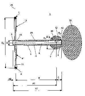
Figs. 1 and 2 illustrate an embodiment of the device which is exemplarily
configured as
an aiming device 1. Said aiming device 1 essentially comprises a rod shaped
member 2
with a central axis 6, an external diameter Dt, a radiopaque first targeting
element 7 with
a diameter DK fixed to said rod shaped member 2 and a radiopaque second
targeting
element 5 with circular central line 11 having a diameter DR > Dt and defining
a plane
perpendicular to said central axis 6. Further, said rod shaped member 2
includes a
central through bore 8 for guiding said drill bit, a rear end 3 and a front
end 4 both
transverse to the central axis 6. Said first targeting element 7 is fixed to
said rod shaped
member 2 close to said front end 4 with its center 46 at a distance C > 0
thereto and
concentrically to said central axis 6.
Said second targeting element 5 is coupled to said rod shaped member 2
concentrically
to said central axis 6 such that said plane defined by said circular central
line 11 is at a
CA 02735812 2011-03-02
WO 2010/025575 PCT/CH2009/000295
13
distance A2 measured from said rear end 3 towards said front end 4. Further,
said
second targeting element 5 is fixedly attached to said rod shaped member 2 by
radiolucent means 9, which are realized in the present embodiment by means of
a
radiolucent disc radially extending between the peripheral surface 20 of said
rod shaped
member 2 and said second targeting element 5. Further, said second targeting
element
is configured as a torus obtained by rotating a circle of radius r1 about said
central
axis 6 of said rod shaped member 2 with said centre of the circle of radius ri
at a
distance DR/2 from said central axis 6.
Said first targeting element 7 has a spherical portion 12 directed towards
said rear end
3 of said rod shaped member 2. Said spherical portion 12 has a radius of
curvature R1
the centre of which coincides with the center 46 of said first targeting
element 7.
Further, said spherical portion 12 is limited by the external surface 20 of
said rod
shaped member 2 towards said rear end 3 of said rod shaped member 2 and
extends
over the median plane of a sphere with radius R1 which is orthogonal to said
central
axis 6 of said rod shaped member 2. Toward said front end 4 said spherical
portion 12
is limited by a front portion 10 with a radius of curvature R2 which is
greater than said
radius of curvature R1 of said spherical portion 12.
Further, said first targeting element 7 is fixed to the peripheral surface 20
of said rod
shaped member 2 concentrically to said central axis 6 with its center 46 at a
distance
A1 measured from said rear end 3 towards said front end 4 and at a distance B
measured from said plane defined by said circular central line 11. Further,
the diameters
Dt of the rod shaped member 2, DK of the spherical first targeting element
7and DR of
the second targeting element are selected in such manner that Dt < DK < DR
Optionally, said aiming device 1 can comprise two cylindrical or prismatical
targets 16.
Said two cylindrical or prismatical targets 16 can be located in said
radiolucent disc
parallel to each other and at an identical distance ATarget to said central
axis 6 of said rod
shaped member 2.
Fig. 3 illustrates another embodiment of said aiming device 1 which differs
from the
embodiment of figs. 1 and 2 only therein that said second targeting element 5
is not
fixed to said rod shaped member 2 but is joinable to a surgical instrument,
particularly to
CA 02735812 2011-03-02
WO 2010/025575 PCT/CH2009/000295
14
a drilling device which is displaceable parallel to said central axis 6 with
respect to said
rod shaped member 2 so that said second targeting element 5 remains coaxial to
said
central axis 6.
Fig. 4 illustrates the steps performed for determining the position of a
circular cylindrical
target 16 in a three-dimensional body 28. Said circular cylindrical target 16
has a lower
density than the surrounding material of said three-dimensional body 28, a
longitudinal
axis 18, a diameter d, a height h and a centre 47. In particular said steps
comprise:
A) acquiring one single image 50 (schematically illustrated in the drawing
plane of fig. 6)
with a lens-shaped projection 42 of said target 16 by means of an image
acquisition
device including an energy emitting source 29 with a central ray 26 and a
receiving
device with an image sensor which is connected to a computer 32 with a display
33.
The angulation range between the central ray 26 and the longitudinal axis 18
of target
16 is restricted in a way that a projection 42 of the target 16 must be
visible on the
image 50;
B) determining the position and orientation of said target 16 from said single
image 50
using a numerical procedure executed with said computer 32, wherein said
numerical
procedure essentially comprises the steps of:
a) automatic detection of said lens-shaped projection 42 of said target 16 in
said
image 50 and determination of the projection points of the two points of
intersection 35, 36 and the first and second apex 37, 38 of said lens-shaped
projection 42 of said target 16;
b) generating a virtual geometric representation of said target 16, with said
diameter d, said longitudinal axis 18, said centre 47 and said height h;
c) determining virtual projection points representing said two points of
intersection 35, 36 and said first and second apex 37, 38 using said virtual
geometric representation of said target 16;
d) iterative determination of the position and angular orientation of said
target 16
by matching said virtual projection points of said virtual geometric
representation
of said target 16 with said two points of intersection 35, 36 and said first
and
second apex 37, 38, wherein said target 16 has three degrees of freedom:
- a position Z on the z-axis of a global system of coordinates 24 measured
between the centre of said energy emitting source 29 and said centre 47.
Said virtual geometric representation of said target 16 can slide along the
CA 02735812 2011-03-02
WO 2010/025575 PCT/CH2009/000295
centre line 43 determined by the centre of projection 44 and the centre of
said energy emitting source 29. Said centre of projection 44 is an
approximation of the centre-line projection 51 for h/H
O. The
coordinates x and y of the target 16 are depending on Z and the centre
line 43. Therefore, one cylindrical target 16 determines five degrees of
freedom but the algorithm needs only three degrees of freedom;
- an angle a between said longitudinal axis 18 and said centre line 43
measured in the y-z plane of said global system of coordinates 24 which is
fix with respect to said image acquisition device 25; and
- an angle p. between said longitudinal axis 18 and said centre line 43
measured in the x-z plane of said global system of coordinates 24.
Examples when using one rotational symmetrical, e.g. cylindrical target only,
mainly drilling, tapping and screw insertion procedures (5 degrees of
freedom):
- guide any type of tool in a predetermined direction with respect to a
work piece or
other object for further machining or processing of said work piece or other
object;
- measurement of alignment of a printed circuit board and register subsequent
drilling of a bore hole;
- fixation, e.g. interlocking of a shaft of an endoprosthesis provided with
screw
holes by means of screws (e.g. shaft of femur component);
- measurement of layer displacement in a multilayer board or panel for
e.g. quality
control; and
- measurement of distortion and/or rotation of a single or multi-layer
work piece or
other object in order to recover a desired shape or orientation or in order to
align
a robotic arm in e.g. a robotic assembly system;
The work piece or other object could be fixed to a support table of e.g. a CNC-
machine
(computerized numerical control ¨ machine) such being mechanically constraint.
Examples when using one non rotational symmetrical target or two or more
targets (6 degrees of freedom):
CA 02735812 2011-03-02
WO 2010/025575 PCT/CH2009/000295
16
-
determination of the alignment of objects that may not be mechanically
constraint
in a predetermined location or angular orientation, for example locating and
manipulating of a cardiac pacemaker, e.g. tightening or loosening of a screw;
and
- handling or manipulating of a work piece or other object with respect to
exact
knowledge of its position and orientation.
The above mentioned numerical procedure includes a numerical approach for
calculating the position of said target 16 and is based on the following
mathematical
relationships:
Numerical approach (for circular cylindrical targets):
The procedure relates to the mathematical condition that the projection of an
image
acquisition device 25 is based on an idealized central perspective. A
punctiform x-ray
source used as energy emitting source 29 sends rays from an origin of known
distance
H to the projection plane 49.
The procedure incorporates the following fundamental steps:
1. Automatic detection of the lens-shaped projection 42 of said target 16 in
said image
50 and determination of significant landmarks, i.e. said two points of
intersection 35, 36
and the first and second apex 37, 38 of the lens-shaped projection 42 of said
target 16
by use of image processing algorithms. Assumption: With h/H
0 the centre-line
projection 51 approximates to the centre of projection 44.
2. Simulation of a virtual geometric representation of said target 16 and of
virtual
projection points corresponding to the above significant landmarks. Iterative
determination of the angular orientation and position of said virtual
geometric
representation by means of a numerical optimization routine; and
3. Simulation of a virtual geometric representation of said aiming device 1
and
projection of target curves 17, 22 (targeting ellipses) into the image 50.
Iterative determination of the orientation of the virtual geometric
representation
A virtual geometric representation of said target 16 is generated with the
known
attributes d (diameter) and h (length). The virtual geometric representation
of said target
CA 02735812 2011-03-02
WO 2010/025575 PCT/CH2009/000295
17
16 has one translational degree of freedom. It can slide along the central
line 43,
determined by the centre of projection 44 and the centre of said energy
emitting source
29. Sliding position is controlled by Z (Figure 4). With further two
rotational degrees of
freedom (a, [3) the position of said virtual geometric representation of said
target 16 is
fully constrained.
Four virtual projection points representing said two points of intersection
35, 36 and said
first and second apex 37, 38 are derived from the orientation of said virtual
geometric
representation of said target 16.
A numerical optimization routine (here least square error minimization) is
used to find a
global minimum for the deviations between said significant landmarks and the
corresponding virtual projection points using three degrees of freedom (DOF)
(a, í3, Z) in
order to carry out the optimized orientation of said virtual geometric
representation of
said target 16. Due to the asymmetry of the lens shaped projection (segments b
and c
appear asymmetrically, due to the nature of a central projection) it is
possible to
calculate a unique solution for the orientation of the target from a single
image.
Target curves (e.g. ellipses)
With determined orientation of the virtual geometric representation of said
target 16 (a,
Z) the device comprising a first and a second targeting element 7, 5 is
mathematically
modelled.
The virtual geometric representation of said device with said first and second
targeting
elements 7, 5 is positioned and oriented with respect to the virtual geometric
representation of said target 16.
The projections of the first and second targeting element 7, 5 (target curves,
i.e. target
ellipses) are visualized in the x-ray image 50 for subsequent targeting.
In the special embodiment illustrated by figs. 5 and 6 said device is an
aiming device 1
comprising coaxially arranged a spherical first targeting element 7 and an
annular
second targeting element 5 with the dimensions DK, DR, B, C. Further, the
virtual
geometric representation of said aiming device 1 is coaxially oriented to the
virtual
CA 02735812 2011-03-02
WO 2010/025575 PCT/CH2009/000295
18
geometric representation of said target 16 and positioned at a distance E from
the
centre 47 of said target 16 (fig. 5).
Exemplarily, with reference to fig. 7 the method for positioning a device is
described for
positioning an aiming device 1 with regard to a through hole 116 in an
endoprosthesis
representing said three-dimensional body 28, wherein said through hole 116 has
a hole
axis 118, a bore diameter D and a bore centre 147.
In the following description:
- said endoprosthesis represents the three-dimensional body 28;
- said through hole 116 represents the circular cylindrical target 16
wherein said
hole axis 118 represents with said longitudinal axis 18 of said target 16;
- said diameter d of said target 16 is represented by the bore diameter D
of said
through hole 116, said height h of said target 16 is represented by the length
L of
said through hole 116 and said centre 47 of said target 16 is represented by
the
bore centre 147 of said through hole 116; and
- the aiming means attached to said aiming device 1 are realized by said
spherical
first targeting element 7 and said annular second targeting element 5.
On the above basis said through hole 116 with said hole axis 118 is used in
said
numerical procedure to generate said virtual geometric representation of said
target 16
with said diameter d, said longitudinal axis 18, said height h and said centre
47.
The aiming device 1 used in the embodiment here differs from the aiming device
of fig.
1 only therein that said first targeting element 7 is spherically configured
with a diameter
DK.
Firstly, the step of acquiring one single image 50 with a projection 42 of
said through
hole 116 by means of an image acquisition device with a projection plane 49 is
performed.
Secondly, the position and angular orientation of said through hole 116 is
determined by
applying said numerical procedure using said single image 50 as described
under fig. 4.
CA 02735812 2011-03-02
WO 2010/025575 PCT/CH2009/000295
19
Then, as illustrated in figs. 5 and 6 said aiming device 1 is positioned by
performing the
further steps of:
1) establishing a virtual geometrical representation of said aiming device 1
coaxial to
said longitudinal axis 18 of said through hole 116 and positioned with said
front end 4 of
said aiming device 1 at a distance E to said centre 147 by means of said
computer 32;
2) determining the virtual position and angular orientation of said spherical
first targeting
element 7 and said annular second targeting element 5 attached to said aiming
device 1
using said virtual geometrical representation of said aiming device 1;
3) depicting a first target curve 17 on said display 33 by means of said
computer 32;
said first targeting curve 17 representing a virtual projection of said
spherical first
targeting element 7 on said projection plane 49;
4) depicting a second target curve 22 on said display 33 by means of said
computer 32;
said second targeting curve 22 representing a virtual projection of said
annular second
targeting element 5 on said projection plane 49; and
5) positioning said aiming device 1 by aligning said spherical first targeting
element 7
with said first targeting curve 17 and by subsequently aligning said circular
central line
11 of said annular second targeting element 5 with said second targeting curve
22 using
an image acquisition device 25; wherein
i) said positioning of said aiming device 1 is performed by firstly manually
aligning
said spherical first targeting element 7 of said aiming device 1 with said
first
targeting curve 17 by translational movement of said aiming device 1; and
ii) by secondly aligning said circular central line 11 of said annular second
targeting element 5 with said second targeting curve 22 by rotational movement
of the aiming device 1.
When said aiming device 1 is correctly positioned with regard to said through
hole 116 a
hole is drilled in the bone 120 surrounding said endoprosthesis.
After drilling the hole in the bone 120 surrounding said endoprosthesis
coaxially to said
hole axis 118 of said through hole 116 an interlocking means, e.g. a bone
screw could
be advanced through the through hole 116 such locking said endoprosthesis with
respect to said bone 120.
CA 02735812 2011-03-02
WO 2010/025575 PCT/CH2009/000295
In order to operate said aiming device 1 during drilling of bore holes through
a material
surrounding said three-dimensional body 28 coaxially to said hole axis 118 of
said
through hole 116 in said three-dimensional body 28 the aiming device 1
comprises a
handle (not shown).
While various descriptions of the present invention are described above, it
should be
understood that the various features can be used singly or in any combination
thereof.
The scope of the present invention is accordingly defined as set forth in the
appended
claims.