Note: Descriptions are shown in the official language in which they were submitted.
CA 02737061 2011-03-11
WO 2010/028508
PCT/CA2009/001282
CATHETER FOR MAGNETIC RESONANCE GUIDED PROCEDURES
Technical Field
[0001] The present disclosure is related to a catheter for magnetic resonance
guided
procedures. In particular, the present disclosure is related to such catheters
that provide
magnetic resonance guidance using magnetic coupling.
Background
[0002] With the emergence of real-time magnetic resonance imaging (MRI)
techniques, the
use of MRI has expanded from static diagnostic imaging to include the
potential to guide a
variety of interventions. Many percutaneous cardiovascular procedures (i.e.,
interventions
performed with a catheter inserted into the vasculature) may benefit from
guidance where
MRI's soft tissue contrast may be exploited. One example is the traversing of
chronic total
occlusions in coronary and peripheral vessels. The presence of chronic total
occlusions is the
leading reason for selection of bypass surgery over less invasive
interventions. Despite the
benefits of percutaneous treatment, clinicians are often unable to traverse
occlusions with
catheter-based devices due to the inadequate imaging capabilities of X-ray
fluoroscopy that is
typically used to image such treatment.
[0003] Reference is now made to FIG. 1. Typically, during percutaneous
interventions two
pieces of equipment are inserted into the vasculature 10. The first is a
catheter 12 that may be
a long thin hollow tube. The second is a guidewire 14, which is typically thin
flexible wire
that may travel through the lumen of the catheter 12. FIG. 1 shows a schematic
diagram
illustrating the use of a conventional guidewire 14 and catheter 12 in the
vasculature 10 of a
patient. Typically, the guidewire 14 is extended from the catheter tip, and
because the
guidewire 14 is usually very flexible, it is the first device to be manoeuvred
through the
vasculature 10. The catheter 12 is advanced over top of the guidewire 14 to
provide
mechanical support, and when pushed, the catheter 12 follows the path of the
guidewire 14.
[0004] Several MRI-guided guidewire tracking and visualization techniques have
been
proposed, which may be classified into two groups. The first group may be
referred to as
"passive techniques" where the device is made visible through the use of
signal voids,
susceptibility artifacts, or off-resonance signals (e.g., those discussed in
References 1-4).
CA 02737061 2011-03-11
WO 2010/028508
PCT/CA2009/001282
These techniques typically are limiting in that the device must lie within the
MR imaging
plane in order to be viewed.
[0005] The second group may be referred to as "active techniques". Active
techniques rely
on an acquisition of the magnetic resonance (MR) signal from small micro-coils
or wires
located on the device in order to determine device position (e.g., as
discussed in References 5
and 6). Active visualization techniques typically do not suffer from the same
limitations as
passive techniques due to the fact that the signal used for device
localization is acquired
independently from that used for anatomical imaging. This enables the device
to be located
even when it lies outside the current imaging plane. Moreover, because the
signal from the
device is a separate signal, it may be colour-overlaid on anatomical images to
create a
"positive contrast" that may be easy to identify and put in an anatomical
context. However,
active visualization of the guidewire may be challenging in that many of the
techniques
developed for catheters and endoscopes (e.g., the use of micro-coils) are
difficult to translate
to guidewires due to the limited thickness of guidewires. Guidewires are thin
wires with a
typical diameter of less than 0.035 inches, whereas catheters and endoscopes
may have a
much larger diameter which allow for accommodation of components necessary for
this
visualization.
[0006] Some current active guidewire designs consist of a loopless antenna
that is formed on
the end of a coaxial cable (e.g., Reference 7). This design includes two
limitations. The first
is that the active wires typically require significant internal structure. A
result of this is that
the mechanical properties of the guidewire do not resemble that of a
conventional bare wire,
which may affect its manoeuvrability in the vasculature. Further, active
guidewires may be
considered to be unsafe because resonant currents may develop on the outside
conductor of
the thin coaxial cable used to carry the MR signal from the loopless antenna
to the input of
the MR scanner (e.g., as discussed in References 8-11). These resonant
currents may create
intense localized heating of tissues located at the ends of the active
guidewire. The same
safety concern exists regarding the use of traditional non-active guidewires
in the MR
scanner.
[0007] Reference is now made to FIG. 2. A design for a MR-compatible guidewire
20 has
been proposed that consists of a short non-resonant length of nitinol
connected to a non-
conducting fibreglass rod (e.g., as discussed in References 12 and 13). The
non-conductive
length may be made of any non-conductive material, including fibreglass,
graphite, carbon
2
CA 02737061 2011-03-11
WO 2010/028508
PCT/CA2009/001282
fibre, or a polymer. FIG. 2 illustrates a schematic diagram of such a
guidewire 20. In this
schematic, the guidewire 20 has a non-resonant conductive length 22 (e.g.,
approximately 10
cm) of nitinol at the distal end attached to a non-conducting length 24 (e.g.,
a fibreglass rod)
that forms the remaining length of the guidewire 20. The length of nifinol 22
is non-resonant
and thus large currents are unable to develop in the guidewire 20. Such a
guidewire 20 is
therefore not susceptible to the heating concerns discussed above.
Visualization of the
guidewire 20 is done passively by doping the conductive length 22 and non-
conductive
length 24 with small iron particles. This creates a susceptibility artifact
that may be seen on
MR images. However, this method suffers from the same limitations as other
passive
visualization methods, including the limitation that the guidewire 20 may be
visualized only
when it is in the imaging plane.
Summary
[0008] A catheter for magnetic resonance (MR) guided procedures is disclosed
that addresses
some of the challenges discussed above.
[0009] In some aspects, there is provided a catheter for magnetic resonance
(MR) guided
procedures comprising: a catheter body having a lumen for accommodating an
intravascular
device; a magnetic coupling component in the catheter body, the magnetic
coupling
component being designed to magnetically couple with a conductive length on
the
intravascular device, the magnetic coupling resulting in a signal; the
catheter having a
connection to deliver the signal to a processor.
[0010] In some aspects, there is provided a combination for magnetic resonance
(MR) guided
procedures comprising: the catheter described above; and a MR-compatible
intravascular
device designed to pass through the lumen of the catheter, the intravascular
device having a
conductive length; wherein the magnetic coupling component in the catheter is
configured to
magnetically couple with the conductive length, magnetic coupling between the
magnetic
coupling component and the conductive length resulting in a signal.
[0011] In some aspects, there is provided a method of monitoring a magnetic
resonance (MR)
guided procedure comprising: providing the combination described above located
in a
patient, the intravascular device having been inserted through the catheter;
inducing a current
in the conductive length; delivering a signal to a processor, the signal
resulting from magnetic
coupling between the magnetic coupling component and the conductive length.
3
CA 02737061 2011-03-11
WO 2010/028508
PCT/CA2009/001282
[0012] There is also provided a use of the catheter and combination described
above for
performing a MR guided procedure.
Brief Description of the Drawings
[0013] FIG. 1 shows an example prior art catheter and guidewire arrangement;
[0014] FIG. 2 shows an example prior art MR compatible guidewire;
[0015] FIG. 3 shows a schematic diagram of an example MR guided guidewire and
catheter;
[0016] FIG. 4 is a schematic illustration of magnetic coupling of an example
magnetic
coupling component;
[0017] FIG. 5 is a schematic illustration of magnetic coupling of another
example magnetic
coupling component;
[0018] FIG. 6 is a schematic illustration modeling magnetic coupling of
another example
magnetic coupling component;
[0019] FIG. 7A shows a schematic modeling an example magnetic coupling
component and
a conductive wire;
[0020] FIG. 7B shows a schematic of an example magnetic coupling component;
[0021] FIG. 8 is an image of an example catheter;
[0022] FIG. 9 is a schematic of an example MR guided guidewire and catheter,
and images
demonstrating the visualization of the guidewire;
[0023] FIG. 10 are charts illustrating signal intensity in example MR guided
guidewires,
compared to theory;
[0024] FIG. 11 are charts illustrating signal intensity in an example MR
guided guidewires,
compared to theory;
[0025] FIG. 12 shows images demonstrating the visualization of an example MR
guided
guidewire using a colour-overlay technique;
4
CA 02737061 2011-03-11
WO 2010/028508
PCT/CA2009/001282
[0026] FIG. 13 shows images and signal plots demonstrating the visualization
of an example
MR guided guidewire using a minimum projection technique;
[0027] FIGS. 14A and 14B are schematic diagrams of example catheters having
additional
circuitry;
[0028] FIG. 15 is a schematic diagram of an example MR guided guidewire and an
example
catheter having intravascular imaging capabilities;
[0029] FIG. 16 is a simulation of an image that may be acquired using an
example MR
guided guidewire and an example catheter having intravascular imaging
capabilities; and
[0030] FIG. 17 is a schematic illustration of an example MR guided guidewire
and an
example catheter having more than one conductive length and magnetic coupling
component.
Detailed Description
[0031] A catheter for MR guided procedures is disclosed, including kits and
methods using
this catheter. As disclosed, a MR signal around a short conductive length on a
device inserted
through the catheter (e.g., a guidewire) is detected through the interaction
of this conductive
length and a magnetic coupling component, such as a coil (e.g., a toroidal-
shaped coil), which
may also be referred to as a "pick-up coil", to which the conductive length is
magnetically
coupled. Although the term "magnetic coupling" is used in this disclosure, it
should be
understood that magnetic coupling refers also to electric coupling, as the
coupling is based on
electromagnetic fields. The magnetic coupling component is located in the wall
of a catheter
through which the MR-compatible guidewire travels. The signal picked up by the
magnetic
coupling component is then delivered to a processor, such as a MR scanner or
other external
electronics, for processing. Signal processing may include filtering,
digitization,
reconstruction or analysis of the signal, as is common in the field of MRI.
The magnetic
coupling component may be connected to the receive chain of the MR scanner
using a
transmission line, such as a conventional coaxial cable located inside the
guide catheter.
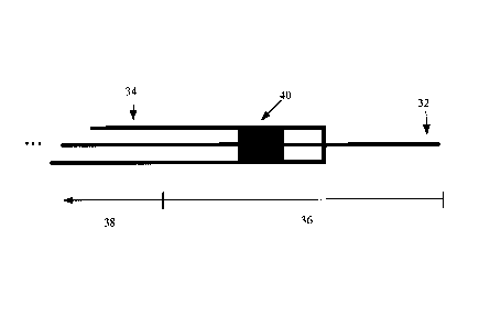
[0032] Reference is now made to FIG. 3 showing a schematic diagram of an
example MR
guided guidewire 32 and catheter 34. The guidewire 32 has a conductive non-
resonant length
36 (e.g., a length of nitinol), and a non-conductive length 38. To perform
active visualization
of MR guided guidewires, a magnetic coupling component 40, in this example a
small
CA 2737061 2017-04-03
WO 2010/028508 PCT/C.
A2009/1811 282
rectangular toroidal coil, embedded in the wall of the catheter 34, is used to
detect currents
induced on the conductive length 36 of the guidewire 32. Although a guidewire
32 is shown
in this example, the conductive length 36 may be provided on other devices
that may pass
through the catheter 34, including balloons, needles and other similar
intravascular devices.
This disclosure will refer to a guidewire 32 as an catiumple of the device
passing through the
catheter 34, but it will be understood that all references to a guidewire 32
also applies to other
devices that may pass through the catheter 34.
[0033] in general, the guidewire may be any suitable MR-eornpatible guidewire
having a
conductive length (e.g., at the distal end) and its remaining length being non-
conductive. The
conductive length should be a non-resonant length (e.g., in order to be MR-
compatible),
which may be dependent on several variables, including the diameter of the
guidewire and
the electrical properties of the guitie catheter, as well as the MR. system it
is to be used in. For
example, a non-resonant length for the conductive length may be in the range
of about 1 to 30
ern. Typically, Stich a guidewire is designed to be MR-compatible by limiting
the conductive
length to be loss than a resonant lenth. Nitinol has been used as the
:material for the
conductive length, in order to best approximate the behaviour of conventional
nitinol
guidewires, however other conductive materials may be used for the conductive
length,
including stainless steel, gold and platinum.
[0034] In general, the catheter is suitably sized to allow the guidewire to
pass thiough its
lumen. The diameter of the catheter may be designed to facilitate
intravascular procedures in
certain parts of the vasculature. For example, the catheter may have a smaller
diameter where
it is designed to be used in the coronary vessels, and may have a larger
diameter where it is
designed to be used in the peripheral vessels. Typically, the average lumen
diameter of the
coronary arteries in an adult is about 1.5 to 2.5 mrn, and the peripheral
lumen diameters (e.g.,
that of the common femoral ark:1y) may be as large as 5 atm. Thus, the
catheter may bc NIZed
to suit these vessels or larger anatomical structures (e.g., the trachea or
the colon), for
example the catheter may have an outer diameter in the range of about 1.5 mm
to about 5 cm,
in some examples in the range of about 3nuri to about 5mm, depending on
intended use.
10035] The catheter has a ntagnctic coupling component (e.g., located at its
distal end). The
magnetic coupling component is designed to be magnetically coupled to the
conductive
length of the guidewire, as will be explained below. The magnetie coupling
component in
some examples is positioned on the catheter to correspond to the likely
position of the
6
CA 02737061 2016-04-05
WO 2010/028508 =
PCT/CA2009/001282
conductive length on the guidewire. The magnetic coupling component may be
made of any
suitable conductive material, such as copper, nitinol, aluminum, or any other
suitable
material, Copper may be useful since the magnetic susceptibility of copper is
such that it does
not produce image artifacts in MR images. The magnetic coupling component may
also
include other materials to provide mechanical support. A.dditional materials
may be bio-
compatible polymers, including polyetheretherkotone, delrin, polyimide,
polyvinylchloride,
polyethylene, polycarb on ate, polysulfone, polypropylene, polytetra
fluoroethylene,
combinations thereof, or any other suitable polymer. The magnetic coupling
component may
also be made -using flexible laminates, for example a flexible copper clad
laminate. Using a
flexible material may result in a flexible magnetic coupling component, which
may help the
catheter to maintain flexibility,
[0036] The magnetic coupling component may be a coil, such as a toroidal coil,
though it is
understood that other component and/or coil shapes can be used to achieve the
magnetic
coupling as explained below. In general, the magnetic coupling com.ponent is
designed so
that it magnetically couples to the conductive length on the guide:wire that
travels through the
catheter. This can be achieved by designing a magnetic coupling component that
produces a
magnetic field that overlaps with the magnetic field produced when a current
flows through
the conductive length, as will be described further below. Mathematically,
this corresponds to
designing a magnetic coupling component such that the dot product (ì.e.,
scalar product) of
the magnetic field produced when unit flows through the conductive length is
non-zero when
integrated over all points in space. In this situation, it may be said that
there is mutual
inductance between the magnetic coupling component and the conductive length.
[00371 This concept is illustrated for the example of the magnetic coupling
component 40
being a single loop coil located adjacent to a conductive wire 42 in Fla 4, In
the example
shown, the conductive wire 42 has current flowing through, giving rise to an
electromagnetic field. One example set of field lines FAIT. is shown for the
conductive wire
42. The magnetic coupling component 40 has =rent flowing through, giving rise
to an
electromapetic field. One example set of field lines F0i is shown for the
magnetic coupling
component. The overlap between and Fepii gives rise to non-zero inductance
between the
magnetic coupling component 40 and the conductive wire 42.
[00381 Based on this general theory, the magnetic coupling component may be
designed
using typical calculations and/or simulations. F'or example, the Target Field
Method, which
7
CA 02737061 2011-03-11
WO 2010/028508
PCT/CA2009/001282
solves for a current distribution that would produce a specified magnetic
field, may be used
(for example, as described in Turner, J Phys. D. Appl. Phys. 19:147-151,
1986.).
[0039] The magnetic coupling component may be sized to suit the diameter of
the catheter as
discussed above. Although the disclosure has referred to a catheter as having
the magnetic
coupling component, other interventional devices through which an
intravascular device can
pass, such as sheaths, may be used to carry the magnetic coupling component,
and the
magnetic coupling component may be sized accordingly to fit these other
devices. For
example, the magnetic coupling component may be in the range of about 0.3mm to
about
5cm in diameter, such as in the range of about 1 mm to about 10mm in diameter.
The
magnetic coupling component may be designed to have a length that does not
interfere or
otherwise affect the behaviour, such as the flexibility, of the catheter. For
example, for a rigid
magnetic coupling component (e.g., a rigid coil), the magnetic coupling
component may be
limited to a length of about 0.1mm to about lOmm, but may have a greater
length where
flexibility of the catheter is not important (e.g., for use in substantially
straight vessels).
Where the magnetic coupling component is flexible, there may be no such limit
on the length
of the magnetic coupling component. A greater length for the magnetic coupling
component
may allow for greater magnetic coupling between the magnetic coupling
component and the
conductive length, which may result in a stronger signal and better imaging.
[0040] Although the catheter has been described as having a magnetic coupling
component at
or near its distal end, the magnetic coupling component may be provided
anywhere along the
length of the catheter. It may be useful to position the magnetic coupling
component close to
where the conductive length of the guidewire is expected to be, as the
magnetic coupling
between the conductive length and the magnetic coupling component typically is
stronger
when the magnetic coupling component is located at or near to the center of
the conductive
length. The coupling between the magnetic coupling component and the
conductive length
typically decreases in strength with radial distance between the conductive
length and the
magnetic coupling component. For example, a radial distance in the range of
about 0.1 mm to
about 1 cm may provide for a suitably strong magnetic coupling.
[0041] The catheter may have more than one magnetic coupling component. For
example,
the catheter may have one magnetic coupling component at or near its distal
end, and
additional one or more magnetic coupling components down its length, such as
the example
illustrated in FIG. 17. As shown, the catheter 120 may have two or more
magnetic coupling
8
CA 02737061 2011-03-11
WO 2010/028508
PCT/CA2009/001282
components 122 along its length. The magnetic coupling components 122 in this
example are
shown together with a device 124 (e.g., a guidewire) passing through the
catheter 120 that has
multiple conductive lengths 126. The conductive lengths 126 on the device 124
are segments
separated by isolating joints 128. The conductive lengths 126 may also be
separated by non-
conductive lengths (not shown). The use of additional magnetic coupling
components 122
may allow the detection of a single conductive length 126 at different points
along the
catheter 120, for example as the device 124 passes through the catheter 120,
or to detect the
position of several conductive lengths 126 on a single device 124.
[0042] In general, a method for visualization of a MR guided guidewire is
disclosed. A MR
compatible device, such as a guidewire, having a non-resonant conductive
length at or near
its distal end is passed through a catheter having a magnetic coupling
component (e.g.,
located at or near its distal end) such that the conductive length is
magnetically coupled to the
magnetic coupling component. During the acquisition of MR signal (e.g., as
part of
conventional MRI), a current is induced in the conductive length. Due to
magnetic coupling
between the conductive length and the magnetic coupling component, this
current induces a
voltage signal across the leads of the magnetic coupling component. The signal
from the
magnetic coupling component is transmitted to the receive chain of the MR
scanner, for
example using conventional transmission lines or a coaxial cable in the
catheter. This signal
may then be processed using conventional signal processing techniques to
obtain an image of
the conductive length. This signal may also be processed in other ways as will
be discussed
further below.
[0043] Instead of using a transmission line to deliver the signal from the
magnetic coupling
component, other signal delivery techniques may be used. For example, the
signal may be
delivered using an optical fibre or other common signal delivery means.
[0044] Using the disclosed catheter, the guidewire does not require any
internal structure
(e.g., any electronic components or cables) as it is not itself being used as
a transmission line.
This avoids the need to add components to a small-diameter wire, and avoids
affecting the
handling behaviour of the guidewire. Safety concerns regarding the use of
conducting
structures are not associated with the guidewire since the conductive length
is kept to a non-
resonant length. The catheter may be used with any guidewire or other
intravascular device
that is MR-compatible and has a conductive length (e.g., at or near its distal
end) that may
pass through the catheter. The magnetic coupling component in the catheter may
be designed
9
CA 02737061 2011-03-11
WO 2010/028508
PCT/CA2009/001282
to magnetically couple and hence detect any such conductive length, as will be
described
below.
[0045] Since the magnetic coupling component is provided in the catheter, size
constraints
which limit possible safety features when a transmission cable is connected
directly to the
guidewire are diminished since the cable is now inside the larger catheter.
Thus, additional
components may be added to the catheter to further improve the safety and/or
signal quality
without burdening the guidewire. For example, RF chokes (e.g., as discussed in
Reference
14), baluns or other devices that reduce currents on the outer conductor of
the cables may be
incorporated into the catheter to further reduce any safety concerns. Thus,
the disclosed
catheter provides the benefits of active visualization for MR guided
procedures yet retains the
safety associated with passive MR-compatible guidewires.
Theory and design
[0046] A theory of operation is now presented. The present disclosure is not
bound or in any
way limited by the theory presented. This theory may be useful in designing
the MR guided
guidewire and/or catheter. With reference to FIG. 5, consider a short
conducting segment of
wire of length L positioned adjacent to a magnetic coupling component, in this
example a
coil, that is magnetically coupled to the wire such that a mutual inductance M
exists between
the wire and the coil.
[0047] The sensitivity to magnetization surrounding the conductive length of
the guidewire
can be analyzed through the use of reciprocity and the calculation of the
current induced
along the conducting segment given a input current I at the magnetic coupling
component or
its peripheral circuitry.
[0048] A simplified lumped-element model of the system is depicted in FIG. 6.
In this
example, the magnetic coupling component is a coil. Here, Zgw(z) is the
complex impedance
of the wire at the location z of the coil, Ig,(z) is the current in the
conductive length at the
location z of the coil, M is the mutual inductance between the conductive
length and the coil,
and Zpue is the complex impedance of the coil. Other local tuning elements
present are in this
model. The impedance of the conductive length at a particular z location
Zg,,(z) is dependant
on several factors including (but not limited to) the length of the conductive
length and the
CA 02737061 2011-03-11
WO 2010/028508
PCT/CA2009/001282
surrounding environment and can be numerically calculated using numerical
methods such as
the Method of Moments (MoM). The current in the coil (Ipuc) can be solved
using
conventional circuit analysis techniques and once known, the current
distribution along the
entire length of the conductive length can be determined using numerical
methods.
[0049] The spatial sensitivity to MR signal in the vicinity of the conductive
length can be
calculated given the current distribution along the conductive length by
calculating the
component of the magnetic field perpendicular to the static field of the MRI
produced by the
current in the guidewire, for example using the law of Biot-Savart or any
other suitable
conventional methods.
[0050] The equations governing the mutual inductance and the current in the
magnetic
coupling component may be used to design the magnetic coupling component For
example,
the dimensions of the magnetic coupling component may be adjusted where a
certain distance
between the magnetic coupling component and the conductive length is desired.
Using the
above theoretical description and lumped-element circuit element model, a
variety of
magnetic coupling components (e.g., different coil configurations) and circuit
configurations
may be designed for different applications, having different geometries and
dimensions, in
order to achieve the presently disclosed MR guided guidewire and catheter. It
should be
noted that the current on the guidewire is dependent on circuitry connected to
the magnetic
coupling component and a person skilled in the art would know how to apply the
model for
different configurations and adapt the model and the corresponding equations
accordingly.
Design example
[0051] One example of a magnetic coupling component designed to magnetically
couple to a
conducting length is a rectangular-shaped toroidal coil with N turns each of
length b, width a,
and distance s from the conductive length. With this particular magnetic
coupling component
design, an intravascular device passing through the centre of the toroidal
coil will
magnetically couple with the magnetic coupling component. An illustration of
this example
magnetic coupling component, in the form of a coil, is shown in FIG. 7A. For
simplicity,
only one turn is shown. The mutual inductance M between the coil with N turns
can be
shown to be:
pAib ln(s + a)
M = [eqn 1]
2a-
11
CA 02737061 2011-03-11
WO 2010/028508
PCT/CA2009/001282
[0052] Along with the impedance of the magnetic coupling component and the
properties of
the conducting segment, one can use the theory above to predict how the
configuration will
behave. Although this is only one example, any other suitable magnetic
coupling component
(e.g., having a coil design) can be designed to further increase the mutual
coupling M to
improve the signal acquired from the magnetic coupling component.
[0053] Other examples of a magnetic coupling component, for example based on
the theory
described above, may include (but are not limited to) single or multiple loops
of wire and
single or multiple loops of conductive ribbon.
[0054] FIG. 7B illustrates an example of a suitable magnetic coupling
component 70. In this
example, the magnetic coupling component 70 is generally in the shape of a
cylinder with a
hole through its length. In this example, the magnetic coupling component 70
includes two
concentric conductive tubes 72, 74, that are joined to each other at one end
of the cylinder
(not shown). The conductive tubes 72, 74 are spaced apart by a non-conductive
material. The
material separating the two tubes, in some examples could be air or
alternatively could be
some type of plastic or any other type of suitably non-conductive supporting
material. In
operation, a signal (in this example, denoted Vsignal) is measured as a
voltage across the two
conductive tubes 72, 74 at the end where the conductive tubes 72, 74 are not
joined. In some
examples, the magnetic coupling component 70 may have dimensions that are
similar to the
coil design described further below. For example, the outside diameter of the
magnetic
coupling component 70 may be designed such that it fits inside a catheter and
may be in the
range of about 0.3mm to about 5cm. The length of the magnetic coupling
component 70 may
be in the range of about 0.1mm to about 10cm. To improve efficiency of
magnetic coupling,
the diameter of the inner conductive tube 74 may be configured to be as small
as possible
while still allowing the intended interventional device to pass through it.
Additional circuitry,
for example capacitors, may be added to the magnetic coupling component 70 to
form a
resonant circuit, according to conventional methods.
[0055] Compared to a coil design, for example the design described below, this
example
magnetic coupling component 70 may exhibit a lower degree of magnetic coupling
with the
interventional device, resulting in lower efficiency. However the magnetic
coupling
component 70 may provide a lower resistance, resulting in greater efficiency.
Any efficiency
gains or loses associated with these properties of the magnetic coupling
component 70 may
be modeled, for example using the theory described above. The design of the
magnetic
12
CA 02737061 2011-03-11
WO 2010/028508
PCT/CA2009/001282
coupling component 70 may be relatively easier to manufacture on a smaller
scale, for
example by simply plating a machined piece of plastic, compared to a coil
design.
Examples
[0056] An example of the MR guided guidewire and a catheter having a suitable
magnetic
coupling component is shown in FIG. 8. In this example, the magnetic coupling
component is
a toroidal pick-up coil, having a width of 1 mm, length of 5mm and 12 turns,
built using 36
AWG insulated magnet wire (e.g., copper wire) and embedded in the wall of a
typical 6F
diagnostic catheter (e.g., MP1 from Cordis). The magnetic coupling component
was
connected to electronic circuitry, in this case a matching network that was
located at the
proximal end of the catheter, and then to the MR scanner via a length of 0.3mm-
diameter
coaxial cable. This catheter was used with a MR-compatible guidewire having a
nitinol
conductive length of length 15cm, which may be passed through the lumen of the
catheter.
[0057] The catheter and guidewire were placed in a 0.4% saline bath and images
were
acquired in cross-sectional planes through a portion of the wire that extended
from the
catheter tip. These images are shown in FIG. 9. An SPGR MRI pulse sequence was
used to
acquire these images, with TR=50ms, TE=6ms, FA=30, FOV=12cm,
Resolution=4691.un.
Significant MR signal in the region immediately surrounding the wire may be
seen thereby
making the guidewire visible.
[0058] Reference is now made to FIG. 10. In addition to the above
demonstration, further
experiments were done to compare the behaviour of the technique to the theory
described
above. Five lengths (5, 10, 15, 20, and 25cm) of 0.018"-diameter nitinol wire
were extracted
from a conventional guidewire (e.g., Glidewire, Terumo) and were centred in
the magnetic
coupling component, in this example a coil. The coil and wire were submersed
in 0.4%
saline. Images were acquired in cross-sectional planes through the axis of the
guidewire in
front of the coil with the wires aligned along the direction of the static
field. The average
signal intensity inside a circular region of interest (0.15cm2) centred about
the wire was
measured in each of the images and results were compared to theory. Signal
around the wire
was found to increase as the length of the wire approached a resonant length,
as indicated in
FIG. 10b. It should be noted that the signal in the region around the wire
decreases as the
imaging plane approaches the tip of the wire. This is due to the current
distribution in the
13
CA 02737061 2011-03-11
WO 2010/028508
PCT/CA2009/001282
wire which approaches zero at the wire ends and is maximum at the centre of
the wire. The
results were found to generally match those predicted by theory, as indicated
in FIG. 10a.
[0059] Reference is now made to FIG. 11. The 15cm wire was placed in the coil
and the coil
was moved off-center by various amounts (Ocm, 20cm, 30cm). Images were
acquired along
the length of the wire to investigate the associated signal behaviour when the
coil is
positioned at different positions along the wire. The effects of positioning
the coil at off-
centre locations along the wire were also found to match those predicted by
theory, as
indicated in FIGS. lla and 1 lb.
Viewing in anatomical context
[0060] Reference is now made to FIG. 12, which illustrates an example of how
the disclosed
device may be used to visualize a guidewire in an anatomical context using a
colour-overlay
technique. In order to visualize the guidewire in an anatomical context (e.g.,
as may be
required for guidance purposes) one may colour-overlay the images acquired
from the
magnetic coupling component onto anatomical images acquired using conventional
surface
coils in a MR system. In a phantom example, FIG. 12a) shows a conventional
image obtained
from convention MR surface imaging coils. FIG. 12b) shows an example image of
the
guidewire obtain using the disclosed device, with a red colour. FIG. 12c)
shows the images
imposed on each other. The signal from the magnetic coupling component may be
transmitted to the MR scanner as a channel separate from the surface coils.
This may allow
the magnetic coupling component signal to be processed directly together with
the signal
from the surface coils using conventional image processing software, obtaining
an anatomical
image including indication of the guidewire. Alternatively, the magnetic
coupling component
signal may be processed separately from the surface coil signals, so that
additional or
different processing techniques may be applied to the magnetic coupling
component signal,
and the resultant image information from the magnetic coupling component may
then be
superimposed on the anatomical image from the surface coil, using conventional
post-
processing techniques.
[0061] Reference is now made to FIG. 13. The position of the guidewire may
also be found
through the identification of a small signal void created by the presence of
the guidewire.
With active tracking techniques such as this, a region of high signal
intensity surrounds the
small signal void. This technique calculates the position information of the
guidewire based
14
CA 02737061 2016-04-05
WO 2010/02S508
PCT/CA2009/001282
on the imago obtained from the magnetic coupling component. One method of
finding the
position of the void with high accuracy is to mask the image based on an
intensity threshold
and perform a minimum-intensity projection of the masked image. In the example
shown in
FIG. 13), the original image showing the location of the guidewire is
threshold masked, so
that the high-intensity signal indicating the location of the guidewire is
isolated. In FIG. 13),
the mask is inverted to obtain a void corresponding to the location of the
guidewire (the
eorresponding signal is shown in FIG. 13). In FIG. 13), the void is identified
using
minimum intensity projections, The location of the minima corresponds to the
void position.
This process, or any other similar process, may be done automatically and/or
in real-time, for
example using convention image processing software. This technique calculates
the location
of the void position reflecting the position of the guidevvire, Once this
information has been
calculated, the position of the g-uidewire may be displayed on anatomical
images, such as by
superimposing on the image obtained from surface coils, with any mark or
symbol, including
one or more 2-dimensional or 3-dimensionai shapes (e.g., as shown in FIG, 16,
described
further below).
Additional components
[00621 Additional components may also be incorporated into the disclosed
catheter. For
example, electronic circuits such as flexible circuit boards and elements such
as capacitors
may be included in the catheter to tune the magnetic coupling component, in
order to increase
the strength of the signal. Possible components include electronic components
such as an
amplifier circuit, a tuning circuit, a detuning circuit, a matching network, a
filter circuit, an
encoding circuit, and a current suppression circuit A safety component may
also be added,
for example a RF choke or a balm.. Components may also include preamplifiers
to
dynamically amplify the signal from the magnetic coupling component before it
ts
transmitted through the coaxial cable. Components may also include diodes to
dettme the
magnetic coupling component during the RF transmission phase of the MR imaging
sequence, to avoid overheating of the magnetic coupling component. Components
included in
the catheter may also be designed to apply an alternating voltage to the
magnetic coupling
component to induce currents on the conductive length of the guidewire. For
example, this
may be used to oppose and thereby suppress currents induced on the conductive
length of the
guidovire during the transmit phase of the MR imaging sequence, Components may
also
CA 02737061 2011-03-11
WO 2010/028508
PCT/CA2009/001282
provide for filtering of the signal or encoding of the signal before it is
received at the
processor.
[0063] Reference is now made to FIGS. 14A and 14B. These are schematic
diagrams
showing how example additional components, in this case electronic circuitry,
may be added
to the disclosed catheter 140. As shown, there is a transmission line 142
between the
magnetic coupling component 144 (in this example, a coil) and the MR scanner
146 for
transmission of the signal detected at the magnetic coupling component. In
FIG. 14A, the
catheter 140 is provided with additional circuitry 148 (for example, a
matching network
and/or preamplifiers) near the proximal end of the catheter 140, via the
transmission line 142
(e.g., a coaxial cable). The signal from the magnetic coupling component 144
reaches the
additional circuitry 148 (e.g., for signal preprocessing) before being
directed into the MR
scanner 146 for image acquisition. In FIG. 14B, the additional circuitry 148
is still provided
via the transmission line 142, but is embedded within the catheter 140, for
example proximal
to the magnetic coupling component 144. Embedding the circuitry 148 within the
catheter
140 may make for a more compact device, but may limit the size and/or number
of additional
circuitry 148 added. Embedding the circuitry 148 within the catheter 140 also
may allow pre-
processing of the signal from the magnetic coupling component 144 to take
place before the
signal travels down the length of the transmission line 142. This may improve
the signal-to-
noise ratio of the signal and the visualization provided.
[0064] The catheter may be fabricated to include other devices or components.
As described
above, additional components such as RF-chokes may be included to increase the
safety of
the catheter. Another example is the inclusion of radio-opaque markers, for
example at the
distal end of the guidewire and/or catheter, to make the guidewire and/or
catheter more
visible under X-ray fluoroscopy.
[0065] In some examples, the catheter includes one or more additional imaging
coils.
Reference is now made to FIG. 15, which shows a schematic diagram of an
example catheter
150 for MR guided procedures having intravascular imaging capabilities. In
this example, the
catheter 150 is additionally provided with one or more intravascular imaging
coils 152, in this
example distal to the magnetic coupling component 154. In the example shown, a
MR-
compatible guidewire 156 passes through the catheter 150 into an occlusion 158
in a
vasculature 160 of a patient. The imaging coils 152 may allow the acquisition
of high-
resolution images at an imaging plane 162 close in front of the catheter 150,
and may provide
16
CA 02737061 2011-03-11
WO 2010/028508
PCT/CA2009/001282
details that are not clear or obscure using surface coils of the MR system
alone. Although not
shown, there may be additional transmission lines to deliver the intravascular
imaging signal
to the MR scanner. Such a device may be useful for revascularization of a
chronic total
occlusion 158. As the guidewire 156 is advanced from the catheter 150, the
position of the
guidewire 156 may be indicated on the intravascular image. This technique may
help to guide
manipulation of the guidewire 156, for example to ensure that the guidewire
156 is
intraluminal before advancing another device over the guidewire 156.
[0066] FIG. 16 is a simulation of an image that may be acquired using a MR-
compatible
guidewire and the disclosed catheter with intraluminal imaging capabilities,
for example as
described above. Here, the position of the guidewire, as determined using the
magnetic
coupling component, is shown using a "+" marker. The position of the marker
may be
calculated using the small signal void as described above, or by using any
other suitable
techniques, and the marker may then be superimposed on the intravascular image
acquired
using the intravascular imaging coils in the catheter. Thus, a clear image is
provided to help
guide manipulation of the guidewire.
Imaging using magnetic coupling component
[0067] In addition to using the magnetic coupling component to detect the
position of the
conductive length, this arrangement may also be used to obtain anatomical
images in the
region surrounding a MR-compatible guidewire passing through the catheter. The
signal
immediately surrounding the conductive length has a large signal intensity. As
such, instead
of or in addition to using this signal to detect the position of the
conductive length, this signal
may be used to acquire images in the region around the conductive length. The
signal may be
used to produce a spatial map of MR signal, and this map may be used to
produce images of
the region around the conductive length. For example, the vessel wall, plaque,
or occlusive
materials in regions located adjacent to and beyond the tip of the guide
catheter may be
viewed. In some examples, this catheter and guidewire arrangement can be
inserted into the
venous system to obtain anatomical images of neighbouring arteries.
[0068] Using this imaging technique in conjunction with conventional MR
techniques (e.g.,
spin relaxation, blood oxygenation shift), one may also assess properties of
the MR signal in
the environment immediately adjacent to the conductive length. This may
include spectral
measurements, or the measurement of relaxation times or chemical shifts, as is
commonly
17
CA 02737061 2011-03-11
WO 2010/028508
PCT/CA2009/001282
known in the field. The MR signal detected in this way may also be used for
other purposes,
including different types of imaging techniques currently used for MR.
Applications
[0069] The MR-guided revascularization of occlusive arterial disease is one
application that
illustrates a use of the disclosed catheter. In this application, a guidewire
is passed through an
occluded artery to re-establish blood flow. While the guidewire is advanced
through the
lesion it may be important to ensure that the guidewire is intraluminal. This
may be difficult
to perform under conventional fluoroscopy guidance due to inadequate soft
tissue contrast
and the inability to distinguish between the lesion and vessel wall. MR is
able to produce
images with better soft-tissue contrast and small imaging coils may be placed
at or near the
distal tip of a guide catheter to produce high-resolution images depicting the
occlusive
material and vessel wall in front of the catheter. When combined with the
disclosed catheter
having a magnetic coupling component, and using the image-overlay techniques
described
above, the position of the guidewire may be displayed on high-resolution
anatomical images
to ensure that it is intraluminal. This may be enhanced by providing an
imaging coil in the
catheter in order to provide higher-resolution intravascular images.
[0070] In this disclosure, a short conductive length in a MR-compatible
guidewire may be
actively visualized through the reception of a MR signal in a magnetic
coupling component
on a catheter without the guidewire being connected directly to the MR
scanner. Moreover, it
enables visualization of the guidewire without requiring the addition of any
internal structure
modifications introduced for the purpose of imaging. This is different from
other active
guidewires and needles, for example those described in the patent literature
(such as
described in U.S. Patent No. 6,675,033), which include a coaxial transmission
line
electrically connected to the receive chain of the MR scanner where the outer
conductor has
one conductor folded back at one end to form a dipole antenna.
[0071] The present disclosure may also be distinguished from other external
devices that
have been proposed. Hillenbrand et al. (Reference 15) have proposed the use of
a bazooka
balun located outside the body to visualize and suppress currents on a
guidewire. This is
accomplished by inductively coupling the guidewire to the balun. Because this
is an external
device, it is unable to visualize "MR-compatible" guidewires (e.g., guidewires
having a
18
CA 02737061 2011-03-11
WO 2010/028508
PCT/CA2009/001282
mostly non-conductive length) because the conducting structure needs to be
long enough so
that it exits the patient's body.
[0072] Another device was recently proposed by Zanchi et al. (Reference 16)
that has a
single-loop external coil that is used to detect corrects on a guidewire. The
AC signal across
the coil is then optically transmitted outside the magnet room and so that the
magnitude of the
signal can be monitored. Again this is an external device located and cannot
be used to
monitor currents on MR-compatible guidewires.
References
[0073] 1. Weiss S, Kuehne T, Brinkert F, Krombach G, Katoh M, Schaeffter T,
Guenther RW, Buecker A. In vivo safe catheter visualization and slice tracking
using an
optically detunable resonant marker. Magn Reson Med 2004;52(4):860-868.
[0074] 2. Omary RA, Unal 0, Koscielski DS, Frayne R, Korosec FR, Mistretta
CA,
Strother CM, Grist TM. Real-time MR imaging-guided passive catheter tracking
with use of
gadolinium-filled catheters. J Vasc Interv Radiol 2000;11(8):1079-1085.
[0075] 3. Miguel ME, Hegde S, Muthurangu V, Corcoran BJ, Keevil SF, Hill
DL,
Razavi RS. Visualization and tracking of an inflatable balloon catheter using
SSFP in a flow
phantom and in the heart and great vessels of patients. Magn Reson Med
2004;51(5):988-
995.
[0076] 4. Kozerke S, Hegde S, Schaeffter T, Lamerichs R, Razavi R, Hill DL.
Catheter
tracking and visualization using 19F nuclear magnetic resonance. Magn Reson
Med
2004;52(3):693-697.
[0077] 5. Dumoulin CL, Souza SP, Darrow RD. Real-time position monitoring
of
invasive devices using magnetic resonance. Magn Reson Med 1993;29(3):411-415.
[0078] 6. Hillenbrand CM, Elgort DR, Wong EY, Reykowski A, Wacker FK, Lewin
JS,
Duerk JL. Active device tracking and high-resolution intravascular MRI using a
novel
catheter-based, opposed-solenoid phased array coil. Magn Reson Med
2004;51(4):668-675.
[0079] 7. Ocali 0, Atalar E. Intravascular magnetic resonance imaging using
a loopless
catheter antenna. Magn Reson Med 1997;37(1):112-118.
19
CA 02737061 2011-03-11
WO 2010/028508
PCT/CA2009/001282
[0080] 8. Liu CY, Farahani K, Lu DS, Duckwiler G, Oppelt A. Safety of MRI-
guided
endovascular guidewire applications. J Magn Reson Imaging 2000;12(1):75-78.
[0081] 9. Nitz WR, Oppelt A, Renz W, Manke C, Lenhart M, Link J. On the
heating of
linear conductive structures as guide wires and catheters in interventional
MRI. J Magn
Reson Imaging 2001;13(1):105-114.
[0082] 10. Yeung CJ, Atalar E. A Green's function approach to local rf
heating in
interventional MRI. Med Phys 2001;28(5):826-832.
[0083] 11. Yeung CJ, Atalar E. RF transmit power limit for the barewire
loopless catheter
antenna. J Magn Reson Imaging 2000;12(1):86-91.
[0084] 12. Krueger S, Schmitz S, Ruhl KM, Spuentrup E, Katoh M, Linssen M,
Schade
H, Weiss S, Buecker A. Evaluation of an MR-compatible guidewire made in a
novel micro-
pultrusion process. Proceedings 15th Scientific Meeting, International Society
for Magnetic
Resonance in Medicine 2007:291.
[0085] 13. Kraemer N, Krueger S, Schmitz S, Linssen M, Schade H, Weiss S,
Guenther
R, Buecker A, Krombach G. Preclinical Evaluation of a Novel Fiber Compound MR
Guide
Wire. Proceedings 16th Scientific Meeting, International Society for Magnetic
Resonance in
Medicine 2008:905.
[0086] 14. Ladd ME, Quick HH. Reduction of resonant RF heating in
intravascular
catheters using coaxial chokes. Magn Reson Med 2000;43(4):615-619.
[0087] 15. Hillenbrand CM, Reykowski EY, Wong EY, Rafie S, Nitz W, Duerk
JL. The
Bazooka Coil: A Novel Dual-Purpose Device for Active Visualization and
Reduction of
Cable Currents in Electrically Conductive Endovascular Instruments.
Proceedings 13th
Scientific Meeting, International Society for Magnetic Resonance in Medicine
2005:197.
[0088] 16. Zanchi M, Venook R, Pauly J, Scott G. An Optically-Coupled
System for
Quantitative Monitoring of MRI-Induced RF Currents into Long Conductors.
Proceedings
16th Scientific Meeting, International Society for Magnetic Resonance in
Medicine
2008:897.
CA 02737061 2016-04-05
WO 20101028508
PCDCA2009/001282
[0089] Although this disclosure has referred to the conductive length as being
provided on a
guictewire, and the magnetic coupling component as being provided in a
catheter, a person
skilled in the art would understand that the conductive length and magnetic
coupling
component may be incorporated into other devices and combinations. For
example, the
conductive length may be incorporated into a non-conductive needle and the
magnetic
coupling component may be incorporated into a sheath for the needle. All
examples and
embodiments provided in this disclosure are for the purpose of illustration
only and are not
intended to be limiting. A person 81.c:fl1ed in the art would understand that
variations and
modifications are possible within the scope of this disclosure.
21