Note: Descriptions are shown in the official language in which they were submitted.
CA 02737407 2011-03-15
WO 2010/035001 PCT/GB2009/002283
METHOD FOR PRESERVING POLYPEPTIDES USING A SUGAR AND
POLYETHYLENEIMINE
Field of the Invention
The invention relates to methods of preserving a polypeptide from thermal
degradation and desiccation. The invention also relates to products comprising
such
preserved polypeptides.
Background to the Invention
Some biological molecules are sufficiently stable that they can be isolated,
purified and then stored in solution at room temperature. However, this is not
possible for many materials and techniques involving storage at low
temperature,
addition of stabilisers, freeze-drying, vacuum-drying and air-drying have been
tried to
ensure shelf preservation.
Despite the availability of these techniques, some biological materials still
show unsatisfactory levels of stability during storage and some techniques
lead to
added cost and inconvenience. For example, refrigerated transportation and
storage is
expensive, and any breaks in temperature control can result in reduced
efficacy of the
biological molecule. Further, refrigerated transport is often not available
for the
transport of medicines in countries in the developing world.
Also, the stresses of freeze-drying or lyophilisation can be very damaging to
some biological materials. Freeze drying of biopharmaceuticals involves
freezing
solutions or suspensions of thermosensitive biomaterials, followed by primary
and
secondary drying. The technique is based on sublimation of water at subzero
temperature under vacuum without the solution melting. Freeze-drying
represents a
key step for manufacturing solid protein and vaccine pharmaceuticals. The rate
of
water vapour diffusion from the frozen biomaterial is very low and therefore
the
process is time-consuming. Additionally, both the freezing and drying stages
introduce stresses that are capable of unfolding or denaturing proteins.
CA 02737407 2011-03-15
WO 2010/035001 2 PCT/GB2009/002283
WO 90/05182 describes a method of protecting proteins against denaturation
on drying. The method comprises the steps of mixing an aqueous solution of the
protein with a soluble cationic polyeletrolyte and a cyclic polyol and
removing water
from the solution. Diethylaminoethyldextran (DEAE-dextran) and chitosan are
the
preferred cationic polyelectrolytes, although polyethyleneimine is also
mentioned as
suitable.
WO-A-2006/0850082 reports a desiccated or preserved product comprising a
sugar, a charged material such as a histone protein and a dessication- or
thermo-
sensitive biological component. The sugar forms an amorphous solid matrix.
However, the histone may have immunological consequences if the preserved
biological component is administered to a human or animal.
WO 2008/114021 describes a method for preserving viral particles. The
method comprises drying an aqueous solution of one or more sugars, a
polyethyleneimine and the viral particles to form an amorphous solid matrix
comprising the viral particles. The aqueous solution contains the
polyethyleneimine
at a concentration of 15 M or less based on the number-average molar mass NO
of
the polyethyleneimine and the sugar concentration or, if more than one sugar
is
present, total sugar concentration is greater than 0.1M. WO 2008/114021 was
published after the priority date of the present application.
Summary of the Invention
It has now been found that polypeptide preparations mixed with an aqueous
solution containing one, two or more sugars and a polyethyleneimine (PEI) are
preserved well on drying such as on freeze-drying. A relatively low
concentration of
PEI and a relatively high sugar concentration are employed. The polypeptide
may be
a hormone, growth factor, peptide or cytokine; an antibody or antigen-binding
fragment thereof; an enzyme; or a vaccine immunogen. The invention can also be
applied to vaccine immunogens such as a subunit vaccine, conjugate vaccine or
toxoid.
CA 02737407 2011-03-15
WO 2010/035001 3 PCT/GB2009/002283
Accordingly, the present invention provides a method for preserving a
polypeptide comprising:
(i) providing an aqueous solution of one or more sugars, a
polyethyleneimine and said polypeptide wherein the concentration of
polyethyleneimine is 25 M or less based on the number-average
molar mass (Mn) of the polyethyleneimine and the sugar concentration
or, if more than one sugar is present, total sugar concentration is greater
than 0.1 M; and
(ii) drying the solution to form an amorphous solid matrix comprising said
polypeptide.
The invention further provides:
a dry powder comprising a preserved polypeptide, obtainable by the method of
the invention;
a preserved product comprising a polypeptide, one or more sugars and
polyethyleneimine, which product is in the form of an amorphous solid;
a sealed vial, ampoule or syringe containing such a dry powder or preserved
product; and
- use of an excipient comprising:
(a) sucrose, stachyose or raffinose or any combination thereof; and
(b) polyethylenimine at a concentration based on Mn of 25 M or less, for
example 51iM or less;
for the preservation of a polypeptide.
Brief Description of the Figures
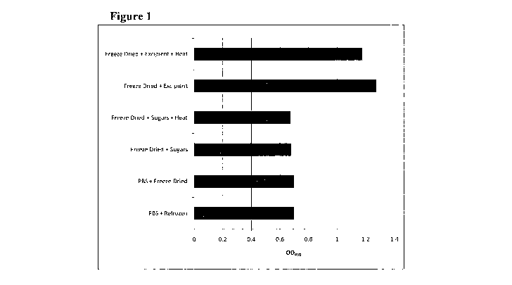
Figure 1 shows the results obtained in Example 1. The results demonstrate
protection of human calcitonin (hCT) from freeze-drying and/or heat treatment,
when
using an excipient with final concentrations of 1.03M sucrose, 0.09M raffinose
and
21nM PEI (based on an Mn of 60,000). Figure 1 shows the averaged result of
detectable hCT as measured by ELISA, after subjecting the samples to the
following
treatments:
CA 02737407 2011-03-15
WO 2010/035001 4 PCT/GB2009/002283
1. Calcitonin resuspended in PBS and frozen
2. Calcitonin resuspended in PBS and freeze dried
3. Calcitonin + sugar mix (sucrose and raffmose) freeze dried
4. Calcitonin + sugar mix (sucrose and raffinose) freeze dried + heated
5. Calcitonin + excipient (preservation mixture composed of sucrose,
raffinose and PEI) freeze dried (invention)
6. Calcitonin + excipient (preservation mixture composed of sucrose,
raffinose and PEI) freeze dried and heat treated (invention)
Figure 2 shows the results obtained in Example 2. The ability of a
preservation
mixture (excipient) according to the invention to stabilize G-CSF against heat
treatment was assessed by monitoring the ability of G-CSF to stimulate ERKl/2
phosphorylation. HL60 cells were serum starved for 24 hours and then
stimulated for
5 minutes with the treatments indicated (100ng/ml G-CSF). Whole cell extracts
were
resolved by SDS-PAGE and then transferred to nylon membranes, which were
immunoprobed with antibodies against phosphorylated and total ERKI/2.
- Panel A shows: Control (serum starved + PBS), UT G-CSF (untreated G-CSF)
and freeze thaw G-CSF (standard G-CSF mixed with excipient and frozen)
samples.
- Panel B shows: Control (serum starved + PBS), UT G-CSF (untreated G-CSF)
and Excipient/HT G-CSF (G-CSF mixed with excipient then heated) samples.
- Panel C shows: Control (serum starved + PBS), UT G-CSF (untreated G-CSF)
and G-CSF Excipient/FD (G-CSF mixed with excipient and freeze dried)
samples.
- Panel D shows: Control (serum starved + PBS), UT G-CSF (untreated G-CSF)
and G-CSF Excipient/FD/HT (G-CSF mixed with excipient, freeze dried and
heat treated) samples.
Figure 3 depicts the results from Example 3. The residual activity of anti-
human tumor necrosis factor-a antibodies (rat monoclonal anti-TNFa, Invitrogen
CA 02737407 2011-03-15
WO 2010/035001 5 PCT/GB2009/002283
Catalogue No.: SKU#RHTNFA00) was assessed in an ELISA after the indicated
treatment:
1. anti-hTNFa rat mAb (test) - no treatment + PBS (4 C)
2. anti-hTNFa rat mAb - freeze dried + excipient and stored at 4 C
3. anti-hTNFa rat mAb - freeze dried + excipient and heat treated at 65 C for
24 hours
4. anti-hTNFa rat mAb - heat treated + PBS at 65 C for 24 hours
The excipient contained a final concentration of 0.91M sucrose, 0.125M
raffinose and 25nM PEI (based on Mn of 60,000). The results show that the
inclusion
of excipient prior to freeze drying of the antibody enabled the said antibody
to
withstand to a significantly higher level, heat challenge for significantly
longer
periods.
Figure 4 shows the preservation of luciferase in Example 4 after freezing and
then freeze-drying overnight, in an excipient (preservation mixture)
containing a final
concentration of 1.092M sucrose, 0.0499M stachyose and either 27nM, 2.7nM and
0.27 nM PEI (Sigma catalogue number P3143, Mn 60,000). As can be clearly seen,
there is improved thermal stability of Luciferase when dried in the presence
of the
excipient.
Figure 5 shows the preservation of beta-galactosidase activity in Example 5
following freeze-drying in an excipient (preservation mixture) containing a
final
concentration of 0.97 M sucrose, 0.13M raffmose and 13 M, 2.6 M, 0.261M, 26nM
or 2.6nM PEI (Sigma catalogue number P3143, Mõ 60,000). This Example clearly
demonstrates that there is significant improvement in the thermal stability of
beta-
galactosidase when dried in the excipient.
Figure 6 shows the results of the experiment of Example 6 evaluating a range
of excipients to provide thermostabilisation of anti-human TNFa antibody.
Samples
of antibody in excipient containing various concentrations of sucrose (Suc),
raffinose
(Raf) and PEI were freeze-dried and then heated at 45 C for 1 week.
Figure 7 shows the effects of excipient composition on the amount of anti-
TNFa measured after freeze-drying (FD) in Example 7. HPLC peak areas are
CA 02737407 2011-03-15
WO 2010/035001 6 PCT/GB2009/002283
depicted. No antibody was measured when freeze-dried in PBS. A significant
amount of anti-TNFa antibody was lost when freeze-dried in sugars alone. A
much
greater amount of anti-TNFa was measured when the antibody was freeze-dried
with
sugars and PEI.
Figure 8 depicts the result of the experiment of Example 8. Anti-TNFa
antibody was freeze-dried in IM sugar (0.9M sucrose and 0.1 M raffmose) and
0.0025nM PEI.
Figure 9 compares the thermal stability of freeze-dried influenza
haemagglutinin (HA) against liquid control samples (Liquid PBS) as tested in
Example 9. Samples of HA protein were prepared in PBS or an excipient mixture
of
1M sucrose/lOOmM raffinose/16.6nM PEI (based on Mn). The mixture was then
lyophilised (FD), secondary drying being carried out between -32 C and 20 C
over a
3 day cycle. After lyophilisation, one of the samples was thermally challenged
at
80 C for 1 hour (FD HT excipient).
Figure 10 shows the effects of sugars and PEI on luciferase freeze-dried with
bovine serum albumin (BSA) in Example 10. This six-part Figure shows the
effects
on luciferase activity of sugar mix (sm) and PEI - alone and together - when
added
before or after freeze-drying (FD). Prior to analysis, freeze-dried samples
were held
at 45 C for 2 weeks, then at room temperature for a further 2 weeks. Error
bars
shown are standard error of the mean. .
Figure 11 shows the effect of freezing (3-gal in the presence of sugar/PEI
excipients as reported in Example 11. Following freeze-drying, (3-gal activity
was
high in sucrose/raffinose excipients compared to PBS. The presence of PEI at
13.3 M in combination with sucrose/raffinose further enhanced enzyme activity
compared to sucrose/raffinose alone. Error bars show standard error of the
mean.
Figure 12 shows the results obtained in Example 12 of subjecting samples of
horse radish peroxidase (HRP) to freeze-drying and then 2, 4 or 6 heat-freeze
cycles
by removing them from the -20 C freezer and placing them in an incubator at 37
C
for 4 hours before replacing them in the freezer for 20 hours 2, 4 or 6 times.
The
results show for all treatments and storage conditions that HRP activity is
better
CA 02737407 2011-03-15 -
WO 2010/035001 7 PCT/GB2009/002283
maintained in the presence of sucrose, raffinose either with or without PEI,
than PBS
alone. However, the presence of sugars in combination with PEI at the initial
freeze-
drying stage significantly reduces loss of HRP activity.
Figure 13 depicts the results obtained in Example 13. The activity of wet,
dried and freeze-dried alcohol oxidase in the presence and absence of
excipients is
shown:
- DO to D16: days incubated at 37 C (for dried and freeze-dried samples);
- No MeOH: no methanol added (negative control);
- wet: samples stored and tested with desiccation (i.e. fresh);
- FD: freeze-dried;
- D: dried;
- G1&G2: excipient mix conditions Gibson 1 & 2 respectively according to
Example 10 of WO 90/05182; and
- Si and S2: excipient mix conditions Stabilitech 1 and 2 respectively
according to the present invention.
Figure 14 shows an assessment of the level of phosphorylated ERK1/ERK2 in
HL-60 cells induced by recombinant human G-CSF in Example 14. G-CSF was
mixed with an excipient containing sucrose, raffinose and PEI, then freeze
dried (FD)
and heat treated at 56 C (HT).
Figure 15 shows the recovery of IgM in Example 15 after freeze-drying in
various excipients and thermal challenge. The error bars represent standard
error.
Figure 16 shows the level of phosphorylated ERK1/ERK2 in HL-60 cells
induced by recombinant human G-CSF in Example 16. G-CSF was mixed with an
excipient containing sucrose, raffinose and PEI, then freeze dried (FD) and
heat
treated at 37 C or 56 C (HT).
Detailed Description of the Invention
Summary
The present invention relates to the preservation of an active agent by
contacting the active agent with a preservation mixture. The active agent may
be a
CA 02737407 2011-03-15
WO 2010/035001 8 PCT/GB2009/002283
polypeptide such as a hormone, growth factor, peptide or cytokine; an antibody
or
antigen-binding fragment thereof; or an enzyme. The active agent may be a
vaccine
immunogen such as a subunit vaccine, conjugate vaccine or toxoid.
The preservation mixture is an aqueous solution of PEI and one, two or more
sugars. Low concentrations of PEI and relatively high concentrations of sugar
are
used. The resulting solution in which the active agent is present is then
dried to form
an amorphous solid matrix comprising the active agent. The matrix is storage
stable
at ambient temperature. If an aqueous solution comprising the active agent is
required
for administration, it is reconstituted from the solid matrix immediately
prior to use.
The invention thus enables the structure and function of the active agent to
be
preserved during the drying step and storage. Biological activity of the
active agent
following drying can thus be maintained. The preserved active agent
demonstrates
improved thermal and desiccation resistance allowing extension of shelf life,
ease of
storage and transport and obviating the need for a cold chain for
distribution. The
preservation mixture can thus provide protection as a cryoprotectant
(protection
against freeze damage), lyoprotectant (protection against desiccation) and/or
a
thermoprotectant (protection against temperatures higher or lower than 4 C).
Polypeptides
Any polypeptide is suitable for use in the invention. For example, the
polypeptide may be a small peptide of less than 15 amino acids such as 6 to 14
amino
acids (e.g. oxytocin, cyclosporin), a larger peptide of between 15 and 50
amino acids
(e.g. calcitonin, growth hormone releasing hormone 1-29 (GHRH)), a small
protein of
between 50 and 250 amino acids in length (e.g. insulin, human growth hormone),
a
larger protein of greater than 250 amino acids in length or a multisubunit
protein
comprising a complex of two or more polypeptide chains. The polypeptide may be
a
peptide hormone, growth factor or cytokine. It may be an antigen-binding
polypeptide, receptor inhibitor, ligand mimic or receptor blocking agent.
Typically,
the polypeptide is in substantially pure form. It may thus be an isolated
polypeptide.
For example, the polypeptide may be isolated following recombinant production.
CA 02737407 2011-03-15 1 " , / u L-0 -., , U U z
WO 2010/035001 PCT/GB2009/002283
For example, the polypeptide may be a hormone selected from a growth
hormone (GH), prolactin (PRL), a human placental lactogen (hPL), a
gonadotrophin
(e.g. lutenising hormone, follicle stimulating hormone), a thyroid stimulating
hormone (TSH), a member of the pro-opiomelanocortin (POMC) family, vasopressin
and oxytocin, a natriuretic hormone, parathyroid hormone (PTH), calcitonin,
insulin,
a glucagon, somatostatin and a gastrointestinal hormone.
The polypeptide may be a Tachykinin peptide (e.g. Substance P, Kassinin,
Neurokinin A, Eledoisin, Neurokinin B), a vasoactive intestinal peptide (e.g.
VIP
(Vasoactive Intestinal Peptide; PHM27), PACAP (Pituitary Adenylate Cyclase
Activating Peptide), Peptide PHI 27 (Peptide Histidine Isoleucine 27), GHRH 1-
24
(Growth Hormone Releasing Hormone 1-24), Glucagon, Secretin), a pancreatic
polypeptide-related peptide (e.g. NPY, PYY (Peptide YY), APP (Avian Pancreatic
Polypeptide), PPY (Pancreatic PolYpeptide), an opioid peptide (e.g.
Proopiomelanocortin (POMC) peptides, Enkephalin pentapeptides, Prodynorphin
peptide, a calcitonin peptide (e.g. Calcitonin, Amylin, AGGO1) or another
peptide
(e.g. B-type Natriuretic Peptide (BNP)).
The polypeptide may be a growth factor selected from a member of the
epidermal growth factor (EGF) family, platelet-derived growth factor family
(PDGF),
fibroblast growth factor family (FGF), Transforming Growth Factors-3 family
(TGFs-
(3), Transforming Growth Factor-a (TGF-a), Erythropoietin (Epo), Insulin-Like
Growth Factor-I (IGF-I), Insulin-Like Growth -Factor-II (IGF-II). Typically,
the
growth factor is a Transforming growth factor beta (TGF-0), a Nerve growth
factor
(NGF), a Neurotrophin, a Platelet-derived growth factor (PDGF), Erythropoietin
(EPO), Thrombopoietin (TPO), Myostatin (GDF-8), a Growth differentiation
factor-9
(GDF9), Acidic fibroblast growth factor (aFGF or FGF-1), Basic fibroblast
growth
factor (bFGF or FGF-2), Epidermal growth factor (EGF) or a Hepatocyte growth
factor (HGF).
The polypepide may be a cytokine selected from Interleukin-1 (IL-1),
Interleukin-2 (IL-2), Interleukin-6 (IL-6) Interleukin-8 (IL-8), Tumor
Necrosis Factor-
a (TNF-a), Tumor Necrosis Factor-(3 (TNF-0), Interferon-y (INF-y) and a Colony
CA 02737407 2011-03-15 rL 1 / U ID LUU / U
WO 2010/035001 10 PCT/GB2009/002283
Stimulating Factor (CSF). Typically the cytokine is a Granulocyte-colony
stimulating
factor (G-CSF) or a Granulocyte-macrophage colony stimulating factor (GM-CSF).
The polypeptide may be a blood-clotting factor such as Factor VIII, Factor V,
von Willebrand factor or coagulation factor III.
Antibodies
An antibody for use in the invention may either be a whole antibody or an
antigen-binding fragment thereof.
Whole antibodies
In one embodiment, the antibody is an immunoglobulin (Ig) monomer, dimer,
tetramer, pentamer, or other oligomer. Each antibody monomer may comprise four
polypeptide chains (for example, a conventional antibody consisting of two
identical
heavy chains and two identical light chains). Alternatively, each antibody
monomer
consists of two polypeptide chains (for example, a heavy chain antibody
consisting of
two identical heavy chains).
The antibody can be any class or isotype of antibody (for example IgG, IgM,
IgA, IgD or IgE) or any subclass of antibody (for example IgG subclasses IgGl,
IgG2, IgG3, IgG4 or IgA subclasses IgAl or IgA2). Typically, the antibody is
an IgG
such as an IgGI, IgG2 or IgG4 antibody. Usually, the antibody is an IgG1 or
IgG2
antibody.
Typically the antibody or antigen-binding fragment is of mammalian origin.
The antibody may thus be a primate, human, rodent (e.g. mouse or rat), rabbit,
ovine,
porcine, equine or camelidae antibody or antibody fragment. The antibody or
antibody fragment may be of shark, origin.
The antibody may be a monoclonal or polyclonal antibody. Monoclonal
antibodies are obtained from a population of substantially homogenous
antibodies that
are directed against a single determinant on the antigen. A population of
polyclonal
antibodies comprises a mixture of antibodies directed against different
epitopes.
CA 02737407 2011-03-15 I v 1 / ~_a
11
WO 2010/035001 PCT/GB2009/002283
Antigen-binding fragments
The antigen-binding fragment can be any fragment of an antibody which
retains antigen-binding ability, for example a Fab, F(Ab')2, Fv, disulphide-
linked Fv,
single chain Fv (scFv), disulphide-linked scFv, diabody, linear antibody,
domain
antibody or multispecific antibody. Such fragments comprise one or more
antigen
binding sites. In one embodiment, the antigen-binding fragment comprises four
framework regions (e.g. FR1, FR2, FR3 and FR4) and three complementarity-
determining regions (e.g. CDR1, CDR2 and CDR3). Methods suitable for detecting
ability of a fragment to bind an antigen are described herein and are well
known in the
art, for example immunoassays and phage display.
The antibody binding fragment may be a monospecific, bispecific or
multispecific antibody. A multispecific antibody has binding specificity for
at least
one, at least two, at least three, at least four or more different epitopes or
antigens. A
bispecific antibody is able to bind to two different epitopes or antigens. For
example,
a bispecific antibody may comprise two pairs of VH and VL, each VH/VL pair
binding
to a single antigen or epitope. Methods for preparing bispecific antibodies
are known
in the art, for example involving coexpression of two immunoglobulin heavy
chain-
light chain pairs, fusion of antibody variable domains with the desired
binding
specificities to immunoglobulin contant domain sequences, or chemical linkage
of
antibody fragments.
The bispecific antibody "diabody" comprises a heavy chain variable domain
connected to a light chain variable domain in the same polypeptide chain (VH-
VL).
Diabodies can be generated using a linker (e.g. a peptide linker) that is too
short to
allow pairing between the two domains on the same chain, so that the domains
are
forced to pair with the complementary domains of another chain and create a
dimeric
molecule with two antigen-binding sites.
A suitable scFv antibody fragment may comprise VH and VL domains of an
antibody wherein these domains are present in a single polypeptide chain.
Generally,
the Fv polypeptide further comprises a polypeptide linker between the VH and
VL
domains, which enables the scFv to form the desired structure for antigen
binding.
CA 02737407 2011-03-15
12 9a"Q ' 0 022
WO 2010/035001 PCT/GB2009/002283
A domain antibody for use in the methods of the invention may essentially
consist of a light chain variable domain (e.g. a VL) or of a heavy chain
variable
domain (e.g. a VH). The heavy chain variable domain may be derived from a
conventional four-chain antibody or from a heavy chain antibody (e.g. a
camelidae
VHH).
Modifications
The whole antibody or fragment thereof may be associated with other
moieties, such as linkers, which may be used to join together two or more
fragments
or antibodies. Such linkers may be chemical linkers or can be present in the
form of a
fusion protein with a fragment or whole antibody. The linkers may thus be used
to
join together whole antibodies or fragments, which have the same or different
binding
specificities.
In a further embodiment, the antibody or antigen-binding fragment is linked to
a further moiety such as a toxin, therapeutic drug (e.g. chemotherapeutic
drug),
radioisotope, liposome or prodrug-activating enzyme. The type of further
moiety will
depend on the end use of the antibody or antigen-binding fragment.
The antibody or antigen-binding fragment may be linked to one or more small
molecule toxins (e.g. calicheamicin, maytansine, trichothene and CC 1065) or
an
enzymatically active toxin or fragment thereof (e.g. diphtheria toxin,
exotoxin A chain
from Pseudomonas aeruginosa, ricin A chain, abrin A chain, modeccin A chain,
alpha-sarcin, Aleuritesfordii proteins, dianthin proteins, curcin, crotin,
gelonin,
mitogellin, restrictocin, phenomycin, enomycin or tricothecenes).
Radioisotopes suitable for linking to the antibody or antigen-binding
fragments include, but are not limited to Tc99, At211, I1311I125, Y90, Re186,
Re'88, Sm'53,
Bi212 and P32.
The antibody or antigen-binding fragment may be linked for example, to a
prodrug-activating enzyme that converts or is capable of converting a prodrug
to an
active anti-cancer drug. For example, alkaline phosphatase can be used to
convert
phosphate-containing prodrugs into free drugs, arylsufatase may be used to
convert
CA 02737407 2011-03-15
WO 2010/035001 13 PCT/GB2009/002283
sulfate-containing prodrugs into free drugs, cytosine deaminase may be used to
convert non-toxic 5-fluorocytosine into the anti-cancer drug 5-fluorouracil;
and
proteases such as serratia protease, thermolysin, subtilisin,
carboxypeptidases and
cathepsins are useful for converting peptide-containing prodrugs into free
drugs. The
enzyme may be a nitroreductase which has been identified as useful in the
metabolism
of a number of prodrugs in anti-cancer gene therapy. Alternatively, antibodies
or
antigen-binding fragments with enzymatic activity can be used to convert
prodrugs
into free active drugs.
A suitable chemotherapeutic agent may include, but is not limited to an
alkylating agent such as thiotepa and cyclosphosphamide; an alkyl sulfonate
such as
busulfan, improsulfan and piposulfan; an aziridine such as benzodopa,
carboquone,
meturedopa and uredopa; a nitrogen mustard such as chlorambucil,
chlornaphazine,
ifosfamide, melphalan; a nitrosurea such as carmustin and fotemustine; an anti-
metabolite. such as methotrexate and 5-fluorouracil (5-FU); a folic acid
analogue such
as denopterin and pteropterin; a purine analogue such as fludarabine and
thiamiprine;
a pyrimidine analogue such as ancitabine, azacitidine, carmofur and
doxifluridine; a
taxoid such as paclitaxel and doxetaxel; and pharmaceutically acceptable
salts, acids
or derivatives of any of the above.
In another embodiment, the antibody or antibody fragment may be PEGylated.
Thus, one or more polyethylene glycol molecules may be covalently attached to
the
antibody molecule or antibody fragment molecule From one to three polyethylene
glycol molecules may be covalently attached to each antibody molecule or
antibody
fragment molecule. Such PEGylation is predominantly used to reduce the
immunogenicity of an antibody or antibody fragment and/or increase the
circulating
half-life of the antibody or antibody fragment.
Chimeric, humanized or human antibodies
In one embodiment the antibody or antigen-binding fragment is a chimeric
antibody or fragment thereof comprising sequence from different natural
antibodies.
For example, the chimeric antibody or antigen-binding fragment may comprise a
CA 02737407 2011-03-15
WO 2010/035001 14 PCT/GB2009/002283
portion of the heavy and/or light chain identical or homologous to
corresponding
sequences in antibodies of a particular species or antibody class, while the
remainder,
of the chain is identical or homologous to corresponding sequences in
antibodies of
another species or antibody class. Typically, the chimeric antibody or antigen-
binding
fragment comprises a chimera of mouse and human antibody components.
Humanized forms of non-human antibodies are chimeric antibodies that
contain minimal sequence derived from non-human immunoglobulin. A suitable
humanized antibody or antigen-binding fragment may comprise for example,
immunoglobulin in which residues from a hypervariable region (e.g. derived
from a
CDR) of the recipient antibody or antigen-binding fragment are replaced by
residues
from a hypervariable region of a non-human species (donor antibody) such as
mouse,
rat, rabbit or non-human primate having the desired specificity, affinity
and/or
capacity. In some instances, some framework region residues of the human
immunoglobulin may be replaced by corresponding non-human residues.
As an alternative to humanization, human antibodies or antigen-binding
fragments can be generated. For example, transgenic animals (e.g. mice) can be
produced that are capable, upon immunization, of producing a full repertoire
of
human antibodies in the absence of endogenous immunoglobulin production. For
example, homozygous deletion of the antibody heavy-chain joining region (JH)
gene
in chimeric and germ-line mutant mice can result in complete inhibition of
endogenous antibody production. Human germ-line immunoglobulin genes can be
transferred to such germ-line mutant mice to result in the production of human
antibodies upon antigen challenge. A human antibody or antigen-binding
fragment
can also be generated in vitro using the phage display technique.
Targets
An antibody or antigen-binding fragment capable of binding any target antigen
is suitable for use in the methods of the present invention. The antibody or
antigen-
binding fragment may be capable of binding to an antigen associated with an
autoimmune disorder (e.g. Type I diabetes, multiple sclerosis, rheumatoid
arthritis,
CA 02737407 2011-03-15
WO 2010/035001 PCT/GB2009/002283
systemic lupus erythematosus, Crohn's disease and myasthenia gravis), an
antigen
associated with a cancer or an inflammatory state, an antigen associated with-
osteoporosis, an antigen associated with Alzheimer's disease, or a bacterial
or viral
antigen.
5 In particular, the target to which an antibody or antigen-binding fragment
may
bind can be a CD antigen, growth factor, growth factor receptor, cell surface
receptor
such as an apoptosis receptor, a protein kinase or an oncoprotein. The
antibody or
antigen-binding fragment, for example a chimeric, humanized or human IgGI,
IgG2
or IgG4 monoclonal antibody or antibody fragment, may thus be capable of
binding to
10 tumour necrosis factor a (TNF-a), interleukin-2 (IL-2), interleukin-6 (IL-
6),
glycoprotein IIb/IIIa, CD33, CD52, CD20, CDI la, CD3, RSV F protein, HER2/neu
(erbB2) receptor, vascular endothelial growth factor (VEGF), epidermal growth
factor
receptor (EGFR), anti-TRAILR2 (anti-tumour necrosis factor-related apoptosis-
inducing ligand receptor 2), complement system protein C5, a4 integrin or IgE.
15 More specifically, in the context of anti-cancer monoclonal antibodies, the
antibody or antigen-binding fragment may be an antibody or antibody fragment
capable of binding to epithelial cell adhesion molecule (EpCAM), mucin-1
(MUC 1 /Can-Ag), EGFR, CD20, carcinoembryonic antigen (CEA), HER2, CD22,
CD33, Lewis Y and prostate-specific membrane antigen (PMSA). Again, the
antibody is typically a chimeric, humanized or human IgGI, IgG2 or IgG4
monoclonal antibody.
Suitable monoclonal antibodies include, but are not limited to: infliximab
(chimeric antibody, anti-TNFa), adalimumab (human antibody, anti-TNFa),
basiliximab (chimeric antibody, anti-IL-2), abciximab (chimeric antibody, anti-
GpIIb/IIIa), daclizumab (humanized antibody, anti-IL-2), gemtuzumab (humanized
antibody, anti-CD33), alemtuzumab (humanized antibody, anti-CD52), edrecolomab
(murine Ig2a, anti-EpCAM), rituximab (chimeric antibody, anti-CD20),
palivizumab
(humanized antibody, RSV target), trastuzumab (humanized antibody, anti-
HER2/neu(erbB2) receptor), bevacizumab (humanized antibody, anti-VEGF),
cetuximab (chimeric antibody, anti-EGFR), eculizumab (humanized antibody, anti-
CA 02737407 2011-03-15
16
WO 2010/035001 PCT/GB2009/002283
complement system protein C5), efalizumab (humanized antibody, anti-CD 1 l a),
ibritumomab (murine antibody, anti-CD20), muromonab-CD3 (murine antibody, anti-
T cell CD3 receptor), natalizumab (humanized antibody, anti-a 4 integrin),
nimotuzumab (humanized IgGI, anti-EGF receptor), omalizumab (humanized
antibody, anti-IgE), panitumumab (human antibody, anti-EGFR), ranibizumab
(humanized antibody, anti-VEGF), ranibizumab (humanized antibody, anti-VEGF)
and I-131 tositumomab (humanized antibody, anti-CD20).
Preparation of antibodies
Suitable monoclonal antibodies may be obtained for example, by the
hybridoma method (e.g. as first described by Kohler et al Nature 256:495
(1975)), by
recombinant DNA methods and/or following isolation from phage or other
antibody
libraries.
The hybridoma technique involves immunisation of a host animal (e.g. mouse,
hamster or monkey) with a desired immunogen to elicit lymphocytes that produce
or
are capable of producing antibodies that specifically bind to the immunogen.
Alternatively, lymphocytes may be immunized in vitro. Lymphocytes are then
fused
with myeloma cells using a suitable fusing agent, such as polyethylene glycol,
to form
a hybridoma cell.
An antibody or antibody fragment can also be isolated from antibody phage
libraries as an alternative to traditional monoclonal antibody hybridoma
techniques
for isolation of monoclonal antibodies. In particular, phage display may be
used to
identify antigen-binding fragments for use in the methods of the invention. By
using
phage display for the high-throughput screening of antigen-antibody binding
interactions, antigen-binding fragments displayed on phage coat proteins can
be
isolated from a phage display library. By immobilising a target antigen on a
solid
support, a phage that displays an antibody capable of binding that antigen
will remain
on the support while others can be removed by washing. Those phages that
remain
bound can then be eluted and isolated, for example after repeated cycles of
selection
or panning. Phage eluted in the final selection can be used to infect a
suitable
CA 02737407 2011-03-15
WO 2010/035001 17 PCT/GB2009/002283
bacterial host from which phagemids can be collected and the relevant DNA
sequence
excised and sequenced to identify the relevant antigen-binding fragment.
Polyclonal antiserum containing the desired antibodies is isolated from
animals using techniques well known in the art. Animals such as sheep, rabbits
or
goats may be used for example, for the generation of antibodies against an
antigen of
interest by the injection of this antigen (immunogen) into the animal,
sometimes after
multiple injections. After collection of antiserum, antibodies may be purified
using
immunosorbent purification or other techniques known in the art.
The antibody or antigen-binding fragment used in the method of the invention
may be produced recombinantly from naturally occurring nucleotide sequences or
synthetic sequences. Such sequences may for example be isolated by PCR from a
suitable naturally occurring template (e.g. DNA or RNA isolated from a cell),
nucleotide sequences isolated from a library (e.g. an expression library),
nucleotide
sequences prepared by introducing mutations into a naturally occurring
nucleotide
sequence (using any suitable technique known, e.g. mismatch PCR), nucleotide
sequence prepared by PCR using overlapping primers, or nucleotide sequences
that
have been prepared using techniques for DNA synthesis. Techniques such as
affinity
maturation (for example, starting from synthetic, random or naturally
occurring
immunoglobulin sequences), CDR grafting, veneering, combining fragments
derived
from different immunoglobulin sequences, and other techniques for engineering
immunoglobulin sequences may also be used.
Such nucleotide sequences of interest may be used in vitro or in vivo in the
production of an antibody or antigen-binding fragment for use in the
invention, in
accordance with techniques well known to those skilled in the art.
For recombinant production of a monoclonal antibody or antigen-binding
fragment, the nucleic acid encoding it is isolated and inserted into a
replicable vector
for further cloning or for expression. The vector components generally
including, but
is not limited to one or more of the following: a signal sequence, an origin
of
replication, one or more marker genes, an enhancer element, a promoter, and a
transcription termination sequence. Suitable host cells for cloning or
expressing the
CA 02737407 2011-03-15 - -- . - - .-
WO 2010/035001 18 PCT/GB2009/002283
DNA in the vectors are prokaryote, yeast, or higher eukaryote cells such as E.
coli and
mammalian cells such as CHO cells. Suitable host cells for the expression of
glycosylated antibody are derived from multi-cellular organisms. Host cells
are
transformed with the expression or cloning vectors for antibody production and
cultured in conventional nutrient media modified as appropriate for inducing
promoters, selecting transformants, or amplifying the genes encoding the
desired
sequences.
When using recombinant techniques, the antibody can be produced
intracellularly or directly secreted into the medium. If the antibody is
produced
intracellularly, as a first step, the particulate debris of either host cells
or lysed cells, is
removed, for example by centrifugation or ultra filtration. Where the antibody
is
secreted into the medium, supernatants from expression systems are generally
first
concentrated using a commercially available protein concentration filter. The
antibody composition prepared from the cells can be purified using, for
example,
hydyoxylapatite chromatography, gel electrophoresis, dialysis and affinity
chromatography.
The purified antibodies may then be isolated and optionally made into antigen-
binding fragments and/or derivatised.
Enzymes
Any protein enzyme is suitable for use in the invention. Such an enzyme
comprises an active site and is capable of binding a substrate. The enzyme may
be a
monomer consisting of one polypeptide chain. Alternatively, the enzyme may be
a
dimer, tetramer or oligomer consisting of multiple polypeptide chains. The
dimer,
tetramer or oligomer may be a homo- or hetero- dimer, tetramer or oligomer
respectively. For example, the enzyme may need to form an aggregate (e.g. a
dimer,
tetramer or oligomer) before full biological activity or enzyme function is
conferred.
The enzyme may be an allosteric enzyme, an apoenzyme or a holoenzyme.
The enzyme may be conjugated to another moiety (e.g. a ligand, antibody,
carbohydrate, effector molecule, or protein fusion partner) and/or bound to
one or
CA 02737407 2011-03-15
WO 2010/035001 19 PCT/GB2009/002283
more cofactors (e.g. coenzyme or prosthetic group).
The moiety to which the enzyme is conjugated may include lectin, avidin, a
metabolite, a hormone, a nucleotide sequence, a steroid, a glycoprotein, a
glycolipid,
or any derivative of these components.
Cofactors include inorganic compounds (e.g. metal irons such as iron,
manganese, cobalt, copper, zinc, selenium, molybdenum) or organic compounds
(e.g.
flavin or heme). Suitable coenzymes include riboflavin, thiamine, folic acid
which
may carry hydride iron (H-) carried by NAD or NADP+, the acetyl group carried
by
coenzyme A, formyl, methenyl or methyl groups carried by folic acid and the
methyl
group carried by S-adenosyl methionine.
In another embodiment, the enzyme may be PEGylated especially if the
enzyme is a therapeutic enzyme that is administered to a patient. Thus, one or
more
polyethylene glycol molecules may be covalently attached to the enzyme
molecule.
From one to three polyethylene glycol molecules may be covalently attached to
each
enzyme molecule. Such PEGylation is predominantly used to reduce the
immunogenicity of an enzyme and/or increase the circulating half-life of the
enzyme.
A suitable enzyme includes any enzyme classified under the International
Union of Biochemistry and Molecular Biology Enzyme classification system of EC
numbers including an oxidoreductase (EC 1), a transferase (EC 2), a hydrolase
(EC
3), a lyase (EC 4), an isomerase (EC 5) or a ligase (EC 6). A typical enzyme
is any
enzyme that is used industrially.
An enzyme that is specific for any type of substrate is suitable for use in
the
present invention. Examples of a suitable enzyme includes a a-galactosidase, 0-
galactosidase, luciferase, serine proteinase, endopeptidase (e.g. cysteine
endopeptidase), caspase, chymase, chymotrypsin, endopeptidase, granzyme,
papain,
pancreatic elastase, oryzin, plasmin, renin, subtilisin, thrombin, trypsin,
tryptase,
urokinase, amylase (e.g. a-amylase), xylanase, lipase, transglutaminase, cell-
wall-
degrading enzyme, glucanase (e.g. (3-glucanase), glucoamylase, coagulating
enzyme,
milk protein hydrolysate, cell-wall degrading enzyme, blood coagulating
enzyme,
hementin, lysozyme, fibre-degrading enzyme, phytase, cellulase, hemicellulase,
CA 02737407 2011-03-15
WO 2010/035001 20 PCT/GB2009/002283
polymerase, protease, mannanase or glucoamylase.
An enzyme preserved according to the invention may thus be a therapeutic
enzyme that is used to treat a disease or other medical condition, an enzyme
used in
industry for the production of bulk products such as glucose or fructose, in
food
processing and food analysis, in laundry and automatic dishwashing detergents,
in the
textile, pulp, paper and animal feed industries, as a catalyst in synthesis or
fine
chemicals, in diagnostic applications such as in clinical diagnosis, in
biosensors or in
genetic engineering.
Therapeutic enzymes to which the present invention can be applied include:
- a DNAase, for example a recombinant DNAase I such as Pulmozyme or
Domase that cleaves the DNA in the pulmonary mucus of children having
cystic fibrosis;
- a gastric lipase such as Meripase which is a recombinant mammalian gastric
lipase for the treatment of lipid malabsorption related to exocrine pancreatic
lipase insufficiency;
- a mannose-terminated glucocerebrosidase such as Cerezyme which is a
recombinant mannose-terminated glucocerebrosidase for the treatment of
Gaucher disease, an inherited disorder that is caused by a deficiency in the
enzyme glucocerebrosidase;
- a-galactosidase which is used in the treatment of the related glycogen
storage
disease Fabry disease;
- an adenosine deaminase (ADA) such as Pegademase that is used to treat ADA
deficiency, a severe combined immunodeficiency;
- a phenylalanine ammonia lyase such as the PEGylated recombinant
phenylalanine ammonia lyase Kuvan that is used for the treatment of
phenylketonuria;
- tissue plasminogen activator, urokinase and streptokinase which are used in
blood fibrinolysis to treat blood clots;
- a urate oxidase such as Elitek (rasburicase) which is a recombinant urate-
oxidase that is produced by a genetically modified yeast and that is used in
the
CA 02737407 2011-03-15
21
WO 2010/035001 PCT/GB2009/002283
treatment or prophylaxis of hyperuricemia in patients with leukaemia or
lymphoma;
L-asparaginase which is used in the treatment of childhood acute
lymphoblastic leukaemia;
- Factor VIIa, used by patients with hemophilia;
Factor IX which is used in the treatment of hemophilia B; and
a superoxide dismutase such as the bovine superoxide dismutase Orgotein that
is used for the treatment of familial amyotrophic lateral sclerosis.
Enzymes for use in food applications such as baking include amylases,
xylanases, oxidoreductases, lipases, proteases and transglutaminase. Enzymes
for use
in fruit juice production and fruit processing include cell-wall-degrading
enzymes.
Enzymes for use in brewing include bacterial a-amylase, f3-glucanase and
glucoamylase in mashing, fungal a-amylase in fermentation and cysteine
endopeptidase in post fermentation. Enzymes for use in dairy applications
include
coagulating enzymes, lipase, lysozyme, milk protein hydrolysates,
transglutaninase,
and (3-galactosidase. Enzymes for use in detergent compositions include
proteases,
amylases, lipases, cellulases and mannanase. Enzymes for use in animal feed
include
fibre-degrading enzymes, phytases, proteases and amylases. Enzymes for use in
pulp
and paper processing include cellulases and hemicellulases.
The enzyme may alternatively be an enzyme used in research and
development applications. For example, luciferases may be used for real-time
imaging of gene expression in cell cultures, individual cells and whole
organisms.
Further, luciferases may be used as reporter proteins in molecular studies,
for example
to test the activity of transcription from specific promoters in cells
transfected with
luciferase. Enzymes may also be used in drug design for example in the testing
of
enzyme inhibitors in the laboratory. Further, enzymes may be used in
biosensors (for
example, a blood glucose biosensor using glucose oxidase).
The luciferase enzyme may be a firefly, beetle or railroad worm luciferase, or
a derivative thereof. In particular, the luciferase may be derived from a
North
American firefly (Phorinuspyralis), Luciola cruciata (Japanese firefly),
Luciola
CA 02737407 2011-03-15
WO 2010/035001 22 PCT/GB2009/002283
lateralis (Japanese firefly), Luciola mingelica (russian firefly), Beneckea
hanegi
(marine bacterial luciferase), Pyrophorus plagiophthalamus (click beetle),
Pyrocelia
miyako (firefly) Ragophthalamus ohbai (railroad worm), Pyrearinus
termitilluminans
(click beetle), Phrixothrix hirtus (railroad worm), Phrixothrix vivianii,
Hotaria
parvula and Photuris pensilvanica, and mutated variants thereof.
Typically the a-galactosidase or (3-galactosidase is derived from bacteria
(such
as Escherichia coil.), a mammal (such as human, mouse, rat) or other
eukaryote.
The enzyme maybe a naturally-occurring enzyme or a synthetic enzyme. Such
enzymes may be derived from a host animal, plant or a microorganism.
Microbial strains used in the production of enzymes may be native strains or
mutant strains that are derived from native strains by serial culture and
selection, or
mutagenesis and selection using recombinant DNA techniques. For example the
microorganism may be a fungus e.g. Thermomyces acermonium, Aspergillus,
Penicillium, Mucor, Neurospora and Trichoderma. Yeasts such as Saccharomyces
cereviseae or Pishiapastoris may also be used in the production of enzymes for
use
in the methods of the present invention.
A synthetic enzyme may be derived using protein-engineering techniques well
known in the art such as rational design, directed evolution and DNA
shuffling.
Host organisms may be transformed with a nucleotide sequence encoding a
desired enzyme and cultured under conditions conducive to the production of
the
enzyme and which facilitate recovery of the enzyme from the cells and/or
culture
medium.
Vaccine immunogens
A vaccine immunogen suitable for use in the invention includes any
immunogenic component of a vaccine. The vaccine immunogen comprises an antigen
that can elicit an immune response in an individual when used as a vaccine
against a
particular disease or medical condition. The vaccine immunogen may be provided
by
itself prior to formulation of a vaccine preparation or it may be provided as
part of a
vaccine preparation. The vaccine immunogen may be a subunit vaccine, a
conjugate
CA 02737407 2011-03-15
23
WO 2010/035001 PCT/GB2009/002283
useful as a vaccine or a toxoid. The vaccine immunogen may be a protein,
bacterial-
specific protein, mucoprotein, glycoprotein, peptide, lipoprotein,
polysaccharide,
peptidoglycan, nucleoprotein or fusion protein.
The vaccine immunogen may be derived from a microorganism (such as a
bacterium, virus, fungi), a protozoan, a tumour, a malignant cell, a plant, an
animal, a
human, or an allergen. The vaccine immunogen is preferably not a viral
particle.
Thus, the vaccine immunogen is preferably not a whole virus or virion, virus-
like
particle (VLP) or virus nucleocapsid. The preservation of such viral particles
is
described in WO 2008/114021.
The vaccine immunogen may be synthetic, for example as derived using
recombinant DNA techniques. The immunogen may be a disease-related antigen
such
as a pathogen-related antigen, tumour-related antigen, allergy-related
antigen, neural
defect-related antigen, cardiovascular disease antigen, rheumatoid arthritis-
related
antigen.
In particular, the pathogen from which the vaccine immunogen is derived may
include human papilloma viruses (HPV), HIV, HSV2/HSV 1, influenza virus (types
A,
B and C), para influenza virus, polio virus, RSV virus, rhinoviruses,
rotaviruses,
hepaptitis A virus, norwalk virus, enteroviruses, astroviruses, measles virus,
mumps
virus, varicella-zoster virus, cytomegalovirus, epstein-barr virus,
adenoviruses, rubella
virus, human T-cell lymphoma type I virus (HTLV-I), hepatitis B virus (HBV),
hepatitis C virus (HCV), hepatitis D virus, poxvirus, vaccinia virus,
Salmonella,
Neisseria, Borrelia, Clamydia, Bordetella such as Bordetella pertussis,
Plasmodium,
Coxoplasma, Pneumococcus, Meningococcus, Cryptococcus, Streptococcus,
Vibriocholerae, Yersinia and in particular Yersinia pestis, Staphylococcus,
Haemophilus, Diptheria, Tetanus, Pertussis, Escherichia, Candida, Aspergillus,
Entamoeba, Giardia and Trypanasoma. The vaccine may further be used to provide
a
suitable immune response against numerous veterinary diseases, such as foot
and
mouth disease (including serotypes 0, A, C, SAT-1, SAT-2, SAT-3 and Asia-1),
coronavirus, bluetongue, feline leukaemia virus, avian influenza, hendra and
nipah
virus, pestivirus, canine parvovirus and, bovine viral diarrhoea virus.
CA 02737407 2011-03-15
24
WO 2010/035001 PCT/GB2009/002283
Tumor-associated antigens include for example, melanoma-associated
antigens, mammary cancer-associated antigens, colorectal cancer-associated
antigens
or prostate cancer-associated antigens
An allergen-related antigen includes any allergen antigen suitable for use in
a
vaccine to suppress an allergic reaction in an individual to which the vaccine
is
administered (e.g. antigens derived from pollen, dust mites, insects, food
allergens,
dust, poisons, parasites).
Subunit vaccine immunogens
A suitable subunit vaccine immunogen includes any immunogenic subunit of a
protein, lipoprotein or glycoprotein derived from a microorganism (for example
a
virus or bacteria). Alternatively, the subunit vaccine immunogen may be
derived
from a disease-related antigen such as a tumour related protein. The subunit
vaccine
immunogen may be a naturally occurring molecule or a synthetic protein
subunit.
The vaccine immunogen may be a full-length viral or bacterial protein,
glycoprotein
or lipoprotein or a fragment of the full-length viral or bacterial protein,
glycoprotein
or lipoprotein.
A viral protein suitable as a subunit vaccine immunogen may be derived from
a structural or non-structural viral protein. A suitable viral subunit
immunogen is
capable of stimulating a subject's immune system even in the absence of other
parts of
the virus. A suitable viral subunit vaccine immunogen includes a capsid
protein,
surface glycoprotein, envelope protein, hexon protein, fiber protein, coat
protein or
immunogenic fragment or derivative of such proteins or glycoproteins.
For example, the viral subunit vaccine immunogen may consist of a surface
protein of the Influenza A, B or C virus. In particular, the vaccine immunogen
may
be a hemagglutinin (HA), neuraminidase (NA), nucleoprotein, M1, M2, NS 1,
NS2(NEP), PA, PB 1, PB 1-F2 and or PB2 protein, or an immunogenic derivative
or
fragment of any of these proteins. The immunogen may be HA1, HA2, HA3, HA4,
HAS, HA6, HA7, HA8, HA9, HA 10, HA 11, HA 12, HA 13, HA 14, HA 15 and/or
HA16, any immunogenic fragment or derivative thereof and any combination of
the
CA 02737407 2011-03-15
WO 2010/035001 25 PCT/GB2009/002283
HA proteins, fragments or derivatives. The neuraminidase may be neuraminidase
1
(N i) or neuraminidase 2 (N2).
The viral subunit vaccine immunogen may be a hepatitis B virus viral
envelope protein or a fragment or derivative thereof For example, the subunit
vaccine immunogen may be the hepatitis B surface antigen (HbsAg) or an
immunogenic fragment or derivative thereof
Typically, the bacterial subunit vaccine immunogen is a bacterial cell wall
protein (e.g. flagellin, outer membrane protein, outer surface protein), a
polysaccharide antigen (e.g. from Neisseria meningitis, Streptococcus
pneumonia),
toxin or an immunogenic fragment or derivative of such proteins,
polysaccharides or
toxins.
Derivatives of naturally occurring proteins include proteins with the
addition,
substitution and/or deletion of one or more amino acids. Such amino acid
modifications can be generated using techniques known in the art, such as site-
directed mutagenesis.
The subunit vaccine immunogen may be a fusion protein comprising a fusion
protein partner linked with for example, a bacterial or viral protein or an
immunogenic fragment or derivative thereof. A suitable fusion protein partner
may
prevent the assembly of viral fusion proteins into multimeric forms after
expression of
the fusion protein. For example, the fusion protein partner may prevent the
formation
of virus-like structures that might spontaneously form if the viral protein
was
recombinantly expressed in the absence of the fusion protein partner. A
suitable
fusion partner may also facilitate purification of the fusion protein, or
enhance the
recombinant expression of the fusion protein product. The fusion protein may
be
maltose binding protein, poly-histidine segment capable of binding metal ions,
antigens to which antibodies bind, S-Tag, glutathione-S-transferase,
thioredoxin, beta-
galactosidase, epitope tags, green fluorescent protein, streptavidin or
dihydrofolate
reductase.
A subunit vaccine immunogen may be prepared using techniques known in the
art for the preparation of for example, isolated peptides, proteins,
lipoproteins, or
CA 02737407 2011-03-15
WO 2010/035001 26 PCT/GB2009/002283
glycoproteins. For example, a gene encoding a recombinant protein of interest
can be
identified and isolated from a pathogen and expressed in E. coli or some other
suitable
host for mass production of proteins. The protein of interest is then isolated
and
purified from the host cell (for example by purification using affinity
chromatography).
In the case of viral subunit immunogens, the subunit may be purified from the
viral particle after isolating the viral particle, or by recombinant DNA
cloning and
expression of the viral subunit protein in a suitable host cell. A suitable
host cell for
preparing viral particles must be capable of being infected with the virus and
of
producing the desired viral antigens. Such host cells may include
microorganisms,
cultured animal cells, trangenic plants or insect larvae. Some proteins of
interest may
be secreted as a soluble protein from the host cell. In the case of viral
envelope or
surface proteins, such proteins may need to be solubilized with a detergent to
extract
them from the viral envelope, followed by phase separation in order to remove
the
detergent.
A subunit vaccine immunogen may be combined in the same preparation and
preserved together with one, two three or more other subunit vaccine
immunogens.
Toxoids
The invention can be applied to toxoids. A toxoid is a toxin, for example
derived from a pathogen, animal or plant, that is immunogenic but has been
inactivated (for example. by genetic mutation, chemical treatment or by
conjugation to
another moiety) to eliminate toxicity to the target subject. The toxin may be
for
example, a-protein, lipoprotein, polysaccharide, lipopolysaccharide or
glycoprotein.
The toxoid may thus be an endotoxin or an exotoxin that has been toxoided.
The toxoid may be a toxoid derived from a bacterial toxin such as tetanus
toxin, diphtheria toxin, pertussis toxin, botulinum toxin, Cdifficile toxin,
Cholera
toxin, shiga toxin, anthrax toxin, bacterial cytolysins or pneumolysin and
fragments or
derivatives thereof. The toxoid may therefore be tetanus toxoid, diphtheria
toxoid or
pertussis toxoid. Other toxins from which a toxoid can be derived include
poisons
CA 02737407 2011-03-15
WO 2010/035001 27 PCT/GB2009/002283
isolated from animals or plants, for example from Crotalis atrox. Typically,
the
toxoid is derived from botulinum toxin or anthrax toxin. For example, the
botulinum
toxin may be derived from Clostridium botulinum of serotype A, B, C, D, E, F
or G.
The vaccine immunogen derived from a botulinum toxin may be combined in the
same preparation and preserved together with one or more other vaccine
immunogens
derived from a botulinum toxin (eg a combination of immunogens derived from
botulinum serotypes A, B, C, D, E, F or G, such as for example A, B and E).
The anthrax toxin may be derived from a strain of Bacillus anthracis. The
toxoid may consist of one of more components of the anthrax toxin, or
derivatives of
such components, such as protective antigen (PA), the edema factor (EF) and
the
lethal factor (LF). Typically the toxoid derived from the anthrax toxin
consists of
protective antigen (PA).
The toxoid may be conjugated to another moiety, for example as a fusion
protein, for use as a toxoid vaccine. A suitable moiety in a conjugate toxoid
includes
a substance that aids purification of the toxoid (e.g hisitidine tag) or
reduces toxicity
to a target subject. Alternatively, the toxoid may act as an adjuvant by
increasing the
immunogenicity of an antigen to which it is attached. For example, the B
polysaccharide of Haemophilus influenzae may be combined with diptheria
toxoid.
A vaccine immunogen may be combined in the same preparation and
preserved together with one, two three or more vaccine immunogens. For
example, a
diphtheria toxoid may be preserved with tetanus toxoid and pertussis vaccine
(DPT).
Diptheria toxoid may be preserved with just tetanus toxoid (DT), or diphtheria
toxoid
may be preserved with diphtheria toxoid, tetanus toxoid and acellular
Pertussis
(DTaP).
Techniques for the preparation of toxoids are well known to those skilled in
the art. Toxin genes may be cloned and expressed in a suitable host cell. The
toxin
product is then purified and may be converted to toxoid chemically, for
example using
formalin or glutaraldehyde. Alternatively, a toxin gene may be engineered so
that it
encodes a toxin having reduced or no toxicity e.g. by addition, deletion
and/or
substitution of one or more amino acids. The modified toxin can then be
expressed in
CA 02737407 2011-03-15
WO 2010/035001 28 PCT/GB2009/002283
a suitable host cell and isolated. The toxicity of toxin genes may also be
inactivated
by conjugation of toxin genes or fragments thereof to a further moiety (e.g.
polysaccharide or polypeptide).
Conjugate vaccine immunogens
A conjugate vaccine immunogen may be a conjugate of an antigen (for
example a polysaccharide or other hapten) to a carrier moiety (for example a
peptide,
polypeptide, lipoprotein, glycoprotein, mucoprotein or any immunostimulatory
derivative or fragment thereof) that stimulates the immunogenicity of the
antigen to
which it is attached. For example, the conjugate vaccine immunogen may be a
recombinant protein, recombinant lipoprotein or recombinant glycoprotein
conjugated
to an immunogen of interest (for example a polysaccharide).
The conjugate vaccine immunogen may be used in a vaccine against
Streptococcus pneumonia, Haemophilus influenza, meningococcus (strains A, B,
C,
X, Y and W135) or pneumococcal strains. For example, the vaccine may be for
example, the heptavalent Pneumococcal CRM197 Conjugate Vaccine (PCV7), an
MCV-4 or Haemophilus influenzae type b (Hib) vaccine.
A conjugate vaccine immunogen may be combined in the same preparation
and preserved together with one, two three or more other conjugate vaccine
immunogens.
Methods for the preparation of conjugate polysaccharide-protein conjugates
are well known in the art. For example, conjugation may occur via a linker
(e.g. B-
propionamido, nitrophenyl-ethylamine, haloalkyl halides, glycosidic linkages).
Preservation mixture
The preservation mixture of the present invention comprises an aqueous
solution of one or more sugars and a polyethyleneimine (PEI). The aqueous
solution
may be buffered. The solution may be a HEPES solution, phosphate-buffered
saline
(PBS) or pure water.
CA 02737407 2011-03-15
WO 2010/035001 29 PCT/GB2009/002283
Sugars suitable for use in the present invention include reducing sugars such
as glucose, fructose, glyceraldehydes, lactose, arabinose and maltose; and non-
reducing sugars such as sucrose. The sugar may be a monosaccharide,
disaccharide,
trisaccharide, or other oligosaccharides. The term "sugar" includes sugar
alcohols.
Monosaccharides such as galactose and mannose; dissaccharides such as
lactose and maltose; trisaccharides such as raffinose and tetrasaccharides
such as
stachyose are envisaged. Trehalose, umbelliferose, verbascose, isomaltose,
cellobiose, maltulose, turanose, melezitose and melibiose are also suitable
for use in
the present invention. A suitable sugar alcohol is mannitol.
Preferably, the aqueous solution is a solution of one, two or three sugars
selected from sucrose, raffinose and stachyose. In particular, sucrose is a
disaccharide
of glucose and fructose; raffinose is a trisaccharide composed of galactose,
fructose
and glucose; and stachyose is a tetrasaccharide consisting of two Da-galactose
units,
one Da-glucose unit and one D(3-fructose unit sequentially linked. A
combination of
15' sucrose and stachyose and especially sucrose and raffinose is preferred.
Preservation of biological activity is particularly effective when at least
two
sugars are used in the preservation mixture of the present invention.
Therefore, the
solution of one or more sugars comprises a solution of at least 2, at least 3,
at least 4
or at least 5 sugars. Combinations of 2, 3, 4, 5, 6, 7, 8, 9, 10, etc sugars
are envisaged.
Preferably, the solution of two or more sugars comprises sucrose and
raffinose, or
sucrose and stachyose.
PEI is an aliphatic polyamine characterised by the repeating chemical units
denoted as -(CH2-CH2-NH)-. Reference to PEI herein includes a
polyethyleneimine
homopolymer or copolymer. The polyethyleneimine copolymer may be a random or
block copolymer. For example, PEI may consist of a copolymer of
polyethyleneimine
and another polymer such as polyethylene glycol (PEG). The polyethyleneimine
may
be linear or branched.
Reference to PEI also includes derivatised forms of a polyethyleneimine. A
polyethyleneimine contains nitrogen atoms at various positions. Nitrogen atoms
are
present in terminal amino groups, e.g. R-NH2, and in internal groups such as
groups
CA 02737407 2011-03-15
WO 2010/035001 30 PCT/GB2009/002283
interrupting an alkyl or alkylene group within the polymer structure, e.g. R-
N(H)-R',
and at the intersection of a polymer branch, e.g. R-N(-R')-R" wherein R, R'
and R"
may be alkylene groups for example. Alkyl or aryl groups may be linked to the
nitrogen centres in addition to or instead of hydrogen atoms. Such alkyl and
aryl
groups may be substituted or unsubstituted. An alkyl group would be typically
a C1-
C4 alkyl group, e.g. methyl, ethyl, propyl, isopropyl, butyl, sec.butyl or
tert.butyl. The
aryl group is typically phenyl.
The PEI may be a polyethyleneimine that has been covalently linked to a
variety of other polymers such as polyethylene glycol. Other modified versions
of
PEI have been generated and some are available commercially: branched PEI 25
kDa, jetPEI , LMW-PEI 5.4 kDa, Pseudodendrimeric PEI, PEI-SS-PEI, PEI-SS-PEG,
PEI-g-PEG, PEG-co-PEI, PEG-g-PEI, PEI-co-L lactamide-co-succinamide, PEI-
co-N-(2-hydroxyethyl-ethylene imine), PEI-co-N-(2-hydroxypropyl)
methacrylamide,
PEI-g-PCL-block-PEG, PEI-SS-PHMPA, PEI-g-dextran 10 000 and PEI-g-
transferrin-PEG, Pluronic85 /Pluronicl23 -g-PEI. The PEI may be permethylated
polyethyleneimine or polyethyleneimine-ethanesulfonic acid.
PEI is available in a broad range of number-average molar masses (Mn) for
example between 300Da and 800kDa. Preferably, the number-average molar mass is
between 300 and 2000Da, between 500 and 1500Da, between 1000 and 1500Da,
between 10 and I OOkDa, between 20 and I OOkDa, between 30 and I OOkDa,
between
40 and IOOkDa, between 50 and IOOkDa, between 60 and IOOkDa, between 50 and
70kDa or between 55 and 65kDa. A relatively high Mn PEI of approximately 60kDa
or a relatively low Mn of 1200Da is suitable.
Preferably, the weight-average molar mass (Mw) of PEI is between 5001)a and
1000kDa. Most preferably, the M, of PEI is between 500Da and 2000Da, between
1000Da and 1500Da, or between 1 and 1000kDa, between 100 and 1000kDa, between
250 and 1000kDa, between 500 and 1000kDa, between 600 and 1000kDa, between
750 and 1000kDa, between 600 and 800kDa, between 700 and 800kDa. A relatively
high MW of approximately 750kDa or a relatively low MW of approximately 1300Da
is
suitable.
CA 02737407 2011-03-15
WO 2010/035001 31 PCT/GB2009/002283
The weight-average molar mass (Mw) and number-average molar mass (Mn) of
PEI can be determined by methods well known to those skilled in the art. For
example, M,,, may be determined by light scattering, small angle neutron
scattering
(SANS), X-ray scattering or sedimentation velocity. Mn may be determined for
example by gel permeation chromatography, viscometry (Mark-Houwink equation)
and colligative methods such as vapour pressure osometry or end-group
titration.
Various forms of PEI are available commercially (e.g. Sigma, Aldrich). For
example, a branched, relatively high molecular weight form of PEI used herein
with
an Mn of approximately 60kDa and a M,,, of approximately 750kDa is available
commercially (Sigma P3143). This PEI can be represented by the following
formula:
NH2 NNH2
H N
N ~/~ N N "/ N
H H
n
H2N __- N N H2
A relatively low molecular weight form of PEI used herein is also available
commercially (e.g. Aldrich 482595) which has a MW of 1300Da and Mn of 1200Da.
In the present invention, a preservation mixture comprising an aqueous
solution of PEI and one, two or more sugars is provided. Typically, the active
agent
is admixed with the preservation mixture to provide the aqueous solution for
drying.
The concentrations of PEI and sugar that are employed for a particular active
agent
will depend upon the active agent. The concentrations can be determined by
routine
experimentation. Optimised PEI and sugar concentrations which result in the
best
CA 02737407 2011-03-15
32
WO 2010/035001 PCT/GB2009/002283
stability can thus be selected. The PEI and sugar can act synergistically to
improve
stability.
The concentration of sugar in the aqueous solution for drying is greater than
0.1 M. Preferably, the concentration of the sugar in the aqueous solution for
drying or,
if more than one sugar is present, the total concentration of sugar in the
aqueous
solution for drying, is at least 0.2M, 0.3M, 0.4M, 0.5M, 0.6M, 0.75M, 0.9M, 1M
or
2M up to saturation e.g. saturation at room temperature or up to 3M, 2.5M or
2M.
The sugar concentration or the total concentration if more than one sugar is
present
may be from 0.5 to 2M. When more than one sugar is present, each sugar may be
present at a concentration of from 0.2M, 0.3M, 0.4M, 0.5M, 0.6M, 0.75M, 0.9M,
1M
or 2M up to saturation e.g. saturation at room temperature or up to 3M, 2.5M
or 2M.
The concentration of PEI in the aqueous solution for drying is generally in
the
range of 20 M or less or preferably 15 M or less based on M. The PEI
concentration may be I O M or less based on M. Such concentrations of PEI are
particularly effective at preserving biological activity.
In a preferred embodiment of the invention, the PEI is provided at a
concentration based on Mn of less than 5 M, less than 500nM, less than lOOnM,
less
than 40nM, less than 25nM, less than lOnM, less than 5nM, less than 1nM, less
than
0.5nM, less than 0.25nM, less than 0.1nM, less than 0.075nM, less than 0.05nM,
less
than 0.025Nm or less than 0.0025 nM. Typically the PEI concentration based on
Mn
is 0.0025nM or more, 0.025nM or more, or O.lnM or more. A suitable PEI
concentration range based on Mn is between 0.0025nM and 5 M, or between 0.025
and 200nM. Further preferred concentration ranges are between O.lnM and 5 M
and
between 0.1 nM and 200nM.
Preferably, the PEI concentration based on MW is less than 5 M, less than
1 M, less than 0.1 M, less than 0.01 M, less than 5nM, less than 4nM, less
than
2nM, less than 1nM, less than 0.5nM, less than 0.25nM, less than O.lnM, less
than
0.05nM, less than 0.02nM, less than 0.002nM or less than 0.1 nM. Typically the
PEI
concentration based on M,,, is 0.00001 nM or more, 0.001 nM or more or 0.01 nM
or
CA 02737407 2011-03-15
WO 2010/035001 33 PCT/GB2009/002283
more. A suitable PEI concentration range based on M,, is between 0.0000 1 and
20nM, between 0.0001 and 20nM or between 0.0001 and 5nM.
Typically, it is found that relatively high molecular weight PEI is effective
at
lower concentrations than relatively low molecular weight PEI. Thus:
- Where a relatively high MW PEI is used, for example in the range of 20 to
1000kDa, a concentration of PEI of between 0.00 1 and 5nM based on MW is
preferred. Where a relatively low MW PEI is used, for example in the range of
300Da to l OkDa, a concentration of PEI of between 0.0001 and 10 M is
preferred.
- Where a relatively high Mn PEI is used, for example in the range of 20 to
1000kDa, the concentration of PEI based on Mn is preferably between 0.00 1
and I OOnM. Where a relatively low Mn, is used, for example in the range of
1Da to l OkDa, a concentration of PEI of between 0.0001 and 10 M is used.
In an embodiment, the preservation mixture initially contacted with the active
agent comprises PEI at a concentration based on Mn of less than 2 M and a
solution
of one or more sugars at a concentration of at least 0.1M, at least 0.2M, at
least 0.3M,
at least 0.4M, at least 0.5M, at least 0.75M, at least 0.9M, at least 1M, or
at least 2M.
When the solution of one or more sugars comprises two or more sugars, the
most effective concentration of PEI will be dependent on the particular type
of sugar
used in the preservation mixture. For example, when one of the two or more
sugars is
sucrose and the other is stachyose, PEI at a concentration based on Mn of less
than
2 M, in particular at a concentration between 0.025nM and 2 M, is effective at
preservation. In a preferred embodiment, the method of the invention involves
admixing the active agent with an aqueous solution of (i) one or more sugars
wherein
one of these sugars is sucrose and the other is stachyose and (ii) PEI at a
concentration
based on Mn of less than 2 M.
When the aqueous solution of two or more sugars comprises an aqueous
solution of sucrose and raffinose, the preferred concentration of PEI is found
to be
less than 2 M, or in the range between 0.0025nM and 2 M. Therefore in a
further
embodiment, the method of the invention involves admixing the active agent
with an
CA 02737407 2011-03-15
34
WO 2010/035001 PCT/GB2009/002283
aqueous solution of (i) sucrose and raffinose and (ii) PEI at a concentration
between
0.0025nM and 2 M. Preferably, when a relatively high molecular weight PEI is
used,
for example between 10 and 100kDa based on Mr,, the concentration of PEI based
on
Mn is between 0.1 and I OOnM.
Whilst using a combination of two sugars in the preservation mixture, the
present inventors investigated the effect of different molar concentration
ratios of
these sugars on the preservation of the active agent. Specific molar
concentration
ratios of one sugar to another were particularly effective but the exact ratio
depended
on the types of sugar used. Therefore in one embodiment of the invention in
which
one of the two or more sugars comprises sucrose, the concentration of sucrose
relative
to the other sugar is at a ratio of molar concentrations of between 3:7 and
9:1,
preferably at a ratio of at least 4:6, at least 50:50, at least 6:4, at least
7:3, at least 8:2
or at least 9:1. In the case of sucrose and stachyose, a ratio of molar
concentrations of
sucrose: stachyose of at least 3:7, at least 4:6, at least 50:50, at least
6:4, at least 7:3,
at least 3:1, at least 8:2 or at least 9:1 demonstrated particularly effective
preservation.
Preferably, the solution of two or more sugars comprises a solution of sucrose
and
stachyose at a ratio of molar concentrations of between 50:50 and 8:2.
In a further embodiment, the preservation mixture of the present invention
comprises an aqueous solution of (i) two or more sugars in which one of the
sugars is
sucrose and the concentration of sucrose relative to the other sugar is at a
ratio of
molar concentrations between 3:7 and 9:1 and (ii) PEI at a concentration of
less than
100nM or at a concentration based on Mn between 0.025 and 100nM.
Preservation
The preservation techniques of the present invention are particularly suited
to
preservation of an active agent against desiccation, freezing and/or thermal
challenge.
Preservation of an active agent is achieved by drying the active agent admixed
with
the preservation mixture of the present invention. On drying, an amorphous
solid is
formed. By "amorphous" is meant non-structured and having no observable
regular
or repeated organization of molecules (i.e. non-crystalline).
CA 02737407 2011-03-15
WO 2010/035001 35 PCT/GB2009/002283
Typically, drying is achieved by freeze-drying, snap-freezing, vacuum drying,
spray-drying or spray freeze-drying. Spray freeze-drying and especially freeze-
drying
are preferred. By removing the water from the material and sealing the
material in a
vial, the material can be easily stored, shipped and later reconstituted to
its original
form. The active agent can thus be stored and transported in a stable form at
ambient
temperature without the need for refrigeration.
Freeze-drying
Freeze-drying is a dehydration process typically used to preserve perishable
material or make the material more convenient for transport. Freeze-drying
represents
a key step for manufacturing solid protein and vaccine pharmaceuticals.
However,
biological materials are subject to both freezing and drying stresses during
the
procedure, which are capable of unfolding or denaturing proteins. Furthermore,
the
rate of water vapour diffusion from the frozen biological material is very low
and
therefore the process is time-consuming. The preservation technique of the
present
invention enables biological materials to be protected against the desiccation
and/or
thermal stresses of the freeze-drying procedure.
There are three main stages to this technique namely freezing, primary drying
and secondary drying. Freezing is typically performed using a freeze-drying
machine.
In this step, it is important to cool the biological material below its
eutectic point, the
lowest temperature at which the solid and liquid phase of the material can
coexist.
This ensures that sublimation rather than melting will occur in the following
steps.
Alternatively, amorphous materials do not have a eutectic point, but do have a
critical
point, below which the product must be maintained to prevent melt-back or
collapse
during primary and secondary drying.
During primary drying the pressure is lowered and enough heat supplied to the
material for the water to sublimate. About 95% of the water in the material is
sublimated at this stage. Primary drying may be slow as too much heat could
degrade
or alter the structure of the biological material. In order to control the
pressure, a
CA 02737407 2011-03-15
WO 2010/035001 36 PCT/GB2009/002283
partial vacuum is applied which speeds sublimation. A cold condenser chamber
and/or condenser plates provide a surface(s) for the water vapour to re-
solidify on.
In the secondary drying process, water molecules adsorbed during the freezing
process are sublimated. The temperature is raised higher than in the primary
drying
phase to break any physico-chemical interactions that have formed between the
water
molecules and the frozen biological material. Typically, the pressure is also
lowered
to encourage sublimation. After completion of the freeze-drying process, the
vacuum
is usually broken with an inert gas, such as nitrogen, before the material is
sealed.
Snap freezing
In one embodiment, drying is achieved by freezing the mixture, such as by
snap freezing. The term "snap freezing" means a virtually instantaneous
freezing as is
achieved, for example, by immersing a product in liquid nitrogen. In some
embodiments it refers to a freezing step, which takes less than 1 to 2 seconds
to
complete.
Vacuum drying
In certain embodiments, drying is carried out using vacuum desiccation at
around 1300Pa. However vacuum desiccation is not essential to the invention
and in
other embodiments, the preservation mixture contacted with the polypeptide is
spun
(i.e. rotary desiccation) or freeze-dried (as further described below).
Advantageously,
the method of the invention further comprises subjecting the preservation
mixture
containing the active agent to a vacuum. Conveniently, the vacuum is applied
at a
pressure of 20,000Pa or less, preferably 10,000Pa or less. Advantageously, the
vacuum is applied for a period of at least 10 hours, preferably 16 hours or
more. As
known to those skilled in the art, the period of vacuum application will
depend on the
size of the sample, the machinery used and other parameters.
CA 02737407 2011-03-15
WO 2010/035001 37 PCT/GB2009/002283
Spray-drying and spray freeze-drying
In another embodiment, drying is achieved by spray-drying or spray feeze-
drying the active agent admixed with the preservation mixture of the
invention. These
techniques are well known to those skilled in the art and involve a method of
drying a
liquid feed through a gas e.g. air, oxygen-free gas or nitrogen or, in the
case of spray
freeze-drying, liquid nitrogen. The liquid feed is atomized into a spray of
droplets.
The droplets are then dried by contact with the gas in a drying chamber or
with the
liquid nitrogen.
Amorphous solid matrix
The admixture of an active agent and preservation mixture is dried to form an
amorphous solid matrix. The admixture can be dried to various residual
moisture
contents to offer long term preservation at greater than refrigeration
temperatures e.g.
within the range from about 4 C to about 45 C, or lower than refrigeration
temperatures e.g. within the range from about 0 to -70 C or below. The
amorphous
solid matrix may thus have moisture content of 5% or less, 4% or less or 2% or
less
by weight.
In one embodiment of the invention, the amorphous solid is obtained in a dry
powder form. The amorphous solid may take the form of free-flowing particles.
It is
typically provided as a powder in a sealed vial, ampoule or syringe. If for
inhalation
the powder can be provided in a dry powder inhaler. The amorphous solid matrix
can
alternatively be provided as a patch.
Drying onto a solid support
In a further embodiment of the invention, the admixture comprising active
agent is dried onto a solid support. The solid support may comprise a bead,
test tube,
matrix, plastic support, microtiter dish, microchip (for example, silicon,
silicon-glass
or gold chip), or membrane. In another embodiment, there is provided a solid
support
onto which an active agent preserved according to the present invention is
dried or
attached.
CA 02737407 2011-03-15
38
WO 2010/035001 PCT/GB2009/002283
Measuring polypeptide preservation
Preservation in relation to a polypeptide such as a hormone, growth factor,
peptide or cytokine refers to resistance of the polypeptide to physical or
chemical
degradation, aggregation and/or loss of biological activity such as the
ability to
stimulate cell growth, cell proliferation or cell differentiation, ability to
stimulate cell
signalling pathways, bind hormone receptors or preserve epitopes for antibody
binding, under exposure to conditions of desiccation, freezing, temperatures
below 0
C, below -5 C, below -10 C, below -15 C, below -20 C or below -25 C, freeze-
drying, room temperature, temperatures above -10 C, above -5 C, above 0 C,
above
5 C, above 10 C, above 15 C, above 20 C, above 25 C or above 30T. The
preservation of a polypeptide may be measured in a number of different ways.
For
example the physical stability of a polypeptide may be measured using means of
detecting aggregation, precipitation and/or denaturation, as determined, for
example
upon visual examination of turbidity or of colour and/or clarity as measured
by UV
light scattering or by size exclusion chromatography.
The assessment of preservation of biological activity of the polypeptide will
depend on the type of biological activity being assessed. For example, the
ability of a
growth factor to stimulate cell proliferation can be assessed using a number
of
different techniques well known in the art, (such as cell culture assays that
monitor
cells in S-phase, or the incorporation of base analogs (e.g. bromodeoxyuridine
(BrdU)) as an indication of changes in cell proliferation. Various aspects of
cell
proliferation, or cell differentiation may be monitored using techniques such
as
immunofluorescence, immunoprecipitation, immunohistochemistry.
The assessment of preservation of epitopes and formation of antibody-
polypeptide complexes may be determined using an immunoassay e.g. an Enzyme-
linked Immunosorbant assay (ELISA).
CA 02737407 2011-03-15
39
WO 2010/035001 PCT/GB2009/002283
Uses of the preserved polypeptides of the invention
The amorphous form of the preserved polypeptide enables the polypeptide to
be stored for prolonged periods of time and maximises the shelf-life of the
polypeptide. The potency and efficacy of the polypeptide is maintained. The
particular use to which a polypeptide preserved according to the present
invention is
put depends on the nature of the polypeptide. Typically, however, an aqueous
solution of the polypeptide is reconstituted from the dried amorphous solid
matrix
incorporating the polypeptide prior to use of the polypeptide.
In the case of a therapeutic polypeptide such as a hormone, growth factor,
peptide or cytokine, an aqueous solution of the polypeptide can be
reconstituted by
addition of for example Sterile Water for Injections or phosphate-buffered
saline to a
dry powder comprising the preserved polypeptide. The solution of the
polypeptide
can then be administered to a patient in accordance with the standard
techniques. The
administration can be by any appropriate mode, including parenterally,
intravenously,
intramuscularly, intraperitoneally, transdermally, via the pulmonary route, or
also,
appropriately by direct infusion with a catheter. The dosage and frequency of
administration will depend on the age, sex and condition of the patient,
concurrent
administration of other drugs, counter indications and other parameters to be
taken
into account by the clinician.
Generally, a therapeutic polypeptide preserved according to the invention is
utilised in purified form together with pharmacologically appropriate
carriers.
Typically, these carriers include aqueous or alcoholic/aqueous solutions,
emulsions or
suspensions, any including saline and/or buffered media. Parenteral vehicles
include
sodium chloride solution, Ringers dextrose, dextrose and sodium chloride and
lactated
Ringers. Suitable physiologically-acceptable adjuvants, if necessary to keep a
polypeptide complex in suspension may be chosen from thickeners such as
carboxymethylcellulose, polvinylpyrrolidine, gelatine and alginates.
Intravenous
vehicles include fluid and nutrient replenishers and electrolyte replenishers
such as
those based on Ringers dextrose. Preservative and other additives, such as
antimicrobials, antioxidants, chelating agents and inert gases may also be
present.
CA 02737407 2011-03-15
WO 2010/035001 40 PCT/GB2009/002283
Other polypeptides preserved according to the invention can, as noted above,
be used as diagnostic agents.
Measuring antibody or antigen-binding fragment preservation
Preservation in relation to an antibody or antigen-binding fragment refers to
resistance of the antibody or antigen-binding fragment to physical or chemical
degradation and/or loss of biological activity such as protein aggregation or
degradation, loss of antigen-binding ability, loss of ability to neutralise
targets,
stimulate an immune response, stimulate effector cells or activate the
complement
pathway, under exposure to conditions of desiccation, freezing, temperatures
below 0
C, below -5 C, below -10 C, below -15 C, below -20 C or below -25 C, freeze-
drying, room temperature, temperatures above -10 C, above -5 C, above 0 C,
above
5 C, above 10 C, above 15 C, above 20 C, above 25 C or above 30 C.
The preservation of an antibody or antigen-binding fragment thereof may be
measured in a number of different ways.
For example, the physical stability of antibodies may be measured using
means of detecting aggregation, precipitation and/or denaturation, as
determined, for
example upon visual examination of turbidity and/or clarity as measured by
light
scattering or by size exclusion chromatography.
Chemical stability of antibodies or antigen-binding fragments may be assessed
by detecting and quantifying chemically altered forms of the antibody or
fragment.
For example changes in the size of the antibody or fragment may be evaluated
using
size exclusion chromatography, SDS-PAGE and/or matrix-assisted laser
desorption
ionization/time-of-flight mass spectrometry (MALDI/TOF MS). Other types of
chemical alteration including charge alteration, can be evaluated using
techniques
known in the art, for example, by ion-exchange chromatography or isoelectric
focussing.
The preservation of biological activity of the antibody or antigen-binding
fragment may also be assessed by measuring the ability of the antibody or
antigen-
binding fragment for example, to bind antigen, raise an immune response,
neutralise a
CA 02737407 2011-03-15
WO 2010/035001 41 PCT/GB2009/002283
target (e.g. a pathogen), stimulate effector functions (e.g. opsonization,
phagocytosis,
degranulation, release of cytokins or cytotoxins) or activate complement
pathway.
Suitable techniques for measuring such biological functions are well known in
the art.
For example an animal model may be used to test biological functions of an
antibody
or antigen-binding fragment. An antigen-binding assay such as an immunoassay,
may
be used for example to detect antigen-binding ability.
Determining whether the antibody binds an antigen in a sample may be
performed by any method known in the art for detecting binding between two
protein
moieties. The binding may be determined by measurement of a characteristic in
either the antibody or antigen that changes when binding occurs, such as a
spectroscopic change. The ability of a preserved antibody or antigen-binding
fragment to bind an antigen may be compared to a reference antibody (e.g. an
antibody with the same specificity of the preserved antibody or antigen-
binding
fragment, that has not been preserved according to the methods described
herein).
Generally the method for detecting antibody-antigen binding is carried out in
an aqueous solution. In particular embodiments, the antibody or antigen is
.immobilized on a solid support. Typically, such a support is a surface of the
container
in which the method is being carried out, such as the surface of a well of a
microtiter
plate. In other embodiments, the support may be a sheet (e.g. a nitrocellulose
or
nylon sheet) or a bead (e.g. Sepharose or latex).
In a preferred embodiment, the preserved antibody sample is immobilized on a
solid support (such as the supports discussed above). When the support is
contacted
with antigen, the antibody may bind to and form a complex with the antigen.
Optionally, the surface of the solid support is then washed to remove any
antigen that
is not bound to the antibody. The presence of the antigen bound to the solid
support
(through the binding with the antibody) can then be determined, indicating
that the
antibody is bound to the antigen. This can be done for example by contacting
the
solid support (which may or may not have antigen bound to it) with an agent
that
binds to the antigen specifically.
CA 02737407 2011-03-15
WO 2010/035001 42 PCT/GB2009/002283
Typically the agent is a second antibody which is capable of binding the
antigen in a specific manner whilst the antigen is bound to the first
immobilised
sample antibody that also binds the antigen. The secondary antibody may be
labelled
either directly or indirectly by a detectable label. The second antibody can
be labelled
indirectly by contacting with a third antibody specific for the Fc region of
the second
antibody, wherein the third antibody carries a detectable label.
Examples of detectable labels include enzymes, such as a peroxidose (e.g. of
horseradish), phosphatase, radioactive elements, gold (or other colloid metal)
or
fluorescent labels. Enzyme labels may be detected using a chemiluminescence or
chromogenic based system.
In a separate embodiment, the antigen is immobilised on a solid support and
the preserved antibody is then contacted with the immobilised antigen. The
antigen-
antibody complexes may be measured using a second antibody capable of binding
antigen or the immobilised antibody.
Heterogeneous immunoassays (requiring a step to remove unbound antibody
or antigen) or homogenous immunoassays (not requiring this step) may be used
to
measure the ability of preserved antibody or antigen-binding fragments to bind
antigen. In a homogenous assay, in contrast to a heterogeneous assay, the
binding
interaction of candidate antibody with an antigen can be analysed after all
components
of the assay are added without additional fluid manipulations being required.
Examples include fluorescence resonance energy transfer (FRET) and Alpha
Screen.
Competitive or non-competitive heterogeneous immunoassays may be used. For
example, in a competitive immunoassay, unlabelled preserved antibody in a test
sample can be measured by its ability to compete with labelled antibody of
known
antigen-binding ability (a control sample e.g. an antibody sampled before
desiccation,
heat treatment, freeze-drying and/or storage). Both antibodies compete to bind
a
limited amount of antigen. The ability of unlabelled antibody to bind antigen
is
inversely related to the amount of label measured. If an antibody in a sample
is able
to inhibit the binding between a reference antibody and antigen, then this
indicates
that such an antibody is capable of antigen-binding.
CA 02737407 2011-03-15
WO 2010/035001 43 PCT/GB2009/002283
Particular assays suitable for measuring the antigen-binding ability of the
preserved antibodies of the invention include enzyme-linked immunoassays such
as
Enzyme-Linked ImmunoSorbent Assay (ELISA), homogenous binding assays such as
fluorescence resonance energy transfer (FRET), Fluorescence Polarization
Immunoassay (FPIA), Microparticle Enzyme Immunoassay (MEIA),
Chemiluminescence Magnetic Immunoassay (CMIA), alpha-screen surface plasmon
resonance (SPR) and other protein or cellular assays known to those skilled in
the art
for assaying antibody-antigen interactions.
In one embodiment, using the ELISA assay, an antigen is brought into contact
with a solid support (e.g. a microtiter plate) whose surface has been coated
with an
antibody or antigen-binding fragment preserved according to the present
invention (or
a reference antibody e.g. one that has not been preserved according to the
method of
the invention). Optionally, the plate is then washed with buffer to remove non-
specifically bound antibody. A secondary antibody that is able to bind the
antigen is
applied to the plate and optionally, followed by another wash. The secondary
antibody can be linked directly or indirectly to a detectable label. For
example, the
secondary antibody may be linked to an enzyme e.g. horseradish peroxidase or
alkaline phosphatase, which produces a colorimetric produce when appropriate
substrates are provided.
In a separate embodiment, the solid support is coated with the antigen and the
preserved antibody or antigen-binding fragment is brought into contact with
the
immobilised antigen. An antibody specific for the antigen as preserved
antibody may
be used to detect antigen-antibody complexes.
In a further embodiment, the binding interaction of the preserved antibody and
a target is analysed using Surface Plasmon Resonance (SPR). SPR or
Biomolecular
Interaction Analysis (BIA) detects biospecific interactions in real-time
without
labelling any of the interactants. Changes in the mass at the binding surface
(indicative of a binding event) of the BIA chip result in alterations of the
refractive
index of light near the surface (the optical phenomenon of surface plasmon
resonance
CA 02737407 2011-03-15
WO 2010/035001 44 PCT/GB2009/002283
(SPR)). The changes in the refractivity generate a detectable signal, which
are
measured as an indication of real-time reactions between biological molecules.
Information from SPR can be used to provide an accurate and quantitative
measure of the equilibrium disassociation constant (DD), and kinetic
parameters,
including K,,õ and Koff for the binding of a biomolecule to a target.
Typically, the ability of an antibody to form antibody-antigen complexes
following preservation according to the present invention and incubation of
the
resulting product at 37 C for 7 days is at least 10%, at least 20%, at least
30%, at least
40%, at least 50%, at least 60%, at least 70%, at least 80% or at least 90% of
the
ability of the antibody to form such complexes prior to such incubation, or
indeed
prior to preservation according to the present invention and such incubation.
Uses of preserved antibodies or antigen-binding fragments thereof
Preserved antibodies or antigen-binding fragments thereof may be employed
in in vivo therapeutic and prophylactic applications, in vitro and in vivo
diagnostic
applications and in in vitro assay and reagent applications.
In diagnostic applications, body fluids such as blood, urine, saliva, sputum,
gastric juices, other blood fluid components, urine or saliva, or, body
tissue, may be
assayed for the presence and amount of antigen that binds to the preserved
antibodies
or antigen-binding fragments. The assay may be performed by a number of
routine
methods known in the art such as immunoassays (e.g. RIA, ELISA).
For example, a sample of bodily fluid may be added to an assay mixture
containing the antibody and a marker system for detection of antigen-bound
antibody.
By comparing the results obtained using a test sample with those obtained
using a
control sample, the presence of an antigen specific to a particular disease or
condition
may be determined. Such methods for qualitatively or quantitatively
determining the
antigen associated with a particular disease or condition may be used in the
diagnosis
of that disease or condition.
Other techniques may be used in diagnostic applications such as Western
analysis and in situ protein detection by standard immunohistochemical
procedures,
CA 02737407 2011-03-15
WO 2010/035001 45 PCT/GB2009/002283
wherein the preserved antibody or antigen-binding fragment may be labelled as
appropriate for the particular technique used. Preserved antibodies or antigen-
binding
fragments may also be used in affinity chromatography procedures when
complexed
to a chromatographic support, such as a resin.
Diagnostic applications include human clinical testing in hospitals, doctors
offices and clinics, commercial reference laboratories, blood banks and the
home.
Non-human diagnostics applications include food testing, water testing,
environmental testing, bio-defence, veterinary testing and in biosensors.
Preserved antibodies or antigen-binding fragments may also be used in
research applications such as in drug development, basic research and academic
research. Most commonly, antibodies are used in research applications to
identify and
locate intracellular and extracellular proteins. The preserved antibodies or
antigen
binding fragments described herein may be used in common laboratory techniques
such as flow cytometry, immunoprecipitation, Western Blots,
immunohistochemistry,
immunofluorescence, ELISA or ELISPOT.
Preserved antibodies or antigen-binding fragments for use in diagnostic,
therapeutic or research applications may be stored on a solid support. In
diagnostic
applications for example, a patient sample such as bodily fluid (blood, urine,
saliva,
sputum, gastric juices etc) may be preserved according to the methods
described
herein by drying an admixture comprising the patient sample and preservation
mixture
of the present invention onto a solid support (e.g. a microtiter plate, sheet
or bead).
Preserved patient samples (e.g. serum) may then be tested for the presence of
antibodies in the sample using for example, immunoassays such as ELISA.
Alternatively, antibodies or antigen-binding fragments of interest may be
preserved according to the methods described herein by drying an admixture
comprising the antibody or antigen-binding fragment and preservation mixture
of the
present invention onto a solid support. Patient samples may be tested for the
presence
of particular antigens by contacting the patient sample with a solid support
onto which
the antibodies or antigen-binding fragments of interest are attached. The
formation of
antigen-antibody complexes can elicit a measurable signal. The presence and/or
CA 02737407 2011-03-15
WO 2010/035001 46 PCT/GB2009/002283
amount of antigen-antibody complexes formed may be used to indicate the
presence
of a disease, infection or medical condition or provide a prognosis.
For therapeutic applications, the preserved antibodies or antigen-binding
fragments described herein will typically find use in preventing, suppressing
or
treating inflammatory states, allergic hypersensitivity, cancer, bacterial or
viral
infection and/or autoimmune disorders (including for example, but not limited
to,
Type I diabetes, multiple sclerosis, rheumatoid arthritis, systemic lupus
erythematosus, Crohn's disease and myasthenia gravis).
The antibody may itself be a therapeutic agent or may target a therapeutic
agent or other moiety to a particular cell type, tissue or location. In one
embodiment,
preserved antibodies or antigen-binding fragments of the invention are
conjugated to
radioisotopes, toxins, drugs (e.g. chemotherpeutic drugs), enzyme prodrugs or
liposomes for the treatment of a variety of diseases or conditions.
Measuring enzyme preservation
Preservation in relation to an enzyme refers to resistance of the enzyme to
physical degradation and/or loss of biological activity such as protein
degradation,
reduced catalytic activity, loss of ability to bind substrate, reduced product
production, enzyme efficiency (e.g. reduced kcat/Km) or rate of reaction,
under
exposure to conditions of desiccation, freezing, temperatures below 0 C,
below -5 C,
below -10 C, below -15 C, below -20 C or below -25 C, freeze-drying, room
temperature, temperatures above -10 C, above -5 C, above 0 C, above 5 C, above
10 C, above 15 C, above 20 C, above 25 C or above 30 C. The preservation of
an
enzyme may be measured in a number of different ways. For example the physical
stability of an enzyme may be measured using means of detecting aggregation,
precipitation and/or denaturation, as determined, for example upon visual
examination
of turbidity or of colour and/or clarity as measured by UV light scattering or
by size
exclusion chromatography.
The preservation of catalytic activity of the enzyme may be assessed using an
enzyme assay to measure the consumption of substrate or production of product
over
CA 02737407 2011-03-15
WO 2010/035001 47 PCT/GB2009/002283
time. The catalytic activity of a preserved enzyme may be compared with a
reference
enzyme having the same specificity that has not been preserved according to
the
present invention.
Changes in the incoporation of radioisotopes, fluorescence or
chemiluminescence of substrates, products or cofactors of an enzymatic
reaction or
substances bound to such substrates, products or cofactors, may be used to
monitor
the catalytic activity of the enzyme in such assays.
For example, a continuous enzyme assay may be used (e.g. a
spectrophotometric assay, a fluorimetric assay, calorimetric assay,
chemiluminescent
assay or light scattering assay) or a discontinuous enzyme assay (e.g. a
radiometric or
chromatographic assay). In contrast to continuous assays, discontinuous assays
involve sampling of the enzyme reaction at specific intervals and measuring
the
amount of product production or substrate consumption in these samples.
For example, spectrophotometric assays involve the measurement of changes
in the absorbance of light between products and reactants. Such assays allow
the rate
of reaction to- be measured continuously and are suitable for enzyme reactions
that
result in a change in the absorbance of light. The type of spectrophotometric
assay
will depend on the particular enzyme/substrate reaction being monitored. For
example, the coenzymes NADH and NADPH absorb UV light in their reduced forms,
but do not in their oxidised forms. Thus, an oxidoreductase using NADH as a
substrate could therefore be assayed by following the decrease in UV
absorbance as it
consumes the coenzyme.
Radiometric assays involve the incorporation or release of radioactivity to
measure the amount of product made over the time during an enzymatic reaction
(requiring the removal and counting of samples). Examples of radioactive
isotopes
suitable for use in these assays include 14C, 32P, 35C and 125I. Techniques
such as
mass spectrometry may be used to monitor the incorporation or release of
stable
isotopes as substrate is converted into product.
CA 02737407 2011-03-15
WO 2010/035001 48 PCT/GB2009/002283
Chromatographic assays measure product formation by separating the reaction
mixture into its components by chromatography. Suitable techniques include
high-
performance liquid chromatography (HPLC) and thin layer chromatography.
Fluorimetric assays use a difference in the fluorescence of substrate from
product to measure the enzyme reaction. For example a reduced form may be
fluorescent and an oxidised form non-fluorescent. In such an oxidation
reaction, the
reaction can be followed by a decrease in fluorescence. Reduction reactions
can be
monitored by an increase in fluorescence. Synthetic substrates can also be
used that
release a fluorescent dye in an enzyme catalysed reaction.
Chemiluminescent assays can be used for enzyme reactions that involve the
emission of light. Such light emission can be used to detect product
formation. For
example an enzyme reaction involving the enzyme luciferase involves production
of
light from its substrate luciferin. Light emission can be detected by light
sensitive
apparatus such as a luminometer or modified optical microscopes.
Uses of the preserved enzymes of the invention
The amorphous form of the preserved enzyme enables the enzyme to be stored
for prolonged periods of time and maximises the shelf-life of the enzyme. The
potency and efficacy of the enzyme is maintained. The particular use to which
an
enzyme preserved according to the present invention is put depends on the
nature of
the enzyme. Typically, however, an aqueous solution of the enzyme is
reconstituted
from the dried amorphous solid matrix incorporating the enzyme prior to use of
the
enzyme.
In the case of a therapeutic enzyme for example, an aqueous solution of the
enzyme can be reconstituted by addition of for example Water for Injections or
phosphate-buffered saline to a dry powder comprising the preserved enzyme. The
solution of the enzyme can then be administered to a patient in accordance
with the
standard techniques. The administration can be by any appropriate mode,
including
parenterally, intravenously, intramuscularly, intraperitoneally,
transdermally, via the
pulmonary route, or also, appropriately by direct infusion with a catheter.
The dosage
CA 02737407 2011-03-15
WO 2010/035001 49 PCT/GB2009/002283
and frequency of administration will depend on the age, sex and condition of
the
patient, concurrent administration of other drugs, counter indications and
other
parameters to be taken into account by the clinician.
Generally, a therapeutic enzyme preserved according to the invention is
utilised in purified form together with pharmacologically appropriate
carriers.
Typically, these carriers include aqueous or alcoholic/aqueous solutions,
emulsions or
suspensions, any including saline and/or buffered media. Parenteral vehicles
include
sodium chloride solution, Ringers dextrose, dextrose and sodium chloride and
lactated
Ringers. Suitable physiologically-acceptable adjuvants, if necessary to keep a
polypeptide complex in suspension may be chosen from thickeners such as
carboxymethylcellulose, polvinylpyrrolidine, gelatine and alginates.
Intravenous
vehicles include fluid and nutrient replenishers and electrolyte replenishers
such as
those based on Ringers dextrose. Preservative and other additives, such as
antimicrobials, antioxidants, chelating agents and inert gases may also be
present.
Other enzymes preserved according to the invention can, as noted above, be
used as diagnostic agents, in biosensors, in the production of bulk products
such as
glucose or fructose, in food processing and food analysis, in laundry and
automatic
dishwashing detergents, in the textile, pulp, paper and animal feed
industries, as a
catalyst in the synthesis of fine chemicals, in clinical diagnosis or in
research
applications such as genetic engineering.
Measuring vaccine immunogen preservation
Preservation in relation to a vaccine immunogen refers to resistance of the
vaccine immunogen to physical or chemical degradation and/or loss of
biological
activity such as protein degradation, loss of ability to stimulate a cellular
or humoral
immune response or loss of ability to stimulate antibody production or bind
antibodies
under conditions of desiccation, freezing, temperatures below 0 C, below -5 C,
below
-10 C, below -15 C, below -20 C or below -25 C, freeze-drying, room
temperature,
temperatures above -10 C, above -5 C, above 0 C, above 5 C, above 10 C, above
15 C, above 20 C, above 25 C or above 30 C.
CA 02737407 2011-03-15
WO 2010/035001 50 PCT/GB2009/002283
The preservation of a vaccine immunogen may be measured in a number of
different ways. For example, antigenicity may be assessed by measuring the
ability of
a vaccine immunogen to bind to immunogen-specific antibodies. This can be
tested
in various immunassays known in the art, which can detect antibodies to the
vaccine
immunogen. Typically an immunoassay for antibodies will involve selecting and
preparing the test sample, such as a sample of preserved vaccine immunogen (or
a
reference sample of vaccine immunogen that has not been preserved in
accordance
with the methods of the present invention) and then incubating with antiserum
specific to the immunogen in question under conditions that allow antigen-
antibody
complexes to form.
Further, antibodies for influenza haemagglutinin and neuraminidase can be
assayed routinely in the haemagglutanin-inhibition and neuraminidase-
inhibition tests,
an agglutination assay using erythrocytes, or using the single-radial
diffusion assay
(SRD). The SRD is based on the formation of a visible reaction between the
antigen
and its homologous antibody in a supporting agarose gel matrix. The virus
immunogen is incorporated into the gel and homologous antibodies are allowed
to
diffuse radially from points of application through the fixed immunogens.
Measurable opalescent zones are produced by the resulting antigen-antibody
complexes.
Uses of preserved vaccine immunogens
A preserved vaccine immunogen of the present invention is used as a vaccine.
For example, a preserved subunit vaccine immunogen, conjugate vaccine
immunogen
or toxoid immunogen is suitable for use as a subunit, conjugate or toxoid
vaccine
respectively. As a vaccine the preserved vaccine immunogens of the invention
may
be used for the treatment or prevention of a number of conditions including
but not
limited to viral infection, sequelae of viral infection including but not
limited to viral-,
animal- or insect-induced toxicity, cancer and allergies. Such antigens
contain one or
more epitopes that will stimulate a host's immune system to generate a humoral
and/or cellular antigen-specific response.
CA 02737407 2011-03-15
WO 2010/035001 51 PCT/GB2009/002283
The preserved vaccine immunogen of the invention may be used as a vaccine
in the prophylaxis or treatment of infection by viruses such as human
papilloma
viruses (HPV), HIV, HSV2/HSV1, influenza virus (types A, B and C), para
influenza
virus, polio virus, RSV virus, rhinoviruses, rotaviruses, hepaptitis A virus,
norwalk
virus, enteroviruses, astroviruses, measles virus, mumps virus, varicella-
zoster virus,
cytomegalovirus, epstein-barr virus, adenoviruses, rubella virus, human T-cell
lymphoma type I virus (HTLV-I), hepatitis B virus (HBV), hepatitis C virus
(HCV),
hepatitis D virus, poxvirus, and vaccinia virus. The vaccine may further be
used to
provide a suitable immune response against numerous veterinary diseases, such
as
foot and mouth disease (including serotypes 0, A, C, SAT-1, SAT-2, SAT-3 and
Asia-1), coronavirus, bluetongue, feline leukaemia virus, avian influenza,
hendra and
nipah virus, pestivirus, canine parvovirus and bovine viral diarrhoea virus.
Alternatively, the vaccine may be used to provide a suitable immune response
against
animal- or insect-induced toxicity (for example as induced by snake venom or
other
animal poisons). In one embodiment, the vaccine is a multivalent vaccine.
The vaccine compositions of the present invention comprise a vaccine -
immunogen admixed with the preservation mixture of the invention containing
one or
more sugars and PEI. The vaccine composition may further comprise appropriate
buffers and additives such as antibiotics, adjuvants or other molecules that
enhance
presentation of the vaccine immunogen to specific cells of the immune system.
A variety of adjuvants well known in the art can be used in order to increase
potency of the vaccine and/or modulate humoral and cellular immune responses.
Suitable adjuvants include, but are not limited to, oil-in-water emulsion-
containing
adjuvants or water in oil adjuvants, such as mineral oil, aluminium-based
adjuvants,
squalene/phosphate based adjuvants, Complete/Incomplete Freunds Adjuvant,
cytokines, an immune stimulating complex (ISCOM) and any other substances that
act as immunostimulating agents to enhance the effectiveness of the vaccine.
The
aluminium-based adjuvant includes aluminium phosphate and aluminium hydroxide.
An ISCOM may comprise cholesterol, lipid and/or saponin. The ISCOM may induce
a wide range of systemic immune responses.
CA 02737407 2011-03-15
WO 2010/035001 52 PCT/GB2009/002283
The vaccine composition of the present invention can be in a freeze-dried
(lyophilised) form in order to provide for appropriate storage and maximize
the shelf-
life of the preparation. This will allow for stock piling of vaccine for
prolonged
periods of time and help maintain immunogenicity, potency and efficacy. The
preservation mixture of the present invention is particularly suited to
preserve viral
substances against desiccation and thermal stresses encountered during freeze-
drying/lyophilisation protocols. Therefore, the preservation mixture is
suitable for
adding to the vaccine immunogen soon after harvesting and before subjection of
the
sample to the freeze-drying procedure.
To measure the preservation of a vaccine prepared in accordance with the
present invention, the potency of the vaccine can be measured using techniques
well
known to those skilled in the art. For example, the generation of a cellular
or humoral
immune response can be tested in an appropriate animal model by monitoring the
generation of antibodies or immune cell responses to the vaccine. The ability
of
vaccine samples prepared in accordance with the method of the present
invention to
trigger an immune response may be compared with vaccines not subjected to the
same
preservation technique.
The following Examples illustrate the invention.
Example 1 - Stabilizing calcitonin
1. Sample preparation
Vials of desiccated hCT (human calcitonin) were obtained from Sigma (code
T3535) and reconstituted in PBS (Sigma) to a final concentration of 3 g/ l
using the
manufacturer's stated mass content before each experiment.
An aqueous solution of the sugars sucrose and raffmose (sugar mix) and PEI
(Sigma catalogue number: P3143 - solution 50% w/v in water; Mn 60,000) was
prepared as 4 parts 1.82M sucrose solution: 1 part 0.75M raffinose: 1 part PEI
(PEI
concentration of 150nM based on Mn). A 50 1 aliquot of the excipient was added
to
CA 02737407 2011-03-15
53
WO 2010/035001 PCT/GB2009/002283
3 i hCT and the volume brought up to 60pl with PBS. The final concentrations
of the
sugars and PEI were:
sucrose: 1.03M
- raffinose: 0.09M
- PEI: 21 nM (based on Mn of 60,000)
For controls, PBS was used in place of excipient. Multiple 60 1 aliquots were
prepared for testing as follows:
1. Calcitonin resuspended in PBS and frozen
2. Calcitonin resuspended in PBS and freeze-dried
3. Calcitonin + sugar mix freeze-dried
4. Calcitonin + sugar mix freeze-dried + heated (at 45 C for 16 hours)
5. Calcitonin + excipient freeze-dried (invention)
6. Calcitonin + excipient freeze-dried and heat-treated (at 45 C for 16 hours)
(invention)
The 60 l aliquots were distributed into separate glass vials (Adelphi Glass),
and frozen or freeze-dried. The vials were freeze-dried in a Modulyo D freeze-
dryer
(Thermo-Fisher). More specifically, the vials were frozen at -80 C in freeze-
dryer
trays containing 30m1 water with rubber stoppers partially in. Frozen vials
were
transferred to the freeze-dryer stoppering shelf of the pre-cooled freeze
dryer and
dried for 16 hours. Rubber stoppers were lowered fully into the vials under a
vacuum
before removing from freeze dryer.
Vials from both the frozen and the freeze-dried sample groups were then either
0
stored at -20 C or subjected to heat challenge. Desiccated samples were then
reconstituted to their original volume of 60 1 using sterile ddH2O (double
distilled
water). S0 1 of each solution was then used for the first dilution of each
series.
2. ELISA Protocol
A NUNC ELISA plate (MaxiSorpTM Surface) was coated for 2 hr at room
temperature (RT) with l00 1 of purified rabbit anti-human calcitonin
polyclonal
antibody (Abcam, code ab8553) diluted 1:2000 in PBS. Wells were then washed
once
CA 02737407 2011-03-15
54
WO 2010/035001 PCT/GB2009/002283
with PBS before being blocked with l00 1 blocking solution (5% sucrose, 5%
bovine
serum albumin (BSA) solution in PBS; prepared fresh) overnight at 4 C. Plates
were
then washed three times with PBS.
In preparation for the dilution series, 50 l PBS was then added to each well.
hCT samples at a concentration of 0.15ug/ml, prepared as described above in
"Sample
preparation", were then added as 50 1 aliquots to the first well of each
dilution series,
to give an initial concentration of 0.075ug/ul, and diluted 2-fold down each
series.
50 l of solution was discarded from the last dilution point of each series
such that all
wells contained 50 l. Plates were then incubated for 2 hours at room
temperature and
then washed 3 times with PBS.
The secondary, horse-radish peroxidase (HR-P)-conjugated antibody was then
added. l00 1 purified monoclonal HRP-conjugated mouse anti-hCT antibody
(Abcam, code ab11484) at a dilution of 1:2000 in PBS was added to each well
and
incubated for 2 hr at RT. Wells were then washed once with 100 l PBS
containing
0.05 % Tween 20 and then five time with PBS.
Bound active hCT was then quantified. 100 l of freshly prepared colorimetric
reagent mix, TMB (3,3',5,5' tetramethylbenzidine) and H202, was added to each
well
prior to a 30 min incubation in the dark. Plates were then read at 450nm using
an
automated plate reader and the optical density (OD) values exported into
Excel.
3. Results & Discussion
Figure 1 summarizes the results. Figure 1 shows the averaged result of
detectable hCT (using OD at a wavelength of 450nm) as measured by ELISA
following subjecting the samples outlined above to heat challenge for an
extended
period. It can be clearly seen that stabilisation of freeze-dried samples is
dramatically
improved when the excipient of 1.03M sucrose, 0.09M raffinose and 21nM PEI
(based on Mn) has been applied. Interestingly, the combination of sugars and
PEI
substantially protects the freeze-dried sample compared to the positive
control which
was not subjected to freeze-drying or heat challenge, but instead subjected to
a second
freeze.
CA 02737407 2011-03-15
WO 2010/035001 55 PCT/GB2009/002283
Example 2 - Preservation of human recombinant G-CSF
1. Materials and Methods
Materials
An antibody for phospho-specific ERKI/2 was purchased from Sigma (Dorset,
UK) and anti-ERK 2 was obtained from (Zymed UK). PEI (Mõ 60,000; Sigma
catalogue number: P3143), sucrose (Sigma), raffinose (Fluka), PBS (Sigma),
glass
vials (Adelphi glass), rubber stoppers (Adelphi glass) and G-CSF (Sigma).
Sample preparation
A lyophilised sample of G-CSF was reconstituted to a concentration of
10 g/ml. 160 1 of sucrose (1.82M) and 4041 of raffinose (0.75M) were mixed
with
50 1 of PEI (at a concentration of 150nM based on Mõ) to complete the
preservation
mixture. 5041 of the reconstituted G-CSF solution was added and mixed well.
The
final concentrations of the sugars and PEI were:
sucrose: 0.91M
raffinose: 0.125M
PEI: 25nM (based on Mn)
100 l aliquots of the final mixture was distributed into separate vials, and
frozen or freeze-dried. Lyophilisation was carried out overnight as described
in
Example 1. Samples from both the frozen and the freeze-dried groups were then
either
stored at -20 C or heated at 37 C for 72 hours. Following incubation, the
samples
were reconstituted in RPMI prior to use.
Tissue Culture
HL60 cells (shown to be mycoplasma free) were maintained in phenol red
containing RPMI 1640 supplemented with 10% foetal bovine serum (FBS) and 2mM
glutamine. Cells were passaged weekly and medium was replenished every 2-3
days.
CA 02737407 2011-03-15
WO 2010/035001 56 PCT/GB2009/002283
Cell stimulation assays
For stimulation assays HL60 cells were harvested and transferred to serum
free medium at a density of 5 x 10⁵ per well of a 6 well plate. After 24 hours
cells
were stimulated for 5 minutes with the treatments shown in Figure 2 (1OOng/ml
G-
CSF) and as indicated below:
- Figure 2 panel A: Control (serum starved + PBS), UT G-CSF (untreated G-
CSF) and freeze thaw G-CSF (standard G-CSF mixed with excipient and
frozen) samples.
- Figure 2 panel B: Control (serum starved + PBS), UT G-CSF (untreated G-
CSF) and Excipient/HT G-CSF (G-CSF mixed with excipient then heated)
samples.
- Figure 2 panel C: Control (serum starved + PBS), UT G-CSF (untreated G-
CSF) and G-CSF Excipient/FD (G-CSF mixed with excipient and freeze
dried) samples.
- Figure 2 panel D: Control (serum starved + PBS), UT G-CSF (untreated G
CSF) and G-CSF Excipient/FD/HT (G-CSF mixed with excipient, freeze dried
and heat treated) samples.
Whole cell extracts were resolved by SDS-PAGE and then transferred to
nylon membranes, which were immunoprobed with antibodies against
phosphorylated
and total ERK1/2.
Preparation of whole cell extracts for immunoblots
Cell suspensions were harvested (1000rpm for 5 minutes) and washed with
ice-cold PBS. Cell pellets were then lysed in extraction buffer (1% (v/v)
Triton X100,
10mM Tris-HCI, pH 7.4, 5mM EDTA, 50mM NaCl, 50mM sodium fluoride 2mM
Na3 V04 and 1 tablet of Complete inhibitor mix (Boehringer) per l Oml of
buffer)
and homogenised by passage through a 26-gauge needle 6 times.
The lysate was incubated on ice for 10 minutes then clarified by
centrifugation
(14,000 rpm for 10 minutes at 4 C). The protein concentration was then
quantified
CA 02737407 2011-03-15
WO 2010/035001 57 PCT/GB2009/002283
using the BSA reagent (Biorad, Inc.). Equal amounts of protein (50mg) were
resolved
by SDS-PAGE (10% gels) and then subjected to immunoblot analysis. Antigen-
antibody interactions were detected with ECL (Pierce, UK).
2. Results
The results are shown in Figure 2. Under serum starved conditions 70-80% of
cells were arrested in GO. Assessment of the level of phosphorylated ERK1/2
showed
limited expression in serum starved vehicle treated control as expected. G-CSF
(native) was shown to enhance phosphorylation without any effect on total
ERK1/2
levels. Further:
G-CSF mixed with the preservation mixture (excipient) then showed a similar
profile to the native G-CSF as indicated in Figure 2A.
Assessment of the effect of mixing G-CSF with the excipient, followed by
heat treatment indicated a marked loss of activity compared to untreated G-
CSF (Figure 2B).
The combination of G-CSF with the excipient followed by freeze-drying
appeared to maintain the potency of G-CSF compared to the untreated G-CSF
form (Figure 2C).
- Of particular note the excipient combined with freeze-drying appeared to
protect G-CSF against heat inactivation (compare Figure 2D with Figure 2B).
Example 3 - Stabilisation of anti-TNFa antibody
1. Experimental outline
The following samples of anti-human tumor necrosis factor-a antibodies (rat
monoclonal anti-TNFa, Invitrogen Catalogue No.: SKU#RHTNFAOO) were prepared
and their preservation assessed by the retention of their normal functional
activity of
binding hTNFa using an ELISA assay after the indicated treatment:
1. anti-hTNFa rat mAb (test) - no treatment + PBS (4 C) (control)
2. anti-hTNFa rat mAb - freeze dried + excipient and stored at 4 C
CA 02737407 2011-03-15
WO 2010/035001 58 PCT/GB2009/002283
3. anti-hTNFa rat mAb - freeze dried + excipient and heat treated at 65 C for
24
hours
4. anti-hTNFa rat mAb - heat treated + PBS at 65 C for 24 hours
The excipient contained a final concentration of 0.91M sucrose, 0.125M
raffinose and 25nM PEI (Mn 60,000). An ELISA plate (NUNC ELISA plate
(MaxiSorpTm)) was coated with the rat monoclonal antibody (rat hTNFa mAb)
directed against hTNFa. hTNFa was added to the plate and allowed to bind to
the
coated plate. Bound hTNFa was detected with a biotinylated polyclonal rat anti
hTNFa, which subsequently was visualized using a Streptavidin-Horseradish
peroxidase (HRP) conjugate in a colorimetric reaction by adding l00 1 TMB
substrate (3, 3', 5, 5'-tetramethylbenzidine and hydrogen peroxide).
After an incubation period of 30 minutes in the dark, the reaction was stopped
by adding 50 1 IN of hydrochloric acid. ELISA plates were subsequently read
using
an ELISA reader (Synergy HT) at 450nm. Results were plotted into Excel.
2. Method
Materials
NUNC ELISA plate (MaxiSorpTm). Anti-hTNFa rat mAb (Catalogue No.:
SKU#RHTNFAOO, Invitrogen, 200gg/ml). Anti-hTNFa detection kit (TiterZyme
EIA, assay designs, Cat. No.: 900-099)
Excipient preparation
An excipient was prepared by mixing l60 1 of sucrose (1.82M), 4011 of
raffinose (0.75M) and 50 1 of PEI (at a concentration of 150 nM as estimated
using a
Mn of 60,000).
Preparation of samples for freeze-drying (FD)
The following samples were prepared and tested after the indicated period of
time, in the ELISA assay.
1. anti-hTNFa rat mAb (test) - no treatment + PBS (4 C) (control)
CA 02737407 2011-03-15
WO 2010/035001 59 PCT/GB2009/002283
2. anti-hTNFa rat mAb - freeze dried + excipient and stored at 4 C
3. anti-hTNFa rat mAb - freeze dried + excipient and heat treated at 65 C for
24
hours
4. anti-hTNFa rat mAb - heat treated + PBS at 65 C for 24 hours
5011 of undiluted anti-TNFa antibody (rat mAb) was added to 250 1 of the
above excipient preparation. The final concentration of each component in the
excipient mix was 0.91M sucrose, 0.125M raffinose and 25nM PEI (based on Mn of
60,000). l00 1 aliquots were added into freeze-drying vials and subjected onto
a
VirTis Freeze-dryer.
After freeze-drying of samples, vials were stored at 4 C or heat treated for
varying lengths of time and reconstituted in PBS (333 l per l00 1 FD aliquot)
prior to
the assay.
50 1 of control (sample 1 above) rat mAb (1:20 dilution in PBS) and 50 l of
each reconstituted solution were coated onto an ELISA plate overnight at 4 C.
The
rest of the assay was performed according to manufacturers' outline (TiterZyme
EIA, assay designs, Cat. No.: 900-099).
Set up of ELISA
An ELISA plate was coated with S0 1(1:20 dilution) of purified anti-hTNFa
rat mAb and incubated overnight (o/n) at 4 C.
A human TNFa standard was prepared according to manufacturers' outline
(starting concentration at 1000pg/ml) and distributed in duplicate onto the
plate.
A rabbit polyclonal antibody to hTNFa, streptavidin conjugated to horseradish
peroxidase, TMB substrate and stop solution were distributed according to the
commercial kit (TiterZyme EIA, see above) outline. Briefly, after each
incubation
step, four washes were performed before the addition of the next reagent and
incubation for a further 60 min at 37 C. After adding the stop solution,
plates were
read at 450nm. Blank wells (coated with the rat mAb against hTNFa, but no
addition
of recombinant hTNFa) were run in parallel.
CA 02737407 2011-03-15
WO 2010/035001 PCT/GB2009/002283
As a positive control, a pre-coated ELISA strip from the kit was run in
parallel
to verify that all used reagents from commercial kit were functional (data not
shown).
3. Results
'5 Following the treatments outlined above, the ELISA enabled us to assess the
level of remaining antibody activity. The results are shown in Figure 3.
It was clear the inclusion of the excipient preparation prior to freeze drying
of
the antibody enabled the said antibody to withstand to a significantly higher
level,
heat challenge for significantly longer periods. Antibody diluted in PBS and
subjected
10 to heat challenge lost greater than 40% of its efficacy over the same time
period.
Example 4 - Preservation of luciferase
All solutions were prepared in 5m1 glass vials (Adelphi Glass). 180 1 of
15 sucrose (1.82M, Sigma) and 20 l of stachyose (0.75M, Sigma) were added
giving a
total 200gl volume for the sugar mix. 50 l of PEI (Sigma catalogue number
P3143,
Mn 60,000) was then added at various concentrations to complete the
preservation
mixture. Finally, 50 1 of luciferase (Promega) at O.lmg/ml or 50 1 of
phosphate-
buffered saline (PBS, Sigma) was added and the mixture vortexed. The final
20 concentrations of PEI and sugars were:
- PEI: 27nM, 2.7nM or 0.27 nM
sucrose: 1.092 M, and
- stachyose: 0.0499M.
A control containing 300 l of PBS was also set up. All vials were set up in
25 triplicate.
The vials were freeze-dried in a Modulyo D freeze-dryer (ThermoFisher).
More specifically, the vials were frozen at -80 C in freeze-dryer trays
containing
30ml water with rubber stoppers partially in. Frozen vials were transferred to
the
freeze-dryer stoppering shelf of the pre-cooled freeze dryer and dried for 16
hours.
CA 02737407 2011-03-15
61
WO 2010/035001 PCT/GB2009/002283
Rubber stoppers were lowered fully into the vials under a vacuum before
removing
from freeze dryer.
The vials contained a free-flowing freeze-dried powder. The powder was
reconstituted by adding Iml PBS. l00 l of each resulting solution was
transferred to
a 96 well plate. Luciferase assay reagent was added to each well according to
manufacturer's instructions and luminescence was read on a Synergy 2
luminometer.
The results are shown in Figure 4. A students T test was performed to analyse
significance between different excipients using PRISM Graphpad software
version
4.00. The P value summaries are *p< 0.10; * *p<0.05; ***p < 0.005.
Example 5 - Preservation of S-galactosidase
All solutions were prepared in 5m1 glass vials (Adelphi Glass). 160 1 of
sucrose (1.82M, Sigma) and 4O 1 of raffinose (1M, Sigma) were added giving a
total
200 l volume for the sugar mix. 50 1 of PEI (Sigma catalogue number P3143, Mn
60,000) was then added at various concentrations to complete the preservation
mixture. Finally, 50 1 of (3-galactosidase (100 units per ml, Sigma) or 50 1
of
phosphate-buffered saline (PBS, Sigma) was added and the mixture vortexed. The
final concentrations of PEI and sugars were:
- PEI: 13 M, 2.6 M, 0.26 M, 26nM or 2.6nM
- sucrose: 0.97 M, and
- raffinose: 0.13M.
To evaluate the effect of PEI without sugars, S0 l of PEI was added to 250 1
of PBS. A control containing 300 l of PBS was also set up. All vials were set
up in
triplicate.
The vials were freeze-dried in a Modulyo D freeze-dryer (ThermoFisher).
More specifically, the vials were frozen at -80 C in freeze-dryer trays
containing
30m1 water with rubber stoppers partially in. Frozen vials were transferred to
the
freeze-dryer stoppering shelf of the pre-cooled freeze dryer and dried for 16
hours.
CA 02737407 2011-03-15
WO 2010/035001 62 PCT/GB2009/002283
Rubber stoppers were lowered fully into the vials under a vacuum before
removing
from freeze dryer.
The vials contained a free-flowing freeze-dried powder. The powder was
reconstituted by adding lml PBS. 100 l of each resulting solution was
transferred to
a 96 well plate. 3-galactosidase activity was assayed with x-gal as the
substrate.
The results are shown in Figure 5. A students T test was performed to analyse
significance between different excipients using PRISM Graphpad software
version
4.00. The P value summaries are *p< 0.10; **p<0.05; ***p < 0.005.
Example 6 - Stabilisation of anti-TNFa antibody
1. Materials
L929 cells (ECCAC 85011426)
PEI (Sigma P3143, Lot 127K0110, Mn 60,000)
Sucrose (Suc, Sigma 16104, Lot 70040)
Raffinose (Raf, Sigma R0250, Lot 039K0016)
Phosphate buffered saline (PBS, Sigma D8662, Lot 118K2339)
Water (Sigma W3500, Lot 8M0411)
Thiazolyl Blue Tetrazolium Bromide (MTT)
Anti-human TNFa purified antibody (Invitrogen RHTNFAOO, Lots 555790A and
477758B). Stock solution of 200 g per ml PBS prepared and stored at 2-8 C
5m1 glass vials (Adelphi Tubes VCDO05)
14mm freeze-drying stoppers (Adelphi Tubes FDIA14WG/B)
14mm caps (Adelphi Tubes CWPP14)
Total recovery HPLC vials (Waters 18600384C, Lot 0384691830)
2. Method
Preparation of samples
Excipients were prepared in PBS in accordance with the components listed in
Table 1. PEI concentrations are based on Mn. 2501i1 of each excipient mixture
and
10 g of the anti-TNFa antibody in 50 1 PBS were then placed in appropriately
CA 02737407 2011-03-15
WO 2010/035001 63 PCT/GB2009/002283
labelled 5m1 glass vials and vortexed. After vortexing, vials were transferred
to the
stoppering shelf of a VirTis Advantage freeze-dryer (Biopharma Process
Systems).
The final concentrations of sucrose, raffinose and PEI in the vials prior to
freeze-
drying are shown in Table 1.
CA 02737407 2011-03-15
WO 2010/035001 64 PCT/GB2009/002283
d ca co (D E to
U)a(A W~WDC)w~ 0 W (n OC)LLJN E ooiwco rn Wm N 0)
W
EO a (I() EO aN o10_ Oa (NO U 10_ 00a co U N 00 a~ U CO.~ O a0 m... Oa
O00 2N(O0 3c. (6020 C'4 OO (60 2O U10 92q
7n (60~
O(n0 ONO O U),-
of 0
=L O U) co CfO O U) C'7w C. O U) 10 O U) O C 0
3a--U)of- tn 2 g " 2 2 to 2 LE M
~2,.-CL ~r-a-tto E r- a((OO~ E nay (En co E tiara N 1-a
EO O EO N_ UIn_ 0 10 .,_O C') C)0 (0 0 0 U 0.0
O(OoO0('40 0Cl) - 0 = Cl) CD 0 6U)C6w0ov)~0 0 6 =L 0 C! =3 2
Cl))0 0
w uli =3 to LO E to
()2 CCOO Wto (ONDW (NOW (OWN E co (W (D CD C2 to N
WCo N (DW
2raa~ E (Dra E ~a o (nato E 'naN iOa
E (O E N Cn o
to
U C') U N CD 0 U CO
(n0 2NCC)0 ci 2O UN (6O2O > w0 0 7O C60 h /6O
000 ONO O U)O O Cl) CD W- O O Cl) (')W O O U).-R' O O CO 0 QO
U..-.
Loa U) a N (O N E N w CCD N N 2 Lo 2 N in E N N E N LO to E N N.
EC7 Lo E('7-N UtowC'') CO UCow(7-c7 UN.~(7 UCO M O Uao w () -
1OOOLLINpO W =- C-4 CC 01L0 N W 0())O Cs 0W O U(O 6O W 0 UC. C6 OW
O(n O ON Oa O CO b o a OU) (d W C) a O (n r) w O a OCAS w o rm aw 6 w oa
2 '6 2
u) in U) W =L in in t to N E (n co In co N Cn
N 22 2 in E N N E N to N in E N 1. N
E (D-(O E CD -N UO..-- CD to UO CD - C") UN - (O- 0CD ,~ (D -O U~.~ CD-
tOO0W NCOOW Dnj Co OW 0 7N (i(0W0 N 0W O DCn (60W O 7C~ (6p
O(n CM Oa OU) -1 oa 0U)CdW Oa OCl('Q Oa OCn~a' Oa O U)0w Oa
U..- 2
0a' co =L :L in 2 N E a co OD N
2 in 2 2 to in E In N E to In in to E to r` to
2 EN-(n EN-N UCn -CD UO ~nC-() UN .-(V-- UCD N-O Um N-
(C') O N (n 7 LU 0 3 (V C 6 ~ W 0 7 l 4 " W O 7 O W' W O 7 1 5V OCn OION Oa
OU) CV -a' Oa OCq COW Oa OClC a' Oa O(na oa Oco dI oa
(L ca a. a- a- 2 a- m E
co 2 2 2 22 O 22 N E (D 2 Co N 2
=L 12 =L LO E N E to in E E 0 1.
2 E E O (n E (n N Uf O 0 UO O 0 UN 0 0(0- W) O UCO O
OON NON >~ NN O UN l6N 0 3~ ~N O DLL) NN O N ~N
o(no dro OU) 0 c, OU)(da0 Ocnriwo oU)_Do OU)o(o
=8a Fu 2
~(a 2 a 2 2 a 2 2 a 2 g a 2 (En a
X 2 2 2 2 2 in E 2 N E 2 N (En 2 ') E 2 ,`D N 2
2 E a Cn E a N U (n a co U ") w a C') U N Y U co . 7 O U Co
O O to N O U) -> N m (I) O N m to O .' to tO O> U) (() 0 N. O
oin o oNO 0U),-(o oU)(DIXo oU)r'Do OW,o 6U)oo
(n2W U)I W W (n 2 W N 2 2 E W m g W N W
22 a. 2 2
a
(n E a N E - (n a u) E a r2Q.
2E2 U) E2 aN U(n g ID Lo g (+) oN g UCD g 00Co2
(Op R N Co .-=L- l6~ O 3N (6{ 0 7 [6 O U (C? C O h CO
0Cn ON OV) it oClCo o(noi~ 0U)-a 0C)oW
U) W W N E W W N W Lo 2 g a g g to E a N E a (n a Cn E a r` a
E 2 (n E 2 N 010 2 (D 0 u) 2 2 U CD .~ 0 U CD~
-C..~ O O a N (n a UV 10 1 O D N C6 a 0 3 r [ 6 =L c c= 0 1. U
0(n N ON N 0U) N OU)CD 0 N c, U> Cn a' N OU) r- a' N OU)0 N
a U w 22
U ca
~ W 2 2 0- - 0- L
a
2 L (n E E N~ E E to to E E r= I
2E2 (n E2 NU(n.~2 mn(n..2 1')N UCD..-2 00~..g
Cn 0 a N O E `_' =IV (6 1 0 N C6 1 0 '- C6 O D O (6 a O 1` C6
o(n v oN IT OU)r It 0U) (o0 .f OU) c'W - OU) - W v 0U)0 ('7
CA 02737407 2011-03-15
WO 2010/035001 PCT/GB2009/002283
Samples were freeze-dried by the VirTis Advantage freeze-dryer for
approximately 3 days. Samples were frozen at minus 40 C for 1 hour before a
vacuum was applied, initially at 200milliTorre. Shelf temperature and vacuum
were
adjusted throughout the process and the condenser was maintained at minus 42
C.
5 Step 8 was extended until the samples were stoppered before releasing the
vacuum.
The drying cycle used is shown below:
Shelf
Step temp Time Ramp/Hold Vacuum
( C) (mins) (milliTorre)
1 -45 15 H 200
2 -32 600 R 200
3 -20 120 R 200
4 -10 120 R 200
5 0 120 R 200
6 10 120 R 200
7 20 120 R 200
8 20 1250 H 400
Following freeze-drying, glass vials were stoppered under vacuum and
10 transferred to MaxQ 4450 incubator (Thermo Scientific) for heat challenge
at 45 C
for 1 week. Following incubation, samples were prepared for the L929 assay.
Specifically, the samples were reconstituted in sterile distilled water.
L929 assay for assessment of TNFa neutralisation
15 Antibody activity was measured using an anti-TNFa neutralisation assay. For
this, L929 cells (mouse C31-1/An connective tissue) were used. A suspension of
3.5 x
105 cells per ml was prepared in 2% FBS in RPMI, and I00 l of the cell
suspension
was added to each well of a 96 well plate and incubated overnight at 37 C, 5%
C02.
In a separate 96 well plate, neutralisation of the recombinant TNFa was set up
by
20 adding 50 l of 2% FBS in RPMI to each well. 50 l of the control rat anti-
human
TNFa antibody (Caltag) at a concentration of 10 g/ml was added to columns 3-
12. In
CA 02737407 2011-03-15
WO 2010/035001 66 PCT/GB2009/002283
the next row, reconstituted anti-TNFa antibody from freeze-dried product was
also
added at a concentration of l Ogg/ml.
A 1:2 dilution was carried out. 50 l of recombinant human TNFa (Invitrogen)
was added to well columns 2-12. The resulting antibody cytokine mixture was
incubated for 2 hours at 37 C. Following incubation 50 l per well of the
antibody
cytokine solution was transferred to the corresponding well of the plate
containing the
L929 cells. 5081 of 0.25gg/ml actinomycin was added to each well.
Plates were incubated for 24 hours at 37 C, 5% CO2 in a humidified incubator.
A fresh stock of 5m1 of MTT solution at 5gg/ml was made up in PBS. 20 l MTT
solution was added to each well. The cells were then incubated (37 C, 5% CO2)
for
3-4 hours for the MTT to be metabolized. Following incubation, the media was
discarded and the wells were dried.
The formazan product was resuspended in 100 l DMSO, placed on a shaking
table for 5 minutes to thoroughly mix the formazan into the solvent. The plate
was
read on a synergy HT plate reader and the optical density read at 560nm. The
background at 670nm was then subtracted to give the final O.D.
3. Results
The results are shown in Figure 6. This experiment sets out a matrix of
optimisation for excipient concentrations by varying sugar concentrations and
PEI
concentrations. A high O.D. corresponds to good antibody stabilisation and
reflects
an effective neutralisation of the TNFa by the anti-TNFa antibody.
Following a week's challenge at 45 C, higher concentrations of Suc/Raf
appeared to provide increased protection following heat challenge, as shown in
Figure
6. Additionally higher concentrations of PEI used in this experiment also
provided
increased protection when used in combination with higher concentrations of
sugars.
Example 7 - Stabilisation of anti-TNFa antibody
1. Materials
CA 02737407 2011-03-15
67
WO 2010/035001 PCT/GB2009/002283
Same as Example 6.
2. Method
A sucrose solution was prepared by adding l Og sucrose to 10ml PBS in a 50ml
falcon tube to give a stock concentration of 1.8M. The solution was gently
heated in a
microwave to assist dissolution. A raffinose solution was prepared by adding
2.5g
raffmose to 5m1 PBS in a 50m1 falcon tube to give a stock concentration of
0.63M.
The solution was heated in a microwave to allow complete dissolution. Once
fully
dissolved, a sugar mix was prepared by adding 4m1 raffmose solution to 16m1
sucrose
solution.
A PEI solution was prepared by dissolving lg of PEI into 50m1 PBS giving a
concentration of 0.167mM based on Mn. Further dilutions of PEI solution were
prepared in PBS.
Freeze-dried PBS controls were prepared with antibody lot 477758B and all
other samples prepared with antibody lot 555790A. Samples were prepared for
freeze-drying by adding l00 1 sugar mix, 100 l PEI solution and l00 1 anti-
TNFa
antibody to glass vials. The fmal sugar and PEI concentrations of these
samples are
shown below. PFD = prior to freeze-drying; FD = freeze-dried.
Sample ID Sucrose conc (M) Raffinose conc (M) PEI conc (pM)
PFD PBS 0 0 0
PFD Sug 0.24 0.021 0
FD PBS 0 0 0
FD Sug 0.48 0.042 0
FD Sug + PEI 0.48 0.042 2.78
2.78 M
FD Sug + PEI 0.48 0.042 0.278
0.28 M
CA 02737407 2011-03-15
68
WO 2010/035001 PCT/GB2009/002283
Samples were vortexed and freeze-dried using the VirTis Advantage freeze-
dryer (Biopharma Process Systems) as described in Example 6. On completion of
drying samples were stoppered and capped. Sets of samples were analysed after
1
week's heat treatment at 60 C.
Freeze-dried and heat treated samples were re-suspended in 150 l water.
Samples were transferred to HPLC glass vials. l00 1 injections were compared
by
size exclusion HPLC (mobile phase of PBS at ambient temperature) measuring
absorbance at 280nm (flow rate of 0.3m1/min, approx 1200 psi). Peak areas were
determined.
3. Results
The results are shown in Figure 7. No antibody was measured when freeze-
dried in PBS. A significant amount of anti-TNFa antibody was lost when freeze-
dried
in sugars alone. A much greater amount of anti-TNFa antibody was measured when
the antibody was freeze-dried with sugars and PEI.
Example 8 - Stabilisation of anti-TNFa antibody
Following the procedures of Example 6, a PBS sample of the anti-TNFa
antibody was prepared containing 0.9M sucrose, 0.1M raffinose and 0.0025nM
PEI.
The sample was freeze-dried as described in Example 6. The sample was then
heat-
treated at 45 C for 2 weeks. The heat-treated sample was reconstituted RPMI
with
2% FBS. TNFa neutralisation was assessed in the L929 assay described in
Example
6. The result is shown in Figure 8. Good antibody stabilisation had been
achieved.
Example 9 - Stabilisation of influenza haemagglutinin
1. Materials
Polyethyleneimine (P3143, Mn 60,000)
Sucrose (Sigma)
CA 02737407 2011-03-15
WO 2010/035001 69 PCT/GB2009/002283
Raffinose (Fluka)
Dulbecco's phosphate buffered saline (PBS) (Sigma)
Glass vials (Adelphi glass)
Rubber stoppers (Adelphi glass)
UV transparent 96 well microtitre plates (Costar )
Maxisorb 96 well ELISA plates (Nunc)
Citric acid (Sigma)
Rabbit anti-sheep Ig's HRP conjugate (AbCam)
30% H202 solution (Sigma)
Orthophenylenediamine (OPD) tablets (Sigma)
H2SO4 (Sigma)
Polyclonal monospecific sheep anti H1 antibody (Solomon Islands) (NIBSC)
Polyoxysorbitan monolaurate (Tween 20) (Sigma)
Non-fat skimmed milk powder (Marvel)
Bromelain solubilised purified influenza haemagglutinin (HA) from X31 (H3N2)
2. Method
Preparation of samples
1 x 57 g vial of the influenza HA protein was reconstituted with 475 l sterile
distilled water (SDW) to give a stock concentration of 120pg/ml. This stock
was then
further diluted 1/4 into SDW and then 1/6 into PBS or an excipient mixture
comprising a combination of sucrose, raffinose and PEI, and further sterile
distilled
water. This resulted in a final concentration of HA of 5 g/ml in an excipient
comprising final concentrations of 1M sucrose/lOOmM raffinose/16.6nM PEI
(based
on Mn).
200 l aliquots of these solutions were placed into 5m1 vials for freeze-drying
(FD). Lyophilisation and secondary drying was carried out in a VirTis
Advantage
freeze-dryer using the protocol described in Example 6. After freeze-drying,
one of
the freeze-dried samples in excipient was thermally challenged at 80 C in a
water bath
for 1 hour. All samples were then allowed to equilibrate to ambient
temperature, the
CA 02737407 2011-03-15
WO 2010/035001 PCT/GB2009/002283
freeze-dried samples were reconstituted with 2O0pl SDW and all samples were
titrated in two-fold dilution series from an initial concentration of 1 g/ml
by ELISA
as described below.
5 ELISA protocol
50 l of each sample diluted in PBS was added to appropriate wells of a
Maxisorb 96 well ELISA plate (Nunc). The plate was tapped to ensure even
distribution over the well bases, covered and incubated at 37 C for 1 hour. A
blocking buffer was prepared consisting of PBS, 5% skimmed milk powder and
0.1%
10 Tween 20. The plate was washed three times by flooding with PBS, discarding
the
wash and then tapping dry.
A 1 in 200 dilution of sheep anti H1 antibody (polyclonal monospecific sheep
anti H1 antibody, Solomon Islands, NIBSC) in blocking buffer was prepared and
50 l
added to each well. The plate was covered and incubated at 37 C for one hour.
The
15 plates were then washed three times in PBS.
A 1 in 1000 dilution of rabbit and sheep IgG, IgA and -IgM was prepared in
blocking buffer. 50 l of this solution was then added to each well. The plates
were
then covered and incubated at 37 C for one hour., The plates were then washed
three
times in PBS.
20 . A substrate/OPD solution was then prepared by adding OPD
(orthophenylenediamine) to a final concentration of 0.4 g/ml in pH 5.0
citrate/phosphate buffer. 50gl of a 0.4 g/ml 30% H202 solution was then added
to
each assay well and the plate was incubated at ambient temperature for 10
minutes.
The reaction was then stopped by the addition of 50 1 per well of 1M H2S04 and
the
25 absorbents read at 490nm.
3. Results
The results are shown in Figure 9. Liquid PBS represents the control samples
of HA in PBS alone. Substantially more HA was detected by ELISA in the freeze-
CA 02737407 2011-03-15
WO 2010/035001 71 PCT/GB2009/002283
dried HA samples containing the sucrose, raffinose and PEI excipient (FD
excipient
and FD HT excipient) than in the freeze-dried samples without excipient (FD
PBS).
Example 10 - Preservation of Luciferase
1. Method
Luciferase stock was purchased from Promega Corporation (code E1701) and
consisted of 1 mg of purified protein at a concentration of 13.5mg/ml,
correlating to
2.13 x 10⁻⁴ M using an approximate molecular weight of 60kDa. The stock was
thawed and refrozen (untouched, without addition of any excipients) at -45 C
as 4 L
aliquots. These aliquots were subsequently used for all experiments.
Luciferin was purchased from Promega Corporation as a kit that also included
ATP (code E1500). This kit shall henceforth be referred to as luciferin
reagent and
consisted of pairs of vials that required mixing before use. One vial
contained a
lyophilised powder and the other 10ml of a frozen liquid. To produce stocks,
these
vials were mixed and then refrozen as lml aliquots at -20 C in standard 1.5m1
Eppendorf tubes. Vials and reconstituted luciferin reagent were stored at -20
C in an
opaque box and only removed under conditions of near-darkness.
Excipients (described below) and bovine serum albumin (BSA) were
dissolved or diluted into PBS so as to minimise deviation of actual PBS
concentration
across the PBS buffers used. BSA stock was made up at 100mg/ml and
subsequently
diluted to give a working concentration of 1 mg/ml. Wherever used to dilute
luciferase, PBS buffer was always supplemented with lmg/mL BSA; luciferase was
not exposed to any solution unless it was supplemented with 1 mg/mL BSA.
A fixed ratio of sucrose:raffinose (sugar mix or "sm") was used throughout all
experiments, but the final concentration of this ratio was varied. The final
concentrations of sugars and PEI (Sigma P3143, Mn 60,000) used in this
experiment
are shown below.
A sucrose solution was prepared by adding 32g sucrose powder to 32ml PBS
in a 50 ml falcon tube to give a final volume of 52m1, correlating to a final
CA 02737407 2011-03-15
72
WO 2010/035001 PCT/GB2009/002283
concentration of 61.54%. The solution was gently heated in a microwave to
assist
initial solvation but thereafter stored at 4 C. Raffinose solution was
prepared by
adding 4g raffinose to 8m1 PBS in a 50ml falcon tube to give a final volume of
10.2m1
corresponding to a final concentration of 39.2%. The solution was heated in a
microwave to allow complete solvation. Once fully dissolved, the raffinose
solution
would precipitate if stored alone for any length of time at room temperature
or at 4 C.
To produce the final sugar mix, the sucrose and raffinose solutions described
above were mixed in a 4:1 ratio. In practice, 32m1 sucrose solution was mixed
with
8ml raffinose solution. Once composed, sugar mix was stored indefinitely at 4
C and
suffered no precipitation.
The luciferase assay involved the mixing of various concentrations of
luciferase with an undiluted aliquot of luciferin reagent in black opaque 96
well
plates. The initial (linear) phase of this luminogenic reaction was then
immediately
quantified by a luminometer. As per the manufacturer's recommendation,
luciferase
samples were of a 100 i volume and luminescence was initiated by addition of
100 l
of luciferin reagent. All steps involving luciferin reagent were conducted in
near-
darkness.
To compensate for inevitable background noise and to assure confidence, each
sample was assayed (in triplicate) at multiple concentration points that were
expected
to generate a linear response. Due to rapid signal decay only three samples
were
assayed at one time.. These corresponded to the triplicate preparations of
each
concentration point. Once read, triplicate samples corresponding to the next
concentration point were then prepared and assayed. The following five
concentration
points were assayed for each sample:
6 x 10⁻¹⁰ M
5x10⁻¹⁰M
4x 10"10M
3x10⁻¹⁰M
2x10⁻¹⁰ M.
CA 02737407 2011-03-15
WO 2010/035001 73 PCT/GB2009/002283
Detailed Description of the Protocol
Luciferase has an extremely high specific activity that necessitates serial
dilution prior to assay. Since luciferase is extremely fragile, such dilution
is best done
immediately prior to assay. Therefore, at the start of each experiment, one
4g1 aliquot
of untouched stock luciferase at 2.21 x 10⁻⁴ M was removed from -45 C storage
and
immediately placed on ice before being rapidly diluted with 880 1 ice-cold PBS
to
give a concentration of 1 x 10⁻⁶ M.
To achieve the desired working concentration of luciferase, further serial
dilutions were then prepared, as described next. l00 1 of the freshly-prepared
1 x 10⁻⁶
M luciferase solution was added to 900 1 ice-cold PBS to give lml at 1 x 10⁻⁷
M.
100 l of this solution was then added to 900g1 ice-cold PBS to give lml at 1 x
10-8 M.
Between 20 l and 60gl of this solution was then added into Iml ice-cold PBS to
give
the five final stock solutions to be diluted tenfold to give the five working
concentrations shown above (i.e. the final stocks were at 2 to 6 x 10⁻⁹ M). 10
gl of
these stocks was diluted to a final assay volume of 100 gl (with or without
excipients)
using PBS with lmg/mL BSA to make up the volume to 100 pl.
All samples, including aliquots to be freeze-dried and freeze-dried aliquots
that had been resuspended prior to assay, were always of 100 gl volume.
Irrespective
of excipient content or concentration, all 100 gl aliquots contained a final
BSA
concentration of 1 mg/ml. Sugar mix and PEI were tested alone and together at
various concentrations (from 0 to 67% and 1 x 10-0 to 1 x 10⁻⁷ %
respectively), and
added either before or after freeze-drying. In all, the following combinations
were
tested (unless stated otherwise in the `Group' column, excipients were added
prior to
freeze-drying):
Final Sugar Mix Concentration Final PEI Concentration
Group
% Molar % Molar
1 67 0.96M Suc, 0.09M Raf 0
50 0.72M Suc, 0.07M Raf
CA 02737407 2011-03-15
74
WO 2010/035001 PCT/GB2009/002283
40 0.58M Suc, 0.05M Raf
30 0.43M Suc, 0.04M Raf
20 0.29M Suc, 0.03M Raf
0.14M Suc, 0.01M Raf
0 OM Suc, OM Raf
67 0.96M Suc, 0.09M Raf 1.7 x 10
50 0.72M Suc, 0.07M Raf 1.7 x 10-
40 0.58M Suc, 0.05M Raf 1.7 x 10
2 30 0.43M Suc, 0.04M Raf 1.7 x 10" 28.3 M
0.29M Suc, 0.03M Raf 1.7 x 10"
10 0.14M Suc, 0.01M Raf 1.7 x 10
0 OM Suc, OM Raf 1.7 x 10"
67 0.96M Suc, 0.09M Raf 1.7 x 10"
3 50 0.72M Suc, 0.07M Raf 1.7 x 10"1
PEI 40 0.58M Suc, 0.05M Raf 1.7 x 10"1
added 30 0.43M Suc, 0.04M Raf 1.7 x 10"1 28.3 M
after 20 0.29M Suc, 0.03M Raf 1.7 x 10⁻¹
FD 10 0.14M Suc, 0.O1M Raf 1.7 x 10⁻¹
0 OM Suc, OM Raf 1.7 x 10"1
10-11 167 M
1.0x10 16.7 M
1.7x10" 28.3pM
1.0x10" 1.67 M
4 0 1.0x 10" 167nM
1.Ox 10 16.7nM
1.0x 10" 1.67nM
1.0x 10 167pM
1.0x 10⁻⁷ 16.7pM
5 67 0.96M Suc, 0.09M Raf 1.0 x 10" 167 M
CA 02737407 2011-03-15
WO 2010/035001 75 PCT/GB2009/002283
1.0 x 10"1 16.7 M
1.7x10"1 28.3 M
1.0x10⁻² 1.67 M
1.0x10"3 167nM
1.0x10 16.7nM
1.0X 10⁵ 1.67nM
1.0 x 10 167 pM
1.0 x 10"1 16.7 pM
1.0 x 10" 167 M
1.0x10⁻¹ 16.7 M
6
1.7x10⁻¹ 28.3 M
sugar
1.0x10"2 1.67 M
mix
67 0.96M Suc, 0.09M Raf 1.0 x 10" 167 nM
added
after 1.0x 10 16.7 nM
FD 1.0 x 10⁻⁵ 1.67 nM
1.0 x 10" 167 pM
1.0 x 10"7 16.7 pM
Samples were always composed in the following order: to 10 l of luciferase
stock (at 2 to 6 x 10"9 M) was added PBS (with 1 mg/ml BSA) then sugar mix
then
PEI, if either of the latter were indicated in the sample, otherwise they were
excluded
(see above table). In all cases final sample volume was made up to l00 1 with
PBS
containing 1 mg/ml BSA.
Assay Procedure
Three 100 l aliquots of the top concentration (6 x 10"10 M luciferase) were
pipetted into adjacent wells on a precooled black opaque 96 well plate. The 96
well
plate was then placed into the luminometer reading tray. A multichannel
pipette was
then used to add and briefly mix I00 l aliquots of luciferin reagent into the
wells.
CA 02737407 2011-03-15
WO 2010/035001 76 PCT/GB2009/002283
Reading was then initiated immediately. After each reading the 96 well plate
was
immediately returned to ice to re-cool before the next reading. Data was then
saved
prior to the next triplicate samples being prepared and assayed.
Resuspension of Freeze-Dried Samples
Samples for freeze-drying were prepared as 100 l aliquots. Freeze-dried
samples containing sugar mix were resuspended in a lesser volume (due to sugar
mix
contributing volume) to give a final volume of I00gl. It was previously shown
that
23.4 l out of a volume of l00 1 was due to sugar mix when used at a
concentration of
66.7% (data not shown). Accordingly, such samples were resuspended by the
addition of 74.6 l. The volume contributed by sugar mix in samples bearing
less
sugar mix was calculated from the above value and adjusted accordingly to
result in a
final volume of l00 1.
2. Results
The results are shown in Figure 10. Firstly, the optimal sugar mix (sm)
concentration occurs from 20% (0.29M sucrose, 0.03M raffinose) to 30% (0.43M
sucrose, 0.04M raffinose). This holds true both in the absence and the
presence of PEI
(first two data sets). The standard sugar mix concentration is 66.7% (0.96M
sucrose,
0.09M raffinose). Optimal PEI concentration occurs at 1.0 x 10⁻³ % PEI (167nM
based on Mn) in the absence of sugar mix (fourth data set) whilst in the
presence of
66.7% sugar mix (fifth data set) it is maintained from 1.0 x 10⁻¹ % through
1.0 x 10⁻³
% PEI (16.7 M to 167nM based on Mn). Therefore, the lowest optimal excipient
concentration is 20% sugar mix (0.29M sucrose, 0.03M raffinose) and 1.0 x 10⁻³
%
PEI (167nM based on Mn).
The lyoprotectant effects of sugar mix and PEI are synergistic, peaking when
both are added together (second and fifth datasets). This effect is most
marked when
comparing the protection afforded by PEI alone (fourth data set) to that
observed
when sugar mix is coincident (fifth data set). Most significantly, the
presence of PEI
CA 02737407 2011-03-15
WO 2010/035001 77 PCT/GB2009/002283
provides extra lyoprotection compared to using sugar mix alone (first and
second data
sets respectively).
However, this synergistic effect is only observed when both components are
added before lyophilisation. Adding either component after freeze-drying
wholly
negates its contribution relative to when that component was excluded: the
excipients
can protect but not resurrect.
Example 11 - Preservation of S-galactosidase
Preparation of samples
Excipients mixtures containing (3-galactosidase were prepared according to the
table below and vortexed. 10 units of (3-galactosidase were added to each
vial. 200 1
of the vortexed mixture was placed in each appropriately labelled 5ml glass
vials. PEI
was obtained from Sigma (P3143, Mn 60,000).
Vials Label Suc Raf PEI
PBS - - -
Sugar 1 M Suc l 00mM -
control Raf
Sugar, PEI IM Suc l 00mM Raf 13.3 M PEI
13.3mM
After vortexing, vials were frozen at -80 C in freeze-dryer trays containing
30ml water with rubber stoppers partially in. Frozen vials were transferred to
the
freeze-dryer stoppering shelf of the pre-cooled freeze-dryer (Thermo Fisher)
and dried
for 16 hours. The condenser chamber was -70 C. However there was no shelf
control on the freeze-drying unit. Rubber stoppers were lowered fully into the
vials
under a vacuum before removing from freeze-dryer.
Beta-galactosidase assay
Following freeze-drying, vials were reconstituted in lml PBS. l00 1 of the
resulting solution from each vial was added in duplicate (giving a total of 6
readings
CA 02737407 2011-03-15
WO 2010/035001 78 PCT/GB2009/002283
per excipient type) to each well of a flat bottom 96 well plate. The substrate
x-gal
was added as according to the manufacturer's instructions. Briefly, a stock
solution
of 20mg/ml was made in DMSO and used at a 1mg/ml working concentration. l00 1
was added to each well and the solution allowed to develop over 10 minutes.
Following development, absorbance was measured at 630nm on a synergy HT
microplate. Background from blank wells was then subtracted from all the
readings
and results assessed using Prism Graphpad.
Results
The results are shown in Figure 11. This experiment examined the effect of
freeze-drying (3-galactosidase in the presence of sugar/PEI excipients.
Following
freeze-drying, 0-galactosidase activity was high in sucrose/raffinose
excipients
compared to PBS. In sucrose/raffinose excipients containing PEI it was further
enhanced.
Example 12 - Preservation of horse radish peroxidase (HRP)
Type IV horse radish peroxidase (HRP, Sigma-Aldrich) was diluted to 1 g/ml
in:
1. PBS alone
2. 1M Suc/100mM Raf (Smix)
3. 1M Suc/100mM Raf116.6nM PEI (SmixP)
The PEI was obtained from Sigma (P3143, Mn 60,000). The PEI concentration
was based on Mn. 10 x 100 l volumes of each of the above solutions were
prepared
in 5ml freeze-drying vials. Five replicates of each solution were freeze-dried
from
minus 32 C over a 3 day cycle on a VirTis Advantage laboratory freeze-dryer
using
the protocol described in Example 6.
One vial from each solution of the liquid and dried samples was placed at 4 C
while the rest were frozen to -20 C. Samples from each of the liquid and dry
solutions were subjected to 2, 4, and 6 heat-freeze cycles by removing them
from the
-20 C freezer and placing them in an incubator set at 37 C for 4 hours before
CA 02737407 2011-03-15
WO 2010/035001 79 PCT/GB2009/002283
replacing them in the freezer for 20 hrs 2, 4 and 6 times. 1 vial of each was
retained
at -20 C as a control.
When the cycling was completed all the samples, including the -20 C and 4 C
maintained non-cycled controls were allowed to equilibrate to room
temperature. The
freeze-dried samples were then reconstituted with 100gl/vial of deionised
water at
room temperature.
Triplicate 10 1 samples were removed from each vial into wells of a flat
bottomed
ELISA plate (Nunc Maxisorb). To each well was then added 50ul of a
chromogen/substrate solution containing 0.4mg/ml orthophenylene diamine (OPD)
and 0.4ul/ml 30% hydrogen peroxide (H202). Colour was allowed to develop
before
the reaction was stopped by the addition of 50 1/well of 1M sulphuric acid
(H2SO4).
Absorbance was measured at 490nm on a BioTek Synergy HT spectrophotometer and
plotted as optical density (OD).
Results
The results are shown in Figure 12. For all treatments and storage conditions
HRP activity is better maintained in the presence of sucrose/raffinose, either
with or
without PEI, than PBS alone. The pattern of HRP decay following consecutive
heat/freeze cycles appears similar for all suspension media. However, the
presence of
sugars and especially sugars in combination with PEI, at the initial freeze-
drying stage
significantly reduces loss of HRP activity. Excipient-treated samples even
following
6 heat/freeze cycles still maintained more HRP activity than unchallenged
samples in
PBS.
Example 13 - Preservation of alcohol oxidase activity
The aim of this experiment was to compare the efficacy of preservation of
alcohol oxidase activity using the lactitol and PEI stabilizer according to
Example 10
of WO 90/05182 (Gibson et al.) and using the present invention.
CA 02737407 2011-03-15
WO 2010/035001 80 PCT/GB2009/002283
Reagents
(All reagents were purchased from Sigma)
Sodium Dodecyl Sulphate; SDS - catalogue no. L4390
2,2'-azino-bis-3-ethylbenzthiazoline-6-suplhuric acid; ABTS - catalogue no.
A1888
Methanol - catalogue no. 65543
Alcohol oxidase; AoX - catalogue no. A0438
Horseradish peroxidase - catalogue no. P8250
Sugar mix (see Reagent Preparation) -
Lactitol - catalogue no. L3250
PEI - catalogue no. P3143, Mn 60,000
Storage and Preparation
All reagents except for SDS were made up fresh prior to each experiment. All
reagents except for 2mM ARTS and SDS were kept on ice during each experiment.
2mM ABTS and 20% SDS were stored at room temperature.
The 20% working solution of SDS was prepared by adding 5g SDS powder to
23.6m1 PBS solution to give a final volume of 25ml. The powder was fully
driven
into solution by vortexing and then centrifuged to collapse surface foam.
lg ABTS was mixed with 18.2ml PBS to give a 100mM solution. lml of this
solution was added into 50 ml PBS to give the working concentration of 2mM.
A 1% working solution of methanol was used and was prepared by adding 500
l methanol into 50m1 PBS.
A working solution of I OU/ml alcohol oxidase (AoX) was used and was
prepared by resuspending 100U of enzyme into l Oml PBS.
Horseradish peroxidase was used at 250U/ml and was prepared by
resuspending 5kU enzyme into 20 ml PBS.
A 20% working solution of lactitol was prepared by dissolving 5g lactitol into
25m1 PBS. PEI was added to the lactitol as required. The lactitol and PEI
mixture
was mixed with alcohol oxidase as required.
CA 02737407 2011-03-15
81
WO 2010/035001 PCT/GB2009/002283
Sugar mix was composed of a 4:1 (by weight) ratio of sucrose (Sigma, 16104)
to raffinose pentahydrate (Sigma, R0250) and was used at a concentration of
either
67% or 20% in the final excipient mix. 67% sugar mix correlates to final
concentrations of 0.96M sucrose and 0.09M raffinose whilst 20% sugar mix
correlates
to final concentrations of 0.29M sucrose and 0.03M raffinose.
Sucrose solution was prepared by adding 32g sucrose powder to 32m1 PBS in
a 50m1 falcon tube to give a final volume of 52m1 corresponding to a final
concentration of 61.54%. The solution was gently heated in a microwave to
assist
initial solvation but thereafter stored at 4 C. Raffmose solution was
prepared by
adding 4g raffinose to 8m1 PBS in a 50m1 falcon tube to give a final volume of
10.2ml
corresponding to a final concentration of 39.22%. The solution was heated in a
microwave to allow complete solvation. Once fully dissolved, the raffinose
solution
would precipitate if stored alone for any length of time at room temperature
or at 4 C.
To produce the final sugar mix, the sucrose and raffinose solutions described
above were mixed in a 4:1 ratio. In practise, 32ml sucrose solution was mixed
with
8m1 raffinose solution. Once composed, sugar mix was stored indefinitely at 4
C and
suffered no precipitation. PEI was added to the sugar mix as required. The
mixture
of the sugar mix and PEI was mixed with alcohol oxidase as detailed below.
Sample Preparation for Drying and Freeze-Drying
All samples were prepared and assayed in duplicate. All samples were made
up to 100 1 with PBS as required. The order the reagents were added, if
present in a
given sample, was always as follows: to alcohol oxidase stock at IOU/ml was
first
added PBS, then sugar mix or lactitol, then PEI. The actual volume of alcohol
oxidase added to each sample was 10 l of 10U/ml stock. The actual volume of
sugar
mix added to each sample was 20 l (for 20% samples) or 67 l (for 67% samples)
of
neat stock prepared as described above. The actual volume of lactitol added to
each
sample was 25 1 of 20% stock. The actual volume of PEI added to each sample
was
always l0 1 of a given stock concentration: 1% (167 M) stock for Gibson 1 (G1)
CA 02737407 2011-03-15
WO 2010/035001 82 PCT/GB2009/002283
samples, 0.1 % (16.7 M) stock for Gibson 2 (G2) and Stabilitech 1 (S 1)
samples or
0.01% (1.67 M) stock for Stabilitech 2 (S2) samples.
For identical samples being tested on different days, a single master mix was
prepared and then sub-aliquoted to give the final 100 l samples. Dried and
freeze-
dried samples were stored at 37 C after drying until assay time. Controls were
tested
only on day 0 unless stated otherwise. The following samples were prepared and
assayed:
Final
Condition State [AoX] Final Composition of Excipients & Notes
Untouched enzyme (no excipients); ano
No McOHa IU/mL Methanol substrate added during assay: tests
background reading of assay
Untouched enzyme (no excipients); this
Untouched experiment is the positive control and global
activity reference
Gibson 1
(G1) Wet 5% lactitol, 0.1% (16.7 M) PEI
Gibson 2
Controls (G2) 5% lactitol, 0.01% (1.67 M) PEI
Stabilitech 1 67% sugar mix (0.96M sucrose, 0.09M
(S 1) raffinose), 0.01% (1.67 M) PEI
Stabilitech 2 20% sugar mix (0.29M sucrose, 0.03M
(S2) raffmose), 0.001% (167nM) PEI
Dry Stock Dried no excipients; Gibson's positive control and
global activity reference
FD Stock Freeze no excipients (negative control for this
-dried experiment)
Gibson 1
Tests (G1) 5% lactitol, 0.1% (16.7 M) PEI
Dried
Gibson 2
(G2) 5% lactitol, 0.01% (1.67 M) PEI
CA 02737407 2011-03-15
WO 2010/035001 83 PCT/GB2009/002283
Gibson 1
(G 1) 5% Iactitol, 0.1% (16.7 M) PEI
Gibson 2
(G2) 5% lactitol, 0.01% (1.67 M) PEI
Freeze
Stabilitech 1 -dried 67% sugar mix (0.96M sucrose, 0.09M
(Si) raffmose), 0.01% (1.67 M) PEI
Stabilitech 2 20% sugar mix (0.29M sucrose, 0.03M
(S2) raffmose), 0.001 % (167nM) PEI
Drying and Freeze-drying
Drying was performed for 10 hours at 30 C under 50% atmospheric pressure.
Freeze-drying was performed as standard using the following program on a
VirTis
Advantage freeze-dryer:
Step Shelf temp Time (mins) Ramp/Hold Vacuum
( C) (milliTorre)
1 -32 120 H 80
2 -32 1250 H 80
3 -32 380 H 80
4 25 600 R 80
5 25 400 H 10
6 20 300 R 10
Sample Assay
For wet control samples, 1 l alcohol oxidase, excipients and up to 90 1 PBS
were taken into a glass drying/freeze-drying vial to give a final volume of
l00 1.
Dried and freeze-dried samples were instead resuspended to a final volume of
100 l
PBS. 2.8m12mM ABTS was then added to each vial. 10 l peroxidase was then
added to each vial. Vials were then briefly vortexed.
The colorimetric reaction was then initiated by addition of lO 1 1% MeOH to
each vial. Samples were taken every 5 minutes up to 55 min and added into
wells of a
CA 02737407 2011-03-15
WO 2010/035001 84 PCT/GB2009/002283
96 well plate. Plates were prepared in advance and contained 75 l 20% SDS in
each
well to quench the reaction. Plates were read at 405nm after the final time
point.
Enzyme activity was assessed by following the rate of reaction (defined as the
quotient of the change in absorbance with respect to time). Blanking was not
performed since gradients are effectively self-blanking.
Results
The results are shown in Figure 13. The activity of wet, dried and freeze-
dried
alcohol oxidase in the presence and absence of excipients is shown:
- DO to D16: days incubated at 37 C (for dried and freeze-dried samples);
- No MeOH: no methanol added (negative control);
- wet: samples stored and tested without desiccation (i.e. fresh);
- FD: freeze-dried;
D: dried;
- G1&G2: excipient mix conditions Gibson 1 & 2 respectively according to
Example 10 of WO 90/05182; and
S 1 and S2: excipient mix conditions Stabilitech 1 and 2 respectively
according to the present invention.
Only G1 exhibited significant negative effects towards activity (independent
of and prior to any drying) whilst G1 and G2 display the greatest attenuation
of
activity in the wet state independent of and prior to any drying. Freeze-dried
Si
provided the greatest level of protection. G1 and G2 provided significantly
better
protection when freeze-dried than when dried (but still not as good as S 1).
Thus the
protocol of the present invention worked considerably better than the protocol
described for the excipient mixes containing PEI in Example 10 of WO 90/05182
(Gibson et al.).
For at least the first 2 weeks, freeze-dried Si stored at 37 C displayed
essentially the same activity profile as wet untouched stock stored at 4 C.
Therefore,
freeze-dried Si stored even at 37 C did not attenuate activity relative to
cold wet
storage. Furthermore, the rate of activity loss decays over the 2 week time
course
CA 02737407 2011-03-15
WO 2010/035001 85 PCT/GB2009/002283
indicating that a majority of the activity may persist during long-term
storage. This
characteristic is absent from the best result described in WO 90/05182 (Gibson
et al.)
(freeze-dried G2) which had decayed to background by day 5.
Dried G1 and freeze-dried G1 and G2 provided essentially zero protection.
The findings that G1 induced precipitation in the pre-desiccation wet state
and that
both G1 and G2 provided very wanting lyoprotection do not support the view
that the
excipients or protocol in WO 90/05182 (Gibson et al.) provide a good level of
protection.
Freeze-dried S2 provided intermediate protection relative to freeze-dried Si
but unlike freeze-dried G2, this protection was stable throughout the entire 2
week test
period.
Drying or freeze-drying in the absence of excipients totally precluded
detectable activity. This is in direct contrast to the observations made in WO
90/05182 (Gibson et al.). WO 90/05182 (Gibson et al.) even quotes all
excipient
protection efficiencies relative to the dried, excipient-free state. Since
even WO
90/05182 (Gibson et al.) most likely suffered significant activity loss on
drying with
or without excipients, for this experiment it was felt that a fairer approach
would be to
quote results relative to wet, untouched (i.e. standard unadulterated) enzyme.
Example 14 - Preservation of G-CSF
1. Materials
Human recombinant G-CSF (10 g) (MBL JM-4094-10)
37% Formaldehyde (BDH 20910.294)
30% H202 (Riedel-dehaen 31642)
Phospho-ERKI/ERK2 (T202/Y204)
Cell-based ELISA kit (R&D SYSTEMS KCB 1018)
HL-60 cells (ECACC98070106)
RPMI 1640 (Sigma R8758)
Poly-L-Lysine (0.01% solution) (Sigma P4707)
CA 02737407 2011-03-15
WO 2010/035001 86 PCT/GB2009/002283
Trypan blue (SigmaT6146-5G)
Penicillin/streptomycin (GIBCO 15070)
PEI (Sigma P3143, Lot 127K0110; Mn 60,000)
Suc (Sigma 16104, Lot 70040)
Raf (Sigma R0250, Lot 039K0016)
PBS (Sigma D8662, Lot 118K2339)
Water (Sigma W3500, Lot 8M0411)
5m1 glass vials (Adelphi Tubes VCDO05)
14mm freeze drying stoppers (Adelphi Tubes FDIA14WG/B)
14mm caps (Adelphi Tubes CWPP14)
Foetal Bovine Serum (Sigma F7524)
2. Method
The following solutions were prepared:
Solutions/Media Preparation
8% Formaldehyde 2.6 ml of 37% formaldehyde in 9.4 ml of
lx PBS.
Total ERK1/ERK2 (T202/y204) Antibody Reconstituted with 110 l of lx PBS
Primary Antibody Mixture 100 l of the phosphor-ERK1/ERK2
Antibody and l00 1 Total ERK1/ERK2
Antibody to 9.8 ml of Blocking Buffer.
Secondary Antibody Mixture Add l00 1 of the HRP-conjugated antibody
and 100 1 of the AP-conjugated antibody
to 9.8 ml of Blocking Buffer
Substrate F1 Add the content of the substrate F1
Concentrate vial (50u1) to the 10 ml of F1
Diluent in the brown bottle.
1 X Wash Buffer Add 60 ml of Wash Buffer (5x) to the 240
ml of 1 x PBS to prepare 1 x wash buffer.
CA 02737407 2011-03-15
WO 2010/035001 87 PCT/GB2009/002283
Preparation of sugar solutions
Sucrose solution was prepared by adding 32g sucrose powder to 32 ml PBS in
a 50 ml falcon tube to give a final volume of 52m1 correlating to a final
concentration
of 61.54%. The solution was gently heated in a microwave to assist initial
solvation
but thereafter stored at 4 C.
Raffinose solution was prepared by adding 4g raffinose to 8 ml PBS in a 50 ml
falcon tube to give a final volume of 10.2m1 corresponding to a final
concentration of
39.2%. The solution was heated in a microwave to allow complete solvation.
To produce the final sugar mix, the sucrose and raffinose solutions described
above were mixed in a 4:1 ratio. In practise, 32ml sucrose solution was mixed
with
8m1 raffinose solution. Once composed, sugar mix was stored at 4 C and
suffered no
precipitation.
Preparation of PEI solutions
6g of PEI (50% w/v) was added to 500m1 PBS to make 6mg/ml then diluted
Iin 10 to make 600ug/ml. 600 g/ml was then diluted to make 100 g/ml solution
of
PEI. Serial 1 in 10 dilutions were prepared in PBS to a concentration of 0.01
g/ml.
For final concentrations of PEI refer to Table 2 below. Concentrations were
calculated based on Mn.
Preparation of G-CSF to mix, with excipients
1 O g of 98% purified recombinant human G-CSF was reconstituted in 1 ml of
PBS and diluted to 0.2ng/ml in PBS (1 in 50,000 dilution), with a starting
concentration 10 g/ml. The G-CSF was aliquoted in 15 l in Eppendorf tubes and
stored at -20 C for further use.
First dilution: l0 1 of G-CSF at 10 g/ml was added to 990 1 of PBS (1 in 100
dilutions). Second dilution: 10 gl of 1 in 100 dilutions of G-CSF was added to
4.99
ml of PBS (1 in 500 dilutions). For final concentrations of G-CSF refer to
Table 2.
CA 02737407 2011-03-15
88
WO 2010/035001 PCT/GB2009/002283
Preparation of Excipients
Excipients were prepared according to Table 2. The final concentration of G-
CSF was 0.2ng/ml per vial. The final concentration of sucrose, raffinose and
PEI are
shown in Table 2. The excipients were vortexed to mix and IOO l placed in each
appropriately labelled 5ml glass vial. Samples were freeze dried by the VirTis
Advantage freeze dryer for approximately 3 days.
Table 2
Vials* Sue Raf PEI
G-CSF (0.2ng)-EXP/FD/HT/at 56 C for 15 min, lh, 2h 1M O.1M 1.6 M
and ON
G-CSF (0.2ng)-EXP/FD/HT/ at 56 C for 15 min, lh, 2h 1M 0.1M 0.16 M
and ON
G-CSF (0.2ng)-EXP/FD/HT/ at 56 C for15 min, lh, 2h IM O.1M 0.016 M
and ON
* FD = Freeze-dried. HT = Heat-treated.
Resuspension of Samples
Samples were prepared as 100 l aliquots. Freeze-dried samples were
resuspended in 100 l of water.
Day 1
The ELISA assay method described below was followed as general assay
procedure of cell base assay's kit (R&D Systems).
Tissue culture
HL-60 cells were maintained in phenol red containing RPMI 1640
supplemented with 20% foetal bovine serum (FBS), Glutamine and Penicillin
CA 02737407 2011-03-15
WO 2010/035001 89 PCT/GB2009/002283
Streptomycin. HL-60 cells were passaged weekly and medium was replenished
every
2-3 days.
The HL-60 (passage three) were transferred to a centrifuge tube and spun
down at 1300rpm, for 5 minutes at 4 C. The supernatant was poured off into a T-
75
flask. The pellet was resuspended in l Oml cold media.
200 l of cell suspension was transferred into an Eppendorf tube by using a
5m1 pipette. l00 1 of cell suspension was added to 100 l of trypan blue into
another
Eppendorf tube and mixed.
A haematocytometer was used for counting cells and the cell concentration
was adjusted to 5x 10⁵ cells /in l Oml.
Coating plate
100 l/well of 10 g/ml Poly-L-Lysine was added to the microplate. The plate
was covered with seal plate and incubated for 30min, at 37 C. Poly-L-Lysine
was
removed from each well and washed 2 times with 100 l of lx PBS.
100 l/well of HL-60 cell line (5xlO5cells in IOml) was added to the plate.
The plate was covered and incubated at 37 C, 5% CO2 overnight.
Day 2
Cell stimulation
The test sample vials were reconstituted into 100 l of sterile water. The
plate
was washed 3 times with 100 l of lx PBS; each wash step was performed for five
minutes. 90 l/well of the completed RPMI media was added to the plate and then
10
l/well of the reconstituted test samples were added to the plate. The plate
was
covered and incubated for lhour at 37 C at 5% CO2.
Cell fixation
An ELISA plate was washed as before and 100 l/well of 8% Formaldehyde in
lx PBS was added to the plate. The plate was covered and incubated for 20
minutes
at room temperature.
CA 02737407 2011-03-15
WO 2010/035001 90 PCT/GB2009/002283
Formaldehyde solution was removed and the plate washed 3 times with 20O 1
of 1X wash buffer, each wash step was performed for five minutes with gentle
shaking.
Wash buffer was removed and 100 gl/well of Quenching Buffer was added to
the plate. The plate was covered and incubated for 20 minutes at room
temperature.
Quenching Buffer was removed and the plate washed as before and 100 l/well of
Blocking Buffer was added to the plate. The plate was covered and incubated
for 1
hour at room temperature.
Binding of Primary and Secondary Antibodies
Blocking buffer was removed and the plate was washed as before and
100 l/well of the primary antibody mixture was added to the plate. The plate
was
covered and incubated overnight at 4 C.
Day 3
Primary antibody mixture was removed and the plate was washed as before
and 100 1/well of the secondary antibody mixture was added to the plate. The
plate
was covered and incubated for 2 hours at room temperature.
Fluorogenic Detection
Secondary antibody mixture was removed and the cells were washed as before
then followed by 2 washes with 200 l of lx PBS. Each wash step was performed
for
five minutes with gentle shaking.
lx PBS was removed and 75 1/well of substrate (labelled substrate Fl by RnD
Systems) to the plate and the plate was covered and wrapped with foil then
incubated
for 1 hour at room temperature. 75 l/well of the second substrate (labelled
substrate
F2 by RnD Systems) was added to the plate and the plate covered and wrapped
with
foil and incubated for 40 minutes at room temperature.
CA 02737407 2011-03-15
WO 2010/035001 91 PCT/GB2009/002283
Development of ELISA plate
The ELISA plate was read twice, the first read was with excitation at 540nm
and emission at 600nm. The plate was then read at excitation at 360nm and
emission
at 450nm by fluorescence plate reader.
The results were expressed as the absorbance readings at 600 nm represent the
amount of phosphorylated ERK1/ERK2 in the cells, while reading at 450nm
represent
the amount of total ERK1/ERK2 in the cells.
Data analysis
The mean OD600, was calculated of duplicate wells for each sample. The
mean OD450<,n, was calculated of duplicate wells for each sample. The mean
absorbance at 600nrrm and at 450nm was calculated and plotted against test
samples
(excipient and without excipient) containing recombinant human G-CSF.
3. Results
The results are shown in Figure 14. The results indicate that mixing G-CSF
with the excipient which contains 1.6 M, 0.16 M or 0.016 M PEI, together with
sucrose and raffinose, followed by freeze drying and heat treatment resulted
in a
higher level of phosphorylated ERK1/ERK2.
The results confirmed that the freeze-drying excipients appeared to protect G-
CSF against heat inactivation. As clearly shown in a cell-based-bioassay, the
level of
phosphorylated ERK1/ERK2 activation by G-CSF is highest when the excipient
comprising PEI and sugars is used. A positive result in this assay also
confirms that
G-CSF freeze-dried with high level of PEI had greater efficacy. These results
suggest
that sugars in combination with high levels of PEI has greater thermal
protection of
G-CSF at 56 C.
Example 15 - Stabilisation of I2M antibody
1. Methods
CA 02737407 2011-03-15
92
WO 2010/035001 PCT/GB2009/002283
Preparation of test samples
Stocks of IgM purified from human serum (Sigma catalogue no. 18260) were
obtained in buffered aqueous solution (0.05M Tris-HCI, 0.2M sodium chloride,
pH
8.0, containing 15mM sodium azide) and stored at 4 C. Aliquots of 10 m IgM
were
mixed with an excipient composed of PBS, an excipient composed of 1 M sucrose
and
0.1M raffinose in PBS, and an excipient composed of 1M sucrose, 0.1M raffinose
and
16.7 M (1mg/ml) PEI (Sigma catalogue no. 18260) also in PBS in a total volume
of
50 l.
Each formulation treatment was made up in duplicate. Samples were
lyophilised on a VirTis Advantage Freeze Dryer using the protocol described in
Example 6. This program took 3 days after which time the samples were capped.
On
day 3 of the experiment, samples were placed in an environmental chamber with
a
cycling temperature regime of 12 hours at 37 C followed by 10 hours at -20 C
with
an hour of ramping between each temperature.
On day 10 of the experiment, after 7 days of temperature cycling, samples
were reconstituted in lml PBS and analysed by Size Exclusion HPLC.
HPLC Analysis
Test samples and standards were run on a silica based size exclusion column
(TSK-Gel Super SW3000 SEC Column, 4.6mm internal diameter, 30cm length) and
compatible guard column (TSK-Gel PWXL Guard Column, 6.0mm internal diameter,
4.0cm length). The mobile phase was PBS (pH 7.0). Injection volumes of 100 l
were applied to the column with a flow rate of 0.3ml/min at ambient
temperature with
a run time of 25 minutes. Primary detection of IgM and degradants was by
measuring
maximum absorbance between 195 and 290nm.
Transformation of data
Standards of known 1gM concentration (10-0.1 g/ml) were made up in 150 l
PBS. These standards were also analysed by Size Exclusion HPLC and the height
of
the major peak was measured (retention time of between 14.5 and 16.1 minutes)
and a
CA 02737407 2011-03-15
WO 2010/035001 93 PCT/GB2009/002283
least squares regression line produced to describe the data. This equation was
used to
estimate the IgM concentration in test samples and this was then converted to
percentage recovery of IgM relative to the known starting concentration (l0
g/ml).
2. Results
Standard curves for the estimation of IgM content
Size Exclusion HPLC and detection of components using a photodiode array
could detect as little as 0.05 g (0.5 g/ml) of IgM. In the range 10-0.5 g/ml
IgM a
good linear correlation was observed between IgM concentration and major peak
height (R2 = 0.993). Least squares regression analysis was used to describe
the fit (y
= 9136.7x+1659.2, where y = peak height and x = IgM concentration) and the
equation generated used to estimate IgM concentration in test samples.
Thermostability of IgM
Size exclusion HPLC can only give an estimate of the percent recovery of
native IgM. The recovery of IgM under the thermocycling conditions is quite
poor,
yielding less than 5% of starting IgM after only 7 days. The addition of
sugars (1M
sucrose and O.1M raffinose) more than doubled this recovery (12.9%). However,
recovery remained poor. Addition of 16.67 M PEI markedly enhanced the efficacy
of the excipients as thermoprotection, as there was 35.6% recovery of IgM (see
Figure
15).
Example 16 - Preservation of G-CSF
Materials were as in Example 14. Excipients were set up as in Table 3 to
allow for incubation at 56 C as well as 37 C for 1 week following freeze
drying. After
heat challenge, phosphorylation levels of ERK 1/2 were assayed as in Example
14.
Table 3
CA 02737407 2011-03-15
WO 2010/035001 94 PCT/GB2009/002283
Vials Label Suc Raf PEI
1 - G-CSF at 0.2ng/mL /EXP/FD/at 56 C/for 1 IM O.1M 1.6 M
week
2 - G-CSF at 0.2ng/mL /EXP/FD/at 56"C /for 1 1M O.1M 0.16 M
week
3 - G-CSF at 0.2ng/mL /EXP/FD/at 560C /for 1 1M O.1M 0.016 gM
week
1 - G-CSF at 0.2ng/mL /EXP/FD/ at 370C / for IM O.1M 1.6 M
1 week
2 - G-CSF at 0.2ng/mL /EXP/FD/ at 370C / for 1M O.1M 0.16 M
Iweek 3 - G-CSF at 0.2ng/mL /EXP/FD/ at 370C / for 1M O.1M 0.016 M
1 week
The results are shown in Figure 16. The results indicate that G-CSF with an
excipient containing 1.6 M, 0.16 M or 0.016 M PEI, together with sucrose and
, raffmose, protects and stabilises G-CSF during freeze drying and heat
challenge. The
highest level of protection of G-CSF, as reflected in higher levels of ERK 1/2
phosphorylation, was seen when sugars were used in combination with a PEI
final
concentration of 1.6 M. This was evident at both 37 C and 56 C incubations.