Note: Descriptions are shown in the official language in which they were submitted.
CA 02737459 2011-03-16
WO 2010/031111 PCT/AU2009/001213
1.
A SURGICAL ORIENTATION SYSTEM AND ASSOCIATED METHOD
FIELD OF THE INVENTION
The present invention relates to surgical implements and surgical methods and
in
particular to an orientation system and method for use in surgical procedures,
for example
surgery involving prosthetic components.
BACKGROUND OF THE INVENTION
Whilst the following discussion is with respect to hip replacement surgery, a
person skilled in the art will appreciate that the present invention is not
limited to this
particular field of use and may be adapted to use with any bone structure or
various types
of surgery.
Hip replacement surgery involves the use of a prosthetic cup (acetabular cup)
or
a prosthetic ball (femoral stems) or both to restore the ball and cup joint
functionality of
the hip. The ball and cup joint enables the hip to rotate in different
directions to various
degrees (in contrast to the relatively limited rotation of a knee joint).
In 2001, approximately 165,000 total hip replacements were performed,
according to data from the American Academy of Orthopaedic Surgeons, using
figures
from the National Centre for Health Statistics. Historically, hip replacement
(arthroplasty) surgery required up to a 40cm (7 to 12 inches) curved incision
to provide
sufficient access for the surgeon to manually access and manipulate the hip
and femur. A
prosthetic cup was attached to the hip socket or the head of the femur removed
and
replaced with a prosthetic ball, or both.
After the incision is made, the ligaments and muscles are separated to allow
the
surgeon access to the bones of the hip joint. It is typically this part of the
surgery that
makes the ligaments and muscles somewhat weak after surgery. Until they heal,
which
CA 02737459 2011-03-16
WO 2010/031111 PCT/AU2009/001213
2.
often takes about a month to six weeks, the patient must follow special hip
precautions to
prevent dislocation of the new hip joint.
Typical steps in hip replacement surgery include the following:
Removing the Femoral Head: Once the hip joint is entered, the femoral head
is dislocated from the acetabulum. Then the femoral head is removed by cutting
through
the femoral neck with a power saw.
Reaming the Acetabulum: After the femoral head is removed, the cartilage is
removed from the acetabulum using a power drill and a special reamer. The
reamer
1 0 forms the bone in a hemispherical shape to exactly fit the metal shell
of the acetabular
component.
Inserting the Acetabular Component: A trial component, which is an exact
duplicate of the patient's hip prosthesis, is used to ensure that the joint
received will be
the right size and fit. Once the right size and shape is determined for the
acetabulum, the
acetabular component is inserted into place. In the uncemented variety of
artificial hip
replacement, the metal shell is simply held in place by the tightness of the
fit or with
screws to hold the metal shell in place. In the cemented variety, a special
epoxy type
cement is used to "glue" the acetabular component to the bone.
Preparing the Femoral Canal: To begin replacing the femoral head, special
2 0 rasps are used to shape and hollow out the femur to the exact shape of
the metal stem of
the femoral component. Once again, a trial component is used to ensure the
correct size
and shape. The surgeon will also test the movement of the hip joint.
Inserting the Femoral Stem: Once the size and shape of the canal exactly fit
the femoral component, the stem is inserted into the femoral canal. Again, in
the
uncemented variety of femoral component the stem is held in place by the
tightness of the
fit into the bone (similar to the friction that holds a nail driven into a
hole drilled into
wooden board ¨ with a slightly smaller diameter than the nail). In the
cemented variety,
the femoral canal is rasped to a size slightly larger than the femoral stem.
Then the epoxy
type cement is used to bond the metal stem to the bone.
3 0 Attaching the Femoral Head: The metal ball that replaces the femoral
head is
attached to the femoral stem.
The Completed Hip Replacement: Before the incision is closed, an x-ray is
CA 02737459 2011-03-16
WO 2010/031111 PCT/AU2009/001213
3.
taken to make sure the new prosthesis is in the correct position.
Such surgery had a number of problems including:
= a hospital stay of three days or more, post-operative pain and weeks of
rehabilitation;
= each cm of incision has a tenfold increase in the risks of blood clotting
and
infection post surgery; and
= the surgeon was reliant on his experience and eye to ensure accurate
placement of
the cup into the three dimensional hip socket and alignment of the cup with
the
ball/femur to enable proper function of the joint. Misalignment may lead to
post
operative complication such as misalignment of the leg, incorrect leg length
and/or incorrect soft tissue tension. The long term effects of misaligned
prosthetic
components can also include accelerated wear of the components, aseptic
loosening of the components and potentially early repetition of the surgery.
Attempts to overcome these problems include:
= WO 2003/037192 which discloses a jig (impaction tool) for use in bone
surgery
and thus enables the use of a smaller incision. For hip replacement surgery,
the
jig enables the use of a 4 to 7 cm (2 to 3 inch) incision, i.e. keyhole
surgery.
2 0 Other benefits include a shorter stay in hospital, less blood loss,
less pain, fewer
postoperative dislocations and faster recovery; and
= WO 2005/046475 which discloses a gauge to assist the surgeon with
accurate
placement of a prosthetic when using a jig in keyhole surgery as the surgeon
is no
longer able to see the fit of the cup into the hip socket or the fit between
the ball
and cup.
The gauge provided in WO 2005/046475 has enabled efficient use of the
impaction tool of WO 2003/037192. Commercial examples include the NilNav Hip
System available from MAC Surgical. However, the gauge only works in two
3 0 dimensions and there is still a heavy reliance on the surgeon's eye and
experience for
optimal placement of the cup into the hip.
CA 02737459 2011-03-16 '
PCT/AU2009/001213
Received 22 April 2010
4.
There is thus a need to further aids to assist the surgeon during surgery.
SUMMARY OF THE INVENTION
It is an object of the present invention to overcome, or substantially
ameliorate,
one or more of the disadvantages of the prior art, or to provide a useful
alternative.
According to a first aspect of the invention there is provided a surgical
orientation system for assisting a surgeon to orient a prosthetic component
relative to a
patient's anatomy, the system including:
an implement for releasable attachment of a prosthetic component;
an electronic orientation monitor attachable to the implement; and
a brace for releasable attachment to the patient so as to define a reference
point
relative to said anatomy, the reference point being disposed in use externally
of the
patient and being adapted for orientation of the electronic orientation
monitor into a
reference orientation,
wherein the electronic orientation monitor is adapted to acquire reference
orientation information whilst in the reference orientation, and
wherein the electronic orientation monitor is adapted to acquire subsequent
orientation information during manipulation of the implement whilst the
implement is
physically separate from the brace.
In an embodiment the electronic orientation monitor is also adapted to provide
an indication when a subsequent orientation of the electronic orientation
monitor has a
predefined relationship relative to the reference orientation. Optionally, the
electronic
orientation monitor may be adapted to provide an indication so as to guide
manipulation
of the implement such that a subsequent orientation of the electronic
orientation monitor
is guided towards the predefined relationship relative to the reference
orientation.
The electronic orientation monitor may include at least one of: an inertial
sensor;
an accelerometer; a gyroscope, a magnetometer and/or an inclinometer.
In an embodiment the reference point includes a surface defining a reference
plane. This
Amended Sheet
IPEA/AU
CA 02737459 2011-03-16
WO 2010/031111 PCT/AU2009/001213
5.
surface may be part of a docking station adapted to receive the electronic
orientation
monitor and to thereby orient the electronic orientation monitor into the
reference
orientation. In one embodiment the docking station is rotatably disposed on
the brace.
In an embodiment the brace includes a movable jaw for clamping engagement
with the patient. The movable jaw may be disposed at a rear end of the brace
and have at
least one positioning pad for clamping engagement adjacent the patient's
sacrum. This
embodiment also includes at least one positioning pad disposed at a front end
of the brace
for clamping engagement adjacent the patient's pubic crest. Two further
positioning pads
1 0 may be disposed at the front end of the brace for clamping engagement
adjacent the
patient's anterior superior iliac spine. For the embodiment having a rotatable
docking
station, the axis of rotation is preferably parallel to a plane containing the
at least one
positioning pad and the two further positioning pads.
An embodiment of the brace includes a base extending intermediate and
interconnecting the front end and the rear end, said base being adapted in use
to at least
partially support the patient. Preferably the base comprises at least two
selectively
interengagable base members. An alternative embodiment dispenses with the base
and
instead includes an elongate frame extending intermediate and interconnecting
the front
2 0 end and the rear end, the elongate frame being adapted in use for
disposition between the
patient's legs.
According to a second aspect of the invention there is provided a method of
assisting a surgeon to orient a prosthetic component relative to a patient's
anatomy, said
method including the steps of:
providing an implement for releasable attachment of a prosthetic component,
said implement having an electronic orientation monitor disposed thereon;
releasably attaching the patient to a brace so as to define a reference point
relative to said anatomy, said reference point being disposed in use
externally of the
patient;
3 0 using the reference point to orient the electronic orientation monitor
into a
reference orientation;
using the electronic orientation monitor to acquire reference orientation
CA 02737459 2011-03-16
PCT/AU2009/001213
Received 22 April 2010
6.
information whilst in the reference orientation;
manipulating the implement whilst the implement is physically separate from
the
brace such that the prosthetic component is adjacent said anatomy and;
using the electronic orientation monitor to provide an indication when a
subsequent orientation of the electronic orientation monitor has a predefined
relationship
relative to the reference orientation.
An optional step associated with this method is use of the electronic
orientation
monitor to provide an indication so as to guide manipulation of the implement
such that a
subsequent orientation of the electronic orientation monitor is guided towards
the
predefined relationship relative to=the reference orientation.
Another optional step associated with this method includes a step of
ascertaining
a neutral pelvic tilt angle of the patent's pelvis and rotating the reference
point by an
angle corresponding to the neutral pelvic tilt angle. Preferably the step of
ascertaining a
neutral pelvic tilt angle of the patent's pelvis includes forming an x-ray
image of the
patient's pelvis as viewed from the side and ascertaining from the x-ray image
an angle
between a line representing the vertical and a line extending from the
patient's anterior
superior iliac spine to the patient's pubic crest.
Any discussion of documents, acts, materials, devices, articles or the like
which
has been included in this specification is solely for the purpose of providing
a context for
the present invention. It is not to be taken as an admission that any or all
of these matters
form part of the prior art base or were common general knowledge in the field
relevant to
the present invention as it existed in Australia or elsewhere before the
priority date of this
application.
Throughout this specification the word "comprise", or variations thereof such
as
"comprises" or "comprising", will be understood to imply the inclusion of a
stated
element, integer or step, or group of elements, integers or steps, but not the
exclusion of
any other element, integer or step, or group of elements, integers or steps.
Amended Sheet
IPEA/AU
CA 02737459 2011-03-16
WO 2010/031111 PCT/AU2009/001213
7.
The features and advantages of the present invention will become further
apparent from the following detailed description of preferred embodiments,
provided by
way of example only, together with the accompanying drawings.
BRIEF DESCRIPTION OF THE ACCOMPANYING DRAWINGS
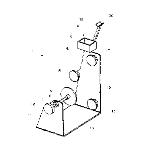
Figure 1 is a perspective view of a brace for use as a part of the preferred
embodiment of the present invention;
Figure 2 is a side view of the brace of figure 1;
Figure 3 is a plan view of the brace of figure 1;
1 0 Figure 4 is a schematic circuit layout of an electronic orientation
monitor for use
as a part of the preferred embodiment of the present invention;
Figure 5 is a circuit diagram of the electronic orientation monitor;
Figure 6 is a schematic side view of the electronic orientation monitor
showing
the physical layout of various electrical components of the monitor;
Figure 7 is a perspective view showing an embodiment of an electronic
orientation monitor attached to an implement for releasable attachment of a
prosthetic
component;
Figure 8 is a schematic side view depicting a measurement of a patient's angle
of pelvic tilt;
2 0 Figure 9 is a perspective view of an alternative embodiment of a
brace;
Figure 10 is a left-side view of the brace of figure 9;
Figure 11 is a plan view of the brace of figure 9;
Figure 12 is another perspective view of the brace of figure 9;
Figure 13 is a perspective view of another embodiment of a brace;
Figure 14 is a perspective view of the embodiment of the brace from figure 13,
along with an implement for releasable attachment of a prosthetic component;
Figure 15 is another perspective depiction of the brace and implement shown in
figure 14; and
Figure 16 is a perspective view of yet another embodiment of a brace with yet
3 0 another embodiment of an implement for releasable attachment of a
prosthetic
component.
CA 02737459 2011-03-16
WO 2010/031111
PCT/AU2009/001213
8.
DETAILED DESCRIPTION OF PREFERRED EMBODIMENTS OF THE
INVENTION
Referring to the drawings, the surgical orientation system of the present
invention is used for assisting a surgeon to orient a prosthetic component
relative to a
patient's anatomy during surgery. In overview, the system comprises the
following
components:
= an implement 1, as shown in figure 7, for releasable attachment of a
prosthetic component;
1 0 = an
electronic orientation monitor 2, as shown in figure 7 attached to the
implement 1 (a cross-section of the electronic orientation monitor 2 is
shown schematically in figure 6 and circuit diagrams are provided in
figures 4 and 5); and
= a brace 3 as shown in figures 1, 2 and 3.
The brace 3 is releasably attachable to the patient so as to define a
reference
point 4 relative to the patient's anatomy. This reference point 4 is external
of the patient
and is used to orient the electronic orientation monitor 2 into a reference
orientation.
Whilst in this orientation the electronic orientation monitor 2 acquires
reference
2 0 orientation information which is used to calibrate electronic
orientation monitor 2 to the
reference orientation. More particularly, the reference point 4 includes at
least one
surface 5 defining a reference plane. This surface 5 forms one of the internal
sides of an
open-topped, box-shaped docking station 6, which is dimensioned to snugly
receive the
electronic orientation monitor 2. This orients the electronic orientation
monitor 2 into the
reference orientation by abuttal of a surface of the outer casing 7 of the
electronic
orientation monitor 2 against the inner surface 5 and against the other inner
surfaces of
the docking station 6.
The preferred embodiment is particularly suited for assisting surgeons to
3 0 properly locate an acetabular cup into a reamed acetabulum during hip
surgery, such as
total or partial hip replacements or revisions. When used in this context, the
brace 3
CA 02737459 2011-03-16
WO 2010/031111
PCT/AU2009/001213
9.
clamps adjacent to the patient's pelvis. The patient is supported by the base
13 of the
brace 3, on his or her side, with the acetabulum that is being operated upon
to the top.
With reference to the perspective view of the brace 3 shown in figure 1, the
patient's head
would be positioned to the right hand side of the brace 3 and the patient's
feet would be
positioned to the left hand side.
The brace 3 includes a movable jaw 8 which is rotatably disposed at an end of
a
shaft 9 that is threadedly engaged with a flange 10 disposed on a side wall 11
at the rear
end of the brace 3. A manual drive wheel 12 is fixedly disposed at the
opposite end of
1 0 the shaft 9 to allow for screwing adjustment of the position of the
movable jaw 8 relative
to the side wall 11. The movable jaw 8 has a positioning pad 14 which is
clamped
adjacent to the patient's sacrum. Three positioning pads (16, 17 and 18) are
disposed in a
triangular arrangement on the opposite side wall 15 at the front end of the
brace 3.
Positioning pad 16 clamps adjacent to the patient's pubic crest. Positioning
pads 17 and
18 clamp adjacent to the patient's anterior superior iliac spine.
Hence, once properly clamped, the brace 3 assumes a known physical
relationship relative to the patient's acetabulum. It therefore follows that
docking station
6 that is part of the brace 3 also assumes a known physical relationship
relative to the
2 0 patient's acetabulum and this known physical relationship is used along
with the
electronic orientation monitor 2 in subsequent surgical steps as described
below to assist
in the accurate orientation of the prosthetic acetabular cup relative to the
reamed
acetabulum.
The calibration process is typically performed whilst the electronic
orientation
monitor 2 is attached to the implement 1. An arm 19 extends diagonally
upwardly from
the front sidewall 15 of the brace 3 and has a cradle 20 disposed at a distal
end. The
cradle 20 is shaped for support of the elongate member 21 which connects the
monitor 2
to the handle 27 of the implement 1 whilst the monitor 2 is docked in the
docking station
3 0 6. Whilst the monitor 2 is docked, the surgeon or an assistant presses
the calibration
button 22 and the electronic orientation monitor acquires the reference
orientation
information required for calibration of the monitor 2 to the reference
orientation. This
CA 02737459 2011-03-16
WO 2010/031111 PCT/AU2009/001213
10.
information is dependent upon the output of a number of sensors disposed
within the
electronic orientation monitor 2, which are capable of monitoring the physical
orientation
of the monitor 2 in three dimensions.
The sensors are solid state integrated circuits (micro-machined electro
mechanical systems) and include various inertial sensors such as a gyroscope
23
(ADXRS150), which provides an output signal that is dependent upon a rate of
rotation
about its vertical axis. To address noise and stability issues, this signal
must be filtered
by a filter which is built around IC1 as shown in figure 5. This signal is
integrated over
-- time so as to provide a yaw angle in degrees of rotation. The technique
used for this
integration is the multiple timebased slices version of Thomas Simpson's Rule
for area
under a curve.
Another inertial sensor is a dual axis accelerometer 24 (ADXL213), which
-- functions as an inclinometer, or tilt sensor, to provide an output signal
that is dependent
upon the inclination of the accelerometer 24 relative to the local
gravitational field. This
provides roll and pitch signals which are stable and may be fed directly to
the
microprocessor 32 without any filtering.
2 0 In alternative embodiments other sensors may also be utilized, such as
a
magnetometer 25, for example, which provides an output signal that is
dependent upon
the direction of the local magnetic field. In other embodiments, a lesser
number and/or
range of types of sensors may be employed. The main issue in this regard is to
ensure
that the electronic orientation module 2 has a sufficient number and range of
types of
-- sensors to provide an acceptable level of spatial orientation accuracy over
an acceptable
time frame.
During calibration, the reference orientation information is stored within a
random access memory. After calibration, during manipulation of the implement
1
3 0 -- (which, in turn causes re-orientation of the monitor 2 that is attached
to the implement 1),
the monitor 2 continues to acquire subsequent orientation information. This
subsequent
orientation information is compared with the stored reference orientation
information and
CA 02737459 2011-03-16
WO 2010/031111 PCT/AU2009/001213
11.
the results of the comparison are used to determine an output for display upon
an array of
light emitting diodes (LEDs) 26 that is disposed on a face of the monitor 2.
The array of
LEDs 26 is driven by the monitor circuitry as shown in figures 4 and 5 to
provide an
indication to the surgeon when the subsequent orientation of the monitor 2 has
a
predefined relationship relative to the reference orientation. This indication
is provided
by lighting a green LED that is centrally disposed within the LED array 26.
In the preferred embodiment the predefined relationship is equality to within
a
predefined tolerance. However, in other embodiments other relationships, such
as the
current orientation forming a predefined angle relative to the reference plane
to within a
predefined tolerance, for example, may be utilized. The tolerance is selected
dependant
upon the desired sensitivity and the desired accuracy of orientation. A
typical tolerance
used in the preferred embodiment is to within 1, 2 or 3 degrees.
In another embodiment which dispenses with the need for a memory, the outputs
of the sensors are zeroed during calibration. These outputs then assume non-
zero values
during subsequent manipulation of the implement 1 and attached monitor 2.
During the
subsequent manipulation, the sensor outputs are monitored to check whether
their values
return to zero (to within the relevant tolerances). When the values return to
zero, it
2 0 means that the monitor has assumed the reference orientation and an
indication is
provided to the surgeon via the green LED.
The monitor 2 is powered by a rechargeable 9 volt battery 28 and includes an
on/off switch 29, one or more programmable microprocessors 32 (PIC16F877A,
sold by
Microchip) and one or more regulators 33. The battery 28 is recharged from an
external
power source via an inductive coupling arrangement. As best shown in figure 6,
the
architecture of the monitor consists of a number of electrically
interconnected levels, each
of which has one or more of the components required for the operation of the
monitor 2.
A non-limiting example of the BASIC code for programming of the microprocessor
32 is
set out below in Annexure A. This programme is compiled and locked into the
microcontroller 32 as protected code. The electronics for the monitor 2 are
housed within
a hermetically sealed outer casing 7.
CA 02737459 2011-03-16
WO 2010/031111 PCT/AU2009/001213
12.
The main electrical components shown in the circuit diagram of figure 5 are as
follows:
SENSORS
Ul: Dual axis Accelerometer - ADXL213EB
U2: Angular Rate Gyroscope - ADXRS150EB
INTEGRATED CIRCUITS
IC1: CMOS Op Amp - LM6062
IC2: Micro Controller - PIC16F877A
IC3: Voltage Regulator - LM7805T
IC4: Voltage Regulator - LM78L05CZ
RESISTORS
R1, R4: 100K, 0.25W, Metal Film
R2, R3: 47K, 0.25W, Metal Film
R5, R7: 22K, 0.25W, Metal Film
R6, R8: 1K5, 0.25W, Metal Film
R8-R22: 100K, 0.25W, Metal Film
VR1: Cermet Trimpot - 10K
CAPACITORS
Cl, C2: 10uF, 16V, Tant
C3, C7, C9: 22uF, 16V, Tant
C4: luF, 50V, Cer
C5: 4u7F, 16V, Tant
C6, C8: 100uF, 16V, Tant
LIGHT EMITTING DIODES
D1, D4, D5, D8, D9, D12: LEDs - 5mm, Super Bright, Red
D2, D3, D6, D7, D10, D11: LEDs - 5mm, Super Bright, Yellow
D21, D22: LEDs - 5mm, Super Bright, Green
MISC
Xi: Resonator - 4MHz
Pbn: Push Switch - SPST
Sw: Toggle Switch - SPST
Bat: Battery - 9V Type 216
As described in the preceding paragraphs, during manipulation of the implement
1, the monitor 2 provides an indication to the surgeon when a subsequent
orientation of
the monitor 2 is equal to the reference orientation to within the relevant
tolerance by
illuminating a green LED. The relative geometry of the brace 3 (including the
docking
CA 02737459 2011-03-16
WO 2010/031111 PCT/AU2009/001213
13.
station 6, and the angle at which the acetabular cup is connected to the
implement 1) is
selected such that the indication is provided to the surgeon when the
acetabular cup is in
an anatomically desirable orientation for insertion into the reamed
acetabulum. Hence,
when the cup is positioned adjacent the reamed acetabulum, and the indication
is given,
the surgeon impacts the cup into the reamed acetabulum.
Whilst the illustrated embodiment makes use of LEDs to provide a visual
indication to the surgeon, it will be appreciated that any other suitable
display means may
be utilized. For example, the LED array 26 may be replaced with a Liquid
Crystal
Display that is adapted to display an indication to assist the surgeon to
achieve the desired
orientation, such as an arrow, a pointer, a bubble, or the like.
In an alternative embodiment (not illustrated) the hardware used to provide a
visual indication to the surgeon is not disposed on the electronic orientation
monitor 2.
Rather, the electronic orientation monitor 2 communicates data to drive a
display on a
remote display means, such as a monitor, which is preferably disposed above
the patient
within a convenient line of sight for the surgeon. It will be appreciated by
those skilled in
the art that the type of communication must be non-invasive so as to avoid
interfering
with other electronic equipment that may be present in the operating theatre.
In one such
2 0 alternative embodiment, the communication is via a wireless protocol
for exchanging
data over a short distance personal area network. An example of such a
wireless protocol
is known to those skilled in the art as "Blue Tooth". The use of a remote
display such as
a monitor allows for more detailed visual indications to be provided to the
surgeon. It
also avoids the possibility that manipulation of the implement 1 may rotate
the array of
light emitting diodes (LEDs) 26 that would otherwise be disposed on the
electronic
orientation monitor 2 out of the line of sight of the surgeon. Another
advantage
associated with separating the display from the electronic orientation monitor
2 is that the
electronics necessary to drive the display does not have to be sterilized
between
operations. Such sterilization processes often involve the use of high
temperatures,
which may damage some of the electronic components in the display.
Yet another embodiment of the electronic orientation monitor incorporates a
CA 02737459 2011-03-16
WO 2010/031111 PCT/AU2009/001213
14.
speaker to provide an audible indication to the surgeon to assist in the
orientation process.
In this embodiment the audible indication consists of a beeping noise, with
the frequency
of the beeping increasing as the surgeon manipulates the implement towards to
the
desired orientation for insertion into the reamed acetabulum. Whilst the
desired
orientation is being maintained to within the relevant tolerances, the beeping
noise
changes to a constant tone to indicate to the surgeon that the acetabulum is
correctly
oriented for insertion into the patient.
The assistance provided to the surgeon by the system of the preferred
embodiment advantageously allows accurate orientation of the acetabular cup
despite the
minimal vision of the acetabulum afforded by minimally invasive surgical
techniques
such as "keyhole" surgery. In other words, the preferred embodiment
advantageously
allows for accurate orientation of the acetabular cup despite the limited
visibility afforded
to the surgeon through a relatively short incision.
In addition to providing the surgeon with an indication when the desired
orientation is reached by illuminating the green LED as described above, the
preferred
embodiment also provides an indication so as to guide manipulation of the
implement 1
towards the desired orientation. This minimizes the guess work and time
required to
2 0 manipulate the implement 1 into the desired orientation. This
indication is provided via
the LED display array 26, which extends in a couple of directions. Hence, if
the
implement 1 must be rotated in a specific rotational direction to achieve the
desired
orientation, then a LED indicative of that rotational direction is
illuminated. The distance
of the illuminated LED from the centre of the array is indicative of the
amount of rotation
required to achieve the desired orientation. Hence, as the implement 1 is
rotated
progressively closer to the desired orientation, LEDs that are progressively
closer to the
centre of the array 26 are illuminated. Additionally, the colour of the LED is
indicative
of the amount of rotation required to achieve the desired orientation, whereby
red LEDs
indicate that a large rotation is required and yellow LEDs indicate that a
lesser amount of
3 0 rotation is required. When the implement 1 is in the desired
orientation (at least, to
within the relevant tolerance), the central green LED is illuminated to
indicate to the
surgeon that the acetabular cup may be impacted into the acetabulum.
CA 02737459 2016-07-05
15.
For maximum possible accuracy, the monitor 2 should not be moved around
more than necessary once it has been calibrated. Additionally, the time
between
calibration and receiving the indication to impact the acetabular cup should
be
minimized. Both of these precautions help to avoid orientational inaccuracies
that may
otherwise creep into the functioning of the electronics in the monitor 2. If,
for some
reason, the monitor 2 is moved excessively after calibration, or if there is a
temporal
delay prior to its use, accuracy may be restored by simply re-calibrating the
monitor 2.
As an alternative to the monitor 2 described above, different electronic
orientation monitors may be used in other embodiments of the present
invention. For
example, the monitor that is disclosed in International Patent Application No.
PCT/US2004/018244, published on 29 December 2004 under publication number
W02004/112610, may be used.
In summary, use of the preferred embodiment of the present system as
illustrated
in figures 1 to 6 to assist a surgeon to orient a prosthetic component
relative to a patient's
anatomy involves the following steps (whether in the order shown, or
otherwise):
= Releasably attaching a prosthetic component and the electronic
orientation monitor 2 to the implement 1. This is achieved using the
connector 30 that is disposed at a distal end of the handle 27;
= Clamping the patient to the brace 3 so as to define an external reference
point 4 relative to the patient's anatomy;
= Using the reference point 4 to orient the electronic orientation monitor
2
into a reference orientation;
= Calibrating the electronic orientation monitor 2 by acquiring reference
orientation information whilst the monitor 2 is in the reference
orientation;
= Manipulating the implement 1 whilst the implement is physically
separate from the brace such that the prosthetic component is adjacent
the relevant anatomy;
= Using the electronic orientation monitor 2 to provide an indication so as
CA 02737459 2011-03-16
WO 2010/031111 PCT/AU2009/001213
16.
to guide manipulation of the implement 1 such that the implement 1 is
guided towards a predefined relationship relative to the reference
orientation; and
= Using the electronic orientation monitor 2 to provide an indication when
the subsequent orientation of the monitor 2 has a predefined relationship
relative to the reference orientation.
One potential difficulty that may be experienced with use of the brace 3 as
illustrated in figures 1 to 3 is positioning of the patient across the solid
base 13.
Typically the brace 3 is firstly placed onto a surface such as a bed such that
the base 13 is
supported by the bed. The patient, who may be anesthetized at this point, is
then lifted
onto the base 13. This can be difficult, particularly if the patient is
unconscious and
therefore unable to assist in the movement process. An alternative embodiment
(not
illustrated) of the brace at least partially addresses this problem by
splitting the base in
half to form two selectively interengagable base members. Mechanical
interengagement
means are provided on the ends of the base members. This allows the patient to
be firstly
placed onto the bed and secondly each base member may be slid under the
patient from
either side. When they meet, the mechanical interengagement means on the two
base
members interengage to form a rigid base. Once the surgery is concluded, the
two base
2 0 members are selectively disengaged from each other and slid out from
either side of the
patient.
An alternative embodiment of the brace, as illustrated in figures 9 to 11,
provides another option for addressing the difficulty in placement of a
patient onto the
brace of figures 1 to 3. In this embodiment of the brace 40, an elongate frame
41 extends
intermediate and interconnects the front end 42 and the rear end 43 of the
brace 40. In
one such embodiment the frame has a generally rectangular cross section, with
dimensions of approximately 25mm by 10mm. Due to its narrow dimensions, the
frame
41 may be positioned so as to extend between the patient's legs. Hence, using
this
3 0 embodiment of the brace 40, the patient is firstly placed on the bed
and secondly the
brace 40 is positioned between the patient's legs. At this point the pads 44
and 45 are
placed in contact with the patient's anterior superior iliac spine and the pad
46 is placed
CA 02737459 2011-03-16
WO 2010/031111 PCT/AU2009/001213
17.
in contact with the patient's pubic crest. Each of pads 44, 45 and 46 are
mounted on a
plate 60, which is connected to the front end 42 of the frame 41. The handle
47 is then
used to rotate externally threaded shaft 48 within internally threaded boss 49
so as to
displace pad 50 toward the patient so as to clampingly engage the patient's
sacrum. The
boss 49 is connected to the rear end 43 of the frame 41.
The embodiment of the brace 40 as illustrated in figures 9 to 11 also solves a
second problem that may be associated with the embodiment illustrated in
figures 1 to 3.
That is, with the embodiment illustrated in figures 1 to 3, the broad flat
base 13 defines
the orientation of the brace 3 as it rests flat on the bed. This may be
undesirable,
particularly if the patient moves but the geometry of the base 13 doesn't
allow the brace 3
to move with the patient. However, it is not necessary for any part of the
brace 40 as
illustrated in figures 9 to 11 to be supported by the bed. Rather, the
position and
orientation of the brace 40 whilst in use is substantially defined by the
patient's pelvic
region to which it is clamped.
Another potential issue that may be associated with the embodiment of the
brace
3 as illustrated in figures 1 to 3 relates to patients who have a natural tilt
to their pelvis,
either forwards or backwards. In practice, pelvic tilt angles may typically
range between
-10 to + 10'; although in some more extreme cases the angle may fall outside
of this
range. The brace 3 as illustrated in figures 1 to 3 has been designed for use
with patients
having a vertically aligned pelvis (i.e. a pelvis which, when in the neutral
position and
when viewed from the side, has the pubic crest in vertical alignment with the
anterior
superior iliac spine). Hence, if this brace 3 is used for a patient who has a
significant
pelvic tilt, then the resulting hip joint may suffer due to mis-alignment of
the prosthetic
femoral head with the prosthetic ascetabular cup. This issue is addressed by
the brace 40
as illustrated in figures 9 to 11. In this brace 40, the docking station 51 is
rotatably
disposed on the brace 40. As best shown on figure 9, the docking station 51 is
mounted
on an arm 52, which is rotatably connected to the rest of the brace 40 via pin
53. The pin
3 0 53 defines the axis of rotation of the arm 52 and of the docking
station 51. This axis of
rotation is parallel to a plane containing positioning pads 44, 45 and 46.
CA 02737459 2011-03-16
WO 2010/031111 PCT/AU2009/001213
18.
In use, the surgeon firstly ascertains the neutral pelvic tilt angle of the
patent's
pelvis by forming an x-ray image of the patient's pelvis as viewed from the
side. This x-
ray image is formed whilst the patient stands with their pelvis held at a
neutral and
comfortable tilt angle (i.e. the patient is asked not to deliberately tilt
their pelvis either
forwards or backwards). The pelvis in such an image is likely to appear
similar to the
pelvis 58 depicted in figure 8. The patient's pelvic tilt angle is ascertained
from the x-ray
image by measurement of the angle between a line 56 representing the vertical
and a line
57 extending from the patient's anterior superior iliac spine 54 to the
patient's pubic crest
55, as shown on figure 8.
Once patient's pelvic tilt angle has been ascertained, the reference point of
the
brace 40, which is in the form of docking station 51, is rotated by the
ascertained angle.
In some such embodiments an angular scale is depicted adjacent to the pin 53
for ease of
reference whilst rotating the docking station 51. Once the docking station has
been
rotated by the required angle, the brace 40 is used in the remaining surgical
steps in
manner described above with reference to the brace illustrated in figures 1 to
3.
However, rather than providing a fixed reference that is only suitable for a
vertically
aligned pelvic (as is the case for brace 3), the docking station 51 of brace
40 provides a
reference that has been tailored to the patient's individual requirements as
dictated by the
2 0 measurement of their pelvic tilt.
The brace 70 illustrated in figures 13 to 15 is generally similar to that
shown in
figures 9 to 12, with the exception of the docking station 71 and the manner
in which the
docking station 71 is attached to the remainder of the brace 70. In this
embodiment the
docking station 71 takes the form of a half-cylindrical cradle 72, which is
shaped to
receive a correspondingly cylindrically shaped member 73 of the implement 74.
As best
shown in figure 14, a lug 75 protrudes from the member 73 so as to mate with
an aperture
76 that is disposed in the cradle 72. These components are aligned such that
when the
member 73 is in the cradle 72 and the lug 75 is in the aperture 76, the
implement 74 is in
3 0 the reference orientation with respect to the patient to whom the brace
70 is clamped.
This allows for calibration of the electronic orientation monitor 82 to take
place in the
manner outlined above with regard to the above-described embodiments.
CA 02737459 2011-03-16
WO 2010/031111 PCT/AU2009/001213
19.
The channel 72 of the docking station 71 is disposed on a first shaft 77. A
locking means 78 connects the first shaft 77 to a second shaft 79. Loosening
of the knob
80 of the locking means 78 allows the first shaft 77 to rotate relative to the
second shaft
79. This rotation of the first shaft 77 causes rotation of the cradle 72
relative to the
remainder of the brace 70 about an axis of rotation that is parallel to the
central
longitudinal axis of the first and second shafts 77 and 79. Such rotation
allows for
adjustment to compensate for the patient's pelvic tilt, in the manner outlined
in more
detail above. When the cradle 72 is at the desired angle, the knob 80 is
tightened such
1 0 that the locking means 78 resists any further relative rotation between
the first and second
shafts 77 and 79 and the angular disposition of the cradle 72 is therefore
fixed relative to
the remainder of the brace.
The end of the second shaft 79 is releasably attachable to the remainder of
the
brace 70 via attachment mechanism 81. More particularly, a projection on the
end of the
second shaft 79 is keyed into a slot 84 on the attachment mechanism 81 so as
to resist
rotation of the second shaft 79 relative to the attachment mechanism 81. Once
the
electronic orientation monitor 82 has been calibrated, the implement 74 may be
removed
from the cradle 72 and the second shaft 79 may be detached from the attachment
mechanism 81. This removes the following components from the brace 70: the
second
shaft 79, the locking means 78, the first shaft 77 and the cradle 72. Removal
of these
components allows the surgeon additional room to move and thereby lessens the
risk of
the surgeon accidentally bumping or snagging any of the detached components
whilst
performing the remainder of the operation.
The implement 74 that is depicted in figures 14 and 15 is adapted for use in
some minimally invasive surgical techniques. The electronic orientation
monitor 82 of
this implement 74 is attached to the member 73 via a lockable universal joint
83, for
example a ball joint. Hence, when the lockable universal joint 83 is unlocked,
the
3 0 electronic orientation monitor 82 may be manipulated into a desired
orientation, for
example in order to ensure that the display array 26 will be visible to the
surgeon whilst
inserting the acetabular cup 84 into the patient's reamed acetabulum. It will
be
CA 02737459 2011-03-16
WO 2010/031111
PCT/AU2009/001213
20.
appreciated that such manipulation of the electronic orientation monitor 82
should only
take place prior to the calibration process. Once the electronic orientation
monitor 82 has
been calibrated, the lockable universal joint 83 should remain locked until
such time as
the prosthetic component 84 has been inserted into the patient. If the
lockable universal
joint 83 is accidentally unlocked after calibration, the lockable universal
joint 83 should
be re-locked and the calibration process commenced afresh.
In another embodiment (not illustrated) the display array 26 is tiltably
mounted
on the electronic orientation monitor 2. This allows for adjustment of the
angle from
which the display array 26 may be viewed without requiring re-orientation of
the
remainder of the electronic orientation monitor 2.
Turning now to the final embodiment depicted in figure 16, the brace is
identical
to that shown in figures 13 to 15, however the implement 90 differs. In this
embodiment
the implement 90 is adapted for use in traditional open hip replacement
surgical
procedures. The electronic orientation monitor 91 is attached to a first
member 92 via a
lockable universal joint 93 that performs the same function as the lockable
universal joint
83 which was described in relation to the preceding embodiment.
2 0 The
first member 92 is attached to a second member 94 via a lockable angular
adjustment mechanism 95 having a locking releasement knob 98. The prosthetic
component, in the form of acetabular cup 96, is releasably attachable to a
distal end of the
second member 94. An impaction surface 97 is disposed on the proximal end of
the
second member 94.
Loosening of the locking releasement knob 98 unlocks the angular adjustment
mechanism 95. This allows for rotation of the second member 94 about an axis
of
rotation that is parallel to the elongate axis of the first member 92.
Rotation about this
axis allows for adjustment of the abduction angle at which the acetabular cup
96 is to be
3 0 inserted into the patient. Unlocking of the angular adjustment
mechanism 95 also allows
for rotation of the second member 94 about an axis of rotation that is
parallel to the
central axis of the locking releasement knob 98. Rotation about this axis
allows for
CA 02737459 2011-03-16
WO 2010/031111 PCT/AU2009/001213
21.
adjustment of the anteversion angle at which the acetabular cup 96 is to be
inserted into
the patient. Once these angles have been set to their desired settings, the
knob 98 is
tightened to ensure that the angular adjustment mechanism 95 maintains the
first and
second members 92 and 94 in the desired angular configuration.
Next the surgeon calibrates the electronic orientation monitor 91 to establish
the
reference orientation. The surgeon then moves the implement 90 and detaches
the cradle
99, the first and second shafts 101 and 102 and the locking means 103 from the
attachment mechanism 100 to provide extra room to move. The acetabular cup 96
is then
positioned adjacent the patient's reamed acetabulum and the orientation of the
implement
90 is manipulated in accordance with the indication provided by the display
array 26.
Once the subsequent orientation of the monitor 91 is equal to the reference
orientation to
within the relevant tolerance, the monitor 91 provides an indication to the
surgeon, who
then impacts a hammer against impaction surface 97 so as to impact the
acetabular cup 96
into the patient's reamed acetabulum.
Once the operation has been completed it is typically necessary to sterilise
the
equipment so as to prepare for the next operation. One method for performing
such
sterilisation is to place the equipment into an autoclave and heat it to a
suitable
2 0 temperature and pressure for a pre-determined time period, for example
132 C at 30 psi
for 10 minutes. In order for the electronics contained within the electronic
orientation
monitor 2 to survive being heated to this temperature some embodiments utilise
outer
cases that are heat-proof or heat-resistant. This may also involve providing
thermal
insulation at or near to the outer casing. An alternative or additional
approach to
providing the heat resistance is to select the individual electronic
components that are
used within the electronic orientation monitor 2 from amongst components that
are
known to have a higher than average heat tolerance.
While a number of preferred embodiments have been described, it will be
3 0 appreciated by persons skilled in the art that numerous variations
and/or modifications
may be made to the invention without departing from the spirit or scope of the
invention
as broadly described. The present embodiments are, therefore, to be considered
in all
respects as illustrative and not restrictive.
CA 02737459 2011-03-16
WO 2010/031111
PCT/AU2009/001213
22.
Annexure A - BASIC Code for Programming of the Microprocessor
11:0$ PROGRAM VI .043'
l-itGS Proof of Concoe.t.Prottrlype Control Phograire
'NOTE 1: Uses boundary knits exciusion zone to reduoe the Z axis zero drtil
_problem'
'Port A pins0 & I are defauit ADC inputs for X & Y -axis 'analog input
signals'
'Port A pip, 2 is default ADC input for 2 axis'
'Port A On 3 is default ADC input foi- stability reference signer
'Port 6 is c..featrit Output mttOr the X & Yaxis LEDs
Port C WO direction is -all oiris are outputaw'
'Port COn 3 is 'for the Green LED`
'Port C .oins 4,5,6,7 are tor the Z axis LEDs
?on D Pin0 is default input port for the F.4ert/Stop pushbutton'
The airtec limits for X axis red LED Weft' w0 (4.1-) 10 ADC oourtts'
'The inner Units for X axis yellow LED cintoff w0(+1--). 4 ADC itS
'The outer limft for Y exis red LED onkiff wl (+/-) 10 ADC courite'
'The inner limits kir Y axis yellow LED ontoff=wi (+/-) 4 ADC counts'
The outer Units for Z axis red LED oriloff = W2 (4-1--) 2000 ADC .C4UMS walin
eadl for/next loop'
The inner &TAB for Z ads yellow LED obleffr- w2 (+/435 ADC oourits within each
for/next loop"
2.6***-klf,fek.51,1-Srd+A.31-*Atiik*A-Ree+d*********Initialisa after R6,8e4+83-
*A-A44r*A-A-1-6-11kat=*.k4711.4.**3µ444k*X-4....,..24-fr**3.***tiabat-***Ok
irk let dit'so %11111111 'set Port C ditedori to output oil all pins'
piMSC; %Oat:N.)00OG 'set all Port C pins to lo (ereen LED &it sail'
test off)'
let pins %00000000 'set all Port B pUs to le LE D.s of)
frse÷tot-ItS*Plin..*****.AF. rdi of ioalaati.40
*icsl=bat=grii-S.4.PAAvbictil=irkk414.6*katviVerffy and Warm up before
run"tets
wk.: let Orisr.m%11111.111 "show :all X & Y axis LEDs'
let oiriso %11111000 'show Z xs and Green LEDV
pause 5000 'pause 5000miSecs'
let OM %00000000 'twitch .DFF all X & axis LEDs'
let ninso %00000000 'switch .OFF (amen LED and Z LEDs"'
do 30 so.ca exercise to 'Oam up the gyro arid tilt systerns"*"'
'' then acquire =docking station data
wup: tor ble = 1 to 100 `do 100 useless loops to warm eW
resdadc10 ), w0 'acquire .X axis signal'
poke $D0,1o0 'put Xaxis LS B -into odd storage'
poke $C1,b1 'pat Xaxis MS13 into cold storwe'
MadednI 0. 1, w0 'acquire Y axis. signal'
poke $C2,b0 'put Ys LSE nto_ cold storage'
poke $C3,b1 Vat Yaks MB o coid storage'
readado1: 02, w0 'acquire Z axis. signal'
poke $C4,b0 'pat Zaxis LM- into cold storage'
poke _$C5,1A 'pat Za>it- MSB into cold storage'
let Onso %11110000 'show sii Z ,9xis LEDS'
pauee 1-25uefor .125mSeixe
pimp14000.00000 'SWitCli OFF Z as LEDs'
pause 125 'patIS-re to$':125m.Secs'
next b10 'continue until all done'
w3 0 ttearthi:, Z axis ts-rar ac.stirtitsigtor
for b1 0 vz, I to 64 "abollinulatv 64 ZEDJS acquisitions'
read ant 10 2, w6 'get Z axis sign& and store it in w6'
let w3 'w.344v6 'add to total in w3`
next bl 0 'continue until ell done'
let we w-3/54 'get 2 axis docking statiori average'
'calculate Zaxis No Barai Top Limit'
CA 02737459 2011-03-16
WO 2010/031111
PCT/AU2009/001213
23.
let w2 =w6-1 'calculate Zaxis.Noise Band Base Limit'
let Z aids dock for drift cl:Ntripensaton"
poke $t.'t,t, b12 'put ZaXtL into cold storage'
poke ..W5, b13 'put :axis MS 6 into cold storage'
poke $C6, b2 'put Zaxis LSE NETL into cold sierage''
poke .$C.:7, b3 put Zaxis MS E NETL into cold tec rage'
poke SC-8, b4 'put Zaxt LSE NEEL into cold storage'
poke $C-9.. b5 Zelee ME NEEL into cold storage'
et w332708 t the initial :Zerr value to mittooint(32768)'
poke TiCA,bf.i 'put Zeit LSE ;into cold storage'
poke SCE.,b7 'put Zed MSE into cold :storage'
high pork: 3 'ready-to go, so switch ON green LED'
serixd ("Dot:Icing Siatkirs Value13,10)
goiitib send
gesub phtn Welt for pushbutton before commencing test
let pins.411111111 'switch ON tel X & Vaitt LEDs'
pause 500 'pause tot 500rri8eca
let pirtsiz%.00000fX10 %witch OFF alt V & V axis LEN'
plosc=%00001000 'Switch ON Green LED and fAktitCh OFF Z axi$ LEDs'
of
assuines at Variables are evadable: for each to routine
Nnevci:zd.ocki$Iv wl NETL
w2NiEL-tor accumulator; w4==b8, i.19; w5io;10, 611; w6.--ouirent value'
"-**b8,µ4Z axis inner loop counter; b9,-- unctied loop cotinte. bi 0z4resetvee
for Xaxis tiag; bl 1..,-resemed for Vats OK tag'
Zexs dock value is readent u w0 during Zakis sobroutine""
Zakis NEM velUe residesh wl airing Zs atibront'
" Zekla NBEL value resides in w2 ittoirio axis
end LEDs routines take apprOX 25 niSees ail-Aiding the sestxd instruction'
the main dale acquisition
main: gesab Zgo
Xgo: tow 0 VtAtitCh OFF the X. axis LEDs only
low I %witch OFF the X axis; LEDs puir
tow 2 %witch OFF the X aKis LEDs on
law 3 `swili:,:h OFF the X axis ;LEDs on
peek $00,b0 'fetch Xarids LSE docking Valile ftOtTI
cdid storage,'
peek $C1,b1 ''fx Itch 'Kakis MSB dociOng value from
cold storage'
readeddt 0 0, w6 'get new V axis data into w6'
let wiziw0+4 'calculate inner; Wis.'
let w2atw0-4 'calculate inner limits'
riwe,t1 than goto if small right tilt skip 0 K2'
if 'w2 then goto x3 'if small left tiit skip to a
blaa0 'idea r the green LED error flag'
goto hot*
K2: high 2 'load tight-yellow-LED-ON data'
let wl=w0+10 'gel the fight eil outer Imit
if vir6-zwi then golo Xxx 'if v,4tiin the limit adjust the green LED
error flag'
high aditsght-Oand-red-LED-ON'
goto Xxx
high I 'load lett=handliellow=LED-ON data`
tat w2 =wilt -10 'get the left tilt outer limit'
if toi5W2 en goto Xxx It within the limit adjust the green LED error
flag'
high 0 load let-nand-red-LED-ON data
Xrx: bl Orz I Set the peen LED error-flag'
CA 02737459 2011-03-16
WO 2010/031111
PCT/AU2009/001213
24.
bthk ipirg.) --==U then gerte done. "..d=bufton held down stop data
acquisftn'
gossb Zgo tail the next Z axis routine'
Yo: low 4 'switch OFF the Y a=Ai.s. LEDs oniy'.
tow S 'awitcil OFF the Y 81.* LElDs only'
tow "stch OFF the Y xs LEDs on
lOW 7 5swltch OFF the Y ac LEL,'s only'
peek $C2,30 'fetch Vaxis LB docking value from ad
storage'
$C3,1>1 'tech Yakis MSB dockiag value from mid
storage'
readad.c10 1, w6 'get new V axis: data: to we'
yt: iet 'calculate i?.trteriids
et w2=v4-4. 'catculate
then gob.) y2 'if back tilt skip to y2'
,r,G=oxw2 then gotoy3 'if trio/vent tilt skip to y3'
b1l=0 'dear the gieen LED error ftaft
goto Yend
y2: ilth S load top--=gilow-LED-ON. data'
let 'get the back tilt outer- Nrnit'
If wt3,4w1 then goto yyy11-. %Albin the iimit adjust the green LED error flag'
high 4 'lead toõo-herid-red-LED-ON`
Ook', YYY
y3: titd15* load .bettorriliellow-LED-ON data'
et w2 "get the forward tilt outer limit'
if wr>w2 then goti Yyy 7it within the lirait adjust the green LED
error .tiag'
fikiti 7 load bottom-red-LEO-ON data'
Yyy: .bl 1=1 'set the green LED error itag'
Yard: goto main 'keep going'
done: settd CIe0 complete ",13,10)
sk:nub send
iet pinsc %LI:0000000 "sWitili off Z akis LEDs'
at pins = %C*000000 'sxkltch off X and V aKis Ted LEDs'
end
..,1-45-11*frel-/i**dr"-T**Jrle-2-**A-iti-*A-A***r0ni El data actiositiori
routine.,
.44 4.44,4vienvit**inVI,kerkita+*keri.**A....+41/44-0t+ Li.40.+2 im(6 sub-
routine d+Aelkik Plealroilnt=Avilnleikist.1.11**.V.***+****40.4..
Zoo: peek $C4,b0 "fetch Zaxls =P=36 docking value from
coki storage'
peek SC5;b1 'fetch Zakis MSB docking value from cold
,-storage'
peek S.C6õO2 'fetch ?r..EiX5 LOB NBTL from .ocild
storage
peek $C7,b3 'fetch Zakis MOB NBTL from cold storage'
peek OCO,b4 'fetch Zak'.ki LSB NBBL from cold
storage"
peek iSt,µ.Ø,65 'fetch Zoo *r.. MBE) NBBL fro.m: cold
storage'
for biS= .1 to 25 'start Z. axis loop counter'
reader:it:10 2w.5 'acquire Z axis analog signet'
if we.i=w1 AND virs>=w2 Hien goto tool 11 within the, noundaty h. ms skip
error routine'
let w w3w 'else, add new value to the error register'
lot w3 re- w3--w2 'subtritict.the ducking wiluie to get the net
accumulated error'
rept : next OS 'continua data acquisition until 25 loops
complete'
let w3 = 'apply mreohon :Mille if nece&saryµ
let pinsu %00000.Mte 'switch oft ali Z axis LEDs'
ifW3..<3276 thee goto 1 lest data; if C-Ctifftti.:10d0445e error,
goto to red/yellow tests'
4/3.3298 then golo7 2 'if clockwise error riot redlyellow tests'
let w3 3278 'alignment OK, check the green LED
error.flaes'
CA 02737459 2011-03-16
WO 2010/031111 PCT/AU2009/001213
25.
if bl Otet OR bii1 then goto tzz 'if Xeit.m, or Yaxis not correct skip to
end of subroutine
high pt.:rt. 3 'switch ON green LED'
goto 21`z.7: 's,kip. to on of etibroutne`
high porto 3 orn& countercilovise 2 do o.ror, show yellow
LED'
w3'-3 7&8 then goto Zrz It not Igo coti oterolook\iviso error, skip
to and of ei_ibroutirte"
high ports 4 Urge cioontersikAthse2 ax,. error, snow red
LED'
goto 177 "skip-to on of subroutine'
72: high r.orto clockwise Z axis error, thow yellow LED'
if w:3-a3a761$ then got Zrz 'if not lawe clockwise on-or, skip to end of
subroutine'
high polio 7 large c.lockwise Z axis error, show rod
LED'
Zzr return 'en:ci of subroutine.'
t&-.5** kerb*Sr1.2.*=*6-6r12-*******64.**AZ axis sub.-routine corn
'Subroutine for push button nueretiori "*"'
plot: if pin 1 then goto pbtn Vail for button to be pushed'
higti, port 3 'switch ON -- on LEO'
pause 50 'pause 50rnSecs for switch dehourice.
obtl: piilD 0 then goto platt -- .well for button to be released'
kw portc 3 'switch OFF green a'D'
return
ubrone for sending data to tormina
send: peek .,'5C0 ,b0 'fetch Xaxis LSE doci,liv vatt.:te from cold
,s.steirage'
peek :t.CI ,b I fetch Xaxis icASE doohing value from cuid
storage
eorixdrRoll "*)
peek $C2,b0 `fetch Yeuris LSE tiOCkitv value from cold
storage'
peek $C3,b1 'feta MS E docking sraiite from cold
storage'
sertxdrPite.,:h "AW," ")
peek SC.4,b0 'fetch. Zee LB decking velue from cold
storage
peek $C5,b1 'fetch 2a-&. MS8 decking value f(tT&I cold
storage'
sertxdCRotetion = 'Azw0," ")
seilyriCTotai Error ."=", "Aq3,13.10)
return