Note: Descriptions are shown in the official language in which they were submitted.
CA 02738019 2011-03-22
WO 2010/039536 PCT/US2009/058041
HMV-132.25
HU 3277
SIRT4 AND USES THEREOF
RELATED APPLICATIONS
This application claims the benefit of priority to United States Provisional
Patent
Application serial number 61/192,892, filed September 23, 2008, the contents
of which is
hereby incorporated by reference.
BACKGROUND OF THE INVENTION
Sir2 (Silent information regulator 2) and its homologs, extend lifespan in
yeast, worms
and flies. Mammals contain seven homologs of sir2 (sirtuins, SIRT1-7) that
possess NAD+-
dependent deacetylase and/or ADP-ribosylation activity. SIRT 1, the closest
mammalian sir2
ortholog, is the most studied sirtuin and has been shown to deacetylate more
than a dozen
substrates to promote metabolic adaptation and cell survival. For example, in
pancreatic beta
cells, SIRT1 represses the expression of mitochondrial uncoupling protein and
increases insulin
secretion. In the liver, SIRT1 activity is up-regulated during fasting,
leading to modulation of
gluconeogenesis through deacetylation of FOXO 1, CRTC2 and PGC-la.
Three of the mammalian sirtuins (SIRT3, SIRT4 and SIRT5) are endogenously
located
in the mitochondria and may play roles as sensors of energy status in this
organelle. SIRT3
deacetylates acetyl-CoA synthetase 2 (AceCS2), glutamate dehydrogenase (GDH)
and
complex I of the electron transport chain in vitro, but SIRT3 knockout mice do
not have an
obvious phenotype under basal conditions. SIRT5 possesses weak deacetylase
activity, and its
in vivo targets remain unidentified. In pancreatic beta cells, SIRT4 regulates
the conversion of
glutamate and glutamine to a-ketoglutarate by ADP-ribosylating and inhibiting
GDH, thereby
repressing insulin secretion from pancreatic beta cells. Nevertheless, SIRT4
is broadly
expressed, and its roles in tissues other than the pancreas have not been
described.
SUMMARY OF THE INVENTION
Mitochondrial function is implicated in a wide variety of disorders,
including, for
example, physiological and pathophysiological stress, obesity, cardiovascular
disease, aging
and age-related disease. It has now been discovered that the mitochondrial
protein SIRT4 is a
key regulator of fatty acid oxidation and plays an important role in the
context of disease, aging
and associated pathologies. Suppression of SIRT4 activity prevents diet-
induced weight gain
by reducing adipose tissue and allows for the maintenance of a lean phenotype
even under
-1-
CA 02738019 2011-03-22
WO 2010/039536 PCT/US2009/058041
HMV-132.25
HU 3277
conditions of a high fat diet. Described herein are methods and compositions
for the regulation
of lipid metabolism, including fatty acid oxidation, the control of weight
gain and the treatment
of metabolic syndromes.
In a one aspect, the invention provides a method of evaluating SIRT4 fatty
acid
oxidation repression activity, the method comprising providing a cell-free
composition
comprising a SIRT4 protein, an enzyme that catalyzes fatty acid oxidation, and
a substrate, and
evaluating fatty acid oxidation activity in the composition. In some
embodiments the substrate
comprises a fatty acid. Optionally, the method also provides the step of
adding a test compound
to the cell-free composition. In some embodiments, the test compound is a
small molecule, an
antibody, or a nucleic acid.
In another aspect, the invention provides a method for measuring an inhibitory
property
of a test compound towards a SIRT4 protein, comprising contacting the SIRT4
protein with the
test compound in the presence of an enzyme that catalyzes fatty acid
oxidation, and a substrate,
measuring the test rate of fatty acid oxidation in the presence of the test
compound, and
comparing the test rate of fatty acid oxidation with a control rate of fatty
acid oxidation
obtained in the absence of the test compound, where an increase in the test
rate relative to the
control rate is indicative of an inhibitory property of the test compound. In
some embodiments,
the test compound is a small molecule, an antibody, or a nucleic acid.
In a further aspect, the invention provides a method for measuring a
stimulatory
property of a test compound towards a SIRT4 protein, including the steps of
contacting the
SIRT4 protein with the test compound in the presence of an enzyme that
catalyzes fatty acid
oxidation, and a substrate, measuring the test rate of fatty acid oxidation in
the presence of the
test compound, and comparing the test rate of fatty acid oxidation with a
control rate of fatty
acid oxidation obtained in the absence of the test compound, where a decrease
in the test rate
relative to the control rate is indicative of a stimulatory property of the
test compound. In some
embodiments, the test compound is a small molecule, an antibody, or a nucleic
acid.
In yet a further aspect, the invention provides a method of treating or
preventing a fatty
acid oxidation disorder (FOD) in a mammalian subject, comprising administering
to the subject
an effective amount of an agent that reduces SIRT4 protein activity. Exemplary
FODs include
obesity, Medium Chain Acyl-CoA Dehydrogenase (MCAD) Deficiency, Short Chain
Acyl-
CoA Dehydrogenase (SCAD) Deficiency, long-chain Acyl-CoA dehydrogenase (LCAD)
-2-
CA 02738019 2011-03-22
WO 2010/039536 PCT/US2009/058041
HMV-132.25
HU 3277
deficiency, Carnitine Palmityltransferase Translocase I & II Deficiency,
Carnitine
acylcamitine translocase deficiency, Very Long Chain Acyl-CoA Dehydrogenase
(VLCAD)
Deficiency, Glutaricaciduria II, EFT Deficiency HMG Carnitine Transport Defect
(Primary
Camitine Deficiency), Long Chain 3-Hydroxyacyl-CoA Dehydrogenase (LCHAD)
Deficiency,
Trifunctional Protein (TFP) Deficiency, 2,4 Dienoyl-CoA Reductase Deficiency,
3-Hydroxy
Acyl CoA Dehydrogenase Deficiency (HADH), Electron Transfer Flavoprotein (ETF)
Dehydrogenase Deficiency, and 3-Hydroxy-3 Methylglutaryl-CoA (HMG) Lyase
Deficiency.
In certain embodiments, the levels of SIRT4 are modulated in a hepatocyte. In
some
embodiments, the agent is an antagonistic nucleic acid that reduces SIRT4
expression. In other
embodiments, the agent comprises a nucleic acid that targets SIRT4 mRNA or an
antibody that
targets SIRT4 protein.
In another aspect, the invention provides a method of evaluating the effect of
a test
compound on SIRT4, the method comprising providing a reaction mixture
comprising SIRT4
and a test compound, and evaluating a fatty acid oxidation activity of SIRT4.
In some
embodiments the test compound is a small molecule. In other embodiments, the
method is
repeated for each of a plurality of test compounds from a chemical library. In
further
embodiments, the reaction mixture is provided in a eukaryotic cell, such as a
hepatocyte. In
still further embodiments, the reaction mixture is provided in a mammalian
subject.
In a further aspect, the invention provides a method of inducing weight gain
or fatty
acid deposition in a mammalian subject, comprising administering to the
subject an effective
amount of an agent that increases SIRT4 protein activity. For example, the
subject is
malnourished.
In yet a further aspect, the invention provides a method of increasing an
activity of a
peroxisome proliferator-activated receptor-alpha (PPAR-a) in a mammalian cell,
comprising
contacting the mammalian cell with a compound that reduces SIRT4 activity.
In another aspect, the invention provides a method of increasing a mammalian
subject's
energy consumption, comprising administering to the subject a SIRT4 inhibitor.
For example,
the subject is overweight, is suffering from or at risk of developing a
mitochondrial-related
disease, or has a metabolic disorder resulting in reduced fatty acid oxidation
and/or increased
fatty acid deposition in the subject's tissue. Mitochondrial-related diseases
include aging,
MELAS syndrome, muscular dystrophy, diabetes, Leber's hereditary optic
neuropathy, Leigh
-3-
CA 02738019 2011-03-22
WO 2010/039536 PCT/US2009/058041
HMV-132.25
HU 3277
syndrome, NARP syndrome, and Myoneurogenic gastrointestinal encephalopathy. In
some
embodiments, the SIRT4 inhibitor is provided in an effective dose such that
fat storage in a
tissue of the subject is reduced. In other embodiments, the SIRT4 inhibitor is
administered to a
liver tissue, a brown adipose tissue, a skeletal muscle tissue, or a
combination thereof.
In a further aspect, the invention provides a method of reducing a cholesterol
level in a
mammalian subject, comprising administering to the subject a SIRT4 inhibitor
in an effective
amount such that a cholesterol level is reduced. For example serum cholesterol
level may be
reduced. In some embodiments, the method also includes administering to the
subject an
effective amount of a peroxisome proliferator-activated receptor-alpha
agonist, such as
ciprofibrate, clofibrate, fenofibrate, bezafibrate, WY14,643, or a combination
thereof.
In another aspect, the invention provides a method of reducing a reactive
oxygen
species (ROS) in a tissue, comprising contacting the tissue with a SIRT4
activator. The ROS
is, for example, an oxygen ion, a free radical, or a peroxide-containing
compound. In some
aspects, the tissue comprises a hepatocyte.
In a further aspect, the invention provides a method of increasing SIRT1
activity in a
cell comprising contacting said cell with a SIRT4 inhibitor. In some
embodiments, said SIRT4
inhibitor is selected from a group consisting of a small molecule, an antibody
and an
antagonistic nucleic acid.
In yet a further aspect, the invention provides a composition comprising a
SIRT4
inhibitor and a peroxisome proliferator-activated receptor-alpha agonist. In
some
embodiments, the peroxisome proliferator-activated receptor-alpha agonist is
ciprofibrate,
clofibrate, fenofibrate, bezafibrate, WY 14,643, or a combination thereof.
BRIEF DESCRIPTION OF THE DRAWINGS
Figure 1 shows the results of quantitative RT-PCR assays depicting the
expression of
SIRT4 (Figure 1A), SIRT3 (Figure 1B), SIRT5 (Figure 1C), Gk (Figure 1D), Cptla
(Figure
1E) and Acot3 (Figure 1F) in hepatocytes taken from WT mice that had been
fasted for the
indicated period of time.
Figure 2 shows the results of microarray analysis of gene expression in whole
liver
of SIRT4 KO mice compared to SIRT4 WT mice. Figure 2A lists the gene ontology
terms
over-represented in the gene expression profile of SIRT4 KO mouse livers.
Figure 2B depicts
the classification of pathways and metabolic processes of all annotated,
differentially expressed
-4-
CA 02738019 2011-03-22
WO 2010/039536 PCT/US2009/058041
HMV-132.25
HU 3277
genes with a p-value of < 0.01. Figure 2C depicts the relative expression of
genes with a p-
value of <0.1 associated with lipid metabolic processes.
Figure 3 shows the primers used in quantitative RT-PCR assays to detect
expression of
Acot3, Asns, Egfr, Lipg, B2m and Rspl6.
Figure 4 shows the results of quantitative RT-PCR assays to detect the
expression of
cptl a, lipg, acot3, asns, egfr, SIRT4 and esr in whole liver taken from fed
or fasted SIRT4 WT
or SIRT4 KO mice.
Figure 5 shows the similarity between the SIRT4 KO liver transcriptome and
published
liver transcriptomes from Gene Expression Omnibus (GEO) and ArrayExpress. WY
PPARa
WT: WT mice treated for 5 days with WY14643 (GSE8295, (Rakhshandehroo et at.,
(2007)
PPAR Research 2007, 26839)), WY PPARa KO: PPARa KO mice treated for 5 days
with
WY14643 (GSE8295 (Rakhshandehroo et at., (2007) PPAR Research 2007, 26839)),
PPARa
KO: WT vs. PPARa KO mice not treated with WY14643 (GSE8295, (Rakhshandehroo et
at.,
(2007) PPAR Research 2007, 26839)), CR: Long term caloric restriction mice vs
control diet
(GSE2431, (Dhahbi et al., (2005) Physiol Genomics 23, 343-350)), PGC-1(3 mut:
PGC-1(3
mutant mice vs. WT mice (GSE6210, (Vianna et at., (2006) Cell Metab 4, 453-
464)), aging 1:
22 mo vs. 4 mo WT Snell dwarf mice (GSE3129, (Boylston et at., (2004) Aging
Cell 3, 283-
296)), aging2: 22 mo vs. 4 mo WT Ames dwarf mice (GSE3150, (Boylston et at.,
(2006) AGE
28, 125-144)), aging3: 130 wks vs 13 wks WT mice (E-MEXP-1504, (Schumacher et
at.,
(2008) PLoS Genet 4, e1000161)). Significance was calculated using
permutation. * p<0.0001.
Figure 6 shows the results of quantitative RT-PCR assays that detect
expression of
PPARa and PPARa target genes in SIRT4 KO and SIRT4 WT livers.
Figure 7A shows immunoblots depicting SIRT4 expression in primary mouse
embryonic fibroblasts (MEFs) from SIRT4 KO and SIRT4 WT mice infected with
control (-)
or SIRT4 expression virus (+). Figure 7B shows the expression of pdk4 in
either SIRT4 KO
or SIRT4 WT MEFs infected with control (-) or SIRT4 expression virus (+) and
either treated
or untreated with 50 M WY14643. Figure 7C shows the expression of pdk4 in
either SIRT4
KO (-/-) or SIRT4 WT (+/+) MEFs and either treated or untreated with 50 M
WY14643.
Figure 8A shows immunoblots depicting expression of SIRT4-Flag (T4), H161A-
SIRT4-Flag (Mut), HA-PPARa and actin in transiently transfected human
embryonic kidney
293T (HEK293T) cells co-transfected with a luciferase reporter driven by three
tandem repeats
-5-
CA 02738019 2011-03-22
WO 2010/039536 PCT/US2009/058041
HMV-132.25
HU 3277
of a consensus PPAR response element (3xPPRE), together with constructs
expressing
PPARa, RXRa. Figure 8B shows luciferase expression in human embryonic kidney
293T
(HEK293T) cells from figure 8A transfected with pCMV control (pCMV), SIRT4-
Flag
(SIRT4) or H161A-SIRT4-Flag (SIRT4 Mut). Figure 8C shows luciferase expression
in H2.35
hepatoma cells transfected with pCMV control (pCMV), SIRT4-Flag (SIRT4) or
H161A-
SIRT4-Flag (SIRT4 Mut).
Figures 9A and 9B show the oxidation of [3H]palmitate (nmol [3H]palmitate / h
/ mg
protein) as analyzed using SIRT4 (-/-) and SIRT4 (+/+) MEFs (Figure 9A) or
SIRT4 (-/-) and
SIRT4 (+/+) primary hepatocytes (Figure 9B). Figure 9C shows the consumption
of
palmitate from culture medium in SIRT4 (-/-) and (+/+) primary hepatocytes.
Figure 10A shows the triglyceride (TG) levels in livers ( g/mg tissue) of
SIRT4 KO
and WT mice after overnight fast (n=6 per genotype). Figure lOB shows the
fatty acid
composition of triglycerides in livers of SIRT4 KO and WT mice after overnight
fast. Data
represent mean SEM (n=6 per genotype). Figure 10C shows the non-esterified
fatty acids
levels (NEFA, M) in plasma of male SIRT4 KO and WT mice on a normal chow
diet, before
(0h) and after fasting (16h and 24h).
Figure 11 shows the overnight weight loss experienced by SIRT4 WT and SIRT4 KO
mice during an overnight fast.
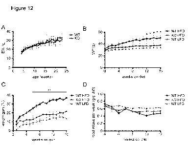
Figure 12A shows growth curves of SIRT4 KO and WT mice on a low fat diet up to
6
months of age (n=10-12 per genotype, data represent mean SEM). Figure 12B
shows the
body weight of SIRT4 KO and WT mice on a high fat diet (HFD, 60% fat, Research
diets) and
WT mice on a low fat diet (LFD, 10% fat, Research diets). Figure 12C shows the
relative
weight gain of SIRT4 KO and WT mice on HFD and WT mice on a LFD. Figure 12D
shows
the weekly food intake (g/g BW) in SIRT4 KO and WT mice on HFD and WT mice on
a LFD.
Figure 13A shows the starting body weights of SIRT4 KO and WT mice on a HFD
and
WT mice on a LFD. Figure 13B shows the starting age of SIRT4 KO and WT mice on
a HFD
and WT mice on a LFD.
Figure 14 shows the daily food intake (g/g body weight) of SIRT4 KO and WT
mice
on a HFD and WT mice on a LFD.
-6-
CA 02738019 2011-03-22
WO 2010/039536 PCT/US2009/058041
HMV-132.25
HU 3277
Figure 15A shows the total fecal output (48h) of SIRT4 KO and WT mice on a
HFD.
Figure 15B shows the total fecal output per bodyweight (48h) of SIRT4 KO and
WT mice on a
HFD.
Figures 16A and 16B show the plasma triglycerides in fed or fasted SIRT4 KO
and
WT mice on a HFD or LFD. Figures 16C and 16D show the plasma NEFA in fed or
fasted
SIRT4 KO and WT mice on a HFD or LFD diet.
Figures 17A and 17B show the blood glucose levels in fed or fasted SIRT4 KO
and
WT mice on a HFD and WT mice on a LFD. Figure 17C shows the liver weights of
SIRT4
KO and WT mice after 16 weeks on HFD or SIRT4 WT mice on a LFD. Figure 17D
shows
the Epididymal white adipose tissue (WAT) weights of SIRT4 KO and WT mice
after 16
weeks on a HFD or SIRT4 WT mice on a LFD. Figures 17E and 17F show the insulin
levels
in plasma of fed or fasted SIRT4 KO and WT mice on a HFD and WT mice on a LFD.
Figure 17G shows the percent weight loss of SIRT4 KO mice on a HFD compared to
WT
mice on a HFD and WT mice on a LFD.
Figure 18A shows a glucose tolerance test (GTT) performed in SIRT4 KO and WT
mice on a HFD or WT mice on a LFD. (n=6 per group). Figure 18B shows the area
under
curve of GTTs from Figure 18A.
Figure 19 shows a Western blot analysis performed on livers of overnight-
fasted SIRT4
KO and SIRT4 WT mice using antibodies directed against phosho-acetyl-CoA
carboxylase (p-
ACC), acetyl-CoA carboxylase (ACC), phosho-AMP-activated kinase (p-AMPK), AMP-
activated kinase (AMPK), SIRT4 and actin.
Figure 20A shows the ATP and ADP levels (nmol/mg tissue) as measured in acid-
soluble fractions from livers of overnight-fasted SIRT4 WT and SIRT4 KO mice.
Figure 20B
shows the ATP/ADP ratio in SIRT4 WT and SIRT4 KO livers, calculated from the
results
presented in Figure 20A.
Figure 21A shows the NAD as measured from livers of SIRT4 KO and SIRT4 WT
mice (fasted, n=6-8). Each data point represents the NAD concentration (pmol
NAD/mg tissue)
of one animal. The line represents the mean NAD concentration. Figure 21B
shows the
NADH as measured from livers of SIRT4 KO and SIRT4 WT mice (fasted, n=6-8).
Each data
point represents the NADH concentration (pmol NADH/mg tissue) in one animal.
The line
represents the mean NADH concentration. Figure 21C shows the NAD/NADH ratio
from
-7-
CA 02738019 2011-03-22
WO 2010/039536 PCT/US2009/058041
HMV-132.25
HU 3277
SIRT4 KO and SIRT4 WT whole liver tissue lysates. Each data point represents
the
NAD/NADH ratio in one animal. The line represents the mean NAD/NADH ratio.
Figure 22 shows a western blot depicting the expression of SIRT1 and actin in
whole
liver lysates from fasted SIRT4 KO and WT mice.
Figure 23 shows the oxidation of [3H]palmitate (nmol [3H]palmitate / h / mg
protein) as
analyzed using SIRT4 WT and SIRT4 KO primary hepatocytes either untreated,
treated with
the SIRT1 inhibitor Ex 527, or treated with etomoxir (ETO).
DETAILED DESCRIPTION OF THE INVENTION
The embodiments and practices of the present invention, other embodiments, and
their
features and characteristics, will be apparent from the description, figures
and claims that
follow, with all of the claims hereby being incorporated by this reference
into this Summary.
Definitions
For convenience, certain terms employed in the specification, examples, and
appended
claims are collected here. Unless defined otherwise, all technical and
scientific terms used
herein have the same meaning as commonly understood by one of ordinary skill
in the art to
which this invention belongs.
The articles "a" and "an" are used herein to refer to one or to more than one
(i.e., to at
least one) of the grammatical object of the article. By way of example, "an
element" means
one element or more than one element.
The terms "test compound" and "agent" are used herein to denote a chemical
compound, a small molecule, a mixture of chemical compounds, a biological
macromolecule
(such as a nucleic acid, an antibody, a protein or portion thereof, e.g., a
peptide), or an extract
made from biological materials such as bacteria, plants, fungi, or animal
(particularly
mammalian) cells or tissues. Test compounds and agents may be identified as
having a
particular activity by screening assays described herein below. The activity
of such test
compounds and agents may render them suitable as a "therapeutic compound" or a
"therapeutic
agent" which is a biologically, physiologically, or pharmacologically active
substance (or
substances) that acts locally or systemically in a subject. A test compound
may be capable of
and useful for binding to, agonizing, antagonizing, or otherwise modulating
(regulating,
modifying, upregulating, downregulating) the activity of a protein or complex
of the invention.
-8-
CA 02738019 2011-03-22
WO 2010/039536 PCT/US2009/058041
HMV-132.25
HU 3277
The term "amino acid" is intended to embrace all molecules, whether natural or
synthetic, which include both an amino functionality and an acid functionality
and capable of
being included in a polymer of naturally-occurring amino acids. Exemplary
amino acids
include naturally-occurring amino acids; analogs, derivatives and congeners
thereof; amino
acid analogs having variant side chains; and all stereoisomers of any of any
of the foregoing.
The term "binding" or "interacting" refers to an association, which may be a
stable
association, between two molecules, e.g., between a polypeptide and a binding
partner or
agent, e.g., small molecule, due to, for example, electrostatic, hydrophobic,
ionic and/or
hydrogen-bond interactions under physiological conditions.
The terms "calorie restricted" and "calorie restriction" include any diet or
feeding
program to a mammal or other organism below ad libitum levels, such as 10%,
20%, 30%,
40%, 50% or more than 50% below ad libitum levels.
The term "chemical entity," as used herein, refers to chemical compounds,
complexes
of two or more chemical compounds, and fragments of such compounds or
complexes. In
certain instances, it is desirable to use chemical entities exhibiting a wide
range of structural
and functional diversity, such as compounds exhibiting different shapes (e.g.,
flat aromatic
rings(s), puckered aliphatic rings(s), straight and branched chain aliphatics
with single,
double, or triple bonds) and diverse functional groups (e.g., carboxylic
acids, esters, ethers,
amines, aldehydes, ketones, and various heterocyclic rings).
The term "complex" refers to an association between at least two moieties
(e.g.
chemical or biochemical) that have an affinity for one another. Examples of
complexes
include associations between antigen/antibodies, lectin/avidin, target
polynucleotide/probe
oligonucleotide, antibody/anti-antibody, receptor/ligand, enzyme/ligand,
polypeptide/
polypeptide, polypeptide/polynucleotide, polypeptide/co-factor,
polypeptide/substrate,
polypeptide/inhibitor, polypeptide/small molecule, and the like. "Member of a
complex"
refers to one moiety of the complex, such as a protein. "Protein complex" or
"polypeptide
complex" refers to a complex comprising at least two polypeptides or proteins.
The terms "comprise" and "comprising" are used in the inclusive, open sense,
meaning
that additional elements may be included.
When using the term "comprising" or "having" herein, it is understood that
this term
may also be replaced by the phrases "consisting essentially of' or "consisting
of," where
-9-
CA 02738019 2011-03-22
WO 2010/039536 PCT/US2009/058041
HMV-132.25
HU 3277
appropriate. For example, "a fragment comprising amino acids 1-100 of sequence
X" should
be read as providing support for "a fragment consisting essentially of amino
acids 1-100 of
sequence X" as well as for "a fragment consisting of amino acids 1-100 of
sequence X."
The term "control" includes any portion of an experimental system designed to
demonstrate that the factor being tested is responsible for the observed
effect, and is therefore
useful to isolate and quantify the effect of one variable on a system. A
control includes a
"reference sample" as described herein.
The term "druggable region", when used in reference to a polypeptide, nucleic
acid,
complex and the like, refers to a region of the molecule which is a target or
is a likely target
for binding a modulator. For a polypeptide, a druggable region generally
refers to a region
wherein several amino acids of a polypeptide would be capable of interacting
with a
modulator or other molecule. For a polypeptide or complex thereof, exemplary
druggable
regions include binding pockets and sites, enzymatic active sites, interfaces
between domains
of a polypeptide or complex, surface grooves or contours or surfaces of a
polypeptide or
complex which are capable of participating in interactions with another
molecule. In certain
instances, the interacting molecule is another polypeptide, which may be
naturally-occurring.
A druggable region may be on the surface of the molecule.
Druggable regions may be described and characterized in a number of ways. For
example, a druggable region may be characterized by some or all of the amino
acids that make
up the region, or the backbone atoms thereof, or the side chain atoms thereof
(optionally with
or without the Ca atoms). Alternatively, in certain instances, the volume of a
druggable
region corresponds to that of a carbon based molecule of at least about 200
amu and often up
to about 800 amu. In other instances, it will be appreciated that the volume
of such region
may correspond to a molecule of at least about 600 amu and often up to about
1600 amu or
more. Alternatively, a druggable region may be characterized by comparison to
other regions
on the same or other molecules. For example, the term "affinity region" refers
to a druggable
region on a molecule (such as a polypeptide of the invention) that is present
in several other
molecules, in so much as the structures of the same affinity regions are
sufficiently the same
so that they are expected to bind the same or related structural analogs. An
example of an
affinity region is an ATP-binding site of a protein kinase that is found in
several protein
kinases (whether or not of the same origin).
-10-
CA 02738019 2011-03-22
WO 2010/039536 PCT/US2009/058041
HMV-132.25
HU 3277
The term "selectivity region" refers to a druggable region of a molecule that
may not
be found on other molecules, in so much as the structures of different
selectivity regions are
sufficiently different so that they are not expected to bind the same or
related structural
analogs. An exemplary selectivity region is a catalytic domain of a protein
kinase that
exhibits specificity for one substrate. In certain instances, a single
modulator may bind to the
same affinity region across a number of proteins that have a substantially
similar biological
function, whereas the same modulator may bind to only one selectivity region
of one of those
proteins.
When used in reference to a druggable region, the "selectivity" or
"specificity' of a
molecule such as a modulator to a druggable region may be used to describe the
binding
between the molecule and a druggable region. For example, the selectivity of a
modulator
with respect to a druggable region may be expressed by comparison to another
modulator,
using the respective values of Kd (i.e., the dissociation constants for each
modulator-
druggable region complex) or, in cases where a biological effect is observed
below the Kd, the
ratio of the respective EC50's (i.e., the concentrations that produce 50% of
the maximum
response for the modulator interacting with each druggable region).
A "form that is naturally occurring" when referring to a compound means a
compound
that is in a form, e.g., a composition, in which it can be found naturally. A
compound is not in
a form that is naturally occurring if, e.g., the compound has been purified
and separated from
at least some of the other molecules that are found with the compound in
nature.
The term "isolated polypeptide" refers to a polypeptide, in certain
embodiments
prepared from recombinant DNA or RNA, or of synthetic origin, or some
combination
thereof, which (1) is not associated with proteins that it is normally found
with in nature, (2) is
isolated from the cell in which it normally occurs, (3) is isolated free of
other proteins from
the same cellular source, (4) is expressed by a cell from a different species,
or (5) does not
occur in nature.
The term "isolated nucleic acid" refers to a polynucleotide of genomic, cDNA,
or
synthetic origin or some combination there of, which (1) is not associated
with the cell in
which the "isolated nucleic acid" is found in nature, or (2) is operably
linked to a
polynucleotide to which it is not linked in nature.
-11-
CA 02738019 2011-03-22
WO 2010/039536 PCT/US2009/058041
HMV-132.25
HU 3277
The terms "label" or "labeled" refer to incorporation or attachment,
optionally
covalently or non-covalently, of a detectable marker into a molecule, such as
a polypeptide.
The term "percent identical" refers to sequence identity between two amino
acid
sequences or between two nucleotide sequences. Identity can each be determined
by
comparing a position in each sequence which may be aligned for purposes of
comparison.
When an equivalent position in the compared sequences is occupied by the same
base or
amino acid, then the molecules are identical at that position; when the
equivalent site occupied
by the same or a similar amino acid residue (e.g., similar in steric and/or
electronic nature),
then the molecules can be referred to as homologous (similar) at that
position. Expression as
a percentage of homology, similarity, or identity refers to a function of the
number of identical
or similar amino acids at positions shared by the compared sequences.
Expression as a
percentage of homology, similarity, or identity refers to a function of the
number of identical
or similar amino acids at positions shared by the compared sequences. Various
alignment
algorithms and/or programs may be used, including FASTA, BLAST, or ENTREZ.
FASTA
and BLAST are available as a part of the GCG sequence analysis package
(University of
Wisconsin, Madison, Wis.), and can be used with, e.g., default settings.
ENTREZ is available
through the National Center for Biotechnology Information, National Library of
Medicine,
National Institutes of Health, Bethesda, Md. In one embodiment, the percent
identity of two
sequences can be determined by the GCG program with a gap weight of 1, e.g.,
each amino
acid gap is weighted as if it were a single amino acid or nucleotide mismatch
between the two
sequences.
Other techniques for alignment are described in Methods in Enzymology, vol.
266:
Computer Methods for Macromolecular Sequence Analysis (1996), ed. Doolittle,
Academic
Press, Inc., a division of Harcourt Brace & Co., San Diego, California, USA.
Preferably, an
alignment program that permits gaps in the sequence is utilized to align the
sequences. The
Smith-Waterman is one type of algorithm that permits gaps in sequence
alignments. See
Meth. Mol. Biol. 70: 173-187 (1997). Also, the GAP program using the Needleman
and
Wunsch alignment method can be utilized to align sequences. An alternative
search strategy
uses MPSRCH software, which runs on a MASPAR computer. MPSRCH uses a Smith-
Waterman algorithm to score sequences on a massively parallel computer. This
approach
improves ability to pick up distantly related matches, and is especially
tolerant of small gaps
-12-
CA 02738019 2011-03-22
WO 2010/039536 PCT/US2009/058041
HMV-132.25
HU 3277
and nucleotide sequence errors. Nucleic acid-encoded amino acid sequences can
be used to
search both protein and DNA databases.
The term "mammal" is known in the art, and exemplary mammals include humans,
primates, bovines, porcines, canines, felines, and rodents (e.g., mice and
rats).
The term "modulation", when used in reference to a functional property or
biological
activity or process (e.g., enzyme activity or receptor binding), refers to the
capacity to either
up regulate (e.g., activate or stimulate), down regulate (e.g., inhibit or
suppress) or otherwise
change a quality of such property, activity or process. In certain instances,
such regulation
may be contingent on the occurrence of a specific event, such as activation of
a signal
transduction pathway, and/or may be manifest only in particular cell types.
A "modulator" may be a polypeptide, nucleic acid, macromolecule, complex,
molecule, small molecule, compound, species or the like (naturally-occurring
or non-
naturally-occurring), or an extract made from biological materials such as
bacteria, plants,
fungi, or animal cells or tissues, that may be capable of causing modulation.
Modulators may
be evaluated for potential activity as inhibitors or activators (directly or
indirectly) of a
functional property, biological activity or process, or combination of them,
(e.g., agonist,
partial antagonist, partial agonist, inverse agonist, antagonist, anti-
microbial agents, inhibitors
of microbial infection or proliferation, and the like) by inclusion in assays.
In such assays,
many modulators may be screened at one time. The activity of a modulator may
be known,
unknown or partially known.
The terms "polynucleotide", and "nucleic acid" are used interchangeably. They
refer
to a polymeric form of nucleotides of any length, either deoxyribonucleotides
or
ribonucleotides, or analogs thereof. Polynucleotides may have any three-
dimensional
structure, and may perform any function, known or unknown. The following are
non-limiting
examples of polynucleotides: coding or non-coding regions of a gene or gene
fragment, loci
(locus) defined from linkage analysis, exons, introns, messenger RNA (mRNA),
transfer
RNA, ribosomal RNA, ribozymes, cDNA, recombinant polynucleotides, branched
polynucleotides, plasmids, vectors, isolated DNA of any sequence, isolated RNA
of any
sequence, nucleic acid probes, and primers. A polynucleotide may comprise
modified
nucleotides, such as methylated nucleotides and nucleotide analogs. If
present, modifications
to the nucleotide structure may be imparted before or after assembly of the
polymer. The
-13-
CA 02738019 2011-03-22
WO 2010/039536 PCT/US2009/058041
HMV-132.25
HU 3277
sequence of nucleotides may be interrupted by non-nucleotide components. A
polynucleotide
may be further modified, such as by conjugation with a labeling component. The
term
"recombinant" polynucleotide means a polynucleotide of genomic, cDNA,
semisynthetic, or
synthetic origin which either does not occur in nature or is linked to another
polynucleotide in
a non-natural arrangement.
A "patient", "subject" or "host" refers to either a human or a non-human
animal.
The term "pharmaceutically acceptable carrier" is art-recognized and refers to
a
pharmaceutically-acceptable material, composition or vehicle, such as a liquid
or solid filler,
diluent, excipient, solvent or encapsulating material, involved in carrying or
transporting any
subject composition or component thereof from one organ, or portion of the
body, to another
organ, or portion of the body. Each carrier must be "acceptable" in the sense
of being
compatible with the subject composition and its components and not injurious
to the patient.
Some examples of materials which may serve as pharmaceutically acceptable
carriers include:
(1) sugars, such as lactose, glucose and sucrose; (2) starches, such as corn
starch and potato
starch; (3) cellulose, and its derivatives, such as sodium carboxymethyl
cellulose, ethyl
cellulose and cellulose acetate; (4) powdered tragacanth; (5) malt; (6)
gelatin; (7) talc; (8)
excipients, such as cocoa butter and suppository waxes; (9) oils, such as
peanut oil, cottonseed
oil, safflower oil, sesame oil, olive oil, corn oil and soybean oil; (10)
glycols, such as propylene
glycol; (11) polyols, such as glycerin, sorbitol, mannitol and polyethylene
glycol; (12) esters,
such as ethyl oleate and ethyl laurate; (13) agar; (14) buffering agents, such
as magnesium
hydroxide and aluminum hydroxide; (15) alginic acid; (16) pyrogen-free water;
(17) isotonic
saline; (18) Ringer's solution; (19) ethyl alcohol; (20) phosphate buffer
solutions; and (21)
other non-toxic compatible substances employed in pharmaceutical formulations.
The term "pharmaceutically-acceptable salts" is art-recognized and refers to
the
relatively non-toxic, inorganic and organic acid addition salts of compounds,
including, for
example, those contained in compositions described herein.
The terms "polypeptide fragment" or "fragment", when used in reference to a
reference
polypeptide, refers to a polypeptide in which amino acid residues are deleted
as compared to
the reference polypeptide itself, but where the remaining amino acid sequence
is usually
identical to the corresponding positions in the reference polypeptide. Such
deletions may occur
at the amino-terminus or carboxy-terminus of the reference polypeptide, or
alternatively both.
-14-
CA 02738019 2011-03-22
WO 2010/039536 PCT/US2009/058041
HMV-132.25
HU 3277
Fragments typically are at least 5, 6, 8 or 10 amino acids long, at least 14
amino acids long, at
least 20, 30, 40 or 50 amino acids long, at least 75 amino acids long, or at
least 100, 150, 200,
300, 500 or more amino acids long. A fragment can retain one or more of the
biological
activities of the reference polypeptide. In certain embodiments, a fragment
may comprise a
druggable region, and optionally additional amino acids on one or both sides
of the druggable
region, which additional amino acids may number from 5, 10, 15, 20, 30, 40,
50, or up to 100
or more residues. Further, fragments can include a sub-fragment of a specific
region, which
sub-fragment retains a function of the region from which it is derived. In
another embodiment,
a fragment may have immunogenic properties. Fragments may be devoid of about
1, 2, 5, 10,
20, 50, 100 or more amino acids at the N- or C-terminus of the wildtype
protein.
The term "small molecule" is art-recognized and refers to a composition which
has a
molecular weight of less than about 2000 amu, or less than about 1000 amu, and
even less than
about 500 amu. Small molecules may be, for example, nucleic acids, peptides,
polypeptides,
peptide nucleic acids, peptidomimetics, carbohydrates, lipids or other organic
(carbon
containing) or inorganic molecules. Many pharmaceutical companies have
extensive libraries
of chemical and/or biological mixtures, often fungal, bacterial, or algal
extracts, which can be
screened with any of the assays described herein. The term "small organic
molecule" refers to
a small molecule that is often identified as being an organic or medicinal
compound, and does
not include molecules that are exclusively nucleic acids, peptides or
polypeptides.
A "sub-cellular fraction" is any portion of a cell or extra-cellular matrix,
as produced by
any fractionation or other method known in the art.
The term "substantially homologous," when used in connection with amino acid
sequences, refers to sequences which are substantially identical to or similar
in sequence with
each other, giving rise to a homology of conformation and thus to retention,
to a useful degree,
of one or more biological (including immunological) activities. The term is
not intended to
imply a common evolution of the sequences.
"Substantially purified" refers to a protein that has been separated from
components
which naturally accompany it. Preferably the protein is at least about 80%,
more preferably at
least about 90%, and most preferably at least about 99% of the total material
(by volume, by
wet or dry weight, or by mole percent or mole fraction) in a sample. Purity
can be measured
-15-
CA 02738019 2011-03-22
WO 2010/039536 PCT/US2009/058041
HMV-132.25
HU 3277
by any appropriate method, e.g., in the case of polypeptides by column
chromatography, gel
electrophoresis or HPLC analysis.
A "target protein" is any protein, peptide, or homolog thereof that is capable
of being
acted upon by a protein having an enzymatic or other activity, such as the
activity of a SIRT4
protein.
A "target mRNA" is any messenger RNA transcript that is capable of being acted
upon
by an antagonistic nucleic acid that reduces expression or levels of the
protein encoded by the
mRNA.
SIRT4 proteins
As used herein, the term "SIRT4" or "SIRT4 protein" refers to proteins, e.g.,
eukaryotic
proteins, e.g., mammalian proteins, comprising a mitochondrial protein having
ADP-ribosyl
transfer case activity, as well as functional domains, fragments (e.g.,
functional fragments),
e.g., fragments of at least 8 amino acids, e.g., at least 8, 18, 28, 64, 128,
150, 180, 200, 220,
240, 260, or 280 amino acids, and variants thereof. Exemplary functional
fragments of SIRT4
can, for example, have ADP-ribosyltransferase activity and/or the ability to
interact with a
SIRT4 binding partner. Exemplary SIRT4 proteins include those designated
GenBank
NM012240 (human SIRT4; SEQ ID NO: 1) and XM_485674 (mouse SIRT4; SEQ ID NO:
2). Homologs of SIRT4 proteins will share 60%, 80%, 85%, 90%, 95%, 98%, 99%
sequence
identity to a known SIRT4 protein and feature an SIRT4 activity, e.g., ADP
ribosylation,
inhibition of fatty acid oxidation, and/or downregulation of glutamate
dehydrogenase.
Eukaryotic SIRT4 proteins may be localized, e.g., to mitochondria. Variants of
SIRT4 proteins
can be produced by standard means, including site-directed and random
mutagenesis.
Exemplary compositions
Compositions comprising an isolated polypeptide or protein described herein,
or a
homolog thereof or may comprise less than about 25%, 10%, or alternatively
about 5%, or
alternatively about I%, contaminating biological macromolecules or
polypeptides. In certain
embodiments, a composition contains a SIRT4 protein. Optionally, a composition
contains a
SIRT4 protein and a SIRT4 - interacting protein. In other embodiments, the
SIRT4 protein is a
variant, such as H161YSIRT4.
-16-
CA 02738019 2011-03-22
WO 2010/039536 PCT/US2009/058041
HMV-132.25
HU 3277
In certain embodiments, a protein described herein is further linked to a
heterologous
polypeptide, e.g., a polypeptide comprising a domain which increases its
solubility and/or
facilitates its purification, identification, detection, and/or structural
characterization.
Exemplary domains, include, for example, glutathione S-transferase (GST),
protein A, protein
G, calmodulin-binding peptide, thioredoxin, maltose binding protein, HA, myc,
poly arginine,
poly His, poly His-Asp or FLAG fusion proteins and tags. Additional exemplary
domains
include domains that alter protein localization in vivo, such as signal
peptides, type III secretion
system-targeting peptides, transcytosis domains, nuclear localization signals,
etc.
A protein described herein may be linked to at least 2, 3, 4, 5, or more
heterologous
polypeptides. Polypeptides may be linked to multiple copies of the same
heterologous
polypeptide or may be linked to two or more heterologous polypeptides. The
fusions may
occur at the N-terminus of the polypeptide, at the C-terminus of the
polypeptide, or at both the
N- and C-terminus of the polypeptide. It is also within the scope of the
invention to include
linker sequences between a protein described herein and the fusion domain in
order to facilitate
construction of the fusion protein or to optimize protein expression or
structural constraints of
the fusion protein. A polypeptide may also be constructed so as to contain
protease cleavage
sites between the fusion polypeptide and polypeptide of the invention in order
to remove the
tag after protein expression or thereafter. Examples of suitable endoproteases
include, for
example, Factor Xa and TEV proteases.
In another embodiment, a protein may be modified so that its rate of
traversing the
cellular membrane is increased. For example, the polypeptide may be fused to a
second
peptide which promotes "transcytosis," e.g., uptake of the peptide by cells.
The peptide may
be a portion of the HIV transactivator (TAT) protein, such as the fragment
corresponding to
residues 37-62 or 48-60 of TAT, portions which have been observed to be
rapidly taken up by
a cell in vitro (Green and Loewenstein, (1989) Cell 55:1179-1188).
Alternatively, the
internalizing peptide may be derived from the Drosophila antennapedia protein,
or homologs
thereof. The 60 amino acid long homeodomain of the homeo-protein antennapedia
has been
demonstrated to translocate through biological membranes and can facilitate
the translocation
of heterologous polypeptides to which it is coupled. Thus, the polypeptide may
be fused to a
peptide consisting of about amino acids 42-58 of Drosophila antennapedia or
shorter
fragments for transcytosis (Derossi et al. (1996) J Biol Chem 271:18188-18193;
Derossi et al.
-17-
CA 02738019 2011-03-22
WO 2010/039536 PCT/US2009/058041
HMV-132.25
HU 3277
(1994) J Biol Chem 269:10444-10450; and Perez et at. (1992) J Cell Sci 102:717-
722). The
transcytosis polypeptide may also be a non-naturally-occurring membrane-
translocating
sequence (MTS), such as the peptide sequences disclosed in U.S. Patent No.
6,248,558.
In another embodiment, a protein described herein is labeled with an isotopic
label to
facilitate its detection and or structural characterization using nuclear
magnetic resonance or
another applicable technique. Exemplary isotopic labels include radioisotopic
labels such as,
for example, potassium-40 (40K), carbon-14 (14C), tritium (3H), sulphur-35
(35S), phosphorus-
32 (32P), technetium-99m (99mTc), thallium-201 (201T1), gallium-67 (67Ga),
indium-111 (1 "In),
iodine-123 (1231), iodine-131 (131I), yttrium-90 (90Y), samarium-153 (153Sm),
rhenium-186
(186Re), rhenium-188 (188Re), dysprosium-165 (165Dy) and holmium-166 (166Ho).
The isotopic
label may also be an atom with non zero nuclear spin, including, for example,
hydrogen-1 (1H),
hydrogen-2 (2H), hydrogen-3 (3H), phosphorous-31 (31P), sodium-23 (23Na),
nitrogen-14 (14N),
nitrogen-15 (15N), carbon-13 (13C) and fluorine-19 (19F). In certain
embodiments, the
polypeptide is uniformly labeled with an isotopic label, for example, wherein
at least 50%,
70%, 80%, 90%, 95%, or 98% of the possible labels in the polypeptide are
labeled, e.g.,
wherein at least 50%, 70%, 80%, 90%, 95%, or 98% of the nitrogen atoms in the
polypeptide
are 15N, and/or wherein at least 50%, 70%, 80%, 90%, 95%, or 98% of the carbon
atoms in the
polypeptide are 13C, and/or wherein at least 50%, 70%, 80%, 90%, 95%, or 98%
of the
hydrogen atoms in the polypeptide are 2H. In other embodiments, the isotopic
label is located
in one or more specific locations within the polypeptide, for example, the
label may be
specifically incorporated into one or more of the leucine residues of the
polypeptide. The
invention also encompasses the embodiment wherein a single polypeptide
comprises two, three
or more different isotopic labels; for example, the polypeptide comprises both
15N and 13C
labeling.
In yet another embodiment, a protein described herein is labeled to facilitate
structural
characterization using x-ray crystallography or another applicable technique.
Exemplary labels
include heavy atom labels such as, for example, cobalt, selenium, krypton,
bromine, strontium,
molybdenum, ruthenium, rhodium, palladium, silver, cadmium, tin, iodine,
xenon, barium,
lanthanum, cerium, praseodymium, neodymium, samarium, europium, gadolinium,
terbium,
dysprosium, holmium, erbium, thulium, ytterbium, lutetium, tantalum, tungsten,
rhenium,
-18-
CA 02738019 2011-03-22
WO 2010/039536 PCT/US2009/058041
HMV-132.25
HU 3277
osmium, iridium, platinum, gold, mercury, thallium, lead, thorium and uranium.
In an
exemplary embodiment, the polypeptide is labeled with seleno-methionine.
A variety of methods are available for preparing a polypeptide with a label,
such as a
radioisotopic label or heavy atom label. For example, in one such method, an
expression
vector comprising a nucleic acid encoding a polypeptide is introduced into a
host cell, and the
host cell is cultured in a cell culture medium in the presence of a source of
the label, thereby
generating a labeled polypeptide. The extent to which a polypeptide may be
labeled may vary.
In still another embodiment, a protein described herein is labeled with a
fluorescent
label to facilitate its detection, purification, or structural
characterization. In an exemplary
embodiment, the polypeptide of the invention is fused to a heterologous
polypeptide sequence
which produces a detectable fluorescent signal, including, for example, green
fluorescent
protein (GFP), enhanced green fluorescent protein (EGFP), Renilla Reniformis
green
fluorescent protein, GFPmut2, GFPuv4, enhanced yellow fluorescent protein
(EYFP),
enhanced cyan fluorescent protein (ECFP), enhanced blue fluorescent protein
(EBFP), citrine
and red fluorescent protein from discosoma (dsRED).
In other embodiments, a protein described herein is immobilized onto a solid
surface,
including, microtiter plates, slides, beads, films, etc. A protein described
herein may be
immobilized onto a "chip" as part of an array. An array, having a plurality of
addresses, may
comprise one or more polypeptides in one or more of those addresses.
In other embodiments, proteins described herein are contained within vessels
useful for
the manipulation of the polypeptide sample. For example, the polypeptide of
the invention
may be contained within a microtiter plate to facilitate detection, screening
or purification of
the polypeptide. The polypeptide may also be contained within a syringe as a
container
suitable for administering the polypeptide to a subject in order to generate
antibodies or as part
of a vaccination regimen. The polypeptides may also be contained within an NMR
tube in
order to enable characterization by nuclear magnetic resonance techniques.
In still other embodiments, the invention relates to a crystallized
polypeptide of the
invention and crystallized polypeptides which have been mounted for
examination by x-ray
crystallography as described further below. In certain instances, a protein
described herein in
crystal form may be single crystals of various dimensions (e.g., micro-
crystals) or may be an
aggregate of crystalline material.
-19-
CA 02738019 2011-03-22
WO 2010/039536 PCT/US2009/058041
HMV-132.25
HU 3277
In certain embodiments, it may be advantageous to provide naturally-occurring
or
experimentally-derived homologs of the polypeptide of the invention. Such
homologs may
function in as a modulator to promote or inhibit a subset of the biological
activities of the
naturally-occurring form of the polypeptide. Thus, specific biological effects
may be elicited
by treatment with a homolog of limited function, and with fewer side effects
relative to
treatment with agonists or antagonists which are directed to all of the
biological activities of
the polypeptide of the invention. For instance, antagonistic homologs may be
generated which
interfere with the ability of the wild-type polypeptide of the invention to
associate with certain
proteins, but which do not substantially interfere with the formation of
complexes between the
native polypeptide and other cellular proteins.
Nucleic acids encoding any of the proteins or homologs described herein are
also
provided herein. A nucleic acid may further be linked to a promoter and/or
other regulatory
sequences, as further described herein. Exemplary nucleic acids are those that
are at least
about 80%, 85%, 90%, 95%, 98%, 99% or 100% identical to a nucleotide sequence
provided
herein or a fragment thereof, such as nucleic acid sequence encoding the
protein fragments
described herein. Nucleic acids may also hybridize specifically, e.g., under
stringent
hybridization conditions, to a nucleic acid described herein or a fragment
thereof.
Also provided herein are molecular complexes, e.g., protein complexes,
comprising a
SIRT4 protein or homolog thereof and a mitochondrial protein, and optionally
other cofactors
or molecules. Such compositions and complexes may be used, e.g., in screening
assays to
identify agents that modulate the interaction between a SIRT4 protein and a
mitochondrial
protein, and the interaction between an ADP ribosyl transferable and target
protein.
Proteins and complexes described herein may exist in solution. A solution may
be a
composition, e.g., pharmaceutical composition, such as comprising a
therapeutically acceptable
diluent.
Proteins or complexes described herein may also exist in crystal form. A
crystallized
complex may include a protein described herein and one or more of the
following: a histone or
homolog thereof, a co-factor (such as a salt, metal, nucleotide,
oligonucleotide or polypeptide),
a modulator, or a small molecule. In another aspect, the present invention
contemplates a
crystallized complex including a polypeptide of the invention and any other
molecule or atom
(such as a metal ion) that associates with the polypeptide in vivo.
-20-
CA 02738019 2011-03-22
WO 2010/039536 PCT/US2009/058041
HMV-132.25
HU 3277
Also provided herein are antibodies that bind specifically to a complex
between a
SIRT4 protein or homolog thereof and a mitochondrial protein or homolog
thereof, but
essentially do not bind specifically to the SIRT4 protein or homolog alone nor
to the
mitochondrial protein or homolog alone. Also provided are antibodies that bind
specifically to
proteins or other biological molecules that are acted on by SIRT4.
Antibodies may be full length antibodies, fragments of antibodies (e.g., Fab
or F(ab')2),
monoclonal antibodies, polyclonal antibodies, single chain antibodies,
chimeric antibodies,
humanized antibodies, human antibodies, mini antibodies or any other form of a
molecule or
complex of molecules that binds specifically to a molecular complex described
herein.
Screening methods
Provided herein are screening methods for evaluating SIRT4 activity and for
identifying test compounds or agents that modulate a SIRT4 activity, such as a
fatty acid
oxidation activity.
For example, the invention provides in part a method of evaluating SIRT4 fatty
acid
oxidation repression activity, the method comprising: providing a cell-free
composition
comprising a SIRT4 protein, an enzyme that catalyzes fatty acid oxidation, and
a substrate,
such as a fatty acid; and evaluating fatty acid oxidation activity in the
composition. Preferably,
the method additionally includes the step of including a test compound in the
cell-free
composition. The test compound may have an inhibitory property towards a SIRT4
protein,
and the invention provides a method including the steps of contacting the
SIRT4 protein with
the test compound in the presence of an enzyme that catalyzes fatty acid
oxidation, and a
substrate, measuring the test rate of fatty acid oxidation in the presence of
the test compound,
and comparing the test rate of fatty acid oxidation with a control rate of
fatty acid oxidation
obtained in the absence of the test compound, wherein an increase in the test
rate relative to the
control rate is indicative of an inhibitory property of the test compound.
Alternatively, the test compound has a stimulatory property towards a SIRT4
protein,
and the invention provides a method including the steps of contacting the
SIRT4 protein with
the test compound in the presence of an enzyme that catalyzes fatty acid
oxidation, and a
substrate, measuring the test rate of fatty acid oxidation in the presence of
the test compound,
and comparing the test rate of fatty acid oxidation with a control rate of
fatty acid oxidation
-21 -
CA 02738019 2011-03-22
WO 2010/039536 PCT/US2009/058041
HMV-132.25
HU 3277
obtained in the absence of the test compound, wherein a decrease in the test
rate relative to the
control rate is indicative of a stimulatory property of the test compound.
The effect of a test compound on SIRT4 is determined by providing a reaction
mixture
comprising SIRT4 and a test compound, and evaluating an activity of SIRT4. The
methods
described herein can be performed in a multiplex or high-throughput format
such that a
plurality of test compounds from a chemical library. The reaction mixture is
provided in vitro,
such as a eukaryotic cell, such as a hepatocyte, brown adipose cell, and/or a
muscle cell.
Alternatively, the reaction mixture is provided in vivo, such as in a
mammalian subject.
Non-limiting examples of tissues from which a cellular composition is obtained
include
liver, muscle, and brown adipose tissue (BAT). The cell or cell lysate may be
from a
eukaryotic cell, e.g., a mammalian cell (such as a human cell), a yeast cell,
a non-human
primate cell, a bovine cell, an ovine cell, an equine cell, a porcine cell, a
sheep cell, a bird (e.g.,
chicken or fowl) cell, a canine cell, a feline cell or a rodent (mouse or rat)
cell. It can also be a
non-mammalian cell, e.g., a fish cell. Yeast cells include S. cerevisiae and
C. albicans. The cell
may also be a prokaryotic cell, e.g., a bacterial cell. The cell may also be a
single-celled
microorganism, e.g., a protozoan. The cell may also be a metazoan cell, a
plant cell or an insect
cell.
The method may further include determining the effect of a test compound or
agent on
a biological activity, e.g., a biological activity of SIRT4 or a complex
thereof.
In certain embodiments, the invention provides contacting a SIRT4 protein with
a
cellular composition containing a target molecule, such as a protein, fatty
acid, nucleic acid or
similar biological moiety, whether naturally or synthetically derived, and a
test compound,
which has an inhibitory property or a stimulatory property directly on SIRT4,
or other
components of the cellular composition that interact with SIRT4.
A screening assay may also comprise using a cell or cell lysate or portion
thereof,
containing a SIRT4 protein and a target molecule; contacting the cell or cell
lysate or portion
thereof with a test compound; and determining whether the interaction between
the SIRT4
protein and the target molecule is affected by the presence of the test
compound. The SIRT4
protein and target molecule may be, e.g., proteins that are encoded by a
heterologous or
exogenous nucleic acid, i.e., a nucleic acid that is not present in a
naturally occurring cell.
Test Compounds
-22-
CA 02738019 2011-03-22
WO 2010/039536 PCT/US2009/058041
HMV-132.25
HU 3277
A compound or test compound can be any chemical compound, for example, a
macromolecule (e.g., a polypeptide, a protein complex, or a nucleic acid) or a
small molecule
(e.g., an amino acid, a nucleotide, an organic or inorganic compound). The
test compound can
have a formula weight of less than about 10 000 grams per mole, less than 5
000 grams per
mole, less than 1 000 grams per mole, or less than about 500 grams per mole.
The test
compound can be naturally occurring (e.g., an herb or a nature product),
synthetic, or both.
Examples of macromolecules are proteins, protein complexes, and glycoproteins,
nucleic acids,
e.g., DNA, RNA (e.g., double stranded RNA or RNAi) and PNA (peptide nucleic
acid).
Examples of small molecules are peptides, peptidomimetics (e.g., peptoids),
amino acids,
amino acid analogs, polynucleotides, polynucleotide analogs, nucleotides,
nucleotide analogs,
nucleosides, glycosidic compounds, organic or inorganic compounds e.g.,
heteroorganic or
organometallic compounds. A test compound can be the only substance assayed by
the method
described herein. Alternatively, a collection of test compounds can be assayed
either
consecutively or concurrently by the methods described herein.
In one embodiment, high throughput screening methods involve providing a
combinatorial chemical or peptide library containing a large number of
potential therapeutic
compounds (potential modulator or ligand compounds). Such "combinatorial
chemical
libraries" or "ligand libraries" are then screened in one or more assays, as
described herein, to
identify those library members (particular chemical species or subclasses)
that display a desired
characteristic activity. The compounds thus identified can serve as
conventional "lead
compounds" or can themselves be used as potential or actual therapeutics.
A combinatorial chemical library is a collection of diverse chemical compounds
generated by either chemical synthesis or biological synthesis, by combining a
number of
chemical "building blocks" such as reagents. For example, a linear
combinatorial chemical
library such as a polypeptide library is formed by combining a set of chemical
building blocks
(amino acids) in every possible way for a given compound length (i.e., the
number of amino
acids in a polypeptide compound). Millions of chemical compounds can be
synthesized
through such combinatorial mixing of chemical building blocks.
Preparation and screening of combinatorial chemical libraries is well known to
those of
skill in the art. Such combinatorial chemical libraries include, but are not
limited to, peptide
libraries (see, e.g., U.S. Pat. 5,010,175; Furka, Int. J. Pept. Prot. Res.
37:487-493 (1991) and
-23-
CA 02738019 2011-03-22
WO 2010/039536 PCT/US2009/058041
HMV-132.25
HU 3277
Houghton et at., Nature 354:84-88 (1991)). Other chemistries for generating
chemical diversity
libraries can also be used. Such chemistries include, but are not limited to:
peptoids (e.g., PCT
Publication No. WO 91/19735), encoded peptides (e.g., PCT Publication No. WO
93/20242),
random bio-oligomers (e.g., PCT Publication No. WO 92/00091), benzodiazepines
(e.g., U.S.
Pat. No. 5,288,514), diversomers such as hydantoins, benzodiazepines and
dipeptides (Hobbs
et at., Proc. Nat. Acad. Sci. USA 90:6909-6913 (1993)), vinylogous
polypeptides (Hagihara et
at., J Amer. Chem. Soc. 114:6568 (1992)), nonpeptidal peptidomimetics with
glucose
scaffolding (Hirschmann et at., J Amer. Chem. Soc. 114:9217-9218 (1992)),
analogous organic
syntheses of small compound libraries (Chen et at., J. Amer. Chem. Soc.
116:2661 (1994)),
oligocarbamates (Cho et al., Science 261:1303 (1993)), and/or peptidyl
phosphonates
(Campbell et at., J Org. Chem. 59:658 (1994)), nucleic acid libraries (see
Ausubel, Berger and
Sambrook, all supra), peptide nucleic acid libraries (see, e.g., U.S. Pat.
5,539,083), antibody
libraries (see, e.g., Vaughn et al., Nature Biotechnology, 14(3):309-314
(1996) and
PCT/US96/10287), carbohydrate libraries (see, e.g., Liang et al., Science,
274:1520-1522
(1996) and U.S. Pat. No. 5,593,853), small organic molecule libraries (see,
e.g.,
benzodiazepines, Baum C&EN, Jan 18, page 33 (1993); isoprenoids, U.S. Pat. No.
5,569,588;
thiazolidinones and metathiazanones, U.S. Pat. No. 5,549,974; pyrrolidines,
U.S. Pat. Nos.
5,525,735 and 5,519,134; morpholino compounds, U.S. Pat. No. 5,506,337;
benzodiazepines,
U.S. Pat. No. 5,288,514, and the like). Additional examples of methods for the
synthesis of
molecular libraries can be found in the art, for example in: DeWitt et at.
(1993) Proc. Natl.
Acad. Sci. U.S.A. 90:6909; Erb et al. (1994) Proc. Natl. Acad. Sci. USA
91:11422;
Zuckermann et at. (1994). J. Med. Chem. 37:2678; Cho et at. (1993) Science
261:1303; Carrell
et at. (1994) Angew. Chem. Int. Ed. Engl. 33:2059; Carell et at. (1994) Angew.
Chem. Int. Ed.
Engl. 33:2061; and Gallop et at. (1994) J Med. Chem. 37:1233.
Some exemplary libraries are used to generate variants from a particular lead
compound. One method includes generating a combinatorial library in which one
or more
functional groups of the lead compound are varied, e.g., by derivatization.
Thus, the
combinatorial library can include a class of compounds which have a common
structural
feature (e.g., framework).
Devices for the preparation of combinatorial libraries are commercially
available (see,
e.g., 357 MPS, 390 MPS, Advanced Chem Tech, Louisville Ky.; SYMPHONY.TM.,
Rainin,
-24-
CA 02738019 2011-03-22
WO 2010/039536 PCT/US2009/058041
HMV-132.25
HU 3277
Woburn, Mass.; 433A Applied Biosystems, Foster City, Calif.; 9050 Plus,
Millipore, Bedford,
Mass.). In addition, numerous combinatorial libraries are themselves
commercially available
(see, e.g., ComGenex, Princeton, N.J.; Asinex, Moscow, RU, Tripos, Inc., St.
Louis, Mo.;
ChemStar, Ltd, Moscow, RU; 3D Pharmaceuticals, Exton, Pa.; Martek Biosciences,
Columbia,
Md.; etc.).
Test compounds can also be obtained from biological libraries; peptoid
libraries
(libraries of molecules having the functionalities of peptides, but with a
novel, non-peptide
backbone which are resistant to enzymatic degradation but which nevertheless
remain
bioactive; see, e.g., Zuckermann, R. N. et at. (1994) J Med. Chem. 37:2678-
85); spatially
addressable parallel solid phase or solution phase libraries; synthetic
library methods requiring
deconvolution; the "one-bead one-compound" library method; and synthetic
library methods
using affinity chromatography selection. The biological libraries include
libraries of nucleic
acids and libraries of proteins. Some nucleic acid libraries encode a diverse
set of proteins
(e.g., natural and artificial proteins; others provide, for example,
functional RNA and DNA
molecules such as nucleic acid aptamers or ribozymes. A peptoid library can be
made to
include structures similar to a peptide library. (See also Lam (1997)
Anticancer Drug Des.
12:145). A library of proteins may be produced by an expression library or a
display library
(e.g., a phage display library).
Libraries of compounds may be presented in solution (e.g., Houghten (1992)
Biotechniques 13:412-421), or on beads (Lam (1991) Nature 354:82-84), chips
(Fodor (1993)
Nature 364:555-556), bacteria (Ladner, U.S. Pat. No. 5,223,409), spores
(Ladner U.S. Pat. No.
5,223,409), plasmids (Cull et at. (1992) Proc Natl Acad Sci USA 89:1865-1869)
or on phage
(Scott and Smith (1990) Science 249:386-390; Devlin (1990) Science 249:404-
406; Cwirla et
at. (1990) Proc. Natl. Acad. Sci. 87:6378-6382; Felici (1991) J. Mol. Biol.
222:301-310).
Methods of Using SIRT4 Polypeptides and Nucleic Acids
Provided herein are methods of determining a SIRT4 activity, particularly with
regard
to fatty acids and mitochondrial proteins such as the oxidative
phosphorylation complex
proteins. Also provided herein are methods for modulating the expression of
genes that are
regulated by SIRT4.
Exemplary methods for determining a SIRT4 activity include contacting a
cellular
composition comprising a target molecule with a SIRT4 protein, and measuring
oxidation
-25-
CA 02738019 2011-03-22
WO 2010/039536 PCT/US2009/058041
HMV-132.25
HU 3277
levels of the target molecule, as described herein. The cellular composition
includes a
mammal, a mammalian cell, a cellular component or sub-cellular fraction. A
cellular
composition may contain a liver cell or a muscle cell, or a cellular component
or sub-cellular
fraction of a liver or muscle cell, or mixtures of same. Advantageously, the
cellular
composition is obtained from a mammal subjected to a physiological stress,
such as a calorie-
restricted diet, a high fat diet, exercise or a combination thereof.
SIRT4 polypeptides and nucleic acids are also useful in methods of determining
mitochondrial function in a mammalian subject based on a determination of the
oxidation state
of a biological molecule (e.g., a protein, lipid, nucleic acid, carbohydrate,
hormone, growth
factor, cytokine, or combination thereof) in a biological sample of the
mammalian subject.
Optionally, the method includes the further step of comparing the oxidation
state of the
mitochondrial biological molecule in the biological sample with an oxidation
state of the
mitochondrial biological molecule in a control or a reference sample. In
certain embodiments,
the reference sample comprises a biological sample obtained from a mammalian
subject
subjected to a physiological stress or has a reduced number of functional
SIRT4 gene copies.
Physiological stress is a calorie-restricted diet, a high fat diet, exercise
or a combination
thereof. In other embodiments, the biological sample is obtained from a
mammalian subject
suffering from or at risk of developing a fatty acid oxidation disorder (FOD),
such as obesity,
Medium Chain Acyl-CoA Dehydrogenase (MCAD) Deficiency, Short Chain Acyl-CoA
Dehydrogenase (SCAD) Deficiency, long-chain Acyl-CoA dehydrogenase (LCAD)
deficiency,
Camitine Palmityltransferase Translocase I & II Deficiency, Carnitine
acylcarnitine
translocase deficiency, Very Long Chain Acyl-CoA Dehydrogenase (VLCAD)
Deficiency,
Glutaricaciduria II, EFT Deficiency HMG Camitine Transport Defect (Primary
Carnitine
Deficiency), Long Chain 3-Hydroxyacyl-CoA Dehydrogenase (LCHAD) Deficiency,
Trifunctional Protein (TFP) Deficiency, 2,4 Dienoyl-CoA Reductase Deficiency,
3-Hydroxy
Acyl CoA Dehydrogenase Deficiency (HADH), Electron Transfer Flavoprotein (ETF)
Dehydrogenase Deficiency, or 3-Hydroxy-3 Methylglutaryl-CoA (HMG) Lyase
Deficiency.
The biological sample comprises, e.g., liver, kidney, brown adipose tissue or
muscle. In some
embodiments, the method further includes the step of administering to the
mammalian subject
a SIRT4 modulator.
-26-
CA 02738019 2011-03-22
WO 2010/039536 PCT/US2009/058041
HMV-132.25
HU 3277
Nucleic acids, e.g., those encoding a protein of interest or functional
homolog thereof,
or a nucleic acid intended to inhibit the production of a protein of interest
(e.g., siRNA or
antisense RNA) can be delivered to cells, e.g., eukaryotic cells, in culture,
to cells ex vivo, and
to cells in vivo. The cells can be of any type including without limitation
cancer cells, stem
cells, neuronal cells, and non-neuronal cells. The delivery of nucleic acids
can be by any
technique known in the art including viral mediated gene transfer, liposome
mediated gene
transfer, direct injection into a target tissue, organ, or tumor, injection
into vasculature which
supplies a target tissue or organ.
Polynucleotides can be administered in any suitable formulations known in the
art.
These can be as virus particles, as naked DNA, in liposomes, in complexes with
polymeric
carriers, etc. Polynucleotides can be administered to the arteries which feed
a tissue or tumor.
They can also be administered to adjacent tissue, whether tumor or normal,
which could
express the demethylase protein.
Nucleic acids can be delivered in any desired vector. These include viral or
non-viral
vectors, including adenovirus vectors, adeno-associated virus vectors,
retrovirus vectors,
lentivirus vectors, and plasmid vectors. Exemplary types of viruses include
HSV (herpes
simplex virus), AAV (adeno associated virus), HIV (human immunodeficiency
virus), BIV
(bovine immunodeficiency virus), and MLV (murine leukemia virus). Nucleic
acids can be
administered in any desired format that provides sufficiently efficient
delivery levels, including
in virus particles, in liposomes, in nanoparticles, and complexed to polymers.
The nucleic acids encoding a protein or nucleic acid of interest may be in a
plasmid or
viral vector, or other vector as is known in the art. Such vectors are well
known and any can be
selected for a particular application. In one embodiment of the invention, the
gene delivery
vehicle comprises a promoter and a demethylase coding sequence. Preferred
promoters are
tissue-specific promoters and promoters which are activated by cellular
proliferation, such as
the thymidine kinase and thymidylate synthase promoters. Other preferred
promoters include
promoters which are activatable by infection with a virus, such as the a- and
0-interferon
promoters, and promoters which are activatable by a hormone, such as estrogen.
Other
promoters which can be used include the Moloney virus LTR, the CMV promoter,
and the
mouse albumin promoter. A promoter may be constitutive or inducible.
-27-
CA 02738019 2011-03-22
WO 2010/039536 PCT/US2009/058041
HMV-132.25
HU 3277
In another embodiment, naked polynucleotide molecules are used as gene
delivery
vehicles, as described in WO 90/11092 and U.S. Patent 5,580,859. Such gene
delivery
vehicles can be either growth factor DNA or RNA and, in certain embodiments,
are linked to
killed adenovirus. Curiel et at., Hum. Gene. Ther. 3:147-154, 1992. Other
vehicles which can
optionally be used include DNA-ligand (Wu et at., J. Biol. Chem. 264:16985-
16987, 1989),
lipid-DNA combinations (Felgner et at., Proc. Natl. Acad. Sci. USA 84:7413
7417, 1989),
liposomes (Wang et at., Proc. Natl. Acad. Sci. 84:7851-7855, 1987) and
microprojectiles
(Williams et at., Proc. Natl. Acad. Sci. 88:2726-2730, 1991).
A gene delivery vehicle can optionally comprise viral sequences such as a
viral origin
of replication or packaging signal. These viral sequences can be selected from
viruses such as
astrovirus, coronavirus, orthomyxovirus, papovavirus, paramyxovirus,
parvovirus,
picornavirus, poxvirus, retrovirus, togavirus or adenovirus. In a preferred
embodiment, the
growth factor gene delivery vehicle is a recombinant retroviral vector.
Recombinant
retroviruses and various uses thereof have been described in numerous
references including,
for example, Mann et at., Cell 33:153, 1983, Cane and Mulligan, Proc. Nat'l.
Acad. Sci. USA
81:6349, 1984, Miller et al., Human Gene Therapy 1:5-14, 1990, U.S. Patent
Nos. 4,405,712,
4,861,719, and 4,980,289, and PCT Application Nos. WO 89/02,468, WO 89/05,349,
and WO
90/02,806. Numerous retroviral gene delivery vehicles can be utilized in the
present invention,
including for example those described in EP 0,415,73 1; WO 90/07936; WO
94/03622; WO
93/25698; WO 93/25234; U.S. Patent No. 5,219,740; WO 9311230; WO 9310218; Vile
and
Hart, Cancer Res. 53:3860-3864, 1993; Vile and Hart, Cancer Res. 53:962-967,
1993; Ram et
at., Cancer Res. 53:83-88, 1993; Takamiya et at., J. Neurosci. Res. 33:493-
503, 1992; Baba et
at., J. Neurosurg. 79:729-735, 1993 (U.S. Patent No. 4,777,127, GB 2,200,651,
EP 0,345,242
and W091/02805).
A polynucleotide of interest can also be combined with a condensing agent to
form a
gene delivery vehicle. The condensing agent may be a polycation, such as
polylysine,
polyarginine, polyornithine, protamine, spermine, spermidine, and putrescine.
Many suitable
methods for making such linkages are known in the art.
In an alternative embodiment, a polynucleotide of interest is associated with
a liposome
to form a gene delivery vehicle. Liposomes are small, lipid vesicles comprised
of an aqueous
compartment enclosed by a lipid bilayer, typically spherical or slightly
elongated structures
-28-
CA 02738019 2011-03-22
WO 2010/039536 PCT/US2009/058041
HMV-132.25
HU 3277
several hundred Angstroms in diameter. Under appropriate conditions, a
liposome can fuse
with the plasma membrane of a cell or with the membrane of an endocytic
vesicle within a cell
which has internalized the liposome, thereby releasing its contents into the
cytoplasm. Prior to
interaction with the surface of a cell, however, the liposome membrane acts as
a relatively
impermeable barrier which sequesters and protects its contents, for example,
from degradative
enzymes. Additionally, because a liposome is a synthetic structure, specially
designed
liposomes can be produced which incorporate desirable features. See Stryer,
Biochemistry, pp.
236-240, 1975 (W.H. Freeman, San Francisco, CA); Szoka et at., Biochim.
Biophys. Acta
600:1, 1980; Bayer et al., Biochim. Biophys. Acta. 550:464, 1979; Rivnay et
al., Meth.
Enzymol. 149:119, 1987; Wang et al., PROC. NATL. ACAD. SCI. U.S.A. 84: 7851,
1987,
Plant et at., Anal. Biochem. 176:420, 1989, and U.S. Patent 4,762,915.
Liposomes can
encapsulate a variety of nucleic acid molecules including DNA, RNA, plasmids,
and
expression constructs comprising growth factor polynucleotides such those
disclosed in the
present invention.
Liposomal preparations for use in the present invention include cationic
(positively
charged), anionic (negatively charged) and neutral preparations. Cationic
liposomes have been
shown to mediate intracellular delivery of plasmid DNA (Felgner et at., Proc.
Natl. Acad. Sci.
USA 84:7413-7416, 1987), mRNA (Malone et at., Proc. Natl. Acad. Sci. USA
86:6077-6081,
1989), and purified transcription factors (Debs et al., J. Biol. Chem.
265:10189-10192, 1990),
in functional form. Cationic liposomes are readily available. For example, N[1-
2,3-
dioleyloxy)propyl]-N,N,N-triethylammonium (DOTMA) liposomes are available
under the
trademark Lipofectin, from GIBCO BRL, Grand Island, NY. See also Felgner et
at., Proc.
Natl. Acad. Sci. USA 91: 5148-5152.87, 1994. Other commercially available
liposomes
include Transfectace (DDAB/DOPE) and DOTAP/DOPE (Boerhinger). Other cationic
liposomes can be prepared from readily available materials using techniques
well known in the
art. See, e.g., Szoka et at., Proc. Natl. Acad. Sci. USA 75:4194-4198, 1978;
and WO 90/11092
for descriptions of the synthesis of DOTAP (1,2-bis(oleoyloxy)-3-
(trimethylammonio)propane)
liposomes.
Similarly, anionic and neutral liposomes are readily available, such as from
Avanti
Polar Lipids (Birmingham, AL), or can be easily prepared using readily
available materials.
Such materials include phosphatidyl choline, cholesterol, phosphatidyl
ethanolamine,
-29-
CA 02738019 2011-03-22
WO 2010/039536 PCT/US2009/058041
HMV-132.25
HU 3277
dioleoylphosphatidyl choline (DOPC), dioleoylphosphatidyl glycerol (DOPG), and
dioleoylphoshatidyl ethanolamine (DOPE), among others. These materials can
also be mixed
with the DOTMA and DOTAP starting materials in appropriate ratios. Methods for
making
liposomes using these materials are well known in the art.
One or more proteins (e.g., a SIRT4 protein, or a protein that modulates SIRT4
activity)
or nucleic acid (e.g., siRNA) of interest may be encoded by a single nucleic
acid delivered.
Alternatively, separate nucleic acids may encode different protein or nucleic
acids of interest.
Different species of nucleic acids may be in different forms; they may use
different promoters
or different vectors or different delivery vehicles. Similarly, the same
protein or nucleic acid
of interest may be used in a combination of different forms.
Oligonucleotide inhibitors of SIRT4
In certain embodiments of the present invention, oligonucleotide inhibitors of
SIRT4
are used. Oligonucleotide inhibitors include, but are not limited to,
antisense molecules,
siRNA molecules, shRNA molecules, ribozymes and triplex molecules. Such
molecules are
known in the art and the skilled artisan would be able to create
oligonucleotide inhibitors of
SIRT4 using routine methods.
Antisense molecules, siRNA or shRNA molecules, ribozymes or triplex molecules
may
be contacted with a cell or administered to an organism. Alternatively,
constructs encoding
such molecules may be contacted with or introduced into a cell or organism.
Antisense
constructs, antisense oligonucleotides, RNA interference constructs or siRNA
duplex RNA
molecules can be used to interfere with expression of a protein of interest,
e.g., SIRT4 protein.
Typically at least 15, 17, 19, or 21 nucleotides of the complement of the mRNA
sequence are
sufficient for an antisense molecule. Typically at least 15, 19, 21, 22, or 23
nucleotides of a
target sequence are sufficient for an RNA interference molecule. In some
embodiments, an
RNA interference molecule will have a 2 nucleotide 3' overhang. If the RNA
interference
molecule is expressed in a cell from a construct, for example from a hairpin
molecule or from
an inverted repeat of the SIRT4 gene sequence, then the endogenous cellular
machinery may
create the overhangs. siRNA molecules can be prepared by chemical synthesis,
in vitro
transcription, or digestion of long dsRNA by Rnase III or Dicer. These can be
introduced into
cells by transfection, electroporation, intracellular infection or other
methods known in the art.
See, for example: Hannon, GJ, 2002, RNA Interference, Nature 418: 244-25 1;
Bernstein E et
-30-
CA 02738019 2011-03-22
WO 2010/039536 PCT/US2009/058041
HMV-132.25
HU 3277
at., 2002, The rest is silence. RNA 7: 1509-1521; Hutvagner G et at., RNAi:
Nature abhors a
double-strand. Cur. Open. Genetics & Development 12: 225-232; Brummelkamp,
2002, A
system for stable expression of short interfering RNAs in mammalian cells.
Science 296: 550-
553; Lee NS, Dohjima T, Bauer G, Li H, Li M-J, Ehsani A, Salvaterra P, and
Rossi J. (2002).
Expression of small interfering RNAs targeted against HIV-1 rev transcripts in
human cells.
Nature Biotechnol. 20:500-505; Miyagishi M, and Taira K. (2002). U6-promoter-
driven
siRNAs with four uridine 3' overhangs efficiently suppress targeted gene
expression in
mammalian cells. Nature Biotechnol. 20:497-500; Paddison PJ, Caudy AA,
Bernstein E,
Hannon GJ, and Conklin DS. (2002). Short hairpin RNAs (shRNAs) induce sequence-
specific
silencing in mammalian cells. Genes & Dev. 16:948-958; Paul CP, Good PD, Winer
I, and
Engelke DR. (2002). Effective expression of small interfering RNA in human
cells. Nature
Biotechnol. 20:505-508; Sui G, Soohoo C, Affar E-B, Gay F, Shi Y, Forrester
WC, and Shi Y.
(2002). A DNA vector-based RNAi technology to suppress gene expression in
mammalian
cells. Proc. Natl. Acad. Sci. USA 99(6):5515-5520; Yu J-Y, DeRuiter SL, and
Turner DL.
(2002). RNA interference by expression of short-interfering RNAs and hairpin
RNAs in
mammalian cells. Proc. Natl. Acad. Sci. USA 99(9):6047-6052, PCT publications
W02006/066048 and W02009/029688, US published application US2009/0123426, each
of
which is incorporated by reference in its entirety.
Antisense or RNA interference molecules can be delivered in vitro to cells or
in vivo.
Typical delivery means known in the art can be used. Other modes of delivery
can be used
without limitation, including: intravenous, intramuscular, intraperitoneal,
intraarterial, local
delivery during surgery, endoscopic, subcutaneous, and per os. Vectors can be
selected for
desirable properties for any particular application. Vectors can be viral,
bacterial or plasmid.
Adenoviral vectors are useful in this regard. Tissue-specific, cell-type
specific, or otherwise
regulatable promoters can be used to control the transcription of the
inhibitory polynucleotide
molecules. Non-viral carriers such as liposomes or nanospheres can also be
used.
In the present methods, a RNA interference molecule or an RNA interference
encoding
oligonucleotide can be administered to the subject, for example, as naked RNA,
in combination
with a delivery reagent, and/or as a nucleic acid comprising sequences that
express the siRNA
or shRNA molecules. In some embodiments the nucleic acid comprising sequences
that
express the siRNA or shRNA molecules are delivered within vectors, e.g.
plasmid, viral and
-31-
CA 02738019 2011-03-22
WO 2010/039536 PCT/US2009/058041
HMV-132.25
HU 3277
bacterial vectors. Any nucleic acid delivery method known in the art can be
used in the present
invention. Suitable delivery reagents include, but are not limited to, e.g.,
the Mirus Transit
TKO lipophilic reagent; lipofectin; lipofectamine; cellfectin; polycations
(e.g., polylysine),
atelocollagen, nanoplexes and liposomes.
The use of atelocollagen as a delivery vehicle for nucleic acid molecules is
described in
Minakuchi et at. Nucleic Acids Res., 32(13):e109 (2004); Hanai et at. Ann NY
Acad Sci.,
1082:9-17 (2006); and Kawata et at. Mol Cancer Ther., 7(9):2904-12 (2008);
each of which is
incorporated herein in their entirety.
In some embodiments of the invention, liposomes are used to deliver an
inhibitory
oligonucleotide to a subject. Liposomes suitable for use in the invention can
be formed from
standard vesicle-forming lipids, which generally include neutral or negatively
charged
phospholipids and a sterol, such as cholesterol. The selection of lipids is
generally guided by
consideration of factors such as the desired liposome size and half-life of
the liposomes in the
blood stream. A variety of methods are known for preparing liposomes, for
example, as
described in Szoka et at. (1980), Ann. Rev. Biophys. Bioeng. 9:467; and U.S.
Pat. Nos.
4,235,871, 4,501,728, 4,837,028, and 5,019,369, the entire disclosures of
which are herein
incorporated by reference.
The liposomes for use in the present methods can also be modified so as to
avoid
clearance by the mononuclear macrophage system ("MMS") and reticuloendothelial
system
("RES"). Such modified liposomes have opsonization-inhibition moieties on the
surface or
incorporated into the liposome structure. In an embodiment, a liposome of the
invention can
comprise both opsonization-inhibition moieties and a ligand.
Opsonization-inhibiting moieties for use in preparing the liposomes of the
invention are
typically large hydrophilic polymers that are bound to the liposome membrane.
As used
herein, an opsonization inhibiting moiety is "bound" to a liposome membrane
when it is
chemically or physically attached to the membrane, e.g., by the intercalation
of a lipid-soluble
anchor into the membrane itself, or by binding directly to active groups of
membrane lipids.
These opsonization-inhibiting hydrophilic polymers form a protective surface
layer that
significantly decreases the uptake of the liposomes by the MMS and RES; e.g.,
as described in
U.S. Pat. No. 4,920,016, the entire disclosure of which is herein incorporated
by reference.
-32-
CA 02738019 2011-03-22
WO 2010/039536 PCT/US2009/058041
HMV-132.25
HU 3277
Opsonization inhibiting moieties suitable for modifying liposomes are
preferably water-
soluble polymers with a number-average molecular weight from about 500 to
about 40,000
daltons, and more preferably from about 2,000 to about 20,000 daltons. Such
polymers include
polyethylene glycol (PEG) or polypropylene glycol (PPG) derivatives; e.g.,
methoxy PEG or
PPG, and PEG or PPG stearate; synthetic polymers such as polyacrylamide or
poly N-vinyl
pyrrolidone; linear, branched, or dendrimeric polyamidoamines; polyacrylic
acids;
polyalcohols, e.g., polyvinylalcohol and polyxylitol to which carboxylic or
amino groups are
chemically linked, as well as gangliosides, such as ganglioside GM I.
Copolymers of PEG,
methoxy PEG, or methoxy PPG, or derivatives thereof, are also suitable. In
addition, the
opsonization inhibiting polymer can be a block copolymer of PEG and either a
polyamino acid,
polysaccharide, polyamidoamine, polyethyleneamine, or polynucleotide. The
opsonization
inhibiting polymers can also be natural polysaccharides containing amino acids
or carboxylic
acids, e.g., galacturonic acid, glucuronic acid, mannuronic acid, hyaluronic
acid, pectic acid,
neuraminic acid, alginic acid, carrageenan; aminated polysaccharides or
oligosaccharides
(linear or branched); or carboxylated polysaccharides or oligosaccharides,
e.g., reacted with
derivatives of carbonic acids with resultant linking of carboxylic groups.
Preferably, the
opsonization-inhibiting moiety is a PEG, PPG, or derivatives thereof.
Liposomes modified with
PEG or PEG-derivatives are sometimes called "PEGylated liposomes."
The opsonization inhibiting moiety can be bound to the liposome membrane by
any one
of numerous well-known techniques. For example, an N-hydroxysuccinimide ester
of PEG can
be bound to a phosphatidyl-ethanolamine lipid-soluble anchor, and then bound
to a membrane.
Similarly, a dextran polymer can be derivatized with a stearylamine lipid-
soluble anchor via
reductive amination using Na(CN)BH3 and a solvent mixture, such as
tetrahydrofuran and
water in a 30:12 ratio at 60 C.
Liposomes modified with opsonization-inhibition moieties remain in the
circulation
much longer than unmodified liposomes. For this reason, such liposomes are
sometimes called
"stealth" liposomes. Stealth liposomes are known to accumulate in tissues fed
by porous or
"leaky" microvasculature. Thus, tissue characterized by such microvasculature
defects, for
example solid tumors, will efficiently accumulate these liposomes; see
Gabizon, et at. (1988),
Proc. Natl. Acad. Sci., USA, 18:6949-53. In addition, the reduced uptake by
the RES lowers
-33-
CA 02738019 2011-03-22
WO 2010/039536 PCT/US2009/058041
HMV-132.25
HU 3277
the toxicity of stealth liposomes by preventing significant accumulation of
the liposomes in the
liver and spleen.
Antibody inhibitors of SIRT4
Because of their ability to bind to a particular target with high specificity,
antibodies
specific for SIRT4 are able to inhibit SIRT4 activity. Though antibodies are
most often used to
inhibit the activity of extracellular proteins (e.g., receptors and/or
ligands), the use of
intracellular antibodies to inhibit protein function in a cell is also known
in the art (see e.g.,
Carlson, J. R. (1988) Mol. Cell. Biol. 8:2638-2646; Biocca, S. et al. (1990)
EMBO J. 9:101-
108; Werge, T. M. et at. (1990) FEES Lett. 274:193-198; Carlson, J. R. (1993)
Proc. Natl.
Acad. Sci. USA 90:7427-7428; Marasco, W. A. et at. (1993) Proc. Natl. Acad.
Sci. USA
90:7889-7893; Biocca, S. et at. (1994) Biotechnology (NY) 12:396-399; Chen, S-
Y. et at.
(1994) Hum. Gene Ther. 5:595-601; Duan, L et at. (1994) Proc. Natl. Acad. Sci.
USA 91:5075-
5079; Chen, S-Y. et at. (1994) Proc. Natl. Acad. Sci. USA 91:5932-5936;
Beerli, R. R. et at.
(1994) J. Biol. Chem. 269:23931-23936; Beerli, R. R. et at. (1994) Biochem.
Biophys. Res.
Commun. 204:666-672; Mhashilkar, A. M. et at. (1995) EMBO J. 14:1542-155 1;
Richardson,
J. H. et at. (1995) Proc. Natl. Acad. Sci. USA 92:3137-3141; PCT Publication
No. WO
94/02610 by Marasco et al.; and PCT Publication No. WO 95/03832 by Duan et
al.).
Therefore, antibodies specific for SIRT4 are useful agents for the methods of
the present
invention.
Antibodies that specifically bind to SIRT4 can be produced using a variety of
known
techniques, such as the standard somatic cell hybridization technique
described by Kohler and
Milstein, Nature 256: 495 (1975). Additionally, other techniques for producing
monoclonal
antibodies known in the art can also be employed, e.g., viral or oncogenic
transformation of B
lymphocytes, phage display technique using libraries of human antibody genes.
Polyclonal antibodies can be prepared by immunizing a suitable subject with a
polypeptide immunogen. The polypeptide antibody titer in the immunized subject
can be
monitored over time by standard techniques, such as with an enzyme linked
immunosorbent
assay (ELISA) using immobilized polypeptide. If desired, the antibody directed
against the
antigen can be isolated from the mammal (e.g., from the blood) and further
purified by well
known techniques, such as protein A chromatography to obtain the IgG fraction.
At an
-34-
CA 02738019 2011-03-22
WO 2010/039536 PCT/US2009/058041
HMV-132.25
HU 3277
appropriate time after immunization, e.g., when the antibody titers are
highest, antibody-
producing cells can be obtained from the subject and used to prepare
monoclonal antibodies.
Any of the many well known protocols used for fusing lymphocytes and
immortalized
cell lines can be applied for the purpose of generating monoclonal antibodies
specific against
SIRT4 (see, e.g., Galfre, G. et at. (1977) Nature 266:55052; Gefter et at.
(1977) supra; Lerner
(1981) supra; Kenneth (1980) supra). Moreover, the ordinary skilled worker
will appreciate
that there are many variations of such methods which also would be useful.
Typically, an
immortal cell line (e.g., a myeloma cell line) is derived from the same
mammalian species as
the lymphocytes. For example, murine hybridomas can be made by fusing
lymphocytes from a
mouse immunized with an immunogenic preparation of the present invention with
an
immortalized mouse cell line. An example of an appropriate mouse cell lines
are mouse
myeloma cell lines that are sensitive to culture medium containing
hypoxanthine, aminopterin
and thymidine ("HAT medium"). Any of a number of myeloma cell lines can be
used as a
fusion partner according to standard techniques, e.g., the P3-NS1/1-Ag4-1, P3-
x63-Ag8.653 or
Sp2/O-Ag14 myeloma lines. These myeloma lines are available from the American
Type
Culture Collection (ATCC), Rockville, Md. Typically, HAT-sensitive mouse
myeloma cells
are fused to mouse splenocytes using polyethylene glycol ("PEG"). Hybridoma
cells resulting
from the fusion are then selected using HAT medium, which kills unfused and
unproductively
fused myeloma cells (unfused splenocytes die after several days because they
are not
transformed). Hybridoma cells producing a monoclonal antibody of the invention
are detected
by screening the hybridoma culture supernatants for antibodies that bind a
given polypeptide,
e.g., using a standard ELISA assay.
As an alternative to preparing monoclonal antibody-secreting hybridomas, a
monoclonal antibody specific for SIRT4 can be identified and isolated by
screening a
recombinant combinatorial immunoglobulin library (e.g., an antibody phage or
yeast display
library) with the appropriate SIRT4 to thereby isolate immunoglobulin library
members that
bind SIRT4. Kits for generating and screening phage display libraries are
commercially
available (e.g., the Pharmacia Recombinant Phage Antibody System, Catalog No.
27-9400-01;
and the Stratagene Sur)ZAPT M Phage Display Kit, Catalog No. 240612), and
methods for
screening phage and yeast display libraries are known in the art. Examples of
methods and
reagents particularly amenable for use in generating and screening an antibody
display library
-35-
CA 02738019 2011-03-22
WO 2010/039536 PCT/US2009/058041
HMV-132.25
HU 3277
can be found in, for example, Ladner et at. U.S. Patent No. 5,223,409; Kang et
at. International
Publication No. WO 92/18619; Dower et at. International Publication No. WO
91/17271;
Winter et at. International Publication WO 92/20791; Markland et at.
International Publication
No. WO 92/15679; Breitling et at. International Publication WO 93/01288;
McCafferty et at.
International Publication No. WO 92/01047; Garrard et at. International
Publication No. WO
92/09690; Ladner et at. International Publication No. WO 90/02809; Fuchs et
at. (1991)
Biotechnology (NY) 9:1369-1372; Hay et at. (1992) Hum. Antibod. Hybridomas
3:81-85; Huse
et at. (1989) Science 246:1275-1281; Griffiths et at. (1993) EMBO J. 12:725-
734; Hawkins et
at. (1992) J. Mol. Biol. 226:889-896; Clarkson et at. (1991) Nature 352:624-
628; Gram et at.
(1992) Proc. Natl. Acad. Sci. USA 89:3576-3580; Garrard et at. (1991)
Biotechnology (NY)
9:1373-1377; Hoogenboom et at. (1991) Nucleic Acids Res. 19:4133-4137; Barbas
et at.
(1991) Proc. Natl. Acad. Sci. USA 88:7978-7982; and McCafferty et at. (1990)
Nature
348:552-554.
In addition, chimeric and humanized antibodies against SIRT4 can be made
according
to standard protocols such as those disclosed in US patent 5,565,332. In
another embodiment,
antibody chains or specific binding pair members can be produced by
recombination between
vectors comprising nucleic acid molecules encoding a fusion of a polypeptide
chain of a
specific binding pair member and a component of a replicable generic display
package and
vectors containing nucleic acid molecules encoding a second polypeptide chain
of a single
binding pair member using techniques known in the art, e.g., as described in
US patents
5,565,332, 5,871,907, or 5,733,743.
In another embodiment, human monoclonal antibodies directed against SIRT4 can
be
generated using transgenic or transchromosomal mice carrying parts of the
human immune
system rather than the mouse system. In one embodiment, transgenic mice,
referred to herein
as "humanized mice," which contain a human immunoglobulin gene miniloci that
encodes
unrearranged human heavy and light chain variable region immunoglobulin
sequences,
together with targeted mutations that inactivate or delete the endogenous
and x chain loci
(Lonberg, N. et at. (1994) Nature 368(6474): 856 859). The mice may also
contain human
heavy chain constant region immunoglobulin sequences. Accordingly, the mice
express little
or no mouse IgM or x, and in response to immunization, the introduced human
heavy and light
chain variable region transgenes undergo class switching and somatic mutation
to generate
-36-
CA 02738019 2011-03-22
WO 2010/039536 PCT/US2009/058041
HMV-132.25
HU 3277
high affinity human variable region antibodies (Lonberg, N. et at. (1994),
supra; reviewed in
Lonberg, N. (1994) Handbook of Experimental Pharmacology 113:49 101; Lonberg,
N. and
Huszar, D. (1995) Intern. Rev. Immunol. Vol. 13: 65 93, and Harding, F. and
Lonberg, N.
(1995) Ann. N. Y Acad. Sci 764:536 546). These mice can be used to generate
fully human
monoclonal antibodies using the techniques described above or any other
technique known in
the art. The preparation of humanized mice is described in Taylor, L. et at.
(1992) Nucleic
Acids Research 20:6287 6295; Chen, J. et at. (1993) International Immunology
5: 647 656;
Tuaillon et at. (1993) Proc. Natl. Acad. Sci USA 90:3720 3724; Choi et at.
(1993) Nature
Genetics 4:117 123; Chen, J. et al. (1993) EMBO J. 12: 821 830; Tuaillon et
al. (1994) J.
Immunol. 152:2912 2920; Lonberg et at., (1994) Nature 368(6474): 856 859;
Lonberg, N.
(1994) Handbook of Experimental Pharmacology 113:49 101; Taylor, L. et at.
(1994)
International Immunology 6: 579 591; Lonberg, N. and Huszar, D. (1995) Intern.
Rev.
Immunol. Vol. 13: 65 93; Harding, F. and Lonberg, N. (1995) Ann. N.Y. Acad.
Sci 764:536
546; Fishwild, D. et at. (1996) Nature Biotechnology 14: 845 851. See further,
U.S. Pat. Nos.
5,545,806; 5,569,825; 5,625,126; 5,633,425; 5,789,650; 5,877,397; 5,661,016;
5,814,318;
5,874,299; and 5,770,429; all to Lonberg and Kay, and GenPharm International;
U.S. Pat. No.
5,545,807 to Surani et at.
Exemplary methods of treatment and diseases
Provided herein are methods of treatment or prevention of conditions and
diseases that
can be improved by modulating the level or activity of SIRT4.
For example, provided herein are methods of treating or preventing a
mitochondrial
disease in a mammalian subject, comprising administering to the subject an
effective amount of
an agent that modulates SIRT4 protein activity. The mitochondrial disease is,
e.g., a fatty acid
oxidation disorder (FOD) such as obesity, Medium Chain Acyl-CoA Dehydrogenase
(MCAD)
Deficiency, Short Chain Acyl-CoA Dehydrogenase (SCAD) Deficiency, long-chain
Acyl-CoA
dehydrogenase (LCAD) deficiency, Carnitine Palmityltransferase Translocase I &
II
Deficiency, Carnitine acylcarnitine translocase deficiency, Very Long Chain
Acyl-CoA
Dehydrogenase (VLCAD) Deficiency, Glutaricaciduria II, EFT Deficiency HMG
Carnitine
Transport Defect (Primary Carnitine Deficiency), Long Chain 3-Hydroxyacyl-CoA
Dehydrogenase (LCHAD) Deficiency, Trifunctional Protein (TFP) Deficiency, 2,4
Dienoyl-
CoA Reductase Deficiency, 3-Hydroxy Acyl CoA Dehydrogenase Deficiency (HADH),
-37-
CA 02738019 2011-03-22
WO 2010/039536 PCT/US2009/058041
HMV-132.25
HU 3277
Electron Transfer Flavoprotein (ETF) Dehydrogenase Deficiency, or 3-Hydroxy-3
Methylglutaryl-CoA (HMG) Lyase Deficiency. In some embodiments, the levels of
SIRT4 are
modulated in a brown adipose tissue, hepatocyte or a muscle cell. In other
embodiments, the
agent is an antagonistic nucleic acid that reduces SIRT4 expression. For
example, the agent
comprises a nucleic acid that targets SIRT4 mRNA or an antibody that targets
SIRT4 protein.
In certain embodiments, the invention relates to methods of preventing diet-
induced
weight gain in a subject through the administration of an agent that reduces
the level or activity
of SIRT4. In some embodiments, the invention relates to methods of treating
steatosis in a
subject through the administration of an agent that reduces the level or
activity of SIRT4 in the
subject. In other embodiments, the invention relates to methods of treating
lipodystrophies or
other fatty acid storage diseases in a subject through the administration of
an agent that
increases the level of activity of SIRT4 in the subject.
Mitochondrial dysfunction is associated with the onset and progression of
cancer.
Exemplary cancers that may be treated include leukemias, e.g., acute lymphoid
leukemia and
myeloid leukemia, and carcinomas, such as colorectal carcinoma and
hepatocarcinoma. Other
cancers include Acute Lymphoblastic Leukemia; Acute Lymphoblastic Leukemia;
Acute
Myeloid Leukemia; Acute Myeloid Leukemia; Adrenocortical Carcinoma
Adrenocortical
Carcinoma; AIDS-Related Cancers; AIDS-Related Lymphoma; Anal Cancer;
Astrocytoma,
Childhood Cerebellar; Astrocytoma, Childhood Cerebral; Basal Cell Carcinoma,
see Skin
Cancer (non-Melanoma); Bile Duct Cancer, Extrahepatic; Bladder Cancer; Bladder
Cancer;
Bone Cancer, osteosarcoma/Malignant Fibrous Histiocytoma; Brain Stem Glioma;
Brain
Tumor; Brain Tumor, Brain Stem Glioma; Brain Tumor, Cerebellar Astrocytoma;
Brain
Tumor, Cerebral Astrocytoma/Malignant Glioma; Brain Tumor, Ependymoma; Brain
Tumor,
Medulloblastoma; Brain Tumor, Supratentorial Primitive Neuroectodermal Tumors;
Brain
Tumor, Visual Pathway and Hypothalamic Glioma; Brain Tumor; Breast Cancer;
Breast
Cancer and Pregnancy; Breast Cancer; Breast Cancer, Male; Bronchial
Adenomas/Carcinoids;
Burkitt's Lymphoma; Carcinoid Tumor; Carcinoid Tumor,Gastrointestinal;
Carcinoma of
Unknown Primary; Central Nervous System Lymphoma, Primary; Cerebellar
Astrocytoma;Cerebral Astrocytoma/Malignant Glioma; Cervical Cancer; Childhood
Cancers;
Chronic Lymphocytic Leukemia; Chronic Myelogenous Leukemia; Chronic
Myeloproliferative Disorders; Colon Cancer; Colorectal Cancer; Cutaneous T-
Cell Lymphoma,
-38-
CA 02738019 2011-03-22
WO 2010/039536 PCT/US2009/058041
HMV-132.25
HU 3277
see Mycosis Fungoides and Sezary Syndrome; Endometrial Cancer; Ependymoma;
Esophageal
Cancer; Esophageal Cancer; Ewing's Family of Tumors; Extracranial Germ Cell
Tumor;
Extragonadal Germ Cell Tumor; Extrahepatic Bile Duct Cancer; Eye Cancer,
Intraocular
Melanoma; Eye Cancer, Retinoblastoma; Gallbladder Cancer; Gastric (Stomach)
Cancer;
Gastric (Stomach) Cancer; Gastrointestinal Carcinoid Tumor; Germ Cell Tumor,
Extracranial;
Germ Cell Tumor, Extragonadal; Germ Cell Tumor, Ovarian; Gestational
Trophoblastic
Tumor; Glioma; Glioma, Childhood Brain Stem; Glioma, Childhood Cerebral
Astrocytoma;
Glioma, Childhood Visual Pathway and Hypothalamic; Hairy Cell Leukemia; Head
and Neck
Cancer; Hepatocellular (Liver) Cancer, Adult (Primary); Hepatocellular (Liver)
Cancer,
Childhood (Primary); Hodgkin's Lymphoma; Hodgkin's Lymphoma; Hodgkin's
Lymphoma
During Pregnancy; Hypopharyngeal Cancer; Hypothalamic and Visual Pathway
Glioma;
Intraocular Melanoma; Islet Cell Carcinoma (Endocrine Pancreas); Kaposi's
Sarcoma; Kidney
(Renal Cell) Cancer; Kidney Cancer; Laryngeal Cancer; Laryngeal Cancer;
Leukemia, Acute
Lymphoblastic; Leukemia, Acute Lymphoblastic; Leukemia, Acute Myeloid;
Leukemia, Acute
Myeloid; Leukemia, Chronic Lymphocytic; Leukemia; Chronic Myelogenous;
Leukemia,
Hairy Cell; Lip and Oral Cavity Cancer; Liver Cancer, Adult (Primary); Liver
Cancer,
Childhood (Primary); Lung Cancer, Non-Small Cell; Lung Cancer, Small Cell;
Lymphoma,
AIDS-Related; Lymphoma, Burkitt's; Lymphoma, Cutaneous T-Cell, see Mycosis
Fungoides
and Sezary Syndrome; Lymphoma, Hodgkin's; Lymphoma, Hodgkin's; Lymphoma,
Hodgkin's
During Pregnancy; Lymphoma, Non-Hodgkin's; Lymphoma, Non-Hodgkin's; Lymphoma,
Non-Hodgkin's During Pregnancy; Lymphoma, Primary Central Nervous System;
Macroglobulinemia, Waldenstrom's; Malignant Fibrous Histiocytoma of
Bone/Osteosarcoma;
Medulloblastoma; Melanoma; Melanoma, Intraocular (Eye); Merkel Cell Carcinoma;
Mesothelioma, Adult Malignant; Mesothelioma; Metastatic Squamous Neck Cancer
with
Occult Primary; Multiple Endocrine Neoplasia Syndrome; Multiple Myeloma/Plasma
Cell
Neoplasm' Mycosis Fungoides; Myelodysplastic Syndromes;
Myelodysplastic/Myeloproliferative Diseases; Myelogenous Leukemia, Chronic;
Myeloid
Leukemia, Adult Acute; Myeloid Leukemia, Childhood Acute; Myeloma, Multiple;
Myeloproliferative Disorders, Chronic; Nasal Cavity and Paranasal Sinus
Cancer;
Nasopharyngeal Cancer; Nasopharyngeal Cancer; Neuroblastoma; Non-Hodgkin's
Lymphoma;
Non-Hodgkin's Lymphoma; Non-Hodgkin's Lymphoma During Pregnancy; Non-Small
Cell
-39-
CA 02738019 2011-03-22
WO 2010/039536 PCT/US2009/058041
HMV-132.25
HU 3277
Lung Cancer; Oral Cancer; Oral Cavity Cancer, Lip and; Oropharyngeal Cancer;
Osteosarcoma/Malignant Fibrous Histiocytoma of Bone; Ovarian Cancer; Ovarian
Epithelial
Cancer; Ovarian Germ Cell Tumor; Ovarian Low Malignant Potential Tumor;
Pancreatic
Cancer; Pancreatic Cancer; Pancreatic Cancer, Islet Cell; Paranasal Sinus and
Nasal Cavity
Cancer; Parathyroid Cancer; Penile Cancer; Pheochromocytoma; Pineoblastoma and
Supratentorial Primitive Neuroectodermal Tumors; Pituitary Tumor; Plasma Cell
Neoplasm/Multiple Myeloma; Pleuropulmonary Blastoma; Pregnancy and Breast
Cancer;
Pregnancy and Hodgkin's Lymphoma; Pregnancy and Non-Hodgkin's Lymphoma;
Primary
Central Nervous System Lymphoma; Prostate Cancer; Rectal Cancer; Renal Cell
(Kidney)
Cancer; Renal Cell (Kidney) Cancer; Renal Pelvis and Ureter, Transitional Cell
Cancer;
Retinoblastoma; Rhabdomyosarcoma; Salivary Gland Cancer; Salivary Gland
Cancer;
Sarcoma, Ewing's Family of Tumors; Sarcoma, Kaposi's; Sarcoma, Soft Tissue;
Sarcoma, Soft
Tissue; Sarcoma, Uterine; Sezary Syndrome; Skin Cancer (non-Melanoma); Skin
Cancer; Skin
Cancer (Melanoma); Skin Carcinoma, Merkel Cell; Small Cell Lung Cancer; Small
Intestine
Cancer; Soft Tissue Sarcoma; Soft Tissue Sarcoma; Squamous Cell Carcinoma, see
Skin
Cancer (non-Melanoma); Squamous Neck Cancer with Occult Primary, Metastatic;
Stomach
(Gastric) Cancer; Stomach (Gastric) Cancer; Supratentorial Primitive
Neuroectodermal
Tumors; T-Cell Lymphoma, Cutaneous, see Mycosis Fungoides and Sezary Syndrome;
Testicular Cancer; Thymoma; Thymoma and Thymic Carcinoma; Thyroid Cancer;
Thyroid
Cancer; Transitional Cell Cancer of the Renal Pelvis and Ureter; Trophoblastic
Tumor,
Gestational; Unknown Primary Site, Carcinoma of, Unknown Primary Site, Cancer
of,
Unusual Cancers of Childhood; Ureter and Renal Pelvis, Transitional Cell
Cancer; Urethral
Cancer; Uterine Cancer, Endometrial; Uterine Sarcoma; Vaginal Cancer; Visual
Pathway and
Hypothalamic Glioma; Vulvar Cancer; Waldenstrom's Macroglobulinemia; Wilms'
Tumor; and
Women's Cancers.
Any other disease in which epigenetics factors play a role is likely to be
treatable or
preventable by applying methods described herein.
In some embodiments, the present invention relates to methods of inducing
weight gain,
fatty acid deposition or of treating lipodystrophy in a mammalian subject by
administering to
the subject an agent that increases SIRT4 level or activity. Such methods are
useful, for
example, for a subject that is malnourished or underweight.
-40-
CA 02738019 2011-03-22
WO 2010/039536 PCT/US2009/058041
HMV-132.25
HU 3277
In certain embodiments, the present invention relates to methods of reducing a
subject's
cholesterol level by administering an agent that inhibits SIRT4 level or
activity. Such a
method can be used to reduce the cholesterol level in a subject that has an
above-normal
cholesterol level. In some embodiments the subject has a total cholesterol
level of above 180
mg/dL, above 200 mg/dL or above 240 mg/dL.
Also provided herein are methods of increasing SIRT1 activity in a cell by
contacting
the cell with a SIRT4 inhibitor. Increased SIRT1 activity has been
demonstrated to prevent
and treat many age related diseases. Such methods are therefore useful, for
example, for
treating SIRT1 related diseases, including, but not limited to, as age-related
diseases, such as
Type II Diabetes, cardiovascular disease and cancer.
Pharmaceutical Compositions
Pharmaceutical compositions of this invention include any modulator identified
according to the present invention, or a pharmaceutically acceptable salt
thereof, and a
pharmaceutically acceptable carrier, adjuvant, or vehicle.
Methods of making and using such pharmaceutical compositions are also included
in
the invention. The pharmaceutical compositions of the invention can be
administered orally,
parenterally, by inhalation spray, topically, rectally, nasally, buccally,
vaginally, or via an
implanted reservoir. The term parenteral as used herein includes subcutaneous,
intracutaneous,
intravenous, intramuscular, intra articular, intrasynovial, intrasternal,
intrathecal, intralesional,
and intracranial injection or infusion techniques.
Dosage levels of between about 0.01 and about 100 mg/kg body weight per day,
preferably between about 0.5 and about 75 mg/kg body weight per day of the
modulators
described herein are useful for the prevention and treatment of disease and
conditions. The
amount of active ingredient that may be combined with the carrier materials to
produce a single
dosage form will vary depending upon the host treated and the particular mode
of
administration. A typical preparation will contain from about 5% to about 95%
active
compound (w/w). Alternatively, such preparations contain from about 20% to
about 80%
active compound.
Kits
-41 -
CA 02738019 2011-03-22
WO 2010/039536 PCT/US2009/058041
HMV-132.25
HU 3277
The present invention provides kits, for example for screening, diagnosis,
preventing or
treating diseases, e.g., those described herein. For example, a kit may
comprise one or more
polypeptides or one or more modulators, optionally formulated as
pharmaceutical compositions
as described above and optionally instructions for their use. In still other
embodiments, the
invention provides kits comprising one or more one or more polypeptides or one
or more
modulators, optionally formulated as pharmaceutical compositions, and one or
more devices
for accomplishing administration of such compositions.
Kit components may be packaged for either manual or partially or wholly
automated
practice of the foregoing methods. In other embodiments involving kits, this
invention
contemplates a kit including compositions of the present invention, and
optionally instructions
for their use. Such kits may have a variety of uses, including, for example,
imaging, diagnosis,
therapy, and other applications.
All publications, including patents, applications, and GenBank Accession
numbers
mentioned herein are hereby incorporated by reference in their entirety as if
each individual
publication or patent was specifically and individually indicated to be
incorporated by
reference. In case of conflict, the present application, including any
definitions herein, will
control.
The invention now being generally described, it will be more readily
understood by
reference to the following examples, which are included merely for purposes of
illustration of
certain aspects and embodiments of the present invention, and are not intended
to limit the
invention.
EXEMPLIFICATION
Experimental Procedures
Mice and diets
Unless otherwise specified, Male 129/Sv SIRT4 KO (Haigis et at., (2006) Cell
126,
941-954.) and WT litter-mates were used in the studies described herein. Mice
were housed in
12 hour light-dark cycle (7PM lights off and 7AM lights on) in temperature
controlled rooms.
Unless specified as otherwise, Mice were maintained on a normal chow diet
(Picolab diet 5053,
energy content: 13% fat, 25% protein, 62% carbohydrates, Labdiet). High fat
diet (D12492:
60% fat, 20% protein, 20% carbohydrates, Research diets) was provided for 16
weeks (n=6 per
genotype) and a separate group of WT mice (n=6) were fed a low fat diet
(D12450B: 10% fat,
-42-
CA 02738019 2011-03-22
WO 2010/039536 PCT/US2009/058041
HMV-132.25
HU 3277
20% protein, 70% carbohydrate, Research Diets). Food intake and body weight
were measured
weekly at 9AM and daily food intake was analyzed during a period of 5 days
each day at 9AM.
Fasting experiments, to measure sirtuin levels, were performed in 26-week-old
male 129/Sv
mice. Mice were fasted for 0, 10, 12 and 24 hours, starting the fast at 9AM.
Animal care and
experiments were carried out in accordance with both institutional and federal
animal care
regulations and were approved by the Harvard Medical Area Standing Committee
on Animals.
Primary cell culture
Primary hepatocytes were isolated using a two-step perfusion protocol, based
on
previous methods (Lin et at., (2004) Cell 119, 121-135.). Briefly, livers were
perfused first
with Hanks balanced salt solution (HBSS, pH 7.4), containing glucose (1.0
g/1), EDTA (0.2
g/1), HCO3 (2.1 g/1) and KC1(0.4 g/1) for 5 minutes. Next, livers were
perfused for 15 min with
a collagenase buffer (pH 7.4, Invitrogen). After perfusion, livers were
dissected, minced,
filtered, and hepatocytes purified using Percoll (Sigma) and plated (500,000
cells per well) on
collagen coated 6 well plates (BD Biosciences) in DMEM (4.5 g/l glucose)
containing 10%
FBS, 2 mM pyruvate, 2% Pen/strep, 1 mM dexamethasone and 100 nM insulin.
Typical
primary hepatocyte isolations yielded 12-14 x 106 cells per liver with 96%-98%
cell viability
(assessed by Trypan blue exclusion assay). Two hours after plating, medium was
replaced
with maintenance medium (DMEM with 0.2% BSA, 2 mM pyruvate, 2% Pen/strep, 0.1
mM
dexamethasone and 1 nM insulin). Fatty acid oxidation assays were performed
one day after
isolation.
Mouse embryonic fibroblasts (MEFs) were isolated on embryonic day 12.5-14.5
from
heterozygous females that were mated with heterozygous males. Primary MEFs
were cultured
in DMEM containing 10% FBS and 0.1 mM BME and were used between passages 2 and
5.
Luciferase assay
Transactivation assays were carried out in mouse H2.35 hepatoma cells and
human
HEK293T cells transfected with PPARa and RXRa. Cells were cultured in 60mm
dishes and
transfected with pCMV, SIRT4, and SIRT4 mutant (H161A), using Lipofectamine
2000
reagent according to the protocol of the manufacturer. After 24 hours, cells
were co-transfected
with (PPRE)3-luciferase reporter vector (2 g), or PGL3 FF-Luc reporter
control, and Ren-Luc
(200ng). The following day, cells were transferred to 96 well plates.
Following 12 hr treatment
-43-
CA 02738019 2011-03-22
WO 2010/039536 PCT/US2009/058041
HMV-132.25
HU 3277
with WY14643 (0.5, 1 M) or DMSO cells were lysed and assayed for luciferase
activity
(Luciferase assay system, Promega) and normalized to co-expressed renilla
luciferase activity.
Gene expression
RNA was isolated from frozen liver tissue of overnight fasted mice or from
cells using
Trizol (Invitrogen) according to the manufacturer's instructions and further
purified using
RNeasy columns (Qiagen). Amplification and detection of target and reference
cDNA samples
was performed on a Lightcycler 480 (Roche) using Lightcycler 480 Sybr Green I
Mastermix
(Roche). A standard curve was generated for all genes using serial dilutions
of a pool prepared
from all cDNA samples. mRNA levels of target genes were normalized using beta
2
microglobulin (B2m), peptidyl-prolyl isomerase (Ppia) and ribosomal protein 16
(Rps16) as
reference genes. Primer sequences are listed in Supplemental table S2.
Microarray analysis
SIRT4 KO and WT litter-mates (males, n=6 per genotype, 7-8 mo old littermates)
were
fasted overnight for 16 hours and were sacrificed by cervical dislocation. All
samples were
individually hybridized on Affymetrix Mouse Genome 430 2.0 GeneChips by the
Biopolymers
Facility (Harvard Medical School). Data analysis was performed using dCHIP
software.
Differentially expressed genes between WT and SIRT4 KO mice were ranked
according to the
dCHIP calculated p-value that takes into account measurement errors. ErmineJ
(Lee et at.,
(2005) BMC Bioinformatics 6, 269.) was used to calculate overrepresentation of
gene ontology
terms in the data set using dCHIP p-values as gene scores. Transcriptome
similarity analysis
was performed based on previously published methods (Schumacher et at., (2008)
PLoS Genet
4, e1000161). In brief, all analyzed datasets were downloaded from GEO or
ArrayExpress and
significantly differential expressed genes were compared to SIRT4 KO
differentially expressed
genes (p<O. 1). To exclude confounding factors associated with between-
platform and
between-tissue comparisons, we selected adult mouse liver data sets that used
Affymetrix
GeneChip Mouse genome 430 2.0 or Affymetrix GeneChip Murine genome U74
platforms
from Gene Expression Omnibus (GEO) and ArrayExpress. Similarity was scored
using two
criteria: a gene should be significantly different in both gene sets and the
direction of
regulation should be the same. Statistical significance was calculated by
permutation among
annotated unique genes using 10,000 permutations. All comparisons were
performed in
Microsoft Excel using Visual Basic to automate calculations.
-44-
CA 02738019 2011-03-22
WO 2010/039536 PCT/US2009/058041
HMV-132.25
HU 3277
Western blotting
Western blotting was performed using antibodies directed against phospho-ACC
(Ser-
79), ACC, phospho-AMPKa (Thr-172), AMPKa (Cell Signaling). Actin, Flag and HA
antibodies were from Sigma. Antibodies raised against SIRT4 were described
previously
(Haigis et at., (2006) Cell 126, 941-954.).
Fatty acid oxidation
Cells were incubated overnight in culture medium containing 100 M palmitate
(C 16:0) and 1 mM carnitine. In the final 2 hours of incubation, cells were
pulsed with 1.7 Ci
[9,10(n)-3H]palmitic acid (GE Healthcare), and the medium was collected to
analyze the
released 3H20, formed during cellular oxidation of [3H]palmitate. In brief,
medium was TCA
precipitated, supernatants were neutralized with NaOH and loaded onto ion
exchange columns
packed with DOWEX 1X2-400 resin (Sigma). The radioactive product was eluted
with water
and quantitated by liquid scintillation counting. Oxidation of [3H]palmitate
was normalized to
protein content using Bio-Rad DC protein assay. Etomoxir, a specific inhibitor
of CPT1a, was
used to specifically inhibit mitochondrial fatty acid oxidation.
Plasma and liver metabolic parameters
Blood was collected from the tail vein of mice in EDTA coated microvette CB300
tubes (Sarstedt) and plasma was separated by centrifugation. Blood glucose was
read directly
from the tail vein using a glucose meter (OneTouch Ultra 2, Lifescan). Insulin
was analyzed
using Ultra Sensitive Mouse Insulin ELISA (Alpco). Non-esterified fatty acids
(NEFA) in
culture medium were analyzed using a commercial kit (WAKO diagnostics). GTT
was
performed after an overnight fast by injecting mice i.p. with 2 g/kg BW
glucose and blood
glucose was read from the tail vain using a glucose meter. Plasma NEFA,
culture medium
NEFA, triglycerides and total ketone bodies were analyzed using commercial
kits (WAKO
diagnostics). Liver triglycerides and liver fatty acids were analyzed by the
Vanderbilt Mouse
Metabolic Phenotyping Center (MMPC) Lipid Lab.
NAD/NADH and ATP/ADP analysis
NAD, ATP and ADP levels were analyzed in acid-soluble fractions from livers of
SIRT4 WT and KO mice. For NAD analysis, frozen pulverized tissue was extracted
with 7%
cold perchloric acid and O'8-NAD was used as internal control. Samples were
neutralized with
3 M NaOH and 1 M phosphate buffer (pH-9) and centrifuged before separation of
NAD from
-45-
CA 02738019 2011-03-22
WO 2010/039536 PCT/US2009/058041
HMV-132.25
HU 3277
other cellular components by HPLC. NAD peaks were collected according to the
standard's
retention time and dried on lyophilyzer. MALDI-TOF was used to detect distinct
peaks (m/z =
664 or 666) corresponding to isotopomers of NAD. Corrections were applied for
isotopic
abundance.
NADH levels were analyzed by extracting frozen pulverized livers in 0.05 M
NaOH/l
mM EDTA by vortexing and sonication. Additionally, samples were incubated at
60 C for 30
mins. After cooling on ice for 5 mins, and centrifuging, samples were
neutralized with 0.1 M, 1
M HC1 and 300 mM phosphate buffer (pH-4.4). The neutralized sample was
centrifuged and
supernatant was used for enzymatic cycling assay measurements. The sample was
mixed with
cycling assay buffer containing 25 mM Tris-HCl (pH-8), 5 mM MgC12, 50 mM KC1,
2.25 mM
lactate, 54 M resazurin and 0.4 u/mL lactate dehydrogenase. The cycling
reaction was
initiated with the addition of diaphorase and the increase in the resorufin
fluorescence (with
excitation at 560 nm and emission at 590 nm) was measured continuously on a
fluorescent
plate reader. The concentration of NADH was measured fluorometrically using
the cycling
assay described above. Standard curves were obtained by processing the
standard NADH
samples along with the biological samples.
ATP and ADP were analyzed according to previously published methods (Vander
Heiden et at., (1999) Mol Cell 3, 159-167). In brief, acid-soluble fractions
were neutralized
with 2 M K2C03 in 6 M KOH and centrifuged to precipitate insoluble
perchlorate. The
supernatant was used for ATP/ADP measurements using a luciferase-based assay
(Biovision).
Concentrations of ATP and ADP in samples were determined by using standard
curves for
ATP and ADP.
Statistical Analysis
Analysis was performed using an unpaired Student's t test, and significant
differences
are indicated by a single asterisk when p < 0.05 and double asterisks when p <
0.01.
Example 1: SIRT4 is down-regulated during fasting
To investigate whether modulation of SIRT4 activity plays a role in the
response to
nutrient deprivation in the liver, SIRT4 gene expression levels in livers of
fasted 129/Sv mice
were analyzed by quantitative RT-PCR, as described above. The fasting period
was initiated at
the beginning of the light cycle (9AM) and food deprivation was continued for
24 hours. At
the onset of the dark cycle (the period when mice normally start eating),
SIRT4 levels were
-46-
CA 02738019 2011-03-22
WO 2010/039536 PCT/US2009/058041
HMV-132.25
HU 3277
down-regulated by 20% (t=l Oh), and after a 24-hour fast the levels of SIRT4
transcripts were
decreased by half compared with starting fed levels of SIRT4 (p<0.05) (Figure
IA). Because
SIRT3 and SIRT5 are also mitochondrial NAD-dependent sirtuins that could be
involved in
redundant regulation of liver metabolism, expression of SIRT3 and SIRT5 was
also examined
by quantitative RT-PCR. In contrast to the down-regulation of SIRT4 upon
fasting, nutrient
deprivation induced SIRT3 by 1.8-fold (p<0.05) (Figure 1B), but did not
significantly modulate
SIRT5 levels (Figure 1 Q. The 24-hour fasting period suppressed glucokinase
(Gk) expression
and increased expression of carnitine palmitoyltransferase 1 a (Cptla) (Figure
1 D) and acyl-
CoA thioesterase 3 (Acot3) 5.1-fold and 4.8-fold, respectively (Figure lE and
1F),
demonstrating that fasting induces a robust shift from glycolysis to fatty
acid oxidation. Taken
together, these results demonstrate that SIRT4 levels are down-regulated in
wild-type livers
during fasting and implicate that SIRT4 participates in the shift in liver
metabolism during
nutrient deprivation.
Example 2: Loss of SIRT4 enhances lipid catabolism gene expression upon
nutrient
deprivation
To characterize physiological pathways regulated by SIRT4 in the liver upon
fasting,
genome-wide gene expression profiles in SIRT4 knockout (KO) and SIRT4 wild-
type (WT)
mouse livers from 16-hour-fasted mice were analyzed by microarray analysis, as
described
above. SIRT4 KO mice are developmentally normal with no obvious liver
phenotype (Haigis
et at., (2006) Cell 126, 941-954). Analysis of the data revealed that hepatic
gene expression
profiles of SIRT4 KO mice (n=6) were only subtly different than those of SIRT4
WT mice
(n=6). Out of the 22,094 unique genes on the microarray, only 654 genes were
significantly
different between SIRT4 KO and WT mice (p<0.05). Nevertheless, detailed
analysis of over-
represented pathways using ErmineJ clearly revealed that the majority of
differentially
expressed genes in the SIRT4 KO livers encoded mitochondrial proteins (Figure
2A).
Moreover, metabolic pathways, including lipid, acetyl-CoA and tricarboxylic
acid metabolism,
were highly enriched (Figure 2A). These data reinforce further support that
SIRT4 is involved
in the regulation of liver metabolic programs, even at the transcriptional
level. Although
detailed functional classification of the significantly changed genes (p<0.01)
demonstrated that
31 % of the most significantly changed genes encoded proteins involved in
metabolism of
lipids, amino acids and carbohydrates (Figure 2B), other cellular processes,
like transport
-47-
CA 02738019 2011-03-22
WO 2010/039536 PCT/US2009/058041
HMV-132.25
HU 3277
(11 %) and RNA metabolism (9%) were also found to be overrepresented (Figure
2B). Overall,
lipid metabolism genes accounted for 20% of the most significantly changed
genes, the
majority of which were up-regulated by loss of SIRT4 (Figure 2B and 2C).
The above described gene expression analysis further revealed a coordinated up-
regulation of fatty acid catabolic gene expression in SIRT4 KO mice. For
example, expression
of beta oxidation genes (Acadm, Acadl, Hadhcs, Acaala, Acaa2, Acoxl ), lipases
(Lipg, Lipc)
and thioesterases (Acot2, Acot3, Acot4) was all enhanced by loss of SIRT4
(Figure 2C).
Consistent with a profile of increased fatty acid catabolism, genes encoding
proteins involved
in fatty acid synthesis were suppressed (Elovl, Scd3, Abca2).
Because loss of SIRT4 intensified the lipid fasting response in livers, the
expression of
Cptla, Lipg and Acot3 were analyzed in fed and fasted SIRT4 KO mice by
quantitative RT-
PCR using the primers indicated in Figure 3. It was found that Acot3 and Asns
were elevated
in SIRT4 KO animals under both fed and fasted conditions, indicating that
there is
inappropriate lipid catabolism in the fed state (Figure 4). The levels of Lipg
and Cptla were
only elevated during fasting in SIRT4 KO liver (Figure 4). The levels of Egfr
and Esr were
decreased under fed and fasted conditions, demonstrating a down-regulation of
growth
signaling pathways under conditions of high lipid catabolism (fasting). In
sum, the microarray
data analysis indicates a focused, coordinated metabolic shift in SIRT4 KO
liver towards fatty
acid catabolism.
Example 3: Loss of SIRT4 up-regulates PPARa-dependent transcription of lipid
catabolism genes
PPARa is the major transcriptional activator of fatty acid catabolism during
fasting
(Kersten et at., (1999) J Clin Invest 103, 1489-1498; Leone et at., (1999)
Proc Natl Acad Sci
USA 96, 7473-7478). Therefore, whether SIRT4 regulates PPARa-dependent
transcriptional
activity was examined by comparing the gene expression profile of SIRT4 KO
livers with
published liver gene expression profiles from PPARa KO mice and WT mice
treated with
WY14643, a chemical agonist of PPARa activity. A significant overlap between
gene
expression profiles of PPARa activation (mice treated for 5 days with WY
14643, GSE8295)
and SIRT4 KO expression profiles was observed. In contrast, the similarity was
much lower in
PPARa KO mice treated with WY14643 (Figure 5) and the transcriptome of PPARa
KO mice
without activation by WY14643 did not overlap with the SIRT4 KO transcriptome
(Figure 5).
-48-
CA 02738019 2011-03-22
WO 2010/039536 PCT/US2009/058041
HMV-132.25
HU 3277
Furthermore, the SIRT4 KO gene expression profile did not significantly
overlap with
differential liver gene expression profiles from PGC-1(3 mutant, caloric
restriction (CR), high
fat diet, and aging transcriptomes (Figure 5), demonstrating a unique and
specific overlap
between gene expression changes in SIRT4 KO mice and in mice where PPARa is
activated.
These results suggest a coordination of SIRT4 down-regulation during fasting
with
concomitant PPARa activation, an idea we sought to investigate in greater
depth.
To test in a more directed manner whether PPARa activation in fasted SIRT4 KO
mice
is heightened, a set of canonical PPARa target genes were analyzed by
quantitative real time
RT-PCR (Mandard et at., (2004) Cell Mol Life Sci 61, 393-416). The PPARa
target genes
(Lipg, Acot3, Pdk4, Acoxl, Hmgcs2, Mcd and Acadm) were significantly elevated
by 1.3 - 3.5
fold in the livers of fasted SIRT4 KO mice as compared to the livers of fasted
WT mice (Figure
6). All other PPARa target genes analyzed (Acadvl, Cptl a, Cyp4a14 and
Cyp4a10) were also
at least marginally induced. Interestingly, no induction of Ppara expression
itself was detected
(Figure 6), thus ruling out regulation of PPARa targets through induction of
Ppara gene
expression. These findings are consistent with the microarray studies and
demonstrate that a
network of PPARa targets is up-regulated by loss of SIRT4.
Collectively, the differences in gene expression between wild-type and SIRT4
KO
livers indicate a physiological up-regulation of breakdown of acyl-CoAs and
triglycerides to
FFA in SIRT4 deficient mice. The gene expression differences also indicate a
subsequent
increase in fatty acid oxidation and down-regulation of amino acid and protein
synthesis.
Carbons from fatty acids are further metabolized to acetyl-CoA and enter into
the TCA cycle
or are used for ketone production. These results are consistent with findings
that GDH activity
is elevated in SIRT4 KO mice because, like PPAR-a, GDH activity also increases
during
periods of fasting. Like PPAR-a, GDH activity increases in wild-type animals
during CR.
Example 4: PPARa activation is suppressed by SIRT4
The results of the gene expression assays described above indicate that SIRT4
represses
PPARa activity and fatty acid catabolism, and thus suggests down-regulation of
SIRT4 in
fasting promotes PPARa activity and fatty acid catabolism. To determine
whether SIRT4 is a
bona fide repressor of PPARa in a cell-autonomous manner, the induction of the
PPARa target
gene Pdk4 was examined in SIRT4-1- and SIRT4+1+ mouse embryonic fibroblast
(MEF) cell lines
-49-
CA 02738019 2011-03-22
WO 2010/039536 PCT/US2009/058041
HMV-132.25
HU 3277
exposed to the PPARa agonist WY 14643 (WY), as described above. In wild-type
cells,
activation of PPARa by WY 14643 leads to a 2-fold increase in Pdk4 expression
(Figure 7B).
By contrast, the stimulation of Pdk4 expression was more than doubled in MEFs
lacking
SIRT4, compared with WT cells (Figure 7B and 7C). This suggests that SIRT4
negatively
regulates the ability of PPARa to activate transcription of Pdk4. To test this
hypothesis
further, the wild-type and SIRT4-1- MEFs were reconstituted with a retroviral
expression vector
for SIRT4 (Figure 7A). Reconstitution of SIRT4 in previously null MEFs
suppressed the
induction of Pdk4 by WY14643, but had no effect on the induction of Pdk4 by
WY14643 in
wild-type MEFs (Figure 7B). Thus, SIRT4 represses PPARa activation in a cell-
autonomous
manner.
To further test the connection of PPARa transcriptional activity control by
SIRT4,
PPARa transcriptional activity was examined using luciferase reporter assays
in cells with or
without increased levels of SIRT4. Human embryonic kidney (HEK293T) cells
expressing
elevated levels of either SIRT4 or the enzymatically inactive SIRT4 (H 161 A)
mutant protein,
were transfected with a luciferase reporter driven by three tandem repeats of
a consensus PPAR
response element (3xPPRE), together with constructs expressing PPARa, RXRa and
a control
Renilla luciferase reporter (Figure 8A). Increased levels of wild type SIRT4
significantly
reduced WY induced transactivation of PPARa in a dose-dependent manner (Figure
8B). By
contrast, HEK293T cells that expressed H161A-SIRT4 had similar PPARa reporter
activity
compared to cells transfected with the pCMV control plasmid (Figure 8B).
H2.35 mouse hepatoma cells were used to investigate the effect of SIRT4 on
PPARa
activity in hepatic cells that endogenously express PPARa and RXRa. Consistent
with the
reduction of PPARa activity by SIRT4 in human HEK293T cells (Figure 8B), SIRT4
reduced
3xPPRE reporter activity in mouse hepatoma cells as well, whereas H161A-SIRT4
did not
block PPARa promoter activity (Figure 8C). Taken together, these results
validate the model
that the enzymatic activity of SIRT4 is required to achieve repression of
PPARa activity in
multiple cell types.
Example 5: Fatty acid oxidation is increased in primary cells from SIRT4 KO
mice.
Ultimately, the up-regulation of lipid catabolism gene expression during
nutrient
deprivation leads to enhanced rates of fatty acid oxidation. Based on the
observations that
-50-
CA 02738019 2011-03-22
WO 2010/039536 PCT/US2009/058041
HMV-132.25
HU 3277
SIRT4 represses PPARa activity and expression of PPARa target genes, it is
likely that
decreased levels of SIRT4 induce increased fatty acid oxidation from cells. To
test this model,
the rates of oxidation of palmitate, a saturated long chain fatty acid, were
analyzed in primary
MEFs and primary hepatocytes isolated from SIRT4 WT and KO mice. The CPTla
inhibitor,
etomoxir, (Baht and Saggerson, 1989) was used to specifically block
mitochondrial import of
fatty acids. The results of these assays indicated that oxidation rates were
17% higher (p<0.05)
in SIRT4 KO MEFs than in WT MEFs (Figure 9A), and that etomoxir inhibited
fatty acid
oxidation strongly in both cell types. Furthermore, in primary hepatocytes
isolated from
SIRT4 KO mice higher rates (59%, p<0.01) of oxidation of palmitate than in
hepatocytes from
WT mice were observed (Figure 9B). Etomoxir effectively blocked mitochondrial
fatty acid
oxidation in both WT and SIRT4 KO hepatocytes (Figure 9B).
The effect of SIRT4 on fatty acid oxidation was also determined by analyzing
the levels
of fatty acids in culture medium before and after exposure to palmitate.
Consumption of
palmitate from the culture medium was significantly higher in SIRT4 KO
hepatocytes than in
WT hepatocytes, confirming that utilization of palmitate was indeed higher in
SIRT4 KO
hepatocytes (Figure 9C). Thus, decreased SIRT4 in cells ex vivo enhances fatty
acid uptake
and increases rates of fatty acid oxidation. These data functionally
demonstrate that SIRT4
acts as an upstream regulator of fatty acid utilization and oxidation pathways
and confirm that
SIRT4 represses PPARa, a positive regulator of fatty acid oxidation pathways.
Example 6: SIRT4 KO mice on a low fat diet have normal lipid homeostasis and
body
weights
Because PPARa is activated by fatty acids, the possibility that altered lipid
metabolism
profiles could be responsible for the induction of PPARa activity in livers of
SIRT4 KO mice
was investigated. However, total liver triglyceride levels were not different
between SIRT4
KO and WT mice (Figure 1 OA) and fatty acid profiles of the triglyceride
fraction in the liver
were also comparable (Figure I OB), suggesting lipid profiles are not likely
to control
differences observed in PPARa activity. Consistently, fasting plasma NEFA
levels were
similar between SIRT KO and WT mice after a 16h and 24h fast (Figure I OC). On
the other
hand, pre-fasted fatty acid levels in SIRT4 KO mice were lower (562 49 M)
than those in
SIRT4 WT mice (792 70 M), establishing that, overall, the SIRT4 KO mice
circulate more
-51-
CA 02738019 2011-03-22
WO 2010/039536 PCT/US2009/058041
HMV-132.25
HU 3277
fatty acids during fasting than do WT mice (Figure IOC). Weight loss during
fasting of SIRT4
KO and WT mice on a standard low fat diet was not different (Figure 11).
Example 7: SIRT4 loss protects against dietary induced weight gain
Despite the fact that elevated fatty acid catabolism in SIRT4 KO mice could
affect the
body weight of SIRT4 KO mice, no differences in body weights of mice fed a
standard low fat
diet (LFD) up to 6 months of age were observed (Figure 12A). However, the
repression of
PPARa activity by SIRT4 described above suggests that SIRT4 may function to
regulate fat
metabolism during dietary stress. Furthermore, SIRT4 KO mice have higher
expression of
fatty acid oxidation genes, especially in the fasting state.
Ironically, in order to increase the turnover of the high burden of fatty
acids, the livers
of mice fed a high fat diet (HFD) up-regulates a similar transcriptional
programs to when they
are in a fasting state (Savage et at., (2007) Physiol Rev 87, 507-520). In
order to determine
how SIRT4 suppression effects mice under these conditions, SIRT4 KO mice and
WT controls
(males, n=6 per group, Figure 13A and 13B) were fed a HFD, consisting of 60%
of calories
originating from fat. A HFD was maintained for 16 weeks and a control group of
WT mice was
maintained on a LFD (10% of total calories from fat) during the 16 week
period. After 16
weeks on the HFD the body weight of the WT mice increased to 50.1 3.6 g
(mean SEM)
(Figure 12B), representing a 37% increase in starting body weight (Figure
12C). This value
was statistically higher than WT control mice fed a LFD (14% increase in
starting body
weight) (Figure 12C). In contrast, the body weight of SIRT4 KO mice on a HFD
after 16
weeks was 38.0 3.4 g (mean SEM) (Figure 12B), representing only a 22%
increase in
starting body weight, which was similar to WT control mice fed a LFD (Figure
12B and 12C).
Taken together, these results show that loss of SIRT4 activity protects
against weight gain
when on a HFD.
Decreased weight gain is often associated with decreased food intake.
Therefore,
weekly food consumption in animals from all three diet groups was also
analyzed. SIRT4 KO
mice on a HFD did not eat less food than the WT HFD mice. If anything, their
food intake was
slightly higher (Figure 12D). Additionally, when we analyzed daily food intake
over a period
of 5 days, SIRT4 KO mice consumed similar amounts of food to the WT mice
(Figure 14).
The mice fed a LFD consumed more food than mice on a HFD (Figure 12C and
Figure 14),
which is in accordance with the lower energy density of the LFD (3.85 kcal/g
food) than the
-52-
CA 02738019 2011-03-22
WO 2010/039536 PCT/US2009/058041
HMV-132.25
HU 3277
HFD (5.24 kcal/g food). In addition, total fecal output and relative fecal
output in the SIRT4
KO and WT mice were not different (Figure 15A and 15B). Thus, overall lower
body weight
gain of SIRT4 KO mice cannot be attributed to differences in food intake or
gastro-intestinal
food uptake between SIRT4 KO and WT mice.
Example 8: SIRT4 KO mice on a HFD retain a lean physiology
HFD is associated with increases in plasma lipid levels and hyperglycemia
(Almind and
Kahn, 2004). Moreover, depending on the strain of mice and the severity and
duration of the
HFD, mice may develop a diabetic state of insulin resistance and hyperglycemia
(Almind and
Kahn, 2004). To investigate whether loss of SIRT4 improves lipid and glucose
homeostasis,
plasma lipid and glucose parameters in SIRT4 KO and WT mice on a HFD were
analyzed.
Plasma triglyceride (Figure 16A), NEFA (Figure 16C), and ketone body levels
were not
significantly different between SIRT4 KO and WT mice in a fully fed state. In
addition, blood
glucose (Figure 17A), plasma insulin (Figure 17E and 17F) and glucose
clearance (Figure 18)
were comparable between WT and KO mice fed a HFD. On the other hand, after an
overnight
fast, SIRT4 KO mice on a HFD had lower glucose levels (86.7 6.9 mg/dL) than
did WT mice
on a HFD (104.3 4.8 mg/dL) (Figure 17B).
Because SIRT4 KO mice on a HFD have lower body weights than WT mice on a HFD
and demonstrate improved fasting lipid and glucose homeostasis, the weights of
the liver and
adipose tissue after the 16 week HFD period were examined. Liver weights were
not
significantly different between KO and WT mice on a HFD (Figure 17C), but
epididymal
white adipose tissue weight was significantly lower in the SIRT4 KO mice
(Figure 17D). The
latter was comparable to epididymal fat pad weight of WT mice on a LFD (Figure
17D).
Interestingly, when SIRT4 KO and WT animals were fasted, the overnight weight
loss was
significantly greater in SIRT4 KO mice (4.8% 0.7%) than in WT mice (3.0%
0.4%)
(Figure 17G). This indicates that SIRT4 KO mice do not conserve their fat
stores and thus lose
more weight when fasted overnight. In summary, long term HFD feeding or
fasting in SIRT4
KO mice may trigger a state of induced fatty acid oxidation, which is
characterized by
protection from dietary induced weight gain with loss of SIRT4.
Example 9: Mechanism of action of SIRT4
The mechanism through which SIRT4 mediates repression of PPARa and suppresses
fatty acid oxidation was examined. Since PPARa is stimulated by
phosphorylation under
-53-
CA 02738019 2011-03-22
WO 2010/039536 PCT/US2009/058041
HMV-132.25
HU 3277
conditions of high AMP-activated protein kinase (AMPK) activity (Lee et at.,
(2006) Biochem
Biophys Res Commun 340, 291-295), it was tested whether the increase of fatty
acid oxidation
that resulted from the loss of SIRT4 is caused by activation of AMPK. AMPK is
activated by
low ATP/ADP ratio and triggers fatty acid catabolism by phoshorylating and
inhibiting acetyl-
CoA carboxylase (ACC) (Kahn et at., (2005) Cell Metab 1, 15-25). The results
of this assay
indicated that levels of phosphorylated ACC were lower in fasted livers of
SIRT4 KO mice
than in SIRT4 WT mice (Figure 19), suggesting down-regulation of AMPK. ATP and
ADP
levels were then analyzed in fasted SIRT4 KO mouse livers. Although ATP and
ADP levels
per se were not significantly different between SIRT4 KO and WT livers (Figure
20A), the
ATP/ADP ratio was slightly higher in SIRT4 KO mice (3.2 0.2) as compared to
WT mice
(2.7 0.18) (Figure 20B), consistent with the decreased phosphorylation of
ACC. These data
indicate that AMPK signaling is not responsible for the enhanced fatty acid
oxidation
phenotype in SIRT4 KO mice.
Next, it was examined whether SIRT4 could alter nuclear transcription of fatty
acid
oxidation enzymes by altering cross talk between the mitochondria and the
nucleus. It was
tested whether SIRT4-regulated metabolic intermediates from the mitochondria
could impact
PPARa dependent gene transcription. Enzymatic activity of SIRT4 depends on
NAD, and
other sirtuins have been shown to be regulated by the levels of NAD and NADH
(Guarente and
Picard, 2005). Interestingly, NAD levels were higher in SIRT4 KO livers after
fasting (KO:
430 129 pmol/g tissue and WT: 312 95 pmol/g tissue) (Figure 21A), while
NADH levels
were not significantly different (Figure 21 B). This resulted in higher
NAD/NADH ratios in
livers of SIRT4 KO mice (4.4 2.9) as compared to WT mice (2.2 0.7) (Figure
21 Q. This
suggests that the metabolite NAD could be one of the signals arising from the
mitochondria to
trigger enhanced nuclear transcriptional activity in response to SIRT4
suppression. As NAD
concentration impacts sirtuin activity directly, loss of SIRT4 could activate
other sirtuins or
NAD-dependent pathways by increasing intracellular levels of NAD.
Notably, sirtuin SIRT1 has been shown to regulate co-activators and co-
repressors of
PPAR transcription factors. Though the protein level of SIRT1 is normal in
SIRT4 KO mice
(Figure 22), it is likely that increased NAD observed in SIRT4 KO mice (Figure
21A)
promotes SIRT1 deacetylase activity. For example, it is known that PGC-la is
deacetylated
by SIRT1 during nutrient deprivation, enhancing gluconeogenesis (Rodgers et
at., (2005)
-54-
CA 02738019 2011-03-22
WO 2010/039536 PCT/US2009/058041
HMV-132.25
HU 3277
Nature 434, 113-118). In addition, SIRT1 induces lipolysis in adipose cells by
docking with
co-repressors of PPARy, a master regulator of adipose cell development (Picard
et at., (2004)
Nature 429, 771-776). Since PGC-la is deacetylated by SIRT1 during nutrient
deprivation
(Rodgers et at., (2005) Nature 434, 113-118), it is probable that increased
NAD observed in
SIRT4 KO mice promotes SIRT 1-mediated deacetylation of PGC-1 a, thereby
activating
PPARa. Consistent with this model, SIRT1 increases mitochondrial fatty acid
oxidation in
liver and muscle cells, via PGC-la deacetylation ( Rodgers et at., (2005)
Nature 434, 113-
118).
To confirm that SIRT4 suppresses Fatty Acid Oxidation through the suppression
of
SIRT1 activity, primary hepatocytes were isolated from WT or SIRT4 null mice
and assayed
for Fatty Acid Oxidation in the presence or absence of the SIRT1 inhibitor Ex
527.
Confirming the results presented in Figure 9B, in the absence of Ex 527 the
SIRT1 KO
hepatocytes exhibited significantly higher levels of Fatty Acid Oxidation than
the WI
hepatocytes. Addition of the SIRT 1 inhibitor to the WT hepatocytes did not
significantly alter
the rate of Fatty Acid Oxidation (Figure 23). This result is likely due to the
fact that, in these
cells, SIRT1 activity is already inhibited by SIRT4, and therefore addition of
a SIRT1 inhibitor
has no effect. On the other hand, when Ex 527 is added to SIRT4 KO hepatocytes
the level of
Fatty Acid Oxidation is significantly reduced (Figure 23). That a SIRT1
inhibitor is able to
inhibit Fatty Acid Oxidation in SIRT4 KO hepatocytes, but not in WT
hepatocytes, indicates
that SIRT1 is active and contributing to Fatty Acid Oxidation in the SIRT4 KO
hepatocytes,
but is suppressed in the SIRT4 WT hepatocytes. Thus, the results presented
herein indicate
that suppression of SIRT4 results in activation of SIRT 1, likely through the
elevation of
cellular NAD levels.
Example 10: SIRT4 directly regulates fatty acid oxidation and ATP production
by
hepatocytes
Mitochondria utilize both fatty acids and amino acids to contribute to
electron transport
and ATP production, but many questions remain about the regulation of this
process and the
cross-talk between fatty acid and amino acid metabolism. SIRT4 is a regulator
of both nutrient
pathways. SIRT4 is a mitochondrial ADP-ribosyltransferase that inhibits GDH
activity
(impacting amino acid metabolism) and suppresses the expression of genes that
control fatty
-55-
CA 02738019 2011-03-22
WO 2010/039536 PCT/US2009/058041
HMV-132.25
HU 3277
acid oxidation. SIRT4 therefore directly down-regulates fatty acid metabolism
and controls
ATP production from amino acids or fatty acids
SIRT4 KO mice display a coordinated up-regulation of genes involved in fatty
acid
breakdown (Figure 2). SIRT4 KO MEFs demonstrate a stronger response to PPAR-a
agonists
than wild-type MEFs (Figure 7). Data in cultured cells demonstrate that SIRT4
over-
expression suppresses PPAR-a transcriptional activity (Figure 8). SIRT4 thus
suppresses fatty
acid oxidation. The mechanism through which SIRT4 suppresses fatty acid
oxidation is further
tested by measuring fatty acid oxidation from isolated SIRT4 WT or KO
hepatocytes, and
using drugs and/or mediators of RNA interference (RNAi) to probe the
mechanisms behind
these changes. These studies involve isolating primary hepatocytes from wild-
type or SIRT4
KO livers, measuring rates of palmitate oxidation, and performing assays in
the presence of
drugs and/or mediators of RNA interference that perturb fatty acid uptake,
mitochondrial
function or PPAR-a activity.
Primary hepatocytes are isolated using a two-step perfusion protocol that we
have
optimized, based on a previous method. Briefly, the livers are perfused first
with Hanks
balanced salt solution (HBSS, pH 7.4), containing glucose (1.0 g/1), EDTA (0.2
g/1), HCO3 (2.1
g/1) and KC1(0.4 g/1) for 5 minutes. Next, livers are perfused for 15 min with
a collagenase
buffer (pH 7.4, Invitrogen). After perfusion, livers are dissected, minced,
and hepatocytes
purified using Percoll (Sigma) and plated (500,000 cells per well) on collagen
coated 6 well
plates in DMEM (4.5 g/1 glucose) containing 10% FBS, 2 mM pyruvate, 2%
Pen/strep, 1 mM
dexamethasone and 100 nM insulin. Two hours after plating, medium are replaced
with
maintenance medium (DMEM with 0.2% BSA, 2 mM pyruvate, 2% Pen/strep, 0.1 mM
dexamethasone and 1 nM insulin).
To examine the role of SIRT4 in fatty acid catabolism, primary hepatocytes are
incubated with tritiated palmitate, a long chain fatty acid (C 16:0), and its
oxidation is measured
by quantitating the radioactive product (3H20). Freshly isolated primary SIRT4
WT or KO
hepatocytes are used after they have been incubated for one day in maintenance
medium. Cells
are then incubated overnight in maintenance medium containing 100 uM palmitate
and 1 mM
carnitine. In the final 2 hours of incubation, cells are pulsed with 1.7 uCi
[9,10(n)-3H]palmitate
(GE Healthcare), and the medium is collected to analyze the released 3H20
formed during
oxidation of [3H]palmitate. In brief, medium is TCA precipitated, and
supernatants are
-56-
CA 02738019 2011-03-22
WO 2010/039536 PCT/US2009/058041
HMV-132.25
HU 3277
neutralized with NaOH and loaded onto ion exchange columns packed with DOWEX
1X2-400
resin (Sigma). The radioactive product is eluted with water and quantitated
using a
scintillation counter (Beckman LS6500, available in the Pathology Department).
Oxidation of
[3H]-palmitate is normalized to protein content using Biorad DC protein assay,
and data are
represented as fatty acid oxidized/h/mg protein. Experiments are performed in
at least
triplicate, comparing results from at least 6 individual SIRT4 WT and KO
hepatocyte
isolations. When the rate of palmitate oxidation was measured from SIRT4 wild-
type and KO
hepatocytes and it was found that SIRT4 KO hepatocytes displayed a higher rate
of fatty acid
oxidation. Importantly, this result demonstrates that the changes in lipid
catabolic gene
expression have a biological function. This result also demonstrates that
SIRT4 represses fatty
acid oxidation and shows the utility of this system to explore mechanisms
using drugs or
RNAi. Also measured is the oxidation of short chain fatty acids, such as
butyrate, and a
medium chain fatty acid, such as octanoate.
Using isolated hepatocytes, the following types of drugs are used in the fatty
acid
oxidation studies in order to provide mechanistic insight and: 1) dissect the
contribution of
mitochondria versus peroxisomes in fatty acid catabolism, 2) investigate the
role of PPAR-a
and 3) investigate the role of sirtuins. First, drugs that inhibit
mitochondrial fatty acid import
or mitochondrial respiration are used. To inhibit fatty acid transport into
the mitochondria, cells
are pre-incubated with etomoxir, an inhibitor of the mitochondrial fatty acid
transporter CPT1
or L-aminocarnitine, an inhibitor of CPT2. KCN, an inhibitor of mitochondrial
electron
transport, which has also been used to block fatty acid oxidation is also
used. These drugs
block mitochondrial fatty acid oxidation, leaving peroxisomal oxidation
intact.
Also the results presented above demonstrate that SIRT4 represses gene
expression
through PPAR-a. According to this model, PPAR-a function contributes to the
increased
palmitate oxidation that was observed in hepatocytes from SIRT4 KO mice.
Palmitate-driven
beta oxidation studies are performed in hepatocytes that have been pre-
incubated with either a
PPAR-a activator (WY14643) or the PPAR-a inhibitor MK886. WY14643 studies are
performed using DMSO treatment in parallel for a negative control.
The results presented herein indicate that sirtuins likely mediate the
elevated fatty acid
oxidation observed by loss of SIRT4. For these studies, hepatocytes are pre-
incubated with
drugs that block general (all sirtuins) and specific sirtuin enzymatic
activity. Chemical
-57-
CA 02738019 2011-03-22
WO 2010/039536 PCT/US2009/058041
HMV-132.25
HU 3277
compounds such as nicotinamide and sirtinol are used, which have been found to
inhibit ADP-
ribosyltransferase and deacetylase activities. Thus, these compounds block the
activities of all
sirtuins tested to date. Also, EX-527 and AGK2 are used, which specifically
inhibit SIRT1 and
SIRT2, respectively. All drug studies are optimized for both dose and time.
SIRT4 may repress fatty acid catabolism from both peroxisomes and
mitochondria. If
SIRT4 specifically affects mitochondrial fatty acid oxidation, identical rates
of oxidation after
treatment with etomoxir and mitochondrial inhibitors are observed. This result
indicates that
SIRT4 has the capacity to function in the regulation of beta oxidation by
directly interacting
with and repressing mitochondrial proteins involved in lipid uptake or
catabolism.
Example 11: The effect of SIRT4 on mitochondrial bioenergetics
Without being limited by theory, SIRT4 impacts electron transport and ATP
production
from amino acids and fatty acid catabolism. It has been shown that SIRT4
suppresses GDH
enzymatic activity and regulates insulin secretion, a process highly dependent
on mitochondrial
function and ATP production. It is further demonstrated herein that SIRT4
regulates fatty acid
oxidation. This aim proposes the next logical step: to perform a systematic
analysis of how
SIRT4 impacts mitochondrial bioenergetics in response to different nutrients.
For these studies,
we will use diverse approaches to analyze mitochondrial respiration, ATP
production and ROS
production. These experiments represent the first detailed and mechanistic
study of SIRT4
function in mitochondrial bioenergetics.
We perform mitochondrial assays in primary MEFs (with varying levels of SIRT4)
or
primary hepatocytes from SIRT4 WT and SIRT4 KO liver, each of which is
isolated using the
methods described above. The effect of SIRT4 on mitochondrial respiration is
examined using
a Clarke-type oxygen electrode (Hansatech) these cells. The basal respiration
from SIRT4 WT
or KO MEFs or hepatocytes is analyzed by assaying for glucose, amino acids
and/or fatty acids
(palmitate or octanoate). Oligomycin is then added to inhibit coupled
respiration, followed by
the chemical uncoupler carbonyl cyanide P-(trifluoromethoxy) phenylhydrazone
(FCCP) in
order to determine the maximum possible rate of respiration that the
mitochondria can support.
Finally, KCN is added to inhibit mitochondrial respiration. Rotenone and
antimycin A are
used to inhibit complexes I and III, respectively. Rates are normalized to the
protein content
using the Bio-Rad Protein Assay kit.
-58-
CA 02738019 2011-03-22
WO 2010/039536 PCT/US2009/058041
HMV-132.25
HU 3277
To measure SIRT4 impact on the rate of mitochondrial respiration and the
efficiency at
which respiration is coupled to ATP production, the oxygen consumption of
freshly isolated
mitochondria is measured. Various substrates and inhibitors are used to
differentiate the
function of the five key complexes of the electron transport chain. Oxygen
consumption is
analyzed using a Clark oxygen electrode (Hansatech). Complex I respiration is
measured using
pyruvate, glutamate and/or malate as substrates is the presence or absence the
specific
inhibitor, rotenone. Complexes II + III use the substrate succinate; addition
of antimycin
inhibit complex III. Ascorbate is used as substrate for Complex IV and cyanide
as the
inhibitor. Palmitate, which requires fatty acid oxidation, is also used.
Substrates are added to
respiring mitochondria, with and without ADP, to measure respiration rates and
determine the
P/O ratio (molecules of ATP synthesized per 2e- transferred from substrates to
1/2 02). The
P/O ratio, reflecting the degree of respiratory coupling, is calculated as the
amount of ADP
added, divided by the amount of oxygen used in the conversion of ADP to ATP.
Function of
complex V (H+-translocating ATP synthase) is determined by comparing
respiration in the
presence of ADP with or without an uncoupler (FCCP) present. These studies of
the
respiration of each component of the electron transport chain are sensitive
measures of
mitochondrial function that allow the identification of complexes affected by
SIRT4.
The Seahorse XF24 Extracellular Flux Analyzer provides a complementary
approach to
analyzing mitochondrial function in living cells. The XF24 Analyzer
simultaneously measures
oxygen consumption rate (OCR) and extracellular acidification rate (ECAR) in a
small number
of intact cells. As most of the oxygen consumed by cells is used by
mitochondria during
electron transport, OCR is a good measure of mitochondrial respiration.
Likewise,
acidification of cell culture media is largely due to the production of lactic
acid by glycolysis or
pyruvate overload. Therefore, ECAR is a good measure of glycolysis or
mitochondrial
dysfunction. Lactic acid production also increases with mitochondrial
dysfunction. SIRT4 WT
or KO MEFs or hepatocytes (30,000 per well) are cultured in the wells of a
specialty 24 well
plate (embedded with oxygen and pH fluorescent biosensors, coupled to a fiber-
optic
waveguide and designed by Seahorse Bioscience). The day of the assay, cells
are incubated in
assay buffer (6 wells per genotype), containing non-buffered DMEM (supplied by
Seahorse
Bioscience). OCR and ECAR are recorded for basal rates, after the addition of
the
mitochondrial uncoupler 2,4-dinitrophenol (DNP, 100 mM), and after the
addition of rotenone
-59-
CA 02738019 2011-03-22
WO 2010/039536 PCT/US2009/058041
HMV-132.25
HU 3277
(or oligomycin) (1 mM). Studies are performed using palmitate, glucose or
glutamine to drive
respiration through pyruvate, fatty acid oxidation of amino acid metabolism. A
significant
advantage of the Seahorse XF24 Analyzer is its ability to measure oxygen
consumption in 24
wells simultaneously without disturbing the normal environment of cultured
cells; oxygen
consumption is measured in intact cells attached to their normal culture
vessel in a specialized
culture plate. Because the cells remain viable one plate is analyzed, washed
and then
reanalyzed using a new set of substrates.
The effect of SIRT4 on mitochondrial ATP production is examined in SIRT4 WT or
KO
hepatocytes that have been incubated overnight in culture medium containing,
e.g., 3 or 17 mM
glucose, palmitate, or glutamine. ATP production is measured in living cells
using a luciferase
assay, which yields luminescence upon ATP hydrolysis (PerkinElmer). The
samples are
homogenized and centrifuged at 10,000 g for 15 min at 4 C and the supernatant
is collected for
ATP analysis. The pellet is used for measurement of protein content. ATP
measurements are
performed in a luminometer (96-well plate reader to measure the reaction of
ATP with luciferin
at 562 nm. Standard ATP solution is used to construct a standard curve to
calculate cellular
ATP content. Standards and samples are analyzed in triplicate, and the results
are expressed as
nmol/mg protein. This experiment is also performed using inhibitors of
mitochondrial
respiration as a negative control.
The effects of SIRT4 on levels of key mitochondrial proteins such as
cytochrome c,
complex IV subunits I (encoded by mtDNA) and IV (encoded by nuclear DNA)
determined
with Western blots. The levels of other OXPHOS enzymes are measured depending
on the
results of the activity assays described herein. Mitochondrial lysates are
prepared and analyzed
by Western blot as described previously. Equal amounts of protein are loaded
in each lane of
an 8 or 16% Tris-glycine gel and separated by SDS-PAGE. Proteins are
transferred to
nitrocellulose membranes, incubated in blocking buffer, and treated with
primary antibodies
obtained from commercial sources or non-commercial antibodies available at the
MAMMAG
Center (UC-Irvine, in collaboration with Dr. Doug Wallace). Appropriate
secondary
antibodies are then applied, and protein bands are visualized using enhanced
chemiluminescence reagent and Hyperfilm (GE Healthcare). Protein bands are
identified
based on predicted molecular weights and the position of positive control
bands. Levels of
mitochondrial porin also are measured on each blot to verify equal protein
loading in every
-60-
CA 02738019 2011-03-22
WO 2010/039536 PCT/US2009/058041
HMV-132.25
HU 3277
lane. UN-SCAN-IT software (Silk Scientific Inc., UT) is used for quantitative
densitometric
analysis of immunoreactive bands. The results from these mitochondrial studies
reveal the
effect of SIRT4 on mitochondrial respiration, lactic acid production as an
indicator of
glycolytic rate, ROS production, and the efficiency of ATP production. Because
assays are
performed using glucose, palmitate or glutamate as substrates, the data
provide important
mechanistic information about how ATP production from the metabolism of fats
and amino
acids is regulated by SIRT4. It is believe that loss of SIRT4 in the liver
leads to increased
oxidative respiration. Glutamine alteration of any of the measured
mitochondrial functions in
SIRT4 KO MEFs, indicates that GDH activity is involved ; this is tested in
cells deleted for
SIRT4 and GDH using RNAi experiments, similar to studies performed in MINE
cells. If data
indicate that palmitate alters mitochondrial functions specifically in SIRT4
KO cells, PPAR-a
activity is determined by treating cells with PPAR-a inhibitor MK886 or
agonist WY14643.
Interestingly, PPAR-a agonists mimic many effects of CR, one of which is to up-
regulate liver
B-oxidation and mitochondrial respiration. Data show SIRT4 interacts with ANT,
which
supplies ADP for ATP synthase. This represents a connection between
mitochondrial
respiration and fatty acid metabolism. Elevated amino acid and fatty acid
catabolism lead to
increased ketone body formation instead of or in addition to changes in
mitochondrial
bioenergetics. Ketones produced from fatty acids provide other tissues with
energy during
times of nutrient deprivation. Ketone production is measured from SIRT4 WT and
KO
primary hepatocytes using palmitate as the substrate.
Example 12: Identification of novel SIRT4 interacting proteins in hepatocytes
A SIRT4 complex is purified from a liver cell line, HepG2, to identify novel
SIRT4
interacting proteins in hepatocytes that are directly involved in fatty acid
metabolism and/or
energy production.
HepG2 cell lines that maintain the stable expression of pCMV vector control,
SIRT4-
FLAG, or the H161Y SIRT4-FLAG variant are created. To generate stable lines,
cells are
transfected with control or SIRT4 plasmids, which contain neomycin resistance,
and then
selected using G418. Stable expression is verified by Western blotting using
antibodies against
the FLAG epitope.
To identify proteins that interact with SIRT4 in HepG2 cells, anti-FLAG
immunoprecipitations are performed using SIRT4-FLAG or H161Y SIRT4-FLAG stable
cells
-61-
CA 02738019 2011-03-22
WO 2010/039536 PCT/US2009/058041
HMV-132.25
HU 3277
and using cells containing pCMV as a negative control. Briefly, cells are
lysed in NP-40
buffer, and cleared lysates are incubated with resin conjugated to anti-M2
FLAG (Sigma).
Then, resin is washed in NP-40 buffer and complexes will be eluted using FLAG
peptide. All
purification steps are performed in the cold room, in the presence of protease
inhibitors,
dithiothreitol (DTT), and phosphatase inhibitors. The elution is analyzed by
SDS-PAGE,
stained by Coomassie, followed by Mass spectrometry of bands unique to SIRT4
or H161Y
SIRT4 (Taplin Mass Spectrometry Core Facility, Harvard Medical School). These
experiments are generally repeated 3-5 times to determine the consistency of
interaction.
Interactions are verified by Western blotting elutions with antibodies.
Using the methods described above, a specific SIRT4-containing complex is
purified.
The active site variant, H161Y SIRT4, is used to stabilize interactions
between SIRT4 and its
substrates, compared with WT SIRT4, resulting in more bands in the H161Y SIRT4
elution.
To reduce non-specific interactions complexes are immunoprecipitated from
isolated
mitochondria, instead of whole cell lysates. By starting with only 1000
mitochondrial proteins,
the nonspecific binding of "sticky" cytosolic and nuclear proteins are
eliminated. The washing
step is optimized by increasing the salt concentration stepwise (from 150 mM
to 300 mM) and
adjusting the detergent for lysis. Also, tandem affinity purification using
sequential
immunoprecipitations of FLAG and HA epitopes are used.
To analyze SIRT4 interacting proteins, a SIRT 1-7-FLAG IP is performed to test
the
sirtuin type specificity of these interacting protein. Particularly
interesting are interactors that
function directly in fatty acid metabolism and/or bioenergetics, because these
interacting
proteins provide insight for how SIRT4 regulates fatty acid oxidation. Once
relevant
interactions are verified by Western blotting, how their interactions with
SIRT4 change with
nutrient availability is examined. Finally, to test whether these interacting
proteins are
substrates of SIRT4, ADP-ribosylation assays using radioactive [32P]-NAD, are
performed as
described herein.
Equivalents
Those skilled in the art will recognize, or be able to ascertain using no more
than
routine experimentation, many equivalents of the specific embodiments of the
invention
described herein. Such equivalents are intended to be encompassed by the
following claims.
-62-