Note: Descriptions are shown in the official language in which they were submitted.
CA 02738044 2011-03-22
WO 2010/034107 PCT/CA2009/001311
IMAGING DEVICE FOR DENTAL INSTRUMENTS AND METHODS FOR
INTRA-ORAL VIEWING
FIELD OF THE INVENTION
The present invention relates to an apparatus and method in the field of
dentistry and in particular to optical tools for diagnosis and imaging.
BACKGROUND OF THE INVENTION
The use of handheld dental instruments such as drills and scalers are
impeded by the fact that the area to be treated can be hard to reach and that
the instrument itself can block the optimal field of view of the area to be
treated. Although mirrors are routinely used by dental professionals to
circumvent this situation, they are both a nuisance by using up the free hand
of the dental professional and hard to interpret due to image reversal.
Nevertheless, it is still common for dental professionals to use handheld
mirrors to visualize certain areas of a patient's mouth. It is known in the
art to
provide dental professionals with a camera for intra-oral use to eliminate the
need for handheld mirrors.
Oral cavity cameras are used in the field of dentistry to allow dental
professionals to store images of patient's mouth and to follow disease
progression. Oral cavity cameras also allow the dental professional to show
patients hard to view areas of their mouth before, during and after treatment.
Furthermore, spectroscopic imaging cameras are used for tooth color
determination allowing for proper color matching of implants. Additionally, 3-
D
camera-scanners are used to generate digital images of mouth architecture to
facilitate computer-assisted prosthesis fabrication.
1
CA 02738044 2011-03-22
WO 2010/034107 PCT/CA2009/001311
It is also known in the art to provide the dental professional with common
handheld dental instruments integrating oral cavity cameras to view the
patient's mouth in real-time and to concomitantly perform dental work. For
example, U.S. Pat. Nos. 5,634,790, 5,052,924, 5,049,070 all disclose dental
instruments (drills) incorporating cameras which allow the dental professional
to benefit from enhanced viewing of the operative field in real-time. These
three objects of the prior art all pertain to imaging devices using fibre-
optics to
relay the image to a sensor located inside the handpiece of the dental
instrument. Furthermore, in all three patents, the dental instrument must be
designed and/or significantly modified to allow for incorporation of a camera.
In U.S. Pat. Pub. No. 2008/0090199, Noguchi teaches an apparatus
combining a visual camera with optical coherence tomography (OCT) to view
and diagnose certain dental pathologies. In the device, OCT is combined with
an optical camera in order to provide an apparatus which both depicts images
of the surface of patient's teeth and gums as well as an inner picture of
these
tissues. Noguchi also teaches an imaging device where the image sensing
component is in the distal end of the device. It is important to note that the
device of Noguchi is exclusively an imaging device and does not teach a
method to add imaging capability to a standard dental instrument.
In U.S. Pat. No 5,328,365 Jacoby teaches an instrument for endoscopic
subgingival identification of dental tartar while concomitantly removing the
tartar using the same instrument. Like most endoscopic devices, Jacoby's
apparatus contains imaging sensing capability located outside the endoscopic
device at the distal end of the fibre optic bundle. Most endoscopic devices
are
designed in this way because cameras are larger than the anatomical
passageway into which they are inserted. Furthermore, in a typical endoscopic
device where the image sensing capability is removed from the image capture
2
CA 02738044 2011-03-22
WO 2010/034107 PCT/CA2009/001311
capability, a portion of the reflected light and resolution is lost due its
passage
through a fibre optic bundle.
SUMMARY OF THE INVENTION
It has been discovered that dental professionals can benefit from adding
image viewing capability to several standard dental instruments such as a
drill,
a curette, a scaler, an extractor, a surgical instrument, a mirror and a
toothbrush. A viewing device is provided where all camera components such
as illumination, image capture and image processing are integrated into a
structure allowing easy attachment and detachment from the tip of a dental
instrument to enhance viewing of the operative field. Easy attachment and
detachment of an imaging device can also allow the removal of an imaging
device prior to sterilization of a dental instrument.
It has also been discovered that incorporating all optical components in the
distal tip of a dental instrument allows for greater flexibility and
manoeuvring of
an imaging device due to the absence of a bulky fibre optic bundle between
image capture, processing and display.
It has also been discovered that, when an intra oral camera is combined with a
dental drill, the angle between the camera's line-of-sight (i.e. optical axis)
and
the drill bur axis needs to be as small as possible in order to maintain a
proper
view of the "drilling" area as the end of the drill enters into the tooth.
The invention uses small imaging chips. In addition to removing the need for
fibre optics, using a miniaturized camera has the added benefits of reducing
the size of the viewing area of the assembly hence allowing it to conveniently
fit on a dental handpiece without compromising the visual field and physical
access to the field, thus also allowing to define a low angle between the
longitudinal axis of the instrument and the working tip, utilising very small
3
CA 02738044 2011-03-22
WO 2010/034107 PCT/CA2009/001311
electric conductors to attach the camera to the rest of the electronic
compartment, obtaining a good depth (few mm) of the field of view and a small
focussing distance (few mm) between the imaging sensor and the field to
image and finally lowering the cost of the device when compared to more
complex optics.
It is an object of the present invention to provide a device and method which
allows dental professionals to visualize in real-time the inside of a
patient's
mouth without the need for mirrors. It is a further object of the present
invention to design a system and method allowing for easy attachment and
detachment of a camera to standard dental instruments. In the present
invention, a camera where all required optical components (lens, sensor,
illumination) are encapsulated for sterilization purposes (autoclaving) and
attached to the tip region of an instrument. Furthermore, coarse camera
adjustments can be made by moving the camera along the tip of the dental
instrument and fine adjustments can be performed by mounting, for example,
the encapsulated camera on a ball joint.
It is another object of the present invention to provide a device for adding
imaging capability to a dental instrument comprising an adapter with a
chamber adapted for receiving and removing an imaging device, the adapter
secured to a dental instrument to isolate the imaging device from the dental
instrument in said chamber such that a non-sterile imaging device can be used
with a sterile dental instrument without compromising sterility of the dental
instrument and a removable imaging device wherein electronic image
detection occurs at the instrument head.
It is an object of the present invention to provide a dental instrument with
optical means to localize specific hard to view/reach areas of the mouth,
after
which the optical means can be removed through a quick release mechanism
and the dental work such as drilling or scaling can be performed without
optical means.
4
CA 02738044 2011-03-22
WO 2010/034107 PCT/CA2009/001311
It is object of the present invention to provide an optical device that is
attached
to the instrument or embedded into the instrument that contains all optical
components for illumination, image capture, image processing and image
transmission at the working end of a dental instrument. When transmitting an
image away from the working end of a dental instrument, having all optical
components at the working end allows for transmission of an electrical signal
to an image viewer as opposed to an optical signal when a fibre optic bundle
is
used. On one hand, electric wires are more convenient and flexible that fibre
optic bundles and on the other hand, capturing an image directly at the
working end of the dental instrument allows for greater resolution and
decreased loss of signal.
It is another object of the present invention to provide an optical device
which
is easily attachable to the working end of a dental instrument to perform
dental
work and easily detachable thereafter. In order to fit an optical device on
the
working end of a dental instrument, it must be designed in such a way that the
optical device is small enough not to inhibit normal use of the dental
instrument.
It is also an object of the present invention to provide a system which allows
the position and angle of the optical device to be adjusted on the working end
of a dental instrument and this can be achieved by a magnet or clip
attachment or any other means which allows easy attachment and
detachment. It will become clear from the description (Fig.4) why this is an
important consideration in the case of an attached optical device.
It is yet another object of the present invention to provide a chip that
generates
a video signal directly inside a casing in order to reduce noise on the video
signal and therefore enable electrical wiring to relay image data without loss
or
damage of signal due to electromagnetic field generated by the dental
instrument.
5
CA 02738044 2011-03-22
WO 2010/034107 PCT/CA2009/001311
In other embodiments, the optical device can be attached to dental
instruments such as those used for extractions, soft tissue restorative
surgery
instruments, periodontics instruments such as scalers and curettes, diagnosis
instruments to record location of the area to monitor or to treat, intra-oral
cameras with physical reference support to keep a good focus and to facilitate
handling and 3D imaging acquisition devices while using the instrument.
Additionally, attaching an imaging device onto many dental instruments allows
dental professionals to work on or treat patients without physically being in
the
office, allows dental professionals to show their patients the treatment being
performed as well as before-after images of the work area, allows for
occlusion verification tool from lingual aspects, allows to monitor for
temporal
disorders inside an occlusal plate.
In yet other embodiments, there is provided a dental drill comprising an
intraoral camera adapted to provide images of a drill site, the intraoral
camera
comprising an image sensor integrated circuit chip having an optoelectronic
area and image processing area, the optoelectronic image acquisition area
being offset from a center of the chip, the chip being arranged at a distal
end
of the drill with offset the optoelectronic image acquisition area closer to a
drill
bur axis, wherein an angle between the drill bur axis and an optical axis of
the
camera is reduced by the offset.
In still other embodiment of the present invention, there is provided a dental
handpiece comprising an optoelectronic device for intraoral optical
diagnostics
and/or imaging, a socket located at a distal end for removably receiving and
connecting the device, an electrical cable connected between the socket and a
proximal end of the handpiece.
In some aspects of the present invention, there is provided a method of
protecting an optical window or camera using hydrophilic material to establish
a liquid layer thus enhancing the quality of images captured.
6
CA 02738044 2011-03-22
WO 2010/034107 PCT/CA2009/001311
In other aspects of the present invention, there is provided a dental
handpiece
comprising an imaging device mounted with tongue/cheek retractors to
observe a work area inside the mouth that is otherwise blocked by tongue and
cheeks or by tongue and cheek retractors.
In yet other aspects of the present invention, there is provided an imaging
device with a thin wall close to the viewing window of the imaging device to
enable/facilitate evacuation of water. This is especially useful when a dental
drill bur is rotating at high speed wherein the generated vortex increases the
accumulation water at the viewing window.
In yet other applications of the imaging device, it can be used instead of a
microscope if sufficient zooming power is available, it can be used to isolate
field devices (e.g. Isolite ), it can be attached to a finger of the operator
when
using endodontic files, it can be used to obtain endoscopic stereoscopic
vision, it can be used to obtain pocket measurements by focussing on the
probe with color coded depth.
BRIEF DESCRIPTION OF THE DRAWINGS
FIG.1 is a schematic side view of an imaging device attached to a dental
instrument.
FIG.2 illustrates an encapsulated imaging device relative to the distal end of
a
dental instrument such as a drill.
FIG.3 illustrates a magnified view of the physical arrangement of
optoelectronic components of an imaging device capsule.
FIG.4 illustrates a dental instrument with a camera performing work inside the
mouth to highlight usefulness of an adjustable camera.
7
CA 02738044 2011-03-22
WO 2010/034107 PCT/CA2009/001311
FIG.5 illustrates one embodiment of the invention where an imaging device is
easily attachable and adjustable on the working head of a dental instrument
such as a drill.
FIG. 6 illustrates one embodiment of the invention where the image capture,
processing, transmission, control, and viewing components are depicted.
FIG. 7 pictures the imaging device with standard dental instruments.
FIG. 8 is a schematic side view of an assembled preferred embodiment of the
present invention.
FIG. 9 illustrates an exploded side view of the various components of a
preferred embodiment.
FIG. 10 is a schematic representation of another embodiment of the
optoelectronic components of an imaging device.
DETAILED DESCRIPTION OF THE EMBODIMENTS
Referring to the drawings and for illustrative purposes, the present invention
can be embodied by the device shown in FIG.1 which illustrates the
attachments 50 of the imaging device 55 to a dental instrument such as a drill
52 with a working tip such as a bur 20. Attachment of the imaging device 55
to the dental instrument 52 can be achieved by a mounting device 42
composed of an attachment body casing made of materials such as silicon or
plastic and comprising a means to easily attach, position and detach the
imaging device on a dental instrument. The electrical wires 8 allowing
transmission between the optical device head 51 and control unit 57 can be
secured to the dental instrument 52 with an easy clipping mechanism 50
whereas the optical device head 51 can be secured to the dental instrument
head 52 by a permanent magnet found on the mounting device 42. Magnetic
8
CA 02738044 2011-03-22
WO 2010/034107 PCT/CA2009/001311
mounting of the device head to the instrument head is advantageous as it
allows for optimal positioning of the device head 51 on the dental instrument
head 52. The device head 51 contains at least one optical window 53 which
allows for both illumination and image capture as well as all the necessary
encapsulated miniaturized optical components described in more detail in
FIG.2. Keeping the outside of viewing window 53 clean can be achieved by a
standard air-water spray, by manually cleaning it with a cloth or by using
permanent or temporary anti-fogging material such as oil or grease or a
hydrophilic agent to create a layer of liquid, thus preventing fog. The inside
of
the tube can be cleaned, as required, using small brush and/or compressed
air.
In the embodiment of FIG. 1, the window 53 is a flat window. However, in other
embodiments, the window can be a lens that provides optical power for
imaging.
The image sensor assembly 2 has protruding connectors 7 which allow it to be
connected to the distal casing 57 through electrical wires 8 inside a flexible
supporting tube 56 to protect the wires 8 and help guide the imaging device
into the tube. This protective tubing can be made of insulating material such
as
that used for coaxial wires. The proximal casing 57 contains electromagnetic
interference shielding, an electronic module for imaging and illumination
control such as controllers for LED (light-emitting diode) intensity,
exposure,
aperture correction, gamma correction, gain control, white balance, color
saturation, zoom function, image rotation function as well as other electronic
controls 9 and a wireless RF transmitter 10 to send the image wirelessly to an
external viewer or recorder. Alternatively, the image data can be sent to a
viewer through an electrical wire (FIG.6) or infra-red data transmission.
Image
rotation is an important functional aspect of the device and several methods
can be used to achieve image rotation such as directly adjusting camera
position, manually turning the viewer or by electronic processing. The
9
CA 02738044 2011-03-22
WO 2010/034107 PCT/CA2009/001311
electronic processing can be used to automatically rotate the image axis both
horizontally and vertically depending on the location of the instrument inside
the mouth. Orientation of the dental instrument can be determined
automatically by an accelerometer for example.
The proximal end of the imaging device is adapted to provide a power source
54 such as that from external alternative current or internal power from
batteries 11 or induction means. In the present embodiment, a connector 58 is
provided which allows easy connection and disconnection of the distal from
proximal ends of the device so as to permit sterilization of the dental
instrument and distal end of the imaging device either by autoclaving or
chemical sterilization such as ozone or ethylene without having to sterilize
sensitive electronic equipment found in the casing 57.
In the embodiment illustrated in FIG.1, the dental handpiece has a tube that
is
removable from the handpiece for receiving the camera. The tube includes an
optical window at its end. The camera can be separated from the tube to allow
sterilization of the tube and the dental handpiece without ever needing to
sterilize the camera. Inserting of the camera into the tube and the sealing at
connector 58 eliminates any exposure or transfer of pathogens between the
camera and the oral cavity environment to protect the health of the patient.
The tube may be removable from the handpiece as shown, or it can be
integrated into the handpiece in other embodiments. The tube and its end
window can be made from materials that can withstand autoclaving.
In some embodiments, the image sensor chip, lens and illumination LED
package is removably connected in a socket at the distal end of the
handpiece. The socket can provide an electrical connection between the
camera and the proximal controller and/or display unit and/or casing 57. The
socket can allow the camera to be removed, if desired, before sterilizing the
handpiece. For example the camera can be chemically sterilized and the
CA 02738044 2011-03-22
WO 2010/034107 PCT/CA2009/001311
handpiece autoclaved. The socket can also allow the camera unit to be easily
replaced if it fails. The distal camera unit comprising the image sensor chip,
LED and imaging lens can be manufactured at low cost.
Image data can be recorded directly in a memory storage device found inside
the electronic casing 57. Alternatively, image data can be sent to a computer
found in the dentist's office. An optical button can be conveniently added to
wires 8 consisting of an emitter and receiver for detecting disturbances in
light
transmission. This optical button could have multiple functions such as
adjusting illumination intensity; acquire image data and controlling optical
parameters.
The proximal casing 57 can be mounted to the proximal end of the handpiece.
In the embodiment of Figure 6, the display screen is separate from the
handpiece. In some embodiments, the casing 57 comprises a liquid crystal
display screen. In some of such embodiments, the casing 57 is mounted to the
handpiece in a rotatable manner, for example rotatable about the proximal
handle of the dental drill. Thus if a drill is being used in different
orientations,
namely with the bur up or down, the screen can be rotated to face the dentist.
It will be appreciated that in embodiments in which a tube is removably added
to the handpiece, the tube can fit between the thumb or index finger and the
handle when the user grips the instrument.
In embodiments where the display screen is mounted on the proximal end of
the handle of the dental instrument, the screen can be located proximal of the
user's grip on the handle and be in a good position for viewing without
interfering with the use of the instrument.
FIG.2 illustrates the miniature encapsulated imaging sensor assembly 2
relative to the distal end of a dental instrument 52 and the working tip 20 of
a
rotating bur. Adjustments can be made to optimally position the image sensor
assembly 3 in such a way as to minimize the angle 40 (FIG.2) between the
11
CA 02738044 2011-03-22
WO 2010/034107 PCT/CA2009/001311
long axes of the working tip bur and the line-of-sight of the camera
intersecting
the distal end of the working tip bur in a focal plane defined by 41. A lens
can
be adjusted so as to focus substantially at the end of the working end drill
bur
41.
FIG.3 illustrates the distal end of the miniature encapsulated imaging device
1
where an asymmetrical housing contains all optical components such as an
imaging sensor assembly 2, namely an Omni-Vision OV6920 which has a very
small footprint (2mm x 2mm x 1mm), the imaging area of the image sensor 3
which is a CMOS-type sensor but can also be a CCD-type sensor located on a
chip/printed circuit board (PCB) 88, a focusing lens 4 which can be made of
glass or plastic and adjusted by an adjusting the position of the lens or
alternatively by altering the position of the imaging device on the dental
instrument.
The Omni-Vision OV6920 integrated circuit has its image sensor region 3 in
one corner of the chip surface with the rest of the chip serving image
processing or transmission functions 88. The offset image sensor region is
used to place the optical axis closer to one side of the encapsulated assembly
so that the angle 40 can be reduced. The offset image sensor also makes it
possible to locate small illumination LEDs over the chip in the non-image
sensor area so that the illumination source and the imaging optics can be
packaged as close as possible to one another and decreasing total footprint of
the imaging device.
The optical components also comprise a light emitting diode (LED) area 5
which can contain one or more LEDs of one or more wavelengths such as
white, ultra-violet, infra-red, laser or other for illuminating or diagnosing
the
work area, an infra-red filter 6 or any other filter to prevent entry of
unwanted
wavelengths, and wires to electrically connect an image sensor to an image
viewer and/or recorder. The light source 5 can illuminate a different field
than
that detected by an optical window to prevent specular reflection when a prism
12
CA 02738044 2011-03-22
WO 2010/034107 PCT/CA2009/001311
or mirror is used to divert an image to the detector. The imaging device 1
capsule can be made of aluminum or any other metal or heat resistant plastic
and can be epoxy-filled to resist temperatures associated with standard
autoclaving or chemical sterilization by ozone or ethylene. Furthermore, the
capsule should be air tight and impermeable to liquids. Stereoscopic images
can be acquired by placing another imaging device on the dental instrument.
FIG.4 sketches a dental instrument with a camera performing work inside the
mouth of a patient to highlight one of the advantages of having an imaging
device with adjustable positioning with respect to the dental instrument. It
can
be appreciated that the camera position on the dental instrument in FIG.4B
would be more comfortable for the patient than that of 4A due its optimal
localisation thereby limiting skin extension. Furthermore, if dental work is
performed in the back of the mouth, it is not ideal to have the camera
positioned at the top of the dental instrument for accessibility reasons.
FIG.5 pictures one embodiment of the invention where an encapsulated
imaging device 51 is attached to a dental instrument head 52, in this case a
drill with a drill bur 20. It can be appreciated from the image that the
encapsulated optical device can be displaced along the circumference of the
instrument head as can be seen by the arrows in the top view. Image quality
and patient comfort are two reasons why it is important to optimally place the
imaging device on the dental instrument head. Indeed, it is an object of the
present invention to provide an imaging device where all optical components
can be found at the working end of a dental instrument, hence putting the
image sensor in direct path of the image and bypassing the need to transmit
the image through a fibre optic bundle.
While not shown in Figure 5, in some embodiments, the drill can be provided
with a flexible shroud that prevents a patient's cheek or tongue from coming
into contact with the bur. By providing an intra-oral camera within the field
of
13
CA 02738044 2011-03-22
WO 2010/034107 PCT/CA2009/001311
operation, the shroud can be used without concern as to its impact on seeing
the field of operation since the camera image is used to guide the dentist.
The attachment of the distal camera unit to the handpiece distal end can also
be done using an adapter that mates or clips securely to the distal end of the
handpiece. Several notches into which the imaging device can be placed
along the circumference of the dental instrument head can allow for more
secure fastening of the imaging device to the dental instrument head 52.
FIG.6 pictures the imaging components 71 such as the optical device head
containing the image capture and processing components which is connected
to the Image control components 79 through electrical wires 8. These image
control components 79 can be found either inside the handpiece if the device
is embedded into a dental instrument or conveniently attached to the
handpiece if the device is in the attachable format. The data is then
transferred
through an electrical wire in real-time to the image viewer 76. The device
shown in FIG.6 is battery powered through battery 77.
FIG.7A shows the imaging device with respect to two standard dental
instruments such as a dental drill (FIG.7B) and a scaler (FIG.7C) to show the
relative size of each and to highlight the multiple uses of an adaptable
imaging
device.
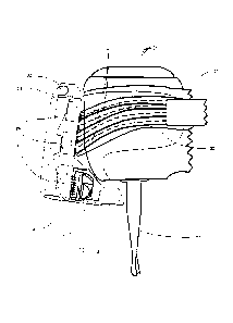
FIG.8 shows a completely assembled side view image of one preferred
embodiment of the invention while FIG.9 shows the same embodiment
separated/exploded into its component parts. This embodiment comprises a
drill head adapter 26 which can be secured to a drill head 52, and comprises a
chamber 27 for receiving a removable imaging device 25 that can be
conveniently inserted and removed, using a snap-in mechanism for example,
into the drill head adapter 26. A snug piece 23 which can be made of a
material such as silicone and used to insure impermeability and a snug fit and
proper electrical contact between the imaging device 25 and the drill head
14
CA 02738044 2011-03-22
WO 2010/034107 PCT/CA2009/001311
adapter 26 through connection mechanism 4 and 21. The cover 22 and snug
piece 23 can be combined into one single cover piece. The cover piece 22
allows for non-sterilized removable imaging device 25 to be combined with a
dental drill without compromising sterility of the dental drill. The snug
piece 23
and cover 22 can be used to seal off the imaging device 25 in the drill head
adapter, rendering it impermeable to aqueous contaminants such as water
saliva and blood but more importantly allowing the dental instrument to be
sterile on the outside despite non-sterile components inside the chamber 27 of
the drill head adapter 26. The snug piece 23 must be rigid enough to allow
pressure on cover 22 to actuate, through protruding section 29, a button 39
(such as an on/off or image adjustment buttons) of the image device 25. Snug
piece 23 can also be attached to the removable imaging device through a
hinge in order to decrease the number of independent components. The
securing mechanism 31 allows the drill head adapter to be securely fastened
to the drill and can act as a passage and/or connector for the wiring 8.
This type of assembly allows sterilization of all components of the dental
drill
imaging device that can come in contact with a patient including the cover 22
which is snapped back into place (without the imaging device 25) prior to
sterilization. In this embodiment, electronic components 9 can be combined to
the imaging device 25 in order to minimize the number of wires exiting the
drill
head adapter. It will be appreciated by those skilled in the art that this
drill
head adapter for adding imaging capability can be adapted to any other dental
instrument such as a curette, a scaler, a mirror, a toothbrush, a surgical
instrument and an extractor.
A battery and wireless transmitter can be placed inside the drill head adapter
(or the removable imaging device 25) in order to eliminate the need for wires.
However, if wires are used such as in FIG.8, the wires can enter drill head
adapter to provide, in addition to external image control, the required
electric
current for function. As seen in FIG.8, the drill head adapter is fitted onto
the
CA 02738044 2011-03-22
WO 2010/034107 PCT/CA2009/001311
drill head and comprises a mirror 12. The drill head adapter and mirror can be
permanently secured to the dental drill with autoclave resistant glue such as
epoxy. All chemical and/or heat sensitive components can be included in the
removable imaging device 25 to avoid damage caused by sterilization.
As can be seen in FIGS.8 and 9, the drill head adapter comprises but does not
cover the mirror 12. The mirror 12 allows for the angle between the drill bur
20
and the line-of-sight of the camera to be minimized. It will be appreciated by
those skilled in the art that when the angle 40 is too wide and the tip of the
drill
penetrates inside the surface of the tooth, the end of the drill bur is no
longer
in the line-of-sight of the camera (i.e. visible), which would then capture
the
tooth surface rather than the drilling part of the drill bur. Alternatively,
the
imaging device 1 can be placed directly adjacent to the drill bur however this
would be more cumbersome than a small mirror. The removable imaging
device comprises all components shown outside the dotted line (i.e. the
chamber 27 of the drill head adapter 26) with the exception of the cover 22.
The imaging capability 22-26, is positioned at the distal end of the dental
handpiece for space optimization purposes and to facilitate use of the dental
instrument. The removable imaging device 25 makes electrical contacts with
the drill head adapter 26 through electrically conducting/connecting elements
4
and the wires 24 which are part of the drill head adapter 26. The electrically
conducting elements can be made of any conducting material such as copper
or stainless steel. In order to reduce interference and prevent short circuits
between the wires 24, separation grooves/walls 21 can be provided.
The part of the sheath for receiving the removable imaging device surrounding
the drill head adapter which faces the drill bur can be made of a thin and/or
translucent material such that light emitting diodes inside the removable
imaging device 25 (which can be made of the same material) can illuminate
the work field without specular reflection in the mirror. It will be
appreciated
16
CA 02738044 2011-03-22
WO 2010/034107 PCT/CA2009/001311
that the material must be autoclavable because it is part of the drill head
adapter which undergoes sterilization with the dental instrument.
FIG. 10 is a schematic representation of alternative embodiment of the
optoelectronic components of an imaging device 80 as shown in FIG.3. In this
embodiment, a barrel lens 4 is placed in the middle of the chip/PCB 88 thus
allowing for LEDs 5a and 5b to be placed on both sides of the lens 4 and
detector 3. This allows to better control of illumination intensity and
waveband
such that multiple illumination colors can be used. Electronic components 90
are sandwiched between the PCB 88 and the silicone cover 92.
Imaging capabilities on a dental instrument can find numerous applications in
the dental field. For example, miniature imaging capabilities on a toothbrush
can be exploited for teaching purposes, especially for children learning how
to
brush their teeth. Also, imaging capability can be added to articulation paper
used to verify occlusion (bite) or cracked teeth.
Furthermore, having viewing capabilities on a dental drill can help in the
determination of drilling quantity, quality and to evaluate cavity
preparation.
Indeed, when a dental professional drills a tooth to remove carious material,
knowledge about when to stop drilling can be obtained from both feeling the
drill bur on the tooth but also by observing the visual characteristics of the
drilled/shredded material projecting from the drilling site.
Imaging capability on a standard mirror can provide a useful way for a dental
professional to examine the mouth of a patient, whilst recording visual images
of the patient's mouth. These images can be used for reconstruction of a 3D
images of the patient's mouth and teeth. Indeed, images from the dental
professional's exploration can be stitched together and used to evaluate
treatment quality and/or progress over time.
An imaging device in place of a mirror can be provided including a small
screen outside the mouth on the back end of the mirror handle. A standard
17
CA 02738044 2011-03-22
WO 2010/034107 PCT/CA2009/001311
mirror can be on the opposite side of the imaging device. This solves the
problem or at least slows the process of water droplets disturbing the camera
image, as typically happens with a mirror. A hydrophilic material can be
provided to protect the imaging device and to enhance the quality of images
captured.
By using an appropriate detector/CCD, the imaging device can be used in the
infrared and ultra-violet spectra in order to detect or capture images where
IR
and UV can be of informational and/or diagnostic value. Specific LEDs can be
used for pulsing in specific wavebands in order to obtain differential images
indicative of diseases such as caries and periodontal disease. In fact by
having a wide waveband detector and either waveband specific LEDs or
filters, the imaging device can act as a miniature spectrophotometer. The
imaging device can be used to illuminate specific points with structured light
in
order to evaluate the amount of light adjacent to an illumination point.
It will be appreciated by those skilled in the art that image stabilisation
techniques and components could be useful when imaging capability is added
to a dental drill with high vibration. Furthermore, although image focussing
techniques and image processing techniques are known in the art, they can be
applied advantageously within the present intraoral camera for added
functionality.
18