Note: Descriptions are shown in the official language in which they were submitted.
CA 02741089 2011-04-18
WO 2010/048446 PCT/US2009/061733
MODULATION OF AXON DEGENERATION
FIELD OF THE INVENTION
This invention relates generally to treatment of neurological disorders and
nervous system injuries. The invention specifically concerns the use of
modulators of
particular target proteins and processes in methods to inhibit neuron and axon
degeneration.
BACKGROUND OF THE INVENTION
Neuron or axon degeneration plays a central role in the proper development of
the nervous system and is a hallmark of many neurodegenerative diseases
including,
for example, amyotrophic lateral sclerosis (ALS), Alzheimer's disease, and
Parkinson's disease, as well as traumatic injury to the brain and spinal cord.
These
diseases and injuries are devastating to patients and caregivers, and also
result in great
financial burdens, with annual costs currently exceeding several hundred
billion
dollars in the United States alone. Most current treatments for these diseases
and
conditions are inadequate. Adding to the urgency of the problems created by
these
diseases is the fact that many such diseases are age-related, and thus their
incidence is
increasing rapidly as population demographics change. There is a great need
for the
development of effective approaches to treating neurodegenerative diseases and
nervous system injuries.
SUMMARY OF THE INVENTION
The invention provides methods for inhibiting degeneration of a neuron or a
portion thereof (e.g., the neuron cell body, an axon, or a dendrite). The
methods
involve administering to the neuron or portion thereof an agent that
modulates: (i) the
activity or expression of a target protein in the neuron or portion thereof,
or (ii) a
process in the neuron or portion thereof.
Examples of proteins that can be targeted in the methods of the invention
include dual leucine zipper-bearing kinase (DLK), glycogen synthase kinase 3(3
(GSK3(3), p38 mitogen-activated protein kinase (p38 MAPK), (3-catenin,
transcription
1
CA 02741089 2011-04-18
WO 2010/048446 PCT/US2009/061733
FAILIN I
GNE REFERENCE: PR4267R1-WO
ATTORNEY DOCKET NO. 50474/02OW02
factor 4 (TCF4), epidermal growth factor receptor (EGFR), phosphoinositide 3-
kinase
(P13K), cyclin-dependent kinase 5 (cdk5), adenylyl cyclase, c-Jun N-terminal
kinase
(JNK), BCL2-associated X protein (Bax), Ih channel, calcium/calmodulin-
dependent
protein kinase kinase (CaMKK), G-proteins, G-protein coupled receptors,
transcription factor 4 (TCF4), or 3-catenin, while examples of processes that
can be
targeted are transcription and protein synthesis.
The neuron or portion thereof can consist of or can be within a neuron
selected
from the group consisting of a cerebellar granule neuron, a dorsal root
ganglion
neuron, a cortical neuron, a sympathetic neuron, and a hippocampal neuron.
The agents can be, for example, inhibitors of the target protein or process.
Further, the agents can be, for example, selected from the group consisting of
antibodies, polypeptides, peptides, peptibodies, nucleic acid molecules, short
interfering RNAs (siRNAs), polynucleotides, aptamers, small molecules, and
polysaccharides. In the case of antibodies, the antibodies can be monoclonal
antibodies, chimeric antibodies, humanized antibodies, human antibodies, or
antibody
fragments (e.g., an Fv, Fab, Fab', or F(ab')2 fragment). In the case of small
molecules, the agent can be, for example, selected from the group consisting
of
MG132, SB 415286, GSK30 inhibitor I, GSK3(3 inhibitor VII, GSK3(3 inhibitor
VIII,
GSK33 inhibitor XII, Lithium Chloride, SB 202190, SB 239063, SB 239069, SB
203580, SB 203580 HCI, AG 556, AG 555, AG 494, PD168393, Tyrphostin B44,
Tyrphostin B42 (AG 490), LY 294022, Anisomycin, Cycloheximide, Roscovitine,
Forskolin, NKH 477, Actinomycin D, SP600125, Bax Channel Blocker, ZD7288,
STO-609, bortezomid, disulfiram, pamapimod, gefitinib, erlotinib, lapatinib
ditosylate, demeclocycline hydrochloride, gentamicin sulfate, neomycin
sulfate,
paromomycin sulfate, and pharmaceutically acceptable salts thereof.
The neuron or portion thereof can be present in a subject, such as a human
subject. The subject can, for example, have or be at risk of developing a
disease or
condition selected from the group consisting of (i) disorders of the nervous
system,
(ii) conditions of the nervous system that are secondary to a disease,
condition, or
therapy having a primary effect outside of the nervous system, (iii) injuries
to the
nervous system caused by physical, mechanical, or chemical trauma, (iv) pain,
(v)
ocular-related neurodegeneration, (vi) memory loss, and (vii) psychiatric
disorders.
2
CA 02741089 2011-04-18
WO 2010/048446, PCT/US2009/061733
rA1EA1
GNE REFERENCE: PR4267R1-WO
ATTORNEY DOCKET NO. 50474/020W02
Examples of disorders of the nervous system include amyotrophic lateral
sclerosis (ALS), trigeminal neuralgia, glossopharyngeal neuralgia, Bell's
Palsy,
myasthenia gravis, muscular dystrophy, progressive muscular atrophy, primary
lateral
sclerosis (PLS), pseudobulbar palsy, progressive bulbar palsy, spinal muscular
atrophy, inherited muscular atrophy, invertebrate disk syndromes, cervical
spondylosis, plexus disorders, thoracic outlet destruction syndromes,
peripheral
neuropathies, prophyria, Alzheimer's disease, Huntington's disease,
Parkinson's
disease, Parkinson's-plus diseases, multiple system atrophy, progressive
supranuclear
palsy, corticobasal degeneration, dementia with Lewy bodies, frontotemporal
dementia, demyelinating diseases, Guillain-Barre syndrome, multiple sclerosis,
Charcot-Marie-Tooth disease, prion disease, Creutzfeldt-Jakob disease,
Gerstmann-
Straussler-Scheinker syndrome (GSS), fatal familial insomnia (FFI), bovine
spongiform encephalopathy, Pick's disease, epilepsy, and AIDS demential
complex.
Examples of pain include chronic pain, fibromyalgia, spinal pain, carpel
tunnel
syndrome, pain from cancer, arthritis, sciatica, headaches, pain from surgery,
muscle
spasms, back pain, visceral pain, pain from injury, dental pain, neuralgia,
such as
neuogenic or neuropathic pain, nerve inflammation or damage, shingles,
herniated
disc, torn ligament, and diabetes.
Examples of conditions of the nervous system that are secondary to a disease,
condition, or therapy having a primary effect outside of the nervous system
include
peripheral neuropathy or neuralgia caused by diabetes, cancer, AIDS,
hepatitis, kidney
dysfunction, Colorado tick fever, diphtheria, HIV infection, leprosy, lyme
disease,
polyarteritis nodosa, rheumatoid arthritis, sarcoidosis, Sjogren syndrome,
syphilis,
systemic lupus erythematosus, and amyloidosis.
Examples of injuries to the nervous system caused by physical, mechanical, or
chemical trauma include nerve damage caused by exposure to toxic compounds,
heavy metals, industrial solvents, drugs, chemotherapeutic agents, dapsone,
HIV
medications, cholesterol lowering drugs, heart or blood pressure medications,
and
metronidazole. Additional examples include burn, wound, surgery, accidents,
ischemia, prolonged exposure to cold temperature, stroke, intracranial
hemorrhage,
and cerebral hemorrhage.
3
CA 02741089 2011-04-18
WO 2010/048446, PCT/US2009/061733
rtiIL1\1
GNE REFERENCE: PR4267R1-WO
ATTORNEY DOCKET NO. 50474/020W02
Examples of psychiatric disorders include schizophrenia, delusional disorder,
schizoaffective disorder, schizopheniform, shared psychotic disorder,
psychosis,
paranoid personality disorder, schizoid personality disorder, borderline
personality
disorder, anti-social personality disorder, narcissistic personality disorder,
obsessive-
compulsive disorder, delirium, dementia, mood disorders, bipolar disorder,
depression, stress disorder, panic disorder, agoraphobia, social phobia, post-
traumatic
stress disorder, anxiety disorder, and impulse control disorders.
Examples of ocular-related neurodegeneration include glaucoma, lattice
dystrophy, retinitis pigmentosa, age-related macular degeneration (AMD),
photoreceptor degeneration associated with wet or dry AMD, other retinal
degeneration, optic nerve drusen, optic neuropathy, and optic neuritis.
Examples of
glaucoma include primary glaucoma, low-tension glaucoma, primary angle-closure
glaucoma, acute angle-closure glaucoma, chronic angle-closure glaucoma,
intermittent
angle-closure glaucoma, chronic open-angle closure glaucoma, pigmentary
glaucoma,
exfoliation glaucoma, developmental glaucoma, secondary glaucoma, phacogenic
glaucoma, glaucoma secondary to intraocular hemorrhage, traumatic glaucoma,
neovascular glaucoma, drug-induced glaucoma, toxic glaucoma, and glaucoma
associated with intraocular tumors, retinal deatchments, severe chemical burns
of the
eye, and iris atrophy.
Contacting of the neuron or portion thereof with the agent, according to the
methods of the invention, can involve administering to a subject a
pharmaceutical
composition including the agent. The administering can be carried out by, for
example, intravenous infusion; injection by intravenous, intraperitoneal,
intracerebral,
intramuscular, intraocular, intraarterial or intralesional routes; or topical
or ocular
application. Further, the methods of the invention can include administering
to a
subject one or more additional pharmaceutical agents.
In other examples, the neuron or portion thereof treated according to the
methods of the invention is ex vivo or in vitro (e.g., a nerve graft or nerve
transplant).
The invention also includes methods of identifying agents for use in
inhibiting
degeneration of a neuron or a portion thereof. These methods involve
contacting a
neuron or portion thereof with a candidate agent in an assay of axon or neuron
4
CA 02741089 2011-04-18
WO 2010/048446, PCT/US2009/061733
YAILINI
GNE REFERENCE: PR4267R1-WO
ATTORNEY DOCKET NO. 50474/02OW02
degeneration (e.g., anti-nerve growth factor (NGF) antibodies, serum
deprivation/KCl
reduction, and/or rotenone treatment). Detection of reduced degeneration of
the
neuron or portion thereof in the presence of the candidate agent, relative to
a control,
indicates the identification of an agent for use in inhibiting degeneration of
a neuron
or portion thereof. The candidate agent can be, for example, selected from the
group
consisting of antibodies, polypeptides, peptides, peptibodies, nucleic acid
molecules,
short interfering RNAs (siRNAs), polynucleotides, aptamers, small molecules,
and
polysaccharides.
The invention also includes pharmaceutical compositions and kits that contain
one or more agent that can be used to inhibit degeneration of a neuron or a
portion
thereof, as described herein. The pharmaceutical compositions and kits can
include,
for example, one or more agent selected from the group consisting of MG132, SB
415286, GSK3(3 inhibitor I, GSK30 inhibitor VII, GSK30 inhibitor VIII, GSK30
inhibitor XII, Lithium Chloride, SB 202190, SB 239063, SB 239069, SB 203580,
SB
203580 HCI, AG 556, AG 555, AG 494, PD168393, Tyrphostin B44, Tyrphostin B42
(AG 490), LY 294022, Anisomycin, Cycloheximide, Roscovitine, Forskolin, NKH
477, Actinomycin D, SP600125, Bax Channel Blocker, ZD7288, STO-609,
bortezomid, disulfiram, pamapimod, gefitinib, erlotinib, lapatinib ditosylate,
demeclocycline hydrochloride, gentamicin sulfate, neomycin sulfate,
paromomycin
sulfate, and pharmaceutically acceptable salts thereof. The pharmaceutical
compositions and kits can optionally include one or more pharmaceutically
acceptable
excipients. Further, the pharmaceutical compositions and kits can optionally
include
instructions for use of the compositions and kits in methods for inhibiting
degeneration of a neuron or portion thereof.
In any of the methods, compositions, and kits of the invention, the agent may
be a DLK signaling inhibitor (e.g., a siRNA molecule targeting DLK comprising,
e.g.,
the sequence of, desirably, GCACTGAATTGGACAACTCTT (SEQ ID NO: 1),
GAGTTGTCCAATTCAGTGCTT (SEQ ID NO: 2),
GGACATCGCCTCCGCTGATTT (SEQ ID NO: 3), or
ATCAGCGGAGGCGATGTCCTT (SEQ ID NO: 4), or
GCAAGACCCGTCACCGAAATT (SEQ ID NO: 5),
TTTCGGTGACGGGTCTTGCTT (SEQ ID NO: 6), GCGGTGTCCTG
CA 02741089 2011-04-18
WO 2010/048446, PCT/US2009/061733
rvILI'NI
GNE REFERENCE: PR4267R1-WO
ATTORNEY DOCKET NO. 50474/02OWO2
GTCTACTATT (SEQ ID NO: 7), or TAGTAGACCAGGACACCGCTT (SEQ ID
NO: 8); an antibody, such as, antibody 317, antibody 318, antibody 319,
antibody 320,
antibody 321, antibody 322, an siRNA molecule targeting the JNK1 sequence of
TTGGATGAAGCCATTAGACTA (SEQ ID NO: 9), an siRNA molecule targeting
the JNK2 sequence of ACCTTTAATGGACAACATTAA (SEQ ID NO: 10) or
AAGGATTAGCTTTGTATCATA (SEQ ID NO: 11), an siRNA targeting the JNK3
sequence of CCCGCATGTGTCTGTATTCAA (SEQ ID NO: 12), SP600125, JNKV
inhibitor, JNKVIII inhibitor, SC-202673, SY-CC-401, SP600125, As601245, XG-
102, myricetin, T278A DLK, 5281 A DLK, S 152A DLK, and the leucine zipper
domain of DLK), a GSK3(3 inhibitor (e.g., SB415287, GSK3(3 inhibitor I,
GSK3(3 inhibitor VII, GSK30 inhibitor VIII, GSK33 inhibitor XII, and lithium
chloride), an EGFR pathway inhibitor (e.g., erlotinib, tyrphostin B44,
tyrphostin
B42/AG490, AG555, AG494, PD168393, S13203580, S13239063, S13202190,
SB239069, STO-609, and SP600125), or a G-protein inhibitor (e.g., SCG292676
and
pertussis toxin).
In any of the above-described methods, the administering of an agent results
in
at least a 10% decrease (e.g., at least 15%, 20%, 25%, 30%, 35%, 40%, 45%,
50%.
55%, 60%, 65%, 70%, 75%, 80%, 85%, 90%, 95%, or even 100% decrease) in one or
more (e.g., 1, 2, 3, 4, 5, 6, 7, 8. 9, or 10) symptoms of a disorder of the
nervous
system; condition of the nervous system that is secondary to a disease,
condition, or
therapy having a primary effect outside of the nervous system; injury to the
nervous
system caused by physical, mechanical, or chemical trauma; pain; ocular-
related
neurodegeneration; memory loss; or psychiatric disorder. Non-limiting examples
of
such symptoms include tremors, slowness of movement, ataxia, loss of balance,
depression, decreased cognitive function, short-term memory loss, long-term
memory
loss, confusion, changes in personality, language difficulties, loss of
sensory
perception, sensitivity to touch, numbness in extremities, muscle weakness,
muscle
paralysis, muscle cramps, muscle spasms, significant changes in eating habits,
excessive fear or worry, insomnia, delusions, hallucinations, fatigue, back
pain, chest
pain, digestive problems, headache, rapid heart rate, dizziness, blurred
vision,
shadows or missing areas of vision, metamorphopsia, impairment in color
vision,
6
CA 02741089 2011-04-18
WO 2010/048446 PCT/US2009/061733
YAIEAVI
GNE REFERENCE: PR4267R1-WO
ATTORNEY DOCKET NO. 50474/02OWO2
decreased recovery of visual function after exposure to bright light, and loss
in visual
contrast sensitivity.
In any of the above-described methods, the administering results in at least a
10% decrease (e.g., at least 15%, 20%, 25%, 30%, 35%, 40%, 45%, 50%, 55%, 60%,
65%, 70%, 75%, 80%, 85%, 90%, 95%, or even 100% decrease) in the likelihood of
developing a disorder of the nervous system; condition of the nervous system
that is
secondary to a disease, condition, or therapy having a primary effect outside
of the
nervous system; injury to the nervous system caused by physical, mechanical,
or
chemical trauma; pain; ocular-related neurodegeneration; memory loss; or
psychiatric
disorder, compared to a control population of subjects that are not
administered said
agent.
The invention also provides methods for activating degeneration of a neuron or
portion thereof. These methods involve administering to a neuron or portion
thereof
an agent that modulates: (i) the activity of a target protein in the neuron or
portion
thereof, or (ii) a process in the neuron or portion thereof. The target
protein can be,
for example, selected from the group consisting of dual leucine zipper-bearing
kinase
(DLK), glycogen synthase kinase 33 (GSK30), p38 mitogen-activated protein
kinase
(p38 MAPK), epidermal growth factor receptor (EGFR), phosphoinositide 3-kinase
(P13K), cyclin-dependent kinase 5 (cdk5), adenylyl cyclase, c-Jun N-terminal
kinase
(JNK), BCL2-associated X protein (Bax), Ih channel, calcium/calmodulin-
dependent
protein kinase kinase (CaMKK), a G-protein, a G-protein coupled receptor,
transcription factor 4 (TCF4), or (3-catenin, while the process can be, for
example,
transcription or protein synthesis. Further, the agent can be, for example, an
activator
of the target protein or process (as is the case for the targets listed above,
with the
exception of adenylyl cyclase). In the methods of activiating degeneration of
a neuron
or portion thereof described above, the modulation of a target protein may he
an
increase in the activity or expression of GSK313, a decrease in the activity
or
expression of (3-catenin, and/or a loss in the activity or expression of TCF4.
The invention also provides purified antibodies that specifically bind to the
phosphorylated form of DLK (e.g., antibody 318, antibody 319, antibody 320,
antibody 321, or antibody 322) and inhibitory nucleic acids (e.g., siRNA) that
comprise the sequence of, desirably, GCACTGAATTGGACAACTCTT (SEQ ID
7
CA 02741089 2011-04-18
WO 2010/048446, PCT/US2009/061733
VA I EIN1
GNE REFERENCE: PR4267R1-WO
ATTORNEY DOCKET NO.50474/02OW02
NO: 1), GAGTTGTCCAATTCAGTGCTT (SEQ ID NO: 2), GGACATCGCCTC
CGCTGATTT (SEQ ID NO: 3), or ATCAGCGGAGGCGATGTCCTT (SEQ ID NO:
4), or GCAAGACCCGTCACCGAAATT (SEQ ID NO: 5), TTTCGGTGACGGGTC
TTGCTT (SEQ ID NO: 6), GCGGTGTCCTGGTCTACTATT (SEQ ID NO: 7), or
TAGTAGACCAGGACACCGCTT (SEQ ID NO: 8) that mediate a decrease in the
expression of DLK.
BRIEF DESCRIPTION OF THE DRAWINGS
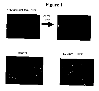
Figure 1 shows that treatment of neurons for 20 hours with anti-NGF
antibodies results in axon degeneration. The two images in the top row show
neurons
visualized with Tuj 1 (neuron specific R-tubulin) antibodies, with and without
20 hours
of treatment with anti-NGF antibodies. The two images in the bottom row show
neurons visualized with actin antibodies incubated with or without (control)
50 g/ml
anti-NGF antibodies.
Figure 2 shows that varicosities form in neurons cultured with anti-NGF
antibodies for 1, 3, 6, 9, 12, or 16 hours.
Figure 3 shows that axons cultured with anti-NGF antibodies for 16 hours lack
elongated mitochondria and show accumulation of mitochondria in varicosities.
Figure 4 shows that, in axons cultured with anti-NGF antibodies for 0 to 48
hours, the microtubule network is not disassembled before the actin or
neurofilament
networks.
Figure 5 illustrates Wallerian degeneration, which takes place in axons
severed
from neuron cell bodies (top panel; Raff et al., Science 296(5569):868-871,
2002), and
shows that there is a significant delay in axon degeneration after lesion in
Wallerian
Degeneration Slow (WIdS) mutants, as compared to controls (bottom panel; Araki
et
al., Science 305(5686):1010-1013, 2004).
Figures 6A-6D show that a proteasome inhibitor and a GSK inhibitor prevent
axon degeneration in an anti-NGF antibody-based NGF withdrawal assay.
Figures 7A-7D show that a p38 MAPK inhibitor and an adenylyl cyclase
activator prevent axon degeneration in an anti-NGF antibody-based NGF
withdrawal
assay.
8
CA 02741089 2011-04-18
WO 2010/048446, PCT/US2009/061733
rAILINI
GNE REFERENCE: PR4267R1-WO
ATTORNEY DOCKET NO. 50474/02OW02
Figures 8A-8D show that a transcription inhibitor and an EGFR kinase
inhibitor prevent axon degeneration in an anti-NGF antibody-based NGF
withdrawal
assay.
Figures 9A-9D show that a JNK inhibitor and Bax Channel Blocker prevent
axon degeneration in an anti-NGF antibody-based NGF withdrawal assay.
Figures I OA-l OD shows that an Ih channel blocker and a CAMKK inhibitor
prevent axon degeneration in an anti-NGF antibody-based NGF withdrawal assay.
Figure 11 is a graph showing the activities of inhibitors of GSK3 (30 M
SB415286), EGFR kinase (10.tM AG555), p38 MAPK (30 M SB239063),
CAMKK (15 M STO-609), and JNK (10 pM SP600125) when added at the time of
NGF withdrawal (t = 0) or at 3, 6, 9, or 12 hours after NGF withdrawal.
Figure 12 illustrates a Campenot chamber, in which somal (cell body) and
axonal environments are separated.
Figure 13 shows that axon degeneration is localized and proceeds without
apoptosis in Campenot chamber studies in which NGF withdrawal took place in
the
axon-containing chamber. Degeneration is visualized by tubulin
immunofluorescence.
Figure 14 shows that there are no signs of degeneration in the cell body
compartment in the Campenot chamber-based assay illustrated in Figure 13.
Figure 15 shows that cell bodies (left panels), but not axons (right panels),
in
the presence of 30 M SB415 (GSK inhibitor; GSKi) or 15 M Act D
(transcription
inhibitor; TXNi) were protected from local degeneration.
Figure 16 shows that axons (right panels), but not cell bodies (left panels),
exposed to anti-NGF antibodies in the presence of 10 M AG555 (EGFR inhibitor;
EGFRi) or 30 M SB239 (p38 inhibitor; p38i) were protected from local
degeneration.
Figure 17 is a graph showing quantification of axon degeneration in Campenot
chambers in which anti-NGF antibodies were added to the axonal environment in
the
presence or absence (DMSO) of 15 M actinomycin D (ActD), 30 pM SB415286
(SB415), 10 M AG555, or 30 M SB239063 (SB239) in the axonal (Axon) or the
somal environment (Cell).
Figure 18 is a model based on data from the screens described herein.
9
CA 02741089 2011-04-18
WO 2010/048446, PCT/US2009/061733
VAILINI
GNE REFERENCE: PR4267R1-WO
ATTORNEY DOCKET NO. 50474/02OW02
Figure 19 shows that cell bodies appear smaller when NGF is removed from
the axon compartment in a Campenot chamber.
Figure 20 shows that many neurons deprived of NGF in the axon compartment
have increased cleavage of caspase-3 and show nuclear condensation. (Neuron
health
may be affected by the membrane stain DiI.)
Figure 21 is a graph showing GSK3 activity (as measured by decreased levels
of phosphorylated GSK3(3) at the start of NGF withdrawal (t = 0) and after 1,
3, 6, 9,
and 12 hours of NGF withdrawal in the cell body or axon compartment.
Figure 22 is a graph showing JNK activity (as measured by increased levels of
phosphorylated JNK) at the start of NGF withdrawal (t = 0) and after 1, 3, 6,
9, and 12
hours of NGF withdrawal in the cell body or axon compartment.
Figure 23 shows that a large number of axons with varicosities, as well as
fragmented axons, were observed when the cell body inhibitors (30 M SB415
(GSKi) and 15 M Act D (TXNi)) were added to the cell body compartment. The
addition of the axon inhibitors (10 M AG555 (EGFRi) and 30 M SB239 (p38i))
to
the axon compartment showed fewer varicosities, and the axons seemed to go
straight
to fragmentation.
Figure 24 shows that there may be more functional mitochondria, but still no
elongated mitochondria, in NGF-deprived neurons treated with GSK, EGFR, and
p38
inhibitors.
Figure 25 shows that the GSK inhibitor SB415 can delay axon degeneration
after lesion.
Figure 26 shows that, after global NGF withdrawal, 10 p.M or 25 M GSK
inhibitor blocks axon degeneration, but does not block cell death.
Figure 27 shows that EGFR expression is increased in a section of SODI
mouse (Tg) spinal cord stained with anti-EGFR antibodies (right panel), as
compared
to a non-transgenic control (NTG; left panel).
Figure 28 shows that EGFR is normally expressed in neurons (motor neurons)
and that the level of phosphorylated EGFR (pEGFR) is increased in ALS SOD1
mouse model (SOD 1-Tg) compared to a non-transgenic control (Non-Tg).
Figure 29 shows that the number of axons is decreased in the ALS SOD 1
mouse model (SOD 1-Tg) as compared to a non-transgenic control (Non-Tg), and
that
CA 02741089 2011-04-18
WO 2010/048446, PCT/US2009/061733
rvirivi
GNE REFERENCE: PR4267R1-WO
ATTORNEY DOCKET NO.50474/02OW02
the phosphorylated EGFR (pEGFR) in the ALS SOD1 model partially co-localizes
with axons.
Figure 30 shows that the small molecule inhibitors of JNK (5 M SP600125),
CaMKK (5 M STO-609), EGFR (1 M or 10 M AG555), p38 (5 M SB239063),
and GSK (10 p.M SB415286) protect cerebellar granule neurons from serum
deprivation/KCl reduction.
Figure 31 shows that the small molecule inhibitors of EGFR, GSK, CaMKK,
JNK, and p38 protect hippocampal neurons against 10 M rotenone.
Figure 32 shows that the small molecule inhibitors of EGFR, GSK, CaMKK,
JNK, and p38 protect cortical neurons against 10 M rotenone.
Figure 33 shows that ErbB receptorsare detected on axons in dorsal root
ganglion neurons by immunocytochemistry using antibodies specific for EGFR
(top
left panel), Her2 (top right panel), Her3 (bottom left panel), and Her4
(bottom right
panel).
Figure 34 shows that EGFR is expressed in axons of dorsal root ganglion
neurons using immunocytochemistry.
Figure 35 shows that 100 g/ml, EGF does not induce axon degeneration
when added to dorsal root ganglion neurons and that addition of 100 g/mL EGF
induces phosphorylation of ERK in the treated neurons.
Figure 36 shows that the EGFR ectodomain (50 g/mL) does not block axon
degeneration induced by NGF withdrawal in dorsal root ganglion neurons.
Figure 37 shows that 3.4 M, 11.1 4M, 33.3 M, and 100 M Tarceva
(erlotinib) blocks degeneration in dorsal spinal cord explants.
Figure 38 shows that dual leucine zipper-bearing kinase (DLK) acts upstream
from JNK in axon degeneration. Transfection of a plasmid encoding wild type
DLK
in 293 cells results in JNK activation (as measured by increased levels of
phosphorylated JNK) compared to cells mock-transfected with a control plasmid
or a
plasmid encoding kinase-dead DLK (DLK-KD). Knockdown of DLK expression by
siRNA in dorsal root ganglion neurons protects axons from degeneration induced
by
NGF withdrawal. The knockdown of DLK expression using DLK siRNA was
confirmed using quantitative PCR as compared to a control siRNA (bottom right
panel).
11
CA 02741089 2011-04-18
WO 2010/048446, PCT/US2009/061733
rAILIVI
GNE REFERENCE: PR4267R1-WO
ATTORNEY DOCKET NO. 50474/02OW02
Figure 39 shows that siRNA knockdown of DLK signaling delays local axon
degeneration.
Figures 40A and 40B depict the results of experiments assessing the impact of
DLK knockdown on NGF withdrawal-induced sympathetic neuron degeneration using
phase contrast microscopy to visualize neurons.
Figure 41 shows that knockdown of DLK expression using DLK siRNA
(DLK) protects sympathetic neurons from camptothecin- and vincristine-induced
apoptosis compared to neurons treated with a control siRNA (NT).
Figure 42 shows that transfection of sympathetic neurons with a plasmid
encoding kinase-dead DLK (KD) protects the neurons from NGF withdrawal-induced
apoptosis compared to neurons transfected with a plasmid encoding wild type
DLK
(DLK).
Figures 43A-43D show the results of experiments assessing the binding
specificities of the anti-pDLK antibodies described in Example 15A. Figure 43A
shows Western blot analyses of the binding of each of the anti-pDLK antibodies
described herein to DLK, DLK in the presence of a dominant negative DLK, and
control kinase MLK3. Figure 43B shows immunofluorescent microscopic images of
the binding of anti-pDLK antibodies 318 and 319 to cultured 293T cells
transformed
with DLK (upper two images) or control kinase MLK3 (lower two images). Figures
43C and 43D show Western blots using JNK and phospho-JNK antibodies.
Figures 44A and 44B show binding of anti-pDLK antibodies (antibody 318) to
spinal cord sections in wild type and SOD 1 mutant mice at end stage of
disease
(Figure 43A) and at the onset of symptoms (Figure 44B). Figure 44C depicts
Western
blot analyses of pDLK, pJNK, and pcJUN levels in human Alzheimer's disease
patient cortical samples.
Figures 45A and 45B depict the results of experiments assessing the impact of
DLK silencing on phosphorylation of JNK in response to NGF withdrawal stress
in
sympathetic neurons and dorsal root ganglion neurons; and vincristine-induced
stress
in cortical neurons, as described in Example 15C and Example 14B.
Figure 46 shows the protective effect of JNK inhibitors on DRG explants
subjected to NGF withdrawal stress, as described in Example 15C.
Figure 47 depicts the results of experiments assessing the impact of silencing
12
CA 02741089 2011-04-18
WO 2010/048446, PCT/US2009/061733
rriir 4I
GNE REFERENCE: PR4267R1-WO
ATTORNEY DOCKET NO.50474/02OW02
JNK1, JNK2, JNK3, individually, and JNK2 and JNK3 together in DRG neurons on
axon degeneration observed upon NGF withdrawal stress, as described in Example
15C.
Figure 48A shows the affect of DLK siRNA and control siRNA on the
survival of cortical neurons. Figure 48B shows the affect of DLK siRNA and
control
siRNA on the survival of sympathetic neurons.
Figure 49 is a set of immunomicrographs showing the ability of an inhibitor of
G-coupled protein receptors (SCH 202676; 10 M or 100 M) to prevent NGF
withdrawal-induced degeneration in DRGs.
Figure 50 is a set of immunomicrographs showing the ability of SCH 202676
(0.1 M or 1 M) to prevent NGF withdrawal-induced degeneration in DRGs.
Figure 51 is a set of immunomicrographs showing the ability of 0.01 g/ml,,
0.1 g/mL, or 1 g/mL pertussis toxin (an inhibitor of G-protein signaling) to
prevent
NGF withdrawal-induced degeneration in DRGs.
Figure 52 is a set of immunomicrographs of rat hippocampal neurons showing
the effect of expression of active mutant GSK (GSK3S9A), wild type TCF4, and
mutant inactive TCF4 on degeneration.
DETAILED DESCRIPTION OF THE INVENTION
A. Definitions
The term "target" is used herein to refer to proteins and processes that, when
modulated by agents impacting their activities, inhibit or decrease axon
degeneration.
Most of the targets described herein, when contacted with an agent that
inhibits their
activity, inhibit or decrease axon degeneration, but the targets of the
present invention
also include proteins and processes that, when activated, inhibit or decrease
axon
degeneration. Exemplary targets of the invention are as follows: dual leucine
zipper-
bearing kinase (DLK), glycogen synthase kinase 3(3 (GSK30), p38 mitogen-
activated
protein kinase (p38 MAPK), epidermal growth factor receptor (EGFR),
phosphoinositide 3 kinase (PI3K), cyclin-dependent kinase 5 (Cdk5), adenylyl
cyclase, c-Jun N-terminal kinase (JNK), BCL2-associated X protein (Bax), Ih
channel,
calcium/calmodulin-dependent protein kinase kinase (CaMKK), a G-protein, a G-
protein coupled receptor, transcription factor 4 (TCF4), (3-catenin,
transcription, and
13
CA 02741089 2011-04-18
WO 2010/048446, PCT/US2009/061733
VA I C.IVI
GNE REFERENCE: PR4267R1-WO
ATTORNEY DOCKET NO.50474/02OW02
protein synthesis. A selection of common alternate designations for several of
these
targets is listed in Table 1. The targets include native, human sequences and
homologues of these sequences from monkeys, mice, rats, and other non-human
mammals, including all naturally occurring variants, such as alternatively
spliced and
allelic variants and isoforms, as well as soluble forms thereof. Exemplary,
non-
limiting sequence references are also provided in Table 1. Additional
sequences,
including sequences of various target isoforms, variants, homologues, and
fragments
may also be considered as targets, according to the present invention.
Table 1
Target Alternate names Exemplary Genbank
Accession numbers
Gycogen synthase GSK-3(3; GSK-3 beta 3; GSK3beta isoform; CAG38748
kinase 3 beta (GSK30) GSK-3a, GSK-3b2 NP_002084
NP 063937
(3-catenin Catenin beta-1, beta-catenin, CTNNB, CTNBI NP_001091679
NP 001091680
NP 001895
TCF4 Transcription factor 4, E2-2, ITF2, SEF2, SEF2- AA125085
1, SEF2-IA, SEF2-1B NP 001077431
NP 003190
EAW63024
EAW63023
EAW63022
EAW63021
EAW63020
EAW63019
EAW63018
EAW63017
AA125086
Q9NQBO
p38 Mitogen-activated p38alpha (MAPK14, CSBP2, Crkl, Csbpl, NP_002736.3
protein kinase MGC102436, Mxi2, PRKM14, PRKM15, p38, NP620407
p38-alpha, p38MAPK, p38a,
p38alpha, OTTMUSP00000021706; cytokine
suppressive anti-inflammatory drug binding
protein 1; mitogen activated protein kinase 14;
p38 MAP kinase alpha; p38 MAPK; p38 alpha;
tRNA synthetase cofactor p38)
p38beta (MAPK 11,DKFZp586C 1322, P38b,
Prkm11, Sapk2, Sapk2b, p38-2,
p38beta2, mitogen activated protein kinase 11;
protein kinase, mitogen activated kinase, 11,
p38beta)
p38delta (MAPK13, SAPK4,
Serk4, OTTMUSP00000028863;
SAPK/Erk/kinase 4; mitogen activated protein
kinase 13; p38 delta MAP kinase)
p38gamma (MAPK12, AW 123708, Erk6,
Prkml2, Sapk3, mitogen activated protein kinase
12; stress activated protein kinase 3)
14
CA 02741089 2011-04-18
WO 2010/048446 PCT/US2009/061733
rAiIiINI
GNE REFERENCE: PR4267R1-WO
ATTORNEY DOCKET NO. 50474/02OW02
Epidermal growth EGFR (ERBB, ERBB1, HER1, PIG61, AAG35789
factor receptor (EGFR) mENA, avian erythroblastic leukemia viral (v- NP 005219
erb-b) oncogene homolog; cell growth inhibiting NP 958439
protein 40; cell proliferation-inducing protein 61; NP_958440
epidermal growth factor receptor) NP_958441
ErbB2 (CD340, HER-2, HER-2/neu, HER2,
NEU, NGL, TKRI, c-erb B2/neu protein; erbB-2;
herstatin; neuroblastoma/glioblastoma derived
oncogene homolog; v-erb-b2 avian erythroblastic
leukemia viral oncogene homolog 2
(neuro/glioblastoma derived oncogene homolog))
ErbB3 (ErbB-3, HER3, LCCS2, MDA-BF-1,
MGC88033, c-erbB-3, c-erbB3, erbB3-S, p180-
ErbB3, p45-sErbB3, p85-sErbB3, erbB-3; lethal
congenital contracture syndrome 2; v-erb-b2
avian erythroblastic leukemia viral oncogene
homolog 3)
ErbB4 (HER4, MGC 138404, p 180erbB4, avian
erythroblastic leukemia viral (v-erb-b2) oncogene
homolog 4; receptor tyrosine-protein kinase erbB-
4; tyrosine kinase-type cell surface receptor
HER4; v-erb-a avian erythroblastic leukemia viral
oncogene homolog-like 4; v-erb-a erythroblastic
leukemia viral oncogene homolog 4)
Mitogen-activated Jun N-terminal kinase (1); JNK; JNKI; PRKM8; NP_003609
protein kinase 8 (JNK) SAPK1; JNK1A2; JNK2IB1/2; mitogen- CAG38817
activated protein kinase-8; MAPK8; JNK-46 AAH65516
JNK-2; Jun N-terminal kinase (2); MAPK9; NP002741
JNK2; SAPK; p54a; JNK2A; JNK2B; PRKM9; NP_620634
JNK-55; JNK2BETA; p54aSAPK; JNK2ALPHA NP_620635
JNK-3; Jun N-terminal kinase (3); MAPKIO; NP620637
JNK3; JNK3A; PRKM 10; p493F 12; FLJ 12099;
FLJ33785; MGC50974; p54bSAPK
Calcium/calmodulin- CAMKKA; CAMKK alpha protein; CaM-KK NP_115670
dependent protein alpha; CaM-kinase IV kinase; CaM-kinase kinase NP_757343
kinase beta alpha; CaMKK 1; CaMKK alpha; NP_757344
(CaMKbeta) calcium/calmodulin-dependent protein kinase
kinase alpha; calcium/calmodulin-dependent
protein kinase I alpha; calcium/calmodulin-
dependent protein kinase kinase 1, alpha
CAMKKB; CAMKK beta protein; CaM-KK beta; NP_006540
CaM-kinase kinase beta; CaMKK beta; NP 705719
calcium/calmodulin-dependent protein kinase NP 705720
kinase beta; NP 757363
calcium/calmodulin-dependent protein kinase NP 757364
beta; NP_757365
calcium/calmodulin-dependent protein kinase NP_757380
kinase 2, beta; calcium/calmodulin-dependent
protein kinase kinase 2, beta
dual leucine zipper- DLK, DLK1,fetal antigen I (FA1), PG2, PREF-1, NP_003827
bearing kinase PREF1, ZOG, delta-]ika protein dlk, pG2, ABC26857
preadipocyte factor I EAW81713
EAW81712
EAW81711
AAH 14015
AAH13197
AAH07741
P80370
CA 02741089 2011-04-18
WO 2010/048446, PCT/US2009/061733
YAIL1NI
GNE REFERENCE: PR4267R1-WO
ATTORNEY DOCKET NO. 50474/02OW02
Phosphoinositide 3 P13-kinase, P13K, Phosphotidylinositol 3 kinase CAA74194
kinase
Cyclin-dependent Cdk5 NP004926
kinase 5
Adenylyl cyclase Adenylyl cyclase I (ADCY1); 3',5'-cyclic AMP NM_021116
synthetase (1); ATP pyrophosphate-lyases (1);
Cat+/calmodulin-activated adenylyl cyclase;
Adenylate cyclase type I; Brain adenylate cyclase
1
Adenylyl cyclase 2 (ADCY2); 3',5'-cyclic AMP NP_065433
synthetase (2); ATP pyrophosphate-lyase (2);
adenylate cyclase type II; adenylate cyclase 2
(brain);
Adenylate cyclase II; Type II adenylate cyclase
Adenylyl cyclase 8 (ADCY8); ATP NP 001106
pyrophosphate-lyase 8; adenylate cyclase type
VIII; adenylyl cyclase 8; Ca(2+)/calmodulin-
activated adenylyl cyclase;
adenylate cyclase 8 (brain);
adenylyl cyclase-8, brain
adenylate cyclase 3 (Adcy3, AC3, mKIAA0511,
adenylyl cyclase 3)
adenylate cyclase 4 (Adcy4, KIAA4004,
mK1AA4004)
adenylate cyclase 5 (Adcy5, AW121902, Ac5)
adenylate cyclase 6 (Adcy6, mKIAA0422)
adenylate cyclase 7 (Adcy7, AA407758,
MGC141539, adenylyl cyclase type VII)
adenylate cyclase 9 (Adcy9, AW125421,
DI6Wsu65e, mKIAA0520)
adenylate cyclase 10 (AdcylO, 4930431D04Rik,
4931412F17, Sacy, sAC,
OTTMUSP00000023839; soluble adenylyl
cyclase; testicular soluble adenylyl cyclase)
Ih Channel HCN1; hyperpolarization activated cyclic NP_066550
nucleotide-gated potassium channel 1; brain
cyclic nucleotide-gated channel 1; BCNG- 1;
HAC-2 NP_001185
HCN2; hyperpolarization activated cyclic
nucleotide-gated potassium channel 2; brain
cyclic nucleotide-gated channel 2; BCNG-2;
HAC-1
HCN3; K1AA1535; MGC131493; NP 065948
OTTHUMP00000034062; hyperpolarization
activated cyclic nucleotide-gated potassium
channel 3; potassium/sodium hyperpolarization-
activated cyclic nucleotide-gated
channe13 NP 005468
HCN4; hyperpolarization activated cyclic
nucleotide-gated potassium channel 4
BCL2-associated X apoptosis regulator BAX; BCL2L4 Q07812
protein (Bax) NP004315
NP 620116
NP 620118
NP 620119
NP 620120
Transcription Actinomycin D
16
CA 02741089 2011-04-18
WO 2010/048446, PCT/US2009/061733
YAIJLI'4I
GNE REFERENCE: PR4267R1-WO
ATTORNEY DOCKET NO. 50474/02OWO2
"Isolated" when used to describe the various proteins disclosed herein, means
a protein that has been identified and separated and/or recovered from a
component of
its natural environment. Contaminant components of its natural environment are
materials that may interfere with uses (e.g., uses in therapy or antibody
production) for
the protein, and may include enzymes, hormones, and other proteinaceous or non-
proteinaceous solutes. In various embodiments, the protein will he purified
(i) to a
degree sufficient to obtain at least 15 residues of N-terminal or internal
amino acid
sequence by use of a spinning cup sequenator, and/or (ii) to homogeneity by
SDS-
PAGE under non-reducing or reducing conditions using Coomassie blue or silver
stain, and/or (iii) to homogeneity by mass spectroscopic or peptide mapping
techniques. Isolated protein includes protein in situ within recombinant
cells, as at
least one component of the natural environment of the protein in question will
not be
present. Ordinarily, however, isolated protein will be prepared by at least
one
purification step. Isolated target proteins as described herein (or fragments
thereof)
can be used to make antibodies as described herein against the target
proteins.
An "isolated" nucleic acid molecule is a nucleic acid molecule that is
identified and separated from at least one contaminant nucleic acid molecule
with
which it is ordinarily associated in the natural source of the nucleic acid in
question.
An isolated nucleic acid molecule is other than in the form or setting in
which it is
found in nature. Isolated nucleic acid molecules therefore are distinguished
from the
nucleic acid molecules as they exist in natural cells. However, an isolated
nucleic
acid molecule includes nucleic acid molecules contained in cells that
ordinarily
express such nucleic acid molecule where, for example, the nucleic acid
molecule is
in a chromosomal location different from that of natural cells. An example of
an
isolated nucleic acid molecule is one lacking 5' and/or 3' flanking sequences
with
which it is contiguous in a natural setting.
As used herein, the terms "antagonist" and "inhibitor" refer to agents capable
of blocking, neutralizing, inhibiting, abrogating, reducing and/or interfering
with one
or more of the activities of targets and/or reducing the expression of one or
more
target proteins (or the expression of nucleic acids encoding one or more
target
proteins) as described herein. They include, for example, antibodies,
polypeptides,
peptides, nucleic acid molecules, short interfering RNAs (siRNAs) and other
17
CA 02741089 2011-04-18
WO 2010/048446, PCT/US2009/061733
YA1 1NI
GNE REFERENCE: PR4267R1-WO
ATTORNEY DOCKET NO. 50474/02OW02
inhibitory RNAs, small molecules (e.g., small inorganic molecules),
polysaccharides,
polynucleotides, aptamers, and peptibodies. Antagonists or inhibitors of
particular
targets as described herein (i.e., targets other than adenylyl cyclase)
generally inhibit
or decrease axon degeneration (e.g., by at least 10%, 15%, 20%, 25%, 30%, 35%,
40%, 45%, 50%, 55%, 60%, 65%, 70%, 75%, 80%, 85%, 90%, 95%, or even 100%
decrease compared to a control that is untreated with the inhibitor), as
described
herein. An inhibitor may decrease the activity and/or expression of a target
protein by
at least 10% (e.g., by at least 15%, 20%, 25%, 30%, 35%, 40%, 45%, 50%, 55%,
60%, 65%, 70%, 75%, 80%, 85%, 90%, or even 100% decrease) as compared to the
expression and/or activity of the target protein that is untreated with the
inhibitor. A
"DLK signaling inhibitor" is an agent capable of decreasing the activity
(e.g., kinase
activity) or the expression of a DLK protein (or a nucleic acid encoding a DLK
protein) and/or deceasing the activity and/or expression of one or more
proteins
involved in a DLK signaling pathway (e.g., JNK1, JNK2, JNK3, eJun (e.g., cJun-
63
and cJun-73), MKK4, and MKK7). Examples of DLK signaling inhibitors include
siRNA molecules that decrease the expression of a nucleic acid encoding DLK
(e.g.,
desirably, a sequence of GCACTGAATTGGACAACTCTT (SEQ ID NO: 1),
GAGTTGTCCAATTCAGTGCTT (SEQ ID NO: 2), GGACATCGCCTCCGCTGA
TTT (SEQ ID NO: 3), or ATCAGCGGAGGCGATGTCCTT (SEQ ID NO: 4), or
GCAAGACCCGTCACCGAAATT (SEQ ID NO: 5), TTTCGGTGACGGG
TCTTGCTT (SEQ ID NO: 6), GCGGTGTCCTGGTCTACTATT (SEQ ID NO: 7), or
TAGTAGACCAGGACACCGCTT (SEQ ID NO: 8)), JNKI (e.g., a sequence
targeting the JNKI sequence of TTGGATGAAGCCATTAGACTA (SEQ ID NO: 9)),
JNK2 (e.g., a sequence targeting the JNK2 sequence of ACCTTTAATGGACAA
CATTAA (SEQ ID NO: 10) or AAGGATTAGCTTTGTATCATA (SEQ ID NO:
11)), JNK3 (e.g., a sequence targeting the JNK3 sequence of CCCGCATGTGTCT
GTATTCAA (SEQ ID NO: 12)), cJun (e.g., cJun-63 and cJun-73), MKK4, and
MKK7. Additional examples of DLK inhibitors include antibodies that bind to a
DLK protein (e.g., antibodies that recognize unphosphorylated or
phosphorylated
DLK, such as the 317, 318, 319, 320, 321, and 322 antibodies described
herein),
JNKI, JNK2, JNK3, cJun (e.g., Jun-63 and cJun-73), MKK4, and/or MKK7;
inhibitors of JNK activity (e.g., SC-202673, SY-CC-401, SP600125, JNKV
inhibitor,
18
CA 02741089 2011-04-18
WO 2010/048446, PCT/US2009/061733
rAILI' I
GNE REFERENCE: PR4267R1-WO
ATTORNEY DOCKET NO. 50474/02OW02
JNKVIII inhibitor, AS601245, and XG-102, as well as Catalog Nos. 420119,
420130,
420131, 420123, 420116, 420118, 420136, 420129, 420135, 420134, 420133,
420140, and 420128 from EMD Biosciences); inhibitors of MKK4 activity (e.g.,
myricetin and the inhibitors described in WO 04/058764), and inhibitors of
MKK7
activity (e.g., inhibitors described in U.S. Patent No. 7,195,894 and WO
04/002532).
A DLK inhibitor may also be a dominant negative form or kinase-dead form of
DLK
protein (or a nucleic acid encoding a dominant negative form or a kinase-dead
form of
DLK protein), such as T278A DLK, S281 A DLK, S 152A DLK, and the leucine
zipper
domain of DLK.
Another example of an inhibitor is a "GSK3(3 inhibitor." GSK3P inhibitor
refers to an agent capable of decreasing the activity and/or expression of
GSK3P (or a
nucleic acid encoding GSK3(3) and/or decreasing the activity and/or the
expression of
one or more proteins (or a nucleic acid encoding the one or more proteins)
that
activate GSK3(3 or the expression or activity of one or more substrates of
GSK3(3.
Non-limiting examples of GSK30 inhibitors include SB415286, GSK3(3 inhibitor
I,
GSK3(3 inhibitor VII, GSK3(3 inhibitor VIII, GSK3(3 inhibitor XII, and lithium
chloride.
An additional example of an inhibitor is a "G-protein inhibitor." A G-protein
inhibitor refers to an agent capable of decreasing the activity and/or
expression of one
or more G-proteins or G-protein coupled receptors (GPCRs) (or the expression
of one
or more nucleic acids encoding a G-protein or a GPCR), and/or decreasing the
activity
and/or expression of one or more proteins downstream of a G-protein or a GPCR.
Non-limiting examples of a G-protein inhibitor include siRNA molecules that
decrease the level of expression of a nucleic acid encoding a G-protein or a
GPCR, an
antibody or peptibody that binds to a G-protein or GPCR, or a small molecule
or
peptide that inhibits the activity of a G-protein or GPCR (e.g., SCH202676 and
pertussis toxin).
Another example of an inhibitor is a "EGFR pathway inhibitor." EGFR
pathway inhibitor refers to an agent capable of decreasing the activity and/or
expression of EGFR protein (or a nucleic acid encoding EGFR) and/or decreasing
the
activity and/or the expression of one or more proteins that function
downstream of
EGFR in the cell (e.g., p38 MAPK, CAMKK, and JNK). Non-limiting examples of
19
CA 02741089 2011-04-18
WO 2010/048446, PCT/US2009/061733
re irivi
GNE REFERENCE: PR4267R1-WO
ATTORNEY DOCKET NO. 50474/02OW02
EGFR pathway inhibitors include inhibitors of EGFR (e.g., erlotinib,
tyrphostin B44,
tyrphostin B42/AG 490, AG555, AG494, and PD168393), inhibitors of p38 MAPK
(e.g., SB203580, SB239063, SB202190, and SB239069), inhibitors of CAMKK (e.g.,
STO-609), and inhibitors of JNK (e.g., SP600125). Additional examples of EGFR
pathway inhibitors include antibodies and peptibodies that bind to EGFR, p38
MAPK,
CAMKK, and/or JNK; and siRNA molecules that decrease the expression of one or
more nucleic acids that encode a protein that functions downstream of EGFR in
the
cell (e.g., EGFR, p38 MAPK, CAMKK, and/or JNK).
An additional example of an inhibitor is a "CAMK(3 inhibitor." A CAMK(3
inhibitor refers to an agent capable of decreasing the activity and/or
expression of
CAMK(3 protein (or a nucleic acid encoding CAMK(3) and/or decreasing the
activity
and/or expression of one or more proteins that function downstream of CAMKf3
in the
cell. Non-limiting examples of CAMK(3 inhibitors include antibodies and
peptibodies
that specifically bind to CAMK(3, and siRNA molecules that decrease the
expression
of one or more nucleic acids that encode CAMK(3 or a protein that functions
downstream of CAMK(3.
Another example of an inhibitor is a "cdk5 inhibitor." A cdk5 inhibitor refers
to an agent capable of decreasing the activity and/or expression of cdk5
protein (or a
nucleic acid encoding cdk5) and/or decreasing the activity and/or expression
of one or
more proteins that function downstream of cdk5 in the cell. Non-limiting
examples of
cdk5 inhibitors include antibodies and peptibodies that specifically bind to
cdk5, and
siRNA molecules that decrease the expression of one or more nucleic acids that
encode cdk5 or a protein that functions downstream of cdk5.
An additional example of an inhibitor is a "TCF4 inhibitor." A TCF4 inhibitor
refers to an agent capable of decreasing the activity and/or expression of
TCF4 protein
(or a nucleic acid encoding TCF4) and/or decreasing the activity and/or
expression of
a gene regulated by TCF4 protein. Non-limiting examples of TCF4 inhibitors
include
antibodies and peptibodies that specifically bind to TCF4 or a protein encoded
by a
gene regulated by TCF4, and siRNA molecules that decrease the expression of
one or
more nucleic acids that encode TCF4 or decrease the expression of an mRNA
encoded
by a gene regulated by TCF4.
CA 02741089 2011-04-18
WO 2010/048446, PCT/US2009/061733
rHiriv 1
GNE REFERENCE: PR4267R1-WO
ATTORNEY DOCKET NO.50474/020WO2
An additional example of an inhibitor is a "(3-catenin inhibitor." A (3-
catenin
refers to an agent capable of decreasing the activity and/or expression of "(3-
catenin
protein (or a nucleic acid encoding "13-catenin) and/or decreasing the
activity and/or
expression of a gene regulated by "(3-catenin protein. Non-limiting examples
of (3-
catenin inhibitors include antibodies and peptibodies that specifically bind
to f 3-
catenin or a protein encoded by a gene regulated by (3-catenin, and siRNA
molecules
that decrease the expression of one or more nucleic acids that encode (3-
catenin or
decrease the expression of an mRNA encoded by a gene regulated by (3-catenin.
An additional example of an inhibitor is an "adenyl cyclase inhibitor." An
adenyl cyclase inhibitor refers to an agent capable of decreasing the activity
and/or
expression of adenyl cyclase protein (or a nucleic acid encoding adenyl
cyclase)
and/or decreasing the activity and/or expression of one or more proteins that
function
downstream of adenyl cyclase in the cell. Non-limiting examples of adenyl
cyclase
inhibitors include antibodies and peptibodies that specifically bind to adenyl
cyclase,
and siRNA molecules that decrease the expression of one or more nucleic acids
that
encode adenyl cyclase or a protein that functions downstream of adenyl
cyclase.
Additional examples of adenyl cyclase inhibitors include small molecules that
inhibit
the activity of adenyl cyclase (e.g., forksolin and NKH 477).
The terms "agonist" or "activator" as used herein refer to agents capable of
increasing or activating one or more of the activities of targets as described
herein,
and include, for example, antibodies, polypeptides, peptides, nucleic acid
molecules,
short interfering RNAs (siRNAs) or other inhibitory RNAs, small molecules
(e.g.,
small inorganic molecules), polysaccharides, polynucleotides, aptamers, and
peptibodies. Agonists or activators of adenylyl cyclase as described herein
generally
inhibit or decrease axon degeneration, while agonists or activators of the
other
particular targets described herein can be considered to activate axon
degeneration.
The term "antibody" herein is used in the broadest sense understood in the art
and specifically covers, for example, intact antibodies, monoclonal
antibodies,
polyclonal antibodies, monospecific antibodies, multispecific antibodies
(e.g.,
bispecific antibodies) formed from at least two intact antibodies, antibody
fragments,
provided that they exhibit the desired biological activity, and intrabodies.
21
CA 02741089 2011-04-18
WO 2010/048446, PCT/US2009/061733
rrilLivI
GNE REFERENCE: PR4267R1-WO
ATTORNEY DOCKET NO. 50474/02OW02
The term "monoclonal antibody" as used herein refers to an antibody obtained
from a population of substantially homogeneous antibodies, i.e., the
individual
antibodies comprising the population are identical except for possible
naturally
occurring mutations that may be present in minor amounts. Monoclonal
antibodies
are highly specific, being directed against a single antigenic site or
epitope.
Furthermore, in contrast to polyclonal antibody preparations, which include
different
antibodies directed against different determinants (epitopes), each monoclonal
antibody is directed against a single determinant on an antigen. In addition
to their
specificity, monoclonal antibodies are advantageous in that they may be
synthesized
so that they are uncontaminated by other antibodies. The modifier "monoclonal"
indicates the character of the antibody as being obtained from a substantially
homogeneous population of antibodies, and is not to be construed as requiring
production of the antibody by any particular method. For example, the
monoclonal
antibodies to be used in accordance with the present invention may be made by
the
hybridoma method first described by Kohler et al., Nature, 256:495 (1975), or
may be
made by recombinant DNA methods (see, e.g., U.S. Patent No. 4,816,567). The
"monoclonal antibodies" may also be isolated from phage antibody libraries
using, for
example, the techniques described in Clackson et al., Nature 352:624-628,
1991, and
Marks et al., J. Mol. Biol. 222:581-597, 1991.
Antibodies specifically include "chimeric" antibodies in which a portion of
the
heavy and/or light chain is identical with or homologous to corresponding
sequences
in antibodies derived from a particular species or belonging to a particular
antibody
class or subclass, while the remainder of the chain(s) is identical with or
homologous
to corresponding sequences in antibodies derived from another species or
belonging to
another antibody class or subclass, as well as fragments of such antibodies,
provided
that they exhibit the desired biological activity (U.S. Patent No. 4,816,567;
and
Morrison et al., Proc. Natl. Acad. Sci. USA. 81:6851-6855, 1984). Chimeric
antibodies of interest herein include primatized antibodies comprising
variable
domain antigen-binding sequences derived from a non-human primate (e.g., Old
World Monkey, Ape, etc.) and human constant region sequences.
"Antibody fragments" comprise a portion of an intact antibody, such as the
antigen-binding or variable region thereof. Examples of antibody fragments
include
22
CA 02741089 2011-04-18
WO 2010/048446, PCT/US2009/061733
rA1VIA1
GNE REFERENCE: PR4267R1-WO
ATTORNEY DOCKET NO. 50474/020W02
Fab, Fab'; F(ab')2, and Fv fragments; diabodies; linear antibodies; and single-
chain
antibody molecules.
The term "multispecific antibody" is used in the broadest sense and
specifically covers an antibody comprising a heavy chain variable domain (VH)
and a
light chain variable domain (VL), where the VHVL unit has polyepitopic
specificity
(i.e., is capable of binding to more than one different epitope on one or more
biological molecules). If the multispecific antibody binds to two epitopes, it
can be
designated as a "bispecific antibody." Multispecific antibodies include, but
are not
limited to, full length antibodies, antibodies having two or more VL and VH
domains,
antibody fragments such as Fab, Fv, dsFv, scFv, diabodies, bispecific
diabodies and
triabodies, antibody fragments that have been linked covalently or non-
covalently.
"Polyepitopic specificity" refers to the ability to specifically bind to two
or more
different epitopes on the same or different target(s).
An "intact" antibody is one that comprises an antigen-binding variable region
as well as a light chain constant domain (CL) and heavy chain constant
domains, CHI,
CH2, and CH3. The constant domains may be native sequence constant domains
(e.g.,
human native sequence constant domains) or amino acid sequence variants
thereof. In
one example, the intact antibody has one or more effector functions.
"Humanized" forms of non-human (e.g., rodent) antibodies are chimeric
antibodies that contain minimal sequence derived from non-human
immunoglobulin.
For the most part, humanized antibodies are human immunoglobulins (recipient
antibody) in which residues from a hypervariable region of the recipient are
replaced
by residues from a hypervariable region of a non-human species (donor
antibody) such
as mouse, rat, rabbit, or nonhuman primate having the desired specificity and
affinity.
In some instances, framework region (FR) residues of the human immunoglobulin
are
replaced by corresponding non-human residues. Furthermore, humanized
antibodies
may comprise residues that are not found in the recipient antibody or in the
donor
antibody. These modifications may be made to further refine antibody
performance.
In general, the humanized antibody will comprise substantially all of at least
one, and
typically two, variable domains (Fab, Fab', F(ab')2, Fabc, Fv), in which all
or
substantially all of the hypervariable loops correspond to those of a non-
human
immunoglobulin and all or substantially all of the FRs are those of a human
23
CA 02741089 2011-04-18
WO 2010/048446, PCT/US2009/061733
YAILAVI
GNE REFERENCE: PR4267R1-WO
ATTORNEY DOCKET NO. 50474/02OW02
immunoglobulin sequence. The humanized antibody optionally also will comprise
at
least a portion of an immunoglobulin constant region (Fe), typically that of a
human
immunoglobulin. For further details see, for example, Jones et al., Nature
321:522-
525, 1986; Riechmann et al., Nature 332:323-329, 1988; and Presta, Curr. Op.
Struct.
Biol. 2:593-596, 1992.
The term "hypervariable region" when used herein refers to the regions of an
antibody variable domain that are hypervariable in sequence and/or form
structurally
defined loops. The hypervariable region comprises amino acid residues from a
"complementarity determining region" or "CDR" (i.e., residues 24-34, 50-56,
and 89-
97 in the light chain variable domain and 31-35, 50-65, and 95-102 in the
heavy chain
variable domain; Kabat et al., Sequences of Proteins of Immunological
Interest, 5th
Ed. Public Health Service, National Institutes of Health, Bethesda, MD, 1991)
and/or
those residues from a "hypervariable loop" (i.e., residues 26-32, 50-52, and
91-96 in
the light chain variable domain and 26-32, 53-55, and 96-101 in the heavy
chain
variable domain; Chothia et al., J. Mol. Biol. 196:901-917, 1987). In both
cases, the
variable domain residues are numbered according to Kabat et al., supra, as
discussed
in more detail below. "Framework" or "FR" residues are those variable domain
residues other than the residues in the hypervariable regions as herein
defined.
A "parent antibody" or "wild-type" antibody is an antibody comprising an
amino acid sequence that lacks one or more amino acid sequence alterations
compared
to an antibody variant as herein disclosed. Thus, the parent antibody
generally has at
least one hypervariable region that differs in amino acid sequence from the
amino acid
sequence of the corresponding hypervariable region of an antibody variant. The
parent polypeptide may comprise a native sequence (i.e., a naturally
occurring)
antibody (including a naturally occurring allelic variant), or an antibody
with pre-
existing amino acid sequence modifications (such as insertions, deletions,
and/or other
alterations) of a naturally occurring sequence. The terms "wild type," "WT,"
"wt,"
and "parent" or "parental" antibody may be used interchangeably.
As used herein, "antibody variant" or "variant antibody" refers to an antibody
that has an amino acid sequence that differs from the amino acid sequence of a
parent
antibody. In one example, the antibody variant comprises a heavy chain
variable
domain or a light chain variable domain having an amino acid sequence that is
not
24
CA 02741089 2011-04-18
WO 2010/048446, PCT/US2009/061733
rA. I riv I
GNE REFERENCE: PR4267R1-WO
ATTORNEY DOCKET NO.50474/02OWO2
found in nature. Such variants necessarily have less than 100% sequence
identity or
similarity with the parent antibody. In one embodiment, the antibody variant
will
have an amino acid sequence from about 75% to less than 100% amino acid
sequence
identity or similarity with the amino acid sequence of either the heavy or
light chain
variable domain of the parent antibody, for example, about 80% to less than
100%,
from about 85% to less than 100%, from about 90% to less than 100%, or from
about
95% to less than 100%. The antibody variant is generally one that comprises
one or
more amino acid alterations in or adjacent to one or more hypervariable
regions
thereof.
An "amino acid alteration" refers to a change in the amino acid sequence of a
predetermined amino acid sequence. Exemplary alterations include insertions,
substitutions, and deletions. An "amino acid substitution" refers to the
replacement of
an existing amino acid residue in a predetermined amino acid sequence with
another,
different amino acid residue.
A "replacement" amino acid residue refers to an amino acid residue that
replaces or substitutes another amino acid residue in an amino acid sequence.
The
replacement residue may be a naturally occurring or non-naturally occurring
amino
acid residue.
An "amino acid insertion" refers to the introduction of one or more amino acid
residues into a predetermined amino acid sequence. The amino acid insertion
may
comprise a "peptide insertion" in which case a peptide comprising two or more
amino
acid residues joined by peptide bond(s) is introduced into the predetermined
amino
acid sequence. Where the amino acid insertion involves insertion of a peptide,
the
inserted peptide may be generated by random mutagenesis such that it has an
amino
acid sequence that does not exist in nature. An amino acid alteration
"adjacent a
hypervariable region" refers to the introduction or substitution of one or
more amino
acid residues at the N-terminal and/or C-terminal end of a hypervariable
region, such
that at least one of the inserted or replacement amino acid residue(s) forms a
peptide
bond with the N-terminal or C-terminal amino acid residue of the hypervariable
region
in question.
A "naturally occurring amino acid residue" is one encoded by the genetic code,
generally selected from the group consisting of: alanine (Ala); arginine
(Arg);
CA 02741089 2011-04-18
WO 2010/048446 PCT/US2009/061733
rttI I
GNE REFERENCE: PR4267R1-WO
ATTORNEY DOCKET NO. 50474/02OW02
asparagine (Asn); aspartic acid (Asp); cysteine (Cys); glutamine (Gln);
glutamic acid
(Glu); glycine (Gly); histidine (His); ioreucine (Ile): leucine (Leu); lysine
(Lys);
methionine (Met); phenylalanine (Phe); proline (Pro); serine (Ser); threonine
(Thr);
tryptophan (Trp); tyrosine (Tyr); and valine (Val).
A "non-naturally occurring amino acid residue" as referred to herein is an
amino acid residue other than those naturally occurring amino acid residues
listed
above, which is able to covalently bind adjacent amino acid residue(s) in a
polypeptide chain. Examples of non-naturally occurring amino acid residues
include
norleucine, ornithine, norvaline, homoserine, and other amino acid residue
analogues
such as those described in Ellman et al., Meth. Enzym. 202:301-336, 1991. To
generate such non-naturally occurring amino acid residues, the procedures of
Noren et
al., Science 244:182, 1989, and Ellman et al., supra, can be used. Briefly,
these
procedures involve chemically activating a suppressor tRNA with a non-
naturally
occurring amino acid residue followed by in vitro transcription and
translation of the
RNA.
Throughout this disclosure, reference is made to the numbering system from
Kabat, E. A., et al., Sequences of Proteins of Immunological Interest
(National
Institutes of Health, Bethesda, MD, 1987 and 1991). In these compendiums,
Kabat
lists many amino acid sequences for antibodies for each subclass, and lists
the most
commonly occurring amino acid for each residue position in that subclass.
Kabat uses
a method for assigning a residue number to each amino acid in a listed
sequence, and
this method for assigning residue numbers has become standard in the field.
The
Kabat numbering scheme is followed in this description. For purposes of this
invention, to assign residue numbers to a candidate antibody amino acid
sequence that
is not included in the Kabat compendium, one follows the following steps.
Generally,
the candidate sequence is aligned with any immunoglobulin sequence or any
consensus sequence in Kabat. Alignment may be done by hand or by computer
using
commonly accepted computer programs, such as the Align 2 program. Alignment
may he facilitated by using some amino acid residues that are common to most
Fab
sequences. For example, the light and heavy chains each typically have two
cysteines
that have the same residue numbers; in VL domain the two cysteines are
typically at
residue numbers 23 and 88, and in the VH domain the two cysteine residues are
26
CA 02741089 2011-04-18
WO 2010/048446, PCT/US2009/061733
rAI LIN I
GNE REFERENCE: PR4267R1-WO
ATTORNEY DOCKET NO. 50474/02OW02
typically at numbers 22 and 92. Framework residues generally, but not always,
have
approximately the same number of residues, however the CDRs will vary in size.
For
example, in the case of a CDR from a candidate sequence that is longer than
the CDR
in the sequence in Kabat to which it is aligned, typically suffixes are added
to the
residue number to indicate the insertion of additional residues. For candidate
sequences that, for example, align with a Kabat sequence for residues 34 and
36 but
have no residue between them to align with residue 35, the number 35 is simply
not
assigned to a residue.
As used herein, an antibody with a "high-affinity" is an antibody having a KD,
or dissociation constant, in the nanomolar (nM) range or better. A KD in the
"nanomolar range or better" may be denoted by XnM, where Xis a number less
than
about 10.
The term "filamentous phage" refers to a viral particle capable of displaying
a
heterogenous polypeptide on its surface and includes, without limitation, fl,
fd, Pfl,
and M13. The filamentous phage may contain a selectable marker such as
tetracycline
(e.g., "fd-tet"). Various filamentous phage display systems are well known to
those of
skill in the art (see, e.g., Zacher et al., Gene 9:127-140, 1980, Smith et
al., Science
228:1315-1317, 1985; and Parmley et al., Gene 73:305-318, 1988).
The term "panning" is used to refer to the multiple rounds of a screening
process that is used in the identification and isolation of phages carrying
compounds,
such as antibodies, with high affinity and specificity to a target.
The term "short-interfering RNA (siRNA)" refers to small double-stranded
RNAs that interfere with gene expression. siRNAs are mediators of RNA
interference, the process by which double-stranded RNA silences homologous
genes.
siRNAs typically are comprised of two single-stranded RNAs of about 15-25
nucleotides in length that form a duplex, which may include single-stranded
overhang(s). Processing of the double-stranded RNA by an enzymatic complex,
for
example, polymerases, results in cleavage of the double-stranded RNA to
produce
siRNAs. The antisense strand of the siRNA is used by an RNA interference
(RNAi)
silencing complex to guide mRNA cleavage, thereby promoting mRNA degradation.
To silence a specific gene using siRNAs, for example, in a mammalian cell, a
base
pairing region is selected to avoid chance complementarity to an unrelated
mRNA.
27
CA 02741089 2011-04-18
WO 2010/048446, PCT/US2009/061733
rr% it r.i14 I
GNE REFERENCE: PR4267R1-WO
ATTORNEY DOCKET NO. 50474/020W02
RNAi silencing complexes have been identified in the art, such as, for
example, by
Fire et al., Nature 391:806-811, 1998, and McManus et al., Nat. Rev. Genet.
3(10):737-747, 2002.
The term "interfering RNA (RNAi)" is used herein to refer to a double-
stranded RNA that results in catalytic degradation of specific mRNAs, and thus
can be
used to inhibit/lower expression of a particular gene.
As used herein, the term "disorder" in general refers to any condition that
would benefit from treatment with the agents or inhibitors of the present
invention,
including any disease or disorder that can be treated by effective amounts of
inhibitors
of the targets described herein (or activators, in the case of adenylyl
cyclase). Non-
limiting examples of disorders to be treated herein include those listed in
section E of
the present application, below.
The terms "treating," "treatment," and "therapy" as used herein refer to
curative therapy, prophylactic therapy, and preventative therapy. Consecutive
treatment or administration refers to treatment on at least a daily basis
without
interruption in treatment by one or more days. Intermittent treatment or
administration, or treatment or administration in an intermittent fashion,
refers to
treatment that is not consecutive, but rather cyclic in nature. Treatment
according to
the methods of the invention can result in complete relief or cure from a
disease or
condition, or partial amelioration of one or more symptoms of the disease or
condition, and can be temporary or permanent.
The phrases "preventing axon degeneration," "preventing neuron
degeneration," "inhibiting axon degeneration," or "inhibiting neuron
degeneration" as
used herein include (i) the ability to inhibit or prevent axon or neuron
degeneration in
patients newly diagnosed as having a neurodegenerative disease or at risk of
developing a new neurodegenerative disease and (ii) the ability to inhibit or
prevent
further axon or neuron degeneration in patients who are already suffering
from, or
have symptoms of, a neurodegenerative disease. Preventing axon or neuron
degeneration includes decreasing or inhibiting axon or neuron degeneration,
which
may be characterized by complete or partial inhibition of neuron or axon
degeneration.
This can be assessed, for example, by analysis of neurological function. The
above-
listed terms also include in vitro and ex vivo methods. Further, the phrases
28
CA 02741089 2011-04-18
WO 2010/048446, PCT/US2009/061733
r.%IL1' I
GNE REFERENCE: PR4267R1-WO
ATTORNEY DOCKET NO.50474/02OWO2
"preventing neuron degeneration" and "inhibiting neuron degeneration" include
such
inhibition with respect to the entire neuron or a portion thereof, such as the
neuron cell
body, axons, and dendrites. The administration of one or more agent as
described
herein may result in at least a 10% decrease (e.g., at least 15%, 20%, 25%,
30%, 35%,
40%, 45%, 50%, 55%, 60%, 65%, 70%, 75%, 80%, 85%, 90%, or even 100%
decrease) in one or more (e.g., 1, 2, 3, 4, 5, 6, 7, 8, or 9) symptoms of a
disorder of the
nervous system; a condition of the nervous system that is secondary to a
disease,
condition, or therapy having a primary effect outside of the nervous system;
an injury
to the nervous system caused by physical, mechanical, or chemical trauma,
pain; an
ocular-related neurodegeneration; memory loss; or a psychiatric disorder
(e.g.,
tremors, slowness of movement, ataxia, loss of balance, depression, decreased
cognitive function, short-term memory loss, long-term memory loss, confusion,
changes in personality, language difficulties, loss of sensory perception,
sensitivity to
touch, numbness in extremities, muscle weakness, muscle paralysis, muscle
cramps,
muscle spasms, significant changes in eating habits, excessive fear or worry,
insomnia, delusions, hallucinations, fatigue, back pain, chest pain, digestive
problems,
headache, rapid heart rate, dizziness, blurred vision, shadows or missing
areas of
vision, metamorphopsia, impairment in color vision, decreased recovery of
visual
function after exposure to bright light, and loss in visual contrast
sensitivity) in a
subject or population compared to a control subject or population that does
not receive
the one or more agent described herein. The administration of one or more
agent as
described herein may result in at least a 10% decrease (e.g., at least 15%,
20%, 25%,
30%,35%,40%,45%,50%,55%,60%,65%,70%,75%,80%,85%,90%,95%, or
even 100% decrease) in the number of neurons (or neuron bodies, axons, or
dendrites
thereof) that degenerate in a neuron population or in a subject compared to
the number
of neurons (or neuron bodies, axons, or dendrites thereof) that degenerate in
neuron
population or in a subject that is not administered the one or more of the
agents
described herein. The administration of one or more agent as described herein
may
result in at least a 10% decrease (e.g., at least 15%, 20%, 25%, 30%, 35%,
40%, 45%,
50%, 55%, 60%, 65%, 70%, 75%, 80%, 85%, 90%, 95%, or even 100% decrease) in
the likelihood of developing a disorder of the nervous system; a condition of
the
nervous system that is secondary to a disease, condition, or therapy having a
primary
29
CA 02741089 2011-04-18
WO 2010/048446 PCT/US2009/061733
rAI IMN I
GNE REFERENCE: PR4267R1-WO
ATTORNEY DOCKET NO. 50474/02OW02
effect outside of the nervous system; an injury to the nervous system caused
by
physical, mechanical, or chemical trauma, pain; an ocular-related
neurodegencration;
memory loss; or a psychiatric disorder in a subject or a subject population
compared
to a control subject or population not treated with the one or more agent
described
herein.
The term "administering" as used herein refers to contacting a neuron or
portion thereof with an inhibitor as described herein. This includes
administration of
the inhibitor to a subject in which the neuron or portion thereof is present,
as well as
introducing the inhibitor into a medium in which a neuron or portion thereof
is
cultured.
The term "neuron" as used herein denotes nervous system cells that include a
central cell body or soma, and two types of extensions or projections:
dendrites, by
which, in general, the majority of neuronal signals are conveyed to the cell
body, and
axons, by which, in general, the majority of neuronal signals are conveyed
from the
cell body to effector cells, such as target neurons or muscle. Neurons can
convey
information from tissues and organs into the central nervous system (afferent
or
sensory neurons) and transmit signals from the central nervous systems to
effector
cells (efferent or motor neurons). Other neurons, designated interneurons,
connect
neurons within the central nervous system (the brain and spinal column).
Certain
specific examples of neuron types that may be subject to treatment according
to the
invention include cerebellar granule neurons, dorsal root ganglion neurons,
and
cortical neurons.
The term "mammal" as used herein refers to any animal classified as a
mammal, including humans, higher non-human primates, rodents, and domestic and
farm animals, such as cows, horses, dogs, and cats. In one embodiment of the
invention, the mammal is a human.
Administration "in combination with" one or more further therapeutic agents
includes simultaneous (concurrent) and consecutive administration, in any
order.
An "effective amount" is an amount sufficient to effect beneficial or desired
therapeutic (including preventative) results. An effective amount can be
administered
in one or more administrations.
CA 02741089 2011-04-18
WO 2010/048446, PCT/US2009/061733
YA11.A1
GNE REFERENCE: PR4267R1-WO
ATTORNEY DOCKET NO. 50474/02OW02
As used herein, the expressions "cell," "cell line," and "cell culture" are
used
interchangeably and all such designations include progeny. Thus, the words
"transformants" and "transformed cells" include the primary subject cell and
cultures
derived therefrom without regard for the number of transfers. It is also
understood
that all progeny may not be precisely identical in DNA content, due to
deliberate or
inadvertent mutations. The term "progeny" refers to any and all offspring of
every
generation subsequent to an originally transformed cell or cell line. Mutant
progeny
that have the same function or biological activity as screened for in the
originally
transformed cell are included.
A "small molecule" is defined herein to have a molecular weight below about
1000 Daltons, for example, below about 500 Daltons. Small molecules may be
organic or inorganic, and may be isolated from, for example, compound
libraries or
natural sources, or may be obtained by derivatization of known compounds.
"Aptamers" are nucleic acid molecules that form tertiary structures that
specifically bind to a target molecule, such as the targets described herein.
The
generation and therapeutic use of aptamers are well established in the art
(see, e.g.,
U.S. Patent No. 5,475,096). Aptamers used in the invention can include
modified
nucleotides (e.g., nucleic acid analogs or derivatives) that are stable from
degradation
in vivo. At a minimum, the nucleic acid molecules are designed to be
sufficiently
stable in vivo, for a sufficient length of time, to allow therapeutic action
to take place
prior to degradation and/or elimination. As non-limiting examples, such
nucleotide
analogs can be selected from the group consisting of phosphorothioate,
boranophosphate, methyl-phosphonate, and 2'-O-methyl analogs, and analogs
thereof.
As a specific example, the analog can be 2'-deoxy-2'-fluoro-RNA (2'-F-RNA).
"Peptibodies" are peptide sequences linked to an amino acid sequence
encoding a fragment or portion of an immunoglobulin molecule. The peptide
sequences may be derived from randomized sequences selected by any method for
specific binding, including but not limited to, phage display technology. In
one
embodiment, the selected polypeptide may be linked to an amino acid sequence
encoding the Fe portion of an immunoglobulin. Peptibodies that specifically
bind to
and modulate the targets described herein, leading to inhibition of neuron
degeneration, are also useful in the methods of the invention.
31
CA 02741089 2011-04-18
WO 2010/048446, PCT/US2009/061733
VAIhAI
GNE REFERENCE: PR4267R1-WO
ATTORNEY DOCKET NO. 50474/020W02
The term "pharmaceutically acceptable salt" is used herein to refer to those
salts which are, within the scope of sound medical judgment, suitable for use
in
contact with the tissues of humans and animals without undue toxicity,
irritation,
allergic response and the like and are commensurate with a reasonable
benefit/risk
ratio. Pharmaceutically acceptable salts are well known in the art. For
example,
Berge et al. describe pharmaceutically acceptable salts in detail in .1.
Pharm_ Sci. 66:1-
19, 1977. The salts can be prepared in situ during the final isolation and
purification
of the compounds of the invention or separately by reacting the free base
group with a
suitable organic acid. Representative acid addition salts include acetate,
adipate,
alginate, ascorbate, aspartate, benzenesulfonate, benzoate, bisulfate, borate,
butyrate,
camphorate, camphersulfonate, citrate, cyclopentanepropionate, digluconate,
dodecylsulfate, ethanesulfonate, fumarate, glucoheptonate, glycerophosphate,
hemisulfate, heptonate, hexanoate, hydrobromide, hydrochloride, hydroiodide, 2-
hydroxy-ethanesulfonate, lactobionate, lactate, laurate, lauryl sulfate,
malate, maleate,
malonate, methanesulfonate, 2-naphthalenesulfonate, nicotinate, nitrate,
oleate,
oxalate, palmitate, pamoate, pectinate, persulfate, 3-phenylpropionate,
phosphate,
picrate, pivalate, propionate, stearate, succinate, sulfate, tartrate,
thiocyanate,
toluenesulfonate, undecanoate, valerate salts and the like. Representative
alkali or
alkaline earth metal salts include sodium, lithium, potassium, calcium,
magnesium
and the like, as well as nontoxic ammonium, quaternary ammonium, and amine
cations, including, but not limited to ammonium, tetramethylammonium,
tetraethylammonium, methylamine, dimethylamine, trimethylamine, triethylaminc,
ethylamine and the like.
B. Screening Assays to Identify and Characterize Inhibitors of Neuron
Degeneration
The invention is based in part on the discovery that certain modulators of the
target proteins and activities listed in Table 1 in section A, above (dual
leucine zipper-
bearing kinase (DLK), glycogen synthase kinase 3R (GSK3(3), p38 mitogen-
activated
protein kinase (p38 MAPK), epidermal growth factor receptor (EGFR),
phosphoinositide 3 kinase (P13K), cyclin-dependent kinase 5 (Cdk5), adenylyl
cyclase, c-Jun N-terminal kinase (JNK), BCL2-associated X protein (Bax), Ih
channel,
32
CA 02741089 2011-04-18
WO 2010/048446, PCT/US2009/061733
rAILI' I
GNE REFERENCE: PR4267R1-WO
ATTORNEY DOCKET NO. 50474/020W02
calcium/calmodulin-dependent protein kinase kinase (CaMKK), a G-protein, a G-
protein coupled receptor, transcription factor 4 (TCF4), (3-catenin,
transcription, and
protein synthesis) are effective inhibitors of neuron (such as axon)
degeneration. The
modulators function as inhibitors of the target proteins and activities, with
the
exception of the modulator of adenylyl cyclase, which is an activator. The
inhibitors
of neuron or axon degeneration are referred to as "inhibitors" herein,
regardless of
their effects on their respective targets, as they are inhibitors of neuron or
axon
degeneration. They are also referred to herein as "agents" that modulate the
activity
of a target protein or activity in the neuron or axon, leading to inhibition
of neuron or
axon degeneration.
The invention includes methods of inhibiting neuron or axon degeneration by
use of inhibitors as described herein. As described in further detail below,
the
methods can be carried out in vivo, such as in the treatment of neurological
disorders
or injuries to the nervous system. The methods can also be carried out in
vitro or ex
vivo, such as in laboratory studies of neuron function and in the treatment of
nerve
grafts or transplants. These methods are described further below, after a
description
of methods for identifying and testing inhibitors used in the invention.
Inhibitors that can be used in the invention include those listed in Table 2
(section C, below), which have been shown in assays described herein to
prevent
neuron or axon degeneration, as well as additional, known inhibitors of the
targets
described herein (see, e.g., Table 3). Additional inhibitors for use in the
invention can
be identified using standard screening methods specific for each target, as
summarized
below. These assays can also be used to confirm the activities of derivatives
of
compounds found to have a desired activity, which are designed according to
standard
medicinal chemistry approaches. After an inhibitor (or activator, in the case
of
adenylyl cyclase) is confirmed as being active with respect to a particular
target, the
inhibitors can be tested in models of neuron or axon degeneration, as
described herein,
as well as in appropriate animal model systems.
Exemplary assays for identifying inhibitors of the targets listed in Table 1,
as
well as for identifying and characterizing additional inhibitors of neuron or
axon
degeneration, which can be used in the methods of the invention, are described
briefly
as follows.
33
CA 02741089 2011-04-18
WO 2010/048446, PCT/US2009/061733
rvlr.iri
GNE REFERENCE: PR4267R1-WO
ATTORNEY DOCKET NO.50474/02OW02
i) Cell-Based and In Vitro Assays for Inhibitors of Neuron or Axon
Degeneration
Assays for confirming that an inhibitor of the targets described herein also
inhibits neuron or axon degeneration, as well as for identifying additional
inhibitors of
neuron or axon degeneration, are described in detail in the Examples, below,
and are
briefly summarized as follows. These assays include (i) anti-Nerve Growth
Factor
(anti-NGF) antibody assays, (ii) serum deprivation/potassium chloride (KC1)
reduction assays, (iii) rotenone degeneration assays, and (iv) vincristine
degeneration
assays. Additional assays for assessing neuron or axon degeneration that are
known in
the art can also be used in the invention.
NGF is a small, secreted protein involved in differentiation and survival of
target neurons. Treatment of cultured neurons with NGF results in
proliferation of
axons, while treating such neurons with anti-NGF antibodies results in axon
degeneration. Treatment of neurons with anti-NGF antibodies also leads to
several
different morphological changes that are detectable by microscopy, and which
can be
monitored to observe the effects of candidate inhibitors. These changes, which
are
described further in Example 1 and illustrated in, for example, Figures 1-4,
include
varicosity formation, loss of elongated mitochondria, accumulation of
mitochondria in
varicosities, cytoskeletal disassembly, and axon fragmentation. Agents that
are found
to counter any of the morphological changes induced by anti-NGF antibodies can
be
considered as candidate inhibitors of neuron or axon degeneration, which may,
if
desired, be tested in additional systems, such as those described herein.
The serum deprivation/KCl reduction assay is based on the use of cultures of
cerebellar granule neurons (CGN) isolated from mouse (e.g., P7 mouse) brains.
In
this assay, the neurons are cultured in a medium including KCI and then are
switched
to medium containing less KCI (Basal Medium Eagles including 5 mM KC1), which
induces neuron degeneration. Agents that are found to block or reduce neuron
degeneration upon KCl withdrawal, which can be detected by, for example,
analysis
of images of fixed neurons stained with a neuronal marker (e.g., anti-class
III beta-
tubulin) can be considered as candidate inhibitors of neuron or axon
degeneration,
which may, if desired, be tested in additional systems, such as those
described herein.
34
CA 02741089 2011-04-18
WO 2010/048446 PCT/US2009/061733
rri it L1 i
GNE REFERENCE: PR4267R1-WO
ATTORNEY DOCKET NO. 50474/020W02
Another model of neuron or axon degeneration involves contact of cultured
neurons with rotenone (2R,6aS,12aS)-1,2,6,6a,12,12a-hexahydro-2-isopropenyl-
8,9-
dimethoxychromeno[3,4-b] furo(2,3-h)chromen-6-one), which is a pesticide and
insecticide that naturally occurs in the roots and stems of several plants,
interferes
with mitochondrial electron transport, and causes Parkinson's disease-like
symptoms
when injected into rats. Agents that are found to block or reduce degeneration
of
neurons cultured in the presence of rotenone, which can be detected by, for
example,
analysis of images of fixed neurons stained with, e.g., an antibody against
neuron
specific beta III tubulin, can be considered as candidate inhibitors of neuron
or axon
degeneration, which may, if desired, be tested in additional systems, such as
those
described herein.
An additional model of neuron or axon degeneration involves contact of
cultured neurons with vincristine, an alkaloid that binds to tubulin dimers
and
prevents assemble of microtubule structures. Agenst that are found to block or
reduce
degeneration of neurons cultured in the presence of vincristine, which can be
detected
by, for example, analysis of images of fixed neurons stained with, e.g., an
antibody
against neuron specific beta III tubulin, can be considered as candidate
inhibitbors of
neuron or axon degeneration, which may, if desired, be tested in additional
systems,
such as those described herein.
In addition to the assays described above, for which the read-out is
inhibition
of neuron or axon degeneration, the invention also employs assays directed at
detecting inhibitors of the targets listed in Table 1, for which the read-out
is, for
example, target binding or target activity. Thus, the invention includes the
use of
screening assays for inhibitors of the targets listed in Table 1, which may be
designed
to identify compounds that bind or complex with the targets, or otherwise
interfere
with their activities. The screening assays include assays amenable to high-
throughput screening of chemical libraries, making them suitable for
identifying small
molecule drug candidates. Generally, binding assays and activity assays are
used.
The assays can be performed in a variety of formats, including, without
limitation,
kinase assays, biochemical screening assays, immunoassays, and cell-based
assays, as
determined to be appropriate, based on the subject target.
CA 02741089 2011-04-18
WO 2010/048446, PCT/US2009/061733
re- irivi
GNE REFERENCE: PR4267R1-WO
ATTORNEY DOCKET NO. 50474/020W02
In binding assays, the interaction is binding, and the complex formed can be
isolated or detected in the reaction mixture. In a particular embodiment,
either the
target polypeptide or the drug candidate is immobilized on a solid phase,
e.g., on a
microtiter plate, by covalent or non-covalent attachments. Non-covalent
attachment
generally is accomplished by coating the solid surface with a solution of the
polypeptide and drying. Alternatively, an immobilized antibody, e.g., a
monoclonal
antibody, specific for the target polypeptide to be immobilized can be used to
anchor
it to a solid surface. The assay is performed by adding the non-immobilized
component, which may be labeled by a detectable label, to the immobilized
component, e.g., the coated surface containing the anchored component. When
the
reaction is complete, the non-reacted components are removed, e.g., by
washing, and
complexes anchored on the solid surface are detected. When the originally non-
immobilized component carries a detectable label, the detection of label
immobilized
on the surface indicates that complexing occurred. Where the originally non-
immobilized component does not carry a label, complexing can be detected, for
example, by using a labeled antibody specifically binding the immobilized
complex.
If the candidate compound is a polypeptide that interacts with but does not
bind to the target, the interaction of the target with the respective
polypeptide can be
assayed by methods well known for detecting protein-protein interactions. Such
assays include traditional approaches, such as, e.g., cross-linking, co-
immunoprecipitation, and co-purification through gradients or chromatographic
columns. In addition, protein-protein interactions can be monitored by using a
yeast-
based genetic system described by Fields and co-workers (Fields et al., Nature
(London) 340:245-246, 1989; Chien et al., Proc. Natl. Acad. Sci. U.S.A.
88:9578-
9582, 1991) as disclosed by Chevray et al., Proc. Natl. Acad. Sci. U.S.A.
89:5789-
5793, 1991. Many transcriptional activators, such as yeast GAL4, consist of
two
physically discrete modular domains, one acting as the DNA-binding domain, and
the
other one functioning as the transcription-activation domain. The yeast
expression
system described in the foregoing publications (generally referred to as the
"two-
hybrid system") takes advantage of this property, and employs two hybrid
proteins,
one in which the target protein is fused to the DNA-binding domain of GAL4,
and
another, in which candidate activating proteins are fused to the activation
domain.
36
CA 02741089 2011-04-18
WO 2010/048446 PCT/US2009/061733
PATENT
GNE REFERENCE: PR4267R1-WO
ATTORNEY DOCKET NO. 50474/02OW02
The expression of a GAL1-lacZ reporter gene under control of a GAL4-activated
promoter depends on reconstitution of GAL4 activity via protein-protein
interaction.
Colonies containing interacting polypeptides are detected with a chromogenic
substrate for (3-galactosidase. A complete kit (MATCHMAKERTn4) for identifying
protein-protein interactions between two specific proteins using the two-
hybrid
technique is commercially available from Clontech. This system can also be
extended
to map protein domains involved in specific protein interactions as well as to
pinpoint
amino acid residues that are crucial for these interactions.
Compounds that interfere with the interaction of the target and other intra-
or
extracellular components can be tested as follows. Usually a reaction mixture
is
prepared containing the target and the intra- or extracellular component under
conditions and for a time allowing for the interaction of the two products. To
test the
ability of a candidate compound to inhibit the interaction of the target, the
reaction is
run in the absence and in the presence of the test compound. In addition, a
placebo
may be added to a third reaction mixture, to serve as a positive control.
Assays for measuring the impact of a candidate inhibitor on the activity of a
protein kinase are known in the art, and include direct phosphorylation
assays,
typically interpreted via radio-labeled phosphate, phosphorylation-specific
antibodies
to a substrate, and cell-based assays that measure the downstream consequence
of
kinase activity, e.g., activation of a reporter gene. Both of these major
strategies, in
addition to alternative assays based on fluorescence polarization, may be used
in
small-scale or high-throughput format to identify, validate, or characterize
an inhibitor
(see, for example, Favata et al., J. Biol. Chem. 273:18623-18632, 1998; Parker
et al.,
J. Biomol. Screening 5:77-99, 2000; Singh et al., Comb. Chem. High Throughput
Screen 8:319-325, 2005; Garton et al., Meth. Enz. 439:491-500, 2008; and
Kupchko et
al., Anal. Biochem. 317:210-217, 2003).
The screening assays specifically discussed herein are for the purpose of
illustration only. A variety of other assays, which can be selected depending
on the
particular target and type of antagonist candidates screened (e.g.,
antibodies,
polypeptides, peptides, non-peptide small organic molecules, nucleic acid
molecules,
etc.) are well known to those skilled in the art and may also be used in the
present
invention.
37
CA 02741089 2011-04-18
WO 2010/048446, PCT/US2009/061733
FAIEINI
GNE REFERENCE: PR4267R1-WO
ATTORNEY DOCKET NO. 50474/02OW02
The assays described herein may be used to screen libraries of compounds
including, without limitation, chemical libraries, natural product libraries
(e.g.,
collections of microorganisms, animals, plants, etc.), and combinatorial
libraries
comprised of random peptides, oligonucleotides, or small organic molecules. In
a
particular embodiment, the assays herein are used to screen antibody libraries
including, without limitation, naive human, recombinant, synthetic, and semi-
synthetic antibody libraries. The antibody library can, for example, be a
phage display
library, including monovalent libraries, displaying on average one single-
chain
antibody or antibody fragment per phage particle, and multi-valent libraries,
displaying, on average, two or more antibodies or antibody fragments per viral
particle. However, the antibody libraries to be screened in accordance with
the
present invention are not limited to phage display libraries. Other display
techniques
include, for example, ribosome or mRNA display (Mattheakis et al., Proc. Natl.
Acad.
Sci. U.S.A. 91:9022-9026, 1994; Hanes et al., Proc. Natl. Acad. Sci. US.A.
94:4937-
4942, 1997), microbial cell display, such as bacterial display (Georgiou et
al., Nature
Biotech. 15:29-34, 1997), or yeast cell display (Kieke et al., Protein Eng.
10:1303-
1310, 1997), display on mammalian cells, spore display, viral display, such as
retroviral display (Urban et al., Nucleic Acids Res. 33:e35, 2005), display
based on
protein-DNA linkage (Odegrip et al., Proc. Acad. Natl. Sci. U.S.A. 101:2806-
2810,
2004; Reiersen et al., Nucleic Acids Res. 33:elO, 2005), and microbead display
(Sepp
et al., FEES Lett. 532:455-458, 2002).
The results obtained in the primary binding/interaction assays herein can be
confirmed in in vitro and/or in vivo assays of axon degeneration.
Alternatively, in
vitro and/or in vivo assays of axon degeneration may be used as primary assays
to
identify inhibitors and antagonists as described herein.
ii) Animal Models of Neuron or Axon Degeneration
In vivo assays for use in the invention include animal models of various
neurodegenerative diseases, such as animal models of amyotrophic lateral
sclerosis
(ALS), Alzheimer's disease, Parkinson's disease, and multiple sclerosis (e.g.,
experimental autoimmune encephalitis (EAE) in mice). In addition, spinal cord
and
traumatic brain injury models can be used. Non-limiting examples of in vivo
assays
that can be used in characterizing inhibitors for use in the invention are
described as
38
CA 02741089 2011-04-18
WO 2010/048446, PCT/US2009/061733
rilr,ivI
GNE REFERENCE: PR4267R1-WO
ATTORNEY DOCKET NO. 50474/02OW02
follows.
In the case of amyotrophic lateral sclerosis (ALS), a transgenic mouse that
expresses a mutant form of superoxide dismutase 1 (SOD1) recapitulates the
phenotype and pathology of ALS (Rosen et al., Nature 362(6415):59-62, 1993).
In
addition to the SOD1 mouse, several mouse models of amyotrophic lateral
sclerosis
(ALS) have been developed and can be used in the invention. These include
motor
neuron degeneration (Mnd), progressive motor neuropathy (pmn), wobbler (Bird
et
al., Acta Neuropathologica 19(1):39-50, 1971), and TDP-43 mutant transgenic
mice
(Wegorzewska et at., Proc. Natl. Acad. Sci. U.S.A., e-published on October 15,
2009).
In addition, a canine model has been developed and can be used in the
invention
(hereditary canine spinal muscular atrophy (HCSMA)).
Animal models that simulate the pathogenic, histological, biochemical, and
clinical features of Parkinson's disease, which can be used in characterizing
inhibitors
for use in the methods of the present invention, include the reserpine
(rabbit; Carlsson
et al., Nature 180:1200, 1957); methamphetamine (rodent and non-human
primates;
Seiden et at., Drug Alcohol Depend 1:215-219, 1975); 6-OHDA (rat; Perese et
al.,
Brain Res. 494:285-293, 1989); MPTP (mouse and non-human primates; Langston et
al., Ann. Neurol. 46:598-605, 1999); paraquat/maneb (mouse; Brooks et al.,
Brain
Res. 823:1-10, 1999 and Takahashi et al., Res. Commun. Chem. Pathol.
Pharmacol.
66:167-170, 1989); rotenone (rat; Betarbet et al., Nat. Neurosci. 3:1301-1306,
2000);
3-nitrotyrosine (mouse; Mihm et al., J. Neurosci. 21:RC149, 2001); and mutated
a-
synuclein (mouse and Drosophila; Polymeropoulos et al., Science 276:2045-2047,
1997) models.
Genetically-modified animals, including mice, flies, fish, and worms, have
been used to study the pathogenic mechanisms behind Alzheimer's disease. For
example, mice transgenic for (3-amyloid develop memory impairment consistent
with
Alzheimer's disease (Gotz et al., Mol. Psychiatry 9:664-683, 2004). Models
such as
these may be used in characterizing the inhibitors.
Several animal models are used in the art to study stroke, including mice,
rats,
gerbils, rabbits, cats, dogs, sheep, pigs, and monkeys. Most focal cerebral
ischemia
models involve occlusion of one major cerebral blood vessel such as the middle
cerebral artery (see, e.g., Garcia, Stroke 15:5-14, 1984 and Bose et al.,
Brain Res.
39
CA 02741089 2011-04-18
WO 2010/048446 PCT/US2009/061733
rA 11b1V 1
GNE REFERENCE: PR4267R1-WO
ATTORNEY DOCKET NO. 50474/02OW02
311:385-391, 1984). Any of these models may also be used in the invention.
C. Inhibitors of Neuron or Axon Degeneration
As described further below, in the Examples, the compounds listed in Table 2,
below, were identified as inhibitors of neuron or axon degeneration. These
compounds, as well as other agents (see, e.g., Table 3) that inhibit (or
activate, in the
case of adenylyl cyclase) the targets and processes listed in Table 2, can be
used in
methods of inhibiting neuron or axon degeneration, according to the invention.
Table 2
Target Compounds Structure
(Source)
Proteasome MG132
(Calbiochem Cat. No.
474790) 0 0
(CAS: 133407-82-6)
H H
0 0
GSK3P SB 415286 H
0
(Tocris Cat. No. 1617) JN02
(CAS: 264218-23-7) HN OH
GSK303 inhibitor I
(Calbiochem Cat. No. \ f
361540) 0 N
(CAS: 327036-89-5) 0
N- S
Pb0
GSK3(3 inhibitor VII 0
(Calbiochem Cat. No. Br
361548)
(CAS: 99-73-0) Br
GSK3(3 inhibitor VIII N 0
(Calbiochem Cat. No. 01 N
361549) 2N s -H H`` 00H3
(CAS: 487021-52-3)
CA 02741089 2011-04-18
WO 2010/048446, PCT/US2009/061733
rAtr.INI
GNE REFERENCE: PR4267R1-WO
ATTORNEY DOCKET NO. 50474/02OW02
GSK3(3 inhibitor XII OH
(Calbiochem Cat. No.
361554)
(CAS: 601514-19-6) O NHS
N'
N N
H
Lithium Chloride LiCI
Other lithium salts (e.g.,
lithium carbonate (e.g.,
LithobidTM) and lithium
citrate)
(CAS: 7447-41-8)
P38 MAPK SB 202190 N
(Calbiochem Cat. No. H
559388) N
(CAS: 152121-30-7) N Z CH
SB 239063 OH
(Calbiochem Cat. No.
559404) N
(CAS: 193551-21-2)
H3CO N
!>
N
F
SB 239069
SB 203580
(Calbiochem Cat. No.
559389) NH 0
(CAS: 152121-47-6) S",
C H3
EGFRK AG 556
(Calbiochem Cat. No. 0
658415) HO
N
(CAS: 149092-35-3) H
HO CN
41
CA 02741089 2011-04-18
WO 2010/048446 PCT/US2009/061733
VAlJ 1N1
GNE REFERENCE: PR4267R1-WO
ATTORNEY DOCKET NO. 50474/02OW02
AG 555
(Calbiochem Cat. No. HO I H
658404)
(CAS: 133550-34-2) HO D CN
AG 494
(Tocris Cat. No. 0619) 0
(CAS: 133550-35-3) HO
H
HO CN
PD168393 /
(Calbiochem Cat. No.
513033) HN \ Br
H
,~1NTO()
Tyrphostin B44 (+ and -
isomers) 0 M e
(Calbiochem Cat. No. HO N
658402)
(CAS: 133550-32-0) HO CN H
AG 490
Tyrphostin B42
(Calbiochem. Cat. No. HO
658401)
(CAS: 133550-30-8) CN
Ho
P13K LY294002
(Calbiochem. Cat. No.
440202)
(CAS: 154447-36-6)
\ O IN~
Cdk5 Seliciclib (R-
roscovitine/CYC202) H
(CAS: 186692-46-6) N\/N N
N
HO
NH
42
CA 02741089 2011-04-18
WO 2010/048446 PCT/US2009/061733
YA 1L1V I
GNE REFERENCE: PR4267R1-WO
ATTORNEY DOCKET NO. 50474/02OW02
Adenylyl Forskolin CH3
cyclase (Calbiochem Cat. No. #0~H-""-~-CH2
344270) (CAS: 66575-29-9)
Ac
l- NKH 477
(Tocris Cat. No. 1603) OHO
(CAS: 138605-00-2) = Me Me O Me
OH
OAc
Me H
Me 0 NMe2
0
Transcription Actinomycin D tsar' sar~
(Calbiochem Cat. No. L-Pro L-MeVal L-Pro L-MeVal
114666)
(CAS: 50-76-0) o-Va 0 o-V`
L-Thr L-Thr
rO O
N' NH2
O O
CH3 CH3
JNK SP600125 N -NH
(Calbiochem Cat. No.
420119)
(CAS: 129-56-6)
0
JNKV Inhibitor
AS601245 N
(Calbiochem Cat. No. N
420129 -NH
H rN
N
CN
JNKVIII Inhibitor NHz
Calbiochem Cat. No. OCH3 CN
420135 0
HyCO ~N N OEt
H
43
CA 02741089 2011-04-18
WO 2010/048446, PCT/US2009/061733
r H l ri' i
GNE REFERENCE: PR4267R1-WO
ATTORNEY DOCKET NO.50474/02OW02
Bax Channel Bax Channel Blocker r
(Tocris Cat. No. 2160)
(CAS: 335165-68-9)
N
OH
N
HN
Ih Channel ZD7288
(Tocris Cat. No. 1000) Et H
(CAS: 133059-99-1) N I N, He
N He
cr Me
CAMK STO-609
(Calbiochem Cat. No. C02H
570250) N
(CAS: 52029-86-4)
N
0
Protein Anisomycin
Synthesis (CAS: 22862-76-6)
HO \1\ OCH3
N
H
Cycloheximide
(CAS: 66-81-9)
NH
OH O
44
CA 02741089 2011-04-18
WO 2010/048446 PCT/US2009/061733
YAII1NI
GNE REFERENCE: PR4267R1-WO
ATTORNEY DOCKET NO. 50474/02OW02
Table 3
Target Compounds Structure CAS/Reference
Proteasome Bortezomib [(1R)-3-methyl-l-({(2S)-3- CAS: 179324-
(VelcadeTM) phenyl-2-[(pyrazin-2- 69-7
ylcarbonyl)amino]propanoyl }
amino)butyl]boronic acid Adams et al.,
Cancer Invest.
22 (2):304
(2004)
Disulfiram 1- CAS: 97-77-8
(AntabusTM/ (diethylthiocarbamoyldisulfany
AntabuseTM) 1)- N,N-diethyl- Lovborg et al.,
methanethioamide International
Journal of
Cancer 118
(6):1577 (2006)
GSK3[3 Lithium Salts CAS: 7447-41-8
(e.g., lithium
carbonate (e.g., Kaladchibachi et
LithobidTM) and al., J. Circadian
lithium citrate) Rhythms 5:3
(2007)
p38 MAPK Pamapimod Pyrido[2,3-d]pyrimidin-7(8H)- CAS: 449811-
one, 6-(2,4-difluorophenoxy)- 01-2
2-[[3-hydroxy- l -(2-
hydroxyethyl)propyl]amino]-8- Hill et al., J.
methyl- Pharmacol Exp.
Therapeutics
Online
publication
(September
2008)
EGFRK Gefitinib N-(3-chloro-4-fluoro-phenyl)- CAS: 184475-
(IressaTM) 7-methoxy- 35-2
6-(3-morpholin-4-
ylpropoxy)quinazolin-4-amine Paez et al.,
Science
304(5676):1497
(2004)
Erlotinib N-(3-ethynylphenyl)-6,7-bis(2- CAS: 183321-
(TarcevaTM) methoxyethoxy) 74-6
quinazolin-4-amine
Shepherd et al.,
N. Engl. J. Med.
353:123 (2005)
CA 02741089 2011-04-18
WO 2010/048446, PCT/US2009/061733
rHir.INi
GNE REFERENCE: PR4267R1-WO
ATTORNEY DOCKET NO.50474/02OW02
Lapatinib N-[3-chloro-4-[(3- CAS: 388082-
ditosylate fluorophenyl)methoxy]phenyl] 78-8
(TykerbTM/ -6-
TyverbTM) [5-[(2- Higa et al., Exp.
methylsulfonylethylamino)met Rev. Anticancer
hyl]-2-furyl] Ther. 7(9):1183
quinazolin-4-amine (2007)
Adenylyl Demeclocycline 2-(amino-hydroxy- CAS: 127-33-3
cyclase hydrochloride methylidene)- 7-chloro-4-
(DeclomycinTM/ dimethylamino- 6,10,11,12a- Roitman et al.,
DeclostatinTM/ tetrahydroxy-4, 4a,5,5a,6,12a- Human
LedermycinTM) hexahydrotetracene- 1,3,12- Psychopharmaco
trione lolgy: Clinical
and
Experimental
13(2):121-125
(1998)
Protein Gentamicin 2-[4,6-diamino-3- [3-amino-6- CAS: 1405-41-0
Synthesis sulfate (1-methylaminoethyl)
(GaramycinTM) tetrahydropyran-2-yl] oxy-2- Kadurugamuwa
hydroxy- cyclohexoxy]-5- et al., J.
methyl- 4-methylamino- Bacteriol.
tetrahydropyran-3,5-diol 175:5798 (1993)
Neomycin (1R,2R,3S,4R,6S)-4,6- CAS: 1404-04-2
sulfate diamino-2-{[3-O-(2,6-
(MycifradinTM, diamino-2,6-dideoxy-[3-L- Hu, Proc. Natl.
Neo-RxTM) idopyranosyl)-(3-D- Acad. Sci. USA
ribofuranosyl]oxy}-3- 95(17):9791
hydroxycyclohexyl2,6- (1998)
diamino-2,6-dideoxy-a-D-
gluco yranoside
Paromomycin (2R,3S,4R,5R,6S)-5-amino-6- CAS: 1263-89-4
sulfate [(1 R,2 S,3 S,4R,6 S)-
(HumatinTM) 4,6-diamino-2- VanLoock et al.,
[(2S,3R,4R,5R)-4- J. Molecular
[(2R,3R,4R,5R,6S)- Biol. 304(4):507
3-amino-6-(aminomethyl)-4,5- (2000)
dihydroxy-oxan-2-yl]
oxy-3-hydroxy-5-
(hydroxymethyl)oxolan-2-
yl]oxy-
3 -hydroxy-cyclohexyl ]oxy-2-
(h droxymethyl)oxane-3,4-diol
46
CA 02741089 2011-04-18
WO 2010/048446, PCT/US2009/061733
rAIEA1
GNE REFERENCE: PR4267R1-WO
ATTORNEY DOCKET NO. 50474/02OW02
D. Making Antibody Inhibitors of Neuron or Axon Degeneration
Antibodies identified by the binding and activity assays of the present
invention can be produced by methods known in the art, including techniques of
recombinant DNA technology.
(i) Antigen Preparation
Soluble antigens or fragments thereof, optionally conjugated to other
molecules, can be used as immunogens for generating antibodies. For
transmembrane
molecules, such as receptors, fragments of these (e.g., the extracellular
domain of a
receptor) can be used as the immunogen. Alternatively, cells expressing the
transmembrane molecule can be used as the immunogen. Such cells can be derived
from a natural source (e.g., cancer cell lines) or may be cells that have been
transformed by recombinant techniques to express the transmembrane molecule.
Exemplary sequences of targets of the invention are referred to in Table 1,
and can be
used in the preparation of antigens for making antibodies for use in the
invention.
Other antigens and forms thereof useful for preparing antibodies will be
apparent to
those in the art.
(ii) Polyclonal Antibodies
Polyclonal antibodies are typically raised in animals by multiple subcutaneous
(sc) or intraperitoneal (ip) injections of the relevant antigen and an
adjuvant. It may
be useful to conjugate the relevant antigen to a protein that is immunogenic
in the
species to be immunized, e.g., keyhole limpet hemocyanin, serum albumin,
bovine
thyroglobulin, or soybean trypsin inhibitor using a bifunctional or
derivatizing agent,
for example, maleimidobenzoyl sulfosuccinimide ester (conjugation through
cysteine
residues), N-hydroxysuccinimide (through lysine residues), glutaraldehyde,
succinic
anhydride, SOC12, or R1N=C=NR, where R and Ri are different alkyl groups.
Animals are immunized against the antigen, immunogenic conjugates, or
derivatives by combining, e.g., 100 g or 5 g of the protein or conjugate
(for rabbits
or mice, respectively) with 3 volumes of Freund's complete adjuvant and
injecting the
solution intradermally at multiple sites. One month later the animals are
boosted with
1/5 to 1/10 of the original amount of peptide or conjugate in Freund's
complete
adjuvant by subcutaneous injection at multiple sites. Seven to 14 days later
the
animals are bled and serum is assayed for antibody titer. Animals are boosted
until
47
CA 02741089 2011-04-18
WO 2010/048446, PCT/US2009/061733
YAILINI
GNE REFERENCE: PR4267R1-WO
ATTORNEY DOCKET NO. 50474/02OW02
the titer plateaus. The animal can be boosted with a conjugate of the same
antigen,
but conjugated to a different protein and/or through a different cross-linking
reagent.
Conjugates also can be made in recombinant cell culture as protein fusions.
Also,
aggregating agents such as alum are suitably used to enhance the immune
response.
(iii) Monoclonal Antibodies
Monoclonal antibodies may be made using the hybridoma method first
described by Kohler et al., Nature 256:495, 1975, or may be made by
recombinant
DNA methods (see, e.g., U.S. Patent No. 4,816,567). In the hybridoma method, a
mouse or other appropriate host animal, such as a hamster or macaque monkey,
is
immunized as hereinabove described to elicit lymphocytes that produce or are
capable
of producing antibodies that will specifically bind to the protein used for
immunization. Alternatively, lymphocytes may be immunized in vitro.
Lymphocytes
then are fused with myeloma cells using a suitable fusing agent, such as
polyethylene
glycol, to form a hybridoma cell (Goding, Monoclonal Antibodies: Principles
and
Practice, pp. 59-103, Academic Press, 1986).
The hybridoma cells thus prepared are seeded and grown in a suitable culture
medium that can contain one or more substances that inhibit the growth or
survival of
the unfused, parental myeloma cells. For example, if the parental myeloma
cells lack
the enzyme hypoxanthine guanine phosphoribosyl transferase (HGPRT or HPRT),
the
culture medium for the hybridomas typically will include hypoxanthine,
aminopterin,
and thymidine (HAT medium), which substances prevent the growth of HGPRT-
deficient cells.
Exemplary myeloma cells are those that fuse efficiently, support stable high-
level production of antibody by the selected antibody-producing cells, and are
sensitive to a medium such as HAT medium. Among these, particular myeloma cell
lines that may be considered for use are murine myeloma lines, such as those
derived
from MOPC-21 and MPC-11 mouse tumors available from the Salk Institute Cell
Distribution Center, San Diego, CA, USA, and SP-2 or X63-Ag8-653 cells
available
from the American Type Culture Collection, Manassas, VA, USA. Human myeloma
and mouse-human heteromyeloma cell lines also have been described for the
production of human monoclonal antibodies (Kozbor, J. Immunol. 133:3001, 1984;
Brodeur et al., Monoclonal Antibody Production Techniques and Applications,
pp.
48
CA 02741089 2011-04-18
WO 2010/048446, PCT/US2009/061733
rtt I L1' I
GNE REFERENCE: PR4267R1-WO
ATTORNEY DOCKET NO.50474/02OW02
51-63, Marcel Dekker, Inc., New York, 1987).
Culture medium in which hybridoma cells are growing is assayed for
production of monoclonal antibodies directed against the antigen. The binding
specificity of monoclonal antibodies produced by hybridoma cells can be
determined
by immunoprecipitation or by an in vitro binding assay, such as
radioimmunoassay
(RIA) or enzyme-linked immunoabsorbent assay (ELISA).
After hybridoma cells are identified that produce antibodies of the desired
specificity, affinity, and/or activity, clones may be subcloned by limiting
dilution
procedures and grown by standard methods (Goding, Monoclonal Antibodies:
Principles and Practice, pp. 59-103, Academic Press, 1986). Suitable culture
media
for this purpose include, for example, D-MEM or RPMI- 1640 medium. In
addition,
the hybridoma cells may be grown in vivo as ascites tumors in an animal.
The monoclonal antibodies secreted by the subclones are suitably separated
from the culture medium, ascites fluid, or serum by conventional
immunoglobulin
purification procedures such as, for example, protein A-Sepharose,
hydroxylapatite
chromatography, gel electrophoresis, dialysis, or affinity chromatography.
DNA encoding the monoclonal antibodies is readily isolated and sequenced
using conventional procedures (e.g., by using oligonucleotide probes that are
capable
of binding specifically to genes encoding the heavy and light chains of the
monoclonal
antibodies). The hybridoma cells serve as a source of such DNA. Once isolated,
the
DNA may be placed into expression vectors, which are then transfected into
host cells
such as E. coli cells, simian COS cells, Chinese hamster ovary (CHO) cells, or
myeloma cells that do not otherwise produce immunoglobulin protein, to obtain
the
synthesis of monoclonal antibodies in the recombinant host cells. Recombinant
production of antibodies will be described in more detail below.
In a further embodiment, antibodies or antibody fragments can be isolated
from antibody phage libraries generated using the techniques described, for
example,
in McCafferty et al., Nature 348:552-554, 1990.
Clackson et al., Nature 352:624-628, 1991 and Marks et al., J. Mol. Biol.
222:581-597, 1991, describe the isolation of murine and human antibodies,
respectively, using phage libraries. Subsequent publications describe the
production
of high affinity (nM range) human antibodies by chain shuffling (Marks et al.,
49
CA 02741089 2011-04-18
WO 2010/048446 PCT/US2009/061733
rH l Ell I
GNE REFERENCE: PR4267R1-WO
ATTORNEY DOCKET NO. 50474/02OW02
Bio/Technology 10:779-783, 1992), as well as combinatorial infection and in
vivo
recombination as a strategy for constructing very large phage libraries
(Waterhouse et
al., Nucl. Acids. Res. 21:2265-2266, 1993). Thus, these techniques are viable
alternatives to traditional monoclonal antibody hybridoma techniques for
isolation of
monoclonal antibodies.
The DNA also may be modified, for example, by substituting the coding
sequence for human heavy- and light-chain constant domains in place of the
homologous murine sequences (U.S. Patent No. 4,816,567; Morrison et al., Proc.
Natl. Acad. Sci. U.S.A. 81:6851, 1984), or by covalently joining to the
immunoglobulin coding sequence all or part of the coding sequence for a non-
immunoglobulin polypeptide.
Typically, such non-immunoglobulin polypeptides are substituted for the
constant domains of an antibody, or they are substituted for the variable
domains of
one antigen-combining site of an antibody to create a chimeric bivalent
antibody
comprising one antigen-combining site having specificity for an antigen and
another
antigen-combining site having specificity for a different antigen.
(iv) Humanized and Human Antibodies
A humanized antibody has one or more amino acid residues introduced into it
from a source that is non-human. These non-human amino acid residues are often
referred to as "import" residues, which are typically taken from an "import"
variable
domain. Humanization can be essentially performed following the method of
Winter
and co-workers (Jones et al., Nature 321:522-525, 1986; Riechmann et al.,
Nature
332:323-327, 1988; Verhoeyen et al., Science 239:1534-1536, 1988), by
substituting
rodent CDRs or CDR sequences for the corresponding sequences of a human
antibody. Accordingly, such "humanized" antibodies are chimeric antibodies
(U.S.
Patent No. 4,816,567) wherein substantially less than an intact human variable
domain
has been substituted by the corresponding sequence from a non-human species.
In
practice, humanized antibodies are typically human antibodies in which some
CDR
residues and possibly some FR residues are substituted by residues from
analogous
sites in rodent antibodies.
The choice of human variable domains, both light and heavy, to be used in
making the humanized antibodies is very important to reduce antigenicity.
According
50,
CA 02741089 2011-04-18
WO 2010/048446, PCT/US2009/061733
rA 1 L'1vl
GNE REFERENCE: PR4267R1-WO
ATTORNEY DOCKET NO. 50474/02OW02
to the so-called "best-fit" method, the sequence of the variable domain of a
rodent
antibody is screened against the entire library of known human variable-domain
sequences. The human sequence that is closest to that of the rodent is then
accepted
as the human framework (FR) for the humanized antibody (Sims et al., J.
Immunol.
151:2296, 1993; Chothia et al., J. Mol. Biol. 196:901, 1987). Another method
uses a
particular framework derived from the consensus sequence of all human
antibodies of
a particular subgroup of light or heavy chains. The same framework may be used
for
several different humanized antibodies (Carter et al., Proc. Natl. Acad. Sci.
US.A.
89:4285, 1992; Presta et al., J. Immnol. 151:2623, 1993).
It is further important that antibodies be humanized with retention of high
affinity for the antigen and other favorable biological properties. To achieve
this goal,
according to an exemplary method, humanized antibodies are prepared by a
process of
analysis of the parental sequences and various conceptual humanized products
using
three-dimensional models of the parental and humanized sequences. Three-
dimensional immunoglobulin models are commonly available and are familiar to
those skilled in the art. Computer programs are available that illustrate and
display
probable three-dimensional conformational structures of selected candidate
immunoglobulin sequences. Inspection of these displays permits analysis of the
likely
role of the residues in the functioning of the candidate immunoglobulin
sequence, i.e.,
the analysis of residues that influence the ability of the candidate
immunoglobulin to
bind its antigen. In this way, FR residues can be selected and combined from
the
recipient and import sequences so that the desired antibody characteristic,
such as
increased affinity for the target antigen(s), is achieved. In general, the CDR
residues
are directly and most substantially involved in influencing antigen binding.
Alternatively, it is now possible to produce transgenic animals (e.g., mice)
that
are capable, upon immunization, of producing a full repertoire of human
antibodies in
the absence of endogenous immunoglobulin production. For example, it has been
described that the homozygous deletion of the antibody heavy-chain joining
region
(JH) gene in chimeric and germ-line mutant mice results in complete inhibition
of
endogenous antibody production. Transfer of the human germ-line immunoglobulin
gene array in such germ-line mutant mice will result in the production of
human
antibodies upon antigen challenge. See, e.g., Jakobovits et al., Proc. Natl.
Acad. Sci.
51
CA 02741089 2011-04-18
WO 2010/048446, PCT/US2009/061733
rAIL1NI
GNE REFERENCE: PR4267R1-WO
ATTORNEY DOCKET NO. 50474/02OW02
U.S.A. 90:2551, 1993; Jakobovits et al., Nature 362:255-258, 1993; Bruggermann
et
al., Year in Immunol. 7:33, 1993; and Duchosal et al., Nature 355:258, 1992.
Human
antibodies can also be derived from phage-display libraries (Hoogenboom et
al., J.
Mol. Biol. 227:381, 1991; Marks et al., J. Mol. Biol. 222:581-597, 1991;
Vaughan et
al., Nature Biotech. 14:309, 1996). Generation of human antibodies from
antibody
phage display libraries is further described below.
(v) Antibody Fragments
Various techniques have been developed for the production of antibody
fragments. Traditionally, these fragments were derived via proteolytic
digestion of
intact antibodies (see, e.g., Morimoto et al., J. Biochem. Biophys. Meth.
24:107-117,
1992 and Brennan et al., Science 229:81, 1985). However, these fragments can
now
be produced directly by recombinant host cells. For example, the antibody
fragments
can be isolated from the antibody phage libraries discussed above.
Alternatively,
Fab'-SH fragments can be directly recovered from E. coli and chemically
coupled to
form F(ab')2 fragments (Carter et al., Bio/Technology 10:163-167, 1992). In
another
embodiment as described in the example below, the F(ab')2 is formed using the
leucine zipper GCN4 to promote assembly of the F(ab')2 molecule. According to
another approach, F(ab')2 fragments can be isolated directly from recombinant
host
cell culture. Other techniques for the production of antibody fragments will
be
apparent to the skilled practitioner. In other embodiments, the antibody of
choice is a
single chain Fv fragment (scFv) (see WO 93/16185).
(vi) Multispecific Antibodies
Multispecific antibodies have binding specificities for at least two different
epitopes, where the epitopes are usually from different antigens. While such
molecules normally will only bind two different epitopes (i.e., bispecific
antibodies,
BsAbs), antibodies with additional specificities such as trispecific
antibodies are
encompassed by this expression when used herein.
Methods for making bispecific antibodies are known in the art. Traditional
production of full-length bispecific antibodies is based on the coexpression
of two
immunoglobulin heavy chain-light chain pairs, where the two chains have
different
specificities (Millstein et al., Nature 305:537-539, 1983). Because of the
random
assortment of immunoglobulin heavy and light chains, these hybridomas
(quadromas)
52
CA 02741089 2011-04-18
WO 2010/048446, PCT/US2009/061733
rti I Iiv I
GNE REFERENCE: PR4267R1-WO
ATTORNEY DOCKET NO. 50474/02OWO2
produce a potential mixture of 10 different antibody molecules, of which only
one has
the correct bispecific structure. Purification of the correct molecule, which
is usually
done by affinity chromatography steps, is rather cumbersome, and the product
yields
are low. Similar procedures are disclosed in WO 93/08829, and in Traunecker et
al.,
EMBO J. 10:3655-3659, 1991. According to a different approach, antibody
variable
domains with the desired binding specificities (antibody-antigen combining
sites) are
fused to immunoglobulin constant domain sequences. The fusion can be with an
immunoglobulin heavy chain constant domain, comprising at least part of the
hinge,
CH2, and CH3 regions. The first heavy-chain constant region (CH1) containing
the
site necessary for light chain binding can be present in at least one of the
fusions.
DNAs encoding the immunoglobulin heavy chain fusions and, if desired, the
immunoglobulin light chain, are inserted into separate expression vectors, and
are co-
transfected into a suitable host organism. This provides for great flexibility
in
adjusting the mutual proportions of the three polypeptide fragments in
embodiments
when unequal ratios of the three polypeptide chains used in the construction
provide
the optimum yields. It is, however, possible to insert the coding sequences
for two or
all three polypeptide chains in one expression vector when the expression of
at least
two polypeptide chains in equal ratios results in high yields or when the
ratios are of
no particular significance.
In one example of this approach, the bispecific antibodies are composed of a
hybrid immunoglobulin heavy chain with a first binding specificity in one arm,
and a
hybrid immunoglobulin heavy chain-light chain pair (providing a second binding
specificity) in the other arm. It was found that this asymmetric structure
facilitates the
separation of the desired bispecific compound from unwanted immunoglobulin
chain
combinations, as the presence of an immunoglobulin light chain in only one
half of
the bispecific molecule provides for a facile way of separation. This approach
is
disclosed in WO 94/04690. For further details of generating bispecific
antibodies see,
for example, Suresh et al., Methods Enzymol. 121:210, 1986.
According to another approach described in WO 96/27011, the interface
between a pair of antibody molecules can be engineered to maximize the
percentage
of heterodimers that are recovered from recombinant cell culture. The
interface may
comprise at least a part of the CH3 domain of an antibody constant domain. In
this
53
CA 02741089 2011-04-18
WO 2010/048446, PCT/US2009/061733
VAILINI
GNE REFERENCE: PR4267R1-WO
ATTORNEY DOCKET NO. 50474/02OW02
method, one or more small amino acid side chains from the interface of the
first
antibody molecule are replaced with larger side chains (e.g., tyrosine or
tryptophan).
Compensatory "cavities" of identical or similar size to the large side
chain(s) are
created on the interface of the second antibody molecule by replacing large
amino acid
side chains with smaller ones (e.g., alanine or threonine). This provides a
mechanism
for increasing the yield of the heterodimer over other unwanted end-products
such as
homodimers.
Bispecific antibodies include cross-linked or "heteroconjugate" antibodies.
For example, one of the antibodies in the heteroconjugate can be coupled to
avidin,
the other to biotin. Such antibodies have, for example, been proposed to
target
immune system cells to unwanted cells (U.S. Patent No. 4,676,980), and for
treatment
of HIV infection (WO 91/00360 and WO 92/200373). Heteroconjugate antibodies
may be made using any convenient cross-linking methods. Suitable cross-linking
agents are well known in the art and are disclosed in U.S. Patent No.
4,676,980, along
with a number of cross-linking techniques.
Techniques for generating bispecific antibodies from antibody fragments have
also been described in the literature. For example, bispecific antibodies can
be
prepared using chemical linkage. Brennan et al., Science 229:81, 1985 describe
a
procedure in which intact antibodies are proteolytically cleaved to generate
F(ab')2
fragments. These fragments are reduced in the presence of the dithiol
complexing
agent sodium arsenite to stabilize vicinal dithiols and prevent intermolecular
disulfide
formation. The Fab' fragments generated are then converted to
thionitrobenzoate
(TNB) derivatives. One of the Fab'-TNB derivatives is then reconverted to the
Fab'-
thiol by reduction with mercaptoethylamine and is mixed with an equimolar
amount
of the other Fab'-TNB derivative to form the bispecific antibody. The
bispecific
antibodies produced can be used as agents for the selective immobilization of
enzymes.
Fab'-SH fragments can also be directly recovered from E. coli, and can be
chemically coupled to form bispecific antibodies. Shalaby et al., J. Exp. Med.
175:217-225, 1992 describe the production of a fully humanized bispecific
antibody
F(ab')2 molecule. Each Fab' fragment was separately secreted from E. coli and
subjected to directed chemical coupling in vitro to form the bispecific
antibody.
54
CA 02741089 2011-04-18
WO 2010/048446, PCT/US2009/061733
rA I IVIIN i
GNE REFERENCE: PR4267R1-WO
ATTORNEY DOCKET NO. 50474/020W02
Various techniques for making and isolating bispecific antibody fragments
directly from recombinant cell culture have also been described. For example,
bispecific antibodies have been produced using leucine zippers (Kostelny et
al., J.
Immunol. 148(5):1547-1553, 1992). The leucine zipper peptides from the Fos and
Jun
proteins were linked to the Fab' portions of two different antibodies by gene
fusion.
The antibody homodimers were reduced at the hinge region to form monomers and
then re-oxidized to form the antibody heterodimers. This method can also be
utilized
for the production of antibody homodimers. The "diabody" technology described
by
Hollinger et al., Proc. Natl. Acad. Sci. U.S.A. 90:6444-6448, 1993, has
provided an
alternative mechanism for making bispecific antibody fragments. The fragments
comprise a heavy-chain variable domain (VH) connected to a light-chain
variable
domain (VL) by a linker that is too short to allow pairing between the two
domains on
the same chain. Accordingly, the VH and VL domains of one fragment are forced
to
pair with the complementary VL and VH domains of another fragment, thereby
forming two antigen-binding sites. Another strategy for making bispecific
antibody
fragments by the use of single-chain Fv (sFv) dimers has also been reported
(see
Gruber et al., J. Immunol. 152:5368, 1994).
Antibodies with more than two valencies are contemplated. For example,
trispecific antibodies can be prepared (Tuft et al., J. Immunol. 147:60,
1991).
(vii) Effector Function Engineering
It may be desirable to modify an antibody used in the invention with respect
to
effector function, so as to enhance the effectiveness of the antibody. For
example
cysteine residue(s) may be introduced in the Fe region, thereby allowing
interchain
disulfide bond formation in this region. The homodimeric antibody thus
generated
may have improved internalization capability and/or increased complement-
mediated
cell killing and antibody-dependent cellular cytotoxicity (ADCC; see Caron et
al., J.
Exp. Med. 176:1191-1195, 1992 and Shopes, J. Immunol. 148:2918-2922, 1992).
Homodimeric antibodies with enhanced anti-tumor activity may also be prepared
using heterobifunctional cross-linkers as described in Wolff et al., Cancer
Research
53:2560-2565, 1993. Alternatively, an antibody can be engineered which has
dual Fe
regions and may thereby have enhanced complement lysis and ADCC capabilities
(see
Stevenson et al., Anti-Cancer Drug Design 3:219-230, 1989).
CA 02741089 2011-04-18
WO 2010/048446, PCT/US2009/061733
rAILIN1
GNE REFERENCE: PR4267R1-WO
ATTORNEY DOCKET NO. 50474/02OW02
(viii) Antibody- Salvage Receptor Binding Epitope Fusions
In certain embodiments of the invention, it may be desirable to use an
antibody
fragment, rather than an intact antibody, to increase blood brain barrier
penetration,
for example. In this case, it may be desirable to modify the antibody fragment
in order
to increase its serum half-life. This may be achieved, for example, by
incorporation of
a salvage receptor binding epitope into the antibody fragment (e.g., by
mutation of the
appropriate region in the antibody fragment or by incorporating the epitope
into a
peptide tag that is then fused to the antibody fragment at either end or in
the middle,
e.g., by DNA or peptide synthesis).
The salvage receptor binding epitope can constitute a region in which any one
or more amino acid residues from one or two loops of an Fc domain are
transferred to
an analogous position of the antibody fragment. In another example, three or
more
residues from one or two loops of the Fc domain are transferred, while in a
further
example, the epitope is taken from the CH2 domain of the Fc region (e.g., of
an IgG)
and transferred to the CH1, CH3, or Vii region, or more than one such region,
of the
antibody. Alternatively, the epitope is taken from the CH2 domain of the Fc
region
and transferred to the CL region or VL region, or both, of the antibody
fragment.
(ix) Other Covalent Modifications of Antibodies
Covalent modifications of antibodies are included within the scope of this
invention. They may be made by chemical synthesis or by enzymatic or chemical
cleavage of the antibody, if applicable. Other types of covalent modifications
of the
antibody are introduced into the molecule by reacting targeted amino acid
residues of
the antibody with an organic derivatizing agent that is capable of reacting
with
selected side chains or the N- or C-terminal residues. Examples of covalent
modifications are described in U.S. Patent No. 5,534,615, which is
specifically
incorporated herein by reference. One example of a type of covalent
modification of
the antibody comprises linking the antibody to one of a variety of
nonproteinaceous
polymers, e.g., polyethylene glycol, polypropylene glycol, or
polyoxyalkylenes, in the
manner set forth in U.S. Patent Nos. 4,640,835; 4,496,689; 4,301,144;
4,670,417;
4,791,192; or 4,179,337.
56
CA 02741089 2011-04-18
WO 2010/048446, PCT/US2009/061733
FA lr:ivl
GNE REFERENCE: PR4267R1-WO
ATTORNEY DOCKET NO. 50474/02OW02
(x) Generation of Antibodies From Synthetic Antibody Phage Libraries
In a further embodiment, the invention may employ a method for generating
and selecting novel antibodies using a phage display approach. The approach
involves generation of synthetic antibody phage libraries based on single
framework
template, design of sufficient diversities within variable domains, display of
polypeptides having the diversified variable domains, selection of candidate
antibodies with high affinity to target the antigen, and isolation of the
selected
antibodies.
Details of phage display methods can be found, for example, WO 03/102157,
the entire disclosure of which is expressly incorporated herein by reference.
In one example, the antibody libraries used in the invention can be generated
by mutating the solvent accessible and/or highly diverse positions in at least
one CDR
of an antibody variable domain. Some or all of the CDRs can be mutated using
the
methods provided herein. In some embodiments, diverse antibody libraries can
be
generated by mutating positions in CDRH 1, CDRH2, and CDRH3 to form a single
library, or by mutating positions in CDRL3 and CDRH3 to form a single library,
or by
mutating positions in CDRL3 and CDRH1, CDRH2, and CDRH3 to form a single
library.
A library of antibody variable domains can be generated, for example, having
mutations in the solvent accessible and/or highly diverse positions of CDRHI,
CDRH2, and CDRH3. Another library can be generated having mutations in CDRLI,
CDRL2, and CDRL3. These libraries can also be used in conjunction with each
other
to generate binders of desired affinities. For example, after one or more
rounds of
selection of heavy chain libraries for binding to a target antigen, a light
chain library
can be replaced into the population of heavy chain binders for further rounds
of
selection to increase the affinity of the binders.
A library can be created by substitution of original amino acids with variant
amino acids in the CDRH3 region of the variable region of the heavy chain
sequence.
The resulting library can contain a plurality of antibody sequences, in which
the
sequence diversity is primarily in the CDRH3 region of the heavy chain
sequence.
In one example, the library is created in the context of the humanized
antibody
4D5 sequence, or the sequence of the framework amino acids of the humanized
57
CA 02741089 2011-04-18
WO 2010/048446, PCT/US2009/061733
YAIENN1
GNE REFERENCE: PR4267R1-WO
ATTORNEY DOCKET NO. 50474/02OW02
antibody 4D5 sequence. The library can be created by substitution of at least
residues
95-100a of the heavy chain with amino acids encoded by the DVK codon set,
wherein
the DVK codon set is used to encode a set of variant amino acids for every one
of
these positions. An example of an oligonucleotide set that is useful for
creating these
substitutions comprises the sequence (DVK)7. In some embodiments, a library is
created by substitution of residues 95-100a with amino acids encoded by both
DVK
and NNK codon sets. An example of an oligonucleotide set that is useful for
creating
these substitutions comprises the sequence (DVK)6 (NNK). In another
embodiment, a
library is created by substitution of at least residues 95-100a with amino
acids
encoded by both DVK and NNK codon sets. An example of an oligonucleotide set
that is useful for creating these substitutions comprises the sequence (DVK)s
(NNK).
Another example of an oligonucleotide set that is useful for creating these
substitutions comprises the sequence (NNK)6. Other examples of suitable
oligonucleotide sequences can be determined by one skilled in the art
according to the
criteria described herein.
In another embodiment, different CDRH3 designs are utilized to isolate high
affinity binders and to isolate binders for a variety of epitopes. The range
of lengths
of CDRH3 generated in this library is 11 to 13 amino acids, although lengths
different
from this can also be generated. H3 diversity can be expanded by using NNK,
DVK,
and NVK codon sets, as well as more limited diversity at N and/or C-terminal.
Diversity can also be generated in CDRH1 and CDRH2. The designs of CDR-
H 1 and H2 diversities follow the strategy of targeting to mimic natural
antibodies
repertoire as described with modification that focus the diversity more
closely
matched to the natural diversity than previous design.
For diversity in CDRH3, multiple libraries can be constructed separately with
different lengths of H3 and then combined to select for binders to target
antigens. The
multiple libraries can be pooled and sorted using solid support selection and
solution-
sorting methods as described previously and herein below. Multiple sorting
strategies
may be employed. For example, one variation involves sorting on target bound
to a
solid, followed by sorting for a tag that may be present on the fusion
polypeptide (e.g.,
anti-gD tag) and followed by another sort on target bound to solid.
Alternatively, the
libraries can be sorted first on target bound to a solid surface, the eluted
binders are
58
CA 02741089 2011-04-18
WO 2010/048446, PCT/US2009/061733
FAILni
GNE REFERENCE: PR4267R1-WO
ATTORNEY DOCKET NO. 50474/02OW02
then sorted using solution phase binding with decreasing concentrations of
target
antigen. Utilizing combinations of different sorting methods provides for
minimization of selection of only highly expressed sequences and provides for
selection of a number of different high affinity clones.
High affinity binders for the target antigen can be isolated from the
libraries.
Limiting diversity in the HI/H2 region decreases degeneracy about 104 to 105
fold and
allowing more H3 diversity provides for more high affinity binders. Utilizing
libraries
with different types of diversity in CDRH3 (e.g., utilizing DVK or NVT)
provides for
isolation of binders that may bind to different epitopes of a target antigen.
Of the binders isolated from the pooled libraries as described above, it has
been discovered that affinity may be further improved by providing limited
diversity
in the light chain. Light chain diversity is generated in this embodiment as
follows in
CDRLI: amino acid position 28 is encoded by RDT; amino acid position 29 is
encoded by RKT; amino acid position 30 is encoded by RV W; amino acid position
31
is encoded by ANW; amino acid position 32 is encoded by THT; optionally, amino
acid position 33 is encoded by CTG; in CDRL2: amino acid position 50 is
encoded by
KBG; amino acid position 53 is encoded by AVC; and optionally, amino acid
position
55 is encoded by GMA; in CDRL3: amino acid position 91 is encoded by TMT or
SRT or both; amino acid position 92 is encoded by DMC; amino acid position 93
is
encoded by RVT; amino acid position 94 is encoded by NHT; and amino acid
position
96 is encoded by TWT or YKG or both.
In another embodiment, a library or libraries with diversity in CDRIII,
CDRH2, and CDRH3 regions is generated. In this embodiment, diversity in CDRH3
is generated using a variety of lengths of H3 regions and using primarily
codon sets
XYZ and NNK or NNS. Libraries can be formed using individual oligonucleotides
and
pooled or oligonucleotides can be pooled to form a subset of libraries. The
libraries of
this embodiment can be sorted against target bound to solid. Clones isolated
from
multiple sorts can be screened for specificity and affinity using ELISA
assays. For
specificity, the clones can be screened against the desired target antigens as
well as
other nontarget antigens. Those binders to the target antigen can then he
screened for
affinity in solution binding competition ELISA assay or spot competition
assay. High
affinity binders can be isolated from the library utilizing XYZ codon sets
prepared as
59
CA 02741089 2011-04-18
WO 2010/048446 PCT/US2009/061733
YA1EA 1
GNE REFERENCE: PR4267R1-WO
ATTORNEY DOCKET NO. 50474/02OW02
described above. These binders can be readily produced as antibodies or
antigen
binding fragments in high yield in cell culture.
In some embodiments, it may be desirable to generate libraries with a greater
diversity in lengths of CDRH3 region. For example, it may be desirable to
generate
libraries with CDRH3 regions ranging from about 7 to 19 amino acids.
High affinity binders isolated from the libraries of these embodiments are
readily produced in bacterial and eukaryotic cell culture in high yield. The
vectors can
be designed to readily remove sequences such as gD tags, viral coat protein
component sequence, and/or to add in constant region sequences to provide for
production of full length antibodies or antigen binding fragments in high
yield.
A library with mutations in CDRH3 can be combined with a library containing
variant versions of other CDRs, for example CDRL1, CDRL2, CDRL3, CDRH1,
and/or CDRH2. Thus, for example, in one embodiment, a CDRH3 library is
combined with a CDRL3 library created in the context of the humanized 4D5
antibody sequence with variant amino acids at positions 28, 29, 30, 31, and/or
32
using predetermined codon sets. In another embodiment, a library with
mutations to
the CDRH3 can be combined with a library comprising variant CDRH1 and/or
CDRH2 heavy chain variable domains. In one embodiment, the CDRH1 library is
created with the humanized antibody 4D5 sequence with variant amino acids at
positions 28, 30, 31, 32, and 33. A CDRH2 library may be created with the
sequence
of humanized antibody 4D5 with variant amino acids at positions 50, 52, 53,
54, 56,
and 58 using the predetermined codon sets.
(xi) Antibody Mutants
The antibodies generated from phage libraries can be further modified to
generate antibody mutants with improved physical, chemical, and/or biological
properties over the parent antibody. Where the assay used is a biological
activity
assay, the antibody mutant can have a biological activity in the assay of
choice that is
at least about 10 fold better, at least about 20 fold better, at least about
50 fold better,
and sometimes at least about 100 fold or 200 fold better, than the biological
activity of
the parent antibody in that assay. For example, an anti-target antibody mutant
may
have a binding affinity for the target that is at least about 10 fold
stronger, at least
about 20 fold stronger, at least about 50 fold stronger, and sometimes at
least about
CA 02741089 2011-04-18
WO 2010/048446, PCT/US2009/061733
YA I L1V 1
GNE REFERENCE: PR4267R1-WO
ATTORNEY DOCKET NO. 50474/02OW02
100 fold or 200 fold stronger, than the binding affinity of the parent
antibody.
To generate the antibody mutant, one or more amino acid alterations (e.g.,
substitutions) are introduced in one or more of the hypervariable regions of
the parent
antibody. Alternatively, or in addition, one or more alterations (e.g.,
substitutions) of
framework region residues may be introduced in the parent antibody where these
result in an improvement in the binding affinity of the antibody mutant for
the antigen
from the second mammalian species. Examples of framework region residues to
modify include those that non-covalently bind antigen directly (Amit et al.,
Science
233:747-753, 1986); interact with/affect the conformation of a CDR (Chothia et
al., J.
Mol. Biol. 196:901-917, 1987); and/or participate in the VL - V11 interface
(EP 239
400B 1). In certain embodiments, modification of one or more of such framework
region residues results in an enhancement of the binding affinity of the
antibody for
the antigen from the second mammalian species. For example, from about one to
about five framework residues may be altered in this embodiment of the
invention.
Sometimes, this may be sufficient to yield an antibody mutant suitable for use
in
preelinical trials, even where none of the hypervariable region residues have
been
altered. Normally, however, the antibody mutant will comprise additional
hypervariable region alteration(s).
The hypervariable region residues that are altered may be changed randomly,
especially where the starting binding affinity of the parent antibody is such
that such
randomly produced antibody mutants can be readily screened.
One useful procedure for generating such antibody mutants is called "alanine
scanning mutagenesis" (Cunningham et al., Science 244:1081-1085, 1989). Here,
one
or more of the hypervariable region residue(s) are replaced by alanine or
polyalanine
residue(s) to affect the interaction of the amino acids with the antigen from
the second
mammalian species. Those hypervariable region residue(s) demonstrating
functional
sensitivity to the substitutions then are refined by introducing further or
other
mutations at or for the sites of substitution. Thus, while the site for
introducing an
amino acid sequence variation is predetermined, the nature of the mutation per
se need
not be predetermined. The ala-mutants produced this way are screened for their
biological activity as described herein.
61
CA 02741089 2011-04-18
WO 2010/048446 PCT/US2009/061733
rAIiii' I
GNE REFERENCE: PR4267R1-WO
ATTORNEY DOCKET NO.50474/02OW02
Normally one would start with a conservative substitution such as those shown
below under the heading of "preferred substitutions." If such substitutions
result in a
change in biological activity (e.g., binding affinity), then more substantial
changes,
denominated "exemplary substitutions" in the Table 4, or as further described
below
in reference to amino acid classes, are introduced and the products screened.
Table 4
Original Exemplary Preferred
Residue Substitutions Substitutions
Ala (A) val; leu; ile val
Arg (R) lys; gin; asn lys
Asn (N) gin; his; lys; arg gin
Asp (D) glu glu
Cys (C) ser ser
Gln (Q) asn asn
Glu (E) asp asp
Gly (G) pro; ala ala
His (H) asn; gin; lys; arg arg
Ile (I) leu; val; met; ala; phe; leu
norleucine
Leu (L) norleucine; ile; val; met; ala; ile
phe
Lys (K) arg; gin; asn arg
Met (M) leu; phe; ile leu
Phe (F) leu; val; ile; ala; tyr leu
Pro (P) ala ala
Ser (S) thr thr
Thr (T) ser ser
Trp (W) tyr; phe tyr
Tyr (Y) trp; phe; thr; ser phe
Val (V) ile; leu; met; phe; ala; leu
norleucine
62
CA 02741089 2011-04-18
WO 2010/048446, PCT/US2009/061733
rA I JdNI
GNE REFERENCE: PR4267R1-WO
ATTORNEY DOCKET NO. 50474/02OW02
Even more substantial modifications in the antibodies biological properties
are
accomplished by selecting substitutions that differ significantly in their
effect on
maintaining (a) the structure of the polypeptide backbone in the area of the
substitution, for example, as a sheet or helical conformation, (b) the charge
or
hydrophobicity of the molecule at the target site, or (c) the bulk of the side
chain.
Naturally occurring residues are divided into groups based on common side-
chain
properties:
(1) hydrophobic: norleucine, met, ala, val, leu, ile;
(2) neutral hydrophilic: cys, ser, thr, asn, gln;
(3) acidic: asp, glu;
(4) basic: his, lys, arg;
(5) residues that influence chain orientation: gly, pro; and
(6) aromatic: trp, tyr, phe.
Non-conservative substitutions will entail exchanging a member of one of
these classes for another class.
In another embodiment, the sites selected for modification are affinity
matured
using phage display (see above).
Nucleic acid molecules encoding amino acid sequence mutants are prepared by
a variety of methods known in the art. These methods include, but are not
limited to,
oligonucleotide-mediated (or site-directed) mutagenesis (see, e.g., Kunkel,
Proc. Natl.
Acad. Sci. USA 82:488 (1985)), PCR mutagenesis, and cassette mutagenesis of an
earlier prepared mutant or a non-mutant version of the parent antibody.
In certain embodiments, the antibody mutant will only have a single
hypervariable region residue substituted. In other embodiments, two or more of
the
hypervariable region residues of the parent antibody will have been
substituted, e.g.,
from about two to about ten hypervariable region substitutions.
Ordinarily, the antibody mutant with improved biological properties will have
an amino acid sequence having at least 75% amino acid sequence identity or
similarity
with the amino acid sequence of either the heavy or light chain variable
domain of the
parent antibody, for example, at least 80%, at least 85%, at least 90%, or at
least 95%
sequence identity or similarity. Identity or similarity with respect to this
sequence is
defined herein as the percentage of amino acid residues in the candidate
sequence that
63
CA 02741089 2011-04-18
WO 2010/048446, PCT/US2009/061733
rAI VIN1
GNE REFERENCE: PR4267R1-WO
ATTORNEY DOCKET NO. 50474/02OW02
are identical (i.e., same residue) or similar (i.e., amino acid residue from
the same
group based on common side-chain properties, see above) with the parent
antibody
residues, after aligning the sequences and introducing gaps, if necessary, to
achieve
the maximum percent sequence identity. None of N-terminal, C-terminal, or
internal
extensions, deletions, or insertions into the antibody sequence outside of the
variable
domain shall be construed as affecting sequence identity or similarity.
Following production of the antibody mutant, the biological activity of that
molecule relative to the parent antibody is determined. As noted above, this
may
involve determining the binding affinity and/or other biological activities of
the
antibody. In a preferred embodiment of the invention, a panel of antibody
mutants is
prepared and screened for binding affinity for the antigen or a fragment
thereof. One
or more of the antibody mutants selected from this initial screen are
optionally
subjected to one or more further biological activity assays to confirm that
the antibody mutant(s) with enhanced binding affinity are indeed useful, e.g.,
for
preclinical studies.
The antibody mutant(s) so selected may be subjected to further modifications,
oftentimes depending on the intended use of the antibody. Such modifications
may
involve further alteration of the amino acid sequence, fusion to heterologous
polypeptide(s) and/or covalent modifications such as those elaborated below.
With
respect to amino acid sequence alterations, exemplary modifications are
elaborated
above. For example, any cysteine residue not involved in maintaining the
proper
conformation of the antibody mutant also may be substituted, generally with
serine, to
improve the oxidative stability of the molecule and prevent aberrant cross
linking.
Conversely, cysteine bond(s) may be added to the antibody to improve its
stability
(particularly where the antibody is an antibody fragment such as an Fv
fragment).
Another type of amino acid mutant has an altered glycosylation pattern. This
may be
achieved by deleting one or more carbohydrate moieties found in the antibody,
and/or
adding one or more glycosylation sites that are not present in the antibody.
Glycosylation of antibodies is typically either N-linked or O-linked. N-linked
refers
to the attachment of the carbohydrate moiety to the side chain of an
asparagine
residue. The tripeptide sequences asparagine-X-serine and asparagine-X-
threonine,
where X is any amino acid except proline, are the recognition sequences for
enzymatic
64
CA 02741089 2011-04-18
WO 2010/048446 PCT/US2009/061733
YAIEA I
GNE REFERENCE: PR4267R1-WO
ATTORNEY DOCKET NO. 50474/02OW02
attachment of the carbohydrate moiety to the asparagine side chain. Thus, the
presence of either of these tripeptide sequences in a polypeptide creates a
potential
glycosylation site. O-linked glycosylation refers to the attachment of one of
the sugars
N-aceylgalactosamine, galactose, or xylose to a hydroxyamino acid, most
commonly
serine or threonine, although 5-hydroxyproline or 5-hydroxylysine may also be
used.
Addition of glycosylation sites to the antibody is conveniently accomplished
by
altering the amino acid sequence such that it contains one or more of the
above-
described tripeptide sequences (for N-linked glycosylation sites). The
alteration may
also be made by the addition of, or substitution by, one or more serine or
threonine
residues to the sequence of the original antibody (for O-linked glycosylation
sites).
(xii) Recombinant Production of Antibodies
For recombinant production of an antibody, a nucleic acid encoding the
antibody is isolated and inserted into a replicable vector for further cloning
(amplification of the DNA) or for expression. DNA encoding the monoclonal
antibody is readily isolated and sequenced using conventional procedures
(e.g., by
using oligonucleotide probes that are capable of binding specifically to genes
encoding the heavy and light chains of the antibody). Many vectors are
available.
The vector components generally include, but are not limited to, one or more
of the
following: a signal sequence, an origin of replication, one or more marker
genes, an
enhancer element, a promoter, and a transcription termination sequence (e.g.,
as
described in U.S. Patent No. 5,534,615, which is specifically incorporated
herein by
reference).
Suitable host cells for cloning or expressing the DNA in the vectors herein
are
the prokaryote, yeast, or higher eukaryote cells described above. Suitable
prokaryotes
for this purpose include eubacteria, such as Gram-negative or Gram-positive
organisms, for example, Enterobacteriaceae such as Escherichia, e.g., E. coli,
Enterobacter, Erwinia, Klebsiella, Proteus, Salmonella, e.g., Salmonella
typhimurium,
Serrafia, e.g., Serrata marcescans, and Shigella, as well as Bacilli such as
B. subtilis
and B. licheniformis (e.g., B. licheniformis 41P disclosed in DD 266,710),
Pseudomonas such as P. aeruginosa, and Streptomyces. One exemplary E. coli
cloning host is E. coli 294 (ATCC 31,446), although other strains such as E.
coli B, E.
coli X 1776 (ATCC 31,537), and E. coli W3110 (ATCC 27,325) are suitable. These
CA 02741089 2011-04-18
WO 2010/048446 PCT/US2009/061733
FAIEN1
GNE REFERENCE: PR4267R1-WO
ATTORNEY DOCKET NO. 50474/02OW02
examples are illustrative rather than limiting.
In addition to prokaryotes, eukaryotic microbes such as filamentous fungi or
yeast are suitable cloning or expression hosts for antibody-encoding vectors.
Saccharomyces cerevisiae, or common baker's yeast, is the most commonly used
among lower eukaryotic host microorganisms. However, a number of other genera,
species, and strains are commonly available and useful herein, such as
Schizosaccharomyces pombe; Kluyveromyces hosts such as, e.g., K lactis, K.
fragilis
(ATCC 12,424), K bulgaricus (ATCC 16,045), K wickeramii (ATCC 24,178), K
waltii (ATCC 56,500), K drosophilarum (ATCC 36,906), K. thermotolerans, and K.
marxianus; yarrowia (EP 402,226); Pichiapastoris (EP 183,070); Candida;
Trichoderma reesia (EP 244,234); Neurospora crassa; Schwanniomyces such as
Schwanniomyces occidentalis; and filamentous fungi such as, e.g., Neurospora,
Penicillium, Tolypocladium, and Aspergillus hosts such as A. nidulans and A.
niger.
Suitable host cells for the expression of glycosylated antibody are derived
from
multicellular organisms. Examples of invertebrate cells include plant and
insect cells.
Numerous baculoviral strains and variants and corresponding permissive insect
host
cells from hosts such as Spodoptera frugiperda (caterpillar), Aedes aegypti
(mosquito), Aedes albopictus (mosquito), Drosophila melanogaster (fruitfly),
and
Bombyx mori have been identified. A variety of viral strains for transfection
are
publicly available, e.g., the L-1 variant of Autographa californica NPV and
the Bm-5
strain of Bombyx mori NPV, and such viruses may be used as the virus herein
according to the present invention, particularly for transfection of
Spodoptera
frugiperda cells. Plant cell cultures of cotton, corn, potato, soybean,
petunia, tomato,
and tobacco can also be utilized as hosts.
However, interest has been greatest in vertebrate cells, and propagation of
vertebrate cells in culture (tissue culture) has become a routine procedure.
Examples
of useful mammalian host cell lines are monkey kidney CV 1 line transformed by
SV40 (COS-7, ATCC CRL 1651); human embryonic kidney line (293 or 293 cells
subloned for growth in suspension culture, Graham et al., J. Gen. Virol. 36:59
(1977)); baby hamster kidney cells (BHK, ATCC CCL 10); Chinese hamster ovary
cells/-DHFR (CHO, Urlaub et al., Proc. Natl. Acad. Sci. U.S.A. 77:4216,1980);
mouse sertoli cells (TM4, Mather, Biol. Reprod. 23:243-25, 1980); monkey
kidney
66
CA 02741089 2011-04-18
WO 2010/048446, PCT/US2009/061733
YA I LIN I
GNE REFERENCE: PR4267R1-WO
ATTORNEY DOCKET NO. 50474/02OW02
cells (CV 1 ATCC CCL 70); African green monkey kidney cells (VERO-76, ATCC
CRL-1587); human cervical carcinoma cells (HELA, ATCC CCL 2); canine kidney
cells (MDCK, ATCC CCL 34); buffalo rat liver cells (BRL 3A, ATCC CRL 1442);
human lung cells (W138, ATCC CCL 75); human liver cells (Hep G2, HB 8065);
mouse mammary tumor (MMT 060562, ATCC CCL5 1); TRI cells (Mather et al.,
Annals N.Y. Acad. Sci. 383:44-68, 1982); MRC 5 cells; FS4 cells; and a human
hepatoma line (Hep G2).
Host cells are transformed with the above-described expression or cloning
vectors for antibody production and cultured in conventional nutrient media
modified
as appropriate for inducing promoters, selecting transformants, or amplifying
the
genes encoding the desired sequences.
The host cells used to produce antibodies for use in the invention may be
cultured in a variety of media. Commercially available media such as Ham's F10
(Sigma), Minimal Essential Medium (MEM, Sigma), RPMI-1640 (Sigma), and
Dulbecco's Modified Eagle's Medium (DMEM, Sigma) are suitable for culturing
the
host cells. In addition, any of the media described in Ham et al., Meth. Enz.
58:44,
1979, Barnes et al., Anal. Biochem. 102:255, 1980, U.S. Patent Nos. 4,767,704;
4,657,866; 4,927,762; 4,560,655; or 5,122,469; WO 90/03430; WO 87/00195; or
U.S.
Patent No. Re. 30,985 may be used as culture media for the host cells. Any of
these
media may be supplemented as necessary with hormones and/or other growth
factors
(such as insulin, transferrin, or epidermal growth factor), salts (such as
sodium
chloride, calcium, magnesium, and phosphate), buffers (such as HEPES),
nucleotides
(such as adenosine and thymidine), antibiotics (such as GENTAMYCINTM), trace
elements (defined as inorganic compounds usually present at final
concentrations in
the micromolar range), and glucose or an equivalent energy source. Any other
necessary supplements may also be included at appropriate concentrations that
would
be known to those skilled in the art. The culture conditions, such as
temperature, pH,
and the like, are those previously used with the host cell selected for
expression, and
will be apparent to the ordinarily skilled artisan.
When using recombinant techniques, the antibody can be produced
intracellularly, in the periplasmic space, or directly secreted into the
medium. If the
antibody is produced intracellularly, as a first step, the particulate debris,
either host
67
CA 02741089 2011-04-18
WO 2010/048446, PCT/US2009/061733
YAILINI
GNE REFERENCE: PR4267R1-WO
ATTORNEY DOCKET NO. 50474/02OW02
cells or lysed cells, is removed, for example, by centrifugation or
ultrafiltration.
Where the antibody is secreted into the medium, supernatants from such
expression
systems are generally first concentrated using a commercially available
protein
concentration filter, for example, an Amicon or Millipore Pellicon
ultrafiltration unit.
A protease inhibitor such as PMSF may be included in any of the foregoing
steps to
inhibit proteolysis and antibiotics may be included to prevent the growth of
adventitious contaminants.
The antibody composition prepared from the cells can be purified using, for
example, hydroxylapatite chromatography, gel electrophoresis, dialysis, and
affinity
chromatography. The suitability of protein A as an affinity ligand depends on
the
species and isotype of any immunoglobulin Fc domain that is present in the
antibody.
Protein A can be used to purify antibodies that are based on human yl, y2, or
y4 heavy
chains (Lindmark et al., J Immunol. Meth. 62:1-13, 1983). Protein G is
recommended for all mouse isotypes and for human y3 (Guss et al., E1VIBO J.
5:1567-
1575, 1986). The matrix to which the affinity ligand is attached is most often
agarose,
but other matrices are available. Mechanically stable matrices such as
controlled pore
glass or poly(styrenedivinyl)benzene allow for faster flow rates and shorter
processing
times than can be achieved with agarose. Where the antibody comprises a CH3
domain, the Bakerbond ABXTM resin (J. T. Baker, Phillipsburg, NJ) is useful
for
purification. Other techniques for protein purification such as fractionation
on an ion-
exchange column, ethanol precipitation, Reverse Phase HPLC, chromatography on
silica, chromatography on heparin SEPHAROSETM chromatography on an anion or
cation exchange resin (such as a polyaspartic acid column), chromatofocusing,
SDS-
PAGE, and ammonium sulfate precipiation are also available depending on the
antibody to be recovered.
E. Use of Inhibitors of Neuron or Axon Degeneration
Inhibitors of the targets described herein, such as inhibitors identified or
characterized in the screening assays described herein (e.g., the particular
inhibitors
described above) can be used in methods for inhibiting neuron or axon
degeneration.
The inhibitors are, therefore, useful in the therapy of, for example, (i)
disorders of the
nervous system (e.g., neurodegenerative diseases), (ii) conditions of the
nervous
68
CA 02741089 2011-04-18
WO 2010/048446, PCT/US2009/061733
YA I E1V I
GNE REFERENCE: PR4267R1-WO
ATTORNEY DOCKET NO. 50474/020W02
system that are secondary to a disease, condition, or therapy having a primary
effect
outside of the nervous system, (iii) injuries to the nervous system caused by
physical,
mechanical, or chemical trauma, (iv) pain, (v) ocular-related
neurodegeneration, (vi)
memory loss, and (vii) psychiatric disorders. Non-limiting examples of some of
these
diseases, conditions, and injuries are provided below.
Examples of neurodegenerative diseases and conditions that can be prevented
or treated according to the invention include amyotrophic lateral sclerosis
(ALS),
trigeminal neuralgia, glossopharyngeal neuralgia, Bell's Palsy, myasthenia
gravis,
muscular dystrophy, progressive muscular atrophy, primary lateral sclerosis
(PLS),
pseudobulbar palsy, progressive bulbar palsy, spinal muscular atrophy,
progressive
bulbar palsy, inherited muscular atrophy, invertebrate disk syndromes (e.g.,
herniated,
ruptured, and prolapsed disk syndromes), cervical spondylosis, plexus
disorders,
thoracic outlet destruction syndromes, peripheral neuropathies, prophyria,
mild
cognitive impairment, Alzheimer's disease, Huntington's disease, Parkinson's
disease, Parkinson's-plus diseases (e.g., multiple system atrophy, progressive
supranuclear palsy, and corticobasal degeneration), dementia with Lewy bodies,
frontotemporal dementia, demyelinating diseases (e.g., Guillain-Barre syndrome
and
multiple sclerosis), Charcot-Marie-Tooth disease (CMT; also known as
Hereditary
Motor and Sensory Neuropathy (HMSN), Hereditary Sensorimotor Neuropathy
(HSMN), and Peroneal Muscular Atrophy), prion disease (e.g., Creutzfeldt-Jakob
disease, Gerstmann-Straussler-Scheinker syndrome (GSS), fatal familial
insomnia
(FFI), and bovine spongiform encephalopathy (BSE, commonly known as mad cow
disease)), Pick's disease, epilepsy, and AIDS demential complex (also known as
HIV
dementia, HIV encephalopathy, and HIV-associated dementia).
The methods of the invention can also be used in the prevention and treatment
of ocular-related neurodegeneration and related diseases and conditions, such
as
glaucoma, lattice dystrophy, retinitis pigmentosa, age-related macular
degeneration
(AMD), photoreceptor degeneration associated with wet or dry AMD, other
retinal
degeneration, optic nerve drusen, optic neuropathy, and optic neuritis. Non-
limiting
examples of different types of glaucoma that can be prevented or treated
according to
the invention include primary glaucoma (also known as primary open-angle
glaucoma,
chronic open-angle glaucoma, chronic simple glaucoma, and glaucoma simplex),
low-
69
CA 02741089 2011-04-18
WO 2010/048446 PCT/US2009/061733
rAILINI
GNE REFERENCE: PR4267R1-WO
ATTORNEY DOCKET NO. 50474/02OW02
tension glaucoma, primary angle-closure glaucoma (also known as primary closed-
angle glaucoma, narrow-angle glaucoma, pupil-block glaucoma, and acute
congestive
glaucoma), acute angle-closure glaucoma, chronic angle-closure glaucoma,
intermittent angle-closure glaucoma, chronic open-angle closure glaucoma,
pigmentary glaucoma, exfoliation glaucoma (also known as pseudoexfoliative
glaucoma or glaucoma capsulare), developmental glaucoma (e.g., primary
congenital
glaucoma and infantile glaucoma), secondary glaucoma (e.g., inflammatory
glaucoma
(e.g., uveitis and Fuchs heterochromic iridocyclitis)), phacogenic glaucoma
(e.g.,
angle-closure glaucoma with mature cataract, phacoanaphylactic glaucoma
secondary
to rupture of lens capsule, phacolytic glaucoma due to phacotoxic meshwork
blockage, and subluxation of lens), glaucoma secondary to intraocular
hemorrhage
(e.g., hyphema and hemolytic glaucoma, also known as erythroclastic glaucoma),
traumatic glaucoma (e.g., angle recession glaucoma, traumatic recession on
anterior
chamber angle, postsurgical glaucoma, aphakic pupillary block, and ciliary
block
glaucoma), neovascular glaucoma, drug-induced glaucoma (e.g., corticosteroid
induced glaucoma and alpha-chymotrypsin glaucoma), toxic glaucoma, and
glaucoma
associated with intraocular tumors, retinal deatchments, severe chemical burns
of the
eye, and iris atrophy.
Examples of types of pain that can be treated according to the methods of the
invention include those associated with the following conditions: chronic
pain,
fibromyalgia, spinal pain, carpel tunnel syndrome, pain from cancer,
arthritis, sciatica,
headaches, pain from surgery, muscle spasms, back pain, visceral pain, pain
from
injury, dental pain, neuralgia, such as neuogenic or neuropathic pain, nerve
inflammation or damage, shingles, herniated disc, torn ligament, and diabetes.
Certain diseases and conditions having primary effects outside of the nervous
system can lead to damage to the nervous system, which can be treated
according to
the methods of the present invention. Examples of such conditions include
peripheral
neuropathy and neuralgia caused by, for example, diabetes, cancer, AIDS,
hepatitis,
kidney dysfunction, Colorado tick fever, diphtheria, HIV infection, leprosy,
lyme
disease, polyarteritis nodosa, rheumatoid arthritis, sarcoidosis, Sjogren
syndrome,
syphilis, systemic lupus erythematosus, and amyloidosis.
CA 02741089 2011-04-18
WO 2010/048446, PCT/US2009/061733
rAILINI
GNE REFERENCE: PR4267R1-WO
ATTORNEY DOCKET NO. 50474/020W02
In addition, the methods of the invention can be used in the treatment of
nerve
damage, such as peripheral neuropathy, which is caused by exposure to toxic
compounds, including heavy metals (e.g., lead, arsenic, and mercury) and
industrial
solvents, as well as drugs including chemotherapeutic agents (e.g.,
vincristine and
cisplatin), dapsone, HIV medications (e.g., Zidovudine, Didanosine, Stavudine,
Zalcitabine, Ritonavir, and Amprenavir), cholesterol lowering drugs (e.g.,
Lovastatin,
Indapamid, and Gemfibrozil), heart or blood pressure medications (e.g.,
Amiodarone,
Hydralazine, Perhexiline), and Metronidazole.
The methods of the invention can also be used to treat injury to the nervous
system caused by physical, mechanical, or chemical trauma. Thus, the methods
can
be used in the treatment of peripheral nerve damage caused by physical injury
(associated with, e.g., burns, wounds, surgery, and accidents), ischemia,
prolonged
exposure to cold temperature (e.g., frost-bite), as well as damage to the
central
nervous system due to, e.g., stroke or intracranial hemorrhage (such as
cerebral
hemorrhage).
Further, the methods of the invention can be used in the prevention or
treatment of memory loss such as, for example, age-related memory loss. Types
of
memory that can be affected by loss, and thus treated according to the
invention,
include episodic memory, semantic memory, short-term memory, and long-term
memory. Examples of diseases and conditions associated with memory loss, which
can be treated according to the present invention, include mild cognitive
impairment,
Alzheimer's disease, Parkinson's disease, Huntington's disease, chemotherapy,
stress,
stroke, and traumatic brain injury (e.g., concussion).
The methods of the invention can also be used in the treatment of psychiatric
disorders including, for example, schizophrenia, delusional disorder,
schizoaffective
disorder, schizopheniform, shared psychotic disorder, psychosis, paranoid
personality
disorder, schizoid personality disorder, borderline personality disorder, anti-
social
personality disorder, narcissistic personality disorder, obsessive-compulsive
disorder,
delirium, dementia, mood disorders, bipolar disorder, depression, stress
disorder,
panic disorder, agoraphobia, social phobia, post-traumatic stress disorder,
anxiety
disorder, and impulse control disorders (e.g., kleptomania, pathological
gambling,
pyromania, and trichotillomania).
71
CA 02741089 2011-04-18
WO 2010/048446 PCT/US2009/061733
rAIEA l
GNE REFERENCE: PR4267R1-WO
ATTORNEY DOCKET NO. 50474/02OW02
In addition to the in vivo methods described above, the methods of the
invention can be used to treat nerves ex vivo, which may be helpful in the
context of
nerve grafts or nerve transplants. Thus, the inhibitors described herein can
be useful
as components of culture media for use in culturing nerve cells in vitro.
Therapeutic formulations of the inhibitors described herein are prepared for
storage by mixing the inhibitor (such as small molecule or an antibody) having
the
desired degree of purity with optional physiologically acceptable carriers,
excipients,
or stabilizers (see, e.g., Remington's Pharmaceutical Sciences (I 8th
edition), ed. A.
Gennaro, 1990, Mack Publishing Co., Easton, PA), in the form of lyophilized
cake or
aqueous solutions. Acceptable carriers, excipients, or stabilizers are
nontoxic to
recipients at the dosages and concentrations employed, and can include buffers
such
as phosphate, citrate, and other organic acids; antioxidants including
ascorbic acid,
BHA, and BHT; low molecular weight (less than about 10 residues) polypeptides;
proteins, such as serum albumin, gelatin or immunoglobulins; hydrophilic
polymers
such as polyvinylpyrrolidone, amino acids such as glycine, glutamine,
asparagine,
arginine, or lysine; monosaccharides, disaccharides, and other carbohydrates
including
glucose, mannose, or dextrins; chelating agents such as EDTA; sugar alcohols
such as
mannitol or sorbitol; salt-forming counter-ions such as sodium; and/or
nonionic
surfactants such as Tween, Pluronics, or PEG.
Inhibitors to be used for in vivo administration must be sterile, which can be
achieved by filtration through sterile filtration membranes, prior to or
following
lyophilization and reconstitution. Therapeutic compositions may be placed into
a
container having a sterile access port, for example, an intravenous solution
bag or vial
having a stopper pierceable by a hypodermic injection needle.
The inhibitors can be optionally combined with or administered in concert
with each other or other agents known to be useful in the treatment of the
relevant
disease or condition. Thus, in the treatment of ALS, for example, inhibitors
can be
administered in combination with Riluzole (Rilutek), minocycline, insulin-like
growth
factor 1 (IGF- 1), and/or methylcobalamin. In another example, in the
treatment of
Parkinson's disease, inhibitors can be administered with L-dopa, dopamine
agonists
(e.g., bromocriptine, pergolide, pramipexole, ropinirole, cabergoline,
apomorphine,
and lisuride), dopa decarboxylase inhibitors (e.g., levodopa, benserazide, and
72
CA 02741089 2011-04-18
WO 2010/048446, PCT/US2009/061733
rr%I I
GNE REFERENCE: PR4267R1-WO
ATTORNEY DOCKET NO.50474/02OW02
carbidopa), and/or MAO-B inhibitors (e.g., selegiline and rasagiline). In a
further
example, in the treatment of Alzheimer's disease, inhibitors can be
administered with
acetylcholinesterase inhibitors (e.g., donepezil, galantamine, and
rivastigmine) and/or
NMDA receptor antagonists (e.g., memantine). The combination therapies can
involve concurrent or sequential administration, by the same or different
routes, as
determined to be appropriate by those of skill in the art. The invention also
includes
pharmaceutical compositions and kits including combinations as described
herein.
In addition to the combinations noted above, other combinations included in
the invention are combinations of inhibitors of degeneration of different
neuronal
regions. Thus, the invention includes combinations of agents that (i) inhibit
degeneration of the neuron cell body, and (ii) inhibit axon degeneration. As
described
further below, inhibitors of GSK and transcription were found to prevent
degeneration
of neuron cell bodies, while inhibitors of EGFR and p38 MAPK prevent
degeneration
of axons. Thus, the invention includes combinations of inhibitors of GSK and
EGFR
(and/or p38 MAPK), combinations of transcription inhibitors and EGFR (and/or
p38
MAPK), and further combinations of inhibitors of dual leucine zipper-bearing
kinase
(DLK), glycogen synthase kinase 3(3 (GSK30), p38 MAPK, EGFF, phosphoinositide
3-kinase (P13K), cyclin-dependent kinase 5 (cdk5), adenylyl cyclase, c-Jun N-
terminal
kinase (JNK), BCL2-associated X protein (Bax), In channel, calcium/calmodulin-
dependent protein kinase kinase (CaMKK), a G-protein, a G-protein coupled
receptor,
transcription factor 4 (TCF4), and (3-catenin. The inhibitors used in these
combinations can be any of those described herein, or other inhibitors of
these targets.
The route of administration of the inhibitors is selected in accordance with
known methods, e.g., injection or infusion by intravenous, intraperitoneal,
intracerebral, intramuscular, intraocular, intraarterial or intralesional
routes, topical
administration, or by sustained release systems as described below.
For intracerebral use, the compounds can be administered continuously by
infusion into the fluid reservoirs of the CNS, although bolus injection may be
acceptable. The inhibitors can be administered into the ventricles of the
brain or
otherwise introduced into the CNS or spinal fluid. Administration can be
performed
by use of an indwelling catheter and a continuous administration means such as
a
pump, or it can be administered by implantation, e.g., intracerebral
implantation of a
73
CA 02741089 2011-04-18
WO 2010/048446, PCT/US2009/061733
rAIEAI
GNE REFERENCE: PR4267R1-WO
ATTORNEY DOCKET NO. 50474/02OW02
sustained-release vehicle. More specifically, the inhibitors can be injected
through
chronically implanted cannulas or chronically infused with the help of osmotic
minipumps. Subcutaneous pumps are available that deliver proteins through a
small
tubing to the cerebral ventricles. Highly sophisticated pumps can be refilled
through
the skin and their delivery rate can be set without surgical intervention.
Examples of
suitable administration protocols and delivery systems involving a
subcutaneous pump
device or continuous intraccrcbroventricular infusion through a totally
implanted drug
delivery system are those used for the administration of dopamine, dopamine
agonists,
and cholinergic agonists to Alzheimer's disease patients and animal models for
Parkinson's disease, as described by Harbaugh, J. Neural Transm. Suppl. 24:27
1,
1987; and DeYebenes et al., Mov. Disord. 2:143, 1987.
Suitable examples of sustained release preparations include semipermeable
polymer matrices in the form of shaped articles, e.g., films or microcapsules.
Sustained release matrices include polyesters, hydrogels, polylactides (U.S.
Patent No.
3,773,919; EP 58,481), copolymers of L-glutamic acid and gamma ethyl-L-
glutamate
(Sidman et al., Biopolymers 22:547, 1983), poly (2-hydroxyethyl-methacrylate)
(Langer et al., J. Biomed. Mater. Res. 15:167, 1981; Langer, Chem. Tech.
12:98,
1982), ethylene vinyl acetate (Langer, et al., Id), or poly-D-(-)-3-
hydroxybutyric acid
(EP 133,988A). Sustained release compositions also include liposomally
entrapped
compounds, which can be prepared by methods known per se (Epstein et al.,
Proc.
Natl. Acad. Sci. U.S.A. 82:3688, 1985; Hwang et al., Proc. Natl. Acad. Sci.
U.S.A.
77:4030, 1980; U.S. Patent Nos. 4,485,045 and 4,544,545; and EP 102,324A).
Ordinarily, the liposomes are of the small (about 200-800 Angstroms)
unilamelar type
in which the lipid content is greater than about 30 mol % cholesterol, the
selected
proportion being adjusted for the optimal therapy.
An effective amount of an active compound to be employed therapeutically
will depend, for example, upon the therapeutic objectives, the route of
administration,
and the condition of the patient. Accordingly, it will be necessary for the
therapist to
titer the dosage and modify the route of administration as required to obtain
the
optimal therapeutic effect. A typical daily dosage might range from, for
example,
about 1 pg/kg to up to 100 mg/kg or more (e.g., about 1 .tg/kg to 1 mg/kg,
about I
g/kg to about 5 mg/kg, about l mg/kg to 10 mg/kg, about 5 mg/kg to about 200
74
CA 02741089 2011-04-18
WO 2010/048446 PCT/US2009/061733
PATENT
GNE REFERENCE: PR4267R1-WO
ATTORNEY DOCKET NO. 50474/02OW02
mg/kg, about 50 mg/kg to about 150 mg/mg, about 100 mg/kg to about 500 mg/kg,
about 100 mg/kg to about 400 mg/kg, and about 200 mg/kg to about 400 mg/kg),
depending on the factors mentioned above. Typically, the clinician will
administer an
active inhibitor until a dosage is reached that results in improvement in or,
optimally,
elimination of, one or more symptoms of the treated disease or condition. The
progress of this therapy is easily monitored by conventional assays. One or
more
agent provided herein may be administered together or at different times
(e.g., one
agent is administered prior to the administration of a second agent). One or
more
agent may be administered to a subject using different techniques (e.g., one
agent may
be administered orally, while a second agent is administered via intramuscular
injection or intranasally). One or more agent may be administered such that
the one or
more agent has a pharmacologic effect in a subject at the same time.
Alternatively,
one or more agent may be administered, such that the pharmacological activity
of the
first administered agent is expired prior the administration of one or more
secondarily
administered agents (e.g., 1, 2, 3, or 4 secondarily administered agents).
Antibodies of the invention (and adjunct therapeutic agent) can be
administered by any suitable means, including parenteral, subcutaneous,
intraperitoneal, intrapulmonary, intranasal, and, if desired for local
treatment,
intralesional administration. Parenteral infusions include intramuscular,
intravenous,
intraarterial, intraperitoneal, and subcutaneous administration. In addition,
antibodies
can be administered by pulse infusion, particularly with declining doses of
the
antibody. Dosing can be by any suitable route, e.g., by injections, such as
intravenous
or subcutaneous injections, depending in part on whether the administration is
brief or
chronic.
The location of the binding target of an antibody used in the invention can be
taken into consideration in preparation and administration of the antibody.
When the
binding target is an intracellular molecule, certain embodiments of the
invention
provide for the antibody or antigen-binding fragment thereof to be introduced
into the
cell where the binding target is located. In one embodiment, an antibody of
the
invention can be expressed intracellularly as an intrabody. The term
"intrabody," as
used herein, refers to an antibody or antigen-binding portion thereof that is
expressed
intracellularly and that is capable of selectively binding to a target
molecule, as
CA 02741089 2011-04-18
WO 2010/048446 PCT/US2009/061733
PATENT
GNE REFERENCE: PR4267R1-WO
ATTORNEY DOCKET NO. 50474/02OW02
described in Marasco, Gene Therapy 4:11-15, 1997; Kontermann, Methods 34:163-
170, 2004; U.S. Patent Nos. 6,004,940 and 6,329,173; U.S. Patent Application
Publication No. 2003/0104402, and PCT Publication No. WO 03/077945.
Intracellular expression of an intrabody is effected by introducing a nucleic
acid
encoding the desired antibody or antigen-binding portion thereof (lacking the
wild-
type leader sequence and secretory signals normally associated with the gene
encoding
that antibody or antigen-binding fragment) into a target cell. Any standard
method of
introducing nucleic acids into a cell may be used, including, but not limited
to,
microinjection, ballistic injection, electroporation, calcium phosphate
precipitation,
liposomes, and transfection with retroviral, adenoviral, adeno-associated
viral and
vaccinia vectors carrying the nucleic acid of interest.
In another embodiment, internalizing antibodies are provided. Antibodies can
possess certain characteristics that enhance delivery of antibodies into
cells, or can be
modified to possess such characteristics. Techniques for achieving this are
known in
the art. For example, cationization of an antibody is known to facilitate its
uptake into
cells (see, e.g., U.S. Patent No. 6,703,019). Lipofections or liposomes can
also be
used to deliver the antibody into cells. Where antibody fragments are used,
the
smallest inhibitory fragment that specifically binds to the binding domain of
the target
protein is generally advantageous. For example, based upon the variable-region
sequences of an antibody, peptide molecules can be designed that retain the
ability to
bind the target protein sequence. Such peptides can be synthesized chemically
and/or
produced by recombinant DNA technology (see, e.g., Marasco et al., Proc. Natl.
Acad.
Sci. U.S.A. 90:7889-7893, 1993).
Entry of modulator polypeptides into target cells can be enhanced by methods
known in the art. For example, certain sequences, such as those derived from
HIV Tat
or the Antennapedia homeodomain protein are able to direct efficient uptake of
heterologous proteins across cell membranes (see, e.g., Chen et al., Proc.
Natl. Acad.
Sci. U.S.A. 96:4325-4329, 1999).
When the binding target is located in the brain, certain embodiments of the
invention provide for the antibody or antigen-binding fragment thereof to
traverse the
blood-brain barrier. Certain neurodegenerative diseases are associated with an
increase in permeability of the blood-brain barrier, such that the antibody or
antigen-
76
CA 02741089 2011-04-18
WO 2010/048446 PCT/US2009/061733
PATENT
GNE REFERENCE: PR4267R1-WO
ATTORNEY DOCKET NO.50474/02OW02
binding fragment can be readily introduced to the brain. When the blood-brain
barrier
remains intact, several art-known approaches exist for transporting molecules
across
it, including, but not limited to, physical methods, lipid-based methods, and
receptor
and channel-based methods.
Physical methods of transporting the antibody or antigen-binding fragment
across the blood-brain barrier include, but are not limited to, circumventing
the blood-
brain barrier entirely, or by creating openings in the blood-brain barrier.
Circumvention methods include, but are not limited to, direct injection into
the brain
(see, e.g., Papanastassiou et al., Gene Therapy 9:398-406, 2002), interstitial
infusion/convection-enhanced delivery (see, e.g., Bobo et al., Proc. Natl.
Acad. Sci.
U.S.A. 91:2076-2080, 1994), and implanting a delivery device in the brain
(see, e.g.,
Gill et al., Nature Med. 9:589-595, 2003; and Gliadel WafersTM, Guildford
Pharmaceutical). Methods of creating openings in the barrier include, but are
not
limited to, ultrasound (see, e.g., U.S. Patent Publication No. 2002/003 8086),
osmotic
pressure (e.g., by administration of hypertonic mannitol (Neuwelt, E. A.,
Implication
of the Blood-Brain Barrier and its Manipulation, Volumes 1 and 2, Plenum
Press,
N.Y., 1989)), permeabilization by, e.g., bradykinin or permeabilizer A-7 (see,
e.g.,
U.S. Patent Nos. 5,112,596, 5,268,164, 5,506,206, and 5,686,416), and
transfection of
neurons that straddle the blood-brain barrier with vectors containing genes
encoding
the antibody or antigen-binding fragment (see, e.g., U.S. Patent Publication
No.
2003/0083299).
Lipid-based methods of transporting the antibody or antigen-binding fragment
across the blood-brain barrier include, but are not limited to, encapsulating
the
antibody or antigen-binding fragment in liposomes that are coupled to antibody
binding fragments that bind to receptors on the vascular endothelium of the
blood-
brain barrier (see, e.g., U.S. Patent Application Publication No.
2002/0025313), and
coating the antibody or antigen-binding fragment in low-density lipoprotein
particles
(see, e.g., U.S. Patent Application Publication No. 2004/0204354) or
apolipoprotein E
(see, e.g., U.S. Patent Application Publication No. 2004/0131692).
Receptor and channel-based methods of transporting the antibody or antigen-
binding fragment across the blood-brain barrier include, but are not limited
to, using
glucocorticoid blockers to increase permeability of the blood-brain barrier
(see, e.g.,
77
CA 02741089 2011-04-18
WO 2010/048446 PCT/US2009/061733
PATENT
GNE REFERENCE: PR4267R1-WO
ATTORNEY DOCKET NO. 50474/02OW02
U.S. Patent Application Publication Nos. 2002/0065259, 2003/0162695, and
2005/0124533); activating potassium channels (see, e.g., U.S. Patent
Application
Publication No. 2005/0089473), inhibiting ABC drug transporters (see, e.g.,
U.S.
Patent Application Publication No. 2003/0073713); coating antibodies with a
transferrin and modulating activity of the one or more transferrin receptors
(see, e.g.,
U.S. Patent Application Publication No. 2003/0129186), and cationizing the
antibodies (see, e.g., U.S. Patent No. 5,004,697).
Antibody compositions used in the methods of the invention are formulated,
dosed, and administered in a fashion consistent with good medical practice.
Factors
for consideration in this context include the particular disorder being
treated, the
particular mammal being treated, the clinical condition of the individual
patient, the
cause of the disorder, the site of delivery of the agent, the method of
administration,
the scheduling of administration, and other factors known to medical
practitioners.
The antibody need not be, but is optionally formulated with one or more agent
currently used to prevent or treat the disorder in question. The effective
amount of
such other agents depends on the amount of antibodies of the invention present
in the
formulation, the type of disorder or treatment, and other factors discussed
above.
These are generally used in the same dosages and with administration routes as
described herein, or about from 1 to 99% of the dosages described herein, or
in any
dosage and by any route that is empirically/clinically determined to be
appropriate.
For the prevention or treatment of disease, the appropriate dosage of an
antibody (when used alone or in combination with other agents) will depend on
the
type of disease to be treated, the type of antibody, the severity and course
of the
disease, whether the antibody is administered for preventive or therapeutic
purposes,
previous therapy, the patient's clinical history and response to the antibody,
and the
discretion of the attending physician. The antibody is suitably administered
to the
patient at one time or over a series of treatments. Depending on the type and
severity
of the disease, about 1 pg/kg to 15 mg/kg (e.g., 0.1 mg/kg-10 mg/kg) of
antibody can
be an initial candidate dosage for administration to the patient, whether, for
example,
by one or more separate administrations, or by continuous infusion. One
typical daily
dosage might range from about 1 g/kg to 100 mg/kg or more, depending on the
factors mentioned above. For repeated administrations over several days or
longer,
78
CA 02741089 2011-04-18
WO 2010/048446 PCT/US2009/061733
VA I EN l
GNE REFERENCE: PR4267R1-WO
ATTORNEY DOCKET NO. 50474/02OW02
depending on the condition, the treatment would generally be sustained until a
desired
suppression of disease symptoms occurs. One exemplary dosage of the antibody
would be in the range from about 0.05 mg/kg to about 10 mg/kg. Thus, one or
more
doses of about 0.5 mg/kg, 2.0 mg/kg, 4.0 mg/kg, or 10 mg/kg (or any
combination
thereof) may be administered to the patient. Such doses may be administered
intermittently, e.g., every week or every three weeks (e.g., such that the
patient
receives from about two to about twenty, or, e.g., about six doses of the
antibody). An
initial higher loading dose, followed by one or more lower doses may be
administered.
An exemplary dosing regimen comprises administering an initial loading dose of
about 4 mg/kg, followed by a weekly maintenance dose of about 2 mg/kg of the
antibody. However, other dosage regimens may be useful. The progress of this
therapy is easily monitored by conventional techniques and assays.
F. Activation of Neuron or Axon Degeneration
The invention also includes methods for activating or increasing neuron or
axon degeneration. This may be accomplished by use of an activator or agonist
of one
or more of the targets listed in Table 2, above (with the exception of
adenylyl cyclase,
with respect to which an inhibitor can be used to activate neuron or axon
degeneration). Thus, agonists of dual leucine zipper-bearing kinase (DLK),
glycogen
synthase kinase 3(3 (GSK3(3), p38 mitogen-activated protein kinase (p38 MAPK),
epidermal growth factor receptor (EGFR), phosphoinositide 3-kinase (PI3K),
cyclin-
dependent kinase 5 (CdkS), c-Jun N-terminal kinase (JNK), BCL2-associated X
protein (Bax), Ih channel, and calcium/calmodulin-dependent protein kinase
kinase
(CaMKK) can be used in methods of activating or increasing axon degeneration.
Such agonists may be identified or characterized in assays of axon
degeneration, as
described herein. Thus, for example, a candidate agonist can be present in
medium in
which neurons are cultured, in the presence of nerve growth factor, and
assessed for
its effect on degeneration. Agonists of neuron or axon degeneration can be
used in the
prevention and treatment of diseases or conditions including epilepsy and
autism, as
well as any disease or condition that may be characterized by the failure of a
natural
process of axon pruning and degeneration. The agonists may be formulated and
79
CA 02741089 2011-04-18
WO 2010/048446 PCT/US2009/061733
PATENT
GNE REFERENCE: PR4267R1-WO
ATTORNEY DOCKET NO.50474/02OWO2
administered to subjects in need of such treatment using methods such as those
described in section E, above.
G. Articles of Manufacture
In another aspect of the invention, an article of manufacture (e.g., a
pharmaceutical
composition or kit) containing materials useful for the treatment or
prevention of the
disorders and conditions described above is provided. The article of
manufacture includes
a container and a label or package insert on or associated with the container.
Suitable
containers include, for example, bottles, vials, syringes, etc. The containers
may be
formed from a variety of materials such as glass or plastic. The container
holds
a composition that is by itself or when combined with another composition
effective for
treating or preventing the condition and may have a sterile access port (for
example the
container may be an intravenous solution bag or a vial having a stopper
pierceable by a
hypodermic injection needle). At least one active agent in the composition is
an inhibitor
of the invention. The label or package insert indicates that the composition
is used for
treating the condition of choice. Moreover, the article of manufacture may
include (a) a
first container with a composition contained therein, wherein the composition
includes an
inhibitor of the invention; and (b) a second container with a composition
contained therein,
wherein the composition includes a further therapeutic agent. The article of
manufacture
in this embodiment of the invention may further include a package insert
indicating that
the compositions can be used to treat a particular condition. Alternatively,
or additionally,
the article of manufacture may further include a second (or third) container
including a
pharmaceutically-acceptable buffer, such as bacteriostatic water for injection
(BWFI),
phosphate-buffered saline, Ringer's solution and dextrose solution. It may
further include
other materials desirable from a commercial and user standpoint, including
other buffers,
diluents, filters, needles, and syringes.
Further details of the invention are illustrated by the following non-limiting
examples.
H. Examples
As discussed above, neuron or axon degeneration is a common feature of
many neurodegenerative diseases and also occurs as a result of injury or
trauma to the
CA 02741089 2011-04-18
WO 2010/048446 PCT/US2009/061733
PATENT
GNE REFERENCE: PR4267R1-WO
ATTORNEY DOCKET NO. 50474/02OW02
nervous system. The mechanisms that regulate this active process, however, are
just
beginning to be understood. Based on studies employing different models of
neuron
or axon degeneration, it appears that different mechanisms may be involved in
this
process, depending upon the nature of the disorder or injury. To further
characterize
the events that regulate neuron or axon degeneration, a library of small
molecule
compounds targeting known signaling pathways in models of neuron or axon
degeneration were screened. Multiple signaling pathways were identified as
being
necessary for neuron or axon degeneration. Notably, a number of kinases were
identified as mediators of neuron or axon degeneration, and further
mechanistic
studies localized the function of distinct kinases to either the axonal or
cell body
compartments. These pathways were also studied in other degeneration paradigms
with similar results, suggesting common molecular mechanisms leading to neuron
or
axon self-destruction. These experiments are described in the following
Examples.
Example 1
Models of Neuron or Axon Degeneration
Three models of neuron or axon degeneration, which were introduced above in
connection with descriptions of assays that can be used to identify and
characterize
inhibitors of neuron or axon degeneration, as described herein, were used in
the
experiments described below, including anti-NGF antibody, serum
deprivation/KCl
reduction, and rotenone treatment assays.
As described above, treatment of cultured nerves with NGF results in
proliferation of axons, while treating such nerves with anti-NGF antibodies
results in
axon degeneration (Figure 1). Treatment of neurons with anti-NGF antibodies
leads
to several different morphological changes that are detectable by microscopy.
For
example, varicosities formed in nerves cultured with anti-NGF antibodies
before axon
fragmentation and, in some cases, appeared to burst (Figure 2). In another
example,
axons cultured with anti-NGF antibodies lacked elongated mitochondria and
showed
accumulation of mitochondria in varicosities, suggesting that axon transport
is
blocked. A significant number of mitochondria were still active within hours
of axon
fragmentation (Figure 3). A further example relates to cytoskeletal
disassembly.
After axotomy, the mictrotubule network disassembles prior to the actin and
81
CA 02741089 2011-04-18
WO 2010/048446 PCT/US2009/061733
PATENT
GNE REFERENCE: PR4267R1-WO
ATTORNEY DOCKET NO.50474/020WO2
neurofilament networks. In contrast, in the NGF withdrawal model, the
microtubule
network was not disassembled before the actin or neurofilament network (Figure
4).
Another model of axon degeneration is Wallerian degeneration, which is
induced by the occurrence of a lesion in the axon that separates it from the
cell body
(Figure 5) (see, e.g., Raff et al., Science 296(5569):868-871, 2002). In
Wallerian
Degeneration Slow (Wlds) mutants, axon degeneration was significantly delayed,
as
compared to wild type controls (Figure 5; Araki et al.,
Science 305(5686):1010-1013, 2004). The effect of the Wlds mutant is evidence
of
the existence of an axonal self-destruction mechanism, which is weakened in
the Wlds
mutant. Mds protected axons after NGF withdrawal, but did not prevent
apoptosis.
Additional information concerning the anti-NGF antibody, serum
deprivation/KCL reduction, and rotenone treatment assays is provided in
Examples 2-
4 (anti-NGF antibody), 7 (serum deprivation/KCl reduction), and 8 and 9
(rotenone
treatment).
Example 2
Screen for Inhibitors of Neuron or Axon Degeneration
The anti-NGF antibody model described above in Example 1 was used in a
screen for inhibitors of neuron or axon degeneration. A library of more than
400
small molecules (Tocris Bioscience) was tested to see which, if any, modulate
degeneration observed in the presence of anti-NGF antibodies.
Methods and Materials
Mouse E13.5 embryos were dissected and placed into L15 medium
(Invitrogen). The spinal cord was dissected out from the embryos with DRGs
attached. The spinal cords with DRGs attached were placed into L15 medium + 5%
goat serum (Gibco) on ice. The DRGs were removed using a tungsten needle and
the
remaining spinal cord was disposed of. Eight well slides were filled with N3-
F12
solution (23 ml Ham's F12, 1 ml N3 supplement, and 1 ml 1 M glucose) to which
was
added 25 ng/ml NGF (Roche). (N3 supplement was made by dilution of N3 1 OOX
concentrate, which was made by mixing the following ingredients, in the
following
order: 5.0 ml Hank's buffered saline solution (HBSS; Ca, Mg free; Invitrogen),
1.0 ml
bovine serum albumin (10 mg/ml in HBSS = 150 m), 2.0 ml Transferrin (TI 147-
1G,
82
CA 02741089 2011-04-18
WO 2010/048446 PCT/US2009/061733
PATENT
GNE REFERENCE: PR4267R1-WO
ATTORNEY DOCKET NO.50474/02OW02
human, 100 mg/ml in HBSS = 1.1 mM), 1.0 ml sodium selenite (S9133-1 MG, 0.01
mg/ml in HBSS = 58 gM), 0.4 ml putrescine dihydrochloride (P5780-5G, 80 mg/ml
in
HBSS = 500 mM), 0.2 ml progesterone (P8783-5G, 0.125 mg/ml in absolute ethanol
= 400 gM), 0.02 ml corticosterone (C2505-500MG, 2 mg/ml in absolute ethanol =
5.8
mM), 0.1 ml triiodothyonine, sodium salt (T6397-100MG, 0.2 mg/ml in 0.01 N
NaOH = 300 gM), 0.4 ml insulin (16634-250MG, bovine pancreas, 241 U/mg, 25
mg/ml in 20 mM HC1= 4.4 mM), for a total volume of 10.02 ml (can be stored at -
20 C). An N3 supplement stock was made by combining the following: 10 ml
Pen/Strep (100X, Gibco), 10 ml glutamine (200 mM, Gibco), 10 ml MEM vitamins
(100X, Gibco), 10 ml N3 concentrate (100X, see above), for a total volume of
40 ml.
The mixture was filter sterilized using a 0.22 gm filter, and 1-2 ml aliquots
were
stored at -20 C).
The DRGs were sectioned into halves, and placed in the center of each well of
an 8-chamber slide (BD Biocoat PDL/Laminin coated glass, Becton Dickinson).
The
DRGs were permitted to attach to the slide at room temperature for 5-10
minutes,
followed by an overnight incubation at 37 C. Inhibitors of choice were added
at a
concentration of 100 gM (top row) or 10 gM (bottom row) 1 hour before adding
anti-
NGF antibodies. The anti-NGF antibodies were added to the right half of the
slide (4
wells) at a concentration of 25 gg/ml. After incubation for 20 hours at 37 C,
the
slides were fixed with 30% sucrose/8% paraformaldehyde (PFA) by adding 250 gl
of
the fix solution directly to the 250 gl of culture medium. (To make the 30%
sucrose/8% PFA solution, the following ingredients were added to a 600 ml
beaker
including a stir bar: 250 ml 16% PFA (cat# 15710-S, Electron Microscopy
Sciences),
50 ml 10X PBS pH 7.4, and 150 g sucrose. The solution was mixed under low heat
until dissolved, and then 6-8 drops of 1 M NaOH were added to bring the pH to
7.4.
The volume was then brought to 500 ml with water in a graduated cylinder. The
solution was mixed well, placed in aliquots, and frozen.) The slides were
fixed for
30 minutes, followed by washing once with PBS. All cells were labeled with an
actin
stain (Alexa-568 conjugated phalloidin; 1:40; Invitrogen), a membrane dye
(DiO;
1:200; Invitrogen), and a DNA stain (Hoechst 33258; 1:10,000; Invitrogen) in
0.1%
Triton and I% goat serum for 2 hours at room temperature. The stain solution
was
removed and the slides were washed I x with PBS, and coverslipped with 130 gl
of
83
CA 02741089 2011-04-18
WO 2010/048446 PCT/US2009/061733
PATENT
GNE REFERENCE: PR4267R1-WO
ATTORNEY DOCKET NO. 50474/02OW02
mounting medium (Fluoromount G; Electron Microscopy Sciences) and 24 x 60 mm
no. 1 coverslips (VWR).
Results
In these experiments, control neurons (as visualized by actin staining)
underwent significant degeneration upon NGF withdrawal. In contrast, in the
presence of certain small molecules, axon integrity was maintained upon NGF
withdrawal. Figures 6-10 show the results of these studies for a proteasome
inhibitor
and a GSK inhibitor (Figure 6), a p38 MAPK inhibitor and an adenylyl cyclase
activator (Figure 7), a transcription inhibitor and an EGFR kinase inhibitor
(Figure 8),
a JNK inhibitor and a Bax channel blocker (Figure 9), and an Ih channel
blocker and a
CaMKK inhibitor (Figure 10). Examples of specific compounds found to protect
against neurodegeneration, and corresponding targets, are shown in Table 5,
which is
similar to Table 2, above, except that it further includes a column with
comments
concerning observations made with respect to some of the compounds.
Table 5
Target Compounds Comments
Proteasome MG132
GSK3(3 SB 415286
GSK3 (3 inhibitor I
GSK30 inhibitor VII
GSK30 inhibitor VIII
GSK3 inhibitor XII
Lithium Chloride
P38 MAPK SB 202190
SB 239063
SB 239069 Filopodia enriched
growth cones present
SB 203580
SB 203580 HCI Partial
EGFRK AG 556
AG 555
AG 494
PD168393
Tvrphostin B44 Partial
Tyrphostin B42/AG
490
P13K LY294002
(Calbiochem. Cat. No.
440202)
Cdk5 Roscovitine
84
CA 02741089 2011-04-18
WO 2010/048446 PCT/US2009/061733
PATENT
GNE REFERENCE: PR4267R1-WO
ATTORNEY DOCKET NO. 50474/02OW02
Adenylyl cyclase Forskolin
NKH 477 Partial
Transcription Actinomycin D
JNK SP600125
Bax Channel Bax Channel Blocker Cell bodies detach
Ih Channel ZD7288
CAMK STO-609 Partial
Protein Anisomycin
Synthesis
Cycloheximide
Example 3
Characterization of Inhibitors with Respect to Timing of Addition After
Growth Factor Withdrawal
Studies were conducted to assess the activities of the inhibitors when added
at
various time points after NGF withdrawal. In particular, experiments were
carried out
to determine the latest time-point after the start NGF-deprivation at which
candidate
kinases can be inhibited to stop axon degeneration.
Methods and Materials
Primary cells used in this study were Charles River CD-1 E13 Dorsal Root
Ganglia (DRG). The cells were maintained in N3/F 12 (+ 25 ng/ml NGF) medium
(Ham's F12 (23 ml), N3 supplement (1 ml), glucose (1 ml of 1 M glucose stock),
25
ng/ml Nerve Growth Factor 2.5 S, mouse (Roche 11362348001) in Ham's F12
(stock:
50 g/ml -80 C), filter-sterilized prior to use). The experiments also
employed BD
Biocoat PDL/Laminin coated glass 8 well chamber slides (BD 354688), and 24 x
60
mm No. 1 coverslips (VWR 48393 106).
Embryos were dissected in L15 medium (the dissection tools were soaked in
70% isopropanol, and dishes were set up on ice: 10 cm dishes (4 embryos per
dish)
and one 6 cm dish for spinal cords). E13.5 spinal cords were extracted into
DMEM +
10% FBS. >128 DRGs were detached from the spinal cord and sectioned into
halves
with a tungsten needle. 8 well slides were filled with 250 l N3/F12 (25 ng/ml
NGF).
Sectioned DRGs (-4 per well) were placed in a 5 pl volume. Attachment was
allowed for -10 minutes at room temperature, and then the DRGs were incubated
at
37 C overnight. The following conditions were used to test the inhibitors. 25
pg/ml
anti-NGF antibody was added at 22 hours. Inhibitors were added at T= 0, 1, 3,
6, 9,
and 12 hours after anti-NGF addition (1.25 p1 inhibitors added; 5 p1 aliquot
from mass
CA 02741089 2011-04-18
WO 2010/048446, PCT/US2009/061733
YAIhAI
GNE REFERENCE: PR4267R1-WO
ATTORNEY DOCKET NO. 50474/02OW02
mix into -20 C; 18 l in 12 l DMSO; 6 l in 24 l DMSO; 18 kl in 12 gl DMSO;
9
pl in 21 l DMSO; 6 l in 24 gl DMSO). Mixing was done by addition of the
inhibitors and light shaking. The controls were +/- Anti-NGF (+ 1.25 l DMSO),
and
the experiments were done in duplicate.
After 25 hours, 250 l 30% sucrose/8% PFA was added to the 250 l of
culture medium for 30 minutes. Slides were washed with 1 x PBS (stop: 4 C).
Immunofluorescence was carried out as follows. First, blocking was carried out
in 5%
BSA/0.2% Triton for 30 minutes at room temperature. Primary antibodies (Tuj I
(1:1,000)) were incubated with the slides overnight in 2% BSA at 4 C. Slides
were
washed with lx PBS and add a secondary antibody (Goat anti-mouse 488; 1:200)
was
added. Slides were incubated with the secondary antibody for 1 hour at room
temperature in the dark. Slides were washed in 1:10,000 Hoescht in PBS for 10
minutes, washed in PBS in a glass copland for 5 minutes, smacked on a towel to
mildly dry, coverslipped with 200 l of fluoromount G, and stored at 4 C.
Pictures
were taken all at the same settings (i.e., exposure times). The rate of
degeneration was
assessed based on detachment of axons from cell bodies as well.
Results
These studies show that the inhibitors are protective even when added several
hours (3, 6, 9, and 12 hours) after NGF withdrawal (Figure 11). Axon score is
a score
of axon health on a scale from 1 to 10 (0 = looks like anti-NGF control; 10 =
looks
like NGF control; 5 = looks like kinase inhibitor when added at 0 hours. The
inhibitors used in these studies are listed in Table 6.
Table 6
Anti-NGF Inhibitors (stock: 10 mM in DMSO)
Target/Action Inhibitor Concentration Cell Body Axon
Compartment Compartment
1) GSK3 inhibitor SB415286 30 p.M Yes (5) No (5)
2) EGFR kinase AG555 10 M No (4) Yes (4)
inhibitor
3) p38 MAPK SB239063 30 M No (3) Yes (3)
inhibitor
4) CAMKK inhibitor STO-609 15 M No (1) Yes (1)
5) JNK inhibitor SP600125 10 M Yes (1) Yes (1)
86
CA 02741089 2011-04-18
WO 2010/048446 PCT/US2009/061733
rAIEINt
GNE REFERENCE: PR4267R1-WO
ATTORNEY DOCKET NO. 50474/020W02
Example 4
Characterization of Compounds with Respect to Localized Degeneration
Campenot chambers were used to further analyze compounds identified as
inhibiting axon degeneration in Example 2. Campenot chambers allow the
separation
of somal and axonal environments, and permit the induction of localized
degeneration
(see Figure 12 and, e.g., Zweifel et al., Nat. Rev. Neurosci. 6(8):615-625,
2005). In
such chambers, axon degeneration is localized and proceeds without apoptosis
(Figures 13 and 14).
Materials and Methods
Teflon dividers (Tyler Research) were cleaned by washing in water and wiping
them clean of any residual grease. Dividers were then soaked in Nochromix
(Godax
Laboratories)/sulfuric acid overnight, rinsed five times in distilled and
autoclaved
water (SQ water), boiled for 30 minutes, and then air-dried before use.
Mouse laminin (5 g/ml in sterile filtered water; Invitrogen) was added to
PDL coated 35 mm dishes (BD Biosciences) and they were incubated for 1 hour at
37 C, followed by two rinses in SQ water. The dishes were vacuum-dried and
then
air-dried in a laminar flow hood for 15 minutes. Prepared dishes were then
scored
with a pin rake (Tyler Research). Fifty microliters of NBM + MC solution
containing
NGF was applied across the resulting score tracks (The solution was made as
follows:
1750 mg of methycellulose was combined with 480 ml of Neurobasal (Invitrogen),
to
which was added 4.5 ml penicillin/streptomycin, 7.5 ml L-glutamine, 10 ml B-27
serum-free supplement (Invitrogen); the solution was mixed for one hour at
room
temperature, overnight at 4 C, and one further hour at room temperature; the
solution
was then filter sterilized, and 50 ng/ml NGF (Roche) was added prior to use).
High
vacuum grease (VWR) was added to each Teflon divider under a dissection scope.
The laminin coated PDL dishes were inverted and dropped onto the Teflon
divider,
with additional pressure added by use of a toothpick in the non-track-
containing
regions. Dishes were incubated for 1 hour at 37 C. Five hundred microliters of
NBM
+ MC (50 ng/ml NGF) solution was added to each of the side compartments, and a
grease barrier was added in front of the center cell slot.
Free E13.5 spinal cords were dissected from mouse embryos and placed into
NBM + MC (25 ng/ml NGF) solution. DRGs were detached from the spinal cord
87
CA 02741089 2011-04-18
WO 2010/048446 PCT/US2009/061733
PATENT
GNE REFERENCE: PR4267R1-WO
ATTORNEY DOCKET NO. 50474/02OW02
with a tungsten needle. An NBM + MC-lubricated P200 pipette was used to move
DRGs into a 1.5 ml tube. DRGs were pelleted with a tabletop centrifuge for 30
seconds. The supernate was discarded and 0.05% Trypsin/EDTA (cold) was added.
The pellet was resolubilized with a pipette and incubated at 37 C for 15
minutes with
constant agitation (650 RPM). The sample was again centrifuged and the
supernate
discarded. The pellet was resuspended in warm NBM+MC (50 ng/ml NGF) solution
and triturated with a flamed glass pipette 20 times, followed by trituration
with a fire-
bored glass pipette another 20 times. The samples were again centrifuged and
the
resulting pellets were resuspended in 0.5 ml NBM + MC (50 ng/ml NGF) solution.
The cells were diluted to a final concentration of 2.5 x 10⁶ cells/ml. The
cell
suspensions were loaded into a 1 ml syringe with a 22 gauge needle. The center
slot
of the Campenot divider was filled using the syringe (to a volume of at least
50 l).
The Campenot chamber was incubated overnight at 37 C. 2.5 ml NGF + MC (50
ng/ml NGF) solution was added to the center compartment and the grease gate
was
removed. The outer medium (cell body compartment) was replaced after three
days
with 2.5 ml NBM + MC medium (with 25 ng/ml NGF). After five days in culture,
one distal compartment was washed three times with warmed NBM + MC (no NGF)
solution. After the third wash, 500 gl NBM + MC (no NGF) solution was added to
the axon compartment in combination with either 0.5% DMSO or an inhibitor. The
cell body compartment was replaced with 2.5 ml NBM + MC medium (with 25 ng/ml
NGF) containing either 0.5% DMSO or inhibitor.
After 28 hours of anti-NGF antibody treatment, 8%PFA/30% sucrose solution
(see above) was added directly to the culture medium at a 1:1 dilution and
incubated
for 30 minutes. The Teflon divider was removed after the first 15 minutes of
addition.
The system was washed once with 2.5 ml PBS prior to immunostaining. Neurons
were blocked in 5% BSA/0.2% triton in PBS for 30 minutes. The primary antibody
Tuj I (Covance) was added to a final dilution of 1:1000 in PBS containing 2%
BSA
and incubated overnight at 4 C. The dish was washed once with PBS. The
secondary
antibody (Alexa 488 goat anti-mouse antibody (Invitrogen)) was added at a
final
dilution of 1:200 in 2% BSA in PBS and incubated for one hour at room
temperature.
The dish was washed twice with PBS, and a 22 x 22 mm coverslip (VWR) was added
88
CA 02741089 2011-04-18
WO 2010/048446. PCT/US2009/061733
rri i r iv I
GNE REFERENCE: PR4267R1-WO
ATTORNEY DOCKET NO. 50474/020W02
with 350 l of fluoromount G (Electron Microscopy Sciences). The neurons were
visualized by use of a fluorescence microscope.
Results
As described above, E13.5 DRGs were isolated and grown for 5 days in a
Campenot chamber. Fifty g/ml anti-NGF was added to the experimental axon
compartment, with an inhibitor added either to the axon compartment (together
with
the NGF antibody), or the cell body compartment, and the axons were allowed to
degenerate for 28 hours. Another axon compartment was maintained in NGF as a
control. As shown in Figures 15 and 16, when cell bodies were exposed to SB415
(GSKi) or Act D (transcription inhibitor), or when axons were exposed to AG555
(EGFRi) or SB239 (p38i), axons deprived of NGF did not degenerate. In
contrast,
SB415 (GSKi) or Act D (transcription inhibitor) in the NGF-deprived axon
compartment, or AG555 (EGFRi) or SB239 (p38i) treatment in the cell body
compartment, failed to prevent degeneration, suggesting that signaling in
local axon
degeneration is not limited to the axon segment being lost; some inhibitors
are most
effective when applied to the cell body, and others to the axon.
Quantification of
these results is shown in Figure 17. Table 7 provides a summary of axon
degeneration
data from Campenot chambers.
Table 7
Inhibitors in Axon vs. Cell Body Compartment
Concentra Cell Body Axon
Target/Action Inhibitor -tion Compart- Compartment Cellular IC50
ment
Transcription
inhibitor Actinomycin D 15 M Yes (4) No (4)
Protein synthesis Cycloheximide 5 M Yes (2) No (2)
inhibitor
Proteasome inhibitor MG132 0.5 M Yes (1) No (1)
GSK3 inhibitor S13415286 30 [IM Yes (5) No (5) 10 [tM'
GSK3 inhibitor 30 M Yes (2) No (2)
XI
AR-A014418 20 M Yes (1) No (1)
Ih channel blocker ZD7288 5 M Yes (3) No (3)
ErbB kinase AG555 10 M No (4) Yes (4)
inhibitor
PD168393 5 M No (1) Yes (1) 5 nMb
100 nM
p38 MAPK inhibitor SB239063 30 M No (3) Yes (3) 0.35 pMd
M No (1) Yes (1)
CAMKK inhibitor STO-609 15 pM No (1) Yes (1) 3 pMe
Adenylyl cyclase Forskolin 3 M Yes (2) Yes (2)
activator
89
CA 02741089 2011-04-18
WO 2010/048446 PCT/US2009/061733
GNE REFERENCE: PR4267R1-WO
ATTORNEY DOCKET NO. 50474/02OW02
JNK inhibitor SP600125 5 pM Yes (1) Yes (1)
(Note: IC50 information in Table 7 is from published sources and some values
were generated
with non-neuronal cell types)
a - Cross et al., J. Neurochem. 77(1):94-102, 2001; total GSK-3 activity by in
vitro peptide
assay in cerebellar granule neurons.
b - Fry et al., Proc. Natl. Acad. Sci. U.S.A. 95(20):12022-12027, 1998;
heregulin induced
tyrosine phosphorylation in MDA-MB-453 cells.
c - Bose et al., Proc. Natl. Acad. Sci. U.S.A. 103(26):9773-9778, 2006;
protein tyrosine
phosphorylation in 3T3-Her2 cells.
d - Barone et al., J. Pharmacol. Exp. Ther. 296(2):312-321, 2001; LPS-induced
TNF-alpha
production in human monocytes.
e - Tokumitsu et al., J. Biol. Chem. 277(18):15813-15818, 2002; CaMKK activity
after
ionomycin stimulation in transfected HeLa cells.
A model based on data from the screens described above is shown in Figure
18. Preliminary studies showed that cell bodies appeared smaller when NGF was
removed from the axon compartment (Figure 19), and that many of the neurons
locally deprived of NGF were cleaved caspase-3-positive and showed nuclear
condensation (Figure 20). Further studies show that GSK3 (Figure 21) and JNK
(Figure 22) activities peak 6 hours after NGF withdrawal.
Example 5
Characterization of Compounds with Respect to Particular Phenotypes of
Neuron Degeneration
With compounds identified above as being cell body inhibitors (SB415 (GSKi)
and Act D (transcription inhibitor)) applied to the cell body, and compounds
identified
as axon inhibitors (AG555 (EGFRi) and SB239 (p38i)) applied to axons, locally
deprived axons were observed with respect to formation of varicosities using
the
Campenot chamber assay described above. A large number of axons with
varicosities,
as well as fragmented axons, were observed with the cell body inhibitors
(Figure 23).
The axon inhibitors showed fewer varicosities, and the axons appeared to
proceed
straight to fragmentation upon treatment with those inhibitors (Figure 23).
These
observations indicate that EGFR and p38 may be upstream of the formation of
varicosities. Mitochondrial dysfunction and blockade of axon transport were
studied
with GSK, EGFR, and p38 inhibitors. As shown in Figure 24, functional
mitochondria were observed, but none were elongated.
CA 02741089 2011-04-18
WO 2010/048446 PCT/US2009/061733
rtIL1vI
GNE REFERENCE: PR4267R1-WO
ATTORNEY DOCKET NO. 50474/02OW02
Inhibiting GSK in an axon degeneration paradigm where signaling through the
cell body does not occur was also studied. Fifty gM SB415 slightly delayed
degeneration after lesion, so inhibiting GSK3 has a direct effect on the axon,
most
likely through stabilization of the axonal microtubule network. This effect
likely
contributes to the delay in axon degeneration observed after global NGF
withdrawal,
but the role of GSK3b in the cell body appears to be more significant (Figure
25).
Additionally, protection of axons after global NGF withdrawal appears to be
independent of the role of GSK in neuronal death since the GSK inhibitor
blocked
axon degeneration but did not block cell death (Figure 26).
Example 6
Protection Against Peripheral Neuropathy in Patients
Tarceva (erlotinib) is an EGFR kinase inhibitor. Treatment of patients with
paclitaxel and other chemotherapeutic agents is known to lead to peripheral
neuropathy (reviewed in Wilkes, Semin. Oncol. Nurs. 23(3):162-173, 2007).
Patients
treated with a combination of Tarceva and paclitaxel showed significantly
less
peripheral neuropathy (p=0.012; Fisher's exact), as compared to a placebo +
paclitaxel
group (16.3% vs. 26.4% of patients; first 400 patients enrolled; analysis of
common
adverse events).
Example 7
Analysis of EGFR Kinase Levels in an Amyotrophic Lateral Sclerosis (ALS)
Model
Amyotrophic lateral sclerosis ("ALS") is a severely debilitating and fatal
neurodegenerative disease, the pathology of which is characterized by loss of
motor
neurons. Similar pathology occurs in mouse models of ALS containing mutations
in
the Superoxide Dismutase ("SOD") gene (SOD(G93A)). Phosphorylation of the EGF
receptor is associated with axonal degeneration. EGFR levels are altered in
SOD(G93A) mice, and an experiment was performed to determine whether
phosphorylated EGFR levels are also altered in SOD(G93A) mice.
Spinal cords from terminal SOD(G93A) and non-transgenic littermates were
cryosectioned (20 pm thick) onto slides and stored at -80 C. Slides were
thawed to
91
CA 02741089 2011-04-18
WO 2010/048446. PCT/US2009/061733
rt1IJ1i I
GNE REFERENCE: PR4267R1-WO
ATTORNEY DOCKET NO. 50474/02OWO2
room temperature and hydrated in PBS twice for 5 minutes per rinse. Slides
were
blocked in hydrogen peroxide (0.3% in PBS) for 5 minutes, and then were rinsed
twice in PBS for 5 minutes per rinse. Slides were washed twice in PBS-T (PBS
containing 0.1% Triton X100) for 10 minutes per wash. Slides were blocked in
PBS
containing 5% BSA and 0.3% Triton X100 for about 1 hour at room temperature. A
rabbit monoclonal anti-phosphorylated EGFR primary antibody (NTovus
Biologicals)
was diluted 1:500 in I% BSA and 0.3% Triton X100 in PBS and incubated with the
slides overnight at 4 C. The slides were washed four times in PBS-T for 10
minutes
per wash. A biotinylated goat anti-rabbit secondary antibody (Vector Labs) was
diluted 1:300 in 1% BSA and 0.3% Triton X100 in PBS and incubated with the
slides
for 30 minutes - 1 hour. The slides were washed four times in PBS-T for 5
minutes
per wash. Washed slides were incubated in Avidin-biotin complex solution
(Vector
Laboratories) for 30 minutes at room temperature. The slides were washed four
times
in PBS-T for 5 minutes per wash. The slides were then incubated with a
peroxidase
substrate (Diaminobenzidinc (DAB); Sigma) until the desired intensity
developed.
The slides were rinsed in water, coverslipped, and viewed.
As shown in Figures 27 and 28, which show sections of spinal cord stained
with EGFR antibodies, phosphorylated EGFR levels were increased in the SOD
mice,
as compared to the control mice. This observation shows that EGFR kinase
inhibition
may be beneficial in the treatment of ALS in vivo.
Immunohistochemical studies of SOD1 samples were also carried out. Slides
were warmed to room temperature, outlined with an ImmEdge pen, hydrated by 2 x
10
minute washes in PBS at room temperature, and washed for 2 x 10 minutes in
PBSTx
at room temperature. Antibody blocking was carried out for 1-2 hours at room
temperature (5% BSA, 0.3% Tx in PBS; (25 mg BSA in 500 ml PBS + 1.5 ml 10%
Tx)). Samples were incubated with primary antibodies overnight at 4 C in I%
BSA,
0.3% Tx in PBS (NF (mouse, Millipore) 1:200; SM132 (mouse, Covance) 1:300)).
Samples were washed after primary antibody incubation 4 x 10 minutes in PBSTx
(0.1 %) at room temperature. Secondary antibodies were applied (Molecular
Probes,
Alexa conjugated; Donkey anti-Mouse Alexa-488; diluted 1:500 in 1% BSA, 0.3%
Tx
in PBS) and samples were incubated for 2 hours at room temperature. Samples
were
washed after secondary antibody incubation 2 x 10 minutes in PBSTx, and 2 x 10
92
CA 02741089 2011-04-18
WO 2010/048446, PCT/US2009/061733
YAIEA I
GNE REFERENCE: PR4267R1-WO
ATTORNEY DOCKET NO. 50474/02OW02
minutes PBS. Optionally, nuclear counterstaining was carried out during the
second
wash by application of DAPI at 1:10,000 in PBS. Mounting was carried out with
Vectashield mounting medium (Vector Labs).
Figure 29 shows pEGFR remaining in axons in the SOD ALS model. As
shown in the figure, axon number decreases in SOD 1-tg spinal cord, and pEGFR
partially co-localizes with axons in SOD1-tg animals. Thus, axons are lost in
SODI
transgenic mice as compared to non-transgenic controls, and many of the
remaining
exons are immunoreactive for pEGFR.
Example 8
Protection of Cerebellar Granule Neurons from Serum Deprivation/KCl
Reduction-Induced Degeneration
Kinase inhibitors (inhibitors of GSK3, JNK, EGFR, p38, and CaMKK) were
tested for their capacity to protect cultured cerebellar granule neurons
(CGNs)
following serum deprivation/KCl reduction. Briefly, CGNs isolated from P7
mouse
brain were cultured on PDL- and laminin-coated 96-well tissue culture dishes
in
medium containing serum and potassium (Basal Medium Eagles including 29 mM
KCl and 10% FBS) at 37 C. After 24 hours in culture, cells were switched to
"deprivation" medium (Basal Medium Eagles including 5 mM KCI), alone or in
combination with various small molecule inhibitors as shown in Table 8. After
a
further 24 hours in culture, the neurons were fixed with 4% paraformaldehyde
and
stained with a neuronal marker (anti-class III 0-Tubulin, Covance). Image
acquisition
was performed using the ImageXpress automated imaging system (Molecular
Devices). The plate set-up used is summarized in Table 8.
Table 8
1 2 3 4 5 6 7 8 9 10 11 12
A Positive Control 5 mM KCI + 1 M Positive Control (29 5 mM KCI + I
(29KC1+serum) EGFR kinase mM KCI + serum) M EGFR kinase
inhibitor inhibitor
B Negative Control (5 5 mM KCI + 10 M Negative Control (5 5 mM KCI + 10
mM KCI) EGFR kinase mM KCI) M EGFR kinase
inhibitor inhibitor
C 5mMKCI+5 M 5mMKCI+30 M 5mMKCI+5 M 5mMKCI+30
JNK inhibitor EGFR kinase JNK inhibitor M EGFR
inhibitor inhibitor
93
CA 02741089 2011-04-18
WO 2010/048446. PCT/US2009/061733
A L-11 L'1\ I
GNE REFERENCE: PR4267R1-WO
ATTORNEY DOCKET NO. 50474/02OW02
D 5mMKCI+10 5mMKC1+5 M 5mMKC1+10 M 5mMKC1+5
M JNK inhibitor p38 inhibitor JNK inhibitor M p38 inhibitor
E 5mMKCI+20 5mMKC1+10 M 5mMKC1+20 M 5mMKCI+10
M JNK inhibitor p38 inhibitor JNK inhibitor M p38 inhibitor
F 5mMKC1+5 M 5mMKCI+30 M 5mMKC1+5 M 5mMKC1+30
CAMKK inhibitor p38 inhibitor CAMKK inhibitor M p38 inhibitor
G 5mMKC1+15 5mMKC1+10 M 5mMKC1+15 M 5mMKC1+10
M CAMKK GSK3 inhibitor CAMKK inhibitor M GSK3
inhibitor inhibitor
H 5mMKC1+30 5mMKC1+30 5mMKC1+30 5mMKCI+30
gM CAMKK M GSK3 M CAMKK M GSK3
inhibitor inhibitor inhibitor inhibitor
JNK inhibitor = SP600125
CAMKK inhibitor = STO-609
EGFR inhibitor = AG 555
P38 inhibitor = SB 239063
GSK3 inhibitor = SB 415286
Columns 1-6: cell density = 5 x 10⁴/well
Columns 7-12: cell density = 2.5 x 10⁴/well
As shown in Figure 30, the kinase inhibitors were found to be effective at
protecting the CGNs from degeneration, similar to the findings in Examples 2
and 3
for DRGs.
Example 9
Protection of Hippocampal Neurons from Rotenone-Induced Degeneration
Kinase inhibitors (inhibitors of GSK3, JNK, EGFR, p38, and CaMKK) were
tested for their capacity to protect hippocampal neurons from rotenone-
dependent
degeneration. The assay was performed as follows. First, hippocampal neurons
were
dissected. Briefly, E18-E19 embryos were removed from anesthetized pregnant
rats,
placed in Petri dishes containing cold HBSS (see above), and washed in fresh,
cold
HBSS. Embryos were removed from their sacs and placed in fresh, cold HBSS, and
the brains were isolated and placed in fresh, cold HBSS using standard
techniques.
The hippocampus and cortex were isolated from each embryo, and the brains were
cut
sagitally in half to separate the hemispheres. Remaining cerebellum/brain
stem,
olfactory bulbs, non-cortex ventral underlying tissue, and the meninges were
removed.
The hippocampi were removed from the remaining tissue and transferred to a 1.5
ml
tube on ice. The cortical tissues were removed to a separate 1.5 ml tube on
ice. As
much HBSS as possible was removed, and 1 % Trypsin (Hyclone)/0.1% DNase
94
CA 02741089 2011-04-18
WO 2010/048446, PCT/US2009/061733
X1114 I
GNE REFERENCE: PR4267R1-WO
ATTORNEY DOCKET NO. 50474/020W02
(Sigma) in HBSS was added to the remaining tissue in each tube. The tissues
were
each broken up using a fire-polished pipette. The tissues were incubated for
ten
minutes at 37 C, with tapping of the tubes every two minutes. As much solution
as
possible was removed from the tissues, and the tissues were each washed with
0.05%
DNase (Sigma) solution. The samples were triturated in 0.05% DNase (Sigma)
about
20X with each of the following (1) fire polished pipette at 2/3 bore, and (2)
fire
polished pipette at 1/3 bore. The samples were permitted to settle for five
minutes,
after which the supernatants were collected (the debris settled and the
supernatant was
removed with a pipette) and the cells were counted on a hemocytometer.
Nucleofection was performed on the isolated hippocampal neurons. Briefly,
the desired number of cells (0.5-1x10⁶ cells per nucleofcction) were isolated
by
centrifugation. As much of the supernatant solution as possible was removed
and the
pelleted cells from each tube were re-suspended in 20 pI room temperature
nucleofection solution (Amaxa). Each resuspension was added to pre-aliquoted
f3-
actin - GFP "pCAGGS-AFP"DNA (400 ng per run) and tapped gently. The mixtures
were transferred to nucleocuvette wells of a 96 well shuttle plate, and the
plate was
inserted into the nucleofector device. Neurons were nucleofected using a
preset
Amaxa program, CU 110. Immediately following nucleofection, 100 l pre-warmed
CNBM (10 ml B27 (serum-free supplement; Invitrogen), 5 mls IOOX Pen-Strep, 5
mis
I OOX Glutamax, 480 mis NBM (neurobasal medium; Invitrogen)) was added, and
the
cells were subsequently plated at a density of 70,000 cells/well in 8-well PDL-
coated
chamber slides. Neurons were grown for 5-14 days before rotenone addition,
with
changes of medium every 3-5 days.
Ten M rotenone (resuspended in DMSO; VWR) was added to neurons in the
presence or absence of vehicle or experimental compounds. 17-18 hours after
rotenone addition, neurons were fixed in a solution of 4% paraformaldchyde/15%
sucrose for 40 minutes at room temperature. Neurons were washed once with PBS
and mouse anti-Tuj 1 antibody (Covance) was added at a dilution of 1:1000 (in
PBS,
0.1% Triton X-100, 2% goat serum) for an overnight incubation at 4 C. Neurons
were washed 4X with PBS and goat anti-mouse antibody conjugated to Alexa-568
(Alexa) was added at a dilution of 1:200 (in PBS, 2% goat serum) for 30
minutes.
Neurons were again washed 4X with PBS and mounted on slides in Vectashield
CA 02741089 2011-04-18
WO 2010/048446. PCT/US2009/061733
it II1' I
GNE REFERENCE: PR4267R1-WO
ATTORNEY DOCKET NO. 50474/02OW02
mounting medium (Vector Labs). All neurons were visualized with Tuj 1, and
individual neurons were visualized with GFP.
As shown in Figure 31, the kinase inhibitors were found to protect
hippocampal neurons from rotenone-dependent degeneration. Similar studies were
carried out with cortical neurons, and showed that cortical neurons are also
protected
from rotenone-dependent degeneration by kinase inhibitors (Figure 32).
Example 10
Visualization of ErbB Receptor on Axons
To visualize ErbB receptor expression on axons, DRGs were cultured as
described above for the screen. After 24 hours of growth, cells were fixed by
adding
8%PFA/30% sucrose to culture medium at a 1:1 ratio for 30 minutes and washed
lx
with PBS. Fixed cells were incubated in 5% BSA and 0.2% TritonX100 in PBS for
30 minutes, and then incubated overnight in 2% BSA in PBS at 4 C with the
following antibodies: (1) 50 g/ml anti-EGFR (D1-5, Genentech), (2) 1:500 anti-
ErbB2 (Abeam), (3) 24 g/ml anti-ErbB3 (57.88, Genentech), and (4) 1:500 ErbB4
(Abeam). Cells were washed lx with PBS, followed by incubation with a
fluorescently conjugated secondary antibody (1:200, Invitrogen) at room
temperature
for 30 minutes, washed lx with PBS containing Hoechst 33258 (1 g/ml,
Invitrogen),
followed by a final PBS wash, and coverslipped with 250 l of Fluoromount G
(Electron Microscopy Sciences). As shown in Figure 33, ErbBs are detected on
axons
by immunocytochemistry.
Example 11
Characterization of EGFR Expressed on Axons by Use of EGFR Li ands
Experiments were carried out to assess the effects of activation of EGFR by
ligands, including EGF.
Materials and Methods
Dorsal Root Ganglia (DRG) immunoblots were prepared as follows. Primary
cells used in the experiments were Charles River CD-1 E13 Dorsal Root Ganglia
(DRG). The cells were maintained in N3/F12 (+ 25 ng/ml NGF) medium (Ham's F12
(23 ml), N3 supplement (1 ml), glucose (1 ml of 1 M glucose stock), 25 ng/ml
Nerve
96
CA 02741089 2011-04-18
WO 2010/048446. PCT/US2009/061733
rA i EIN i
CNE REFERENCE: PR4267R1-WO
ATTORNEY DOCKET NO. 50474/02OW02
Growth Factor 2.5 S, mouse (Roche 11362348001) in Ham's F12 (stock: 50 g/ml -
80 C), which was filter-sterilized prior to use). The experiments also
employed Triton
lysis buffer (20 mM Tris pH 7.5, 150 mM NaCl, 0.1% Triton-X100, 100x
Phosphatase inhibitor cocktail I (added fresh), 100x Phosphatase inhibitor
cocktail II
(added fresh), 1 tablet/10 ml Protease inhibitor tablet (added fresh)).
DRG explants were prepared on Day 0. Embryo dissections were carried out
in L15 medium (dissection tools were soaked in 70% isopropanol, and dishes
were set
up on ice: 10 cm dishes (4 embryos per dish) and one 6 cm dish for spinal
cords).
E13.5 spinal cords were extracted into DMEM + 10% FBS. 5x 20 DRGs were
detached from the spinal cord. 8 well slides were filled with 250 l N3/F12
(25 ng/ml
NGF) + 7 M cytosine arabinoside. Approximately 20 DRGs were placed in each
well (five total), and attachment was allowed for about 10 minutes at room
temperature. On day 2, media was changed to N3/1712 + 25 ng/ml NGF. On day 3,
ErbB ligands were added (1000x; 100 pg/ml; dilute in SQ water 1:10) for 30
minutes
to 1 hour (SQ water (control); EGF (ErbB1); Neuregulin-1 (ErbB3); Beta-
cellulin
(ErbB 1 /4); Epiregulin (ErbB 1 /4); Anti-NGF (25 g/ml)) and slides were
shaken
slightly.
Immunobloting was carried out by aspiration of media with a pipette tip,
washing twice with ice-cold sterile PBS, and removal of all PBS using an
aspirator
with a pipette tip or kimwipes. Cells were lysed with 40 l Triton lysis
buffer on ice.
Lysate was collected, rotated for 30 minutes at 4 C, spun for 5 minutes at top
speed,
and supernatant was kept (stop: -80 C). About 20 l of sample was loaded. The
sample was prepared by adding lx SDS sample buffer and NuPAGE reducing agent
(10X), and the lysate was boiled. Samples were fractionated by 10% SDS-PAGE
(at
120 Volts)(run for two membranes) with standards (1 l MWM; 1 l and 2 l
spinal
cord lysate). The gels were blotted using the iBlot program 3
(Nitrocellulose), and the
blot was cut at 98 kDa. The top membrane was blocked in 5% BSA in TBST for 1
hour. Primary antibody was placed into 2 ml 5% BSA in TBST for 1 hour
(Calbiochem anti-P-tyrosine HRP conjugate (PY20) 1:10,000). The blot was
washed
with TBST for 3x 5 minutes and developed with ECL. The bottom membrane was
blocked in 5% milk for one hour in PBS. Primary antibodies were placed in 2 ml
5%
BSA in TBST 4 C for two days in vacuum bag. The antibodies used were CS anti-P-
97
CA 02741089 2011-04-18
WO 2010/048446. PCT/US2009/061733
Fit I L'1\1
GNE REFERENCE: PR4267R1-WO
ATTORNEY DOCKET NO.50474/02OW02
Erk rabbit (42/44 kDa) 1:1000; C anti-TUJ1 rabbit (55 kDa) 1:5000 (on a
separate
membrane), and SC anti-Erkl rabbit (42/44 kDa) 1:1000 (on a separate
membrane).
The blot was washed in TBST for 3x 5 minutes. Secondary antibody was added
into
ml 5% milk in TBST 1:5000 and incubated for 1 hour at room temperature (Red
(680) anti-rabbit). The blots were washed with TBST for 3x 5 minutes, and
scanned
in PBS with Odyssey. Quantification was carried out by choosing a background
method and standard bands that minimize negative/counter-intuitive values:
i.e.,
top/bottom, right/left, user-defined (user-defined). The lower the intensity
of the
concentration standard, the fewer negative values.
Results
The goal of these experiments was to see if activation of EGFR by EGF would
be sufficient to drive degeneration. As shown in Figure 34, EGFR is expressed
on
axons. Figure 35 shows that EGF added to neurons maintained in NGF does not
cause degeneration. To ensure that EGF is capable of activating EGFR/ErbBs in
the
neuronal cultures used in these assays, ERK activation was assessed (ERK is a
downstream target of EGFR). As shown in the graph in Figure 35, adding EGF
increased ERK activity, as determined by Western blot. These data show that
activating EGFR is not sufficient to drive degeneration.
Example 12
Characterization of EGFR Expressed on Axons by Use of EGFR Ectodomains
Experiments were carried out to assess the effects of EGFR ectodomains on
activation of EGFR.
Materials and Methods
Materials used in these studies include BD Biocoat PDL/Laminin coated glass
8 well chamber slides (BD 354688); L15 medium (Invitrogen 11415114); albumin
from bovine serum (BSA) (Sigma A7906-500g); fetal bovine serum (Sigma F2442-
100ml); N3 supplement (see above); Fluoromount G (Electron Microscopy Sciences
17984-25); 24 x 60 mm No. 1 coverslips (VWR 48393 106); monoclonal anti-
neuronal class III beta-tubulin (Covance MMS-435P); Nerve Growth Factor 2.5 S,
mouse (Roche 11362348001) in Ham's F12; and Triton X-100 (Sigma T8787-100m1).
Solutions used in these experiments include 25 ml N3/F12 medium (23 ml of
Ham's
98
CA 02741089 2011-04-18
WO 2010/048446. PCT/US2009/061733
inlLt1i
GNE REFERENCE: PR4267R1-WO
ATTORNEY DOCKET NO. 50474/020W02
F12; 1 ml of N3 supplement; 1 ml of 1M glucose; 25 ng/ml of NGF) and 30%
Sucrose/8% PFA (see above).
Ectodomains (R&D Systems) were resuspended in sterile filtered 0.1% BSA in
PBS to 1 mg/ml. Mouse E13.5 embryos were dissected out and place into L15
medium. Using forceps, the ventral region of the embryo was opened, organs
removed, ribs cut away, and spinal cord dissected out with DRGs attached. The
spinal
cords with DRGs attached were placed into L15 medium + 5% goat serum on ice.
DRGs were removed with a tungsten needle and the remaining spinal cord was
disposed of. 8 well slides were filled with N3-F12 plus 25 ng/ml NGF. DRGs
were
sectioned into halves. Sectioned DRGs were placed in the centers of each well
of an
8-chamber slide and DRGs were allowed to attach at room temperature for 5-10
minutes. Ectodomain was added to a final concentration of 50 gg/ml in the top
row.
Slides were placed in a 37 C incubator overnight. Ectodomain was added to the
bottom row to a final concentration of 50 pg/ml. Anti-NGF antibody was added
to the
wells at a concentration of 25 g/ml. EGFR inhibitor AG555 was added at a
concentration of 10 M as a positive control.
After 24 hours of anti-NGF antibody treatment, 8% PFA/30% sucrose was
added directly to the culture medium at a 1:1 dilution for 30 minutes. The
Teflon
divider was removed after the first 15 minutes, and slides were washed once
with
PBS.
Slides were blocked in 5% BSA/0.2% Triton for 30 minutes, and incubated
with primary antibodies (Tuj 1 (1:1,000)) overnight in 2% BSA. Slides were
washed
with PBS once and secondary antibody (Goat anti-mouse 488; 1:200) was added.
Slides were incubated for 1 hour at room temperature, washed 2x with PBS,
coverslipped with 250 pl of fluoromount G, and stored at 4 C.
Results
In addition to EGF, there are many ligands that can activate EGFR. To test
whether EGFR activation during degeneration is ligand dependent, EGFR
ectodomain
from R&D Systems was added at a concentration of 50 g/ml, which should be
sufficient to bind any free ligands that may activate EGFR. The EGFR
ectodomain,
added either 24 hours prior to, or at the same time as adding anti-NGF, does
not block
99
CA 02741089 2011-04-18
WO 2010/048446 PCT/US2009/061733
1 t11L'1\1
GNE REFERENCE: PR4267R1-WO
ATTORNEY DOCKET NO.50474/02OW02
degeneration. This suggests EGFR activation during degeneration is likely
ligand-
independent.
Example 13
The dorsal spinal cord (DSC) explant is another model of neurodegeneration.
DSCs grow out axons over a period of days, but if they are not rescued with
the
addition of a survival factor, they will eventually degenerate. As shown in
Figure 37,
Tarceva (Erlotinib) can delay this degeneration program.
Example 14
A. Inhibition of Dual Leucine Zipper-bearing Kinase
Materials and Methods
DLK Transfection in 293 cell line
293 cells were plated at 30% confluence into 3 wells of a 6-well plate. Cells
were transfected with either control plasmid DNA, a DNA construct expressing
wild
type DLK, or two plasmids, with one expressing wild type DLK and the other
expressing a DLK in which threonine 277 has been mutated to alanine (T278A),
creating a form of the protein without activity (Fugene 6, Roche). Twenty-four
hours
after transfection, cells were washed with PBS, scraped off each plate, and
transferred
to an eppendorf tube. Cells were then spun down and excess media was removed.
Pelleted cells were lysed in 20 mM Tris, 150 mM NaCl, 0.1% Triton X-100, and
placed at 4 C for 30 minutes. Samples were then spun to remove insoluble
particles
and tested for protein concentration in a bicinchoninic acid (BCA) assay
(Promega).
Samples were run on a 10% polyacrylamide gel (Invitrogen) and transferred
using an iBlot device (Invitrogen) according to the manufacturer's
specifications.
Blots were blocked for 1 hour in TBST (Tris-buffered saline + I% Tween 20)
with
5% bovine serum albumin (BSA). After blocking, blots were incubated overnight
in
primary antibodies directed against JNK (Cell Signaling Technologies) or the
phosphorylated form of JNK (Cell Signaling Technologies) in TBST + I% BSA.
After incubation, blots were washed 3 times in TBST and then incubated with
HRP-
conjugated anti-rabbit secondary antibodies for 1 hour at room temperature.
Blots
were once again washed three times and then incubated with ECL (Promega) for I
100
CA 02741089 2011-04-18
WO 2010/048446 PCT/US2009/061733
it11L'1\ I
GNE REFERENCE: PR4267R1-WO
ATTORNEY DOCKET NO.50474/02OW02
minute. Blots were then exposed on film and analyzed.
Degeneration in DRGs after DLK siRNA
Mouse E13.5 embryos were dissected and placed into L15 medium
(Invitrogen). The spinal cord was dissected out from the embryos with DRGs
attached. The spinal cords with DRGs attached were placed into L15 medium + 5%
goat serum (Gibco) on ice. The DRGs were removed using a tungsten needle and
the
remaining spinal cord was disposed. Eight-well slides were filled with N3-F12
solution (23 ml of Ham's F 12, 1 ml of N3 supplement, and 1 ml of 1 M glucose)
to
which was added 25 ng/ml NGF (Roche). (N3 supplement was made by dilution of
N3 100X concentrate, which was made by mixing the following ingredients, in
the
following order: 5.0 ml Hank's buffered saline solution (HBSS; Ca, Mg free;
Invitrogen), 1.0 ml bovine serum albumin (10 mg/ml in HBSS = 150 m), 2.0 ml
Transferrin (T1147-1G, human, 100 mg/ml in HBSS = 1.1 mM), 1.0 ml sodium
selenite (S9133-1MG, 0.01 mg/ml in HBSS = 58 M), 0.4 ml putrescine
dihydrochloride (P5780-5G, 80 mg/ml in HBSS = 500 mM), 0.2 ml progesterone
(P8783-5G, 0.125 mg/ml in absolute ethanol = 400 M), 0.02 ml corticosterone
(C2505-500MG, 2 mg/ml in absolute ethanol = 5.8 mM), 0.1 ml triiodothyonine,
sodium salt (T6397-100MG, 0.2 mg/ml in 0.01 N NaOH = 300 M), 0.4 ml insulin
(16634-250MG, bovine pancreas, 241 U/mg, 25 mg/ml in 20 mM HC1= 4.4 mM), for
a total volume of 10.02 ml (can be stored at -20 C)). An N3 supplement stock
was
made by combining the following: 10 ml Pen/Strep (100X, Gibco), 10 ml
glutamine
(200 mM, Gibco), 10 ml MEM vitamins (100X, Gibco), 10 ml N3 concentrate (100X,
see above), for a total volume of 40 ml. The mixture was filter sterilized
using a 0.22
m filter, and 1-2 ml aliquots were stored at -20 C.
Neurons were trypsinized using 0.05% trypsin-EDTA (Gibco) at 37 C for 30
minutes, spun down and counted. Two hundred thousand cells were electroporated
using an Amaxa 96-well nucleofector with 400 ng control of DLK siRNA (program
DC 100), then split equally onto two wells of an 8-chamber slide (BD Biocoat
PDL/Laminin coated glass, Becton Dickinson). Neurons were permitted to attach
to
the slide at room temperature for 5-10 minutes, followed by incubation for 3
days at
37 C. The anti-NGF antibodies were then added to the right half of the slide
(4 wells)
at a concentration of 25 g/ml. After incubation for 20 hours at 37 C, the
slides were
101
CA 02741089 2011-04-18
WO 2010/048446 PCT/US2009/061733
rA 1 ri' i
GNE REFERENCE: PR4267R1-WO
ATTORNEY DOCKET NO. 50474/02OW02
fixed with 30% sucrose/8% paraformaldehyde (PFA) by adding 250 l of the fix
solution directly to the 250 l of culture medium. (To make the 30% sucrose/8%
PFA
solution, the following ingredients were added to a 600 ml beaker including a
stir bar:
250 ml 16% PFA (cat# 15710-S, Electron Microscopy Sciences), 50 ml l OX PBS pH
7.4, and 150 g sucrose. The solution was mixed under low heat until dissolved.
Then
6-8 drops of 1 M NaOH were added to bring the pH to 7.4. The volume was then
brought to 500 ml with water in graduated cylinder. The solution was mixed
well,
placed in aliquots, and frozen).
The slides were fixed for 30 minutes, followed by washing three times with
PBS. All cells were labeled with a neuron specific (3-tubulin antibody (Tuj 1
(1:1000)
in 2% BSA, 0.1% Triton) at 4 C overnight. The primary antibody was removed and
the slides were washed three times with PBS. Slides were incubated with goat
Alexa
488 anti-rabbit secondary antibody (1:500) for one hour followed by three
washes in
PBS, and then coverslipped with 130 l of mounting medium (Fluoromount G;
Electron Microscopy Sciences) and 24 x 60 mm No. 1 coverslips (VWR).
Quantitative PCR (qPCR) for DLK
E13.5 DRGs were dissected, electroporated with control siRNAs or siRNAs
directed against DLK, and cultured as described above for a period of five
days. RNA
from neurons was then isolated using Purify Total RNA by Qiagen RNeasy Mini
kit,
according to the manufacturer's protocols. 10 ng of total RNA from cells
treated with
control and DLK siRNAs were run in triplicate in quantitative PCR experiments
(Qiagen Quantifect SYBR Green RT-PCR) using DNA primers specific for DLK and
GAPDH (control) (CATCATCTGGGTGTGGGAAG (forward primer) and
AGTTGCAGCATGAGGGCATTC (reverse primer); SEQ ID NOs: 16 and 17).
Amplification curves were analyzed and relative RNA concentrations of each
sample
were calculated relative to controls.
Results
The results of these experiments are shown in Figure 38. Transient
transfection of 293 cells with a plasmid encoding wild type DLK resulted in
the
phosphorylation and activation of JNK, showing that DLK is a kinase upstream
of
JNK in a signaling cascade. Co-transfection of a plasmid encoding a kinase-
dead
DLK reduced the levels of JNK phosphorylation. Knockdown of DLK expression via
102
CA 02741089 2011-04-18
WO 2010/048446 PCT/US2009/061733
rti i L1\ 1
GNE REFERENCE: PR4267R1-WO
ATTORNEY DOCKET NO. 50474/02OW02
siRNA resulted in protection of neurons'against degeneration as did inhibition
of the
downstream kinase JNK. In experiments using cultured neurons transfected with
siRNAs directed against DLK (Dharmicon), neurons transfected with control
siRNAs
(as visualized by actin staining) underwent significant degeneration upon NGF
withdrawal as visualized by TuJI staining (Figures 38 and 39). In contrast, in
neurons
transfected with siRNAs targeted against DLK, axon integrity was maintained
upon
NGF withdrawal. Knockdown of DLK expression in these experiments was
confirmed using quantitative PCR with primers specific to DLK.
B. DLK Knockdown Protects Against Toxin-Induced Neuronal Cell Death
Experiments were performed to assess whether the above-observed protective
effect of DLK inhibition is more generally protective against axon
degeneration/neuronal apoptosis caused by other insults, such as toxin
exposure.
Isolated neurons were exposed to vincristine, a mitotic inhibitor, in the
presence or
absence of siRNA specific for DLK.
Cortical neurons were isolated using the following procedure. Rat El 8
cortices devoid of meninges were dissected into ice-cold neurobasal media
(Invitrogen). Cortices were dissociated in a final concentration of 0.1%
trypsin/PBS
(Worthington) for 30 minutes at 37 C. A final concentration of 0.1% DNase
(Roche)
was added to the tube and tissue was incubated at room temperature for 1
minute.
Tissue was washed once with warm neurobasal media and the tissue was allowed
to
settle in the tube before 1 mL of neurobasal media containing 2% B27
supplement (Invitrogen) was added. Tissue was dissociated by trituration with
a
P 1000 pipet (Rainin). The cells were counted and, for some experiments,
transfected
with siRNAs using a Biosystem 96-well nucleofector (Amaxa).
Cells were plated in 6- or 8-well chambers or 96-well poly-D-lysine coated
dishes (BD Biosciences) at plated cell densitites of 2 x 10⁶ cells/well, 1.25 -
2 x 10⁵
cells/well, or I x 104 cells/well, respectively, for siRNA experiments
(densities that
allow for some cell death normally associated with the electroporation
process).
Neurons were mixed with 20 pL of nucleofection solution (Amaxa) and 600 ng of
siRNA (Qiagen or Dharmacon). Gene silencing was assayed 3-4 days after plating
by
quantitative reverse transcriptase polymerase chain reaction (qRT-PCR).
Briefly,
103
CA 02741089 2011-04-18
WO 2010/048446. PCT/US2009/061733
rtvIr.i'I
GNE REFERENCE: PR4267R1-WO
ATTORNEY DOCKET NO.50474/02OW02
RNA was isolated from transfected neurons using an RNeasy mini kit (Qiagen).
Each
reaction containing 10 ng of RNA was run in triplicate using the Quantifect
SYBR
Green RT-PCR kit (Qiagen) with DLK-, JNKI-, JNK2-, or JNK3-specific primers.
Primers for the housekeeping gene, GAPDH, were used as controls. (All primers
were purchased from Qiagen.) Amplification curves were analyzed for relative
RNA
concentrations in each sample using OCT.
Cortical neurons plated for 3-4 days were treated with 300 nM vincristine, a
microtubule destabilizer, for 6-72 hours. NGF withdrawal-induced apoptosis and
degeneration were assayed between 4-36 hours after the final treatment.
Apoptosis
and degeneration were assayed using both the MTT assay and the lactate
dehydrogenase assay. The MTT (3-(4,5-dimethylthiazol-2-yl)-2,5-
diphenyltetrazolium bromide) assay measures cell viability based on
mitochondrial
function. MTT was added to a total of one tenth of the culture volume to each
well
and incubated at 37 C with 5% CO2 for 90-120 minutes. The MTT-containing
culture
media was aspirated and the cells were resuspended in an equal volume of
solubilization solution (10% triton X-100, 1 drop HCI, 50 mL isopropanol).
Absorbance (570 and 690 nm) was read using a spectrophotometer. Lactate
dehydrogenase (LDH) levels were assayed using a CytoTox non-radioactive LDH
assay kit (Promega) to assess the amount of cell death in assays. Fifty L of
media
from different treatment conditions were used according to the manufacturer's
instructions. For both the MTT and the LDH assays, cell survival/cell death
was
normalized to positive controls.
The results are shown in Table 9. As previously observed in the above-
described DLK knockdown, NGF withdrawal experiments, knockdown of DLK
expression was also protective against vincristine-induced cortical neuron
death in
two different assay systems. This finding indicates that DLK knockdown may be
generally protective against stress-induced neuronal cell death in a number of
different
neuron types.
104
CA 02741089 2011-04-18
WO 2010/048446, PCT/US2009/061733
GNE REFERENCE: PR4267R1-WO
ATTORNEY DOCKET NO. 50474/02OW02
Table 9: Effect of DLK Knockdown on Cortical Neuron Survival upon
Vincristine Challenge as Measured by LDH Assay
Condition % cell death (normalized)
Control (no treatment) 0
20 hour vincristine treatment 100
DLK siRNA (no treatment) 0
DLK siRNA (20 hour vincristine) 11
Condition % Viability (normalized)
Control (no treatment) 100
12 hour vincristine treatment 77
DLK siRNA (12 hr vincristine) 95
C. Effect of DLK Modulation in Sympathetic Neurons
The foregoing analyses demonstrate that DLK inhibition is protective against
toxin-induced or growth factor starvation-induced axon degeneration/apoptosis
in
cortical neurons and dorsal root ganglion neurons. Further experiments were
performed to assess the ability of DLK inhibition to protect sympathetic
neurons from
the apoptosis normally induced by these challenges.
Sympathetic cervical ganglia were dissected from postnatal day 1 Sprague-
Dawley rats (Charles River Labs) and collected in a dish containing
Ultraculture
media (Lonza) with 5% fetal bovine serum (Invitrogen). Ganglia were washed
twice
with serum-free Ultraculture media, and a final concentration of 0.1 % trypsin
(Worthington) was added and incubated with the tissue at 37 C for 30 minutes.
One
percent DNase (Roche) solution in PBS was added to the ganglia and incubated
at
room temperature for 1-2 minutes. Ganglia were washed with Ultraculture and
allowed to settle to the bottom of the tube. All wash media were removed and 1
mL
plating media containing Ultraculture, 5% rat serum, I% penicillin-
streptomycin, I%
glutamax, 7 M cytosine arabinoside (Sigma), and 50 ng/mL nerve growth factor
(2.5S, Cedarlane Laboratory) was added to the tube. Ganglia were dissociated
by
trituration using a pipet (Rainin). The cell slurry was filtered over a 0.45
m cell
strainer (Falcon) and cell number was counted. Sympathetic neurons were plated
at a
density of 5000-7500 cells/well (96-well dish), 200,000 cells/well (6-well
plate), or
100,000 cells/well (8-well chamber slide) and grown for 4-5 days in 5% CO2 at
37 C.
105
CA 02741089 2011-04-18
WO 2010/048446. PCT/US2009/061733
1tVii.1Iq I
GNE REFERENCE: PR4267R1-WO
ATTORNEY DOCKET NO.50474/02OW02
Every other day neurons were fed with fresh plating media containing 5-10
ng/mL
NGF.
NGF withdrawal assays were performed as described in Example 14A.
Vincristine treatments, MTT assays, and LDH assays were performed as described
in
Example 14B.
Lentivirus experiments were performed using two lentivirus vectors: GCMV-
MCS-IRES-eGFP and GCMV-MCS-IRES-dsRed. Both vectors are HIV I strains that
lack the structural viral genes gag, pol, env, rev, tat, vpr, vif, vpu, and
nef. In
addition, there is a partial deletion of the promoter/enhancer sequences
within the 3'
LTR that renders the 5' LTR/promoter self-inactivating following integration.
The
genes provided in trans for both vectors are the structural viral proteins
Gag, Pol, Rev,
and Tat (via plasmid Delta8.9) and the envelope protein VSV-G. These plasmids
are
introduced into HEK293-T cells by co-transfection, and transiently express the
different viral proteins required to generate viral particles. The potential
for
generating wild type or pathogenic lentivirus is extremely low, because it
would
require multiple recombination events amongst three plasmids. In addition, the
virulence factors (vpr, vif, vpu, and nef) have been completely deleted from
both
vectors.
Neurons were infected either immediately at the time of plating or 3-5 days
after plating by adding virus into cell culture media containing 5 g/mL
polybrene.
The next day the virus-containing media is removed and replaced with fresh
media.
Cells are allowed to express the virus for 48 hours before experimentation.
Similar to the protective effect of DLK knockdown on cortical neurons and
DRG, knockdown of DLK protected sympathetic neurons from NGF withdrawal-
induced apoptosis (Figures 40A-40B and Table 10) and toxin-induced apoptosis
(Figure 41 and Table 11).
106
CA 02741089 2011-04-18
WO 2010/048446, PCT/US2009/061733
rHiL1 I
GNE REFERENCE: PR4267R1-WO
ATTORNEY DOCKET NO. 50474/02OW02
Table 10: DLK Knockdown Protects Sympathetic Neurons from NGF
Withdrawal Stress (MTT assay)
Condition % viability (normalized)
Control (with NGF) 100
NGF removed 24 hours 46
DLK siRNA control 133
DLK siRNA NGF removed 93
Table 11: DLK Knockdown Protects Sympathetic Neurons from Toxin-Induced
Degeneration (MTT assay)
Condition % viability (normalized)
Control (no treatment) 100
24 hour vincristine treatment 28
24 hour camptothecin treatment 79
DLK siRNA (24 hour vincristine) 47
DLK siRNA (24 hour camptothecin) 106
Notably, introduction of an excess of kinase-dead DLK was similarly protective
against NGF withdrawal-induced apoptosis in sympathetic neurons (Figure 42 and
Table 12).
Table 12: Dominant Negative DLK was Protective Against NGF Withdrawal-
Induced Degeneration in Sympathetic Neurons (MTT assay)
Condition % viability (normalized)
GFP virus control (with NGF) 77
GFP virus -- NGF removed 36
DLK virus control (with NGF) 62
DLK virus - NGF removed 22
DLK DN virus control (with NGF) 104
DLK DN virus - NGF removed 60
This finding was consistent with the finding in Example 15 that kinase-dead
DLK introduced into 293 cells inhibited JNK phosphorylation in those cells.
One
non-limiting possibility is that kinase-dead DLK may prevent normal DLK
dimerization and self-phosphorylation. This finding suggests that inhibition
of DLK's
JNK-phosphorylating capabilities, either through reduction of DLK expression
or
through introduction of a DLK variant lacking kinase activity, protects many
types of
107
CA 02741089 2011-04-18
WO 2010/048446 PCT/US2009/061733
GNE REFERENCE: PR4267R1-WO
ATTORNEY DOCKET NO.50474/02OW02
neurons against axon degeneration and apoptosis in response to toxin exposure
or
growth factor withdrawal.
Example 15
Having determined that RNA silencing of DLK and introduction of kinase-
dead DLK were each protective against NGF withdrawal-triggered or toxin-
induced
degeneration of cultured neurons, further experiments were performed to
identify the
role of DLK in neuronal degeneration pathways. DLK is a MAP kinase kinase
kinase
in the multiple lineage kinase (MLK) family. In contrast to other members of
the
MLK family, the expression of DLK is restricted to the nervous system (Gallo
et al.,
Nat. Rev. viol. Cell Biol. 3(9):663-672, 2002; Bisson et al., Cell Cycle
7(7):909-916,
2008; Holzman et al., J. Bio. Chem. 269(49):30808-30817, 1994). DLK is a
member
of a signal transduction pathway activating JNK1-3 and cJun that results in
apoptosis/inflammation under certain conditions. Increased JNK/cJun activity
has
been linked to a variety of neural disorders, including Parkinson's disease,
glaucoma,
Alzheimer's disease, ALS, stroke, and Huntington's disease through examination
of
patient samples or experiments in animal models of disease (Oo et al., J.
Neurochem.
72(2):557-564, 1999; Ries et al.,.I. Neurochem. 107(6):1578-1588, 2008; Vlug
et al.,
Eur. J. Neurosci. 22(8):1881-1894, 2005; Morfini et al., Nat. Neurosci.
12(7):864-
781, 2009; Perrin et al., Exp. Neurol. 215(1):191-200, 2009; Levkovitch-Verbin
et al.,
Eye Res. 80(5):663-670, 2005; Tezel et al., Brain Res. 996(2):202-212, 2004;
Kuan et
al., Proc. Natl. Acad. Sci. U.S.A. 100(25):15184-15189, 2003; Yang et al.,
Nature
389(6653):865-870, 1997; Hunot et al., Proc. Natl. Acad Sci. US.A. 101(2):665-
670,
2004; Thakur et al., J. Neurosci. Res. 85(8):1668-1673, 2007). Potential
correlation
of DLK to neural disorders and the involvement of one or more JNK/cJun
activation
pathways was investigated.
A. Antibodies Specific for Phosphorylated DLK
DLK is a MAP kinase and, like other MAP kinases, it is phosphorylated
within the activation loop in its activated state. In order to be able to
distinguish
readily between activated and resting (non-activated) DLK, antibodies were
generated
that are specific for the phosphorylated form of DLK ("p-DLK"). To do this,
specific
108
CA 02741089 2011-04-18
WO 2010/048446, PCT/US2009/061733
rf11 ELI 1
GNE REFERENCE: PR4267R1-WO
ATTORNEY DOCKET NO. 50474/02OW02
residues in the active site were mutated to alanine, which cannot be
phosphorylated.
These constructs were then transfected into 293 cells using the same protocol
as
described in Example 14, part A (above), and tested for their abilities to
phosphorylate
the downstream kinase JNK (Figure 43 C). This analysis confirmed that
threonine 278
(T278) and serine 281 (S281) were required for activity. Antibodies were
generated
against peptides in the activation loop of the DLK protein that contained
these
residues using standard in vivo immunization techniques in rabbits. The
peptide
sequences used for immunization were CKELSDKSpTKMpSFAG (SEQ ID NO: 13)
for antibodies 317 and 318, and KMpSFAGTVAWMAKKC (SEQ ID NO: 14) for
antibodies 319 and 320, and CGTSKELSDKSpTKM (SEQ ID NO: 15) for antibodies
321 and 322. The procedure was performed at Yenzym, using the "p-site"
protocol,
which includes purification on columns of phosphorylated and unphosphorylated
peptide to generate selectivity. Polyclonal antibodies from six immunized
rabbits
were isolated and screened for binding to DLK and p-DLK.
Six of the obtained polyclonal antibodies were subjected to Western blot and
immunohistochemistry analyses to determine their abilities to detect p-DLK,
and to
distinguish it from non-phosphorylated DLK or other phosphorylated kinases and
phosphorylated MLK3. For cultured cells, the media was removed and the cells
were
washed once with ice-cold PBS. Cells were scraped in cold PBS and were
transferred
to an eppendorf tube on ice. Cells were quickly pelleted and excess buffer was
removed. The pellets were either snap-frozen on dry ice and stored at -80 C,
or were
immediately lysed in fresh lysis buffer (20 mM Tris, 150 mM NaCl, 0.1 % Triton
X-
100) containing phosphatase (Sigma P5726 and P2850) and protease inhibitors
(Roche 11836153001). Cells were lysed on a rotator at 4 C for 30 minutes and
then
pelleted at 4 C. Protein concentrations were determined using a BCA assay
(Pierce).
Protein lysates were mixed with sample buffer (Invitrogen), boiled for 5
minutes, and then loaded onto a denaturing 4-12% gradient gel (Invitrogen).
Samples
were transferred onto nitrocellulose (Invitrogen) and blocked in 5% milk/TBST
blocking solution (Tris-buffered saline + 0.05% Triton X-100) for 1 hour. The
blot
was incubated with a phospho-DLK antibody at a dilution of 1:1000 in TBST with
5%
BSA on a shaking platform overnight at 4 C. Blots were washed three times for
5
minutes with TBST, and incubated with a rabbit secondary horseradish
peroxidase
109
CA 02741089 2011-04-18
WO 2010/048446 PCT/US2009/061733
r/- 1 L1 I
GNE REFERENCE: PR4267R1-WO
ATTORNEY DOCKET NO. 50474/02OW02
(HRP)-conjugated antibody (Jackson Labs, 1:5000) for 1-2 hours at room
temperature.
Membranes were washed three times in TBST and developed with Westdura
chemiluminescent substrate (Pierce).
293T cells were plated onto coverslips contained in a 12-well culture dish for
immunostaining. The cells were then transfected with either mDLK or mMLK3 as
previously described and then fixed with 4% PFA, 30% sucrose 24 hours after
transfection. Cells were blocked and permeabilized with 5% BSA, 0.3% Triton X-
100 for an hour, and then incubated with the primary antibody at 1:500
concentration
in I% BSA overnight. Next the cells were gently rinsed with PBS three times
and
conjugated with Alexa488 anti-rabbit secondary antibody for 1 hour. This step
was
followed by three PBS washes, with the final wash containing DAPI (1:5000) to
stain
nuclei. The coverslip was then carefully lifted out of the culture and mounted
onto a
slide with the surface with cells facing down on the slide, and the slides
were
visualized using fluorescence microscopy.
Antibodies 318, 319, 320, 321, and 322 each detected p-DLK and p-DLK/DN-
DLK with no (antibodies 318 and 319) or very limited (antibodies 320, 321, and
322)
binding to phosphorylated MLK3 (Figure 43A). In contrast, antibody 317
recognized
all three proteins. Antibodies 318 and 319 showed the strongest binding to p-
DLK,
coupled with minimal p-MLK3 binding, and thus were used for further analysis.
The antibodies were also able to specifically bind to p-DLK in the context of
a
cell, as measured by immunohistochemistry. Both antibodies 318 and 319 stained
DLK-transfected 293T cells, with significantly reduced binding observed in
MLK3-
transfected 293T cells (Figure 43B). This binding is significantly reduced in
293 cells
that have been transfected with DLK mutants having a mutation that results in
a near
complete lack of kinase activity (Figure 43C). As DLK, like other mixed
lineage
kinases, is thought to dimerize and autophosphorylate when transfected in
heterologous systems, these data further confirm that these antibodies indeed
recognize the phosphorylated form of DLK.
B. Activation of DLK During Vincristine-Induced Degeneration of Cortical
Neurons
The antibodies generated as described above were then tested to assess
110
CA 02741089 2011-04-18
WO 2010/048446. PCT/US2009/061733
rtit Ld14 I
GNE REFERENCE: PR4267R1-WO
ATTORNEY DOCKET NO. 50474/02OW02
whether DLK phosphorylation and activity were increased in cortical neurons
that had
been stressed with vincristine. This would be expected, as knockdown of DLK
activity promoted survival. When grown as described above and treated with
vincristine for 1 hour, levels of phosphorylated-DLK were elevated as compared
to
control cultures when analyzed by Western blot (same protocol as described
above)
using antibodies 318, 319, and 320 (Figure 43D). This provided further
validation
that levels of phosphorylated DLK correlate with activity and are increased
under
conditions of neuronal stress.
C. Expression and Activity of DLK in Neuronal Disease Models
Immunohistochemistry assays were performed as follows. SODI transgenic
animals were perfused with 4% paraformaldehyde and the spinal cords were
carefully
removed from the vertebral column and post-fixed overnight at 4 C. The cords
were
then equilibrated in 30% sucrose/PBS before embedding in OCT for
cryosectioning.
Coronal sections (20 m thick) were cut and mounted on cold slides and kept at
-
80 C. Sections were hydrated in PBS and blocked for endogenous peroxidase
activity
with H202 (0.3% in PBS) for 10 minutes, followed by rinsing twice with PBST
(0.1%
Triton X-100). The sections were blocked in 5% BSA, 0.3% Triton X-100 in PBS
for
1 hour, and then incubated with primary antibody in p-DLK in 1% BSA, 0.3%
Triton
X-100 in PBS at 4 C overnight. Slides were washed three times in PBST and then
labeled with biotinylated secondary antibody (1:300) in PBS containing 1% BSA,
0.3% Triton X-100 for 1 hour. The slides were rinsed with PBST followed by a
30-
minute incubation with avidin DH containing ABS reagent (Vector labs), and
finally
incubated in peroxidase substrate (0.05% di-amino bendizine in 10 mM Tris, 150
mM
NaCl, pH 7.6 to which 30% H2O2 was added just before use) in the dark for 10-
15
minutes. The reaction was stopped by rinsing with water, and the sections were
mounted on glass slides and coverslipped. Alzheimer's patient tissue was
purchased
from US Biological. Tissue lysate from hippocampi of Alzheimer's patients and
age-
matched controls were used for this analysis. Analysis was conducted by
Western
blot using the same protocols as described above.
The results in Figure 44A demonstrate that the levels of phosphorylated-DLK
(p-DLK) were highly elevated in the SOD1 mouse of ALS relative to wild type
mice
111
CA 02741089 2011-04-18
WO 2010/048446 PCT/US2009/061733
rt-11 IV,1`4 1
GNE REFERENCE: PR4267R1-WO
ATTORNEY DOCKET NO. 50474/02OW02
late in the course of disease. Evidence of p-DLK was evident in tissue samples
as
early as 14 weeks (Figure 44B), which is prior to the onset of ALS-like
symptoms in
these animals.
Similar results were observed in human Alzheimer's patient samples. The
levels of p-DLK in cortex samples in Alzheimer's disease patients were
increased
relative to control samples (Figure 44C). The levels of phosphorylated JNK and
phosphorylated cJun63 levels were also increased in Alzheimer's disease
patients
relative to control samples (Figure 44C). Thus, DLK is activated in a
recognized
mouse model of ALS, both prior to the onset of symptoms and at the end stage
of
disease, and also in cortical samples from human Alzheimer's patients.
D. Signaling Contributing to Observed DLK Inhibition Effect
1. Molecular Effect of DLK Silencing
As evidenced above in Example 14, DLK silencing protects neurons from
degradation in response to toxin exposure or growth factor withdrawal, and DLK
kinase activity is involved directly or indirectly in JNK phosphorylation.
Experiments
were performed to assess JNK and cJun expression and activity under NGF
withdrawal stress and toxin-dependent stress conditions. The experimental
protocols
used to decrease the expression of DLK (siRNA) and for immunoblotting were the
same as those described above.
Silencing of DLK did not change baseline JNK protein levels or JNK activity
in DRGs or cortical neurons (Figure 45). DLK knockdown however decreased p-
cJun
levels after either NGF withdrawal or vincristine-dependent stress in cortical
neurons,
DRGS, and sympathetic ganglia neurons (Figure 45). DLK knockdown also
decreased p-JNK levels after either NGF withdrawal or vincristine-dependent
stress in
cortical neurons and DRG (Figure 45). It should be noted that the p-JNK
antibody
recognizes the phosphorylated forms of each of JNK1, JNK2, and JNK3. JNKI
phosphorylation does not correlate with stress induction or phosphorylation of
cJun.
DLK knockdown silences stress-induced phosphorylation of JNK2 and JNK3. The
background signal observed in the control samples in these data is likely due
to
baseline phosphorylated JNK1. The results show that DLK silencing or other
inhibition does not cause degradation of JNK or cJun, but does result in a
lesser
112
CA 02741089 2011-04-18
WO 2010/048446, PCT/US2009/061733
ri 111114 11
GNE REFERENCE: PR4267R1-WO
ATTORNEY DOCKET NO. 50474/02OW02
degree of stress-induced phosphorylation (and thus lack of activation) of
those
molecules.
If DLK inhibition protects neurons from degeneration through limiting the
phosphorylation of JNK2 and JNK3, then inhibitors of JNK2 or JNK3 should also
protect neurons from cell death and axon degeneration. The ability of direct
JNK
inhibition to protect neurons against NGF withdrawal-dependent degeneration
was
also assessed. DRG explants were treated with anti-NGF and either pan-JNK
inhibitor JNKV or JNKVIII, prior to assessment in the NGF withdrawal assay
described above. Both inhibitors were able to protect DRG explants from NGF-
dependent degeneration (Figure 46): JNKV (Calbiochem) was most protective at a
concentration of 5 M, and JNKVIII (Calbiochem) at a concentration of 4 M.
To identify which JNK(s) were involved in the observed protective effect,
further knockdown experiments were performed using the same methodology as
described above. Silencing RNAs specific for JNKI alone were not able to
replicate
the effects observed with the pan-JNK inhibitors (Figure 47). Inhibitory RNAs
(siRNAs) for JNK2 or JNK3 alone were not able to protect DRG against axon
degeneration upon NGF withdrawal (Figure 47). The most effective protection
was
observed in the presence of silencing of both JNK2 and JNK3, though even this
protection was not the same extent as siDLK alone (Figure 47).
JNKI is a constitutively active molecule, known to be responsible for axon
maintenance and synapse maturation, whereas JNK2 and JNK3 are known to be
stress-induced and involved in cJun activation and apoptosis/axon degeneration
(Coffey et al., J. Neurosci. 22(11):4335-4345, 2002; Coffey et al., J.
Neurosci.
20(20):7602-7613, 2000). The fact that DLK inhibition results in a larger
neuroprotective effect than JNK1/2/3 inhibition strongly suggests that DLK is
inhibitory to one or more mechanisms of neuron growth and/or survival in
addition to
its role in the JNK/cJUN pathway.
E. Trophic Effects of DLK
A previous study has suggested that a dominant negative kinase-dead mutant
form of DLK (KI 52A) causes trophic effects in dopaminergic neurons of the
substantia nigra pars compacta, whereas a dominant negative form of DLK
containing
113
CA 02741089 2011-04-18
WO 2010/048446, PCT/US2009/061733
YA I EIN I
GNE REFERENCE: PR4267R1-WO
ATTORNEY DOCKET NO.50474/020WO2
only the leucine zipper domain does not cause such effects (Chen et al., J.
Neurosci.
28(3):672-680, 2008). Notably, however, trophism in this study was defined by
an
increased number of DLK (K152A) infected neurons expressing a particular
marker
rather than any functional or morphological differences between infected and
non-
infected neurons. The impact of DLK silencing was accordingly investigated in
cortical neurons and DRGs. The protocols used for these studies were the same
as
those described above.
DLK knockdown was shown to have pronounced trophic effects in both
cortical neurons and sympathetic neurons (observed increase in number of
cells)
(Figures 48A and 48B). Specifically, DRG, cortical, and sympathetic neurons
showed
increased growth/survival in culture when treated with siRNA directed against
DLK
(observed increase in growth) (Table 13).
Table 13: Increased Growth of Multiple Types of Neurons in Culture after DLK
Knockdown (MTT assay)
Condition % viability (normalized)
Cortical neurons control 100
Cortical neurons + DLK siRNA 137
-Sympathetic neurons control 100
Sympathetic neurons + DLK siRNA 139
DRG neurons 100
DRG neurons + DLK siRNA 147
To further examine the role of DLK in neuronal growth, the levels of MAP2,
were
measured, a microtubule associated protein that is specifically localized to
dendrites.
MAP2 expression in cultured neurons is reflective of neuronal maturation and
expression, and is often increased under pro-growth conditions. Inhibition of
DLK
activity using either a dominant-negative leucine zipper form of DLK or a
dominant
negative kinase-dead form of DLK in neurons resulted in induction of MAP2
protein
production. The induction of MAP2 expression indicates that neuronal growth
and
maturation are taking place, and further confirms that DLK knockdown indeed
has a
functional trophic effect on neurons.
114
CA 02741089 2011-04-18
WO 2010/048446 PCT/US2009/061733
YAILLINI
GNE REFERENCE: PR4267R1-WO
ATTORNEY DOCKET NO. 50474/020W02
Example 16
The assays described herein identified several inhibitors of G-proteins and G-
protein coupled receptors (GPCRs) that decrease degeneration in neurons.
Experiments were performed in DRGs to verify the role of G-proteins and GPCRs
in
degeneration pathways. The experimental protocols used in these experiments
are the
same as those described in Example 14. Pertussis toxin and SCH 202676 were
purchased from Tocris Bioscience.
The data demonstrate that SCH 202676, a sulphydryl-reactive compound that
inhibits agonist and antagonist binding to G protein-coupled receptors,
decreases NGF
withdrawal-induced degeneration in DRGs (Figures 49 and 50). Pertussis toxin,
another inhibitor of G-protein signaling, also decreases degeneration in
neurons
following NGF withdrawal (Figure 51). These data indicate that G-proteins and
G-
protein coupled receptors play a role in neuron degeneration.
Example 17
Additional cellular targets, identified in the assays described herein, and
which
play a role in degeneration pathways, are members of the beta-catenin
signaling
pathway and its downstream targets (e.g., transcription factor 4, TCF4).
Experiments
were performed in hippocampal neurons to verify the role of beta-catenin and
TCF4 in
neuron degeneration. The experimental protocols used in these experiments are
the
same as those described in Example 14.
The data show that expression of a constitutively active GSK3, which can
result in loss of beta-catenin, mediates a decrease in neuron viability
(Figure 52; top
right panel). The expression of a dominant negative form of TCF4 also mediates
a
decrease in neuron viability (Figure 52, bottom right panel). These data
indicate that
the beta-catenin signaling pathway is important for the maintenance of neuron
viability. Inhibitors that target the beta-catenin signaling pathway can be
used to
promote neuron degeneration (e.g., inhibitors of beta-catenin expression and
inhibitors
of TCF4 activity.) Conversely, activators or beta-catenin and TCF4 signaling
may
prevent axon degeneration and neuronal cell death.
115
CA 02741089 2011-04-18
WO 2010/048446 PCT/US2009/061733
FA1I N1,
GNE REFERENCE: PR4267R1-WO
ATTORNEY DOCKET NO. 50474/02OW02
Other Embodiments
All publications and patents cited in this specification are herein
incorporated
by reference as if each individual publication or patent were specifically and
individually indicated to be incorporated by reference. Use of singular forms
herein,
such as "a" and "the," does not exclude indication of the corresponding plural
form,
unless the context indicates to the contrary. Although the invention has been
described in some detail by way of illustration and example for purposes of
clarity of
understanding, it will be readily apparent to those of ordinary skill in the
art in light of
the teachings of the invention that certain changes and modifications may be
made
thereto without departing from the spirit or scope of the appended claims.
What is claimed is:
116