Note: Descriptions are shown in the official language in which they were submitted.
PLURIPOTENT STEM CELLS OBTAINED BY NON-VIRAL REPROGRAMMING
BACKGROUND
[0003] Embryonic stem (ES) cells hold great promise in science and
medicine due to
their pluripotent nature, i.e. the ability to replicate indefinitely and
differentiate into cells of
all three germ layers (Thomson et al., Science 282:1145-1147 (1998). The
application of
human ES cells in therapy and regenerative medicine is complicated by the
possibility of
rejection by the recipient's immune system. Human pluripotent cells that are
substantially
genetically identical to a particular recipient are, thus, highly desirable.
Also, genetic identity
may be important for the use of ES cells in designing patient-specific
treatment strategies.
[0004] First attempts to generate pluripotent cells from a post-natal
primate individual
employed somatic nuclear transfer (see, e.g., Byrne, JA et al., Nature 450:497-
502 (2007))
and cell fusion (see. e.g., Yu, J etal., Stem Cells 24:168-176 (2006)).
However, clinical use
of somatic nuclear transfer is impractical due to its low efficiency, while
cell fusion results in
near tetraploid cells. In 2007, two groups of scientists reprogrammed somatic
cells from a
post-natal primate individual into pluripotent stem cells (Yu et al., Science
318:1917-1920
(2007) and Takahashi et al., Cell 131:861-872 (2007)). Both groups delivered
into, and
expressed in, human somatic cells cDNA of four transcription factors using a
viral vector
system for expressing potency-determining transgenes. The transcription
factors of Takahashi
et al. were OCT4, SOX2, c-Myc, and KLF4, while Yu et al. employed OCT4, SOX2,
NANOG,
and LIN28. The
CA 2741090 2018-02-27
CA 02741090 2011-04-18
WO 2010/048567
PCT/US2009/061935
expression of these sets of transcription factors induced human somatic cells
to acquire ES
cell-specific characteristics, including morphology, proliferation, and gene-
and surface
marker expression. Somatic cells reprogrammed in this manner are referred to
as induced
pluripotent (iPS) cells. The existence of iPS cells circumvents the need for
blastocysts and
reduces concerns associated with immune rejection.
[0005] Shortly thereafter, Lowry et al. generated patient-specific iPS cell
lines
through ectopic expression of OCT4, SOX2, c-Myc, and KL,F4 (Lowry et aL, PNAS
105:2883-2888 (2008)) transgenes. More recently, iPS cells have been generated
from a
number of different human and murine somatic cell types, such as epithelial,
fibroblast, liver,
stomach, neural, and pancreatic cells. Further, iPS cells have been
successfully differentiated
into cells of various lineages (e.g., Dimos etal., Science 321:1218-1221
(2008)).
[0006] Current methods for generating iPS cells employ retroviral vectors
such as
those derived from lentivirus. These vectors stably integrate into, and
permanently change, a
target cell's DNA at virtually any chromosomal locus. This untargeted
interaction between
reprogramming vector and genome is associated with a risk of aberrant cellular
gene
expression as well as neoplastic growth caused by viral gene reactivation
(Okita et al. Nature
448:313-317 (2007)).
[0007] Moreover, continued presence and expression of the transgenes can
interfere
with the recipient cell's physiology. Further, ectopic expression of
transcription factors used
to reprogram somatic cells, such as c-Myc, can induce programmed cell death
(apoptosis)
(Askew etal., Oncogene 6:1915-1922 (1991), Evan et al., Cell 69:119-128
(1992)).
Furthermore, continued expression of factors such as OCT4 can interfere with
subsequent
differentiation of iPS cells.
[0008] It is desirable to reprogram somatic cells to a state of higher
potency without
altering the cells' genetic makeup beyond the reprogramming-associated
alterations.
Recently, Stadtfeld et al. generated murine iPS cells using a nonintegrating
adenovirus that
transiently expressed OCT4, SOX2, KLF4, and c-Myc (Stadtfeld et al.,
Sciencexpress, Sep.
25, 2008). To date, primate iPS cells generated without using retroviral
vectors have not
been reported.
Q13\960296.00915\9067411.1 2
CA 02741090 2011-04-18
WO 2010/048567
PCT/US2009/061935
BRIEF SUMMARY
[0009] The present invention is broadly summarized as relating to
reprogramming of
differentiated primate somatic cells to produce primate pluripotent cells.
[0010] In a first aspect, the invention is summarized in that a method for
producing a
primate pluripotent cell includes the step of delivering into a primate
somatic cell a set of
transgenes sufficient to reprogram the somatic cell to a pluripotent state,
the transgenes being
carried on at least one episomal vector that does not encode an infectious
virus, and
recovering pluripotent cells. References herein to a "non-viral" vector or
construct indicate
that the vector or construct cannot encode an infectious virus.
[0011] In a second aspect, the invention relates to an enriched population
of
replenishable reprogrammed pluripotent cells of a primate, including a human
primate,
wherein, in contrast to existing iPS cells, the at least one vector, including
any element
thereof having a viral source or derivation is substantially absent from the
pluripotent cells.
As used herein, this means that the reprogrammed cells contain fewer than one
copy of the
episomal vector per cell, and preferably no residual episomal vector in the
cells. Because
asymmetric partitioning during cell division dilutes the vector, one can
readily obtain
reprogrammed cells from which the vector has been lost. As noted elsewhere
herein, on very
rare occasions a reprogramming vector can integrate into the genome of the
cell, but cells
having an integrated vector can be avoided by screening for absence of the
vector. Further, in
contrast to existing ES cells, the primate pluripotent cells of the invention
are substantially
genetically identical to somatic cells from a fetal or post-natal individual.
Fetal cells can be
obtained from, e.g., amniotic fluid. The cells of the enriched population are
not readily
distinguished from existing primate ES and iPS cells morphologically (i.e.,
round shape, large
nucleoli and scant cytoplasm) or by growth properties (i.e., doubling time; ES
cells have a
doubling time of about seventeen to eighteen hours). Like iPS cells and ES
cells, the
reprogrammed cells also express pluripotent cell-specific markers (e.g., OCT-
4, SSEA-3,
SSEA-4, TRA-1-60, TR4-1-81 , but not SSEA-1). Unlike ES cells, the
reprogrammed cells are
not immediately derived from embryos. As used herein, "not immediately derived
from
embryos" means that the starting cell type for producing the pluripotent cells
is a non-
pluripotent cell, such as a multipotent cell or terminally differentiated
cell, such as somatic
cells obtained from a fetal or post-natal individual. Like iPS cells, the
pluripotent cells
Q6\960296.00915\9067411.1 3
produced in the method can transiently express one or more copies of selected
potency-
determining factors during their derivation.
In another aspect, the invention provides a method of reprogramming primate
somatic cells, the method comprising the steps of: introducing SV40 T Antigen
and at least
one non-viral episomal vector comprising an Epstein Barr oriPiNuclear Antigen-
1 (EBNA-1)
combination and encoding two or more potency-determining factors into the
primate somatic
cells under conditions sufficient to reprogram the cells, wherein the two or
more potency-
determining factors comprise human OCT4 and human SOX2, wherein the primate
somatic
cells are obtained from a post-natal individual; and culturing the cells to
obtain pluripotent
reprogrammed cells having a higher potency level than the primate somatic
cells, the
reprogrammed cells being genetically identical to the post-natal individual
and free of any
non-viral episomal vector component associated with introducing the at least
one non-viral
episomal vector encoding two or more potency-determining factors into the
somatic cells.
[0012] Unless otherwise defined, all technical and scientific terms used
herein have
the same meaning as commonly understood by one of ordinary skill in the art to
which this
invention belongs. Although suitable materials and methods for the practice or
testing of the
present invention are described below, other materials and methods similar or
equivalent to
those described herein, which are well known in the art, can be used.
[0013] Other objectives, advantages and features of the present
invention will become
apparent from the following specification taken in conjunction with the
accompanying
drawings.
BRIEF DESCRIPTION OF THE SEVERAL VIEWS OF THE DRAWINGS
[0014] FIG. 1A-B illustrate the effect on reprogramming efficiency of
different nucleotide
sequences that link transgenes on the vector(s) delivered during the
reprogramming methods.
[0015] FIG. 2A-C illustrate the effect on reprogramming efficiency of c-Myc,
KLF-4, and
SV40 large T antigen gene expression in human newborn foreskin fibroblasts.
[0016] FIG. 3A-C illustrate a suitable construct for carrying transgenes into
somatic cells in
accord with the method, temporal expression of an episomal vector-mediated
transgene, and
the effect of vector quantity on cell survival after nucleofection. [0017]
FIG. 4A-D illustrate
reprogramming of human newborn foreskin fibroblasts with episomal vector-
mediated
transgene expression.
[0018] FIG. 5A-B illustrate related constructs harboring an expression
cassette useful in the
reprogramming methods of the invention.
4
CA 2741090 2018-02-27
DETAILED DESCRIPTION OF PREFERRED EMBODIMENTS
[0019] The present invention broadly relates to novel methods for
reprogramming
differentiated primate somatic cells into reprogrammed primate cells that are
substantially
free of the vectors used in their production by introducing potency-
determining factors on a
non-viral vector that is present during reprogramming, but is substantially
absent from the
reprogrammed cells. As used herein, "reprogramming" refers to a genetic
process whereby
differentiated somatic cells are converted into de-differentiated cells having
a higher potency
than the cells from which they were derived.
4a
CA 2741090 2018-02-27
CA 02741090 2011-04-18
WO 2010/048567
PCT/US2009/061935
[0020] Advantageously, the higher potency cells produced in the method are
euploid
pluripotent cells. As used herein, "pluripotent cells" refer to a population
of cells that express
pluripotent cell-specific markers, have a cell morphology characteristic of
undifferentiated
cells (L e., compact colony, high nucleus to cytoplasm ratio and prominent
nucleolus) and can
differentiate into all three germ layers (e.g., endoderm, mesoderm and
ectoderm). When
introduced into an immunocompromised animal, such as a SCID mouse, the
pluripotent cells
form teratomas that typically contain cells or tissues characteristic of all
three germ layers.
One of ordinary skill in the art can assess these characteristics by using
techniques commonly
used in the art. See, e.g., Thomson et al., supra. Pluripotent cells are
capable of both
proliferation in cell culture and differentiation towards a variety of lineage-
restricted cell
populations that exhibit multipotent properties. Pluripotent cells have a
higher potency than
somatic multipotent cells, which by comparison are more differentiated, but
which are not
terminally differentiated. The pluripotent products of primate somatic cell
reprogramming
methods are referred to herein as "reprogrammed primate pluripotent cells" or
as induced
pluripotent (iPS) cells. Such cells are suitable for use in research and
therapeutic applications
currently envisioned for human ES cells or existing iPS cells.
[0021] Differentiated somatic cells, including cells from a fetal, newborn,
juvenile or
adult primate, including human, individual, are suitable starting cells in the
methods.
Suitable somatic cells include, but are not limited to, bone marrow cells,
epithelial cells,
endothelial cells, fibroblast cells, hematopoietic cells, keratinocytes,
hepatic cells, intestinal
cells, mesenchymal cells, myeloid precursor cells and spleen cells. Another
suitable somatic
cell is a CD29+ CD44+ CD166. CD105+ CD73+ and CD31- mesenchymal cell that
attaches to
a substrate. Alternatively, the somatic cells can be cells that can themselves
proliferate and
differentiate into other types of cells, including blood stem cells,
muscle/bone stem cells,
brain stem cells and liver stem cells. Suitable somatic cells are receptive,
or can be made
receptive using methods generally known in the scientific literature, to
uptake of potency-
determining factors including genetic material encoding the factors. Uptake-
enhancing
methods can vary depending on the cell type and expression system. Exemplary
conditions
used to prepare receptive somatic cells having suitable transduction
efficiency are well-
known by those of ordinary skill in the art. The starting somatic cells can
have a doubling
time of about twenty-four hours.
[0022] The vectors described herein can be constructed and engineered using
methods
generally known in the scientific literature to increase their safety for use
in therapy, to
include selection and enrichment markers, if desired, and to optimize
expression of
QB \960296.00915 9067411.1 5
nucleotide sequences contained thereon. The vectors should include structural
components
that permit the vector to self-replicate in the somatic starting cells. For
example, the known
Epstein Barr oriP/Nuclear Antigen-1 (EBNA-1) combination (see, e.g., Lindner,
S.E. and B.
Sugden, The plasmid replicon of Epstein-Barr virus: mechanistic insights into
efficient,
licensed, extrachromosomal replication in human cells, Plasmid 58:1 (2007)) is
sufficient to
support vector self-replication and other combinations known to function in
mammalian,
particularly primate, cells can also be employed. Standard techniques for the
construction of
expression vectors suitable for use in the present invention are well-known to
one of ordinary
skill in the art and can be found in publications such as Sambrook J, et al.,
"Molecular
cloning: a laboratory manual," (3rd ed. Cold Spring harbor Press, Cold Spring
Harbor, N.Y.
2001.
[0023] In the methods, genetic material encoding a set of potency-
determining factors
is delivered into the somatic cells via one or more reprogramming vectors.
Suitable potency-
determining factors can include, but are not limited to OCT-4, SOX2, LIN28,
NANOG, c-
Myc, KLF4, and combinations thereof. Each potency-determining factor can be
introduced
into the somatic cells as a polynucleotide transgene that encodes the potency-
determining
factor operably linked to a heterologous promoter that can drive expression of
the
polynucleotide in the somatic cell. Although SV40 T Antigen is not a potency-
determining
factor per se, it advantageously introduced into somatic cells as it provides
the cells with a
condition sufficient to promote cell survival during reprogramming while the
potency-
determining factors are expressed. Other conditions sufficient for expression
of the factors
include cell culture conditions described in the examples.
[0024] Suitable reprogramming vectors are episomal vectors, such as
plasmids, that
do not encode all or part of a viral genome sufficient to give rise to an
infectious or
replication-competent virus, although the vectors can contain structural
elements obtained
from one or more virus. One or a plurality of reprogramming vectors can be
introduced into
a single somatic cell. One or more transgenes can be provided on a single
reprogramming
vector. One strong, constitutive transcriptional promoter can provide
transcriptional control
for a plurality of transgenes, which can be provided as an expression
cassette. Separate
expression cassettes on a vector can be under the transcriptional control of
separate strong,
constitutive promoters, which can be copies of the same promoter or can be
distinct
promoters. Various heterologous promoters are known in the art and can be used
depending
on factors such as the desired expression level of the potency-determining
factor. It can be
6
CA 2741090 2018-02-27
CA 02741090 2011-04-18
WO 2010/048567
PCT/US2009/061935
advantageous, as exemplified below, to control transcription of separate
expression cassettes
using distinct promoters having distinct strengths in the target somatic
cells. Another
consideration in selection of the transcriptional promoter(s) is the rate at
which the
promoter(s) is silenced in the target somatic cells. The skilled artisan will
appreciate that it
can be advantageous to reduce expression of one or more transgenes or
transgene expression
cassettes after the product of the gene(s) has completed or substantially
completed its role in
the reprogramming method. Exemplary promoters are the human EFla elongation
factor
promoter, CMV cytomegalovirus immediate early promoter and CAG chicken albumin
promoter, and corresponding homologous promoters from other species. In human
somatic
cells, both EF la and CMV are strong promoters, but the CMV promoter is
silenced more
efficiently than the EFla promoter such that expression of transgenes under
control of the
former is turned off sooner than that of transgenes under control of the
latter.
[0025] The potency-determining factors can be expressed in the somatic
cells in a
relative ratio that can be varied to modulate reprogramming efficiency. For
example, somatic
cell reprogramming efficiency is fourfold higher when OCT-4 and SOX2 are
encoded in a
single transcript on a single vector in a 1:1 ratio than when the two factors
are provided on
separate vectors, such that the uptake ratio of the factors into single cells
is uncontrolled.
Preferably, where a plurality of transgenes is encoded on a single transcript,
an internal
ribosome entry site is provided upstream of transgene(s) distal from the
transcriptional
promoter. Although the relative ratio of factors can vary depending upon the
factors
delivered, one of ordinary skill in possession of this disclosure can
determine an optimal ratio
of factors.
[0026] The skilled artisan will appreciate that the advantageous efficiency
of
introducing all factors via a single vector rather than via a plurality of
vectors, but that as total
vector size increases, it becomes increasingly difficult to introduce the
vector. The skilled
artisan will also appreciate that position of a factor on a vector can affect
its temporal
expression, and the resulting reprogramming efficiency. As such, Applicants
employed
various combinations of factors on combinations of vectors. Several such
combinations are
here shown to support reprogramming.
[0027] After introduction of the reprogramming vector(s) and while the
somatic cells
are being reprogrammed, the vectors can persist in target cells while the
introduced
transgenes are transcribed and translated. Transgene expression can be
advantageously
downregulated or turned off in cells that have been reprogrammed to a
pluripotent state. The
reprogramming vector(s) can remain extra-chromosomal. At extremely low
efficiency, the
QB\960296.00915\9067411.1 7
CA 02741090 2011-04-18
WO 2010/048567
PCT/US2009/061935
vector(s) can integrate into the cells' genome. The reprogramming vector(s)
replicate
coordinately with the recipient cell's genome and, as such, are reasonably
stable for about two
weeks, longer than episomal vectors that cannot replicate their DNA.
Nevertheless, because
the vectors are not partitioned evenly at cell division, in the absence of
selective pressure,
cells lose the episomal vector(s) so one can readily recover vector-free
pluripotent cells in the
method. For example, it usually takes two-to-three weeks for oriP/EBNA-1-based
episomal
plasmids to be stably maintained in somatic cells. During the initial two-to-
three weeks, cells
quickly lose episomal plasmids. Once the cells are stabilized, the cells
continue to lose
episomal vector at ¨5% per generation.
[0028] Pluripotent cells produced in the method can be cultured in any
medium that
supports pluripotent cell growth, including but not limited to a defined
medium, such as
TeSirm (StemCell Technologies, Inc.; Vancouver, Canada), mTeSem (StemCell
Technologies, Inc.) and StemLine serum-free medium (Sigma; St. Louis, Mo.),
or a
conditioned medium such as mouse embryonic fibroblast (MEF)-conditioned
medium. As
used herein, a "defined medium" refers to a biochemically defined formulation
comprised
solely of biochemically-defmed constituents which can include constituents of
known
chemical composition or constituents derived from known sources. As used
herein,
"conditioned medium" refers to a growth medium that is further supplemented
with soluble
factors from cells cultured in the medium. Alternatively, cells can be
maintained on MEFs in
culture medium.
[0029] The invention will be more fully understood upon consideration of
the
following non-limiting Examples.
EXAMPLES
Example 1
Design and construction of expression cassettes
[0030] Suitable expression cassettes structures were created using
conventional
methods by direct polymerase chain reaction (PCR) amplification of open
reading frames
(ORFs) from some or all of the transgenes, using the first and last 20-22
bases of the coding
region as primers, and from the Internal Ribosome Entry Sites listed in Table
1. The sources
of SV40 T Antigen and human telomerase reverse transcriptase, plasmids pBABE-
puro SV40
LT and pBABE-hygro-hTERT, are commercially available from Addgene, Inc,
Cambridge,
MA, as plasmids 13970 and 1773, respectively. The sources of IRES1 and 1RES2,
plasmids
QB\960296.00915\9067411.1 8
CA 02741090 2016-06-17
pIRESpuro3 and pIRES2EGFP, are commercially available from Clontech
Laboratories, Inc.,
Mountain View, CA. Foot-and-mouth disease virus segment 2, was chemically
synthesized.
In-frame expression cassettes are described using the codes set forth below in
Table I. For
example, "E-02S" refers to an expression cassette having an EF I a promoter
upstream of the
OCT4 and SOX2 coding regions, with IRES2 therebetween. Likewise, "C-M2K"
refers to an
expression cassette having a CMV promoter upstream of the c-Myc and K1f4
coding regions,
with IRES2 therebetween. In several constructs, none of which was used in
subsequent
reprogramming, a variant 02S expression cassette ("02S(2)") was employed that
differed
from 02S in that it contained a TK promoter - Hyg - TK polyA cassette (compare
FIG. 5A
and 5B). Cassettes having the indicated structures were selected for
subsequent use in
reprogramming methods by empirical determination of expression levels of
various factors.
The promoter designated as EF2 (SEQ ID NO:12) was a slight variant from the
known EFla
promoter (SEQ ID NO:11) that did not differ from EFla in activity and which
was not used
in subsequent episomal vector reprogramming trials, infra. The F2A is a
peptide linker that
facilitates co-translation of distinct coding regions expressed from a single
transcript. F2A
was tested but was not used in subsequent reprogramming trials using episomal
vectors.
IRES I was tested but was not used in subsequent reprogramming trials using
episomal
vectors.
[0031] The relative effects of various promoters, IRES sequences, and
transgene
arrangements on the expression of the upstream and downstream ORFs were
evaluated by
separately cloning various transgene expression cassettes into pSin4, a
modified lentivirus-
based vector, to test their ability to reprogram human somatic cells after
transfection, as
previously described (Yu et al., supra). 293FT cells were transfected with
lentiviral plasmid
vectors expressing OCT4 and SOX2 linked by IRES I or IRES2 using SuperFectTM
(Qiagen,
Valencia, CA), as depicted below. Cells were collected two days post-
transfection. FIG. IA
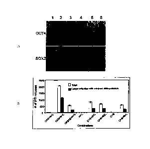
shows a Western blot analysis of OCT-4 and SOX2 in 293FT cells. Lane 1, pSIN4-
EF2-
OCT4-IRESI-S0X2; lane 2, pSIN4-EF2-OCT4-IRES2-SOX2; lane 3, pSIN4-EF2-OCT4-F2A-
SOX2; lane 4, pSIN4-EF2-OCT4-IRES1-PURO; lane 5, pSIN4-EF2-SOX2-IRES1-PURO;
lane 6, no plasmid (control). Mouse anti-human OCT4 monoclonal antibody
(1:500, Santa
Cruz Biotechnology, Inc., Santa Cruz. CA, sc-5279) and goat anti-human SOX2
polyclonal
antibody (1:500, R&D Systems, Minneapolis, MN AF2018) were used to detect the
relative
expression of OCT4 and SOX2 respectively.
[0032] FIG. 1B shows reprogramming using linked potency-determining factors
in
0.2 x 10⁶ mesenchymal cells derived (Yu et al., supra) from OCT4 knock-in
human ES cells
9
CA 02741090 2016-06-17
(US Patent Application No. 2006/0128018 and Zwaka and Thomson, Nature
Biotechnology
21:319-321(2003)). This line was maintained under neomycin selection
(geneticin: 100
ng/ml, Invitrogen Corp.). Human iPS cell colonies were counted on day 16 post-
transduction. The gene combinations were pSIN4-EF2-0C74-IRES1-SOX2 (01S);
pSIN4-
EF2-OCT4-IRES2-S0X2 (02S); pSIN4-EF2-OCT4-F2A-SOX2 (0F2AS); pSIN4-EF2-
NANOG-IRES1-LIN28 (Ni L); pSIN4-EF2-NANOG-IRES2-LIN28 (N2L); pSIN4-EF2-OCT4-
IRES1-PURO (0); pSIN4-EF2-S0X2-IRES1-PURO (S); pSIN4-EF2-NANOG-IRESI-PURO
(N); pSIN4-EF2-LIN28-IRES1-PURO (L). The abbreviation used for each lentiviral
plasmid
vector is shown in parentheses after the vector name.
Example 2
Reprogramming human newborn foreskin fibroblasts using lentiviral constructs
[0033] Preliminary reprogramming experiments were conducted by introducing
lentiviral vectors into human neonatal foreskin fibroblasts. FIG. 2A shows
that NANOG has
a profound positive effect on reprogramming efficiency when OCT4, SOX2, LIN28,
and c-
MYC are also introduced, and that in combination with OCT4, SOX2, and LIN28,
NANOG
can support reprogramming, even in the absence of c-MYC or ICLF4, Lentiviral
constructs
used were pSIN4-EF2-OCT4-IRES2-SOX2 (02S); pSIN4-EF2-NANOG-IRES2-LIN28 (N2L);
pSIN4-EF2-LIN28-IRES1-PURO (L); pSIN4-CMV-c-Myc-IRES1-PURO (M); pSIN4-EF2-
KLF4-IRES1-PURO (K). Twenty-one days after transduction, alkaline phosphatase-
positive
human iPS cell colonies were counted. The number of iPS cell colonies were
derived from
an input of 2.5 x 10⁴ human newborn foreskin fibroblasts (passage 9). The
light gray bars
represent the total number of reprogrammed colonies formed having typical
human ES cell
morphology; dark gray bars indicate the number of large colonies with minimal
differentiation.
[0034] FIG. 2B evidences reprogramming using linked potency-determining
factors.
Lentiviral constructs used were pSIN4-EF2-c-Myc-IRES2-KLF4 (EF2-M2K); pSIN4-
CMV-c-
Myc-IRES2-KLF4 (CMV-M2K); pSIN4-EF2-KLF4-IRES2-c-Myc (EF2-K2M); pSIN4-CMV-
KLF4-IRES2-c-Myc (CMV-K2M); pSIN4-CMV-c-Myc-IRES2-LIN28 (M2L); pSIN4-EF2-
NANOG-IRES2-KLF4 (N2K). Fourteen days after transduction, alkaline phosphatase-
positive human iPS cell colonies were counted. The number of iPS cell colonies
were
derived from an input of approximately 7.0 x 10⁴ foreskin fibroblasts (passage
12). The
CA 02741090 2011-04-18
WO 2010/048567
PCT/US2009/061935
asterisk indicates that most of the alkaline phosphatase-positive colonies
appeared
morphologically loose.
[0035] FIG. 2C shows the effect of SV40 large T antigen gene expression on
reprogramming efficiency. SV40 large T antigen prevents c-Myc-induced in
murine
fibroblasts (Hexmelcing et al., PNAS 91:10412-10416 (1994)) and enhances
reprogramming
efficiency (Hanna et al., Cell 133:250-264 (2008); Mali etal., Stem Cells doi:
10.1634/stemcells.2008-0346 (2008)). Abbreviations of gene combinations are
the same as
in FIG. 2B, with the addition of SV40 large T antigen (T). c-Myc also promotes
cell
proliferation. Twelve days after transduction, alkaline phosphatase-positive
human iPS cell
colonies were counted. The number of iPS cell colonies were derived from an
input of
approximately ¨3.5 x 10⁴ foreskin fibroblasts (passage 17). Fig. 2C
demonstrates that if
present at levels achieved during lentiviral-based reprogramming, T antigen
inhibits fmal
stages of iPS cell derivation. In contrast, see infra, wherein T antigen does
not have this
effect when present for the temporal expression time and/or level achieved
during
reprogramming using episomal vectors. In addition, T antigen prevents c-Myc-
induced
apoptosis but does not adversely affect c-Myc-induced cell proliferation.
Example 3
Reprogramming of human newborn foreskin fibroblasts using non-viral episomal
constructs
[0036] Human newborn foreskin fibroblasts (Cat# CRL-2097111, ATCC) were
maintained in foreskin fibroblast culture medium (DMEM (Cat# 11965,
Invitrogen)
supplemented with 10% heat-inactivated fetal bovine serum (FBS, HyClone
Laboratories,
Logan, UT), 2 mM Glutamax, 0.1 mM non-essential amino acids, and 0.1 mM13-
mercaptoethanol).
[0037] Various combinations of potency-determining factors provided as
transgene
expression cassettes constructed as in Example 1 and as detailed below in
Table 3 were
introduced into somatic cells using an episomal construct pCEP4-EGFP (as shown
in Fig.
3A) resulting in reprogramming with varying efficiency. pCEP4-EGFP was created
from
commercially available mammalian episomal expression vector pCEP4 (Invitrogen
Corp.,
Carlsbad, CA) by inserting the EGFP coding region between the pCEP4 BamHI and
NheI
sites. The episomal vectors of Table 2 were created by inserting the
designated expression
cassettes into pCEP4-EGFP or into a related backbone lacking Pcmv (designated
pEP4). See
Fig. 3A and Table 2 footnotes for cloning sites into which expression
cassettes were inserted.
[0038] Vectors were introduced into the fibroblasts via a single
nucleofection event,
using Human Dermal Fibroblasts Nucleofector Kit (Normal Human Dermal
Fibroblasts,
QB \960296.00915\ 9067411.1 11
CA 02741090 2016-06-17
Amaxa, Inc. Cat. No. VPD-1001), in accord with the manufacturer's
instructions. After
nucleofection, the transfected fibroblasts (¨ 0.8 to 1.0 x 10⁶ cells each)
were immediately
plated onto three 10 cm dishes seeded with irradiated mouse embryonic
fibroblasts (MEF).
Foreskin fibroblast culture medium was replaced every other day. After four
days, the
foreskin fibroblast culture medium was replaced with human ES cell culture
medium
(DMEM/F12 culture medium supplemented with 20% KnockOut serum replacer, 0.1 mM
non-essential amino acids (all from Invitrogen Corp.), 1 mM Glutamax, 0.1 mM
13-
mercaptoethanol and 100 ng/ml zebrafish basic fibroblast growth factor (zbEGF)
as
previously described (Amit etal., Developmental Biology 227:271-278 (2006);
Ludwig et
al., Nature Methods 3:637-646 (2006)). When the seeded MEF could no longer
sustain the
reprogramming culture, about 8 to 10 days after plating, human ES cell culture
medium
conditioned with irradiated MEF was used instead. When appropriate (about 2-3
weeks after
transfection), the cultures were stained for alkaline phosphatase as an
indication of human
iPS colony development.
[0039] To determine suitable parameters for introducing transgene
constructs,
temporal expression was initially evaluated by measuring EGFP level over time
after
introduction of EGFP from pEGFP-N2 (control) and pCEF'4-EGFP episomal vector
into
293FT cells was evaluated (Fig. 38).
[0040] The effect of the amount of transgene construct introduced on human
newborn
foreskin fibroblast cell survival was also evaluated in preliminary
experiments. FIG. 3C
shows the effect of amount of pCEP4-EGFP episomal vector used on nucleofection
efficiency and survival of human newborn foreskin fibroblasts, estimated from
cell
confluence on the day after nucleofection. Approximately 1 x 10⁶ nucleofected
foreskin
fibroblasts were plated into each well of a 6-well plate. Gray lines represent
non-transfected
control fibroblasts; black lines represent transfected fibroblasts.
[0041] Fig. 4A depicts schematic transgene expression constructs from Table
3
containing various expression cassettes that when introduced in certain
combinations into
human newborn foreskin fibroblasts result in reprogramming of the fibroblasts
to pluripotent
cells. Three combinations of introduced episomal reprogramming vectors have
yielded
reprogrammed pluripotent cells: (1) pEP4-E-02S-E-T2K, pEP4-E-02S-E-N2K and
pCEP4-
C-M2L; (2) pEP4-E-02S-C-K2M-E-N2L and pEP4-E-02S-E-T2K; and (3) pEP4-E-02S-E-
N2L, pEP4-E-02S-E-T2K and pEP4-E-02S-E-M2K. Table 3 indicates the amount of
each
12
CA 02741090 2016-06-17
vector used in each successful combination. One vector in each successful
reprogramming
combination encoded T antigen under control of the EF la promoter.
[0042] FIG. 4B shows a bright-field microscopy image of a typical colony
with
morphological changes observed 18 days after episomal vector transfection.
FIG. 4C shows
a bright-field microscopy image of an alkaline phosphatase-positive colony 18
days after
episomal vector transfection.
[0043) Twenty-five to thirty days after transfection, the reprogramming
cultures were
passaged once to fresh 10 cm MEF dishes (1:3 ratio), due to the presence of
many non-iPS
cell colonies with morphologies similar to human iPS cell colonies. Colonies
were then
picked for further analysis. FIG. 4D shows a bright-field microscopy image of
a human iPS
cell colony 6 days after the first passage of day 28 post-transfection
reprogramming culture.
The scale bar represents 0.1 mm. Reprogrammed cells were maintained for
subsequent
analysis in feeder-free culture on MatrigelTM (BD Biosciences, Bedford, MA)
with
conditioned medium as previously described (Xu et al., Nat. Biotechnol. 19:971
(2001)).
[0044] Advantageously, the reprogramming efficiency of greater than 1% of
the
newborn foreskin fibroblast cells reprogrammed was achieved, at significantly
lower
reprogramming time than was achieved using four gene combinations.
[0045] The scope of the claims should not be limited by the preferred
embodiments
set forth in the examples, but should be given the broadest interpretation
consistent with the
description as a whole.
13
CA 02741090 2016-06-17
TABLE 1-- Reprogramminz genes and translation elements
Gene Symbol Abbr. Source SEQ ID NO Accession # or sequence
OCT4 0 hESC 1 NM_002701
SOX2 S hESC 2 NM_003106
NANOG N hESC 3 NM 024865
LN28 L hESC 4 NM_024674
c-Myc M hESC 5 NM_002467
KLF4 K hESC 6 NM_004235
pBABE-puro
SV40 T T 7 EF579667
SV40 LT p
_ .
pBABE-hygro-
7ERT TERT 8 NM 198253
hTERT
IRES1 1 pIRESpuro3 --
' IRES2 2 pIRES2EGFP --
F2A F2A (synthesized) ' 9
CMV . C ' 10
EFla E 11
EF2a - ' 12
14
CA 02741090 2016-06-17
TABLE 2: Episomal constructs
# Name Size (bp)
1 pCEP4-EGFP 10984
21' pEP4-E-02S(2) 13523
3b 'pEP4-E-M2K 14293
4 a pCEP4-M2K 13643
5b pEP4-E-K2M 14268
pCEP4-K2M 13636
7b pEP4-E-N2K 13819
8 b pEP4-E-T2K 15071
9 a pCEP4-M2L 12852
pEP4-E-N2L 13020
11 b pEP4-E-T2L 14284
12 pEP4-E-02S-C-M2K 16038
13 pEP4-E-02S-E-M2K 16680
14 pEP4-E-02S-C-K2M 16010
pEP4-E-02S-E-K2M 16652
16 C pEP4-E-02S-E-N2K 16206
17 C 'pEP4-E-02S-E-T2K 17458
18' pEP4-E-02S-E-N2L 15415
19 pEP4-E-02S-E-T2L 16679
20G pEP4-02S-C-M2L 15247
21' pEP4-E-02S-E-K2T 17474
22 C pEP4-E-02S-C-M2L-E-N2K 19956
23 pEP4-E-02S-C-M2K-E-N2L 19956
24 C pEP4-E-02S-C-K2M-E-N2L 19949
pEP4-E-02S-C-M2L-E-T2K 21220
26C 'pEP4-E-02S-C-M2K-E-T2L 21220
27 C pEP4-E-02S-C-K2M-E-T2L 21213
28' 'pEP4-E-02S-C-M2L-E-K2T 21224
CA 02741090 2016-06-17
a All linlced gene cassettes were cloned into the pCEP4-EGFP between BamHI and
NheI
restriction sites.
b All linked gene cassettes plus the EFla promoter were cloned into the pCEP4-
EGFP
between BamHI and SpeI (19) restriction sites.
All expression cassettes were cloned into the pCEP4-EGFP between BamHI and
Nrul
restriction sites.
16
CA 02741090 2016-06-17
TABLE 3: Combinations of episomal constructs tested for reprogramming activity
Equivalent of Morph. AP+ colony
pCEP4-EGF(pg) Test # Plasmids pig Changes /plate
EXPERIMENT 1
_
6.3 1 pEP4-E-02S-C-M2K 9.2
+/- . 0
6.3 2 pEP4-E-02S-K2Neo 9.3 +/- 0
6.3 pCEP4-M2L 7.4
6.3 , 3 pEP4-E-02S-E-N2K 9.3 +/- 0
6.3 pCEP4-M2L 7.4
6.3 4 pEP4-E-02S-E-T2K 10 i-H- 0
6.3 pCEP4-M2L 7.4
,
6.3 . 5 pEP4-E-02S-E-TERT2K 10.8 . +/- 0
6.3 pCEP4-M2L 7.4
6.3 6 pEP4-E-02S-C-M2L 8.7 +1- 0
6.3 pEP4-E-N2K . 7.9 .
6.3 7 pEP4-E-02S-C-M2L 8.7 , + 0 '
6.3 pEP4-E-T2K 8.6
6.3 8 2EP4-E-02S-C-M2L , 8.7 , +/- 0
6.3 pEP4-E-TERT2K 9.4
EXPERIMENT 2
3.3 , 1 pEP4-E-02S-C-M2K 5.0 +/- 0
3.3 2 pEP4-E-02S-E-M2K 5.0 +/- 0
3.3 . 3 1EP4-E-02S-C-K2M 5.0 +/- 0
3.3 4 pEP4-E-02S-E-K2M 5.0 +/- 0
2.5 5 pEP4-E-02S(2) 3.0 , +/- , 0
2.5 pCEP4-M2K 3.0 ..
2.5 6 pEP4-E-02S(2) 3.0 +/- 0
2.3 pEP4-E-M2K 3.0
,
2.5 7 _pEP4-E-02S(2) 3.0 +/- 0
2.5 pCEP4-K2M 3.0
2.5 8 pEP4-E-02S(2) 3.0 . +1- 0
2.3 pEP4-E-K2M 3.0
1.7 9N pEP4-E-02S(2) 2.0 +/- 0
1.5 pEP4-E-N21( 2.0
1.7 pCEP4-M2L 2.0 +/- 0
1.7 . lON pEP4-E-02S(2) 2.0 +/- 0
1.7 pEP4-E-N2L 2.0
1.7 pCEP4-M2K 2.0
-
1.7 , 11N 2EP4-E-02S(2) 2.0 +/- 0
1.7 2EP4-E-N2L 2.0
1.5 pEP4-E-M2K 2.0
17
CA 02741090 2016-06-17
1.7 12N yEP4-E-02S(2) 2.0 +/- 0
1.7 pEP4-E-N2L 2.0
1.7 pCEP4-1(2M 2.0
1.7 13N pEP4-E-02S(2) 2.0 +/- 0
1.7 pEP4-E-N2L 2.0
1.5 pEP4-E-K2M . 2,0
2.3 14N pEP4-E-02S-E-N2K 3.5 +/- 0
2.1 pCEP4-M2L 2.5
2.5 15N pEP4-E-02S-E-N2L 3.5 +/- 0
2.1 pCEP4-M2K 2.5
2.5 16N pEP4-E-02S-E-N2L 3.5 +/- 0
1.9 pEP4-E-M2K 2.5
2.5 , 17N pEP4-E-02S-E-N2L 3.5 +/- 0
2.1 pCEP4-K2M 2.5 ,
2.5 18N pEP4-E-02S-E-N2L 3.5 +/- 0
1.9 pEP4-E-K2M 2.5
EXPERIMENT 3
1.7 ' 9T pEN-E-02S(2) 2.0 -H- 0
1.4 yEP4-E-T2K 2.0
1.7 pCEP4-M2L 2.0
1.7 10T pEP4-E-02S(2) 2.0 + 0
1.5 pEP4-E-T2L 2.0
, .
1.7 pCEP4-M2K 2.0
1.7 11T yEP4-E-02S(2) 2.0 + 0
1.5 pEP4-E-T2L 2.0 ,
. .
1.5 pEP4-E-M2K 2.0
1.7 , 12T pEP4-E-02S(2) . 2.0 +/- 0
1.5 pEP4-E-T2L , 2.0 .
1.7 yCEP4-K2M 2.0 ,
1.7 13T pEP4-E-02S(2) 2.0 +/- 0
1.5 pEP4-E-T2L 2.0
1.5 pEP4-E-K2M 2.0
, _.
2.2 14T 9EP4-E-02SET2K 3.5 -i-H- 0
2.1 pCEP4-M2L 2.5
2.3 15T pEP4-E-02S-E-T2L 3.5 , + 0
1
2. yCEP4-M2K 2.5 .
, _ .
2.3 16T pEP4-E-02S-E-T2L 15 + 0 .
1.9 pEP4-E-M2K 2.5
,
2.3 17T pEP4-E-02S-E-T2L 3.5 +/- 0
,
2.1 pCEP4-K2M 2.5
, ,
2.3 18T pEP4-E-02S-E-T2L 3.5 +1- 0
1.9 ,yEP4-E-K21V1 2.5
P, ;:44*.$,',.;:.0
Pir ' IIV:C:! ' ',': -114:4 fili*'..4.. S.:$:' 1;1'44 r,=::4-.;:*" '4' , i'
.'' .-.'' rAlii:M'1;::# ;''''::- ','' ii'L:V4
18
CA 02741090 2016-06-17
EXPERIMENT 4
pEP4-E-02S-C-M2K-E-
6 1 2L 10.9 +/- 0
ye EP4-E-02S-C-M2K-E-
4 2 2L 7.3 i 1 I 0 .
2 ye EP4-E-02S-E-T2K 3.2
p EP4-E-02 S-C-K2M-E-
___ 6 3 2L 10.9 +/- 0
N
,.. '..,.. .. ,it. , ,-,-, , . i' 4 ii, 27 -2-:,te-::.%.k.4. ' ''""t-
,',7:-:-: ':71
:a.: ' - - c ',, , II! ,1..4 = . ".. ,,...424,!'. - ,
3 5 p EP4-E-02S-E-N2L 4.2 +/- 0
3 ye EP4-E-02S-E-M2K 4.6
1 ,: : .i., ..,. 2- : = -f ._ , _:i
=.#7,,E_:,;. "i: :d,-, . ,oliw,,,t4,. ,,,..,..--,--,i7,,,;.NT., -.;.. -
,..wy,...:,N,-1.1i,,14,-.,,
..,..:-.. . ....,. . , _,_,I. ,_ ;;.,...
..-*.' =--,..= ...=--e-''.v. ::::<;-"k -=': ' ' t t'4*: 4 '''',4;'
3 =-= -- . "T;'= ' =, , 74. 44:410/* 7 iteei:.; .. .-
,..,,. ,:..v, i: . ;;,. - '.. - !' = , = , .- ,! ,'' ' i' 4 -",-=
3 7 ye EP4-E-02S-E-N2L 4.2 +/- 0
3 1 e EP4-E-02S-C-M2K 4.4
3 8 le EP4-E-02S-E-N2L 4.2 + 0 ,
2 p EP4-E-02S-E-T2K 3.2
3 y e EP4-E-02S-C-M2K 4.4
3 9 ye EP4-E-02S-E-N2L 4.2 +/- 0
3 y e EP4-E-02S-E-K2M 4.5
3 10 p EP4-E-02S-E-N2L 4.2 +/- 0
2 ye EP4-E-02S-E-T2K 3.2
,
3 is EP4-E-02S-E-K2M 4.5
3 11 le EP4-E-02S-E-N21. 4.2 +/- 0
,
3 ye EP4-E-02S-C-K2M 4.4
3 12 p EP4-E-02S-E-N2L 4.2 + 0
2 leEP4-E-02S-E-T2K 3.2 ,
3 1 . EP4-E-02S-C-K2M 4.4
le EP4-E-02S-C-M2L-E-
2 13 2K 3.9 + 0
4 y e EP4-E-02S-E-N2K 5.9
p EP4-E-02S-C-M2K-E-
6 14 L 11.6 + 0
le EP4-E-02S-C-M2K-E-
3 15 2L 5.8 + 0
3 le EP4-E-02S-E-N2K 4.4
. ,
p EP4-E-02 S-C-1C2M-E-
6 16 2L 11.6 +/- 0
I. EP4-E-02S-C-K2M-E-
3 17 2L 5.8 + 0
3 y e EP4-E-02S-E-N2K 44
19
CA 02741090 2016-06-17
pEP4-E-02S-C-M2L-E-
6 18 K2T 11.6 +/- 0
pEP4-E-02S-C-M2L-E-
3 19 K2T 5.8 +/-
3 pEP4-E-02S-E-N2K 4.4
3 20 pEP4-E-02S-E-K2T 4.8 +/-
3 pEP4-E-02S-E-N2K 4.4
2 _pEP4-E-02S-C-M21, 2.8 _
+/-: No or very few colonies with morphological change were observed (fig:
FIG. 4B).
-F, and Different number (from less to more) of colonies with
morphological change
were observed.