Note: Descriptions are shown in the official language in which they were submitted.
CA 02741431 2016-04-05
1
INTRODUCER FOR DEPLOYING A STENT GRAFT
IN A CURVED LUMEN
Technical Field
The present invention relates to an introducer for deploying a stent graft
within a curved lumen. It also relates to a method of deploying a stent graft
within
a curved lumen, and to a stent graft.
Background
Stent grafts are used to replace or repair vessels of the body such as the
arteries. A stent graft is usually formed from a tubular body of a
biocompatible
graft with one or more stents mounted into or onto the tubular body to provide
support therefor. The stents may be balloon expandable stents or self-
expanding
stents.
Endovascular methods have been proposed for treatment of an aneurysm
of the aorta particularly where the aneurysm is adjacent the aorta
bifurcation.
However, when an aneurysm occurs higher up in the aorta, in the region of the
descending aorta adjacent the thoracic arch or in the ascending aorta,
endovascular techniques for treating these aneurysms are somewhat more
difficult
because of the tight curvature of the thoracic arch, the occurrence of major
arteries
in the region and the proximity to the heart. Placement of a substantially
cylindrical prosthesis in such a curved region can cause problems.
Stent grafts are typically deployed using endovascular techniques on an
introduction device in which the stent graft is retained in a radially
contracted
condition by a sheath. Upon withdrawal of the sheath and release of any
retention
arrangement where provided, for example in cases in which the stent graft has
self-expanding stents, the stent graft can expand under the action of the self-
expanding stents towards the vessel walls to redefine the blood flow path. The
introduction device is withdrawn after deployment.
Currently, stent grafts are deployed in curved lumens by causing these to
follow the curvature imparted to the introducer. However, this can result in
the
stent graft not sitting properly in the blood vessel and in the lumen of the
CA 02741431 2016-04-05
2
prosthesis being closed off or reduced in diameter. Kinks can also occur
along the length of the prosthesis and these can cause problems with
restriction of flow in the lumen.
Furthermore, when deploying a stent graft that is substantially
cylindrical in a curved aorta there is a danger that the proximal end of the
stent graft, that is, the end nearest the heart, will not lie flat against the
walls of the aorta (i.e., is not positioned perpendicularly to the wall of the
vessel) and blood can flow underneath the edge of the graft, particularly on
the inner side of the curve of the thoracic arch and cause the stent graft to
buckle and close off thereby causing serious problems.
US 6,974,471, US 2004/0073289, US 7,318,835, US 7,279,003
disclose prior art prostheses for implantation within a curved body lumen.
In general this application relates to the placement of prostheses in
the aorta in the region known as the thoracic arch where the aorta leaves
the heart and curves over in approximately a semi-circle to the descending
aorta and then into the abdominal aorta and then into the lower limbs via
the iliac arteries. The invention is, however, not so restricted and can
relate
to placement of prostheses within or in place of lumens in any portion of a
human or animal body, though it is particularly relevant to curved lumens,
particularly tightly curved lumens.
Summary
Certain exemplary embodiments provide an introducer for deploying
a stent graft in a curved lumen, the introducer including: a carrier for a
stent graft including a plurality of stents; a release mechanism including a
constraining mechanism, wherein the constraining mechanism includes at
last one release wire; stent graft mounted on the carrier, the stent graft
including at least one wire-receiver co-operating with the release wire of the
CA 02741431 2016-04-05
2a
_
introducer, wherein the wire-receiver and the release wire radially constrain
at least a portion of at least one stent of the stent graft, wherein at least
an
adjacent portion of an adjacent stent of the stent graft is not radially
constrained by the release wire, wherein at least one of the constrained
5 stent portions is not provided at the proximal end of the most proximal
stent
and is not provided at the distal end of the most distal stent; the release
wire and the wire receiver together operable to enable the constrained stent
to expand so as to overlap with the interior of at least a portion of an
adjacent stent in an expanded portion of the stent graft.
10 Other exemplary embodiments provide a method of deploying a
stent graft within a curved lumen including: (a) delivering a stent graft
including a plurality of stents to the site of deployment; (b) expanding a
first
portion of the stent graft and maintaining in a radially constrained
configuration at least a portion of at least one stent, wherein at least one
of
15 the constrained stent portion is not provided at the proximal end of the
most
proximal stent and wherein at least one of the constrained stent portions is
not provided at the distal end of the most distal stent of the stent graft;
(c) after expansion of the first portion of the stent graft, allowing the
constrained stent to expand such that it overlaps with the interior of at
least
20 a portion of an adjacent stent in the expanded portion of the stent
graft.
Other exemplary embodiments provide use of a stent graft
comprising a plurality of stents suitable for deployment within a curved
lumen, the stent graft comprising: a first portion suitable for expansion; and
further comprising at least a further portion of at least one stent that is
not
25 at the proximal end of the most proximal stent of the stent graft, and
is not
at the distal end of the most distal stent of the stent graft, that is a
radially
constrained configuration, but which is suitable for expansion after
CA 02741431 2016-04-05
2b
expansion of the first expanded portion of the stent graft, such that it is
suitable for overlap with the interior of at least a portion of an adjacent
stent
in the expanded portion of the stent graft.
The present invention seeks to provide an improved introducer and
method for deploying a stent graft within a curved lumen.
According to a first aspect of the present invention, there is provided
an introducer for deploying a stent graft in a curved lumen, the introducer
including: a carrier for a stent graft including a plurality of stents; a
release
mechanism including a constraining mechanism operable to maintain at
least a portion of at least one stent of the stent graft in a radially
constrained configuration during deployment whilst allowing at least a
portion of the stent graft to expand, wherein the portion of the constrainable
stent is radially constrained substantially entirely therearound; the release
mechanism operable to enable the constrained stent to
CA 02741431 2011-04-21
WO 2010/062362
PCT/US2009/005912
3
expand so as to overlap with the interior of at least a portion of an adjacent
stent in
the expanded portion of the stent graft.
The introducer enables a substantially cylindrical stent graft to be deployed
in a curved lumen without the need to match the curve of the stent graft to
the
lumen either prior to, or during, deployment. Furthermore, the stent graft can
be
accommodated within a curved lumen without the stents bunching together and
creating gaps that might cause blood leakage.
In a preferred embodiment, the constrainable stent is not the most proximal
stent of the stent graft or the most distal stent of the stent graft. This
assists in
enabling the ends of the stent graft to be securely anchored within the
vessel.
The constraining mechanism may include a release wire for co-operating
with a wire-receiving mechanism provided on the constrainable stent.
More than one stent may be maintained in a constrained configuration
during deployment whilst at least a portion of the stent graft expands.
According to a second aspect of the present invention, there is provided a
method of deploying a stent graft within a curved lumen including: (a)
delivering a
stent graft including a plurality of stents to the site of deployment; (b)
expanding a
first portion of the stent graft and maintaining in a radially constrained
configuration
at least a portion of at least one stent, wherein the portion of the
constrainable
stent is radially constrained substantially entirely therearound; (c) after
expansion
of the first portion of the stent graft, allowing the constrained stent to
expand such
that it overlaps with the interior of at least a portion of an adjacent stent
in the
expanded portion of the stent graft.
This method allows a substantially cylindrical stent graft to be deployed
within a curved lumen, irrespective of the extent of the curvature of the
lumen and
without the need to match the curvature of the stent graft with the curvature
of the
lumen either prior to, or during, deployment.
Preferably, the constrained stent is not the most proximal stent of the stent
graft or the most distal stent of the stent graft. This facilitates proper
anchoring of
the stent graft.
CA 02741431 2011-04-21
WO 2010/062362 PCT/US2009/005912
4
According to a third aspect of the present invention, there is provided a
stent graft for deployment within a curved lumen by means of the above-
described
introducer and/or by means of the above-described method, the stent graft
including a plurality of stents, the stent graft including a mechanism for
allowing at
least a portion of at least one stent to be radially constrained during
deployment
whilst a portion of the stent graft is expanded, wherein the portion of the
constrainable stent is radially constrained substantially entirely
therearound.
The stent graft is typically substantially cylindrical prior to deployment,
but is
able to be securely located within a curved lumen, irrespective of the extent
of the
curvature of the lumen and without the need to match curvature of the stent
graft
with the curvature of the lumen either prior to, or during, deployment.
Preferably, the mechanism is not provided at the most proximal stent of the
stent graft or at the most distal stent of the stent graft. This facilitates
secure
anchoring of the stent graft within the lumen.
The mechanism may include at least one wire-receiver for co-operating with
a release wire of the introducer. The wire-receiver may be a loop of material,
such
as a loop of thread.
In an embodiment, the mechanism may be provided at only one end of the
constrainable stent, for example, it may be provided only at the proximal end
of the
constrainable stent. In another embodiment, the mechanism may be provided at
both the proximal end and the distal end of the constrainable stent.
The stent graft may include more than one constrainable stent. In an
embodiment, it may include at least one constrainable stent in which the
mechanism is provided only at one of its ends, and at least one constrainable
stent
in which the mechanism is provided at both its proximal end and its distal
end.
CA 02741431 2011-04-21
WO 2010/062362 PCT/US2009/005912
Brief Description of the Drawings
Embodiments of the present invention are described below, by way of
example only, with reference to the accompanying drawings, in which:
Figures 1 and 2 show an example of an implant deployment device;
5 Figure 3 shows a prior art stent graft for deployment in a curved lumen;
Figure 4 shows a stent graft in accordance with an embodiment of the
present invention;
Figure 5 shows an end view of the stent graft of Figure 4;
Figure 6 shows an embodiment of an introducer for deploying the stent graft
of Figure 4;
Figure 7 shows deployment of the stent graft of Figure 4 in a curved body
lumen;
Figure 8 shows the stent graft of Figure 4 deployed in a curved body lumen;
Figure 9 shows deployment of another embodiment of a stent graft in a
curved body lumen;
Figure 10 shows the stent graft of Figure 6 deployed in a curved body
lumen;
Figure 11 shows deployment of another embodiment of a stent graft in a
curved body lumen;
Figure 12 shows the stent graft of Figure 10 deployed in a curved body
lumen; and
Figure 13 illustrates one possible arrangement of a stent graft and an
implant deployment device.
Detailed Description
It is to be understood that the Figures are schematic and do not show the
various components in their actual scale. In many instances, the Figures show
scaled up components to assist the reader.
In this description, when referring to a deployment assembly, the term distal
is used to refer to an end of a component which in use is furthest from the
surgeon
during the medical procedure, including within a patient. The term proximal is
CA 02741431 2011-04-21
WO 2010/062362 PCT/US2009/005912
6
used to refer to an end of a component closest to the surgeon and in practice
in or
adjacent an external manipulation part of the deployment or treatment
apparatus.
On the other hand, when referring to an implant such as a stent or stent
graft, the term proximal refers to a location which in use is closest to the
patient's
heart, in the case of a vascular implant, and the term distal refers to a
location
furthest from the patient's heart.
Referring to Figures 1 and 2, an implant deployment device 10 includes an
external manipulation section 12, a proximal attachment region 14 and a distal
attachment region 16. The proximal attachment region 14 and the distal
attachment region 16 secure the two ends of the implant 18. During the medical
procedure to deploy the implant 18, the proximal and distal attachment regions
14
and 16 will travel through the patient's vasculature, in this example, to a
desired
deployment site. The external manipulation section 12 at the proximal end of
the
implant deployment device 10, which is operated by a surgeon to manipulate the
introducer, remains outside of the patient throughout the procedure.
The distal attachment region 16 of the implant deployment device 10
includes a dilator tip 20, which is typically provided with a bore 22 therein
for
receiving a guide wire (not shown) of conventional type. The longitudinal bore
22
also provides a channel for the introduction of medical reagents. For example,
it
may be desirable to supply a contrast agent to allow angiography to be
performed
during placement and deployment phases of the medical procedure.
An inner catheter or cannula 24, conventionally made from a flexible thin
walled metal tube, is fastened to the dilator tip 20. The inner catheter 24 is
flexible
so that the implant deployment device 10 can be advanced along a relatively
tortuous vessel, such as a femoral artery, and so that the distal end of the
implant
deployment device 10 can be longitudinally and rotationally manipulated. The
inner catheter 24 carries a stent 18 or other device to be implanted in the
patient.
The catheter 24 extends through the implant deployment device 10 to the
manipulation section 12, terminating at a connection device 26, in
conventional
manner.
CA 02741431 2011-04-21
WO 2010/062362 PCT/US2009/005912
7
The connection device 26 is designed to accept a syringe to facilitate the
introduction of reagents into the inner catheter 24 and for this purpose is
typically
provided with a threaded luer lock connection.
Where provided, a pusher sheath or rod 30 (hereinafter referred to as a
pusher member), typically made from a plastics material, is mounted coaxial
with
and radially outside of the inner catheter 24. The pusher member 30 is "thick
walled", that is the thickness of its wall is preferably several times greater
than that
of the guide wire catheter 24. In some instances, the pusher member 30 and the
inner catheter 24 are the same component, possibly having different outer
diameters at the location at which the stent 18 is to be carried.
A sheath 32 extends coaxially over and radially outside of the pusher
member 30. The pusher member 30 and the sheath 32 extend distally to the
manipulation region 12.
The implant 18, which may be a stent, a stent graft or any other implant or
prosthesis deliverable by the implant deployment device 10, is retained in a
compressed condition by the sheath 32. The sheath 32 extends proximally to a
sheath manipulator and haemostatic sealing unit 34 of the external
manipulation
section 12. The haemostatic sealing unit 34 includes a haemostatic seal (not
shown) and a side tube 36 held to the unit 34 by a conventional luer lock 38.
The sheath manipulator and haemostatic sealing unit 34 also includes a
clamping collar (not shown) that clamps the sheath 32 to the haemostatic seal
and
a silicone seal ring (not shown) that forms a haemostatic seal around the
pusher
member 30. The side tube 38 facilitates the introduction of medical fluids
between
the pusher member 30 and the sheath 32. Saline solution is typically used.
During assembly of the implant deployment device 10, the sheath 32 is
advanced over the proximal end of the dilator tip 20 of the proximal
attachment
region 16 while the implant 18 is held in a compressed state by an external
force.
A suitable distal attachment (retention) section (not visible in this view) is
coupled
to the pusher member 30 and retains a distal end 40 of the prosthesis 18
during
the procedure. The distal end of the prosthesis 18 may be provided with a loop
of
material (not shown) through which a distal restraining wire 42 extends. The
distal
CA 02741431 2011-04-21
WO 2010/062362 PCT/US2009/005912
8
restraining wire also extends through an aperture (not shown in Figures 1 and
2) in
the proximal attachment section 40 into an annular region 44 between the inner
catheter 24 and the pusher member 30. The distal restraining wire 42 extends
through the annular space 44 to the manipulation region 12 and exits the
annular
space 44 at a distal wire release mechanism 46.
A proximal portion of the external manipulation section 12 includes at least
one restraining wire actuation section 50 mounted on a body 48, in turn
mounted
onto the pusher member 30. The inner catheter 24 passes through the body 48.
The distal wire release mechanism 46 and the proximal wire release mechanism
50 are mounted for slidable movement on the body 48.
Clamping screws 52 prevent inadvertent early release of the prosthesis 18.
A haemostatic seal (not shown) is included so that the release wires can
extend
out through the body 48 without unnecessary blood loss during the medical
procedure.
A proximal portion of the external manipulation section 12 includes a pin
vice 54 mounted onto the proximal end of the body 48. The pin vice 54 has a
screw cap 56. When screwed in, vice jaws (not shown) of the pin vice 54 clamp
against or engage the inner catheter 24. When the vice jaws are engaged, the
inner catheter 24 can only move with the body 48 and hence it can only move
with
the pusher member 30. With the screw cap 56 tightened, the entire assembly can
be moved together as one piece.
Once the implant deployment device 10 is in the desired deployment
position, the sheath 32 is withdrawn and the proximal and distal wire release
mechanisms 50, 46 are released to allow the prosthesis 18 to expand.
For some procedures, the sheath 32 may be left in place after expansion of
the implant 18. The pusher member 30 and inner catheter 24 may be withdrawn
and replaced by a further component, using the sheath 32 as a guide.
Figure 3 illustrates a prior art stent graft 18' for deployment within a
curved
body lumen. The stent graft 18' comprises a graft material tube 1 which is
substantially cylindrical. The graft material tube 1 has a proximal end 2 and
a
distal end 3. The graft 1 has a number of self expanding zig-zag or well-known
CA 02741431 2014-01-31
9
Gianturco Z-stents 4 positioned at intervals along the length of the tube and
providing the force necessary to open the graft 1 out to the walls of the
aorta when
deployed. The stents 5 and 6 at the distal end 3 and proximal end 2
respectively
are inside the graft 1 and the other intermediate stents 4 are on the outside
of the
graft 1.
The stent graft 18' includes a length reduction arrangement comprising an
elastic material 8 such as a silicone rubber or similar material which is
fastened at
9 at the proximal end 2 of the prosthesis 18' and joined at 1110 near the
distal end
3 of the prosthesis 18'. The length reduction arrangement can also comprise a
shape memory metal such as Nitinol, a nickel titanium alloy, which is heat set
in a
curved configuration.
Upon deployment, the ends of the graft 18' are released from a
deployment device and the elastic material 8 takes up its shortened rest
position
so that the points 9 and 10 move closer together which causes the graft to
form a
curved shape. The curved stent graft 18' can then sit within a body lumen
having
the same curve.
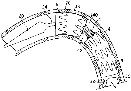
Figures 4 and 5 illustrate a stent graft 18 in accordance with an
embodiment of the present invention. The stent graft 18 is formed from a
tubular
piece of biocompatible graft material 1 having, in this example, six Z-stents
4, 4', 5,
6 disposed along its length. In this embodiment the stents 5, 6 found at the
distal
end 3 and the proximal end 2 of the tubular piece of graft material 1 are
located on
the inside of the tube 1, whereas the intermediate stents 4, 4' are located on
the
outside of the tube 1.
In this embodiment, one of the stents 4' is provided, at substantially
equally spaced locations therearound, with loops of, in this embodiment,
suture
material 140. The loops of suture material 140 are able to engage with a
release
wire 42 of an introducer 10 for deployment of the stent graft 18. The purpose
of
this is described below.
CA 02741431 2014-01-31
For deployment of the stent graft 18 illustrated in Figures 4 and 5, the stent
graft 18 is loaded in a radially compressed condition onto an inner catheter
24 of a
deployment device 10' such as that shown in Figure 6. The introducer 10'
illustrated in Figure 6 is similar to that shown in Figures 1 and 2. However,
the
5 introducer 10' shown in Figure 6 includes a release mechanism including
trigger
wires 42' able to engage with the suture loops 140 of the stent graft 18. The
compressed stent graft 18 is then covered by the sheath 32 in a conventional
manner for deployment.
The stent graft 18 is delivered to the site of deployment, which in this
10 example is within a curved body lumen (such as the thoracic arch). Once
the
implant deployment device 1010 is in the desired deployment position, the
sheath
32 is withdrawn and the stent graft 18 is allowed to expand (see Figure 7).
However, the engagement between the release wires 42'42 and the suture loops
140 retains a single stent 4' in a constrained configuration. The constrained
stent
4' is typically constrained by over 50% of its expanded configuration, and may
be
constrained by up to 70 or 80%. This will depend upon the spacing between the
stents. In practice it will be kept in its fully constrained condition,
whereby it is
constrained around the catheter 24 of the implant deployment device 10' 10. In
an
embodiment, however, the constrained stent 4' may expand partially prior to
release of the constraining mechanism. In a preferred embodiment the partial
expansion comprises the constrained stent 4' expanding to no more than 50% of
its fully deployed diameter.
Next, the release wires 42' are released from the suture loops 140 to allow
the constrained stent 4' to expand.
Once the constrained stent 4' has expanded, the pusher member 30 and
inner catheter 24 may be withdrawn leaving the expanded stent graft 18 in
place
(see Figure 8).
Figures 7 and 8 illustrate the improved positioning effect of this
deployment process. Figure 7 shows the stent graft 18 partially expanded after
CA 02741431 2014-01-31
11
withdrawal of the sheath 32. Only the constrained stent 4' remains in its
compressed state by means of the release wires 42' and the suture loops 140.
The stent graft 18 is located such that the constrained stent 4' is positioned
at the
tightest part of the bend of the curved body vessel 70. As such, the stents 4,
5, 6,
which are allowed to expand as soon as the sheath 32 is withdrawn, engage
against the walls of the body vessel 70 effectively because the vessel is not
too
curved at the location where the stents 4, 5, 6 of the expanded portion are
located.
It can be seen from Figure 7, however, that the stent 4 located immediately
proximally of the constrained 4' and the stent 4 located immediately distally
of the
constrained stent 4' are positioned such that they are closer together on the
inside
part of the curved body lumen 70 than they are towards the outside of the
curve.
This results from the constrained stent 4' drawing the graft material 1 and
the
adjacent stents 4 towards it. As a result, the adjacent stents 4 are able to
locate
within the vessel 70 closer together on the inside of the curve than they
would
have if the stent 4' between them had not been constrained whilst they expand.
Consequently, when the constrained stent 4' is allowed to expand it overlaps
80
with its adjacent stents 4 on the inside of the curve of the curved body
vessel 70.
Because This is because the gap between the adjacent stents 4 is less than the
length of the constrained stent 4'. Because the stents 4 adjacent to the
constrained stent 4' were allowed to expand first, these properly engage the
wall
of the vessel 70, and the expanded constrained stent 4' engages with the
interior
of the adjacent stents 4 providing an area of overlap 80.
A second embodiment is illustrated in Figures 9 and 10. The difference
between the embodiment of Figures 9 and 10 and that of Figures 7 and 8 is that
the constrained stent is constrained only at its proximal end so that it forms
a
"cone-shape" prior to release, but after expansion of the remainder of the
stent
graft 18. Again, when the constrained stent 4' is allowed to expand by removal
of
the release wire 42' from the suture loops 140, the constrained stent 4'
expands
such that it overlaps with the interior of the stent 4 immediately proximal to
the
CA 02741431 2014-01-31
12
constrained stent 4'. As shown in Figure 10, the result is a single region of
overlap
80 between the constrained stent 4' and its immediately proximal stent 4.
Constraining only the proximal end of the constrained stent 4' can provide
a positioning of the stent graft 18 that maximises blood flow through the
stent graft
18 after deployment.
In a third embodiment, more than one constrained stent 4' is provided with
suture loops 140. In such an embodiment, each constrained stent 4' may include
suture loops 140 at both its proximal ends and distal ends (c.f. Figure 7),
or, as
illustrated in Figure 11, (and in particular, where two adjacent constrained
stents 4'
are provided) each constrained stent 4' is provided with suture loops only at
one
end, in this example, the proximal ends. As illustrated in Figure 12 this
arrangement leads to a plurality of regions of overlaps 80 with constrained
stents
4' overlapping with the interior of an immediately proximal stent 4, 6.
An advantage of the above-described embodiments is that a substantially
straight stent graft 18 can be deployed in a curved vessel. The stents 4
adjacent
the constrained stent expand first and are properly anchored within the
vessel. As
the constrained stent 4' expands during a second stage of deployment, the
stent
graft 18 can be used in any type of vessel, whether straight, having only a
slight
bend, or having a sharp bend. Proper curvature of the stent graft 18 within
the
vessel is therefore obtained. In addition, the curve of the stent graft 18
does not
have to be matched to the curve of the vessel prior to deployment.
Furthermore,
the surgeon or clinician does not have to ensure that the stent graft is
deployed in
a particular orientation to match the curve of the vessel as is the case with
prior art
prostheses. Constraining a stent 4' creates a neck portion, which imparts
increased flexibility to the stent graft 18. This assists in enabling the
stent graft 18
to conform to the curvature of a vessel 70 irrespective of the extent of the
curvature of the vessel.
Of course, the skilled person will appreciate that the different types of
constrained stent 4' may be combined within a single stent graft 18 where
CA 02741431 2014-01-31
12a
appropriate. Furthermore, the constrained stent 4' may be located at any point
along the stent graft 18, depending on the particular requirements. For
example,
the constrained stent 4' may be at the proximal end, in the middle, or in any
one
or more of the stents along the stent graft 18. In another modification, it is
envisaged that every stent of the stent graft 18 could be constrained,
preferably
only at one end, which would preferably be at the proximal end of each stent
4'.
In an embodiment, the constrained stent 4' is not the stent 6 at the proximal
end 2
of the stent graft 18. This is because the stent 6 at the proximal end 2 of
the stent
graft 18 can be useful for anchoring and positioning of the stent graft 18.
CA 02741431 2015-09-28
13
In another modification, the release wire 42' may be the same as the wire
42 that holds the proximal end Of the stent graft to the distal end of the
introducer.
In another modification, the suture loops 140 of different constrained stents
140 can co-operate with different release wires 42'. This enables greater
control
over the deployment process where desired by allowing different constrained
stents 4' to be released in a particular desired order.
The suture loops 140 could be provided on the graft material 1 instead of on
the stent 4'. The suture loops 140 could additionally or alternatively be
provided to
co-operate with release wires 42 inside the tubular graft 1 instead of
outside.
In another example of a restraining mechanism illustrated in Figure 13, a
thread of suture material 130 having loops 132 at each end may be provided
around the stent 4' of the stent graft 18, and which, when pulled tight,
constrains
the stent 4'. The loops 132 at each end of the thread of suture material 130
overlap one another and a release wire 42' is threaded therethrough, thereby
maintaining an overlap of the loops 132 and maintaining the stent 4' in its
constrained configuration. Withdrawal of the release wire 42' allows the loops
132
at each end of the thread of suture material 130 to separate and the
constrained
stent 4' to expand. In the example illustrated in Figure 13 the inner catheter
24 is
provided with an aperture 134 through which the suture material 132 extends in
order to engage with the release wire 42'. Other suitable arrangements may be
envisaged.
The term thread as used herein is intended to include any filamentary
material which can perform the stated function and could, for example, be of
conventional suture material, a multi-filamentary structure formed of yarns
for
example and of a natural or synthetic material such as cotton, other
biocompatible
material or a polymer material such as polyester, or a mono-filamentary
structure
of a natural material, other biocompatible material, a metal such as gold or
an
alloy such as Nitinol.