Note: Descriptions are shown in the official language in which they were submitted.
POSITION DEPENDENT INTERFERENCE CANCELLATION
FIELD OF THE INVENTION
The present invention relates generally to invasive
probes, and specifically to determining the position of a
medical probe inside a body cavity.
BACKGROUND
A wide range of medical procedures involve placing
objects, such as sensors, tubes, catheters, dispensing devices
and implants, within the body. Position sensing systems have
been developed for tracking such objects. Magnetic position
sensing is one of the methods known in the art. In magnetic
position sensing, magnetic field generators are typically
placed at known positions external to the patient. One or
more magnetic field sensors within the distal end of a probe
generate electrical signals in response to these magnetic
fields, which are processed in order to determine the position
coordinates of the distal end of the probe. These methods and
systems are described in U.S. Patents 5,391,199, 6,690,963,
6,484,118, 6,239,724, 6,618,612 and 6,332,089, in PCT
International Publication WO 1996/005768, and in U.S. Patent
Application Publications 2002/0065455 Al, 2003/0120150 Al and
2004/0068178 Al.
U.S. Patent 6,370,411 describes a probe having two parts:
a catheter of minimal complexity which is inserted into a
patient's body, and a connection cable that connects between
the proximal end of the catheter and the console. The catheter
comprises a microcircuit that carries substantially only
information specific to the catheter, which is not in common
with other catheters of the same model. The cable comprises an
access circuit which receives the information from the
catheter and passes it in a suitable form to the console. In
some embodiments, the cable operates with all catheters of a
specific model or type, and therefore when a catheter is
1
CA 2741995 2017-06-01
replaced, there is no need to replace the cable. Catheters
that are planned for one-time use do not require replacement
of the cable, which does not come in contact with patients.
U.S. Patent Application Publication 2006/0074289 Al
discusses an endoscopic probe, whose handle has an orientation
sensor that generates signals indicative of the orientation of
the handle in an external frame of reference. The output of
the orientation sensor may be used to sense movement of the
handle relative to its initial position and orientation at the
beginning of the endoscopic procedure.
SUMMARY OF THE INVENTION
An embodiment of the present invention that is described
herein provides a method for position tracking, including:
receiving signals from a main position transducer at a
distal end of a medical probe via wiring traversing the probe
to a connector at a proximal end of the probe, for connection
to a processor, which processes the signals to find a first
position of the distal end;
collecting calibration data with respect to an
interference introduced into the signals at the connector as a
function of a position of the proximal end;
measuring a second position of an auxiliary position
transducer at the proximal end of the probe;
canceling the interference in the signals responsively to
the measured second position and the calibration data; and
calculating the first position based on the signals,
after canceling the interference.
In some embodiments, the medical probe includes a
catheter. In an embodiment, the signals are generated by the
main position transducer in response to one or more magnetic
fields that are applied in a vicinity of the probe and sensed
2
CA 2741995 2017-06-01
CA 02741995 2011-06-03
by the main position transducer. In another embodiment, the
auxiliary position transducer is fitted adjacent to the
connector. The auxiliary position transducer and the connector
may be coupled to a handle of the probe. In another
embodiment, collecting the calibration data includes placing
the proximal end at a plurality of positions relative to a
source of the interference, collecting auxiliary position
signals from the auxiliary position transducer indicative of
the respective positions of the proximal end, and measuring
the interference as a function of the auxiliary position
signals.
In yet another embodiment, measuring the second position
includes applying one or more magnetic fields in a vicinity of
the proximal end, receiving from the auxiliary position
transducer signals that are generated by the auxiliary
position transducer responsively to the magnetic fields, and
calculating the second position based on the received signals.
In still another embodiment, the method includes presenting
the calculated first position to an operator.
There is additionally provided, in accordance with an
embodiment of the present invention, apparatus, including:
a medical probe, which includes a distal end including a
main position transducer, a proximal end including an
auxiliary position transducer, a connector connecting the
distal end to the proximal end, and wiring traversing the
probe and coupling the main position transducer to the
connector; and
a processor, which is configured to receive from the main
position transducer over the wiring signals, which are
indicative of a first position of the distal end, to collect
calibration data with respect to an interference introduced
into the signals at the connector as a function of a position
of the proximal end, to measure a second position of the
auxiliary position transducer, to cancel the interference in
3
CA 02741995 2011-06-03
= .
the signals responsively to the measured second position and
the calibration data, and to calculate the first position
based on the signals, after canceling the interference.
There is also provided, in accordance with an embodiment
of the present invention, a computer software product,
operated in conjunction with a medical probe that includes a
distal end including a main position transducer, a proximal
end including an auxiliary position transducer, a connector
connecting the distal end to the proximal end, and wiring
traversing the probe and coupling the main position transducer
to the connector, the product including a non-transitory
computer-readable medium, in which program instructions are
stored, which instructions, when read by a computer, cause the
computer to receive from the main position transducer over the
wiring signals, which are indicative of a first position of
the distal end, to collect calibration data with respect to an
interference introduced into the signals at the connector as a
function of a position of the proximal end, to measure a
second position of the auxiliary position transducer, to
cancel the interference in the signals responsively to the
measured second position and the calibration data, and to
calculate the first position based on the signals, after
canceling the interference.
The present invention will be more fully understood from
the following detailed description of the embodiments thereof,
taken together with the drawings in which:
BRIEF DESCRIPTION OF THE DRAWINGS
Figs. 1 and 2 are schematic, pictorial illustrations of a
medical position tracking system that uses interference
cancellation, in accordance with an embodiment of the present
invention; and
Figure 3 is a flow diagram that schematically illustrates
a method of measuring the position of a catheter using
4
CA 02741995 2011-06-03
= ,
interference cancellation, in accordance with an embodiment of
the present invention.
DETAILED DESCRIPTION OF EMBODIMENTS
OVERVIEW
Various diagnostic and therapeutic procedures, such as
intracardiac electrical mapping and cardiac ablation, use an
invasive probe that is inserted into a patient's body. In
these procedures, it is sometimes important to ascertain the
location of the probe within a body cavity. The location can
be determined by a console which processes signals from a
position transducer fitted in the distal tip.
Probe assemblies are sometimes implemented with a
disposable distal part (e.g., the part of the catheter to be
inserted in the body cavity) and a reusable proximal part
(e.g., a cable carrying signals from the distal part to a
processing console). The distal and proximal parts of the
probe are typically connected to one another using a
connector. The connector may be fitted, for example, in a
handle of the probe. In this
"split handle" configuration,
wires conveying the signals from the position transducer in
the distal tip to the console may be shielded against
interference pickup, e.g., using shielded and/or twisted pair
wiring. In the vicinity of the connector, however, continuous
shielding may be difficult to achieve, because the wiring may
need to be unwound in order to connect to the connector pins.
In some position tracking systems, the position
transducer in the distal tip generates signals in response to
a magnetic field that is generated by external field
generators. In many
practical implementations, the signals
sent over the wiring in the probe are weak in comparison with
the external magnetic field. As a result, the wiring may pick
up interference from the external magnetic field, and this
interference may distort the position measurements of the
5
CA 02741995 2011-06-03
system. Since, as noted above, shielding may be degraded in
the vicinity of the connector, interference pickup in that
area may be particularly severe.
Embodiments of the present invention provide methods and
systems for canceling interference that is picked-up in the
vicinity of the connector. In some embodiments, an additional
auxiliary position transducer is fitted in the handle, in
close proximity to the connector. The signals produced by the
auxiliary position transducer are indicative of the location
and orientation of the handle (and thus of the connector). In
a preparatory calibration procedure, the interference is
measured as a function of the handle position, according to
the signals produced by the auxiliary position transducer.
During an actual medical procedure, the console receives
position measurements from the position transducer the distal
tip (referred to as a main position transducer), as well as
from the auxiliary position transducer in the handle. The
console determines the position of the distal tip by canceling
out the interference in the signals received from the main
position transducer using the calibration data, based on the
signals received from the auxiliary position transducer in the
handle. Thus, the position of the distal tip can be measured
with high accuracy, even in the presence of strong
interference.
SYSTEM DESCRIPTION
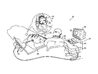
Figure 1 is an illustration of a medical position
tracking system 20 that uses interference cancellation, in
accordance with an embodiment of the invention. System 20 may
be based, for example, in the CARTOrm system, produced by
Biosense Webster Inc. (Diamond Bar, California). System 20
comprises a medical probe 22, such as a catheter, and a
control console 24. In the embodiment described hereinbelow,
it is assumed that probe 22 is used for diagnostic or
6
CA 02741995 2011-06-03
. ,
therapeutic treatment, such as mapping electrical potentials
in a heart 26 or performing ablation of heart tissue.
Alternatively, probe 22 may be used, mutatis mutandis, for
other therapeutic and/or diagnostic purposes in the heart or
in other body organs.
An operator 28, such as a cardiologist, inserts probe 22
through the vascular system of a patient 30 so that a distal
end 32 of probe 22 enters a chamber of the patient's heart 26.
Holding probe 22 at a handle 34, operator 28 advances the
probe, positioning a distal tip 36 at a desired location.
Handle 34 couples probe 22 to a cable 38, which connects to
console 24 via a suitable connector. The configuration of
probe 22, and particularly handle 34, is shown in greater
detail in Fig. 2 below.
Console 24 uses magnetic position sensing to determine
position coordinates of distal tip 36 inside heart 26. To
determine the position coordinates, a driver circuit 40 in
console 24 drives field generators 42 to generate magnetic
fields within the body of patient 30.
Typically, field
generators 42 comprise coils, which are placed below the
patient's torso at known positions external to patient 30.
These coils generate magnetic fields in a predefined working
volume that contains heart 26.
Magnetic field transducers
that are coupled to distal tip 36 and handle 34 generate
electrical signals in response to these magnetic fields. A
signal processor 44 in console 24 processes the electrical
signals in order to determine the position coordinates of
distal tip 36 and handle 34, typically including both location
and orientation coordinates. As discussed supra, processor 44
can cancel out the interference in the signals received from a
main position transducer in distal tip 36, based on the
signals received from an auxiliary position transducer in
handle 34. Both position transducers are shown in Figure 2
below.
7
CA 02741995 2011-06-03
=
Processor 44 typically comprises a general-purpose
computer, with suitable front end and interface circuits for
receiving signals from probe 22 and controlling the other
components of console 24. Processor 44 may be programmed in
software to carry out the functions that are described herein.
The software may be downloaded to console 24 in electronic
form, over a network, for example, or it may be provided on
non-transitory tangible media, such as optical, magnetic or
electronic memory media. Alternatively, some or all of the
functions of processor 44 may be carried out by dedicated or
programmable digital hardware components, or using a
combination of hardware and software elements.
An input/output (I/O) interface 46 enables console 24 to
interact with probe 22. Based on the signals received from
probe 22 (via interface 46 and other components of system 20),
processor 44 drives a display 48 to present operator 26 with
an image 50 showing the position of distal tip 36 in the
patient's body, as well as status information and guidance
regarding the procedure that is in progress.
Alternatively or additionally, system 20 may comprise an
automated mechanism (not shown) for maneuvering and operating
probe 22 within the body of patient 30. Such mechanisms are
typically capable of controlling both the longitudinal motion
(advance/retract) of probe 22 and transverse motion
(deflection/steering) of distal end 32. In such embodiments,
processor 44 generates a control input for controlling the
motion of probe 22 based on the signals provided by the
magnetic field transducers in the probe and the handle, as
explained further hereinbelow.
Figure 2 is another schematic, pictorial illustration of
system 20, in accordance with an embodiment of the present
invention. Fig. 2 shows the configuration of probe 22, and in
particular handle 34, in greater detail. As can be seen in the
figure, handle 34 connects probe 22 to cable 38, and comprises
8
CA 02741995 2011-06-03
, =
a distal part 52 and a proximal part 54 that mate via a
suitable connector 56.
Proximal part 54 of the handle and
cable 38 are sometimes referred to as the proximal part of the
probe. Distal part 56 of the handle, and catheter 22, are
sometimes referred to as the distal part of the probe.
Distal tip 36 comprises a main position transducer 58,
which generates a signal to console 24 that is indicative of
the position coordinates of the distal tip relative to field
generators 42. An auxiliary position transducer 60 is fitted
in proximal part 54 of handle 34, and generates a signal to
console 24 that is indicative of the position coordinates of
the handle relative to field generators 42. Each of position
transducers 58 and 60 may comprise one or more miniature
coils, and typically comprise multiple coils oriented along
different axes. Alternatively, position transducers 58 and 60
may comprise either another type of magnetic transducer, an
electrode which serves as a position transducer, or position
transducers of other types, such as impedance-based or
ultrasonic position transducers. Although Figure 2 shows a
probe with a single position transducer in distal tip 36,
embodiments of the present invention may utilize probes with
more than one position transducer in the distal tip and/or
distal end 32. When distal tip 36 is positioned in heart 26
during a medical procedure, processor 44 uses the signals
received from position transducers 58 and 60 to calculate the
position of the distal tip.
As discussed supra, position transducers 58 and 60 may
generate weak signals due to their configuration. An
amplifier 62 coupled to proximal part 54 amplifies the signals
received from position transducers 58 and 60. The "split
handle" configuration shown in Figure 2 permits components
such as amplifier 62 and auxiliary position transducer 60 to
be contained in proximal part 54, which is reusable, while
probe 22 is disposed of after use. Further aspects of split-
9
CA 02741995 2011-06-03
= .
handle configurations are addressed in U.S. Patent 6,370,411,
cited above.
In an alternative embodiment, the roles of position
transducers 58, 60 and magnetic field generators 42 may be
reversed. In other
words, driver circuit 40 may drive
magnetic field generators in position transducers 58 and 60,
so as to generate magnetic fields. Coils 42 may be configured
to sense the fields and generate signals indicative of the
amplitudes of the components of these magnetic fields. In
this embodiment, processor 44 receives and processes the
signals from coils 42 in order to determine the position
coordinates of distal tip 36 within heart 26.
Although Figures 1 and 2 show a particular system
configuration, other system configurations can also be
employed to implement embodiments of the present invention,
and are thus considered to be within the spirit and scope of
this invention. For
example, the methods described
hereinbelow may be applied using position transducers of other
types, such as impedance-based or ultrasonic position
transducers. The term
"position transducer" as used herein
refers to an element mounted on probe 22 or handle 34 which
causes console 24 to receive signals indicative of the
coordinates of the respective element. The
position
transducer may thus comprise a receiver on the probe or the
handle, which generates a position signal to the control unit
based on energy received by the transducer; or it may comprise
a transmitter, emitting energy that is sensed by a receiver
external to the probe or the handle. Furthermore, the methods
described hereinbelow may similarly be applied in mapping and
measurement applications using not only catheters, but also
probes of other types, both in the heart and in other body
organs and regions.
CA 02741995 2011-06-03
'
POSITION MEASUREMENT USING INTERFERENCE CANCELLATION
Cable 38 conveys signals from main position transducer 58
to console 24 via handle 34. As discussed hereinabove, cable
38 may pick up interference that may distort the signals of
the main position transducer. As a result, console 24 may err
is calculating the position of distal tip 36. The interference
picked-up by cable 38 may be caused by the relatively strong
magnetic fields generated by generators 42, by various
electrical signals in the vicinity of the probe, or by any
other source.
Cable 38 typically comprises shielded, twisted-pair wires
in order to avoid such undesired interference pickup. In the
vicinity of connector 56, however, the shielding performance
may be degraded because of the interconnection to the
connector pins. Thus, some residual interference is sometimes
picked-up in the vicinity of the connector.
System 20 reduces the effect of interference pickup in
connector 56 by pre-calibrating and canceling this
interference using auxiliary position transducer 60. In some
embodiments, processor 44 first measures the interference
pickup as a function of the position (location and
orientation) of handle 34 relative to the source of the
interference. Processor 44 then uses this calibration data for
canceling the interference in the signals received from main
position transducer 58 during an actual medical procedure. The
position of distal tip 36 can thus be calculated with high
accuracy, even in the presence of strong interference.
Moreover, the disclosed techniques may permit relaxing of the
shielding requirements of cable 38.
Figure 3 is a flow diagram that schematically illustrates
a method of measuring the position of distal tip 36 of probe
22 using interference cancellation, in accordance with an
embodiment of the present invention. At a
preliminary
calibration step 70, operator 28 positions handle 34 in
11
CA 02741995 2011-06-03
' =
multiple positions (locations and orientations) relative to
field generators 42 (or other interference source). At each
handle position, processor 44 measures the interference pickup
at connector 56 as a function of the position of handle 34 (as
measured by auxiliary position transducer 60). Processor 44
thus calibrates the interference amplitude as a function of
the output of the auxiliary position transducer in the handle.
The measured interference as a function of handle position is
referred to as calibration data. Main position transducer 58
is typically disabled during the calibration procedure.
During a medical procedure, operator 28 manipulates
handle 34 to position probe 22 in heart 26, at a probe
positioning step 72. Processor 44 receives position signals
from main position transducer 58 indicating the position of
distal tip 36, at a main measurement step 74. Additionally,
processor 44 receives position signals from auxiliary position
transducer 60 indicating the position of handle 34, at an
auxiliary measurement step 76.
Processor 44 cancels the interference in the signal
received from main position transducer 58 based on the
measured position of handle 34, at an interference
cancellation step 78. Typically, processor 44 queries the
calibration data with the current position of the handle, as
measured at step 76, so as to determine the expected
interference level at this handle position. Processor 44 then
subtracts the expected interference level from the signal of
main position transducer 58, measured at step 74 above.
After canceling the interference, processor 44 computes
the position of distal tip 36, at a tip positioning step 80.
The calculation is performed using the position signal
received from the main position transducer, after the
interference has been canceled out from the signal. Finally,
processor 44 presents image 50 on display 48, so as to display
12
CA 02741995 2011-06-03
=
the location of distal tip 36 to operator 28, at an output
step 82. The method returns to step 72 above.
Alternatively or additionally, the position measurements
and interference cancellation scheme may be used in closed-
loop control of an automated mechanism for maneuvering and
operating probe 22, as described hereinabove, to ensure that
the automated mechanism positions distal tip 36 in the proper
location.
Although the embodiments described herein refer mainly to
interference cancellation in medical position tracking
systems, the disclosed techniques can be used for canceling
position-dependent interference in various other applications.
The corresponding structures, materials, acts, and
equivalents of all means or steps plus function elements in
the claims below are intended to include any structure,
material, or act for performing the function in combination
with other claimed elements as specifically claimed. The
description of the present disclosure has been presented for
purposes of illustration and description, but is not intended
to be exhaustive or limiting to the disclosure in the form
disclosed. Many modifications and variations will be apparent
to those of ordinary skill in the art without departing from
the scope and spirit of the disclosure. The embodiment was
chosen and described in order to best explain the principles
of the disclosure and the practical application, and to enable
others of ordinary skill in the art to understand the
disclosure for various embodiments with various modifications
as are suited to the particular use contemplated.
It is intended that the appended claims cover all such
features and advantages of the disclosure that fall within the
spirit and scope of the present disclosure. As
numerous
modifications and changes will readily occur to those skilled
in the art, it is intended that the disclosure not be limited
to the limited number of embodiments described herein.
13
CA 02741995 2011-06-03
. .
Accordingly, it will be appreciated that all suitable
variations, modifications and equivalents may be resorted to,
falling within the spirit and scope of the present disclosure.
14