Note: Descriptions are shown in the official language in which they were submitted.
CA 02742149 2011-04-28
DESCRIPTION
TITLE OF INVENTION
SENSOR CHIP, BIOSENSOR SYSTEM, METHOD FOR MEASURING
TEMPERATURE OF BIOLOGICAL SAMPLE, METHOD FOR MEASURING
TEMPERATURE OF BLOOD SAMPLE, AND METHOD FOR MEASURING
CONCENTRATION OF ANALYTE IN BLOOD SAMPLE
TECHNICAL FIELD
[0001]
The present invention relates to a sensor chip, a biosensor system, a
method for measuring temperature for a biological sample, a method for
measuring temperature for a blood sample, and a method for measuring a
concentration of an analyte in a blood sample.
BACKGROUND ART
[00021
A portable biosensor system provided with a measuring device having a
calculating unit and a sensor chip detachable from the measuring device is
used
for measuring an analyte concentration, for example a blood glucose
concentration (blood glucose value) in a blood sample. The analyte
concentration is calculated by an optical method or an electrochemical method
based on an amount of a reductant or an oxidant produced by an oxygen cycling
reaction mediated by an oxidoreductase that uses the analyte as a substrate.
1
CA 02742149 2011-04-28
The speed of the oxygen cycling reaction depends on the temperature that
promotes the reaction (reaction temperature). As a result, the concentration
of
the analyte is preferably corrected with reference to the reaction
temperature.
[00031
The reaction temperature for example is measured by a temperature
sensor disposed in the measuring device (Patent Literature 1). However, in the
biosensor system according to Patent Literature 1, the inner portion
temperature
of the measuring device is measured, and therefore the measured reaction
temperature does not accurately reflect the temperature of the blood sample.
As a result, an error may result in the measurement of the analyte
concentration.
[00041
Patent Literature 2 - 4 disclose a biosensor system for improving the
measurement accuracy of the reaction temperature. The biosensor system in
Patent Literature 2 and 3 includes a heat conduction member in proximity to
the
blood sample retention unit of the sensor chip, and detects the temperature of
the blood sample transmitted through the heat conduction member with a
temperature sensor disposed in the measuring device. Since the biosensor
system in Patent Literature 2 and 3 includes a resin plate disposed between
the
heat conduction member and the blood sample retention unit, the heat
conduction member does not come into contact with the blood sample. The
biosensor system in Patent Literature 4 includes a temperature sensor and a
2
CA 02742149 2011-04-28
heat conduction member disposed in a mounting unit of the measuring device for
mounting of the sensor chip, and therefore transmits the temperature of the
blood sample to the temperature sensor through the heat conduction member.
CITATION LIST
PATENT LITERATURE
[00051
Patent Literature 1: Japanese Patent Application Laid-Open No.
2003-156469
Patent Literature 2: Japanese Patent Application Laid-Open No.
2001-235444
Patent Literature 3: Japanese Patent Application Laid-Open No.
2003-42995
Patent Literature 4: Pamphlet of PCT International Application No.
2003/062812
SUMMARY
[00061
TECHNICAL PROBLEM
When a user with a biosensor system moves into a location that has a
large temperature difference (for example, moves from an external location in
summer or winter into a building), the measuring device will be incapable of
tracking the sharp variation in the environmental temperature, and for a
certain
period of time, will maintain a higher temperature or lower temperature than
3
CA 02742149 2011-04-28
the environment of the current location. For example, when moving the
measuring device from a 40 C or a 10 C environment to a 25 C environment, a
period of approximately 30 minutes may be required until the temperature of
the
measuring device reaches 25 C (Patent Literature 1).
[00071
It is difficult to completely eliminate the effect of the temperature of the
measuring device when measuring the reaction temperature by a temperature
sensor in a measuring device. Thus when there is a sharp change in the
temperature of the environment in which the sensor is used, an error will tend
to
be produced in the measurement of an analyte concentration when using the
biosensor system disclosed in Patent Literature 2 -4.
[00081
Since the temperature of the blood sample in the biosensor system
disclosed in Patent Literature 2 -4 is communicated by heat transfer through
the
resin plate and the heat conduction member to the temperature sensor, the
measured reaction temperature does not accurately reflect the temperature of
the blood sample.
[00091
The present invention has the object of providing a biosensor system and
a sensor chip for application to the biosensor system that measures a
temperature of a blood sample and suppresses the production of a measurement
error resulting from the temperature of a use environment. Furthermore the
4
CA 02742149 2011-04-28
present invention has the object of providing a measurement method that
improves the measurement accuracy of an analyte concentration in a blood
sample.
SOLUTION TO PROBLEM
[00101
A sensor chip according to a first aspect of the present invention is a
sensor chip for measuring the temperature of a biological sample and includes
temperature electrodes having at least a working electrode and an counter
electrode for measuring the temperature of the biological sample, and having a
direct current voltage applied thereto, and a capillary configured to
introduce the
biological sample to the temperature electrodes. The working electrode and/or
the counter electrode in the temperature electrodes are disposed to make
contact
with the biological sample introduced into the capillary. The direct current
voltage is set to reduce an effect of hematocrit on a temperature measurement
result of hematocrit during application of the direct current voltage.
[00111
In this sensor chip, a predetermined direct current voltage is applied to
the temperature electrodes so that the effect of hematocrit is low during
measurement of the biological sample temperature by the temperature
electrodes.
In this manner, temperature measurement of a biological sample is
5
CA 02742149 2011-04-28
enabled without reference to a hematocrit value in the biological sample. As a
result, the temperature measurement accuracy for a biological sample can be
improved, and the accuracy in relation to various types of corrections using
the
temperature of the biological sample can also be improved.
[00121
A sensor chip according to a second aspect of the present invention
includes the sensor chip according to the first aspect, and the uptake amount
of
the biological sample into the capillary is 51iL or less, and the application
time of
the direct current voltage to the temperature electrodes is 15 seconds or
less.
[00131
A sensor chip according to a third aspect of the present invention
includes the senor chip according to the first or the second aspect, and the
predetermined direct current voltage is within a range in which the solvent of
the biological sample is subjected to electrolysis.
[00141
A sensor chip according to a fourth aspect of the present invention
includes the senor chip according to any one of the first to the third aspect,
and is
disposable.
[00151
A sensor chip according to a fifth aspect of the present invention is a
sensor chip for measuring the concentration of an analyte in a blood sample,
and
includes temperature electrodes disposed to make contact with the blood
sample,
6
CA 02742149 2011-04-28
and having at least a working electrode and an counter electrode for measuring
the temperature of the blood sample, and a concentration measuring unit
configured to measure a feature related to a concentration of the analyte in
the
blood sample.
[00161
In this manner, direct measurement of the temperature of a blood sample
is enabled in contrast to a conventional sensor chip provided with temperature
electrodes that measure the heat transmitted through a resin plate, heat
conduction member, or the like. As a result, the production of a measurement
error caused by the temperature of the use environment can be suppressed, and
an improvement in the measurement accuracy of the analyte concentration in a
blood sample is enabled.
[00171
A sensor chip according to a sixth aspect of the present invention
includes the sensor chip according to the fifth aspect, and the concentration
measuring unit is formed from analysis electrodes including at least a working
electrode and an counter electrode.
[00181
A sensor chip according to a seventh aspect of the present invention
includes the sensor chip according to the sixth aspect, and the temperature
electrodes and the analysis electrodes are provided separately.
[00191
7
CA 02742149 2011-04-28
In this manner, accurate measurement of a concentration of an analyte in
a blood sample is enabled.
[00201
A sensor chip according to an eighth aspect of the present invention
includes the sensor chip according to the sixth or the seventh aspect, and
further
includes a sample introduction port and a capillary configured to introduce a
blood sample from the sample introduction port to the temperature electrodes
and the analysis electrodes. The temperature electrodes are disposed at a
position closer to the sample introduction port than the analysis electrodes.
[00211
A sensor chip according to a ninth aspect of the present invention
includes the sensor chip according to any one of the fifth to the eighth
aspect,
and the temperature electrodes are disposed to not make contact with at least
one of the oxidoreductase or the electron mediator.
[00221
In this manner, the temperature of the blood sample can be accurately
measured.
[00231
A sensor chip according to a tenth aspect of the present invention
includes the sensor chip according to any one of the fifth to the ninth
aspect, and
the concentration measuring unit further includes a reaction reagent that
induces an oxidation-reduction reaction, and the temperature electrodes are
8
CA 02742149 2011-04-28
disposed to not make contact with the reaction reagent that induces the
oxidation-reduction reaction.
[0024]
In this manner, contact of the reaction reagent with the temperature
electrodes can be avoided, and accurate measurement of the blood sample
temperature is enabled.
[0025]
A sensor chip according to an eleventh aspect of the present invention
includes the sensor chip according to any one of the fifth to the ninth
aspect, and
is disposed to not make contact with any reagent.
[0026]
In this manner, contact of any reagent with the temperature electrodes
can be avoided, and accurate measurement of the blood sample temperature is
enabled.
[0027]
A sensor chip according to a twelfth aspect of the present invention
includes the sensor chip according to the sixth aspect, and the working
electrode
of the temperature electrodes is common to at least either the working
electrode
or the counter electrode of the analysis electrodes.
[0028]
A sensor chip according to a thirteenth aspect of the present invention
includes the sensor chip according to the sixth aspect, and the counter
electrode
9
CA 02742149 2011-04-28
of the temperature electrodes is common to at least either the working
electrode
or the counter electrode of the analysis electrodes.
[00291
A sensor chip according to a fourteenth aspect of the present invention
includes the sensor chip according to any one of the sixth to the eighth
aspect,
and the concentration measuring unit includes at least one electrode in
addition
to the working electrode and the counter electrode, and at least one of the
electrodes of the concentration measuring unit other than the working
electrode
and the counter electrode is common to at least one of the working electrode
and
the counter electrode of the temperature electrodes.
[00301
The electrodes included in the concentration measuring unit according to
the twelfth to the fourteenth aspects may be combined with at least one of the
working electrode and the counter electrode of the temperature electrodes.
The sensor chip according to the twelfth and the thirteenth aspects may
include a plurality of working electrodes and/or a plurality of counter
electrodes
as analysis electrodes. At least one of the plurality of working electrodes
and/or
counter electrodes may be combined with the working electrode and/or counter
electrode of the temperature electrodes.
[00311
An example of an electrode other than a working electrode and counter
electrode according to the fourteenth aspect includes
CA 02742149 2011-04-28
- a hematocrit measuring electrode;
- a measuring electrode for an amount or concentration of a reducing
substance;
a detection electrode for detecting the introduction of blood; and
- a measuring electrode other than a electrode for measuring an amount
or concentration of a reducing substance, hematocrit, or glucose
concentration.
[0032]
A sensor chip according to a fifteenth aspect of the present invention
includes the sensor chip according to the sixth aspect, and the surface area
of the
working electrode in the temperature electrodes is either the same or smaller
than the surface area of the counter electrode in the temperature electrodes.
[0033]
A sensor chip according to a sixteenth aspect of the present invention
includes the sensor chip according to any one of the fifth to the fifteenth
aspect,
and at least hematocrit is included as a feature in relation to the
concentration of
the analyte.
[0034]
A sensor chip according to a seventeenth aspect of the present invention
includes the sensor chip according to any one of the fifth to the sixteenth
aspect,
and at least a concentration or an amount of a reducing substance is included
as
a feature in relation to the concentration of the analyte.
[0035]
11
CA 02742149 2011-04-28
A method for measuring a temperature of a biological sample according
to an eighteenth aspect of the present invention measures a temperature of a
biological sample by a sensor chip including temperature electrodes formed
from
a working electrode and an counter electrode, and a capillary. The method
includes an introduction step of introducing a biological sample by the
capillary
to the temperature electrodes, an application step of applying a direct
current
voltage to the temperature electrodes, and an adjustment step of adjusting the
direct current voltage applied in the application step to a first voltage. The
first
voltage is set so that the effect of hematocrit on the temperature measurement
result during application of the first voltage to the temperature electrodes
is
reduced.
[00361
This method enables temperature measurement of a biological sample
without reference to a hematocrit value in the biological sample. As a result,
the accuracy of the temperature measurement of the biological sample can be
increased, and the accuracy in relation to various corrections using the
temperature of the biological sample can also be increased.
[00371
A method for measuring a temperature according to a nineteenth aspect
of the present invention includes the method for measuring a temperature
according to the eighteenth aspect, and a direct current voltage that enables
a
reduction of the effect of hematocrit on the temperature measurement result is
12
CA 02742149 2011-04-28
measured and stored in advance, and the adjustment step adjusts to the first
voltage based on the stored direct current voltage.
[00381
A method for measuring a temperature of a biological sample according
to a twentieth aspect of the present invention includes the method for
temperature measurement of a biological sample according to the eighteenth or
the nineteenth aspect, and the uptake amount of the biological sample in the
introduction step is 51iL or less, and the application time of the direct
current
voltage in the application step is 15 seconds or less.
[00391
A method for measuring a temperature of a blood sample according to a
twenty first aspect of the present invention measures a temperature of a blood
sample using a sensor chip including temperature electrodes formed from a
working electrode and an counter electrode. The method includes a step of
applying a voltage to the temperature electrodes in contact with the blood
sample, a step of acquiring data a related to the temperature of the blood
sample
based on a dimension of a current flowing in the blood sample by application
of
the voltage, and a step of calculating a temperature t of the blood sample
based
on the data a.
[00401
A temperature t of the blood sample is calculated based on data a related
to the temperature of the blood sample that can be acquired by application of
a
13
CA 02742149 2011-04-28
voltage to the temperature electrodes in contact with the blood sample.
In this manner, since the temperature t of the blood sample can be
calculated based on data a related to the temperature of the blood sample that
can be accurately acquired, the production of a measurement error caused by
the
temperature of the use environment can be suppressed.
[00411
A method for measuring a concentration of an analyte in a blood sample
according to a twenty second aspect of the present invention includes a step
of
acquiring data a related to the temperature of the blood sample based on the
dimension of a current flowing in the blood sample by application of a voltage
to
the pair of electrodes in contact with the blood sample, a step of acquiring
data b
related to a concentration of the analyte based on the dimension of a current
flowing in the blood sample by a reaction mediated by an oxidoreductase that
uses the analyte in the blood sample as a substrate, and a step of measuring a
concentration that determines the analyte concentration in the blood sample
based on the data a and the data b.
[00421
Herein, the data a is acquired by directly measurement of the
temperature of the blood sample without interposing a resin plate or a heat
conduction member, and the analyte concentration in the blood sample is
determined based on the data a related to the temperature of the blood sample
and the data b related to the concentration of the analyte.
14
CA 02742149 2011-04-28
[0043]
In this manner, the measurement accuracy of the analyte concentration
in the blood sample can be improved.
[0044]
A method for measuring a concentration of an analyte in a blood sample
according to a twenty third aspect of the present invention includes the
method
for measuring a concentration of an analyte in a blood sample according to the
twenty second aspect, and the concentration measurement step includes a step
of
correcting the data b based on the data a.
[0045]
In this manner, the measurement accuracy of the concentration of the
analyte in the blood sample can be improved.
[0046]
A method for measuring a concentration of an analyte in a blood
sample according to a twenty fourth aspect of the present invention includes
the
method for measuring a concentration of an analyte in a blood sample according
to the twenty second aspect, and the concentration measurement step includes a
step of calculating a concentration x of an analyte in a blood sample based on
the
data b, and a step of correcting the concentration x based on the data a.
[0047]
In this manner, the measurement accuracy of the concentration of the
analyte in the blood sample can be improved.
CA 02742149 2011-04-28
[00481
A method for measuring a concentration of an analyte in a blood sample
according to a twenty fifth aspect of the present invention includes the
method
for measuring a concentration of an analyte in a blood sample according to the
twenty second aspect, and the concentration measurement step includes a step
of
calculating a temperature t of the analyte in the blood sample based on the
data
a, and a step of correcting the data b based on the temperature t.
[00491
In this manner, the measurement accuracy of the concentration of the
analyte in the blood sample can be improved.
[00501
A method for measuring a concentration of an analyte in a blood sample
according to a twenty sixth aspect of the present invention includes the
method
for measuring a concentration of an analyte in a blood sample according to the
twenty second aspect, and the concentration measurement step includes a step
of
calculating a temperature t of an analyte in a blood sample based on the data
a,
a step of calculating a concentration x of the analyte in a blood sample based
on
the data b, and a step of correcting the concentration x based on the
temperature
t.
[00511
In this manner, the measurement accuracy of the concentration of the
analyte in the blood sample can be improved.
16
CA 02742149 2011-04-28
[00521
A method for measuring a concentration of an analyte in a blood sample
according to a twenty seventh aspect of the present invention includes the
method for measuring a concentration of an analyte in a blood sample according
to any one of the twenty second to the twenty sixth aspect, and the step of
acquiring the data a is performed in advance of the step of acquiring the data
b.
[00531
In this manner, the temperature at the time of acquiring the data b can
be more accurately reflected.
[00541
A method for measuring a concentration of an analyte in a blood sample
according to a twenty eighth aspect of the present invention includes the
method
for measuring a concentration of an analyte in a blood sample according to the
twenty second aspect, and the concentration measurement step includes a step
of
acquiring data c related to the temperature of the blood sample based on the
dimension of a current flowing in the blood sample by application of a
predetermined voltage to the pair of electrodes in contact with the blood
sample
after acquisition of the data b, and a step of calculating data d related to
the
temperature of the blood sample based on the data a and the data c, and a step
of
correcting the data b based on the data d.
[00551
In this manner, the temperature at the time of acquiring the data b can
17
CA 02742149 2011-04-28
be more accurately reflected, and the analyte concentration measurement
accuracy for the blood sample can be improved.
[00561
A method for measuring a concentration of an analyte in a blood sample
according to a twenty ninth aspect of the present invention includes the
method
for measuring a concentration of an analyte in a blood sample according to the
twenty second aspect, and the concentration measurement step includes a step
of
calculating the temperature t of the blood sample based on the data a, a step
of
calculating the concentration x of the analyte in the blood sample based on
the
data b, the step of measuring an environmental temperature tl on a periphery
of
the blood sample, a step of comparing the difference between the temperature t
and the environmental temperature t1 with a temperature threshold Z, and a
step of correcting the concentration x based on the temperature t when the
relation I t - tl I > Z is satisfied, and correcting the concentration x based
on the
temperature t1 when the relation I t - tl I < Z is satisfied.
[00571
Herein, the concentration x of the analyte in the blood sample is
calculated based on the data b, and the temperature t of the blood sample is
calculated based on the data a. The environmental temperature tl in the
periphery of the blood sample is measured. Then the difference between the
temperature t and the environmental temperature tl is compared with a
temperature threshold Z, and correction is performed as described below.
18
CA 02742149 2011-04-28
[00581
When I t - t1 I > Z is satisfied, the concentration x is corrected based on
the temperature t
When I t - t1 I < Z is satisfied, the concentration x is corrected based on
the temperature tl
In this manner, since the concentration x can be corrected using an
appropriate temperature in response to an external temperature environment, a
measurement accuracy for the analyte concentration in the blood sample can be
improved.
[00591
A method for measuring a concentration of an analyte in a blood sample
according to a thirtieth aspect of the present invention includes the method
for
measuring a concentration of an analyte in a blood sample according to any one
of the twenty second aspect to the twenty ninth aspect, and a temperature is
contained in the data a related to the temperature of the blood sample, and a
glucose concentration is contained in the data b related to the concentration
of
the analyte.
[00601
Herein, the temperature is included as a feature of the data acquired as
data a, and the glucose concentration is included as a feature of the data
acquired as the data b.
[00611
19
CA 02742149 2011-04-28
A method for measuring a concentration of an analyte in a blood sample
according to a thirty first aspect of the present invention includes the
method for
measuring a concentration of an analyte in a blood sample according to the
thirtieth aspect, and hematocrit is included in the data b related to the
concentration of the analyte.
[0062]
Herein, hematocrit is included as a feature of the data acquired as the
data b.
[0063]
A method for measuring a concentration of an analyte in a blood sample
according to a thirty second aspect of the present invention includes the
method
for measuring a concentration of an analyte in a blood sample according to the
thirtieth or thirty first aspect, and the concentration or amount of the
reducing
substance is contained in the data b related to the concentration of the
analyte.
[0064]
Herein, the amount or concentration of the reducing substance is
included as a feature of the data acquired as the data b.
[0065]
A method for measuring a concentration of an analyte in a blood sample
according to a thirty third aspect of the present invention includes the
method
for measuring a concentration of an analyte in a blood sample according to any
one of the thirtieth to the thirty second aspect, and at least two features of
the
CA 02742149 2011-04-28
data included in the data a and the data b are measured at the same time.
[00661
Herein, when the data a and the data b are measured, at least two
features of the data are measured at the same time. For example, the
concentration or the amount of the reducing substance and the glucose
concentration are measured at the same time.
[00671
A method for measuring a concentration of an analyte in a blood sample
according to a thirty fourth aspect of the present invention includes the
method
for measuring a concentration of an analyte in a blood sample according to any
one of the thirtieth to the thirty second aspect, and independent measurement
of
the respective data included in the data a and the data b is executed.
[00681
Herein, when the data a and the data b are measured, two or more
features are not measured at the same time, but are measured separately. The
order of measuring the features may be arbitrary.
[00691
A method for measuring a concentration of an analyte in a blood sample
according to a thirty fifth aspect of the present invention includes the
method for
measuring a concentration of an analyte in a blood sample according to any one
of the thirtieth to the thirty second aspect, and the measurement of the data
contained in the data a and the data b is performed in order of temperature,
21
CA 02742149 2011-04-28
glucose concentration, concentration or amount of the reducing substance, and
hematocrit.
[0070]
Herein, the order of measuring the data is specified. In this manner,
effective results can be obtained with respect to speed, accuracy, and burden
on
the electrodes.
[0071]
A method for measuring a concentration of an analyte in a blood sample
according to a thirty sixth aspect of the present invention includes the
method
for measuring a concentration of an analyte in a blood sample according to any
one of the thirtieth to the thirty fifth aspect, and the measurement of the
data
contained in the data a and the data b is performed through independent
electrodes.
[0072]
Herein, when measuring the data contained in the data a and the data b,
such measurement is performed by respectively independent electrodes.
[0073]
A biosensor system according to a thirty seventh aspect of the present
invention has the sensor chip according to any one of the first to the
seventeenth
aspects, and a measuring device including a control circuit applying a voltage
to
the temperature electrodes of the sensor chip. The biosensor system measures a
concentration of an analyte in a blood sample. The biosensor system includes a
22
CA 02742149 2011-04-28
voltage application unit configured to apply a voltage to the temperature
electrodes in accordance with the control circuit, a temperature measuring
unit
configured to acquire the data a related to the temperature of the blood
sample
based on the dimension of a current flowing in the temperature electrodes in
contact with the blood sample, an analyte measuring unit acquiring data b
related to the concentration of the analyte based on the dimension of a
current
flowing in the blood sample depending on a reaction mediated by an
oxidoreductase that uses the analyte in the blood sample as a substrate, and a
concentration determination unit configured to determine an analyte
concentration in the blood sample based on the data a and the data b.
[0074]
Herein, data a is acquired by direct measurement of the temperature of
the blood sample and not through a resin plate or a heat conduction member.
The concentration determination unit determines the analyte concentration in
the blood sample based on the data a related to the temperature of the blood
sample and the data b related to the analyte concentration.
[0075]
In this manner, the production of a measurement error resulting from the
temperature of the use environment can be suppressed, and thereby the
measurement accuracy of the analyte concentration in the blood sample can be
improved.
[0076]
23
CA 02742149 2011-04-28
A biosensor system according to the thirty eighth aspect of the present
invention includes the biosensor system according to the thirty seventh
aspect,
and the concentration determination unit includes a first analyte correction
unit
configured to correct the data b based on the data a.
[00771
Herein, the first analyte correction unit corrects the data b related to the
concentration of the analyte in the blood sample based on the data a acquired
by
direct measurement of the temperature of the blood sample and not through a
resin plate or a heat conduction member.
[00781
In this manner, the production of a measurement error resulting from the
temperature of the use environment can be suppressed, and thereby the
measurement accuracy of the analyte concentration in the blood sample can be
improved.
[00791
A biosensor system according to the thirty ninth aspect of the present
invention includes the biosensor system according to the thirty seventh
aspect,
and the concentration determination unit includes a calculating unit
configured
to calculate the concentration x of the analyte of the blood sample based on
the
data b, and a second analyte correction unit configured to correct the
concentration xbased on the data a.
[00801
24
CA 02742149 2011-04-28
Herein, the analyte correction unit calculates the concentration x of the
analyte in the blood sample based on the data b, and then the second analyte
correction unit corrects the concentration x based on the data a acquired by
direct measurement of the temperature of the blood sample.
[0081]
In this manner, the production of a measurement error resulting from the
temperature of the use environment can be suppressed, and thereby the
measurement accuracy of the analyte concentration in the blood sample can be
improved.
[0082]
A biosensor system according to the fortieth aspect of the present
invention includes the biosensor system according to the thirty seventh
aspect,
and the concentration determination unit includes a calculating unit
configured
to calculate the temperature t of the blood sample based on the data a, and a
third analyte correction unit configured to correct the data b based on the
temperature t.
[0083]
Herein, the calculating unit calculates the temperature t of the blood
sample based on the data a acquired by direct measurement of the temperature
of the blood sample, and then the third analyte correction unit corrects the
data
b based on the temperature t.
[0084]
CA 02742149 2011-04-28
In this manner, the production of a measurement error resulting from the
temperature of the use environment can be suppressed, and thereby the
measurement accuracy of the analyte concentration in the blood sample can be
improved.
[00851
A biosensor system according to the forty first aspect of the present
invention includes the biosensor system according to the thirty seventh
aspect,
and the concentration determination unit includes a calculating unit
configured
to calculate the temperature t of the blood sample based on the data a, a
calculating unit configured to calculate the concentration x of the blood
sample
based on the data b, and a fourth analyte correction unit configured to
correct
the concentration xbased on the temperature t.
[00861
Herein, the calculating unit calculates the temperature t of the blood
sample based on the data a acquired by direct measurement of the temperature
of the blood sample, and calculates the concentration x of the analyte in the
blood
sample based on the data b, and then the fourth analyte correction unit
corrects
the concentration xbased on the temperature t.
[00871
In this manner, the production of a measurement error resulting from the
temperature of the use environment can be suppressed, and thereby the
measurement accuracy of the analyte concentration in the blood sample can be
26
CA 02742149 2011-04-28
improved.
[0088]
A biosensor system according to the forty second aspect of the present
invention includes the biosensor system according to any one of the thirty
seventh aspect to the forty first aspect, and after acquisition of the data a
related
to the temperature of the sample by the temperature measuring unit, the data b
related to the concentration of the analyte is acquired by the analyte
measuring
unit.
[0089]
In this manner, the temperature when acquiring the data b can be more
accurately reflected.
[0090]
A biosensor system according to a forty third aspect of the present
invention includes the biosensor system according to the thirty seventh
aspect,
and the concentration determination unit includes a temperature measuring
unit configured to acquire data c related to the temperature of the blood
sample
based on the dimension of a current flowing in the blood sample by application
of
a predetermined voltage to the pair of electrodes in contact with the blood
sample after acquisition of the data b, a computing unit configured to
calculate
data d related to the temperature of the blood sample based on data a and the
data c, and a calculating unit configured to calculate the concentration x of
the
analyte corrected in response to the temperature of the blood sample based on
27
CA 02742149 2011-04-28
the data d.
[0091]
In this manner, after acquiring the data b, data c related to the
temperature of the blood sample is acquired by the same acquisition method as
the data a, and the computing unit calculates the data d related to the
temperature of the blood sample based on the data a and the data c. Then the
calculating unit corrects the concentration xbased on the data d.
[0092]
In this manner, the temperature at the time of acquisition of the data b
can be more accurately reflected, and the measurement accuracy of the analyte
concentration in the blood sample can be improved.
[0093]
A biosensor system according to a forty fourth aspect of the present
invention includes the biosensor system according to the thirty seventh
aspect,
and the concentration determination unit includes a temperature calculating
unit configured to calculate the temperature t of the blood sample based on
the
data a, a concentration calculating unit configured to calculate the
concentration
x of the analyte in the blood sample based on the data b, an environmental
temperature measuring unit configured to measure an environmental
temperature tl in a periphery of the blood sample, a comparison unit
configured
to compare the difference between the temperature t and the environmental
temperature tl with a temperature threshold Z, and a correction unit
configured
28
CA 02742149 2011-04-28
to correct the concentration x based on the temperature t when the relation I
t -
t1 I > Z is satisfied, and correcting the concentration x based on the
temperature
tl when the relation I t - ti I < Z is satisfied.
[00941
Herein, the concentration x of the analyte in the blood sample is
calculated based on the data b, and the temperature of the blood sample is
calculated based on the data a. The environmental temperature t1 in the
periphery of the blood sample is measured. Then the difference between the
temperature t and the environmental temperature tl is compared with a
temperature threshold Z, and correction is performed as described below.
[00951
When I t - t1 I > Z is satisfied, the concentration x is corrected based on
the temperature t
When I t - t1 I < Z is satisfied, the concentration x is corrected based on
the temperature tl
In this manner, since the concentration x can be corrected using an
appropriate temperature in response to an external temperature environment, a
measurement accuracy for the analyte concentration in the blood sample can be
improved.
[00961
A biosensor system according to a forty fifth aspect of the present
invention includes the biosensor system according to the any one of the thirty
29
CA 02742149 2011-04-28
seventh aspect to the forty fourth aspect, and a temperature is contained in
the
data a related to the temperature of the blood sample, and a glucose
concentration is contained in the data b related to the concentration of the
analyte.
[0097]
Herein, the temperature is included as a feature of the data acquired as
data a, and the glucose concentration is included as a feature of the data
acquired as the data b.
[0098]
A biosensor system according to a forty sixth aspect of the present
invention includes the biosensor system according to the forty fifth aspect,
and
hematocrit is included in the data b related to the analyte concentration.
[0099]
Herein, hematocrit is included as a feature of the data acquired as the
data b.
[0100]
A biosensor system according to a forty seventh aspect of the present
invention includes the biosensor system according to the forty fifth aspect or
forty sixth aspect, and the concentration or amount of the reducing substance
is
contained in the data b related to the concentration of the analyte.
[0101]
Herein, the amount or concentration of the reducing substance is
CA 02742149 2011-04-28
included as a feature of the data acquired as the data b.
[01021
A biosensor system according to a forty eighth aspect of the present
invention includes the biosensor system according to the any one of the forty
fifth
to the forty seventh aspect, and further includes a sequence control unit
configured to control the control circuit so that at least two features of the
data
included in the data a and the data b are measured at the same time.
[01031
Herein, when the data a and the data b are measured, the sequence
control unit controls the control circuit so that at least two features of the
data
are measured at the same time. For example, the sequence control unit controls
the control circuit so that the concentration or the amount of the reducing
substance and the glucose concentration are measured at the same time.
[01041
A biosensor system according to a forty ninth aspect of the present
invention includes the biosensor system according to the any one of the forty
fifth
to the forty seventh aspect, and further includes a sequence control unit
configured to control the control circuit so that independent measurement of
the
respective data included in the data a and the data b is executed.
[01051
Herein, when the data a and the data b are measured, the sequence
control unit controls the control circuit so that two or more features of the
data
31
CA 02742149 2011-04-28
are not measured at the same time, but are measured separately. The order of
measuring the features may be arbitrary.
[01061
A biosensor system according to a fiftieth aspect of the present invention
includes the biosensor system according to the any one of the forty fifth to
the
forty seventh aspect, and further includes a sequence control unit configured
to
control the control circuit so that the measurement of the data contained in
the
data a and the data b is performed in order of temperature, glucose
concentration, concentration or amount of the reducing substance, or
hematocrit.
[01071
Herein, the order of measuring the data is specified. In this manner,
effective results can be obtained with respect to speed, accuracy, and burden
on
the electrodes.
[0108)
A biosensor system according to a fifty first aspect of the present
invention includes the biosensor system according to the any one of the forty
fifth
to the fiftieth aspect, and further includes an electrode selection unit
configured
to control the control circuit so that the measurement of the data contained
in
the data a and the data b is performed through independent electrodes.
[01091
Herein, when measuring the data contained in the data a and the data b,
the electrode selection unit controls the control circuit so that such
measurement
32
CA 02742149 2011-04-28
is performed by respectively independent electrodes.
ADVANTAGEOUS EFFECTS
[01101
According to the sensor chip, the biosensor system, the method for
measuring a temperature of a blood sample, and a method for measuring a
concentration of an analyte in a blood sample according to the present
invention,
the production of a measurement error resulting from the temperature of a use
environment is suppressed, and improvement of the measurement accuracy of an
analyte concentration in a blood sample is enabled.
BRIEF DESCRIPTION OF DRAWINGS
[01111
FIG. 1 is a perspective view of a biosensor system according to a first
embodiment of the present invention.
FIG. 2 is a partial perspective view of a biosensor chip according to the
first embodiment of the present invention.
FIG. 3 is a through-view plan view of a biosensor chip according to the
first embodiment of the present invention.
FIG. 4 is a circuit diagram in a biosensor system according to the first
embodiment of the present invention.
FIG. 5 is a flowchart illustrating a method for measuring an analyte
concentration in a blood sample in the biosensor system according to the first
33
CA 02742149 2011-04-28
embodiment of the present invention.
FIG. 6(a) and 6(b) is a flowchart illustrating a method for measuring an
analyte concentration in a blood sample in the biosensor system and a circuit
diagram in a biosensor system according to another embodiment of the present
invention.
FIG. 7(a) and 7(b) is a flowchart illustrating a method for measuring an
analyte concentration in a blood sample in the biosensor system, and a circuit
diagram in a biosensor system according to another embodiment of the present
invention.
FIG. 8(a), 8(b) and 8(c) are graphs illustrating the variation
characteristics of a current obtained by use of the biosensor chip according
to the
first embodiment of the present invention.
FIG. 9 is a partial perspective view of a sensor chip according to the first
embodiment of the present invention.
FIG. 10 is a through-view plan view of a sensor chip according to the first
embodiment of the present invention.
FIG. 11(a), 11(b) and 11(c) are graphs illustrating the current
characteristics of a current corresponding to FIG. 8 according to Working
Example 1.
FIG. 12 is a graph illustrating the current characteristics obtained in
relation to a predetermined temperature according to Working Example 1.
FIG. 13(a), 13(b), and 13(c) are graphs illustrating the current
34
CA 02742149 2011-04-28
characteristics obtained in relation to a predetermined applied voltage and a
predetermined hematocrit value when the temperature in Working Example 7 is
4 degrees.
FIG. 14(a), 14(b), and 14(c) are graphs illustrating the current
characteristics obtained in relation to a predetermined applied voltage and a
predetermined hematocrit value when the temperature in Working Example 7 is
13 degrees.
FIG. 15(a), 15(b), and 15(c) are graphs illustrating the current
characteristics obtained in relation to a predetermined applied voltage and a
predetermined hematocrit value when the temperature in Working Example 7 is
21 degrees.
FIG. 16(a), 16(b), and 16(c) are graphs illustrating the current
characteristics obtained in relation to a predetermined applied voltage and a
predetermined hematocrit value when the temperature in Working Example 7 is
30 degrees.
FIG. 17(a), 17(b), and 17(c) are graphs illustrating the current
characteristics obtained in relation to a predetermined applied voltage and a
predetermined hematocrit value when the temperature in Working Example 7 is
38 degrees.
FIG. 18 is a graph illustrating the relationship with a current value
obtained in relation to a predetermined temperature in Working Example 10.
FIG. 19 is a perspective view illustrating the inter-electrode distance in
CA 02742149 2011-04-28
the sensor chip according to Working Example 11.
FIG. 20(a) - 20D are graphs illustrating a response current value by
hematocrit, and by inter-electrode distance when the blood sample is 11 C in
Working Example 11.
FIG. 21(a) - 21(d) are graphs illustrating a response current value by
hematocrit, and by inter-electrode distance when the blood sample is 21 C in
Working Example 11.
FIG. 22(a) - 22(d) are graphs illustrating a response current value by
hematocrit, and by inter-electrode distance when the blood sample is 30 C in
Working Example 11.
FIG. 23(a) and 23(b) is a perspective view illustrating a sensor chip
according to Working Example 12.
FIG. 24(a) and 24(b) are graphs illustrating a response current value by
hematocrit, and by electrode shape when the blood sample is 11 C in Working
Example 12.
FIG. 25(a) and 25(b) are graphs illustrating a response current value by
hematocrit, and by electrode shape when the blood sample is 21 C in Working
Example 12.
FIG. 26(a) and 26(b) are graphs illustrating a response current value by
hematocrit, and by electrode shape when the blood sample is 30 C in Working
Example 12.
FIG. 27(a) and 27(b) is a perspective view illustrating a sensor chip
36
CA 02742149 2011-04-28
according to Working Example 13.
FIG. 28(a) - 28(d) are graphs illustrating a response current value by
hematocrit, and by lead width when the blood sample is 30'C in Working
Example 13.
FIG. 29 is a perspective view illustrating the capillary height in the
sensor chip in Working Example 14.
FIG. 30(a) and 30(b) are graphs illustrating a response current value by
hematocrit, and by capillary height when the blood sample is 11 C in Working
Example 14.
FIG. 31(a) and 31(b) are graphs illustrating a response current value by
hematocrit, and by capillary height when the blood sample is 21 C in Working
Example 14.
FIG. 32(a) and 32(b) are graphs illustrating a response current value by
hematocrit, and by capillary height when the blood sample is 30 C in Working
Example 14.
FIG. 33(a) and 33(b) are graphs illustrating a response current value by
palladium resistance when the blood sample is 4 C in Working Example 15.
FIG. 34(a) and 34(b) are graphs illustrating a response current value by
palladium resistance when the blood sample is 13 C in Working Example 15.
FIG. 35(a) and 35(b) are graphs illustrating a response current value by
palladium resistance when the blood sample is 21 C in Working Example 15.
FIG. 36(a) and 36(b) are graphs illustrating a response current value by
37
CA 02742149 2011-04-28
palladium resistance when the blood sample is 30 C in Working Example 15.
FIG. 37(a) and 37(b) are graphs illustrating a response current value by
palladium resistance when the blood sample is 38 C in Working Example 15.
FIG. 38 is a graph illustrating response current value by glucose
concentration when the blood sample is 24 C in Working Example 15.
FIG. 39 is a graph illustrating response current value by ascorbic acid
concentration when the blood sample is 24 C in Working Example 17.
FIG. 40 is a graph illustrating response current value by temperature
when the blood sample is introduced in an environment of 24 C in Working
Example 18.
FIG. 41 is a perspective view illustrating the upward orientation and
downward orientation of the sensor chip according to Working Example 19.
FIG. 42 is a graph illustrating a response current value when blood is
attached in an upward orientation and a downward orientation in an
environment of 24 C according to Working Example 19.
FIG. 43 is a graph illustrating a response current value when a distal
end portion of the sensor chip is held between the fingers and not held
between
the fingers in an environment of 24 C according to Working Example 20.
FIG. 44 illustrates a measurement sequence in Working Example 21.
FIG. 45(a) is a graph illustrating a response current value for glucose
measured in Working Example 21, and FIG. 45(b) is a graph illustrating a
response current value for temperature and Hct measured in Working Example
38
CA 02742149 2011-04-28
21.
FIG. 46(a) is a graph illustrating a response current value for
temperature measurement in Working Example 21, and FIG. 45(b) is a graph
illustrating a response current value by temperature when temperature is
measured in Working Example 21.
FIG. 47 illustrates another measurement sequence in Working Example
21.
FIG. 48(a) and FIG. 48(b) is a flowchart illustrates a measurement
method for analyte concentration in a blood sample in a biosensor system
according to a first modified example according to the present invention.
FIG. 49(a) and FIG. 49(b) is a flowchart illustrates a measurement
method for analyte concentration in a blood sample in a biosensor system
according to the first modified example according to the present invention.
FIG. 50(a) and FIG. 50(b) is a circuit diagram for a biosensor system
according to the first modified example according to the present invention.
FIG. 51(a) and FIG. 51(b) is a circuit diagram for a biosensor system
according to the first modified example according to the present invention.
FIG. 52 is a circuit diagram for a biosensor system according to a second
modified example according to the present invention.
FIG. 53 is a circuit diagram for a biosensor system according to an
embodiment of the present invention.
39
CA 02742149 2011-04-28
DESCRIPTION OF EMBODIMENTS
[01121
The biosensor system according to the present invention acquires the
temperature of the analyte from the blood sample by a measuring unit disposed
in the sensor chip.
[01131
FIG. 1 illustrates an example of a biosensor system according to the
present invention. The biosensor system 100 includes a rectangular
parallelepiped measuring device 101 and a sensor chip 200. A mounting port
102 configured as a rectangular hole is formed in a side wall surface of the
measuring device 101. The sensor chip 200 is connected to the measuring
device 101 that is detachably attached to the mounting port 102. The display
unit 103 that displays the measurement results is disposed in a substantially
central portion of one major surface of the measuring device 101.
[01141
FIG. 2 is a partial perspective view of the sensor chip 200. FIG. 3 is a
plan view thereof. In the sensor chip 200, a cover 203 is disposed on an
insulating plate 201 through a spacer 202 that forms a rectangular notch 204,
and leaves one end portion of the insulating plate 201 (the right end in FIG.
2).
[01151
Each member 201, 202, 203 is integrated for example by adhesion or
thermal welding. After integration of each of the members, the notch 204 of
the
CA 02742149 2011-04-28
spacer 202 functions as a capillary 40 that retains the blood sample. The
capillary 40 has an elongated shape along the long side of the sensor chip
200,
and communicates with an outer portion on one end portion of the spacer 202
(the left end portion in FIG. 2 and FIG. 3). In other words, the capillary 40
communicates with the blood sample introduction port 17 that opens onto an
outer portion of the sensor chip 200. The cover 203 includes a discharge port
16
in proximity to the opposite end to the side near the blood sample
introduction
port 17 in the capillary 40. In this manner, the blood sample is easily
aspirated
by capillary action from the blood sample introduction port 17 into an inner
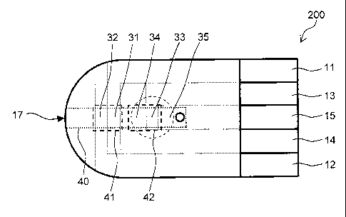
portion of the capillary 40.
[01161
Respective portions (portions 31, 32, 33, 34, 35) of the electrodes (voltage
application portion) 11, 12, 13, 14, 15 are disposed on an insulating plate
201 to
face the capillary 40. The portion 31 of the electrode 11 and the portion 32
of
the electrode 12 are disposed at a position in closer proximity to the blood
sample
introduction port 17 than the portion 33 of the electrode 13 and the portion
34 of
the electrode 14.
[01171
A reaction reagent layer 20 is formed on the insulating plate 201 to cover
the whole of the portion 33 of the electrode 13 and to partially cover the
portion
34 of the electrode 14 and the portion 35 of the electrode 15. The reaction
reagent layer 20 includes an oxidoreductase that uses the analyte in the blood
41
CA 02742149 2011-04-28
sample as a substrate, and an electron mediator.
[01181
The reaction reagent layer 20 is formed at a position separated from the
portion 31 of the electrode 11 and the portion 32 of the electrode 12. It is
preferred that a reagent including an oxidoreductase or an electron mediator
is
not disposed on the portion 31 of the electrode 11 and the portion 32 of the
electrode 12, and more preferably no reagent is disposed.
[01191
In an opposite configuration to the above, when the portion 33 of the
electrode 13 and the portion 34 of the electrode 14 are disposed at a position
in
closer proximity to the blood sample introduction port 17 than the portion 31
of
the electrode 11 and the portion 32 of the electrode 12, if the blood sample
is
introduced from the blood sample introduction port 17, the sample may reach
the
portion 33 of the electrode 13 and the portion 34 of the electrode 14 due to
flow in
the reaction reagent layer 20 on the portion 33 of the electrode 13 and the
portion 34 of the electrode 14. Therefore, this configuration should be
avoided.
[01201
The sensor chip 200 includes a measuring unit 41 (measuring unit A).
The measuring unit A is configured from an electrode system (temperature
electrodes) formed by the portion 31 of the electrode 11 and the portion 32 of
the
electrode 12, and a space in a portion of the capillary 40 that contains the
portion
31 and the portion 32.
42
CA 02742149 2011-04-28
[0121]
The sensor chip 200 includes a measuring unit 42 (measuring unit B).
The measuring unit B is configured from an electrode system (analysis
electrodes) formed by the portion 33 of the electrode 13 and the portion 34 of
the
electrode 14, and a space in a portion of the capillary 40 that contains the
reaction reagent layer 20 in addition to the portion 33 and the portion 34.
[0122]
In the temperature electrodes of the measuring unit A, the electrode 11
functions as a working electrode and the electrode 12 functions as an counter
electrode. In the analysis electrodes of the measuring unit B, the electrode
13
functions as a working electrode and the electrode 14 functions as an counter
electrode.
The measuring unit A (temperature measuring unit) acquires the data a
related to the temperature of the blood sample based on the amount of current
flowing in the temperature electrodes. The substance that exhibits an
electrochemical reaction on the temperature electrodes is mainly a component
of
the blood sample, or may be water, or may be a blood-cell component such as
red
blood cells or white blood cells.
[0123]
The measuring unit B (analyte measuring unit) acquires the data b
related to the concentration of the analyte in the blood sample based on the
amount of current flowing in the analysis electrodes. The substance that
43
CA 02742149 2011-04-28
exhibits an electrochemical reaction on the analysis electrodes is mainly an
electron mediator that exchanges electrons with the oxidoreductase. The data b
acquired in the measuring unit B is corrected based on the temperature using
the data a. The concentration of the analyte is calculated using the data b
after
correction.
[0124]
One or both of the portion 33 of the electrode 13 and the portion 34 of the
electrode 14 may function as one or both of the portion 31 of the electrode 11
and
a portion 32 of the electrode 12. However it is preferred that these
electrodes
are provided separately.
[0125]
The portion 35 of the electrode 15 is disposed in proximity to the inner
end portion of the capillary 40, that is to say, in proximity to the opposite
end to
the end that communicates with the outer portion. Application of voltage
between the electrode 15 and the electrode 13 facilitates detection when the
blood sample is introduced to an inner portion of the capillary 40. The
voltage
may be applied between the electrode 14 and the electrode 15 in substitution
for
the electrode 13.
[0126]
The electrodes 11, 12, 13, 14, 15 are connected with respective leads (not
illustrated). One end of the lead is exposed to an outer portion of the sensor
chip 200 on the end portion of the insulating plate 201 that is not covered by
the
44
CA 02742149 2011-04-28
spacer 202 and the cover 203 to thereby enable application of a voltage
between
each electrode.
[01271
The analyte in the blood sample may be a substance other than a blood
cell, and for example includes glucose, albumin, lactic acid, bilirubin, and
cholesterol. The oxidoreductase may be a substance that uses the target
analyte as a substrate. The oxidoreductase may be exemplified by glucose
oxidase, glucose dehydrogenase, lactate oxidase, lactate dehydrogenase,
bilirubin
oxidase, and cholesterol oxidase. The amount of the oxidoreductase in the
reaction reagent layer is 0.01 - 100 units (U), preferably 0.05 - 10 U, and
more
preferably 0.1 - 5 U.
[01281
The reaction reagent layer 20 preferably contains an electron mediator
that has a function of exchanging electrons produced by an oxidation reaction
with an electrode, such as potassium ferricyanide, p-benzoquinone,
p-benzoquinone derivatives, oxidized phenazine methosulfate, methylene blue,
ferricinium and ferricinium derivatives. The reaction reagent layer 20 may
include a water soluble polymer compound to increase molding characteristics
of
the reaction reagent layer. The water soluble polymer compound may be
exemplified from at least one selected from the group consisting of
carboxymethyl cellulose and salts thereof, hydroxyethyl cellulose,
hydroxypropylcellulose, methylcellulose, ethylcellulose, ethylhydroxyethyl
CA 02742149 2011-04-28
cellulose, carboxymethyl cellulose and salts thereof, polyvinylalcohol,
polyvinylpyrrolidone, polyamino acids such as polylysine, polystyrenesulfonic
acid and salts thereof, gelatin and derivatives thereof, polyacrylic acid and
salts
thereof, polymethacrylate and salts thereof, starch and derivatives thereof,
maleic anhydride polymers and salts thereof, and agarose gel and derivatives
thereof.
[0129]
The material of the insulating plate 201, the spacer 202 and the cover
203 is exemplified by polyethylene terephthalate, polycarbonate, polyimide,
polyethylene, polypropylene, polystyrene, polyvinyl chloride,
polyoxymethylene,
monomer-cast nylon, polybutylene terephthalate, resins such as methacrylate
resin and ABS resin, and glass.
[0130]
The electrodes 11, 12, 13, 14, and 15 for example are configured from a
known conductive material such as palladium, platinum, gold, silver, titanium,
copper, nickel, and carbon.
FIG. 4 illustrates an example of a circuit configuration for measuring an
analyte concentration in a blood sample in the biosensor system 100. The
measuring device 101 includes a control circuit 300 that applies a voltage
between at least two electrodes of the electrodes 11, 12, 13, 14 and 15 in the
sensor chip 200, and a display unit 400 that displays the measurement result.
[0131]
46
CA 02742149 2011-04-28
The control circuit 300 includes five connectors 301a, 301b, 301c, 301d,
301e, a switching circuit 302, a current/voltage conversion circuit 303, an
analog/digital (A/D) conversion circuit 304, a reference voltage power source
305,
and a computing unit 306. The control circuit 300 enables switching of the
potential applied to the electrodes to enable use of one electrode as a
cathode or
as an anode through the switching circuit 302.
[0132]
The computing unit (concentration determination unit) 306 includes a
known central processing unit (CPU) and a conversion table for determining an
analyte concentration in a blood sample based on the data a and the data b.
The computing unit 306 uses a correction coefficient based on the
environmental
temperature to correct the analyte concentration by reference to the
conversion
table above. More specifically, after referring to the conversion table for
preliminary measurement and provisionally calculating the analyte
concentration, the computing unit 306 corrects the analyte concentration by
reference to a conversion table for temperature correction.
[0133]
As illustrated in FIG. 5, the measurement of the analyte concentration in
the blood sample using the biosensor system 100 for example is executed as
described below.
Firstly, the CPU in the computing unit 306 commands the electrode 13 to
connect with the current/voltage conversion circuit 303 through the connector
47
CA 02742149 2011-04-28
301b and the electrode 15 to connect with the reference voltage power source
305
through the connector 301c.
[0134]
Thereafter, the CPU commands the application of a predetermined
voltage to both electrodes (step Si). For example, when the voltage is denoted
by the electrode 15 as the positive electrode and the electrode 13 as the
negative
electrode, the voltage is 0.01 - 2.OV, preferably 0.1 - 1.OV, and more
preferably
0.2 - 0.5V. The voltage is applied from insertion of the sensor chip into the
measuring device 101 until the introduction of the blood sample into an inner
portion of the capillary 40. When the blood sample is introduced into the
capillary 40 from the blood sample introduction port of the sensor chip 200, a
current flows between the electrode 15 and the electrode 13. The CPU detects
that the capillary 40 is filled with the blood sample by discrimination of an
increase amount in the current per unit time during this period. The current
value is converted to a voltage value by the current/voltage conversion
circuit
303 and then is converted to a digital value by the A/D conversion circuit 304
and
and input to the CPU. The CPU detects that the blood sample is introduced
into the inner portion of the capillary based on the digital value.
[0135]
After introduction of the blood sample, for example, the analyte in the
blood sample and oxygen, and oxygen and the electron mediator are reacted
within a range of 0 - 60 seconds, preferably 0 - 15 seconds, and more
preferably
48
CA 02742149 2011-04-28
0 - 5 seconds.
[01361
Then, the data a is acquired in the following manner (step S2).
[01371
Firstly, the voltage switching circuit 302 is operated by command of the
CPU, the electrode 11 is connected with the current/voltage conversion circuit
303 through the connector 301a, and the electrode 12 is connected with the
reference voltage power source 305 through the connector 301e. Then the CPU
commands application of a predetermined voltage between the electrodes in the
measuring unit A. As described below, when the voltage is denoted using the
electrode 11 as the positive electrode and the electrode 12 as the negative
electrode, the voltage is in the range of 0.1 - 5.OV, preferably 1.0 - 3.OV,
and
more preferably 1.5 - 2.5V. The voltage application time is in the range of
0.1 -
30 seconds, preferably from 0.5 - 10 seconds, and more preferably 1 - 5
seconds.
A signal commanding acquisition of the data a is output from the control
circuit
to the measuring unit A, to thereby cause the current/voltage conversion
circuit
303 to convert the current amount between both electrodes resulting from
application of the voltage to a voltage amount. Thereafter, the voltage amount
is converted to a digital value by the A/D conversion circuit 304, inputted to
the
CPU, and stored in the memory of the computing unit 306 as the data a.
[01381
Thereafter, the data b is acquired as described below (step S3).
49
CA 02742149 2011-04-28
[01391
Firstly, the voltage switching circuit 302 is operated by command of the
CPU, the electrode 13 is connected with the current/voltage conversion circuit
303 through the connector 301b, and the electrode 14 is connected with the
reference voltage power source 305 through the connector 301d. Then, the CPU
commands commencement of the measurement sequence in the measuring unit
B. The voltage applied at this time is denoted using the electrode 13 as the
positive electrode and the electrode 14 as the negative electrode, and is in
the
range of 0.05 - 1.OV, preferably 0.1 - 0.8V, and more preferably 0.2 - 0.6V.
The
voltage application time is from 0.1 - 30 seconds, preferably from 0.1 - 15
seconds, and more preferably 0.1 - 5 seconds. A signal commanding acquisition
of the data b is output from the control circuit to the measuring unit B, and
thereby cause the current/voltage conversion circuit 303 to convert the
current
amount flowing between both electrodes as a result of the voltage application
to
a voltage amount. Thereafter the voltage is converted to a digital value by
the
A/D conversion circuit 304, inputted to the CPU, and stored in the memory of
the
computing unit 306 as data b. From the point of view of enhancing the
measurement speed of the analyte concentration, the control circuit preferably
applies the signal commanding acquisition of the data b to the measuring unit
B
within a range of at least 0.5 seconds and less than 5 seconds from the time
that
the blood sample is introduced into the capillary 40 of the sensor chip.
[01401
CA 02742149 2011-04-28
The data b may be acquired prior to acquisition of the data a. However,
prior to acquisition of the data b, since a sufficient period is required for
dissolution of the sample, oxygen reaction of the electron mediator with
oxygen,
and the like, the data b is preferably acquired after acquisition of the data
a.
Furthermore, the data b and the data a may be acquired simultaneously.
However, since a voltage is applied simultaneously to two groups of electrode
systems in one solution system, there may be interference between the
respective
currents. Consequently, separate acquisition of the data a and acquisition of
the data b is preferred.
[01411
As illustrated in FIG. 6(a), the temperature when acquiring the data b is
more accurately reflected in the temperature measurement results by
respectively acquiring data related to temperature of the blood sample before
and after the acquisition of the data b. In other words, the biosensor system
100 applies a predetermined voltage to both electrodes (step S101), acquires
the
data a related to the temperature of the blood sample (step S102), and then
acquires the data b related to the concentration of the analyte in the blood
sample (step S103). Thereafter, the data c related to the temperature of the
blood sample is re-acquired (step S104). Then, the computing unit 306
calculates the data d by calculation of the average of the data a and the data
c
(step S105), and calculates the analyte concentration by correcting the
temperature in the data b using the data d (step S106). As illustrated in FIG.
51
CA 02742149 2011-04-28
6(b), the computing unit (concentration determination unit) 306 (refer to FIG.
4)
in the biosensor system 100 includes a temperature measuring unit 307 that
acquires the data c related to the temperature of the blood sample based on
the
dimension of the current flowing through the temperature electrodes that is in
contact with the blood sample after acquisition of the data b, a computing
unit
308 that calculate the data d related to the temperature of the blood sample
based on the data a and the data c, and a concentration calculating unit 309
that
uses the data d to calculate the concentration x of the analyte that is
corrected in
response to the temperature of the blood sample.
[01421
Then the computing unit 306 refers to the conversion table and
determines the analyte concentration in the blood sample based on the data a
the data b (step S4). The determined analyte concentration is displayed on the
display unit 400. If a temperature conversion table is prepared in relation to
the data a, the computing unit 306 can calculate the temperature of the blood
sample, and can display the temperature on the display unit 400. A computing
program used in this determination may be suitably designed in response to the
data structure of the conversion table. When numerical data displaying a
complete correspondence with the data a and the data b is not stated in the
conversion table, the computing unit 306 may determine the analyte
concentration using data stated in the conversion table and a known
interpolation method using data that approximates the data a and the data b.
52
CA 02742149 2011-04-28
[01431
If required, use of the electrode 11 and the electrode 12 may be used as
an electrode for temperature measurement applications and an electrode for
other analyte applications. The other analyte application for example includes
measurement of a hematocrit value in the blood sample, and measurement of a
reducing substance such as ascorbic acid, uric acid, bilirubin, acetaminophen,
and the like. A method of using the electrode 11 or the electrode 12 as the
working electrode (positive electrode), the electrode 13 or the electrode 14
as the
counter electrode (negative electrode) is known.
[01441
In the present invention, the voltage between the temperature electrodes
in the measuring unit A is affected by the configuration of the sensor chip
such
as the electrode material or the electrode surface area, and therefore it is
necessary to determine an optimal applied voltage in advance. The current
amount acquired when applying a voltage that diverges from an optimal value is
affected by the hematocrit value (Hct value) in the blood sample. An Hct value
means a numerical value expressing the ratio of the content of blood cells in
blood.
[01451
When the optimal voltage value is denoted as Vm, a voltage value higher
than the optimal voltage value is denoted as Vh, and a voltage value lower
than
the optimal voltage value is denoted as Vl, the change in the current amount
53
CA 02742149 2011-04-28
expressed by (Vl<Vm<Vh) is illustrated in FIG. 8. When a voltage value Vl that
is lower than the optimal voltage value is applied, as illustrated in FIG.
8(a), the
current amount increases as the value Hct increases. Conversely, when the
voltage value Vh is higher than the optimal voltage value is applied, as
illustrated in FIG. 8(c), the current amount increases as the Hct value
decreases.
When the optimal voltage value Vm is used, as illustrated in FIG. 8(b), a
fixed
current amount is exhibited irrespective of the Hct value. A conspicuous
estrangement of the current amount resulting from the Hct value is exhibited
under high temperature conditions and a high current amount. Therefore the
upper limiting temperature in the temperature measurement region is
preferably determined in advance. The Vm range is 0.1 - 5.0 V, preferably 1.0 -
3.0 V, and more preferably 1.5 - 2.5 V.
[01461
In the present invention, the current amount flowing between the
temperature electrodes in the measuring unit A is affected by the electrode
surface area. A higher current amount is obtained when either of the surface
area of a portion 31 of the electrode 11 (working electrode) and the surface
area
of a portion 32 of the electrode 12 (counter electrode) is increased. However
it is
preferred to increase the surface area of the portion 32 that is on the
counter
electrode side. More specifically, the range of the proportion of the surface
area
of the working area/the surface area of the counter electrode is preferably 1 -
0.25.
54
CA 02742149 2011-04-28
[0147]
Even when there is a rapid change in the environmental temperature of
the sensor, the biosensor system according to the embodiment enables highly
accurate measurement of the analyte concentration. As a result, there is no
necessity to provide an environmental temperature measuring unit such as a
thermistor in the measuring device.
[0148]
However, the state or configuration of the sensor may result in a low
accuracy in relation to the current amount obtained by the measuring unit A.
For example, in a sensor that has a small surface-area capillary 40, although
the
capacity of the blood sample required for measurement may be reduced, the
surface area of the temperature electrodes in the measuring unit A must be
reduced. Therefore, the current amount obtained in the measurement A is
decreased, and as a result, it is predicted that the accuracy of the current
amount obtained in the measuring unit A will be reduced. In this case, as
illustrated in the circuit configuration diagram in FIG. 53, the environmental
temperature measuring unit 315 may be provided in the measuring device. The
number of environmental temperature measuring units 315 may be only one, or
may be two or more. When two or more environmental temperature measuring
units 315 are provided, respective environmental temperature measuring units
315 guarantee a more accurate measurement result for the environmental
temperature by mutually monitoring of accuracy.
CA 02742149 2011-04-28
[01491
Furthermore, when temperature data obtained by the measuring unit A
in the sensor is compared with the temperature data obtained from a thermistor
provided in the measuring device, temperature correction may be executed and
the respective temperature change can be monitored, an optimal temperature
can be selected, and used for temperature correct. Furthermore, a method may
be used in which the temperature is corrected by reference to the difference
between the temperature of the measuring unit A and the temperature of the
thermistor, or a method in which a plurality of temperature differences is
acquired, and an optimal temperature correction value is selected. Of course,
a
method of utilizing data for average values and not temperature differences
may
be executed.
[01501
In the biosensor system 100 illustrated in FIG. 53, the computing unit
306 compares the temperature t acquired by the measuring unit A and the
temperature tl acquired by the environmental temperature measuring unit 315
in the measuring device (step S43), and uses the temperature t acquired by the
measuring unit A only when there is an error between the two. That is to say,
as illustrated in FIG. 7(a), the computing unit 306 calculates the temperature
t
based on the data a (step S41). The computing unit 306 calculates the
concentration x based on the data b (step S42). The environmental temperature
measuring unit 315 measures the environmental temperature tl ( step S43).
56
CA 02742149 2011-04-28
[01511
When there is no difference between the outer environmental
temperature and the blood sample temperature, the computing unit 306 uses the
temperature t1 (step S45) since the environmental temperature measuring unit
315 has a high measurement accuracy.
When there is a difference between the outer environmental temperature
and the blood sample temperature as a result of a sharp variation in the
temperature, the environmental temperature measuring unit 315 cannot adapt
to the difference. Therefore, the temperature t acquired by the measuring unit
A is adopted (step S46). More specifically, the temperature threshold Z is
preset.
The computing unit 306 is compares the value for I t - t1 I with the
temperature
threshold Z (step S44). When the value for I t - t1 I is higher than or equal
to
the temperature threshold Z, the computing unit 306 corrects the concentration
x
based on the temperature t (step S45). When smaller than the temperature
threshold Z, the concentration x is corrected based on the environmental
temperature t1 (step S46).
[01521
The range of the temperature threshold Z is determined in consideration
of the accuracy of the environmental temperature measuring unit of the
measuring device and the accuracy of the measuring unit A in the sensor chip,
and is in the range of 0.01 - 5.0'C, preferably 0.1 - 2.0'C, and more
preferably
0.2-1.0 C.
57
CA 02742149 2011-04-28
[0153]
As illustrated in FIG. 7(b), the computing unit (concentration
determination unit) 306 in the biosensor system 100 (refer to FIG. 4 and FIG.
52)
includes a temperature calculating unit 310 and a concentration calculating
unit
311. The temperature calculating unit 310 calculates the temperature t of the
blood sample based on the data a. The concentration calculating unit 311
calculates the concentration x of the analyte of the blood sample based on the
data b.
[0154]
The measuring device includes an environmental temperature measuring
unit 312, a comparison unit 313, and a correction unit 314. The environmental
temperature measuring unit 312 measures the peripheral environmental
temperature tl of the blood sample. The comparison unit 313 compares the
difference between the temperature t and the environmental temperature tl
with the temperature threshold value Z. The correction unit 314 corrects the
concentration x based on the temperature t when the expression I t - t1 I > Z
is
satisfied, and corrects the concentration x based on the environmental
temperature t1 when the expression I t - t1 I < Z is satisfied.
[0155]
[Working Examples]
The invention will be described in further detail below with reference to
the embodiments.
58
CA 02742149 2011-04-28
[0156]
[Working Example 1]
A sensor chip 210 is prepared as illustrated in FIG. 9 and FIG. 10. The
capillary is designed with a width of 1.2 mm, a length (depth) of 4.0 mm, and
a
height of 0.15 mm. The insulating plate is formed from polyethylene
terephthalate. After palladium is deposited by vapor deposition onto the
insulating plate, the respective electrodes were formed by formation of a slit
in
the palladium layer with a laser so that the surface area of the portion 31 of
the
electrode 11 is 0.12 mm2, and the portion 32 of the electrode 12 is 0.48 mm2.
[0157]
Three types of blood samples having Hct values respectively of 25%, 45%
and 65% were prepared. The temperature of the blood sample was taken to be
23'C. These blood samples were introduced into the capillary of separate
sensor chips. Thereafter, the electrode 11 was used as the working electrode
(positive electrode) and the electrode 12 was used as the counter electrode
(negative electrode), and a voltage of 2.OV, 2.2V, or 2.4V was applied between
the
electrodes (temperature electrodes). The current flowing between the working
electrode and the counter electrode (response current) due to application of
the
voltage is measured.
[0158]
The measurement results are illustrated in the graphs in FIG. 11(a), FIG.
11(b), and FIG. 11(c).
59
CA 02742149 2011-04-28
When the applied voltage is 2.OV, as illustrated in FIG. 11(a), the
response current increases as the Hct value increases. These results
correspond
to FIG. 8(a).
[01591
As illustrated in FIG. 11(b), when the applied voltage is 2.2V, the
response current is fixed irrespective of the Het value. These results
correspond
to FIG. 8(b).
As illustrated in FIG. 11(c), when the applied voltage is 2.4V, the
response current increases as the Hct value decreases. These results
correspond to FIG. 8(c).
[01601
Next, an experiment using a blood sample with an Hct 45% at 4 C - 38 C
was performed. At each temperature, the blood sample was introduced into the
capillary of separate sensor chips. Thereafter, the electrode 11 was used as
the
working electrode (positive electrode) and the electrode 12 was used as the
counter electrode (negative electrode), and the response current was measured
when a voltage of 2.2V was applied between the electrodes (temperature
electrodes). The measurement results are illustrated in the graph in FIG. 12.
As illustrated in FIG. 12, the response current increases as the temperature
increases.
[01611
The results in FIG. 11 and FIG. 12 demonstrate that a blood sample
CA 02742149 2011-04-28
temperature can be detected by applying a large voltage of 2.2V between the
electrode 11 and the electrode 12 and thereby measuring the response current.
[01621
[Working Example 21
The sensor chip having the configuration described in Working Example
1 was used, and a blood sample at a temperature of 23 C and an Hct value of
45% was introduced into the capillary of the sensor chip. Thereafter, the
electrode 11 was used as the working electrode (positive electrode) and the
electrode 12 was used as the counter electrode (negative electrode), and the
response current was measured when a voltage of 2.2V was applied between the
electrodes (temperature electrodes). Table 1 below illustrates the current
value
after three seconds from initiation of voltage application. The current value
in
Working Example 2 was 1.88 pA.
[01631
Table 1
Electrode Surface Area (mm2) Current Value Current Increase
Working Counter (pA) Rate (%)
Electrode electrode
Working 0.12 0.48 1.88 -
Example 2
Working 0.24 0.48 2.47 32
Example 3
Working 0.48 0.48 3.13 67
Example 4
Working 0.12 0.96 3.08 65
Example 5
Working 0.24 0.96 3.65 94
61
CA 02742149 2011-04-28
Example 6
[0164]
[Working Example 31
An electrode was formed so that the surface area of the portion 31 of the
electrode 11 of the sensor chip is 0.24 mm2, and the surface area of the
portion 32
of the electrode 12 of the sensor chip is 0.48 mm2. Other conditions are the
same as the sensor chip described in Working Example 2. Table 1 below
illustrates the current value after three seconds from initiation of voltage
application. The current value in Working Example 3 was 2.47 pA. When
compared with Working Example 2, the current value exhibits a 32% increase.
The surface area of the working electrode in the sensor chip in Working
Example
3 is twice as large when compared with Working Example 2.
[0165]
[Working Example 4]
An electrode was formed so that the surface area of the portion 31 of the
electrode 11 of the sensor chip is 0.48 mm2, and the surface area of the
portion 32
of the electrode 12 of the sensor chip is 0.48 mm2. Other conditions are the
same as the sensor chip described in Working Example 2. Table 1 below
illustrates the current value after three seconds from initiation of voltage
application. The current value in Working Example 4 was 3.13 pA. When
compared with Working Example 2, the current value exhibits a 67% increase.
The surface area of the working electrode in the sensor chip in Working
Example
62
CA 02742149 2011-04-28
4 is four times as large when compared with Working Example 2 and twice as
large when compared with Working Example 3. In other words, it is shown that
the current value increases as the surface area of the working electrode
increases.
[01661
[Working Example 51
An electrode is formed so that the surface area of the portion 31 of the
electrode 11 of the sensor chip is 0.12 mm2, and the surface area of the
portion 32
of the electrode 12 of the sensor chip is 0.96 mm2. Other conditions are the
same as the sensor chip described in Working Example 2. Table 1 below
illustrates the current value after three seconds from initiation of voltage
application. The current value in Working Example 5 was 3.08 uA. When
compared with Working Example 2, the current value exhibits a 65% increase.
The surface area of the working electrode in the sensor chip in Working
Example
5 is twice as large when compared with Working Example 2. In other words, it
is shown that the current value increases as the surface area of the counter
electrode increases. When compared with Working Example 3, the increase rate
in the current value only reaches 32% under the condition that the surface
area
of the working electrode is two times. Therefore a higher response value is
obtained by increasing the surface of the counter electrode more than the
working electrode.
[01671
63
CA 02742149 2011-04-28
[Working Example 61
An electrode is formed so that the surface area of the portion 31 of the
electrode 11 of the sensor chip is 0.24 mm2, and the surface area of the
portion 32
of the electrode 12 of the sensor chip is 0.96 mm2. Other conditions are the
same as the sensor chip described in Working Example 2. Table 1 below
illustrates the current value after three seconds from initiation of voltage
application. The current value in Working Example 6 was 3.65 pA. When
compared with Working Example 2, the current value exhibits a 94% increase.
The surface area of the working electrode and the counter electrode in the
sensor
chip in Working Example 6 is twice as large when compared with Working
Example 2. In other words, the current value is also increased in proportion
to
an increase in the electrode surface area when the ratio of the electrode
surface
areas is the same.
[0168]
[Working Example 7]
A sensor chip as described in Working Example 1 is prepared. Fifteenth
types of blood samples being combinations of three Hct values respectively of
25%, 45% and 65% and five temperatures of 4 C, 13T, 21'C, 30'C, and 38'C
were prepared.
[0169]
These blood samples were introduced into the capillary of separate
sensor chips. Next, the electrode 11 was used as the working electrode
(positive
64
CA 02742149 2011-04-28
electrode) and the electrode 12 was used as the counter electrode (negative
electrode), and a voltage of 2.1V, 2.15V, or 2.2V was applied between the
electrodes (temperature electrodes) to thereby measure the response current at
that time.
[01701
FIG. 13 to FIG. 17 are graphs illustrating the response current at
respective temperature conditions and applied voltages. The temperature
conditions and the applied voltage conditions in each graph are as illustrated
below.
(Temperature Condition)
FIG. 13(a), 13(b), 13(c): 4 C
FIG. 14(a), 14(b), 14(c): 13'C
FIG. 15(a), 15(b), 15(c): 210C
FIG. 16(a), 16(b), 16(c): 300C
FIG. 17(a), 17(b), 17(c): 380C
(Applied Voltage Condition)
FIG. 13(a), FIG. 14(a), FIG. 15(a), FIG. 16(a), FIG. 17(a): 2100mV
FIG. 13(b), FIG. 14(b), FIG. 15(b), FIG. 16(b), FIG. 17(b): 2150mV
FIG. 13(c), FIG. 14(c), FIG. 15(c), FIG. 16(c), FIG. 17(c): 2200mV
Under the low temperature conditions of 4'C and 13'C in which the
response current is small, a response current that is not dependent in the Hct
value is exhibited in the same manner under any of the applied voltage
CA 02742149 2011-04-28
conditions.
[0171]
Under the high temperature conditions of 30'C and 38'C that have a
large response current, a trend is observed for the response current to vary
in
response to the Het value. In particular, a conspicuous difference is observed
in
the region of 4 seconds or less under an applied voltage condition of 2.1V and
the
region of 3 seconds or more under an applied voltage condition of 2.2V when
compared with an applied voltage of 2.15V.
[0172]
Consequently, it is important to determine an optimal application voltage
conditions with reference to the response current in the high-temperature
region
so that the response current is not dependent upon the Hct value under
different
temperature conditions. The optimal application voltage determined in the
above manner in Working Example 7 is 2.15V. The current value after three
seconds is 1.93 pA when the blood sample is introduced at a Hct value of 45%
and a temperature of 21'C as shown in Table 2.
[0173]
Table 2
Electrode Surface Area (mm2) Optimal Applied Current Value
Working Counter Voltage (V) (pA)
Electrode electrode
Working 0.12 0.48 2.15 1.93
Example 7
Working 0.20 0.40 2.1 1.69
Example 8
66
CA 02742149 2011-04-28
Working 0.30 0.30 2.05 1.48
Example 9
[01741
[Working Example 81
An electrode was formed so that the surface area of the portion 31 of the
electrode 11 of the sensor chip is 0.20 mm2, and the surface area of the
portion 32
of the electrode 12 of the sensor chip is 0.40 mm2. Other conditions are the
same as the sensor chip described in Working Example 1. As described in
Working Example 7, the optimal applied voltage in Working Example 8
determined with reference to the response current in the high-temperature
region is 2.1V. At this time, as illustrated in Table 2, the current value
after
three seconds is 1.69 pA when a blood sample with a Hct value of 45% and a
temperature of 21 C is introduced.
[01751
[Working Example 91
An electrode was formed so that the surface area of the portion 31 of the
electrode 11 of the sensor chip is 0.30 mm2, and the surface area of the
portion 32
of the electrode 12 of the sensor chip is 0.30 mm2. Other conditions are the
same as the sensor chip described in Working Example 1.
[01761
As described in Working Example 7, the optimal applied voltage is
determined with reference to the response current in the high-temperature
67
CA 02742149 2011-04-28
region. The optimal applied voltage in Working Example 9 is 2.05V. At this
time, as illustrated in Table 2, the current value after three seconds is 1.48
pA
when a blood sample with a Hct value of 45% and a temperature of 21'C is
introduced. The results of Working Examples 7, 8 and 9 demonstrate that the
dimension of the response current varied and the optimal applied current is
different when the electrode surface area is different. Furthermore, under a
condition in which the sum of the surface area of the working electrode is the
same as that of the surface area of the counter electrode, a larger response
current is obtained when the surface area of the counter electrode is large.
[0177]
[Working Example 10]
A sensor chip is prepared as illustrated in FIG. 2 and FIG. 3. The
capillary is designed with a width of 1.2 mm, a length (depth) of 4.0 mm, and
a
height of 0.15 mm. The insulating plate is formed from polyethylene
terephthalate, and palladium is deposited by vapor deposition onto the
insulating plate. Thereafter, the respective electrodes are formed by
formation
of a slit in the palladium layer with a laser so that the surface area of the
portion
31 of the electrode 11 is 0.30 mm2, and the portion 32 of the electrode 12 is
0.48
mm2.
[0178]
The reaction reagent layer is formed as follows. An aqueous solution
including glucose dehydrogenase, potassium ferricyanide (Kanto Kagaku Co.,
68
CA 02742149 2011-04-28
Ltd.), taurine (Nakalai Tesque), glucose dehydrogenase was prepared. The
concentration of glucose dehydrogenase is adjusted to a concentration of 2.0
U/sensor. A concentration of 1.7 mass% of potassium ferricyanide, and 1.0
mass% of taurine was dissolved in the aqueous solution to thereby obtain a
reagent liquid. After coating of the reagent liquid onto the polyethylene
terephthalate plate, drying is performed at a humidity of 45% and a
temperature
of 21T.
[0179]
The Hct value of the blood sample is 25%, 45% and 65%, and the glucose
concentration is 40mg/dl, 80mg/dl, 200mg/dl, 400mg/dl, and 1,600mg/dl. The
temperature of the blood sample was 4 C, 13'C, 22T, 30T, and 39T.
[0180]
The application voltage between the electrodes and the application time
is set as follows. 2.075V was applied to both electrodes (temperature
electrodes)
being the electrode 11 (positive electrode ) and electrode 12 (negative
electrode)
for 3 seconds from immediately after introduction of the blood sample. From 3
seconds to five seconds, 0.25V was applied to both electrodes (analysis
electrode)
being the electrode 13 (positive electrode ) and electrode 14 (negative
electrode),
and at five seconds from introduction of the blood sample, the measurement is
completed.
[0181]
Table 3 and the graph illustrated in FIG. 18 illustrate the response
69
CA 02742149 2011-04-28
current value after three seconds between the temperature electrodes. The
response current value after 3 seconds does not depend on the Hct value but
rather depends on the temperature. The response current value after three
seconds is converted to the temperature of the blood sample using the table
illustrated in FIG. 18. A difference is not observed in the response current
value after three seconds at different glucose concentrations. Table 4 below
illustrates the response current value after 5 seconds between the analysis
electrodes. The response current value after 5 seconds increases together with
increases in the glucose concentration at each temperature, or increases
together
with increases in the temperature at each glucose concentration. When the
temperature is known, the table illustrated in Table 4 below may be used as a
conversion table for glucose concentration to thereby enable conversion of the
response current value after 5 seconds to a glucose concentration for the
blood
sample.
[01821
Table 3
Current value after 3 seconds Hematocrit
(pA) 25% 45% 65%
4 C 0.83 0.82 0.83
13 C 1.16 1.19 1.22
Blood 22'C 1.66 1.63 1.64
Temperature 30 C 2.13 2.12 2.16
39'C 2.76 2.81 2.80
[01831
CA 02742149 2011-04-28
Table 4
Current value after 5 seconds Glucose Concentration (mg/dl)
(pA) 40 80 200 400 600
4 C 1.46 2.35 4.52 6.85 8.31
13'C 1.75 2.85 5.60 9.22 11.89
Blood 22'C 2.15 3.56 6.89 12.03 15.81
Temperature 30 C 2.48 4.35 8.44 14.82 20.20
39'C 2.93 4.96 10.50 17.81 23.85
[01841
[Working Example 111
Four types of sensor chips having the configuration illustrated in FIG. 9
and FIG. 10 were prepared. In the first to the four types of sensor chips, the
inter-electrode distance illustrated in FIG. 19 is respectively 100 um, 300
um,
500 pm, and 700 pm.
[01851
Nine types of blood samples being combinations of three Hct values
respectively of 25%, 45% and 65% and three temperatures of 11'C, 21T, and 30
C were prepared.
Next, after introduction of the blood samples above into the capillary in
the sensor chips above, a 2.2V voltage was applied between the electrodes
(temperature electrodes), and the respective response currents were measured.
[01861
The measurement results are illustrated in the graphs in FIGs 20(a) -
20(d), FIGs 21(a) - 21(d), and FIGs 22(a) - 22(d). FIGs 20(a) - 20(d)
illustrate
71
CA 02742149 2011-04-28
the response current value in an 11 C blood sample by inter-electrode
distance
and by hematocrit. FIGs. 21(a) - 21(d) illustrate the response current value
in
a 21'C blood sample by inter-electrode distance and by hematocrit. FIGs. 22(a)
- 22(d) illustrate the response current value in a 30 C blood sample by
inter-electrode distance and by hematocrit.
[0187]
The graphs above do not exhibit a significant difference in the response
current value when the inter-electrode distance is varied. The results of
Working Example 11 demonstrate that the response current exhibits almost no
effect due to the inter-electrode distance.
[0188]
[Working Example 12]
Two types of sensor chips having different electrode shapes were
prepared.
A first type of sensor chip has the configuration illustrated in FIG. 9, FIG.
10 and FIG. 23(a). In the first type of sensor chip, the surface area of the
portion 31 (working electrode) of the electrode 11 is 0.24 mm2, and the
surface
area of the portion 32 (counter electrode) of the electrode 12 of the sensor
chip is
0.96 mm2, and the inter-electrode distance is 300 um.
[0189]
A second type of sensor chip has the configuration illustrated in FIG.
23(b). In the second type of sensor chip, the surface area of the portion 31
72
CA 02742149 2011-04-28
(working electrode) of the electrode 11 is 0.24 mm2, and the portion 32
(counter
electrode) of the electrode 12 has a shape that is formed separately at two
positions in FIG. 23(b). The surface area of the two portions of the portion
32
are respectively 0.48 mm2. The total value for the portion 32 of the electrode
12
is 0.96 mm2. In the second type of sensor chip, the inter-electrode distance
is
300 pm.
[0190]
Nine types of blood samples being combinations of three Hct values
respectively of 25%, 45% and 65% and three temperatures of 11T, 21T, and 30
'C were prepared.
Next, after introduction of the blood samples above into the capillary in
the sensor chips above, a 2.2V voltage was applied between the electrodes
(temperature electrodes), and the respective response currents were measured.
[0191]
The measurement results are illustrated in the graphs in FIGs.
24(a)-24(b), FIGs. 25(a)-25(b), and FIGs. 26(a)-26(b). FIGs. 24(a)-24(b)
illustrate the response current value in an 11 C blood sample by electrode
shape
and by hematocrit. FIGs. 25(a)-25(b) illustrate the response current value in
a
21'C blood sample by electrode shape and by hematocrit. FIGs. 26(a)-26(b)
illustrate the response current value in a 30 C blood sample by electrode
shape
and by hematocrit.
[0192]
73
CA 02742149 2011-04-28
The graphs above do not exhibit a significant difference in the response
current value when the electrode shape distance is varied. The results of
Working Example 12 demonstrate that the response current exhibits almost no
effect due to the electrode shape.
[0193]
[Working Example 13]
Four types of sensor chips having different lead widths in the counter
electrode 12 were prepared. The respective types of sensor chip have the
configuration illustrated in FIG. 2, FIG. 3 and FIG. 27(a). In each type of
sensor chip, the surface area of the portion 31 (working electrode) of the
electrode 11 is 0.30 mm2, and the portion 32 (counter electrode) of the
electrode
12 is 0.30 mm2, and the inter-electrode distance is 100 um. In the first to
the
fourth types of sensor chip, the lead width in the counter electrode 12
illustrated
in FIG. 27(b) is respectively 0.5 mm, 1.0 mm, 1.5 mm, 2.0 mm.
[0194]
Three types of blood samples were prepared. The Hct values for a first
to a third type of blood sample are respectively 25%, 45% and 65% and a
temperature for each type of blood sample is 23 C (room temperature).
[0195]
Next, after introduction of the blood samples above into the capillary in
the sensor chips above, a 2.05V voltage was applied between the electrodes
(temperature electrodes), and the respective response currents were measured.
74
CA 02742149 2011-04-28
[0196]
The measurement results are illustrated in the graphs in FIGs
28(a)-28(d). These graphs demonstrate that the response current exhibits
almost no change even at different hematocrit values. Furthermore, a
significant difference is not observed in the response current value when the
lead
width is varied. The results of Working Example 13 demonstrate that the
response current exhibits almost no effect due to the lead width (resistance).
[0197]
[Working Example 141
Two types of sensor chips were prepared. A first and a second type of
sensor chip have the configuration illustrated in FIG. 9 and FIG. 10. In the
first and the second type of sensor chip, the surface area of the portion 31
(working electrode) of the electrode 11 is 0.12 mm2, and the surface area of
the
portion 32 (counter electrode) of the electrode 12 of the sensor chip is 0.48
mm2,
and the inter-electrode distance is 300 gm. In the first type and the second
type
of sensor chip, the thickness of the spacer 202 illustrated in FIG. 29
(capillary
height) is respectively 0.15 mm and 0.09 mm.
[0198]
Nine types of blood samples being combinations of three Hct values
respectively of 25%, 45% and 65% and three temperatures of 11T, 21'C, and 30
C were prepared.
Next, after introduction of the blood samples above into the capillary in
CA 02742149 2011-04-28
the sensor chips above, a 2.2V voltage was applied between the electrodes
(temperature electrodes), and the respective response currents were measured.
[0199]
The measurement results are illustrated in the graphs in FIGs. 30(a) -
30(b), FIGs. 31(a) - 31(b), and FIGs. 32(a) - 32(b). FIGs. 30(a) - 30(b)
illustrate
the response current value in an 11'C blood sample by capillary height and by
hematocrit. FIGs. 31(a) - 31(b) illustrate the response current value in a 21
C
blood sample by capillary height and by hematocrit. FIGs. 32(a) - 32(b)
illustrate the response current value in a 30 C blood sample by capillary
height
and by hematocrit.
[0200]
The graphs above do not exhibit a significant difference in the response
current value even when the capillary height is varied. The results of Working
Example 14 demonstrate that the response current exhibits almost no effect due
to the capillary height.
[0201]
[Working Example 15]
Two types of sensor chips were prepared. The respective types of sensor
chip have the configuration illustrated in FIG. 9 and FIG. 10. In each type of
sensor chip, the surface area of the portion 31 (working electrode) of the
electrode 11 is 0.12 mm2, and the surface area of the portion 32 (counter
electrode) of the electrode 12 of the sensor chip is 0.48 mm2, and the
76
CA 02742149 2011-04-28
inter-electrode distance is 100 uum. The surface resistance of the palladium
vapor-deposited plate of the first type and the second type of sensor chip is
respectively 115 P/o and 60 9/o.
[0202]
Fifteen types of blood samples being combinations of three Hct values
respectively of 25%, 45% and 65% and the temperatures of 4 C, 13'C, 21'C, 30
C, and 38 C were prepared.
Next, after introduction of the above blood samples above into the above
capillary in the sensor chips, a 2.15V voltage was applied between the
electrodes
(temperature electrodes), and the respective response currents were measured.
[0203]
The measurement results are illustrated in the graphs in figures (a) and
(b) in FIGs. 33 - 37. FIG. 33(a) and 33(b) illustrate the response current
value
in a 4 C blood sample by palladium resistance. FIG. 34(a) and 34(b) illustrate
the response current value in a 13'C blood sample by palladium resistance.
FIG. 35(a) and 35(b) illustrate the response current value in a 21 C blood
sample
by palladium resistance. FIG. 36(a) and 36(b) illustrate the response current
value in a 30 C blood sample by palladium resistance. FIG. 37(a) and 37(b)
illustrate the response current value in a 38'C blood sample by palladium
resistance.
[0204]
The graphs above do not exhibit a significant difference in the response
77
CA 02742149 2011-04-28
current value when palladium resistance is varied. The results of Working
Example 15 demonstrate that the response current exhibits almost no effect due
to the palladium resistance. There is no necessity to explain that when a
known conductive material such as platinum, gold, silver, titanium, copper,
nickel, and carbon is applied to the plate, the same effect is obtained.
[0205]
[Working Example 161
Sensor chips were prepared to have the configuration illustrated in FIG.
9 and FIG. 10. In the sensor chips, the surface area of the portion 31
(working
electrode) of the electrode 11 is 0.12 mm2, the surface area of the portion 32
(counter electrode) of the electrode 12 of the sensor chip is 0.48 mm2, and
the
inter-electrode distance is 100 gm.
[0206]
Three types of blood samples were prepared by adding a glucose
concentrate to blood having a Hct value of 45% and a temperature of 24'C. The
glucose concentrations of the first to the third blood sample are respectively
0
mg/dL, 205 mg/dL, and 640 mg/dL.
[0207]
Next, the above blood samples were introduced into the capillaries of the
respective sensor chips above. Thereafter, a voltage of 2.15V was applied
between the electrodes (temperature electrodes), and the respective response
currents were measured.
78
CA 02742149 2011-04-28
[0208]
The measurement results are illustrated in the graph in FIG 38. FIG.
38 illustrates the response current value in a 24 C blood sample by glucose
concentration. The graphs above do not exhibit a significant difference in the
response current value when glucose concentration is varied. The results of
Working Example 16 demonstrate that the response current exhibits almost no
effect due to glucose concentration. When the present invention is applied to
a
blood glucose sensor (glucose sensor), since the measurements are not affected
by
the glucose concentration, it is shown that application is possible without
problems.
[0209]
[Working Example 171
Sensor chips were prepared as in FIG. 9 and FIG. 10 so that the surface
area of the portion 31 (working electrode) of the electrode 11 is 0.12 mm2,
the
surface area of the portion 32 (counter electrode) of the electrode 12 of the
sensor
chip is 0.48 mm2, and the inter-electrode distance is 100 um.
[0210]
Three types of blood samples with different ascorbic acid concentrations
were prepared by adding an ascorbic acid concentrate to blood having a Hct
value of 45% and a temperature of 24 C. The glucose concentrations of the
first
to the third blood sample are respectively 0 mg/dL, 10 mg/dL, and 20 mg/dL.
[0211]
79
CA 02742149 2011-04-28
Next, the above blood samples were introduced into the capillaries of the
respective sensor chips above. Thereafter, a voltage of 2.15V was applied
between the electrodes (temperature electrodes), and the respective response
currents were measured.
[02121
The measurement results are illustrated in the graph in FIG 39. FIG.
39 illustrates the response current value in a 24 C blood sample by ascorbic
acid
concentration. The graphs above do not exhibit a significant difference in the
response current value when ascorbic acid concentration is varied. That is to
say, in the present working example, the measurement accuracy for blood
glucose
level was not affected by the serum concentration of ascorbic acid, that is a
reducing substance. Therefore it is shown that the sensor chip according to
the
present working example can be used without problems as a blood glucose level
sensor.
[02141
[Working Example 181
Sensor chips were prepared to have the configuration illustrated in FIG.
9 and FIG. 10. In the sensor chips, the surface area of the portion 31
(working
electrode) of the electrode 11 is 0.12 mm2, the surface area of the portion 32
(counter electrode) of the electrode 12 of the sensor chip is 0.48 mm2, and
the
inter-electrode distance is 100 gm.
[02141
CA 02742149 2011-04-28
Two types of blood samples having different temperatures were prepared.
A first type of blood sample has a Hct value of 45% and a temperature of 4 C.
A
second type of blood sample has a Hct value of 45% and a temperature of 42T.
[02151
Next, one minute after moving the above blood samples to a 24'C
environment, the samples were introduced into the capillaries of the
respective
sensor chips described above. Thereafter, a voltage of 2.15V was applied
between the electrodes (temperature electrodes), and the respective response
currents were measured.
[02161
The measurement results are illustrated in the graph in FIG 40. The
dotted line in FIG. 40 illustrates the response current value when introducing
blood at 24 C to a 24 C environment (hereinafter referred to as "normal
introduction"). The solid line in FIG. 40 illustrates the response current
value
when introducing blood at 4 C to a 24 C environment (hereinafter referred to
as
"4 C introduction"). The broken line in FIG. 40 illustrates the response
current
value when introducing blood at 42'C to a 24'C environment (hereinafter
referred to as "42 C introduction").
[02171
The graphs illustrate that during a time period soon after the
measurement period, the temperature exhibited by the 4 C introduction is low
in
comparison to the temperature exhibited by the normal introduction, and the
81
CA 02742149 2011-04-28
temperature exhibited by the 42'C introduction is high in comparison to the
temperature exhibited by the normal introduction. Over the passage of time
during the measurement period, the temperature difference between the 42 C
introduction and the 4 C introduction disappears. The fact that the
temperature difference disappears due to the passage of the measurement period
is thought in both cases to result from the movement of the blood sample at 4
C
or 42'C to a 24 C environment, and therefore over the passage of time, both
samples shift to 24 C that is the temperature of the sensor chip.
[0218]
According to Working Example 18, it is shown that measurement of
temporal variation in relation to the temperature of the blood sample is
possible.
Furthermore, the sensor chip is provided with a temperature electrode
that is disposed to make contact with the blood sample, and measures the
temperature of the blood sample. Therefore, when the sensor chip is used, a
temperature for the blood sample that takes into consideration temporal
variation can be obtained, and this value can be used to correct the glucose
concentration and the like. In other words, the accuracy of various types of
corrections can be improved.
[0219]
[Working Example 191
Sensor chips were prepared to have the configuration illustrated in FIG.
9 and FIG. 10. In the sensor chips, the surface area of the portion 31
(working
82
CA 02742149 2011-04-28
electrode) of the electrode 11 is 0.12 mm2, the surface area of the portion 32
(counter electrode) of the electrode 12 of the sensor chip is 0.48 mm2, and
the
inter-electrode distance is 100 um.
[02201
A blood sample was prepared. The blood sample has a Hct value of 45%.
As illustrated in FIG. 41, approximately 3 uL of blood was dripped in
advance into the sensor chip. The blood was dripped onto an upper portion of
the cover 203. Dripping blood in this manner is hereinafter referred to as
"upward orientation".
[02211
Approximately 10 uL of blood was dripped in advance into the other
sensor chip. The blood was dripped onto a lower portion of the insulating
plate
201. Dripping blood in this manner is hereinafter referred to as "downward
orientation".
[02221
Next, the above blood samples were introduced in a 24 C environment
into the capillaries 204 of the respective sensor chips. Thereafter, a voltage
of
2.15V was applied between the electrodes (temperature electrodes), and the
respective response currents were measured.
[02231
The measurement results are illustrated in the graph in FIG 42. The
broken line in FIG. 42 illustrates the response current when dripping blood in
83
CA 02742149 2011-04-28
advance in an upward orientation in a 24 C environment. The solid line in FIG.
42 illustrates the response current when dripping blood in advance in a
downward orientation in a 24'C environment. The dotted line in FIG. 42
illustrates the response current when dripping blood in advance in both an
upward orientation and a downward orientation in a 24 C environment
(hereinafter referred to as "normal introduction").
[0224)
The graphs illustrate that in comparison to normal introduction, the
response current value is low during an upward orientation and during a
downward orientation. This is thought to be due to the fact that the
temperature of the blood sample in the capillary 204 is reduced by the heat of
evaporation of blood in an upward orientation and during a downward
orientation that becomes excessively attached to an outer range of the
capillary
204.
[02251
Working Example 19 enables comprehension of the effect of heat of
evaporation as illustrated in FIG. 42.
The sensor chip is provided with a temperature electrode that is disposed
to make contact with the blood sample, and that measures the temperature of
the blood sample. Therefore, a temperature for the blood sample that takes
into
consideration heat of evaporation can be obtained, and this value can be used
to
correct the glucose concentration and the like. In other words, the accuracy
of
84
CA 02742149 2011-04-28
various types of corrections can be improved.
[02261
[Working Example 201
Sensor chips were prepared to have the configuration illustrated in FIG.
9 and FIG. 10 in that the surface area of the portion 31 (working electrode)
of the
electrode 11 is 0.12 mm2, the surface area of the portion 32 (counter
electrode) of
the electrode 12 of the sensor chip is 0.48 mm2, and the inter-electrode
distance
is 100 um. A blood sample with a Hct value of 45% was prepared.
[02271
Immediately after the distal end of the sensor chip gripped in the fingers
for 5 seconds is mounted onto the measuring device, and immediately after the
distal end of the sensor chip not gripped in the fingers is mounted onto the
measuring device, the blood sample above is introduced in a 24 C environment.
Thereafter, a voltage of 2.15V was applied between the electrodes (temperature
electrodes), and the respective response currents were measured.
[02281
The measurement results are illustrated in the graph in FIG 43. The
solid line in FIG. 43 illustrates the response current when the distal end of
the
sensor chip is gripped in the fingers for 5 seconds in a 24 C environment. The
solid line in FIG. 43 illustrates the response current when the distal end of
the
sensor chip is not gripped in the fingers for 5 seconds in a 24 C environment
(hereinafter referred to as normal introduction").
CA 02742149 2011-04-28
[02301
According to Working Example 20, an error in the finger tip temperature
as illustrated in FIG. 43 can be comprehended.
The sensor chip according to the present invention is provided with a
temperature electrode that is disposed to make contact with the blood sample,
and that measures the temperature of the blood sample. Therefore, a
temperature for the blood sample that takes into consideration finger-tip
temperature can be obtained, and this value can be used to correct the glucose
concentration and the like. In other words, the accuracy of various types of
corrections can be improved.
[02311
[Working Example 211
Sensor chips as described in Working Example 10 were prepared as
illustrated in FIG. 2 and FIG. 3 in that the surface area of the portion 31
(working electrode) of the electrode 11 is 0.30 mm2, the surface area of the
portion 32 (counter electrode) of the electrode 12 of the sensor chip is 0.48
mm2,
and the inter-electrode distance is 100 um. Blood samples with a glucose
concentration of 209 mg/dL, Hct values of 25%, 45%, and 65% were prepared at a
temperature of 22'C.
[02321
Next, after introduction of the blood samples into the capillary of the
sensor chips as described above, a predetermined voltage was applied between
86
CA 02742149 2011-04-28
predetermined electrodes in the order illustrated in FIG. 44. In other words,
from 0 seconds to 3.0 seconds, a voltage of 2075 mV is applied to electrode 11
and
electrode 12 (electrodes 11-12 in FIG. 44). Then from 3.0 seconds to 5.0
seconds,
a voltage of 250 mV is applied to electrode 13 and electrode 14 (electrodes 13-
14
in FIG. 44). Then from 5.1 seconds to 5.5 seconds, a voltage of 2500 mV is
applied to electrode 11 and electrode 13 (electrodes 11-13 in FIG. 44). The
respective response currents were measured.
[02331
The measurement results are illustrated by the graph in FIG. 45(a) and
FIG. 45(b). These graphs illustrate that a response current value according to
the hematocrit value can be obtained when using glucose or Hct (hematocrit) as
a measurement target. Furthermore as illustrated in FIG. 46(a), a response
current value can be obtained in relation to a predetermined temperature as
illustrated in FIG. 46(b) in relation to temperature.
[02341
According to Working Example 21, it is shown that measurement in
sequence is possible in relation to respective features such as glucose,
temperature or Hct.
The measurement sequence of glucose, temperature and Hct is not fixed
to the sequence above, and may be executed in an arbitrary sequence. For
example, the sequence of temperature, Hct and glucose is possible.
[02351
87
CA 02742149 2011-04-28
As illustrated in FIG. 47, measurement is possible in relation to features
including glucose, temperature, Hct and a reducing substance. In other words,
as illustrated in FIG. 47, a voltage may be applied from 0 seconds to 3.0
seconds
to electrode 11 and electrode 12 (electrodes 11-12 in FIG. 47), from 3.0
seconds to
4.95 seconds to electrode 12 and electrode 14 (electrodes 12-14 in FIG. 47),
then
substantially at the same time, from (3 seconds to 5.0 seconds), to electrode
13
and electrode 14 (electrodes 13-14 in FIG. 47), and from 5.1 seconds to 5.5
seconds to electrode 11 and electrode 13 (electrodes 11-13 in FIG. 47). This
configuration also obtains a response current that corresponds to the
respective
conditions.
[02361
When measuring two or more features at the same time, care is required
to avoid mixing combinations of the working electrode and the counter
electrode.
For example, when measuring glucose at the same time as temperature, it is
preferred to measure the response current of the glucose measurement with
electrode 13 and electrode 14 (electrodes 13 - 14 in FIG. 47), and the
response
current of the temperature measurement with electrode 11 and electrode 12
(electrodes 11 - 12 in FIG. 47). When the glucose response current flows
between electrodes 13 - 12 or the temperature response current flows between
electrodes 11 - 14, the desired response current cannot be obtained. As a
result,
when measuring two or more features at the same time, it is important to
select
suitable combinations of electrodes for application of voltage, suitable
application
88
CA 02742149 2011-04-28
voltage and application time in order to avoid the mixing as described above.
[0237]
[Modified Example 11
As illustrated in FIG. 6(a), in another embodiment, the step of
determining the analyte concentration in the blood sample in step S4
(concentration determination step) was explained with reference to an example
including step 5101 to step S106. However the invention is not limited in this
regard.
[0238]
For example, as illustrated in FIG. 48(a), the concentration
determination step S4 may include a step 141 for correcting the data b based
on
the data a. The computing unit (concentration determination unit) 306 in the
biosensor system 100 (refer to FIG. 4) includes a first analyte correcting
unit 321
configured to correct the data b based on the data a as illustrated in FIG.
50(a).
[0239]
Furthermore, as illustrated in FIG. 48(b), the concentration
determination step S4 may include a step S241 for calculating of the
concentration x of the analyte in the blood sample based on the data b and a
step
S242 for correcting the concentration x based on the data a. The computing
unit (concentration determination unit) 306 in the biosensor system 100 (refer
to
FIG. 4) includes a concentration calculating unit 331 configured to calculate
a
concentration x of an analyte in the blood sample based on data b, and a
second
89
CA 02742149 2011-04-28
analyte correcting unit 332 configured to correct a concentration x based on
the
data a as illustrated in FIG. 50(b).
[0240]
As illustrated in FIG. 49(a), the concentration determination step S4 may
include a step S341 for calculating the temperature t of the blood sample
based
on the data a, and a step S342 for correcting the data b based on the
temperature
t. The computing unit (concentration determination unit) 306 in the biosensor
system 100 (refer to FIG. 4) includes a temperature calculating unit 341
configured to calculate a temperature t of the blood sample based on data a,
and
a third analyte correcting unit 342 configured to correct the data b based on
the
temperature t as illustrated in FIG. 51(a).
[0241]
As illustrated in FIG. 49(b), the concentration determination step S4 may
include a step S441 for calculating the temperature t of the blood sample
based
on the data a, and a step S442 for calculating the concentration x of the
analyte
in the blood sample based on the data b. The computing unit (concentration
determination unit) 306 in the biosensor system 100 (refer to FIG. 4) includes
a
temperature calculating unit 351 configured to calculate a temperature t of
the
blood sample based on data a, a concentration calculating unit 352 configured
to
calculate a concentration x of the analyte in the blood sample based on the
data b,
and a fourth analyte correcting unit 353 configured to correct the
concentration x
based on the temperature t as illustrated in FIG. 51(b).
CA 02742149 2011-04-28
[02421
[Modified Example 21
The control circuit 300 in the above embodiment as illustrated in FIG. 52
may be further provided with a sequence control unit 501 and an electrode
selection unit 502.
[02431
The sequence control unit 501 may control the control circuit 300 to
simultaneously measure at least two features when measuring temperature,
glucose, hematocrit, or a reducing substance. Furthermore the sequence control
unit 501 may control the control circuit 300 to perform independent
measurements when measuring temperature, glucose, hematocrit, or a reducing
substance. The sequence of measuring these respective features is arbitrary.
The sequence control unit 501 may control the control circuit 300 to perform
independent measurements in the sequence of temperature, glucose and a
reducing substance, and hematocrit when measuring temperature, glucose,
hematocrit, or a reducing substance.
[02441
The electrode selection unit 502 may control the control circuit 300 to
perform measurements through independent electrodes when measuring
temperature, glucose, hematocrit, or a reducing substance.
INDUSTRIAL APPLICABILITY
91
CA 02742149 2011-04-28
[02451
During measurement of an analyte in a blood sample, the present
invention suppresses the production of a measurement error caused by
temperature when executing measurements, and therefore has useful value in
broad technical areas that require high measurement accuracy.
REFERENCE NUMBERS
[02461
11, 12, 13, 14, 15 ELECTRODE (VOLTAGE APPLICATION PORTION)
16 DISCHARGE PORT
17 BLOOD SAMPLE INTRODUCTION PORT
REACTION REAGENT LAYER
31 PORTION OF ELECTRODE 11 FACING CAPILLARY
32 PORTION OF ELECTRODE 12 FACING CAPILLARY
15 33 PORTION OF ELECTRODE 13 FACING CAPILLARY
34 PORTION OF ELECTRODE 14 FACING CAPILLARY
35 PORTION OF ELECTRODE 15 FACING CAPILLARY
40 CAPILLARY
41 MEASURING UNIT A (TEMPERATURE MEASURING
20 UNIT)
42 MEASURING UNIT B (ANALYTE MEASURING UNIT)
100 BIOSENSOR SYSTEM
92
CA 02742149 2011-04-28
101 MEASURING DEVICE
102 MOUNTING PORT
103 DISPLAY UNIT
200 SENSOR CHIP
201 INSULATING PLATE
202 SPACER
203 COVER
204 NOTCH
210 SENSOR CHIP
300 CONTROL CIRCUIT
301a, 301b, 301c, 301d, 301e CONNECTOR
302 SWITCHING CIRCUIT
303 CURRENT/VOLTAGE CONVERSION CIRCUIT
304 ANALOG/DIGITAL (A/D) CONVERSION CIRCUIT
305 REFERENCE VOLTAGE POWER SOURCE
306 COMPUTING UNIT (CONCENTRATION
DETERMINATION UNIT)
307 TEMPERATURE MEASURING UNIT
308 COMPUTING UNIT
309 CONCENTRATION CALCULATING UNIT
310 TEMPERATURE CALCULATING UNIT
311 CONCENTRATION CALCULATING UNIT
93
CA 02742149 2011-04-28
312 ENVIRONMENTAL TEMPERATURE MEASURING
UNIT
313 COMPARISON UNIT
314 CORRECTION UNIT
315 ENVIRONMENTAL TEMPERATURE MEASURING
UNIT
321 FIRST ANALYTE CORRECTION UNIT
331 CONCENTRATION CALCULATING UNIT
332 SECOND ANALYTE CORRECTION UNIT
341 TEMPERATURE CALCULATING UNIT
342 THIRD ANALYTE CORRECTION UNIT
351 TEMPERATURE CALCULATING UNIT
352 CONCENTRATION CALCULATING UNIT
353 FOURTH ANALYTE CORRECTION UNIT
400 DISPLAY UNIT
501 SEQUENCE CONTROL UNIT
502 ELECTRODE SELECTION UNIT
S STEP
94