Note: Descriptions are shown in the official language in which they were submitted.
CA 02743068 2013-03-11
-1-
FINE-GRAINED FILLER SUBSTANCES FOR PHoTOMETRIC REACTION
FILMS
Field of the invention
The invention relates to a diagnostic test element for
detecting an analyte in a sample of a body fluid and to
a process for producing such a diagnostic test element.
Such diagnostic test elements are used, for example,
for detecting one or more analytes in body fluids such
as whole blood, for example for detecting glucose, uric
acid, ethanol, or lactate, or similar analytes.
However, other applications are also possible.
Prior art
In the prior art, numerous diagnostic test elements are
known which can be used for detecting at least one
analyte in a sample of a body fluid. The at least one
analyte can be, for example, a metabolite. Qualitative
and also, or else, quantitative detection of the
analyte can be carried out. Known analytes are, for
example, glucose, more particularly blood glucose, uric
acid, ethanol, and/or lactate. Other types of analytes
are also alternatively or additionally detectable. The
body fluid can be, for example, whole blood, blood
plasma, interstitial fluid, saliva, urine, or other
types of body fluids. The invention Will now, without
restricting further possible embodiments, be described
essentially with reference to the detection of glucose
in whole blood.
CA 02743068 2011-05-09
- 2 -
Test elements generally comprise at least one detection
reagent for the qualitative and/or quantitative
detection of the analyte. A detection reagent is to be
generally understood to mean a chemical substance or a
mixture of chemical substances which, in the presence
of the at least one analyte, changes at least one
detectable property, more particularly a physically
and/or chemically detectable property. Preferably, this
property change occurs specifically only in the
presence of the at least one analyte to be detected,
but not in the presence of other substances. However,
in practice, it is possible to tolerate an unspecific
property change within certain limits, in the presence
of other chemical substances whose presence in the
sample of the body fluid is generally unlikely and/or
which are present at only a very low concentration.
The at least one property change can be, for example,
the change in an optically detectable property, more
particularly a color change. Examples of diagnostic
test elements having optical detection reagents are
well known in the prior art. For example, EP 0 821 234
Bl, to which reference can be made to a great extent in
the context of the present invention, describes a
diagnostic test support for determining an analyte from
whole blood by means of a reagent system which is
present in the support and which includes a color
formation reagent. The diagnostic test support
comprises a test field which a sample loading side,
onto which the sample is added, and a detection side,
on which an optically detectable change occurs as a
result of the reaction of the analyte with the reagent
system. The test field is configured such that the
erythrocytes present in the sample do not reach the
detection side. Furthermore, the test field has a
transparent slide and a first film layer and also a
second film layer applied to the first film layer. The
first layer located on the transparent slide is in a
CA 02743068 2011-05-09
- 3 -
moist state and thereby exhibits considerably less
light scattering than the second layer lying over it.
The first film layer comprises a filler whose
refractive index is close to the refractive index of
water, whereas the second layer contains a pigment
having a refractive index of preferably at least or
even > 2.0, more particularly of at least 2.2, at a
concentration of preferably at least 25% by weight or
even more than 25% by weight, based on the dried second
layer. For example, the first layer can comprise a
sodium aluminum silicate as a filler.
US 4,312,834 discloses a diagnostic agent for the
detection of a component material. A water-insoluble
film is disclosed which is composed of a film former
and a film opener in the form of fine, insoluble
inorganic or organic particles. The film opener serves
the purpose of rendering the film porous so that
sufficient sample uptake by the film can occur.
Accordingly, by way of example, it is proposed to use
pigments, i.e., particles having a large particulate
size, titanium dioxide pigments for example, as film
openers.
WO 2006/065900 Al describes a test strip or
electrochemical sensor for measuring the amount of an
analyte in a biological fluid. This comprises an enzyme
system for the reaction with the analyte. The reactive
system is blended into a water-soluble, swellable
polymer matrix which comprises small, water-insoluble
particles having a nominal size of about 0.05 to
20 micrometers. Thus, a reduced porosity using small
particulates is described.
However, in practice, the test elements known from the
prior art, more particularly test elements having at
least one test field, have disadvantages and technical
challenges. For instance, it has become apparent that
CA 02743068 2011-05-09
- 4 -
customary test fields, as known in the prior art, may
result in a granular, inhomogeneous color development.
However, such an inhomogeneous color development is
generally irrelevant for conventional analytical test
devices, since these have comparatively large
measurement spots. For example, commercially available
optical blood glucose measurement devices have
measurement spots with a diameter of about 1.5 mm. With
such diameters, a mean coefficient of variation, i.e.,
the ratio of a standard deviation to a mean value for
the measurement, is, for example, typically about 1.5%
over a measurement range of typically about 10 to
600 mg per dl blood glucose.
However, in terms of device technology, a trend toward
analytical measurement devices having smaller
measurement spots should be noted. However, with
smaller measurement spots, inhomogeneities, especially
in the color development of the detection reaction,
become more strongly noticeable. For example, for
measurement spots having a diameter of below 0.5 mm,
the coefficient of variation distinctly increases, more
particularly above the still clinically acceptable
value of about 4%. Since the development of integrated
blood glucose measurement systems is leading to
increasingly smaller blood volumes, down to about
100 nanoliters, measurement spots of 10 micrometers x
10 micrometers in size, especially when using spatially
resolved optics, have to be made possible here.
However, the known test fields generally do not have
the necessary precision for this purpose.
Object of the invention
It is therefore an object of the present invention to
provide a diagnostic test element, a process for
producing a diagnostic test element, and also a process
for detecting an analyte in a sample of a body fluid,
CA 02743068 2013-03-11
- 5 -
all of which avoid at least to a substantial extent the
disadvantages of known diagnostic test elements and of
known processes. More particularly, high-precision
quantitative detection of the at least one analyte
shall also be made possible in very small amounts of
fluid.
Disclosure of the invention
This object is achieved by the invention having the
features of the described embodiments. Advantageous
further developments of the invention, which are
realizable alone or in any combination, are also
presented, There are proposed a diagnostic test element
for detecting an analyte in a sample of a body fluid, a
process for producing a diagnostic test element for
detecting an analyte in a sample of a body fluid, and
also a process for detecting an analyte in a sample of
a body fluid. The diagnostic test element in one or
more of the proposed embodiments may be obtainable as
per a process according to the invention, and the
production process may be used to produce a diagnostic
test element in one or more of the described
embodiments. Accordingly, for the description of
possible embodiments of the diagnostic test element,
reference can be made to the description of the
production process, or vice versa. However, other
embodiments are also possible in principle. The
proposed process for detecting an analyte of a sample
of a body fluid is carried out using a diagnostic test
element in one or more of the embodiments described
hereinafter.
In a first aspect of the present invention, there is
accordingly proposed a diagnostic test element for
detecting an analyte in a sample of a body fluid. For
the possible embodiments of this test element,
reference can be made in principle to the above
. .
CA 02743068 2011-05-09
- 6 -
description of the prior art. Thus, the at least one
analyte can, for example, be detected quantitatively or
qualitatively. The at least one analyte can be in
particular one or more of the analytes glucose, uric
acid, ethanol, lactate, or a combination of these
analytes and/or of other analytes. However, other
analytes are also detectable in principle, for example
one or more of the abovementioned analytes. The sample
of the body fluid can be in particular whole blood.
However, other embodiments are also possible in
principle, and reference can be made to the above
description.
The diagnostic test element comprises at least one test
field having at least one detection reagent. A test
field is to be understood to mean a continuous area of
the detection reagent, more particularly a film having
one or more layers, which, as will be more particularly
elucidated below, can be applied to, for example, at
least one support element. The detection reagent is set
up to perform at least one detectable change in the
presence of the analyte. More particularly, this
detectable change can be a physically and/or chemically
detectable change. Hereinafter, reference will be made
in particular to physical changes in the form of an
optically detectable change, more particularly a color
change. In principle, however, other types of
detectable changes are also alternatively or
additionally conceivable, for example chemically and/or
electrochemically detectable changes. The detection
reagent can in particular comprise at least one enzyme,
for example glucose dehydrogenase (e.g., FAD-, NAW-, or
PQQ-dependent) and/or glucose oxidase. Thus, in
general, there can be used, for example, glucose
detection methods which comprise one or more of the
following enzymatic detection methods or detection
reagents: GOD, GlucDOR (PQQ-dependent GDH and mutants
thereof), FAD-GDH, NAD-dependent GDH with mediator
,
CA 02743068 2011-05-09
- 7 -
(e.g., diaphorase) for transferring the redox
equivalents of NADH to a nitrosoaniline mediator.
Furthermore, the detection reagent can, alternatively
or additionally, comprise one or more mediators, i.e.,
substances capable of transferring electrical charges
from one substance to another. More particularly,
mediators can be used which are suitable for electron
transfer. For example, this substance can be
nitrosoaniline. Furthermore, the detection reagent can,
again alternatively or additionally, comprise at least
one indicator. An indicator can be understood to be a
substance which as such can change at least one
property which can be detected, depending on in which
form this substance is present. For example, substances
can be used which, in an oxidized and a reduced form,
can have different optical properties, for example
different colors. Alternatively or additionally, the
indicator can comprise a substance which, in different
charge states, has different optical properties, for
example different color properties. Thus, in general, a
detection reagent can be understood to be a single
substance or a mixture of substances, for example, as
explained above, a mixture of at least one enzyme, at
least one mediator, and at least one indicator. Such
detection reagents are known in principle from the
prior art, for example from the prior art described
above.
The test field has at least one detection layer
comprising the detection reagent. A system having a
single detection layer can be used, or multiple
detection layers can be used which can be applied on
top of one another, directly or by interposing one or
more further layers. However, particular preference is
given to a system having only a single detection layer.
A layer is to be understood in the context of the
present invention to mean in general an element in
CA 02743068 2011-05-09
- 8 -
which a layer material is applied flat to a support
element or is formed as a freestanding film. The layer
can, but need not necessarily, be closed, but can have,
for example, openings as well. However, particular
preference is given to, as will be more particularly
developed below, a substantially uniform, preferably
hole-free, homogeneous embodiment of the detection
layer having a homogeneous layer thickness. The layer
thickness, i.e., the average thickness of the detection
layer, is preferably 3 to 60 micrometers, more
particularly 5 to 15 micrometers, for example
8 micrometers.
The detection layer comprises particles. Particles are
to be understood in general in the context of the
present invention to mean solid bodies in the
micrometer range or nanometer range which are not
directly connected to one another and which are thus
able to form, for example, a free-flowing powder in the
dry state and without other substances of the detection
layer. Particles can, for example, form in general
solid constituents of aerosols, suspensions, or
powders.
According to the invention, it is proposed that the
particles have a particle size distribution, more
particularly in the dry state of the detection layer,
in which at least 90% of all particles of the detection
layer have an actual particle size of less than
10 micrometers, preferably of less than 3 micrometers,
or even less than 1 micrometer.
The detection layer to which this condition shall apply
is to be understood to mean the entire detection layer
whose change is measureable. More particularly, it can
be, when an optically detectable change such as a color
change for example is measured, the entire optically
recognizable detection layer, optionally right up to a
CA 02743068 2011-05-09
- 9 -
reflection layer or removal layer which is applied to
the detection layer on a sample loading side, as will
be more particularly elucidated below. For example, the
detection layer can, as will be more particularly
elucidated below, be overlaid by at least one further
layer which can have, for example, reflective
properties. The layers do not necessarily have to be
clearly delimited from one another. Locally, the
detection layer is, viewed from the detection side, to
be understood to mean each layer which is measured,
right up to, for example, the reflecting particles
and/or another reflecting object of a directly or
indirectly adjacent layer.
A particle size is to be understood to mean an
equivalent diameter of a particle, i.e., a diameter of
a bead which has a volume and/or a surface similar to
that of the particles. Various processes for
determining the particle size distribution can be used.
There are different cases which have to be
distinguished from one another. For raw materials, such
as for example one or more fillers which may be
constituents of the detection layer, the particle size
can, for example, be determined by means of laser
scattering and/or laser diffraction. In a layer
assembly, for example of the detection layer and
optionally of a removal layer applied thereto, optical
processes can also be used, for example processes which
are based on image recognition. In this way, a particle
size distribution for example, within the detection
layer for example, can be determined down to a range of
3 micrometers to 10 micrometers. On the other hand,
other processes as well can be alternatively or
additionally used, such as for example scanning
electron microscopy of samples, for example microtome
cross sections. By means of such processes, it is
possible to determine, for example, particle sizes and
particle size distributions in the detection layer and,
CA 02743068 2011-05-09
- 10 -
optionally, also in one or more layers applied thereto,
such as the optional removal layer for example. A clear
identification of the particles can also be carried out
by, for example, additionally using energy dispersive
X-ray spectroscopy (EDX). When using electron
microscopy processes, the resolution is typically
sufficient in the nanometer range, where it is possible
to record, for example, all particles having a particle
size > 1 nanometer. In general, apparatuses and
processes for determining the particle size
distribution are known to a person skilled in the art
and commercially available. In the context of the
present invention, optical determination of the
particle size distribution can be used, since
preferably the detectable change is an optically
detectable change. For example, it can be automatic
image analysis of an image of the detection layer, as
will be more particularly developed below. This
automatic image recognition can be carried out, for
example, by capturing an image of at least one part of
the detection layer by means of a camera or another
spatially resolving image detector and then by
recognizing individual particles by means of image
recognition and assigning them to a size distribution.
It is possible in general to use, for example, all
recognized particles to determine the particle size
distribution. However, in practice, since particles are
generally only recognized as such only above a minimum
size, only particles above a predefined minimum size as
well, for example, can be considered in the
determination of the particle size distribution, for
example only above a minimum size of 10 nanometers to
200 nanometers, more particularly a particle size of 50
nanometers to 100 nanometers.
An actual particle size is, in the context of the
present invention, to be understood to mean the
particle size of the particles in the detection layer,
,
CA 02743068 2011-05-09
- 11 -
in the form in which the particles are actually present
in the detection layer. If the particles in the
detection layer are composed of multiple primary
particles, for example in the form of agglomerates
and/or aggregates which adhere together, the equivalent
diameter of the aggregate or agglomerate should be
used, and not the equivalent diameter of the primary
particles. The present invention thus does not include
cases in which the detection layer is produced such
that production thereof makes use of powders which
nominally have the mentioned properties but in which
the particles of the powder, for example during the
production of the detection layer, interact with one
another such that agglomerates and/or aggregates are
present in the final and preferably dry detection layer
and so, altogether for all particles in the finished
detection layer, the mentioned condition is no longer
fulfilled.
More particularly, at least 80% of all particles of the
detection layer can have an actual particle size of
less than 5 micrometers, more particularly of less than
1 micrometer. It is particularly preferred for at least
70% of all particles of the detection layer to have an
actual particle size of less than 900 nanometers,
preferably of less than 800 nanometers.
The particles of the detection layer can have in
particular an average particle size of 10 nanometers to
5 micrometers, preferably of less than 1 micrometer.
The average particle size can be preferably from
20 nanometers to 1 micrometer and particularly
preferably from 20 nanometers to 500 nanometers.
Alternatively or additionally, the average particle
size can be preferably from 70 nanometers to
5 micrometers, more particularly from 70 nanometers to
1 micrometer, and particularly preferably from
70 nanometers to 500 nanometers.
CA 02743068 2011-05-09
- 12 -
The particles of the detection layer can have in
particular an average particle size of less than
1 micrometer, more particularly of less than
500 nanometers, and particularly preferably of up to
300 nanometers, or even less than 100 nanometers, for
example 25 nanometers or less.
An average particle size can be understood to mean, for
example, the median of all particle sizes of the
particle size distribution, which is usually referred
to as d50. This median is selected such that about 50%
of the particles have a particle size below the d50
value, and about 50% of the particles have a particle
size above this median.
The particles can comprise in particular one or more of
the following materials: Si02; diatomaceous earth; a
silicate, more particularly a sodium aluminum silicate;
a metal oxide, more particularly an aluminum oxide
and/or a titanium oxide; a synthetic oxidic material,
more particularly a nanoparticulate oxidic material,
more particularly a nanoparticulate silicon oxide
and/or aluminum oxide and/or titanium oxide; kaolin;
powder glass; precipitated silica; calcium sulfate x
2 H20.
It is particularly preferred for all particles of the
detection layer having a particle size of more than
10 nanometers, more particularly of more
than
20 nanometers or of more than 100 nanometers, to be
inorganic particles. As already defined above, the term
"particle" shall not include an organic film former and
an organic film formed therefrom, since films are
generally not composed of loose particulates which are
not connected with one another, but since films
generally form a continuous layer. However, the
particles of the detection layer can, as will be more
CA 02743068 2011-05-09
- 13 -
particularly developed below, be embedded in at least
one such film former.
The detection layer can have in particular a refractive
index of 1.1 to 1.8, preferably of 1.2 to 1.5. Thus,
the detection layer can have in particular, whether in
a dry state or in a moist state, a refractive index
which is close to the refractive index of water (about
1.33).
The diagnostic test element, more particularly the at
least one detection reagent and/or the at least one
detection layer, can be set up in particular such that
the detectable change is completed within a period
which is less than 60 seconds, preferably less than
40 seconds, and particularly preferably 20 seconds or
less. This period can also be referred to as the
reaction time. If, for example, the detectable change
includes an optical change in the form of a color
change, reaction time can be defined by, for example,
that timespan from the application of the sample to the
test field within which a color reaction is completed
to the extent that the relative reflectance
subsequently changes by less than 1% per half a second.
The relative reflectance can, for example, be the ratio
of the reflectance to a reflectance of a test element
with no sample and/or to a calibration standard. The
reaction time can, for example, be set by appropriate
selection of the test chemistry of the detection
reagent and/or by the total composition of the test
field and/or by the particle size distribution used in
the context of the present invention.
The test field can have in particular a loading side
for applying the sample of the body fluid and a
detection side for detecting a change in the detection
reagent, more particularly an optical change, for
example a color change. In addition, the test field can
CA 02743068 2011-05-09
- 14 -
have at least one removal layer. This removal layer can
have multiple functions. For example, this layer can be
set up for partitioning coarse constituents of the
sample, more particularly for
partitioning
erythrocytes. Alternatively or additionally, the
removal layer can also be set up to cover the inherent
color of the sample, for example the inherent color of
blood. For this purpose, the removal layer can, as is
yet to be explained in detail below, comprise, for
example, at least one pigment, preferably at least one
white pigment. On the other hand, alternatively or
additionally, the removal layer can also be set up to
fulfill a reflective function, for example to reflect a
measurement light which is interspersed into the
detection layer, and/or light emitted in the detection
layer, such as fluorescent light for example.
The removal layer can be arranged in particular on a
side of the detection layer facing the loading side.
For example, the removal layer can be applied directly
or indirectly to the detection layer. An indirect
application can be understood to mean, for example, the
interposition of one or more further layers. The
removal layer can be set up in particular such that
coarse constituents of the sample, more particularly in
the case of whole blood erythrocytes, are not able to
reach the detection side of the detection layer or are
not able to reach the detection layer at all. Coarse
constituents can be understood to mean in general
constituents which have a size, for example a particle
size and/or an equivalent diameter, of more than 1
micrometer, more particularly of more than 5
micrometers. Erythrocytes in particular, which have a
characteristic and intensive inherent color, are able
to interfere with or even prevent the customary color
detection of blood glucose, for example by means of the
detection reagents described above, on the detection
side.
CA 02743068 2011-05-09
- 15 -
The removal layer can in particular be coarse-grained,
i.e., can be likewise particulate, and the particles of
this removal layer can be coarser than the particles of
the detection layer. More particularly, the removal
layer can have particles of more than one micrometer in
size. More particularly, the removal layer can have at
least one pigment, i.e., one particulate dye,
preferably an inorganic dye, with particles having an
average particle size which is above the light
wavelength used for optical detection, for example
above a wavelength of 660 nanometers. More
particularly, the removal layer, as explained above,
can have at least one pigment for optically covering
any inherent color of blood. The removal layer can
comprise in particular at least one white pigment. The
removal layer can comprise, for example, one or more of
the following pigments: titanium dioxide; zirconium
dioxide; barium titanate; barium zirconate; zirconium
silicate. A combination of the mentioned pigments
and/or of other pigments is also possible. Particular
preference is given to the use of zirconium dioxide
and/or titanium dioxide. The pigment preferably has an
average particle size of between 200 nanometers and
400 nanometers for optimal reflection of the light.
Alternatively or additionally, the removal layer can
optionally have at least one filler, preferably a
filler with a refractive index of < 2Ø This filler
makes it possible to confer, for example, a sucking
behavior and/or a transparency on the removal layer.
The at least one filler can comprise, for example,
silica and/or a silicate. For example, the filler can
have an average particle size of < 5 micrometers.
More particularly, the removal layer can have a pigment
with a refractive index of at least 2.0, preferably of
at least 2.2 or even at least 2.5, at a concentration
CA 02743068 2011-05-09
- 16 -
of at least 25% by weight, based on a dried layer,
i.e., a dried removal layer. This pigment can be in
particular titanium dioxide particles and/or zirconium
dioxide particles, or the pigment can comprise these
types of particles. However, other embodiments are also
possible. The titanium dioxide particles or zirconium
dioxide particles can have in particular an average
particle size of, for example, at least approximately
300 nanometers. However, deviations of preferably not
more than 50%, particularly preferably of not more than
10%, may be tolerable. A particle size of 300
nanometers is generally optimal for white pigment
reflecting visible light. Titanium dioxide particles
have in particular light scattering properties so that
the removal layer can also act at the same time as a
reflection layer in order to reflect light radiated
from the detection side. However, alternatively or
additionally, the layer assembly of the test field can
also comprise in addition at least one reflection layer
which can have the mentioned properties.
The diagnostic test element can, as explained above, be
formed as a layer assembly and/or can comprise a layer
assembly. In addition to the at least one detection
layer, it can comprise in addition the at least one
removal layer and/or at least one reflection layer. The
test field can be applied to at least one support
element with its detection side. More particularly, the
diagnostic test element can thus comprise at least one
support element, with the support element preferably
having at least one transparent region. The test field
can be applied to the transparent region with its
detection side. The support element can be, for
example, a flat support element, more particularly a
support element in the form of a strip. For example,
the support element can comprise a plastics layer, a
paper layer, a ceramic layer or a laminate assembly
and/or a combination of the mentioned layers. For
, .
CA 02743068 2011-05-09
- 17 -
example, the support element can be substantially
opaque outside the transparent region so that the
detection side of the test field is perceptible only
through the transparent region. The loading side of the
sample can then be arranged on a side of the test field
facing away from the support element. The diagnostic
test element can be formed such that the sample of the
body fluid is directly applied to the loading side and
so, for example, the loading side is directly
accessible to a user of the diagnostic test element and
the user can, for example, directly drip, dab, or apply
in some other way a sample onto the area of the loading
side which is at least partly accessible.
Alternatively, a transport system may also be provided
which is set up to transport the sample of the body
fluid from a loading site arranged at another location
to the loading side, but this is less preferred.
The detection layer can, as already mentioned
repeatedly above, comprise not only the particles and
the detection reagent but also further substances. The
particles are preferably not identical to the detection
reagent or at least not completely identical to the
detection reagent, and, as described above, the
detection reagent can also be a mixture of multiple
detection reagents or multiple substances which
together form the detection reagent. The detection
layer can be, for example, analogous to the first film
layer described in EP 0 821 234 B1 of the diagnostic
test support, apart from the particle size distribution
described above. Thus, the detection layer can
comprise, for example, at least one organic film
former. For example, this at least one film former can
comprise a polyvinyl propionate dispersion. However,
other film formers can also be alternatively or
additionally used.
,
CA 02743068 2011-05-09
- 18 -
In a second aspect of the present invention, there is
proposed a process for producing a diagnostic test
element for detecting an analyte in a sample of a body
fluid. As explained above, this diagnostic test element
can be more particularly a diagnostic test element as
per one or more of the embodiments described above or
one or more of the exemplary embodiments yet to be
described below. The diagnostic test element has at
least one test field having at least one detection
reagent. The detection reagent is set up to pass
through at least one change in the presence of the
analyte, more particularly an optical change. The test
field has at least one detection layer comprising the
detection reagent. In the process, the detection layer
is generated such that these particles has, with at
least 90% of all particles of the detection layer
having an actual particle size of less than
10 micrometers, preferably of less than 3 micrometers
or even of less than 1 micrometer. For particularly
preferred particle size distributions, reference can be
made to the above description.
The detection layer can be generated in particular by
means of at least one wet chemical process, more
particularly from one or more dispersions, preferably
aqueous dispersions. Such layer-forming processes from
one or more dispersions are known in principle to a
person skilled in the art, and reference can be
exemplarily made in turn to, for example, the
abovementioned prior art, more particularly EP 0 821
234 Bl.
To ensure that the mentioned conditions for the
particle size distribution are present in the finished
detection layer, different processes can be used. More
particularly, at least one powder, for example a
pigment powder, can be used to provide the particles in
the detection layer. This powder may comprise
CA 02743068 2011-05-09
- 19 -
agglomerates of primary particulates, which may already
be directly present in the starting powder or which may
temporarily form as well only during the production
process, for example in the dispersion. However, the
pigment powder in the proposed production process is
processed by means of at least one mechanical
dispersion process in order to break up the
agglomerates at least partly so that the abovementioned
particle size distribution is present in the detection
layer. A dispersion process is generally to be
understood to mean a process in which the powder, for
example the pigment powder, is distributed in at least
one liquid medium, preferably an aqueous medium,
without the powder dissolving in this medium, so that a
dispersion forms. The dispersion can be admixed with
further substances. A mechanical dispersion process is
- in contrast to chemical dispersion processes, which
may nevertheless be additionally used, though this is
less preferred - to be understood to mean a dispersion
process in which the distribution of the powder in the
medium is maintained by means of a mechanical action on
the dispersion. This mechanical action can be effected
in particular such that, with this mechanical action,
high shear forces have an effect on the dispersion and
more particularly on the powder and the agglomerates
present therein, whereof aggregates shall also be
included, and so these are broken up at least partly to
form smaller particles which fulfill the abovementioned
particle size distribution condition.
More particularly, a dissolver can be used for carrying
out the mechanical dispersion process. A dissolver is
generally to be understood to mean an apparatus which
can maintain the distribution of the powder in a
medium, more particularly a substantially homogeneous
distribution, and can exert at the same time high shear
forces on the dispersion. For example, these shear
forces can be exerted by means of two or more surfaces
CA 02743068 2011-05-09
- 20 -
running against one another and closely spaced to one
another, between which the dispersion is received. For
example, dissolvers in the form of disk stirrers are
commercially available, in which high shear forces are
exerted on the dispersion by means of a stirring disk,
which is brought into a rotary motion, and so the
agglomerates are torn apart. Alternatively or
additionally, dissolvers in accordance with a
rotor/stator principle can be used. By means of such
dissolvers, it is thus possible to generate or process
a dispersion from which the detection layer is
generated, and so the abovementioned condition for the
particle size distribution is fulfilled.
Alternatively or additionally, a three-roll mill (also
called a three-roller mill) can be used for carrying
out the mechanical dispersion process, more
particularly for dispersing the fillers. With such a
three-roll mill, use is made of at least three rolls or
cylinders which run against one another at different
speeds. The gap between the rolls or cylinders is
generally set such that it is comparatively small, for
example to 1 mm, down to the nanometer range.
To provide the particles, one possible option, as will
be more particularly elucidated below, is to use
commercially available particles which fulfill the
mentioned condition for the particle size distribution.
However, grinding processes can also be alternatively
or additionally used. For instance, to provide the
particles, use can be made of, for example, at least
one powder, more particularly at least one pigment
powder, the powder being subjected to at least one
grinding step. A grinding step is to be understood to
mean a process in which the powder in a dry or in a wet
state is ground by the action of mechanical forces.
Various grinding processes are known. For instance, the
at least one grinding step can comprise, for example, a
CA 02743068 2011-05-09
- 21 -
wet grinding step, more particularly in a bead mill,
and/or a dry grinding step, more particularly in an air
jet mill. Other grinding processes are also known to a
person skilled in the art and are commercially
available, and so corresponding mills can be selected
which can be adapted to the type of powder and/or the
type of desired particle size distribution.
Use can be made in particular of a powder of a
synthetic oxidic material when providing the particles.
Such synthetic oxidic materials are already in part, as
is yet to be exemplarily explained in detail below,
commercially available in the mentioned particle sizes,
for example from material manufacturers that have
specialized in micromaterials and/or nanomaterials.
More particularly, the at least one synthetic oxidic
material can be a nanoparticulate oxidic material. A
nanoparticulate material is to be understood in the
context of the present invention to mean in general a
material which has particles having an average particle
size of below 100 nanometers. More particularly, the
oxidic material can be silicon oxide and/or aluminum
oxide and/or titanium oxide, for example A1203 and/or
TiO2 and/or Si02. More particularly, the mentioned
oxides, which may also be present as mixed oxides, can
be present in nanoparticulate form.
In a further aspect of the present invention, there is
proposed a process for detecting an analyte in a sample
of a body fluid, more particularly whole blood. Use is
made here of a diagnostic test element in one or more
of the embodiments described above and/or in one or
more of the embodiments yet to be described in detail
below. The sample has a volume of less than 2
microliters, more particularly of less than 0.5
microliters, and particularly preferably of less than
0.3 microliters, for example of 100 nanoliters. More
particularly, the detectable change of the at least one
,
CA 02743068 2011-05-09
- 22 -
detection reagent of the test field of the diagnostic
test element used, as explained above, can be an
optically detectable change. In this case, it is
particularly preferred for a spatially resolving
optical detector to be used for detecting the
detectable change. A spatially resolving optical
detector is to be understood to mean an optical
detector which has a multiplicity of optical sensors
which are able to record regions of the detection side
of the detection layer which are not completely
congruent. More particularly, the spatially resolving
optical detector can comprise at least one image
sensor, i.e., an array of optical detectors which can
be one-dimensional or else two-dimensional. More
particularly, the optical detector can thus comprise a
CCD chip and/or CMOS chip. In addition, the spatially
resolving optical detector can comprise at least one
optical element for imaging the detection side and/or
the detection layer onto an image-sensitive surface of
the spatially resolving optical detector. A spatially
resolving optical detector and the mentioned small
sample volumes make the advantages of the present
invention, which are yet to be described in detail
below, particularly apparent, since conventional
detection layers lead to great uncertainty in the
detection owing to the disadvantageous wetting effects
and the coarseness of the detectable change, more
particularly of the optically detectable change.
The proposed diagnostic test element, the proposed
production process, and the proposed detection process
have numerous advantages compared to known apparatuses
and processes of the type mentioned. For instance, an
important basis for the present invention is the
recognition that the use of small particulates in the
form of particles for producing a detection layer
generally results in aggregate formation, and so the
detection layer is no longer able to profit from the
. .
CA 02743068 2011-05-09
- 23 -
low particulate size of the primary particles. A test
strip which is produced as per known processes of the
prior art from substances having the described
particulate sizes will thus in general have accordingly
only particulates in an aggregated state. The
particulate sizes which are known from customary
production processes and which are used as starting
material in powders are thus only nominal values which
are generally not found again in the actual particle
size distribution in the detection layer. Generally,
the starting material (which is referred to here in
general as a filler or which may contain at least one
filler) has to be finely dispersed in the production of
the detection layer. In the case of some fillers, such
as Aerosils and/or Aeroxides for example, this does not
generally have to be considered in particular, since
such substances have been optimized by the manufacturer
in many cases for easy disk convenience.
In contrast, according to the invention, production of
the diagnostic test element having a detection layer
can be carried out which results in a considerably more
favorable particle size distribution in the finished
detection layer. For example, use can be made of
starting powders having average particle sizes of, for
example, up to 50 nanometers, preferably up to
nanometers, in which dispersing of the particles can
be provided before the production of the detection
layer, for example before the application of a
30 dispersion for producing the detection layer to the
support element, and so the particle sizes fulfill the
mentioned condition. The particle sizes of the primary
particles of the starting powder can, for example,
remain substantially preserved, or agglomeration and/or
aggregation of the primary particles may occur only to
a slight extent during production.
,
CA 02743068 2011-05-09
- 24 -
Starting materials which can be used for producing the
dispersion are commercially available materials, for
example commercially available powders which already
have the desired particulate size or particle size
distribution. However, alternatively or additionally,
it is also conceivable for at least parts of the
starting substances, for example of the at least one
powder, to be initially ground as described above
before the generation of the detection layer in order
to achieve a corresponding particle size distribution
having preferred grain sizes.
Furthermore, it has become apparent that, as will be
more particularly elucidated below, not all materials
are suitable for such a process, since, depending on
the selected material during dispersing, such materials
may also act as thickeners, and this may lead to gel
formation. More particularly, certain materials may
possibly act as thickeners from, for example, a
concentration of more than 3% by weight, more
particularly more than 5% by weight, or even more than
20% by weight, based on the dispersion. However, the
abovementioned materials have been found to be
particularly advantageous, since such a thickening
action does not occur or at least generally does not
occur, at least at concentrations of up to 3% by weight
in the dispersion, preferably of more, for example of
up to 5% by weight or up to 20% by weight.
The present invention is further based on overcoming
technical prejudices which are often advanced against
fine-grained detection layers. For example, the effect
of small particles on depth of shade and reaction time
in detection layers was hitherto unknown. To achieve
the necessary precision, it is necessary in the case
of, for example, optical detection to achieve in
general reflectance differences, also referred to as a
reaction shift, of more than 40%, preferably of more
CA 02743068 2011-05-09
- 25 -
than 50% or even more than 60%, relative reflectance,
based on the blank value of the dry detection layer,
between the glucose concentrations of 10 mg/dl and
600 mg/dl, which typically form a measurement range.
Reflectance is generally to be understood to mean the
diffuse, undirected reflection of waves, more
particularly of light, in contrast to a regular,
directed reflection. Reflectance is often related to
the surface of the detection side and also referred to
as degree of reflectance. Degree of reflectance is to
be understood to mean the ratio of the luminance
remitted by a surface to the luminance of a surface in
a reference white. Reflectance is a customary measured
value in optical test elements, such as the test
elements described in the prior art for example, and
known in this field to a person skilled in the art.
Furthermore, reaction times of less than 10 seconds
have to be achieved in general with customary
diagnostic test elements. Reaction time is to be
understood to mean the time within which a
substantially steady state has occurred after
application of the sample to the test field. However,
particularly in the case of reaction times, it had,
until now, been feared that more densely packed
ingredients of the detection layer need a longer time
for penetrating and dissolving through the sample
fluid. The sample liquid can be, for example, blood or
blood plasma recovered therefrom after removal of the
erythrocytes.
It was all the more surprising that, as is yet to be
explained in detail below, in the case of the preferred
particle size distributions, the reflectance shift
practically did not change compared to coarse-grained
detection layers and also the reaction time remained at
least approximately the same. However, at the same
time, considerably more homogeneous wetting of the test
CA 02743068 2013-03-11
- 26 -
fields was achieved, and as a result, as is likewise
yet to be explained in detail below, the coefficient of
variation in particular was distinctly reduced. By
overcoming the mentioned prejudices, it is thus
possible to generate diagnostic test elements which
have a considerably higher precision compared to
conventional diagnostic test elements and which, at the
same time, are also suitable for measuring very small
volumes of samples, more particularly by using
spatially resolving optical detectors.
Brief descriELlon of the figures
Further details and features of the invention are
revealed in the following description of preferred
exemplary embodiments. The exemplary embodiments are,
at least in part, depicted schematically in the
figures. The same reference symbols in the individual
figures refer to elements which are the same or similar
in function or to elements which correspond to each
other in terms of their functions. The invention is not
restricted to the exemplary embodiments.
In particular:
Figure 1 shows a schematic cross-sectional
view of a diagnostic test element
according to the present invention;
Figures 2A and 2B show examples of wetting a test
field surface of a test field of a
conventional diagnostic test
element (figure 2A) and of a
diagnostic test element according
to the invention (figure 2B);
CA 02743068 2011-05-09
- 27 -
Figure 3 shows reflectance curves of
diagnostic test elements as per
figures 2A and 2B;
Figure 4 shows reflectance curves of further
exemplary embodiments of diagnostic
test elements according to the
present invention;
Figure 5 shows reflectance curves of samples
with ingredients dispersed in
different ways;
Figures 6A to 6D show microscope images of samples
of varying granularity;
Figures 7A and 7B show standard deviations of the
gray values in figures 6A to 6D;
and
Figures 8A and 8B show autocorrelation functions of
the gray values in figures 6A to
6D.
Exemplary embodiments
Figure 1 schematically shows a possible assembly of a
diagnostic test element 110 in a cross-sectional view,
which assembly can also be used in the context of the
present invention. In this exemplary embodiment, the
diagnostic test element 110 comprises a support element
112 which can be, for example, in the form of a strip.
As a whole, the diagnostic test element 110 can thus be
in the form of a test strip.
The support element 112 comprises at least one
transparent region 114. In the region of the
transparent region 114, there is applied to the support
,
CA 02743068 2011-05-09
- 28 -
element 112 a layer assembly which can completely or
partly cover the transparent region 114. In the
exemplary embodiment depicted, this comprises two
layers and forms a test field 116. In the exemplary
embodiment depicted, this test field 116 comprises by
way of example a detection layer 118 having a detection
side 120 facing the support element 112 and the
transparent region 114. In addition, in the exemplary
embodiment depicted, the test field 116 optionally
comprises a removal layer 122 on the side of the
detection layer 118 facing away from the support
element 112. This removal layer 122 serves to remove
coarse constituents of a sample 126 of a body fluid
which, on a loading side 128, can be applied to a test
field surface 124.
The transparent region 114 can, for example, be simply
in the form of an opening, for example a hole, in the
support element 112. In this case in particular, but
also in other embodiments, there can be additionally
applied to the support element 112 a support slide or
another type of support, preferably a transparent
support slide. This optional support slide is indicated
in figure 1 by the reference number 119. This support
slide 119 can, for example, be introduced between the
support element 112 and the detection layer 118 in the
layer assembly shown in figure 1. For example, the
support slide 119 can be part of a reaction film, where
the at least one detection layer 118 and, optionally,
the at least one removal layer 122 are applied to the
support slide 119, for example by means of a printing
process and/or a blade-coating process. Subsequently,
this reaction film is then applied to the actual
support element 112 having the transparent region 114,
and so the detection layer 118 is perceptible through
the transparent region 114.
Alternatively, the transparent region 114 can, however,
also be completely or partially filled with a
,
CA 02743068 2011-05-09
- 29 -
transparent material, for example a transparent
plastics material, and/or the entire support element
112 can be in the form of a transparent support
element. In this case in particular, but also in other
cases, the layer assembly having the at least one
detection layer 118 and, optionally, the at least one
removal layer 122 can also be directly applied to the
support element 112. Alternatively, it is also possible
here to again use a reaction film according to the
above embodiments which is applied to the support
element 112.
It should be pointed out that the assembly depicted in
figure 1 of the diagnostic test element is to be
understood merely as an example and that other types of
assemblies are also possible. For example, multiple
detection layers 118 and/or multiple removal layers 122
or no removal layer 122 at all can be provided. In
addition, the assembly shown in figure 1 can be
supplemented by various other elements which are not
depicted. For example, a spreading mesh can be provided
on the test field surface 124. In addition, parts of
the test field surface 124 can be covered, for example
with a hydrophobic material, in order, for example, to
make only part of the loading side 128 accessible for
applying the sample 126. For possible embodiments of
the diagnostic test element 110, reference can be made,
for example, to the abovementioned EP 0 821 234 B1 or
to other known test strip assemblies.
Example 1
The present invention relates essentially to
configuring and producing the detection layer 118. To
compare diagnostic test elements 110 having the above-
described particle size distribution according to the
invention with conventional diagnostic test elements,
use may be made in principle of layer assemblies, as
,
CA 02743068 2011-05-09
- 30 -
described in EP 0 821 234 B1 for example. However, in
the present example, use is made of layer assemblies of
the test field 116, which are produced as follows:
Sample A:
The comparative sample (sample A) produced is a
diagnostic test element 110 corresponding to the
following assembly:
a) Detection layer:
To produce a dispersion for the detection layer 118,
two part solutions (part solutions 1 and 2) are
initially prepared, and these are then combined to form
a part batch. In this context, the term "solution" is
used irrespective of whether a real solution is
actually present or only a dispersion for example. An
enzyme solution is prepared, and the part batch 1 and
the enzyme solution are mixed to give a coating
composition. To this end, the following is carried out:
Part solution 1: 0.34 g of xanthan gum are preswollen
for 24 h in 35.5 g of 0.02 M glycerol 3-phosphate
buffer, pH 6.5 and mixed with 5.0 g of polyvinyl
propionate dispersion.
Part solution 2: 5.2 g of Transpafill are dispersed for
10 min with an Ultraturrax in 21.5 g of water.
Part batch 1: Both part solutions are combined and,
after addition of 0.15 g of tetraethylammonium
chloride, 0.17 g of N-octanoyl-N-methylglucamide,
0.06 g of N-methyl-N-octadecenyl taurate ("Geropon T
77"), and 0.88 g of PVP (MW: 25 000), are stirred
moderately for 1 h with a paddle stirrer. Then the
following part solutions are added in the order shown:
,
CA 02743068 2011-05-09
- 31 -
= 0.10 g of bis-(2-hydroxyethyl)-(4-
hydroximinocyclohexa-2,5-dienylidene)ammonium
chloride in 1.5 g of water,
= 0.65 g of 2,18-phosphomolybdic acid hexasodium
salt in 1.5 g of water,
whereupon the pH is adjusted to 6.7 with NaOH.
Enzyme solution: 5 mg of PQQ disodium salt and 0.28 g
of GDH (mutant 31) and also 0.16 g of a 1 M CaC12
solution are added to 25.6 g of 0.1 M glycerol 3-
phosphate buffer, pH 6.5 and stirred for > 3 h.
The part batch 1 and enzyme solution are mixed, admixed
with a solution of 20 mg of K3[Fe(CN)6] in 0.4 g of
water and also 1.0 g of 2-methyl-2-butanol, and stirred
for 30 min. This gives a coating composition for
producing the detection layer 118.
The coating composition produced in this way is, with a
grammage of 90 g/m2, applied to a support slide 119 in
the form of a polycarbonate slide having a thickness of
125 micrometers and dried.
Transpafill is a commercially available sodium aluminum
silicate powder from Evonik Industries AG. The
precision-improving effect of N-methyl-N-octadecenyl
taurate ("Geropon T 77") has been described in
EP 0 995 994.
b) Removal layer:
In the present exemplary embodiment, the removal layer
122 is also produced by initially preparing two part
solutions (part solution 1 and part solution 2) and
then combining them. This is carried out as follows:
Part solution 1: A slurry of 1.37 g of Gantrez S 97 in
13.5 g of water is admixed with 2.2 g of 16% NaOH and
CA 02743068 2011-05-09
- 32 -
preswollen overnight. Then 0.40 g of tetraethylammonium
chloride, 0.34 g of N-octanoyl-N-methylglucamide,
0.06 g of N-methyl-N-octadecenyl taurate ("Geropon T
77"), and 1.87 g of PVP (MW: 25 000) are added and
stirred for 1 h.
Part "solution" 2: 14.3 g of titanium dioxide E 1171
from Kronos and 1.95 g of precipitated silica FK 320
from Degussa are dispersed for 10 min with an
Ultraturrax in 36.4 g of water.
After combining the part solutions, there are added
5.7 g of polyvinyl propionate dispersion, 0.15 g of
bis-(2-hydroxyethyl)-(4-hydroximinocyclohexa-2,5-
dienylidene)ammonium chloride in 4.2 g of water, 1.85 g
of 2,18-phosphomolybdic acid hexasodium salt in 4.2 g
of water, 10 mg of K3[Fe(CN)6] in 0.4 g of water, and
the pH is adjusted to 6.8 with NaOH. After addition of
1.0 g of 2-methyl-2-butanol, stirring is carried out
for 1 h.
The name Gantrez is a product name from ISP
International Speciality Products, Cologne, Germany.
Chemically, it is a copolymer of maleic acid and methyl
vinyl ether.
The coating composition produced in this way by
combining part solutions 1 and 2 is then, with a
grammage of 45 g/m2, applied to the first coated
polycarbonate support slide 119 described as above,
i.e., to the detection layer 118, and dried.
Sample B:
To produce a diagnostic test element 110 according to
the invention, a grinding process is carried out in the
detection layer 118 on the raw material Transpafill
which is substantially responsible for the coarseness
CA 02743068 2011-05-09
- 33 -
of this detection layer 118. Optionally, the likewise
coarse-grained raw material diatomaceous earth in the
removal layer 122, which acts as precipitated silica,
can also be subjected to a grinding process. However,
in the removal layer 122, there should be no grinding
of the titanium dioxide, which serves as a white
pigment and should thus exhibit reflectivity for the
radiated light, for example light having a wavelength
of 660 nanometers. This light is, for example, radiated
through the transparent region 114, radiated through
the detection layer 118, and is reflected at the
removal layer 122, and so the removal layer 122 in the
exemplary embodiment depicted in figure 1 can
simultaneously serve as a reflection layer.
To make the abovementioned coarse-grained fillers
Transpafill and, optionally, precipitated silica finer,
they are subjected to a wet grinding step. This can be
carried out with an agitator bead mill, for example for
20 minutes, which correspondingly leads to a
measurement giving a particle size d50 of about
0.3 micrometers and a particle size d90 of about
0.5 micrometers. The value d90 refers to the particle
size at which 90% of the particles are finer than the
value d90.
Samples A and samples B produced in this way enable
various comparative experiments to be carried out.
These comparative experiments can eliminate in
particular the prejudice that more densely packed
ingredients need a longer time for penetrating and thus
need to dissolve through the sample fluid.
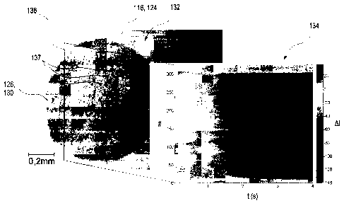
Figures 2A and 2B depict wetting experiments which were
carried out on samples of type A (figure 2A) and
samples of type B (figure 28). These experiments
firstly show the influence of the grinding step on
wetting. The comparative experiments each show test
CA 02743068 2011-05-09
- 34 -
fields 116 having a test field surface 124 to which a
drop 130 of the sample 126 is applied. Subimages 132 in
figures 2A and 2B each show microscope images of the
test field surface 124, whereas subimages 134 show the
change in gray value in the microscope images 132 along
an intersection line 136 through the drop 130 of the
sample 126. The sample 126 used was a test fluid having
a concentration of 50 mg/d1 glucose.
In subimages 134, the pixel position, indicated by #,
along the intersection line 136 is plotted on the
vertical axis in arbitrary units. The horizontal axis
specifies the time t in seconds after application of
the sample 126. In subimage 134, the changes in gray
value are depicted in each case. The right-hand side of
this subimage depicts a scale which specifies the
changes in gray value (AI) in arbitrary units. Figure
2A shows a test field surface 124 of sample A, i.e., a
test field material as is currently used in
commercially available test strips. Figure 2B shows by
contrast a test field 116 having the test field
material according to sample B as per the present
invention.
Without going into the numerical details of the
measurement, particularly subimages 134 of the change
in gray value in figures 2A and 2B show in a direct
comparison that grinding of the test field material
results in a distinctly more homogeneous temporal
change in reflectance characteristics along the
intersection line 136. Accordingly, the initiation of
the reaction, which is specific for the detection of
the analyte to be detected, is effected virtually at
the same time along the intersection line 136 in the
exemplary embodiment as per figure 2B, whereas in the
experiment with unground test field material as per
figure 2A, a strong temporal offset of the initiation
of the reaction can be found. For instance, a temporal
offset between individual locations along the
CA 02743068 2011-05-09
- 35 -
intersection line 136 can occur which can be up to
3 seconds or more. Also, there can be observed
locations along the intersection line 136 at which the
reaction occurs instantaneously, and also locations at
which the reaction does not appear to proceed at all.
Furthermore, in both figures 2A and 2B, a majority of
particles 137 in the detection layer 118 is
perceptible. In the microscope images in subimages 132,
almost solely the detection layer 118 is visible in
each case, since light rays which enter the detection
layer 118 through the transparent region 114 are
reflected no later than at the pigments of the removal
layer 122, more particularly titanium dioxide pigments.
It can be clearly discerned that the particles 137 in
conventional sample A as per figure 2A are considerably
larger and have a broader particle size distribution
than the particles 137 in sample B according to the
invention as per figure 2B. By means of a microscope
image of this kind as per subimage 132 in figures 2A
und 2B, a particle size distribution can also be
readily created by image recognition and automatic
recognition of particles at an appropriate
magnification. Alternatively or additionally, a change
in gray value as per subimages 134 may also be used.
Analytical processes of this kind with automatic image
recognition are known in principle from the field of
image processing to a person skilled in the art.
Overall, an evening-out of the course of the analyte-
specific reaction can thus be observed as the first
positive effect of using a ground test field material.
Furthermore, as is clear from figures 2A and 2B, a
homogenization of the reaction across the wetted test
field surface 124 can be established overall.
CA 02743068 2011-05-09
- 36 -
Furthemore, for the experiments overall, the reaction
time for ground and unground samples remains overall on
average approximately the same. For instance, in all
cases, a reaction time of about 6 to 7 seconds was
established. However, as is clear from the above-
described results, the spatially resolved, local
reaction time for ground test field materials is
strongly evened out, and so local variations in
reaction times can be considerably improved by the
ground test field material.
In a further experiment, experiments on the reflectance
shift are carried out with samples A and B. According
to the prejudice outlined above that ground test field
materials result in incomplete penetration of the
detection layer 118, a distinct reduction in the
reflectance shift would have to be observed for samples
of type B, since only a smaller region of the detection
layer 118 should be penetrated by the sample 126 and is
thus available for the detection reaction.
The results of these reflectance measurements are
depicted in figure 3. The concentration c of glucose in
the sample 126 is shown on the horizontal axis, whereas
the relative reflectance R is plotted on the vertical
axis. EDTA venous blood was used as the sample 126, and
the concentrations of glucose were varied in this test
fluid. The curve 138 in figure 3 shows the remissions
which were measured on a conventional diagnostic test
element, i.e., a sample of type A, whereas the curve
140 shows remissions of a sample according to the
invention of type B. As can be recognized from these
depicted data, there is practically no change in the
reflectance shift, i.e., the change in reflectance
across the entire measurement range, which is typically
between 10 and 600 mg/dl. Wet grinding of the fillers
therefore does not result in impairment of optical
properties and/or detection properties.
,
CA 02743068 2011-05-09
- 37 -
Thus, the experiments depicted in figures 1 to 3
clearly show that the use of ground fillers is not
accompanied by impairment of the properties of the
diagnostic test elements 110 in the form of a
lengthening of the reaction time or in the form of a
worsening of the reflectance shift. At the same time
however, as figures 2A and 2B clearly demonstrate, the
homogeneity and the precision of the measurements can
be distinctly improved by the use of ground test field
chemistry. On an Accu-Chek Active measuring device, it
has already been established in several measurements
that there is an improvement in the coefficient of
variation (CV), which reports the ratio of the standard
deviation to the mean value of the measurements, from
1.5 to 1.2% following the transition of samples of type
A to samples of type B.
As an alternative to the wet grinding described above
according to sample B, grinding can also be
alternatively or additionally carried out with, for
example, a dry grinding step. Accordingly, use can be
made of, for example, an air jet mill, by means of
which particulate sizes of up to, for example,
100 nanometers are achievable in principle.
Furthermore, experiments were carried out in which the
complete coating compositions for the detection layer
118 and the removal layer 122 were ground. In the
detection layer 118, there was no cogrinding of the
detection reagent, more particularly the enzyme,
because of the energy input in the grinding process.
However, such processes brought overall no or only a
slight improvement in the homogeneity, in the
reflectance shift, and in the reaction time. Thus,
grinding the complete coating compositions does not
have any advantage compared to grinding the filler
prebatches. However, the latter is considerably simpler
CA 02743068 2011-05-09
- 38 -
in terms of production technology, since a stock can be
ground.
Example 2
Grinding raw materials for the detection layer 118
represents an additional process step and can increase
the costs of the diagnostic test elements 110.
Therefore, in a second phase, raw materials are tested
which are commercially available and which entail from
the start an average particulate size in the range of
1 micrometer. Available for this purpose is, inter
alia, the Aerosil product range from Evonik Industries
AG. These are hydrophilic, nanoparticulate oxides, more
particularly metal oxides.
For instance, the following substitute materials were
identified as substitutes for the above-described
Transpafill in sample B:
Material: Type: Average
particle size:
Si02: Hardly thickens during 20 nm
dispersion:
Aerosil EG 50, Aerosil 90
Strongly thickens during
dispersion:
Aerosil 200, Aerosil COK 84
Ti02: Aeroxide TiO2 P 25 21 nm
A1203: Aeroxide Alu 65 17 nm
Table 1: Examples of further possible substitute
materials for Transpafill .
The experimentally determinable property of certain
materials that these materials have a thickening effect
on the dispersion during the dispersion procedure is
CA 02743068 2011-05-09
- 39 -
indicated by "thickens during dispersion" or "does not
thicken during dispersion".
For the use of these substitute materials, there is
naturally initially the fear, owing to the
abovementioned prejudice, that the pores may then
ultimately be too small in size to enable penetration
of the detection layer 118, and the reflectance shift
and the reaction times therefore worsen compared to
standard samples.
Accordingly, samples are produced in which, compared to
sample A above, the Transpafill in the detection layer
118 has been replaced 1:1 with the following materials:
Sample C:
S102, Aerosil COK 84 (mixed oxide with 10% A1203),
average particle size 20 nm
Sample D:
Ti02, Aeroxide TiO2 P 25, average particle size
21 nanometers
and
Sample E:
A1203, Aeroxide Alu 65, average particle size
17 nanometers
Sample F:
In a fourth sample in this second example, compared to
sample A, a wet-ground mixture of precipitated silica
and titanium dioxide having an overall average particle
size of 0.3 micrometers is used instead of Transpafill .
,
CA 02743068 2011-05-09
- 40 -
Reflectance measurements are again carried out in part
on these samples, analogously to the experiment as per
figure 3. The results of these measurements are plotted
in figure 4, the data depicted analogously to those in
figure 3. The curve 142 indicates the reflectance of
sample A, the curve 144 indicates the reflectance of
sample D, the curve 146 indicates the reflectance of
sample E, and the curve 148 indicates the reflectance
of sample F.
Initially, it was found that the reaction times for all
the samples were 6 seconds and therefore unchanged
compared to comparative sample A. Furthermore, the
reflectance shifts, as can be clearly discerned in
figure 4, also remain substantially the same across the
measurement range. The curves 142 and 148 are even
overlapping to a great extent in figure 4.
However, it is apparent in this experiment that the
very finely divided fillers should be dispersed close
to their primary particulate size in order to avoid
agglomeration and/or aggregate formation. For this
purpose, conventional dissolvers are used, and use can
be made of, for example, Polytrori or Megatron devices
from Kinematica AG or Ultra-Turrax devices for example
from IKA Maschinenbau.
For Si02 Aerosil types, such as samples C as per the
description above for example, it is apparent that
dissolvers can be used only with difficulty. Dissolvers
are therefore preferably used for samples of types D
and E, i.e., for titanium oxides and aluminum oxides,
having in particular nominal average particle sizes of
the starting powders of 30 nanometers or less. For
samples of type C, in contrast, the use of dissolvers
results in a dispersion only up to concentrations of
about 3% by weight in the wet starting mixture owing to
CA 02743068 2011-05-09
- 41 -
the thickening effect of the Si02 Aerosil types and to
gel formation. However, stirrers with lower shearing
rates can be used here, but which provided in the
experiments virtually the same results as the curves in
figure 4. This indicates that the Aerosils have been
optimized for dispersibility. For Aeroxide TiO2 P 25 and
Aeroxide Alu 65, thickening occurred only negligibly in
the experiments.
As an alternative or in addition to the abovementioned
fillers, ground or already commercially available as
nanoparticulates, a search was made for further
substances which are usable as fillers in the detection
layer 118, for example as a substitute for Transpafille
in the above-described sample A. In addition to the
above-described Aerosil types, the following
substances, among others, were investigated:
= Kaolin
= Powder glass (TROVOtech, Wolfen)
= Precipitated silica
= Calcium sulfate x 2 H20
= Sodium aluminum silicates, such as Sipernat 44 MS
(Degussa/Evonik) for example.
These fillers are either already available in the
desired particle size or particle size distribution or
can be processed by grinding to the required
particulate size or particle size distribution.
The above experiments essentially show that the
manufacturers have adjusted the Aerosil/Aeroxide types
for easy dispersability, and so incorporation is
largely independent of the shearing force.
In order to study more closely the necessity of
dispersing down to primary particulate size, further
experiments are performed. For this purpose, the above
,
CA 02743068 2011-05-09
- 42 -
sample D is studied again in different ways. Therefor,
Aeroxide TiO2 P 25 is dispersed either with a dissolver
(sample G) or with a propeller stirrer having a low
speed. The addition of Aeroxide TiO2 P 25 is carried out
either after mixing beforehand with water to form a
paste (sample H) or by solid introduction into the
thickener solution (xanthan gum) (sample I). The
comparative sample used is, as before, sample A as per
the above description. Altogether, the samples for the
experiment described below are thus as follows:
Sample A': as per sample A above
Sample G: as per sample D, but dispersing of
Aeroxide TiO2 P 25 with a dissolver,
Sample H: as per sample D, but dispersing of
Aeroxide TiO2 P 25 with a propeller
stirrer having a low speed, addition
after mixing beforehand with water to
form a paste, and
Sample I: as per sample D, but dispersing of
Aeroxide TiO2 P 25 with a propeller
stirrer having a low speed by solid
introduction into the thickener solution
(xanthan gum).
In this way, samples G to I are prepared in which
Transpafille is replaced 1:1 by weight with Aeroxide
TiO2 P 25. Otherwise, the diagnostic test elements 110
are produced as described above.
With these samples of diagnostic test elements 110 in
the form of test strips, measurements are carried out
with 15 different glucose concentrations in EDTA venous
blood on the commercially available Accu-Chek Active
CA 02743068 2011-05-09
- 43 -
blood glucose measurement system, where n . 10
individual measurements per concentration are analyzed.
Figure 5 depicts, analogously to the data depicted in
figure 4, reflectance curves for samples A', G, H, and
I. The curve 150 indicates reflectance measurements for
sample A', the curve 152 indicates reflectance
measurements for sample G, the curve 154 indicates
reflectance measurements for sample H, and the curve
156 indicates reflectance measurements for sample I.
The measurement curves show that the depth of shade is
virtually the same for the four different coatings. The
reaction rate as well is in the range of 6 to 8 seconds
for all the samples.
This shows that the Aeroxide TiO2 P 25 in all three
cases, i.e., in samples G, H, and I, is present finely
dispersed, since aggregates of TiO2 have a pigment
character and would attenuate the reaction color. Color
attenuation would be caused by, in the case of
aggregate formation, a pigment being present in the
detection layer 118. Finely dispersed TiO2 is, however,
not a pigment, since in this case the particulate size
is smaller than the light wavelength. This property is,
for example, taken advantage of in sunscreen agents.
The major advantage of the detection films having fine-
grained fillers is the distinctly improved homogeneity
of the reaction colors, which therefore allows the
measurement of smaller areas and thus smaller blood
volumes.
This can be shown again in a comparative experiment in
which different samples are studied. For this purpose,
diagnostic test elements 110 in the form of test strips
are spotted with plasma containing 100 mg/dl glucose,
and the reaction color is measured with a CCD camera.
,
CA 02743068 2011-05-09
- 44 -
Since the individual pixels can be read separately, a
number of pixels are analyzed statistically with regard
to their precision (i.e., with regard to their standard
deviation). Here, 10 pixels (edge length: 10
micrometers) are analyzed at the highest resolution,
i.e., an overall area of 1000 m2. For lower
resolutions, i.e., larger areas, averaging is carried
out over more pixels.
Again, different samples are studied here, analogously
to the above samples:
Sample A": as per sample A above, unground fillers,
comparative sample
Sample J: as per sample A", but Transpafill and
precipitated silica are ground,
Sample A"': as per sample A above,
coarse
ingredients, comparative sample, and
Sample K: replacement of Transpafill with Aeroxide
TiO2 P 25.
Figures 6A to 6C show microscope images of the
measurement spots which are the basis of the subsequent
measurements. Figure 6A shows a microscope image of a
region of sample A" spotted with the solution, i.e.,
of a sample having unground fillers. Figure 6B shows an
analogous image for sample J, i.e., of a sample having
ground test chemistry. Figure 6C shows an image for
sample A"', which essentially represents again a
sample having coarse ingredients and corresponds to
sample A", and figure 6D shows an analogous image for
sample K, in which Transpafille is replaced with
Aeroxide TiO2 P 25.
CA 02743068 2011-05-09
- 45 -
The axes labels in figures 6A to 6D display in each
case the pixel position on a CCD chip in arbitrary
units. Furthermore, measurement fields in figures 6A to
6C are indicated by means of corresponding squares, the
coordinates of which are displayed in the images.
Both figures 7A and 7B depict standard deviations for
the samples of figures 6A to 6D. Plotted on the
vertical axis in each case is the standard deviation s
of the gray values in figures 6A to 6D. This standard
deviation s is specified as a percentage, based on the
average gray value shift between a blank measurement
and a fully reacted sample. This standard deviation s
is specified as a function of the area A plotted on the
horizontal axis, via which averaging was carried out.
7A shows a comparison of samples A" and J, i.e., a
comparison of the standard sample with a sample having
ground test chemistry. The curve 158 displays the
course of the standard deviation for sample A",
whereas the reference number 160 indicates the curve of
sample J having ground test chemistry. In figure 7B,
standard sample A'" (reference number 162) is compared
with sample K (reference number 164) in terms of its
standard deviation.
The measured results show that the standard deviation s
and thus any possible measurement errors strongly
increase below about 30 x 30 pm2, i.e., an area
<0.01 mm2. It can be further recognized that this rise
in the standard deviation for sample J (curve 160)
having ground chemistry is distinctly lower, and the
ground chemistry is therefore advantageous for the
miniaturization of blood volumes, i.e., for
miniaturization of the measurement spot in figures 6A
to 6D. The same can also be said for sample K (curve
164 in figure 7B).
CA 02743068 2011-05-09
- 46 -
Various processes were specified above to determine the
particle size of samples. A further option which can be
alternatively or additionally applied involves
calculating autocorrelation functions via the gray
value distribution in microscope images, such as those
in figures 6A to 6D for example. The autocorrelation
function is a cross-correlation function of a signal
with itself, which is a function of a shift T.
Autocorrelation functions (indicated by ACF) for
samples A" to K are plotted in figures aA and 8B.
Figure 8A shows a comparison of comparative sample A"
(curve 166) with sample J having ground ingredients
(curve 168), and figure 8B shows a comparison of
comparative sample A' ! ! having coarse ingredients
(curve 170) with sample K having fine ingredients
(curve 172). In each case, the autocorrelation function
ACF is shown on the vertical axis, and the shift T of
the autocorrelation function in millimeters is shown on
the horizontal axis. The autocorrelation functions were
determined by analyzing figures 6A to 6D.
It can be discerned in a comparison of the
autocorrelation functions that the coarse fillers
(curves 166, 170) have distinctly broader
autocorrelation functions than the fine-grained fillers
(curves 168, 172). The data depicted show that the
autocorrelation function ACF correlates with the
particle size distribution. Simply from microimages,
such as the images in figures 6A to 6D for example, it
is thus possible to determine the granularity of a
sample. For example, the half-height width of the
autocorrelation functions 166 to 172 can be a measure
of the granularity of the detection layer 118.
Admittedly, direct reading of the particle size
distribution from these curves 166 to 172 is not
possible. By means of one or more calibration
measurements on samples having known particle size
distributions, the particle size or the particle size
,
CA 02743068 2011-05-09
- 47 -
distribution can be directly deduced from the curves
166 to 172.
CA 02743068 2011-05-09
- 48 -
Reference symbol list
110 Diagnostic test element
112 Support element
114 Transparent region
116 Test field
118 Detection layer
119 Support slide
120 Detection side
122 Removal layer
124 Test field surface
126 Sample
128 Loading side
130 Drop
132 Subimage, microscope image
134 Subimage, change in gray value
136 Intersection line
137 Particle
138 Reflectance, sample A
140 Reflectance, sample B
142 Reflectance, sample A
144 Reflectance, sample D
146 Reflectance, sample E
148 Reflectance, sample F
150 Reflectance, sample A'
152 Reflectance, sample G
154 Reflectance, sample H
156 Reflectance, sample I
158 Standard deviation, sample A"
160 Standard deviation, sample J
162 Standard deviation, sample A'"
164 Standard deviation, sample K
166 Autocorrelation, sample A"
168 Autocorrelation, sample J
170 Autocorrelation, sample A"'
172 Autocorrelation, sample K