Note: Descriptions are shown in the official language in which they were submitted.
CA 02743167 2011-05-10
WO 2010/057208 PCT/US2009/064846
WASTE MANAGEMENT SYSTEM
PRIORITY
[0001] This application is a continuation-in-part of International Application
No.
PCT/US2008/070781, filed July 22, 2008, and claims the benefit of priority to
U.S.
Provisional Patent Application No. 61/115,126, filed November 17, 2008, each
of which is
incorporated by reference into this application as if fully set forth herein.
BACKGROUND
[0002] Waste management systems are important in the healthcare field,
particularly
for patients that are unable to care for themselves. Such patients may suffer
from incontinent
diarrhea or like maladies and, due to their condition (e.g., severe burns,
surgical incisions,
etc.), may be susceptible to infections should the fecal matter come in
contact with an open
wound, burn, surgical site, etc. Moreover, healthcare professionals that come
in contact with
the fecal matter while attending to the patient may be susceptible to disease
and/or the
spreading thereof. Thus, a suitable waste management system, at minimum,
substantially
contains fecal matter within a closed system so as to avoid, for example,
substantial skin
breakdown, infection risk, cross-contamination of pathogens, problematic
patient clean-up,
patient discomfort, etc. While fecal management systems are described in the
art, many
known issues remain unsolved or unaddressed.
[0003] The following references relate to fecal management systems or
components
thereof: U.S. Patent No. 5,569,216 to Kim; U.S. Patent No. 6,527,755 to
Salama; U.S. Patent
No. 7,147,627 to Kim et al.; U.S. Patent Application Publication No.
2005/0054996 to
Gregory; U.S. Patent Application Publication No. 2005/0137526 to Machado et
al.; U.S.
Patent Application Publication No. 2006/0189951 to Kim et al.; U.S. Patent
Application
Publication No. 2006/0271087 to Von Dyck et al.; U.S. Patent Application
Publication No.
2007/0049878 to Kim et al.; and U.S. Patent Application Publication No.
2007/0149922 to
Schneider et al., each of which is incorporated by reference in its entirety
into this
application.
[0004] Applicants have recognized that it would be desirable to provide a
waste
management system that is robust, comfortable for the patient, eliminates
known issues and
has features that facilitate its use, embodiments of which are described
herein.
1
CA 02743167 2011-05-10
WO 2010/057208 PCT/US2009/064846
BRIEF SUMMARY
[0005] Accordingly, a waste management system is described herein, the system
including a waste transport device and a waste collection device. The waste
transport device
may include a first connector member configured for releasable connection to a
second
connector member on the waste collection device. The system may also include
an insertion
device to facilitate insertion of the waste transport device into the rectum
of a patient.
[0006] In one embodiment, a waste management system includes a waste transport
device, including a collection member with a distal end opening having a first
cross-sectional
area and a proximal end opening having a second cross-sectional area less than
the first cross-
sectional area, a retention cuff disposed about an outer surface of the
collection member, and
a waste collection device.
[0007] In another embodiment, a waste transport device includes a distal
section
defining a distal end opening having a first cross-sectional area and a
proximal end opening
having a second cross-sectional area less than the first cross-sectional area,
the distal section
including an inflatable retention cuff, a proximal section including a flush
lumen, a connector
coupled to a proximal end of the proximal section, and an intermediate section
connecting the
proximal section to the distal section, the intermediate section including a
transitioning cross-
sectional shape from a proximal end to a distal end. In another embodiment, a
waste
transport device includes a collection member including a lumen connecting a
distal end
opening to a proximal end opening and a retention cuff disposed about an outer
surface of the
collection member, the retention cuff including a pain relief drug.
[0008] In one embodiment, a method of managing the fecal material of a
patient,
includes inserting a distal section of a waste transport system in a collapsed
configuration into
a patient's rectum, the distal section in an expanded configuration defining a
distal end
opening having a first cross-sectional area and a proximal end opening having
a second cross-
sectional area less than the first cross-sectional area, the distal section
including an inflatable
retention cuff, removing the insertion device from the waste transport system,
and inflating
the retention cuff to a first inflated configuration.
[0009] In another embodiment, a method of connecting a waste transport device
to a
waste collection device includes associating a first connector coupled to the
waste transport
device with a second connector coupled to the waste collection device by
aligning an aperture
2
CA 02743167 2011-05-10
WO 2010/057208 PCT/US2009/064846
of the first connector with an aperture of the second connector and pressing
an end of one or
more locking arms of the first connector into slots of the second connector,
and sliding the
first and second connectors to align the apertures with a central lumen of the
waste transport
device and an opening of the waste collection device.
[0010] In another embodiment, a waste management system includes a waste
transport device, including a collection member with a distal end opening
having a first cross-
sectional area and a proximal end opening having a second cross-sectional area
less than the
first cross-sectional area, a lumen fluidly connecting the distal end opening
to the proximal
end opening, a retention cuff disposed about an outer surface of the
collection member, and a
securement device, including means for attaching the securement device to the
waste
transport device.
[0011] In another embodiment, a waste management system includes a waste
transport device, including a collection member with a distal end opening and
a proximal end
opening, a lumen fluidly connecting the distal end opening to the proximal end
opening, a
retention cuff disposed about an outer surface of the collection member, and a
medication
delivery apparatus configured to deliver a therapeutic agent through the waste
transport
device, including an interface port having an opening sealed by a septum.
[0012] These and other embodiments, features and advantages will become more
apparent to those skilled in the art when taken with reference to the
following more detailed
description in conjunction with the accompanying drawings that are first
briefly described.
BRIEF DESCRIPTION OF THE DRAWINGS
[0013] FIG. 1A is a perspective view of a waste management system.
[0014] FIG. lB is a perspective view of the proximal end of the system of FIG.
1 with
the waste transport device separated from the waste collection device.
[0015] FIG. 2A is a longitudinal cross-sectional perspective view of a distal
section of
a waste transport device.
[0016] FIG. 2B is a longitudinal cross-sectional side view of a distal end of
FIG. 2A.
[0017] FIG. 2C is a perspective view of one embodiment of a retention cuff for
a
waste transport device.
3
CA 02743167 2011-05-10
WO 2010/057208 PCT/US2009/064846
[0018] FIG. 2D is a perspective view of another embodiment of a retention cuff
for a
waste transport device.
[0019] FIG. 2E is a perspective view of yet another embodiment of a retention
cuff
for a waste transport device.
[0020] FIG. 2F is a perspective view of FIG. 2B.
[0021] FIG. 2G is a top view of FIG. 2B.
[0022] FIG. 2H is a side view of FIG. 2B.
[0023] FIG. 21 is an axial cross-sectional view of a section of a waste
transport
device.
[0024] FIGS. 3A-D illustrate stages of deflation and folding of a retention
cuff of a
waste transport device.
[0025] FIG. 4A-B are perspective views of a waste transport device with a
proximal
cuff.
[0026] FIG. 4C is one embodiment of an inflatable retention cuff.
[0027] FIGS. 5A-D are perspective views of different embodiments of a
collection
member.
[0028] FIG. 6 is a cross-sectional view of one embodiment of a single piece
collection
member and sphincter section.
[0029] FIGS. 7A-B are perspective views of another embodiment of a single
piece
collection member and sphincter section.
[0030] FIG. 7C is a perspective view of one embodiment of a retention cuff.
[0031] FIG. 7D is a perspective view of one embodiment of a waste management
system.
[0032] FIGS. 7E-G are cross-sectional views of different regions of the waste
management system of FIG. 7D.
4
CA 02743167 2011-05-10
WO 2010/057208 PCT/US2009/064846
[0033] FIG. 8A is a perspective view of another embodiment of a waste
management
system.
[0034] FIG. 8B is a partial view of a single piece collection member and
sphincter
section of the waste management system of FIG. 8A.
[0035] FIG. 8C is a cross-sectional view of the extracorporeal section of the
waste
management system of FIG. 8A.
[0036] FIG. 8D is a cross-sectional view of the waste transport device of the
waste
management system of FIG. 8A.
[0037] FIG. 9A is a perspective view of one embodiment of a waste management
system with a valved connection system.
[0038] FIGS. 9B-D are enlarged views of the connection system of FIG. 9A at
different stages of connection between the waste transport device and waste
collection
device.
[0039] FIGS. 10A-B are perspective views of another embodiment of a connection
system for a waste management system.
[0040] FIGS. 11A-D are perspective views of another embodiment of a connection
system for a waste management system.
[0041] FIG. 12 is a perspective view of yet another embodiment of a connection
system for a waste management system.
[0042] FIGS. 13A-C are perspective view of another embodiment of a connection
system for a waste management system.
[0043] FIGS. 14A-D are perspective views of still another embodiment of a
connection system for a waste management system.
[0044] FIGS. 15A-D are perspective views of one embodiment of an insertion
device
for a waste management system.
[0045] FIGS. 16A-C are perspective views of another embodiment of an insertion
device for a waste management system.
CA 02743167 2011-05-10
WO 2010/057208 PCT/US2009/064846
[0046] FIGS. 17A-D are perspective views of another embodiment of an insertion
device for a waste management system.
[0047] FIGS. 18A-C are perspective views of yet another embodiment of an
insertion
device for a waste management system.
[0048] FIGS. 19A-B are perspective views of still another embodiment of an
insertion
device for a waste management system.
[0049] FIGS. 20A-C are perspective views of another embodiment of an insertion
device for a waste management system.
[0050] FIG. 21 is a perspective view of another embodiment of an insertion
device for
a waste management system.
[0051] FIG. 22 is a perspective view of a securement device for a waste
management
system.
[0052] FIGS. 23A-D are perspective views of the securement device of FIG. 22
positioned onto a waste transport device.
[0053] FIGS. 24A-D are perspective views of a medication delivery apparatus
with an
independent disposable delivery device.
[0054] FIG. 25 is a cross-sectional view of another embodiment of a medication
delivery apparatus with a dedicated drug delivery lumen.
[0055] FIGS. 26A-B are perspective views of a pressure gauge for a waste
management system.
[0056] FIG. 27 is another embodiment of a pressure gauge for a waste
management
system.
[0057] FIGS. 28A-28C illustrate one embodiment of a sheathing apparatus to
facilitate insertion of a waste management system.
[0058] FIG. 29 illustrates one embodiment of a bacterial testing apparatus for
a waste
management system.
6
CA 02743167 2011-05-10
WO 2010/057208 PCT/US2009/064846
DESCRIPTION
[0059] The following description should be read with reference to the
drawings, in
which like elements in different drawings are identically numbered. The
drawings, which are
not necessarily to scale, depict selected embodiments and are not intended to
limit the scope
of the invention. The description illustrates by way of example, not by way of
limitation, the
principles of the invention. This description will clearly enable one skilled
in the art to make
and use the invention, and describes several embodiments, adaptations,
variations,
alternatives and uses of the invention, including what is presently believed
to be the best
mode of carrying out the invention.
[0060] As used herein, the terms "about" or "approximately" for any numerical
values or ranges indicate a suitable dimensional tolerance that allows the
part or collection of
components to function for its intended purpose as described herein. Also, as
used herein, the
terms "patient", "host" and "subject" refer to any human or animal subject and
are not
intended to limit the systems or methods to human use, although use of the
subject invention
in a human patient represents a preferred embodiment.
[0061] The waste management system described herein generally includes a waste
transport device and a waste collection device. The waste transport device
includes a distal
end section, referred to herein as "the rectal section," configured for
disposition in a patient's
rectum to begin transport of fecal material from a patient to a waste
collection device; a
section proximal of the rectal section, referred to herein as "the sphincter
section," configured
for disposition in a patient's anal canal; and a section proximal of the
sphincter section,
referred to herein as "the extracorporeal section," having a majority of its
length outside of
the patient. The proximal end of the waste transport device is configured to
connect to a
waste collection device, including a collection container. In certain
embodiments, the waste
management system includes a connection system for selective coupling of the
waste
transport device to the waste collection device and/or an insertion device to
facilitate
insertion of the waste transport device into a patient. Embodiments of these
and other
features of a waste management system are described herein.
[0062] With reference to FIG. IA, a waste management system 10 includes a
waste
transport device, including a generally tubular body (e.g., catheter) 12,
having a distal end 14
and a proximal end 16, and a waste collection device, including a collection
container 30.
Positioned at the distal end 14 of the body 12 is a rectal section 18,
including a collection
7
CA 02743167 2011-05-10
WO 2010/057208 PCT/US2009/064846
member 32 and a retention cuff 24 disposed about an outer surface of the
collection member
32 (FIG. 2). Proximal of the rectal section is a sphincter section 20,
particularly adapted for
disposition in the anal region of a patient, and an extracorporeal section 22
generally
positioned outside of the patient's body when the system is in use (although a
portion thereof
may be inside). In one embodiment, the collection member 32, sphincter section
20 and
extracorporeal section 22 are made of a material (e.g., silicone) with the
same durometer
(e.g., about 50 Shore A), while the retention cuff 24 is made of a material
(e.g., silicone) with
a different durometer (e.g., about 35 Shore A). In another embodiment, each of
the
aforementioned components are made of a material (e.g., silicone) with the
same durometer
(e.g., about 50 Shore A). A body connector 26 is coupled to a proximal end of
the
extracorporeal section 22 and is configured for quick, secure coupling to a
collection
container connector 28 to place the body 12 in fluid communication with a
collection
container 30. Various examples of connector embodiments are described in
detail below.
The body 12 generally has a plurality of lumens extending along at least a
portion of its
length, including, for example, a central lumen 34 for passage of fecal
material from the
patient to the collection container 30, an inflation lumen 36, a sampling
lumen 38, and a flush
lumen 44, each of which is discussed in detail below.
[0063] With reference to the rectal section 18 of the body 12, shown in FIGS.
2A-2B,
the collection member 32 has a distal opening 31 that, when positioned for
normal use, opens
into the rectum of a patient, and a proximal opening 33 that connects to the
sphincter section
20. In one embodiment, the proximal opening 33 has a cross-sectional area that
is less than a
cross-sectional area of the distal opening. For example, the proximal opening
33 may have
an inner diameter less than the inner diameter of the distal opening 31. Such
a configuration
imparts to the collection member 32 a tapered shape (e.g., a funnel), which is
believed to aid
in the flow of waste material from the patient into the body 12. It is noted
that the tapered
shape according to one embodiment is a frusto-conical shape. In one
embodiment, the
collection member 32 is formed from one or more materials having a durometer
sufficiently
hard to prevent premature closure of the distal opening 31, thereby permitting
safe passage of
fecal material from the patient regardless of forces acting on the collection
member 32. For
example, the collection member may be made from a material selected from
polyurethane,
silicone rubber, natural rubber latex, synthetic rubber, guayule rubber, 80 SH
polydimethylsiloxane, fumed silica, polyvinyl chloride (PVC), and combinations
thereof. In
one embodiment, the collection member 32 includes an annular ring disposed on
the distal
8
CA 02743167 2011-05-10
WO 2010/057208 PCT/US2009/064846
end thereof, the annular ring including a plurality of openings about its
perimeter, which are
connected to lumens through a wall of the collection member that may connect
to one or
more lumens disposed in the sphincter section and/or extracorporeal section.
For example,
the lumens through the wall of the collection member could extend through the
sphincter
section, all of which could connect to the sampling lumen 38.
[0064] In one embodiment, the rectal section 18 includes a split valve/baffle
configured to control the type of fluid permitted to pass therethrough. For
example, the
baffle in one embodiment is configured such that an infusion of medication
into the rectum
will not open (e.g., flow through) the baffle, but a greater volume of fecal
material will open
(e.g., flow through) the baffle. In one embodiment, the baffle includes a
plurality of discs
extending alternately from different sides of a passage of the rectal section
18 (e.g., the
collection member 32) such that the area open for fluid flow is spaced apart
therealong.
Thus, the medication intended for the patient will remain for a longer period
within the
rectum. In another embodiment, a duckbill valve is included in the rectal
section 18 to
control fluid flow therethrough.
[0065] In the embodiment shown in FIGS. 2A-2B, the retention cuff 24 is
disposed
about and attached to an outer surface of the collection member 32 and
includes an inflatable
balloon (e.g., conventional or non-conventional). In some embodiments, the
balloon 24
includes a drug, such as a pain relief drug (e.g., lidocaine). The drug may be
coated on a
surface of the balloon 24 and/or may be incorporated in an inflation liquid
(e.g., lidocaine
mixture) such that the drug gradually diffuses through a wall of the balloon
24. In one
embodiment, the retention cuff 24 includes a balloon, an outer surface of
which is coated
with lidocaine, and which is inflated following insertion into the patient
with a lidocaine or a
mixture including lidocaine. Surfactants and anti-microbial lubricant coatings
may also be
disposed on the retention cuff. Also, a retention cuff balloon may be encased
or otherwise
associated with a foam to maintain the rectal section in its deployed position
within the
patient's rectum and to prevent leakage. The foam may include an absorbant
material and/or
a coating to reduce odor. In one embodiment, a relatively high viscosity foam
is used to
inflate the retention cuff, following introduction into the rectum.
[0066] FIGS. 2C-2E illustrate embodiments of the rectal section 18 with a
retention
cuff 24 and a collection member 32. FIG. 2C is similar to FIG. 2A, including a
collection
member 32 with a frusto-conical shape having a smooth continuous wall (e.g.,
no pleats or
9
CA 02743167 2011-05-10
WO 2010/057208 PCT/US2009/064846
divisions between the proximal and distal openings) and a retention cuff 24
that when inflated
creates a shoulder 11 at the junction between the cuff 24 and the body (e.g.,
sphincter section
20). FIG. 2D shows a cuff 24d with a geometry that more gradually transitions
to the body,
the collection member 32d having a bell-shaped configuration (i.e., frusto-
conical shape with
a flared distal opening) with a smooth continuous wall. FIG. 2E shows cuff 24d
surrounding
a collection member 32e that has a trumpet-shaped configuration (i.e., a
frusto-conical shape
with curved sides and a flared distal opening) with a smooth continuous wall.
The retention
cuff in some embodiments, may also include one or more structural features as
described
more completely below in connection with FIGS. 5A-5D. Also, the retention cuff
may have
a tapered shape when inflated such that the diameter or perimeter increases
from a proximal
end to a distal end.
[0067] FIGS. 3A-3D illustrates one embodiment of a rectal section 18 with a
retention cuff balloon 24, configured to provide an advantageously small
profile for insertion.
FIG. 3A illustrates the cuff 24 in its inflated state. FIG. 3B illustrates the
beginning of
deflation, showing the configuration of the cuff 24, which includes pockets
24' disposed
between spaced apart raised sections 24" about the circumference of the cuff
24. FIG. 3C
illustrates further deflation of the cuff 24 as the raised areas 24" collapse
into the lumen of
the rectal section 18, the cuff easily folded by bringing together the raised
sections 24", as
illustrated in FIG. 3D. It is noted that any of the retention cuffs 24
described herein may have
a similar configuration to provide a small profile for insertion.
[0068] FIGS. 4A-4B illustrate embodiments including a proximal cuff 25, shown
generally in FIG. 4A. The proximal cuff 25, which can include an inflatable
balloon, is
mounted to the body 12 proximally from the retention cuff 24, for example,
along the
extracorporeal section 22. Thus, when the body 12 is properly inserted, the
proximal cuff 25
may be located between the patient's buttocks. The proximal cuff 25 is adapted
to prevent
upward migration of the retention cuff 24 when inserted and deployed in a
patient's rectum
and may optionally include anti-odor, moisturizer and/or lubricant coatings.
In one
embodiment, the proximal cuff has the form of an umbrella, cone, basin, etc.
with the wide
part facing the retention cuff in order to capture material that may leak from
around the body.
The cuff in such an embodiment may be made of a soft, absorbent material and
is configured
to be removable from the body in order to replace when necessary, or
alternatively may be
made of a material that is easily cleaned, such as, for example, a soft
plastic material.
CA 02743167 2011-05-10
WO 2010/057208 PCT/US2009/064846
[0069] FIG. 4B illustrates a variation of a distal section of a body 12, which
includes
a retention cuff 24 and a proximal cuff 25 with a tension member 27 disposed
in the wall of
the body (e.g., along a portion of the sphincter section 20) between the cuffs
24, 25. The
tension member 27 may include one or more elongated members embedded in the
wall of the
sphincter section and/or coupled to an inner or outer wall thereof. The
tension member may
include spaced apart longitudinally oriented members, circumferentially
oriented members,
helically arranged members, combinations thereof, etc. According to one
embodiment,
however, the tension member 27 is a helical coil made of shape memory material
(e.g.,
Nitinol). Adjacent the proximal cuff 25, a sphincter section 29 free of the
tension member is
provided to prevent loss of sphincter tone as discussed above.
[0070] The tension member 27 has a collapsed configuration with a collapsed
perimeter and an expanded configuration with an expanded perimeter greater
than the
collapsed perimeter. In one embodiment, at least a portion of the tension
member 27 is
disposed adjacent the retention cuff 24 such that when the retention cuff 24
is inflated, the
tension member expands from the collapsed perimeter to the expanded perimeter.
Following
inflation of the retention cuff 24 and expansion of the tension member 27, the
proximal cuff
25 is inflated. Due to the shape memory material, the tension member 27 will
attempt to
return to its collapsed configuration, which due to the connection to the
retention cuff 24 will
be resisted. This resistance provides tension between the cuffs 24, 25, which
is believed to
aid in the prevention of leakage and migration of the distal end of the body
12.
[0071] FIG. 4C illustrates one embodiment of an inflatable retention cuff that
is
adapted to occlude the distal opening 31 when activated. Occluding the lumen
34 of the body
12 may be desirable, for example, to temporarily block reflux and to retain
medicants or
drugs in the rectal vault. Known systems accomplish the occlusion function by
including an
internal balloon in the distal end of a catheter. The presence of an internal
balloon may
partially block the lumen of the catheter even when not inflated and/or may
provide a surface
for which fecal material adheres to, resulting in build-up and eventual
blockage of the lumen.
In the embodiment of FIG. 4C, instead of an internal balloon, retention cuff
80 includes a
plurality of lobes 82 that may be expanded from a first inflated
configuration, similar to the
retention cuff 24 described above, to a second inflated configuration (as
shown), converging
to cover the distal opening 31 or otherwise block fluid from reaching the
distal opening 31.
The expansion from the first inflated configuration to the second inflated
configuration can
11
CA 02743167 2011-05-10
WO 2010/057208 PCT/US2009/064846
be accomplished by an infusion system 84 that indicates to the user the state
of the retention
cuff 80 (i.e., deflated, first inflated configuration, second inflated
configuration) based on a
pressure reading of the cuff 80. For example, the infusion system 84 could
provide a readout
of a first volume (e.g., 50 mL) when the lobes 82 are in the first inflated
configuration and a
second volume (e.g., 150 mL) when the lobes 82 are in the second inflated
configuration. In
another embodiment, a sliding cuff or sleeve may be positioned proximal of the
retention cuff
80, the sliding cuff including arms or surfaces configured to force the cuff
80 inward upon
contact therewith such that the distal opening 31 is occluded when the sliding
cuff impinges
on the retention cuff. Thus, for example, after the retention cuff 80 has been
inflated to the
first inflated configuration, the sliding cuff is moved distally at least
partially over the
retention cuff 80 to occlude the opening 31. Thereafter, when it is desired to
remove fecal
matter from the rectum, the sliding cuff is moved proximally out of contact
with the retention
cuff 80 so that the distal opening 31 is no longer occluded.
[0072] FIGS. 5A-5D illustrate different embodiments of the collection member.
FIG.
5A illustrates a collection member 60 with a plurality of spaced apart struts
62 attached at
their distal end to a ring 64, which defines the distal opening 31. The three
struts 62 in the
embodiment shown are twisted to facilitate collapse of the retention cuff 24
for insertion and
removal of the body from the patient. The proximal end of the struts 62 may be
coupled to
the body just proximal of the rectal section 18 (e.g., sphincter section 20)
or may extend
further along a length of the body (e.g., through at least a portion of the
extracorporeal
section 22) to provide structural support thereto. The struts 62 may be
embedded in the wall
of the body section(s) or may be coupled to a surface thereof. In the rectal
section 18, the
twisted struts 62 may, together with the retention cuff 24, define the
collection member
lumen. Alternatively, the collection member lumen may be defined by another
member (e.g.,
a tapered extrusion) to which the struts 62 are attached. The struts 62 and
ring 64 may be
formed from a metal, polymer, or other suitable material that provides
structural support to
the rectal section 18, and may have a circular cross-sectional shape,
rectangular cross-
sectional shape, or any other geometric cross-sectional shape.
[0073] FIG. 5B is another embodiment of a collection member with a plurality
of
spaced apart struts (e.g., four), the struts 66 remaining untwisted (e.g.,
substantially aligned
along a longitudinal axis) and attaching distally to the cuff 24 rather than a
ring. Of course, a
ring could also be included and/or a further member as described above in
connection with
12
CA 02743167 2011-05-10
WO 2010/057208 PCT/US2009/064846
FIG. 5A. The struts 66 have an increasing cross-sectional area from the
proximal end to the
distal end and in the embodiment shown transition from a circular cross-
sectional shape at the
proximal end to an oval cross-sectional shape at the distal end. Other cross-
sectional shapes
are also possible, such as those described above in connection with FIG. 5A,
and are within
the scope of the invention. FIG. 5C is an embodiment of a collection member
including a
helically arranged elongate member in the form of a coil 68. The coil 68
defines a lumen
along a longitudinal axis thereof and may be attached directly to the cuff 64
or an additional
tapered member. In one embodiment, the elongate member forming the coil 68 is
hollow and
is attached directly to the sampling lumen 38 to provide access to the
patient's rectum for
infusion of drugs/fluids and/or extracting samples for testing. FIG. 5D is yet
another
embodiment of a collection member. Collection member 70 has distal sections
removed to
facilitate collapse of the rectal section for insertion and removal. In the
embodiment
illustrated, the removal of sections results in a plurality of petals 72
evenly spaced apart about
the circumference of the collection member 70. It should be noted, however,
that many other
types of patterns, including non-uniform patterns, are also contemplated
herein and within the
scope of the invention.
[0074] With further reference to the embodiment shown in FIGS. 2A-2B, the
sphincter section 20 is disposed between the collection member 32 and the
extracorporeal
section 22. The sphincter section 20 in one embodiment is distinct from the
extracorporeal
section 22 and/or the collection member 32 in that the sphincter section is
configured to
collapse under lower pressures to preserve the tone/strength of the sphincter
when positioned
in the patient for extended periods. For example, in one embodiment, the
material for the
collection member 32 and sphincter section 20 are the same, but the thickness
of the wall of
the sphincter section 20 is less than the wall of the collection member 32. In
other
embodiments, the material for the sphincter section 20 is different from the
material for the
collection member 32 (e.g., more compliant, softer durometer, etc.) In one
embodiment, the
sphincter section 20 is made from a material selected from polyurethane,
silicone rubber,
natural rubber latex, synthetic rubber, 80 SH polydimethylsiloxane, fumed
silica, polyvinyl
chloride (PVC), and combinations thereof.
[0075] The shape of the sphincter section 20 may have a cross-sectional shape
that
transitions from a distal end 21 to a proximal end 23, such as shown in FIGS.
2A-2B and 2F-
2H. For example, the sphincter section 20 in the embodiment shown has a
substantially
13
CA 02743167 2011-05-10
WO 2010/057208 PCT/US2009/064846
circular cross-sectional shape at its distal end 21 and an oval cross-
sectional shape at its
proximal end 23 that attaches to the extracorporeal section 22. In other
words, at its proximal
end 23, the sphincter section 20 has a larger diameter along a first axis
(i.e. the z-axis shown
in FIG. 2B), than along a second axis orthogonal to the first axis (i.e. the y-
axis shown in
FIG. 2B). The oval proximal end 23 of the sphincter section matches the oval
distal end of
the extracorporeal section 22 (FIG. 21), which in one embodiment maintains
this oval cross-
sectional shape from the distal end to the proximal end. The transitional
shape for the
sphincter section 20 is advantageous for resisting rotational motion. That is,
the sections of
the system that have oval cross-sections (e.g., the sphincter section proximal
end 23 and the
extracorporeal distal end) are more resistant to rotation than sections with
circular cross-
sections (e.g., the sphincter section distal end 21 and the collection member
proximal end 33).
In one embodiment, the sphincter section 20 has an hourglass shape. As with
the
embodiment of FIGS. 2A-2B and 2F-2H, the proximal end of the sphincter section
20 may
have an oval cross-sectional shape, while the distal end may have a circular
cross-sectional
shape. Alternatively, each of the proximal and distal ends of the sphincter
section may have a
circular cross-sectional shape to match circular cross-sectional shapes of the
distal end of the
extracorporeal section and the proximal end of the rectal section,
respectively.
[0076] In one embodiment, the sphincter section 20 includes a sealing feature,
such as
a plurality of ribs arranged about the perimeter thereof. The ribs may be
spaced apart and
inflatable such that the ribs are deflated for insertion and inflated upon
deployment. When
inflated, the ribs may provide a seal and/or prevent rotational movement of
the sphincter
section 20. The ribs may be arranged substantially parallel to a longitudinal
axis of the
sphincter section 20, circumferentially about the perimeter of the sphincter
section 20,
diagonally, helically, combinations thereof, etc. In one embodiment, anti-
twist rings are
disposed about sections of the distal end of the system, such as the sphincter
section and the
distal end of the extracorporeal section 22. The rings may be longitudinally
spaced from one
another and may be inflatable similar to the ribs. The rings and/or ribs may
be incorporated
along the distal end of the system to provide an anti-rotating function.
Further, the rings
and/or ribs may include a reinforcing feature, such as a hard material (e.g.,
wire), to prevent
collapse of the system lumen transporting fecal material from the patient. In
one
embodiment, a stiff tube or coiled, flexible spring is disposed in a wall of a
section of the
waste transport device or along an internal surface thereof.
14
CA 02743167 2011-05-10
WO 2010/057208 PCT/US2009/064846
[0077] With reference to FIGS. 2B and 21, inflation lumen 36 and
irrigation/sampling
lumen 38 can be located adjacent and parallel to lumen 34 and on opposite
sides thereof. The
inflation lumen 36 and the irrigation/sampling lumen 38 can each be a flexible
cylindrical
tube extending along and integrally molded with, embedded into, or otherwise
attached to an
inner surface of at least a portion of the rectal section 18, sphincter
section 20 and
extracorporeal section 22. The distal end of the inflation lumen 36 is in
fluid communication
with the interior of the retention cuff balloon 24, while the proximal end of
the inflation
lumen 36 diverges from the extracorporeal section 22 outside of the body when
the system is
properly inserted. An inflation port 40 is attached to the proximal end of the
inflation lumen
and may include a luer-style connector for connection to a syringe or other
device for
selectively inflating and deflating retention cuff balloon 24. The distal end
of the
irrigation/sampling lumen 38 extends through the rectal section 18 and has a
distal opening
positioned adjacent the distal opening 31 of the collection member 32 so that
fluids may be
drawn from the patient therethrough. The proximal end of the
irrigation/sampling lumen 38,
similar to that of the inflation lumen, diverges from the extracorporeal
section 22 and
terminates at an irrigation/sampling port 42. The port 42 may similarly
include a luer-style
connector for connection to a syringe or other device so that, for example, a
medication or
irrigant may be infused into the patient's rectum, or a fecal matter sample
may be extracted
from the patient's rectum. In one embodiment, both the inflation lumen 36 and
sampling
lumen 38 are polyurethane or silicone tubes, having sizes in the range of
about 5 Fr to about
Fr, for example, a 6 Fr inflation lumen and an 8 Fr sampling lumen.
[0078] According to certain embodiments, a pressure or volume indicator may be
used with the waste management systems disclosed herein. It can be important
to ensure that
the retention cuff is inflated with a proper volume of liquid to provide
optimum retention.
Excessive volume in the cuff can lead to excessive pressure exerted by the
external surface of
the cuff on the rectal mucosa. This excessive pressure can damage the mucosa,
and should be
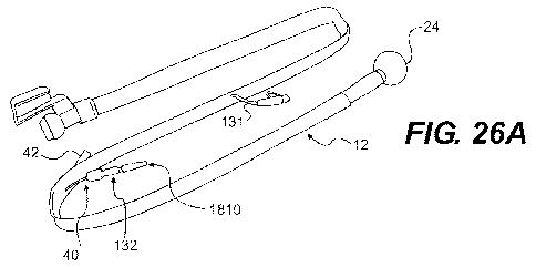
avoided. FIG. 26A illustrates pressure gauge 1810 in fluid communication with
inflation port
40. Once the cuff is infused with, e.g., 40 ml of a liquid and the syringe is
removed from the
luer-style connector, the pressure gauge 1810 is locked onto the connector. As
shown in FIG.
26 B, the gauge 1810 may include indicia 1812, a sealing member 1814, and a
calibrated
spring 1816. Any gauge capable or measuring at least one of volume and
pressure may be
used in accordance with the present disclosure. FIG. 27 illustrates gauge 1850
having
pressure or volume actuated needle 1856, and visible range markings 1852 and
1854. The
CA 02743167 2011-05-10
WO 2010/057208 PCT/US2009/064846
range marking 1852 can designate an acceptable pressure or volume range, and
range
marking 1854 can designate an unacceptable pressure or volume range.
[0079] With reference to FIGS. 26A-B, pilot balloon 132 is in fluid
communication
with retention cuff 24. The pilot balloon 132 is configured to indicate the
inflation status of
the retention cuff 24, and the status is commonly detected by observing the
size of the pilot
balloon or detecting the pressure sensed between two fingers on the surfaces
of the pilot
balloon. Pilot balloons are discussed in U.S. 4,016,885; 4,134,407; and
6,732,734, the
disclosures of which are incorporated by reference herein. According to one
embodiment
(not shown), the pilot balloon has at least one valve, for example a duckbill
valve, within the
chamber thereof. The valve can be calibrated to release fluid from the
retention cuff when
the pressure exerted by the retention cuff on the rectal mucosa exceeds a pre-
defined
pressure. According to certain embodiments, a preferred pressure is 15-40m1
Hg. Thus, the
at least one valve could be configured to release fluid when the pressure
exceeds, e.g., 50, 60,
70, or 80m1 Hg, or any other clinically significant pressure. According to
another
embodiment, waste management system could comprise a plurality of valves in
fluid
communication with the retention cuff 24, each of which is configured to
release a specified
volume of fluid when a specified pressure is reached. Such a system would
provide a
compensatory system that would help minimize the likelihood of tissue damage
from, e.g.,
over-inflation of the retention cuff 24.
[0080] Also, as seen best in FIGS. 2A and 2F, the extracorporeal section 22
can
include a flush lumen 44 disposed along a length of the extracorporeal section
22 in parallel
with the central lumen 34. The flush lumen 44 is configured to flush and clean
the central
lumen 34 as necessary. For example, it may be desired to periodically flush
the lumen 34 of
the body 12 in order to prevent bacterial contamination and to also aid in
reduction of odor
due to fecal build up. In one embodiment, the flush lumen 44 is closed at a
distal end (e.g.,
the distal end of the extracorporeal section 22) and connects at a proximal
end to a flush port
46 coupled to, and extending through, a wall of the extracorporeal section 22
(FIG. 1) that
provides access for a syringe or other device for inputting a desired
cleansing fluid into the
flush lumen 44. A port cover 48 (FIG. 1) of any suitable variety may be used
that is
configured to sealably close and open the flush port 46. Referring to FIG. 2A,
to facilitate
flushing of the central lumen 34, the flush lumen 44 can be perforated with a
plurality of
apertures 50 positioned along the length of the flush lumen 44. In one
embodiment, the
16
CA 02743167 2011-05-10
WO 2010/057208 PCT/US2009/064846
apertures 50 are grouped into aperture groups 52, such as groups of four,
spaced from one
another along the length of the flush lumen. The apertures may be arranged
substantially
linearly, as shown, or may be otherwise disposed, for instance, in circular
patterns, along
separate or continuous curves, etc.
[0081] The collection member 32 and sphincter section 20 may be formed
together
into a single piece, such as member 120, shown in FIG. 6. Member 120 includes
a collection
member 32 that is unattached to the sphincter section 20 at its proximal end,
the attachment
occurring only at a distal end where the member 120 forms a rolling portion
122. Thus, the
wall 121 defining the lumen through member 120 extends from a proximal end 118
to the
distal end of the collection member 32, turning back at the rolling portion
122 toward the
sphincter section 20, and terminating at a distal end 119 where it attaches to
a proximal end
of a retention cuff 24 (the retention cuff 24 having a distal end attached
adjacent to the rolling
portion 122). This configuration permits free motion and movement of the
sphincter section
20 with respect to the retention cuff 24 such that the cuff is not
significantly displaced (if at
all) when the sphincter section 20 is twisted (as represented by arrow 8 and
the dotted lines)
or pulled axially, thereby isolating potential loads from the retention cuff
24, rather than
transferring loads thereto. Applicants believe that by generally preventing
the transfer of
loads from proximal sections of the waste transfer member to the retention
cuff, several
benefits may be realized, such as, for example, minimization of leakage around
the retention
cuff and minimization of pressure exerted on the rectal vault (thereby
reducing the incidence
of pressure necrosis).
[0082] Further, a distal-only attachment configuration enables movement of a
tool
112 over the length of the member 120 to facilitate insertion and removal of
the waste
transport device, as well as "milking" of the collection member 32. In
particular, a tool 112
may include an end piece 116 coupled to an elongate member 114, the end piece
116 having
a cross-section similar to the cross-section of the member 120, a size less
than that of the
collection member 32 in its expanded configuration, and a rigidity greater
than that of the
collection member 32. For example, if the member 120 has a generally hourglass
shape as
shown in FIG. 7, the end piece 116 of the tool 112 can be circular with a
diameter generally
equal to the desired insertion diameter for the collection member 32. Thus,
insertion is
facilitated by merely pushing on the proximal end of the elongate member 114
such that a
force is exerted on the rolling portion 122 from an inner surface thereof by
the end piece 116,
17
CA 02743167 2011-05-10
WO 2010/057208 PCT/US2009/064846
while the distal end of the collection member 32 is maintained in a collapsed
configuration
with a lower profile than that of the collection member in its expanded
configuration.
Following insertion, the tool 112 may be slid in a proximal direction while
the member 120 is
maintained in position in the patient to permit expansion of the collection
member 32 to its
expanded configuration. During use, the member 120 may be "milked" by sliding
the tool
112 over the member 120 and performing successive axial movements, distal to
proximal, to
move the waste through the lumen of the member 120. To remove the waste
transport
device, the tool 112 is slid over the member 120 to the distal end rolling
portion 122 in order
to collapse the collection member 32 to the collapsed configuration having a
suitable
insertion/removal diameter.
[0083] In another embodiment, collection member 32 and sphincter section 20
are
formed into a continuous member 90, shown in FIGS. 7A-7B. In the embodiment
shown,
member 90 includes a stiffening ring 92 around a circumference of the
collection member 32
and relief sections 93 disposed approximately equidistantly between the
inflation lumen 36
and the sampling lumen 38 to facilitate collapse of the collection member 32
for delivery and
withdrawal from the patient's rectum. The relief sections 93 may be raised
portions of the
collection member inner surface, for example, having a semi-circular cross-
section along its
length. The distal end of the collection member includes a lip 96 about the
circumference of
the distal opening 31. Openings 94 in a wall of the collection member are
configured to pass
air or fluid from the inflation lumen 36 to a surrounding retention cuff (it
is noted that the
distal end of the inflation lumen shown open in these figures will be closed
in a final
assembly so that air or fluid will be forced out of the openings 94). The
collection member
32 has a generally frusto-conical shape, while the sphincter section 20 has a
generally
cylindrical shape. FIG. 7C is one embodiment of a retention cuff 123, having a
bulb-like
geometry along a body 124 and a tapered distal end 125. The retention cuff 123
is configured
to fit over the collection member 32 and is attached at distal end 125 to the
distal end of the
collection member 32 and at proximal end 129 to a proximal end of the
collection member
32.
[0084] FIG. 7D illustrates one embodiment of a waste management system 100,
including a waste transport device 101 and a waste collection device 102.
Waste transport
device 101 includes member 90 and retention cuff 25 of FIGS. 7A-C, an
extracorporeal
section 22, a connector housing 126, connector collar 127 and connector ball
valve 160
18
CA 02743167 2011-05-10
WO 2010/057208 PCT/US2009/064846
(described in more detail below), and devices for fluid movement, including
arm flush lumen
131, arm pilot balloon 132 and arm irrigation sleeve 133. Waste collection
device 102
includes a collection container 30, a hub socket 35 configured to receive
connector housing
126, and hub plug 6 tethered to the hub socket 35, the hub plug 6 including
threads for mating
with an interior threaded surface of hub socket 35 in order to seal the
opening of the
collection container 30. FIG. 7E is a cross-sectional view of the collection
container
interface, showing connector housing 126 and hub socket 35 in more detail.
FIG. 7F is a cut-
away view of arm flush lumen 131 and its connection to flush lumen 44 of
extracorporeal
section 22. FIG. 7G is a cross-sectional view of both the arm pilot balloon
132, connected to
inflation lumen 36, and arm irrigation sleeve 133, connected to sampling lumen
38. It is
noted that the hexagonal section of the arm pilot balloon is configured to
bulge outward when
there is line pressure to indicate such to the user.
[0085] Another embodiment of a waste management system is illustrated in FIGS.
8A-8D. Waste management system 110 includes waste transport device 111 with a
relatively
shorter length than waste transport device 101 and a waste collection device
109 with a
different configuration than waste collection device 102. In particular, waste
collection
device 109 has a tubular shape with a proximal opening covered by a sealed
septum 105. An
odor control filter, made of a material such as carbon, may be embedded in the
wall of the
waste collection device 109 or may be a vent disposed therein. The waste
collection device
109 may have a collapsed configuration which expands upon receipt of waste
material
therein, or may have a more rigid configuration (as shown) such that a vent in
a wall thereof
may enhance drainage efficiency.
[0086] The waste transport device 111 includes an extracorporeal section 22
with a
drain tube irrigation port 95, an inflation port 107 and a sampling port 108.
The inflation port
107 is connected to an inflation lumen 36 extending from the inflation port
107 to the
retention cuff 24, while the sampling port 108 is connected to a sampling
lumen 38 extending
from the sampling port 108 to the distal end of the waste transport device
111. The irrigation
port 95, as shown in FIG. 8D, is connected to a flush lumen with patterned
holes along its
length to flush the lumen of the extracorporeal section 22. As fluid is
introduced through the
port 95, the fluid extends along the length of the flush lumen entering into
the lumen of the
extracorporeal section 22 through the patterned holes. The irrigation port 95
in one
embodiment is an EZ-LOK Sampling Port. In another embodiment, an EZ-LOK
19
CA 02743167 2011-05-10
WO 2010/057208 PCT/US2009/064846
Sampling Port is also positioned on the extracorporeal section 22 with access
to the lumen
thereof for periodic sampling of fecal matter therefrom.
[0087] As best seen in FIG. 8B, a continuous member 91 includes both the
sphincter
section 20 and collection member 32. The collection member 32 has a wavy
perimeter with
undulations including peaks and valleys. The valleys 97 form crease lines to
facilitate
collapse of the collection member 32 to a collapsed configuration. A retention
cuff 24
surrounds the collection member 32. Collapsible struts 98 are positioned at
the peaks of the
perimeter, forming a stiffening area to resist collapse of the collection
member during use.
The struts 98, as shown, extend circumferentially away from the perimeter
along an outer
surface of the peak section and form a recessed region along an inner surface
of the peak
section of the collection member. Such a shape is designed to fit in an
insertion tool such that
collapse of the collection member is facilitated. In other embodiments, the
struts 98 may take
a different geometric shape or form, depending on the shape/size of the
insertion tool and/or
desired levels of stiffness for the collection member. The waste transport
device 111 includes
at its proximal end a connection member 103 configured for coupling to
connection member
104 of the waste collection device 109, embodiments of which are described in
more detail
below.
[0088] In the embodiments described herein, the extracorporeal section 22 may
have
a uniform cross-section along its length (e.g., circular, oval, etc.) or a
transitional cross-
section similar to the sphincter section 20 shown in FIGS. 2A-2B. The
extracorporeal section
22 can be formed of a non-collapsible tube constructed of a material that is
sufficiently stiff
in order to maintain its shape during use (e.g., to prevent or minimize
kinking, to facilitate
drainage, etc.), but soft enough to be "milked" by a care professional to
force through fecal
material when necessary. For example, in one embodiment, the extracorporeal
section is
made from a rubber or plastic material that does not collapse under its own
weight. In one
embodiment, the extracorporeal section 22 includes one or more stiffening
structures, such as
inflatable ribs, metal wires or ribbons, axially positioned rings, etc., to
assist in preventing
collapse of the lumen 34. As with the ribs discussed above, the stiffening
structures may be
disposed longitudinally, circumferentially, helically, etc.
[0089] The "milking" in one embodiment is performed by a clamp tool including
opposing first and second arms attached to a handle, the first and second arms
arranged
approximately perpendicular to the handle with a gap therebetween. A portion
of the
CA 02743167 2011-05-10
WO 2010/057208 PCT/US2009/064846
sphincter section 20 or extracorporeal section 22 is placed between the arms
and the handle is
pulled in a proximal direction to move fecal matter through the section
milked. The tool may
include a locking feature such that the first arm locks or is coupled to the
second arm to
clamp a section of the waste transport device.
[0090] The body 12 can be secured to the collection container 30 via
respective
connectors 26 and 28. With reference to FIG. 1B, the collection container 30
is in the form
of a bag, having an opening 54 located on a front side, which provides access
to the interior
thereof. In other embodiments, the collection container 30 may be in other
suitable forms
with one or more openings therein. Because it is desirable to secure the body
12 to the
collection container 30 so that the central lumen 34 is in fluid communication
with the
interior of the collection container 30, the connection system positions the
lumen 34
substantially in axial alignment with the opening 54 when the body 12 is
coupled to the
collection container 30. In one embodiment, the collection container 30 is
configured to
absorb and reduce odor, for example, by providing a ventable section including
activated
charcoal. The activated charcoal can be changed when desired via
interchangeable charcoal
cartridges that are inserted into the collection container 30. The collection
container 30 can
also have a parylene coating, anti-odor coating and/or antimicrobial coating.
In addition, the
collection container 30 can include material in a wall thereof that
absorbs/binds odor.
Suitable examples of coatings/materials include those disclosed in U.S. Patent
No. 6,579,539,
U.S. Patent No. 6,596,401, U.S. Patent No. 6,716,895, U.S. Patent No.
6,949,598, and U.S.
Patent No. 7,179,849, each of which is incorporated by reference in its
entirety into this
application.
[0091] In the embodiment of FIG. 1B, the collection container connector 28
includes
a slide mechanism adapted to receive and retain an annular flange extending
from the body
connector 26. Accordingly, the body 12 can be secured to the collection
container 30 by
sliding the annular flange section of the catheter connector 26 into a slot or
grooved section
of the container connector 28. When it is desired to separate the body 12 from
the collection
container 30, the body connector 26 can be slid upwards, out of the container
connector 28,
thereby disengaging the body 12 from the collection container 30. Because it
is often
desirable to prevent leakage from the body 12 and the collection container 30
upon separation
of the body 12 from the collection container 30, closures valves 56 and 58 can
be provided in
the proximal opening of each. In one embodiment, the closure valves 56 and 58
are split
21
CA 02743167 2011-05-10
WO 2010/057208 PCT/US2009/064846
polymeric coverings, such as septums, that open when fluid pressure acts
thereon from the
fecal matter and/or flush lumen fluid in central lumen 34. In other
embodiments, the valves
open upon connection between the body 12 and collection container 30. For
example, a
mechanism on connector 26 and/or 28 will open one or both of the valves 56, 58
when the
annular flange of the connector 26 is slid into the slot of the connector 28.
[0092] Another embodiment of a connection system for the waste management
system is shown in FIGS. 9A-9D. A catheter connector 126 includes a ball valve
160 that is
rotationally held in the catheter connector 126 and has an internal channel
162 extending
between openings 164 and 166 located on opposite ends of the ball valve 160. A
nub 168
extends from a portion of the ball valve 160. FIG. 9B shows the configuration
of the ball
valve 160 when the connector 126 is in a sealed position and separated from a
collection
container. Here, the openings 164 and 166 and the interior channel 162 do not
align with the
central lumen 34 of the catheter, thereby sealing the proximal opening of the
body 12.
However, in the open position, as illustrated FIGS. 9C and 9D, the ball valve
160 is rotated
so that the channel 162 and openings 164 and 166 are aligned with the central
lumen 34 when
the connector 126 is connected to the collection container connector 128. A
divot 170
located in the container connector 128 is configured to trap and move the nub
168 when the
catheter connector 126 is secured to the container connector 128. As shown in
FIG. 9C and
9D, when the container connector 128 and the catheter connector 126 are
brought together,
the nub 168 is moved rearward, causing the ball valve 160 to rotate to its
open position. In
one embodiment, the connection between the body 12 and the collection
container 130 is
securely held together by a bayonet type mechanism or other types of known
securing
mechanisms.
[0093] As shown in FIG. 9A, the collection container 130 may include a Velcro
strap
172 adapted to be an effective handle and to securely hang the collection
container 130 from
a patient's bed. The Velcro strap 172 can be fastened at one end to the
container connector
128 with a free end including a Velcro strip affixed to one side for
engagement to a
corresponding receiving strip affixed to a part of the strap adjacent the end
fastened to the
container connector 128. Thus, attachment to a patient's bed or other
structure is easily
accomplished by separating the free end of the strap 170 from the receiving
strip, looping it
through an opening in the structure, and reattaching the free end to the
receiving strip.
Alternatively, the collection container 130 may include a hook or other like
member to hang
22
CA 02743167 2011-05-10
WO 2010/057208 PCT/US2009/064846
the collection container 130 from the patient's bed. The collection container
130 may be
substantially opaque with a transparent strip 174 extending from a lower
portion of the
container to an upper portion thereof. The transparent strip 174 can be
located on multiple
sides of the container (e.g. front, first side, second side and back), or only
a single side as
shown. The opaque portion of the container 130 substantially conceals the
contents of the
container, while the transparent strip 174 provides a means to visually
monitor the volume of
waste in the container so that it can be emptied before reaching a maximum
level.
[0094] In another embodiment of a connection system for the waste management
system, a guillotine connection assembly shown in FIGS. l0A-l0E includes a
body connector
226 and a container connector 228. The container connector 228 includes a
first slide 276
held between two sidewalls 278a and 278b and moveable therealong. An upper end
of the
first slide 276 has a tab 280 for griping and a lower portion of the slide 276
includes an
aperture 282. When the first slide 276 is in a closed position, as shown in
FIG. 10A, a
collection container opening is covered by the slide 276. The first slide 276
is moved upward
to place in an open position, in which the slide aperture 282 is aligned with
the collection
container opening. The body connector 226 includes a pair of locking arms 284a
and 284b
extending from the sides of the connector 226. A second slide 286 is held
between the
locking arms 284a and 284b and includes an aperture 288 located on a lower
portion thereof
having approximately the same size and shape as the aperture 282 on the first
slide 276.
[0095] To form a connection between the body 12 and the collection container
230,
the second slide 286 is positioned such that the ends of the locking arms 284a
and 284b are
positioned adjacent corresponding slots 290a and 290b of the container
connector 228 and the
apertures 282 and 288 are aligned. The locking arms, which may include a
feature that
indicates a positive connection (e.g., tactile, audible, etc.), are then
pressed into the slots 290a
and 290b such that the body 12 is coupled to the collection container 230. The
tab 280 is
then pulled in an upward direction, causing both the first slide 276 and the
second slide 286
to move into an open position, in which the lumen 34 of the body 12 is aligned
with the
collection container opening to place the collection container 230 in fluid
communication
with the body 12. In one embodiment, movement of the tab 280 in an upward
direction locks
the connectors 226, 228 together to prevent inadvertent separation during use.
When it is
desired to remove the collection container 230 from the body 12, the tab 280
is pushed in a
downward direction, sealing both the opening of the collection container 230
and the opening
23
CA 02743167 2011-05-10
WO 2010/057208 PCT/US2009/064846
in the body 12 and unlocking the connectors 226, 228 for separation. In one
embodiment, the
locking arms 284a and 284b include a clamping mechanism that can be opened by
pressing a
proximal end toward the connector 226 and closed by releasing the end. Thus,
to release
connector 226 from connector 228, the clamping mechanism on arms 284a, 284b is
opened.
[0096] A variation of a guillotine connection assembly is shown in FIGS. 11A-
11D.
As seen in FIG. 11A, an ostomy bag flap seal 310 seals the opening of the
collection
container 330. A body connector 326 coupled to the body 12 includes a disk 312
positioned
on a side of the connector 326 opposite the face that can be moved between a
sealed position
(shown in FIG. 11B) and an unsealed position (shown in FIG. 11C). Nubs 314a
and 314b
extending from opposing sides of the disk 312 are held in respective tracks
316a and 316b of
the catheter connector 326, permitting the disk 312 to slide in upward and
downward
direction, as shown in various stages in FIG. 11D. When the catheter connector
326 is
separated from a container connector 328, the disk 312 is in the sealed
position. The
connector 326 is attached to the container connector 328 by sliding the track
of connector 326
over the rail of connector 328, by pressing the connector 326 onto the
connector 328, or other
manner of connection known to one skilled in the art. Following connection,
the disk is
pushed up the tracks 316a and 316b to unseal the body proximal opening and
place the body
12 in fluid communication with the collection container 330. FIG 11C
illustrates an
embodiment of a hook/handle 316 attached to the collection container 330,
which may be
integral with the collection container connector 328 and can serve to hold the
collection
container 330 on a patient's bed, as well as providing a handle for the
collection container
330.
[0097] Yet another manner of connecting a catheter to a collection container
is shown
in FIG. 12. A container connector 428 attached to a collection container 430
includes a
housing 410 having an opening 412 to the interior of the collection container
and a cap
member 414. The cap member 414 can be securely snapped onto the housing 410
over the
opening 412 to seal the opening 412. A body connector 426 coupled to the body
12 includes
a reduced diameter section 416 at its proximal end that is configured for
insertion into the
opening 412 of the container connector housing 410. Locking tabs 420a and 420b
are located
on opposite sides of the reduced diameter section 416 and are configured to
slide into
corresponding slots 422a and 422b extending along the interior of the
container connector
housing 410. When fully inserted, the locking tabs 420a and 420b engage
notches (not
24
CA 02743167 2011-05-10
WO 2010/057208 PCT/US2009/064846
shown) in slots 422a and 422b to secure the catheter to the collection
container 430. In
addition, the locking tabs 420a and 420b may produce an audible indication to
the user that
the tabs have been fully inserted into the slots 422a and 422b and that the
connection is
secure. In one embodiment, a ball valve 424 is positioned in the connector
housing 416 that
rotates between a sealed position when the body 12 is separated from the
collection container
430 and an unsealed position when the body 12 is secured to the collection
container 430.
The catheter connector 426 and the collection container housing 410 may also
include one or
more grips 440 to facilitate use. In addition, the connector housing 416 may
include one or
more integrated ports, as shown in FIG. 12. Thus, for example, a first port
442 may be in
fluid communication with the irrigation/sampling lumen 38, a second port 444
may be in
fluid communication with inflation lumen 36, and a third port 446 may be in
fluid
communication with the flush lumen 44. The collection container 430 includes a
rigid,
curved handle 450 affixed to and extending from a top thereof, which may aid a
user in
carrying the collection container 430 for disposal and/or serving as a hook to
quickly and
easily hang the collection container 430 from a patient's bed.
[0098] FIGS. 13A-13C illustrate an embodiment of a connection system similar
to
that of FIG. 12. In this embodiment, a body connector 526, coupled to the body
12, includes
a duckbill valve 510 and a container connector 528 includes a concentric tube
512 with an
angled face that is configured to force the valve 510 open upon contact
therewith. The
duckbill valve 510 is sealed when the body 12 is separated from the collection
container 530
and opens as the end of the body connector 526 is inserted into the container
connector 528.
In one embodiment, a visual indicator is provided with the connection system
to indicate a
proper and secure attachment of the body connector 526 to the container
connector 528. In
the example of FIG. 13, best seen in FIG. 13B, an indicator 514 (e.g., a
raised surface, a
symbol or geometric figure with a different color than the surface on which it
is placed, etc.)
is located on a surface of the reduced diameter section 510 of the body
connector 526. A
complementary feature on the container connector 528, such as an aperture 516
with the same
shape as the indicator 514, provides confirmation to the user of a secure
connection when the
indicator 514 is fully visible through the aperture 516.
[0099] Another example of a connection system is shown in FIGS. 14A-D. A
cylindrically shaped body connector 626 includes a flexible tube 610
positioned inside a
channel. A first annular ring 612 is affixed to a distal end of the flexible
tube 610 and to the
CA 02743167 2011-05-10
WO 2010/057208 PCT/US2009/064846
interior wall of the body connector 626. A second annular ring 614 is affixed
to a proximal
end of the flexible tube 610 and is rotatably held in body connector 626. As
shown in FIG.
14A, the flexible tube 610 is biased in a twisted position to seal the
proximal opening of the
body 12. In order to open the proximal opening, the tube 610 is untwisted as
shown in FIG.
14C. Untwisting the tube 610 is accomplished by first inserting the end of the
body
connector 626 into the container connector 628 of a collection container (FIG.
14D) such that
a tab 616 of the container connector 628 is positioned inside a corresponding
slot 618 on the
body connector 626, located on the second annular ring 614 (FIG. 14A). Next,
the end of the
body connector 626 is rotated, causing the second annular ring 614 and the
proximal end of
the flexible tube 610 to also rotate, thereby unsealing the opening of the
body 12. Various
suitable connection mechanisms can be used to secure the catheter connector
626 to the
container connector 628. For example, FIG. 14D shows a bayonet style
connection
mechanism that gives positive feedback to the user when connection is
complete. In addition,
the collection container opening can be sealed by various suitable mechanisms,
including a
standard ostomy bag flap as discussed above.
[00100] Turning now to FIGS. 15A-15D, one embodiment of an insertion device
for a
waste management system is illustrated. The insertion device 700 is configured
to facilitate
insertion of a waste transport device. Insertion device 700 includes an inner
sleeve 702 and
an outer sleeve 704, each having a generally tubular configuration and flanges
at a proximal
end thereof. The outwardly extending flanges of the outer sleeve 704 are
configured to
prevent over-insertion of the device 700, indicating to a user that maximum
safe insertion has
been reached when the flanges are adjacent a patient's buttocks. The outwardly
extending
flanges of the inner sleeve 702 provide an indication to the user that the
retention cuff has
moved distally through the distal end of the outer sleeve 704 when the outer
sleeve flanges
are adjacent thereto. The proximal end of both the inner sleeve 702 and the
outer sleeve 704
include respective pairs of c-rings 706a, 706b and 708a, 708b positioned on
the respective
flanges. Each pair of c-rings 706a, 706b and 708a, 708b are separated by a
pair of v-cuts 710
and 712 (only one side of v-cuts shown in FIG. 15A). The v-cuts 710 and 712
facilitate
disassembly of the sleeves 702 and 704 from the body 12 post insertion, as the
v-cuts feed
into a split section (e.g., an elongate score from the v-cut to the distal end
of the sleeve) that
separates the sleeve into two pieces. The insertion device 700 is shown on the
body 12 in an
insertion configuration in FIG. 15B, a distal end of the outer sleeve 704
covering the rectal
section 18, the retention cuff 24 held by the outer sleeve 704 in its
collapsed configuration.
26
CA 02743167 2011-05-10
WO 2010/057208 PCT/US2009/064846
In one embodiment, the outer sleeve 704 is configured to compress the
retention cuff 24 in
order to provide a lower profile for the device 700.
[00101] FIG. 15C shows the insertion device 700 as it is retracted from the
rectal
section 18, the end of the outer sleeve having a perforated section to permit
passage of the
rectal section 18 therethrough. Retraction of the outer sleeve 704 may occur
during insertion
due to forces acting on the insertion device 700 or may be manually performed
by a user
following insertion. FIG. 15D shows retraction of the outer sleeve 704 and
initial removal of
the insertion device 700 from the body 12. It should be noted that the
retention cuff may self-
expand following retraction of the outer sleeve 704 in some embodiments, and
in others will
require inflation. Following proper positioning of the body 12 in the patient,
the insertion
device 700 can be disassembled by grasping the pair of c-rings 708a and 708b
and pulling the
outer sleeve 704 apart along its v-cuts 712, and then grasping inner sleeve
702 in a similar
manner and pulling apart and off of the body 12. Removal of the device 700 may
occur after
only a portion of the outer sleeve 704 is retracted from the rectal section 18
or after the device
700 is slid proximally further along the body 12.
[00102] Another embodiment of an insertion device is illustrated in FIGS. 16A-
C. The
insertion device 800 includes a disposable sleeve with an outer portion 804
folded over an
inner portion 802 in a rolling diaphragm arrangement. The inner portion 802
may have a
suitable adhesive disposed on an inner surface thereof to prevent migration of
the device 800
during insertion. A first end of the sleeve, initially inner portion 802,
includes a pair of inner
cuff rings 806a and 806b, while a second end of the sleeve, initially outer
portion 804,
includes a pair of outer cuff c-rings 808a and 808b. FIG. 16A shows the device
800 in an
insertion position with the retention cuff 24 folded and held in a folded
state by sleeve outer
portion 804. To deploy the waste transport device in the patient, the outer
portion 804 is
pulled in a proximal direction, as shown in FIG. 16B, until fully pulled from
over the inner
portion 802, as shown in FIG. 16C. In this position, the retention cuff 24 is
released from its
folded position and allowed to expand and/or inflated. The insertion device
800 is removed
by pulling the c-rings 808a and 808b apart so that the v-cuts 810 expand,
separating the
sleeve into two pieces, and pulling away from the body 12. FIGS. 17A-17D
illustrate a
variation of device 800 with inner cuffs 906a and 906b that have a longer
length than cuffs
806a and 806b. The length of the cuffs (e.g., in the range of about 1 inch to
about 2 inches)
provides clamping to prevent migration, thereby potentially eliminating the
need for an
27
CA 02743167 2011-05-10
WO 2010/057208 PCT/US2009/064846
adhesive on the inner surface of the inner portion 804. In one embodiment, the
insertion
device 800 includes dimpled c-rings to help a user load the catheter
correctly. In another
embodiment, a reduced diameter section 912 is included where the inner portion
802 and
outer portion 804 of the sleeve meet when in the insertion position. As shown
in FIG. 17D,
when the outer portion is pulled in the proximal direction, the reduced
diameter section 912
may indicate to a user when the insertion device 800 is fully unrolled to the
correct point of
insertion. The reduced section 912 also provides a narrower entry for the
device 800 at the
insertion point.
[00103] Another embodiment of an insertion device is illustrated in FIGS. 18A-
18C.
Insertion device 1000 includes a separate inner sleeve 1002 and outer sleeve
1004, similar to
the embodiments of FIG. 15. In this embodiment, a distal end of the outer
sleeve 1004
includes a tapered head 1010 to facilitate insertion by providing a smaller
profile. The
tapered head 1010 can also provide a smooth transition from the sphincter
section 20 of the
catheter and can be constructed of a flexible material with rounded edges to
minimize
discomfort. In addition, the insertion device 1000 can include an increased
diameter limiter
flange 1012 extending from a proximal end of the outer sleeve 1004. The
limiter flange 1012
can be configured to assist a user to locate a proper insertion depth. For
example, the limiter
flange 1012 can be set at a predetermined distance along the insertion device
1000 so that a
person administering the body 12 can use to the limiter flange 1012 as a
reference as to how
far the body 12 is inserted into the patient. The limiter flange 1012 can also
limit the depth
that that the insertion device 1000 can be inserted into the patient. FIG. 18B
shows the
insertion device 1000 in a retracted configuration with the rectal section 18
with retention
cuff 24 released and expanded. As the outer sleeve 1004 is retracted, the head
1010 of the
insertion device 1000 splits apart at a plurality of tear away seams 1024 to
permit relative
distal movement of the body 12.
[00104] The insertion device 1000 is removed from the body 12 by pulling the
outer
sleeve 1002 and inner sleeve 1004 apart at tear zones 1014 and 1016,
respectively, that begin
with corresponding v-cuts 1018 and 1020. In one embodiment, the insertion
device 1000
includes one or more "rip strips" to facilitate disassembly of the insertion
device 1000. An
exemplary rip strip 1022, shown pulled away from the outer sleeve 1004, is
illustrated in FIG.
18C. One or more rip strips may also be included on the inner sleeve 1002.
28
CA 02743167 2011-05-10
WO 2010/057208 PCT/US2009/064846
[00105] Yet another embodiment of an insertion device is illustrated in FIGS.
19A-C.
In this embodiment, the insertion device utilizes a scissor action similar to
that of a
disposable vaginal speculum. A catheter, such as body 12, has a plunger 1102
disposed about
a distal portion thereof, the plunger 1102 including a pair of grips 1104a and
1104b extending
from its proximal end configured for removing the plunger 1102 following
insertion of the
body 12 in the patient. A scissor device 1106 includes a rigid upper arm 1108
and a rigid
lower arm 1110 pivotally connected to one another at pivot point 1112. A soft,
flexible
sleeve 1114 is positioned around an upper portion of the scissor device 1106,
which covers
the pinch points of the scissor device 1106 and provides a protective cover
for at least a
portion of the body 12 and/or plunger 1102 during insertion. As shown in FIG.
19B,
squeezing handles 1116 and 1118 together causes the pivotal movement of the
upper arm
1108 away from the lower arm 1110, which may be limited by the sleeve 1114.
For
insertion, the distal portion of the body 12 and plunger 1102 are inserted
into a proximal
opening of the scissor device 1106, either before or after the scissor device
1106 is inserted
into a patient. The handles of the device 1106 are then squeezed together to
facilitate
insertion of the body into the patient. The plunger grips 1104a, 1104b extend
outward from
the plunger and may be spaced from a distal end of the body 12 to indicate to
the user a
proper depth of insertion or to indicate the maximum safe depth of insertion
when the grips
1104a, 1104b come into contact with a proximal end of the device 1106. One or
more tear-
away bands may be included on the plunger 1102 to facilitate removal.
[00106] Still another embodiment of an insertion device is illustrated in
FIGS. 20A-
20C. Insertion device 1200 includes an upper sleeve 1202 attached along one or
more tear
away seams 1210 to a lower sleeve 1204. A spacer 1212 is provided to indicate
proper or
safe insertion depth of the device. A handle 1214 is provided at the proximal
end of the
lower sleeve 1204 to facilitate handling and insertion. The upper sleeve 1202
in one
embodiment is made of a material that more flexible than the lower sleeve
1204. In the
insertion configuration shown in FIG. 20A, the insertion device 1200
compresses the folded
retention cuff 24 to provide a lower profile for easier insertion into the
patient. Following
insertion, the device 1200 is removed by first shearing back the upper sleeve
1202, as shown
in FIG. 20B, which splits a tip 1206 to expose the retention cuff 24 and allow
it to expand
(e.g., unfold). Then, the upper sleeve 1202 is removed from the patient, as
shown in FIG.
20C, followed by removal of the lower sleeve 1204.
29
CA 02743167 2011-05-10
WO 2010/057208 PCT/US2009/064846
[00107] FIG. 21 illustrates an insertion device 1300 configured similar to a
sheath
introducer or tampon applicator. The distal end of the body 12 is inserted
into the device
1300, which may have a lubricious coating on an outer surface thereof. The
distal end 1302
has a plurality of petals that together maintain the folded profile of the
retention cuff 24, but
which split apart when the body 12 is pushed in a distal direction to permit
passage of the
body therethrough. To remove, the device 1300 is slid in a proximal direction
along the
body. The device 1300 may also include visual depth markers and/or an
anchoring
mechanism.
[00108] FIGS. 28A-28B illustrate a device 1610 for facilitating the insertion
of a waste
management system into the rectal vault. Ease of insertion, patient comfort,
and clinician
time can be important in a hospital setting. It could be advantageous to
provide a device that
simplifies insertion of a waste management system while minimizing discomfort
to a patient.
One way to accomplish the foregoing is to provide the retention cuff 24 in a
folded, low-
profile shape with a leading edge to provide rapid deployment and insertion
with minimal
steps. According to one embodiment, the waste management system is packaged
such that
the cuff 12 is pre-folded into sheath 1612. A lubricating agent may be
provided into the
interior of sheath 1612, and thus onto cuff 24, via lubrication port 1614.
That is, the sheath
may be removed from the waste management system packaging and, once the
patient is
prepped and ready for insertion, a lubricating agent may be provided through
port 1614. This
can be done with, for example, a syringe with a luer-type fitting
complementary to the luer-
type fitting of port 1614 on the sheath 1612. The lubricating agent exits the
port 1614 into
the interior of the sheath 1612, and coats at least a portion of the outer
surface of folded cuff
24. The trans-sphincteric section 20 may then be grasped, as shown in FIG.
28B, and
removed from the sheath 1612 as illustrated in FIG. 28C. Upon removal from
sheath 1612,
cuff 24 is pre-folded, lubricated, and ready for insertion into the rectum of
a patient.
[00109] FIG. 22 illustrates one embodiment of a securement device for a waste
management system. The securement device prevents undesired migration (both
inward and
outward) of the waste transport device and also protects the perianal area of
a patient. In
particular, due to a very weak sphincter tone the patient may have a tendency
to expel the
waste transport device following insertion and during use. Thus, the
securement device
described herein prevents the waste transport device from axial and rotational
movement,
CA 02743167 2011-05-10
WO 2010/057208 PCT/US2009/064846
leading to several benefits including, for example, longer indwell times, leak-
minimization,
proper seating of the waste transport device retention cuff, etc.
[00110] The securement device 1400 shown in FIG. 22 includes a securement
patch
1402 and securement tab 1410. The securement patch 1402 has a distal surface
1404 onto
which an active ingredient material matrix is disposed, including one or more
active agents,
such as, for example, hydrocolloid, antibiotic, antifungal, antimicrobial,
anti-odor, skin-
conditioning, as well as a substrate that adheres to the skin of a patient.
The adhesive patch
may include release profiles for healing skin breakdown. The use of an active
agent is
believed to address the common incidence of decubitus ulcers and skin
breakdown around the
perianal and buttocks area of a patient in need of a waste management system
as described
herein. Examples of devices currently used to address these conditions include
TegadermTM
Dressings from 3MTM and DuoDERM Dressings from ConvaTec. Thus, employment of
an
active ingredient material matrix in the securement patch 1402 imparts
benefits to the
securement device 1400 including, for example, the protection of a patient's
skin, prevention
of future skin breakdown, healing of patient's skin, prevention of infection,
control of odor,
etc. It should be appreciated that while the securement patch 1402 is
illustrated with an
active ingredient material matrix disposed on its distal surface, the adhesive
matrix may
instead be separate from the securement patch 1402 and applied by a clinician
directly to the
patient's skin.
[00111] The securement patch 1402, illustrated as having a clover shape, is
flexible in
order to conform to the shape of a patient's buttocks area and to permit
patient movement
without disconnecting; however other shapes are envisioned and within the
scope of the
invention. Also, while the securement patch 1402 is illustrated as having a
flat profile,
curved profiles are also within the scope of the invention. The securement
patch 1402
includes a slitted section or discontinuation 1406 on one side thereof that
connects to an
opening 1408 having a diameter the same as, or slightly larger than, a tubular
body of a waste
transport device, thereby permitting positioning and attachment of the
securement device
1400 to an installed waste transport device, as described in more detail
below.
[00112] The securement tab 1410 has opposing flaps attached to the securement
patch
1402 on opposite sides of the slitted section 1406. In the embodiment shown,
the securement
tab 1410 has an inner surface onto which an adhesive is disposed for
attachment of the
securement device 1400 to the waste transport device. In one embodiment, the
adhesive used
31
CA 02743167 2011-05-10
WO 2010/057208 PCT/US2009/064846
for the securement tab 1410 is such that the securement device 1400 can be
readily detached
from the tubular body 12 to permit multiple securement devices to be utilized
over the course
of treatment for a patient. In other embodiments, mechanical means of
attachment instead of,
or in addition to, the adhesive means may be employed. For example, the
securement device
1400 could include mechanical means such as hooks, loops (e.g., Velcro
securement), or
ties that work together with features built into the drain tube and/or
securement patch 1402.
In this embodiment, the opposing flaps are bowed outward from a longitudinal
axis of the
securement device to facilitate axial sliding along the waste transport device
for precise
positioning (i.e., to prevent the inner surface from contacting the surface of
the waste
transport device prior to desired positioning).
[00113] FIGS. 23A-D illustrate the securement patch 1402 being positioned onto
the
tubular body 12 of a waste transport device. In FIG. 23A, the securement tab
1410 is
positioned above the tubular body 12 toward the distal end thereof (i.e.,
adjacent a patient's
buttocks following insertion of the waste transport device as described
herein) so that the
slitted section 1406 is facing the tubular body 12 and the opposing flaps of
the securement tab
1410 are flayed outward such that the distance between them is greater than
the diameter of
the tubular body 12. In FIG. 23B, the securement patch 1400 is mounted onto
the tubular
body 12 by pressing the slitted section 1406 thereover so that the tubular
body 12 is
substantially within the opening 1408. FIG. 23B illustrates the next
positioning step of
axially moving the securement device along the tubular body 12 as necessary
until the
adhesive securement patch 1402 is in contact with the patient's buttocks. In
FIG. 23C, the
opposing flaps of the securement tab 1410 are pressed into contact with the
tubular body 12,
the adhesive on the inner surface 1412 of each flap bonding to the outer
surface of the tubular
body 12. In one embodiment, the securement patch 1402 and securement tab 1410
have a
non-stick strip of material that is first removed so that the adhesive surface
thereof is
exposed. Thus, in the embodiment shown, the securement patch 1400 is attached
to both the
patient, via the securement patch, and the waste transport device, via the
securement tab.
FIG. 23D shows the waste transport device from an end perspective view with
securement
device 1400 mounted thereon.
[00114] It is noted that the securement patch can be incorporated into any of
the
embodiments described herein and could be initially separate from the waste
transport device
to be attached following proper insertion and positioning thereof or could be
attached prior to
32
CA 02743167 2011-05-10
WO 2010/057208 PCT/US2009/064846
insertion. Further, the securement patch could be a component sold separately
for use with a
wide variety of waste management systems or could be included with a kit
including a waste
management system as described herein.
[00115] In another embodiment of a waste management system, a waste transport
device includes a medication delivery apparatus. FIG. 24A shows one embodiment
of a
medication delivery apparatus 1500, including an integrated low profile
interface port 1502
and a separate, independent disposable delivery device 1510, which permits
dispensing
medication or a therapeutic agent to a patient on an as-needed basis. The
interface port 1502
includes an access face 1504 with an opening sealed by an embedded septum
1506, the
septum 1506 including a slit or passage configured to permit insertion of the
delivery device
1510 therethrough while preventing exposure and leakage of fecal matter. The
septum 1506
also acts as a "squeegee" to clean the surface of the disposable delivery
device 1510 as it is
withdrawn from the interface port 1502 following use. In other embodiments,
the interface
port could incorporate different types of one-way valve elements in addition
to, or in place of,
the septum. Also, in the embodiment illustrated, the delivery device 1510
includes a
nebulizing element 1512 at its distal end and an occlusion balloon 1514 just
proximal of the
nebulizing element 1512. While the nebulizing element is illustrated, it is
noted that other
types of nozzle elements known to one skilled in the art are envisioned and
within the scope
of the invention.
[00116] A shaft 1516 having an outer wall enclosing at least two lumens
connects the
distal end elements to a proximal end syringe port 1520. The syringe port 1520
includes a
balloon port 1522, in fluid communication with a first lumen in the shaft 1516
connecting to
the occlusion balloon 1514, and a drug delivery port 1524, in fluid
communication with a
separate second lumen in the shaft 1516 connecting to the nebulizing element
1512. The
shaft 1516 is semi-rigid to permit easy insertion through the septum 1506 into
the lumen of
the waste transport device and travel through the lumen of the collection
member without
causing damage to the waste transport device during insertion and withdrawal.
FIG. 24A
illustrates the disposable delivery device 1510 with the occlusion balloon
1514 in a collapsed
state, while FIG. 24B illustrates the disposable delivery device 1510 with the
occlusion
balloon 1514 in an inflated or expanded state.
[00117] In the embodiment shown in FIG. 24A, the medication delivery apparatus
1500 provides access to the drainage lumen of the waste transport device. The
disposable
33
CA 02743167 2011-05-10
WO 2010/057208 PCT/US2009/064846
delivery device 1510 is shown with a pre-shaped bend in the shaft 1516 so that
when fully
inserted, the inserted portion is substantially parallel with the drainage
lumen, the proximal
end of the shaft extending through the interface port 1504. However, in other
embodiments,
the shaft 1516 could be sufficiently flexible such that no pre-shaped bend is
necessary. The
fully inserted disposable delivery device 1510 is shown in FIG. 24C with the
occlusion
balloon inflated or expanded, thereby preventing fecal matter from entering
the drainage
lumen of the waste transport device during delivery of a medication. By
sealing the rectal
vault, the medication is suspended therein to maximize efficacy of medication
delivery.
Typical medications include Lactulose and Kexolate, which are administered in
a bolus form.
The nebulizing element 1512 improves delivery of medication for optimal uptake
by the
mucosal tissue in the rectal vault via a distinct nebulizing nozzle, which can
alternatively be
configured to directionally spray, coat, or otherwise administer drug to the
rectal vault in an
improved manner. With better delivery and uptake by the mucosal tissue,
greater efficacy is
possible.
[00118] The medication delivery apparatus 1500 with a disposable delivery
device
1510 is capable of being implemented at any time following positioning of the
waste
transport device. This modular aspect provides a distinct advantage over fully
integrated
systems that require indwelling and non-removable components within the lumen
of the
waste transport device, as it can be implemented only when necessary based on
individual
patient requirements. Moreover, the nozzle or nebulizing element 1512 permits
enhanced
delivery of medication or a therapeutic agent to the patient by providing to
the clinician an
ability to control both the depth of delivery into the colon and the type of
delivery from the
nozzle element. Accordingly, uptake of the delivered medication is further
optimized due to
the fact that blood flow in the rectal vault tissue increases distal of the
sphincter.
[00119] FIG. 24D is a cross-sectional view of the distal end of the waste
transport
device of FIG. 24C, showing the nebulizing element 1512 extending beyond the
distal
opening of the collection member with the occlusion balloon in an inflated
state. The
medication delivery apparatus 1500 is implemented by inserting the delivery
device 1510
through the access face 1504 and passage of the interface port 1502 and into
the lumen of the
waste transport device. Insertion of the delivery device 1510 continues until
the nebulizing
element 1512 is preferably at least partially distal of the distal end of the
waste transport
device. Completion of insertion is indicated when the bend in the shaft of the
delivery device
34
CA 02743167 2011-05-10
WO 2010/057208 PCT/US2009/064846
contacts the lumen wall of the waste transport device (in the embodiment with
a pre-shaped
bend) and/or when a visual indicator on the shaft (e.g., a marking such as a
ring or line)
reaches the septum 1506 or some other designated point on the interface port.
Once properly
positioned, inflation fluid is delivered through the balloon port 1522 of the
syringe port 1520
to the occlusion balloon 1514 until the balloon is inflated to the desired
size to occlude the
lumen of the waste transport device (e.g., the lumen of the collection member
of the waste
transport device). Following expansion of the occlusion balloon, a medication
is delivered
under pressure through the drug delivery port 1524 of the syringe port 1520 to
exit the
nebulizing element 1512 into the patient. After completion of medication
delivery, the
occlusion balloon 1514 is deflated and the delivery device is removed through
the septum
1506 of the interface port and disposed in a suitable container.
[00120] While FIGS. 24A-D illustrate the combination of an interface port 1502
and a
disposable drug delivery device 1510, other combinations are also possible and
within the
scope of the invention. For example, different types of instruments could be
inserted through
the interface port 1502 before, after, or instead of, the drug delivery
device, including, for
example, instruments capable of aerosolizing fluids and/or power injecting
fluids,
endoscopes, visualization devices, etc. Moreover, enemas can be administered
through the
interface port 1502. As mentioned, a flexible scope could be inserted through
the interface
port 1502 to visualize the indwelling components of the waste transport device
and
investigate any seating, leakage, or pressure necrosis issues. Such
visualization inside the
rectal vault permits the clinician to diagnose potential problems with the
device that may be
addressed without discarding the entire waste management system.
[00121] One skilled in the art will appreciate that providing modular catheter
delivery
permits the delivery system to be tuned and utilized for specific drugs and
patient conditions,
thereby expanding the type of medications administered via the rectal route.
For instance, a
typical medication delivery method involves delivery of medications via the
oral naso-gastric
tube route. Because the amount of fluids administered is significant (as it
also includes
feeding nutrition), patients generally require endotracheal tubes for
delivery. However, it is
known that these fluids in the stomach have a higher risk of aspiration
pneumonia. On the
other hand, the rectal medication delivery method and approach described
herein permits
reduction of the oral medication threshold, thereby reducing aspiration risk.
CA 02743167 2011-05-10
WO 2010/057208 PCT/US2009/064846
[00122] FIG. 25 shows another embodiment of a medication delivery apparatus.
In
this embodiment, rather than providing access to the drainage lumen of a waste
transport
device, an integrated interface port 1532 provides access through septum 1506
to a dedicated
lumen 1530 having a distal end that extends through the collection member. The
lumen 1530
permits the infusion of medication without a delivery device (e.g., delivery
device 1510) and
could potentially be configured to stiffen the sphincter section of the waste
transport device.
[00123] It would be desirable to have a method for quickly and conveniently
identifying certain bacteria, such as Clostridium difficile, in patient stool.
Infections from C.
difficile can spread rapidly and can be difficult to control. Clostridium
difficile-associated
disease can be especially dangerous to elderly and immunocompromised patients,
and
infection can spread rapidly throughout a hospital It could be advantageous to
provide rapid
and simple test for bacteria, where the test apparatus comprises a cassette
inserted into a port
of a waste management system. As illustrated in FIG. 29, catheter 12 can
include at least one
self-sealing port 1712. Testing strips 1711 on cassettes 1710 can be inserted
into the port
1712 for a defined length of time, withdrawn and, ideally, quickly indicate
the presence or
absence of a particular bacterium. One suitable non-limiting example of a C.
difficile test is
the C. difficile Toxin A test available from Oxoid Limited (Hampshire, U.K.).
[00124] While the invention has been described in terms of particular
variations and
illustrative figures, those of ordinary skill in the art will recognize that
the invention is not
limited to the variations or figures described. In addition, where methods and
steps described
above indicate certain events occurring in certain order, those of ordinary
skill in the art will
recognize that the ordering of certain steps may be modified and that such
modifications are
in accordance with the variations of the invention. Additionally, certain of
the steps may be
performed concurrently in a parallel process when possible, as well as
performed sequentially
as described above. Therefore, to the extent there are variations of the
invention, which are
within the spirit of the disclosure or equivalent to the inventions found in
the claims, it is the
intent that this patent will cover those variations as well. Finally, all
publications and patent
applications cited in this specification are herein incorporated by reference
in their entirety as
if each individual publication or patent application were specifically and
individually put
forth herein.
36