Note: Descriptions are shown in the official language in which they were submitted.
CA 027 44175 2016-04-14
WO 2009/158520 PCT/US2009/048696
=
SURGICAL RETRIEVAL DEVICE RADIALLY DEPLOYABLE FROM A
. COLLAPSED POSITION TO A SNARE OR CAUTERIZATION LOOP
FIELD OF THE INVENTION
The
disclosed device relates to retrieval devices for use in surgery. More
particularly the disclosed
device relates to a design and assembly for an expandable surgical instrument
for employment
within internal body cavities. The device features a capture basket that is
deployable in a
retracted position and which will double as a cauterization tool while in the
retracted position.
The device being biased to expand while concurrently held in the retracted
position will, upon
release of the moans of restraint of the plurality of biased members, form a
capture component.
BACKGROUND OF THE INVENTION
Laparoscopic or similar surgeries where a tube with a light and a camera lens
at the end
(laparoscope) is employed to examine organs, check for abnormalities, or
perform minimally
invasive surgeries are a desirable alternative to prior surgical techniques
requiring large
incisions. In a similar fashion, procedures in gastroenterology employ such
devices to search for
and remove colorectal polyps which form on the lining of the intestine.
Such procedures generally employ a small camera adjacent to the instruments
inserted
through small incisions in the patient's body, or into cavities of the
patient's body. Such
procedures may involve removal of tissue for a specific ailment, such as the
gall bladder, or may
be exploratory in nature where tissue samples are taken and removed from the
body for
examination and testing. Just a few such operations include but are not
limited to, a
polypectomy, a bronchoscopy, a bulboscopy, a colonoscopy, a duodenoscopy, an
endoseopy and
a gastroscopy. Rather than a catheter type device, when used for low invasive
procedures
through the lower intestine such devices are also specialized as for Endoscopy
or in many other
specialized versions including but not limited to a gastroscopc, or
colonoscope, or sigmoidiseope
or bronchoscope. These types of devices generally have the video component
following a
collinear path in a common flexible conduit.
Manually operable surgical devices employed for such procedures inside a
patient's body
by a surgeon from a position outside the patient arc widespread and well
known. In a
conventional procedure, the cutting and retrieval components employed by the
surgeon are
located at the distal end of the surgical instrument. In a conventional polyp
removal procedure,
an endoseope is inserted into an internal cavity of a patient, and manipulated
to search for any
abnormal tissue growths such as polyps. If tissue such as a polyp is located
for removal, a wire
extending through an elongated pathway in the biopsy channel of the endoscope
is translated
1
CA 02744175 2011-05-18
WO 2009/158520
PCT/US2009/048696
toward the distal end of the device to project a cauterization loop connected
to the wire from the
distal end of the pathway running through the endoscope. Using a video
display, the surgeon
then manipulates the loop and the endoscope from outside of the patient and
engages the loop
with the polyp. The wire is positioned around the base of the polyp whereafter
an electrical
current is communicated to the loop to cut and cauterize the region.
As can be ascertained, in such a procedure, where tissue is removed for
sampling or as an
object of the procedure, it is imperative that the surgeon is able to view the
tissue in question at
the distal end of the surgical device deployed into the patient. Viewing is
conventionally
achieved over a fiberoptic lifflc from a lens to a video display viewed by the
surgeons outside the
body of the patient.
Avoiding interference with the view of the surgeon, is particularly important
when small
tissue samples are being removed such as a polypectomy because the polyps
being removed are
small and easily missed. Further, once removed, they must be located and
retrieved with a
retrieval component.
Unfortunately, conventional capturing devices for this purpose, are formed as
nets or
netting in a fine mesh. The mesh, especially in the small confines of body
cavities such as the
intestine, can severely impair the view by blocking it from camera view.
Further, the
interconnecting mesh of such devices
communicates electrically or offers the potential for such, and they are
inhibited from
functioning as both the snare or tissue removal instrument as well as the
catch basket in
combination. Other wire formed baskets employed for the purpose impart the
same visual
impairments as mesh type capture devices.
As such, using conventional mesh net style retrieval components, or wire-
formed capture
components, subsequent to locating and removing a polyp or other tissue
portion by the
electrified cauterization component, is a difficult task due to the impairment
of the view of the
internal cavity.
Additionally, most such net style or wire formed retrieving components, are
not easily
positionable around the tissue, or around a lost surgical device, because the
netting which is
engaged around the mouth of the net, impairs or prevents positioning and
rotation of the mouth
in the small confines of an intestine or other small body cavity.
The device and method herein disclosed and described features a cutting or
cauterizing
component to which a radially deployable capturing component is initially
engaged in a retracted
position. The capturing component is formed of radially oriented members
extending from
mouth portion which forms the cutting or cauterizing component. The shape of
the members is
infinite as is the number and positioning thereof. The members can thus
collapse to form
virtually any shape planar component such as an oval, a crescent, a hexagon,
or any other
geometric shape to which the radially deployable members or members can be
formed.
The radial deployment is also infinitely variable in both the number of
deployable
members or wires, and their deployment around the axis formed by the distal
end of the wire
2
CA 02744175 2011-05-18
WO 2009/158520
PCT/US2009/048696
engaging them. Thus the capture component can be deployed radially around the
axis from a
full 360 degrees down to a minimum required to form a capture component such
as 100 degrees
or more preferably 180 degrees of radial deployment.
Unlike most netting or mesh basket collection components, or other wire or
member-
formed capture components, the radially oriented wires forming the capture
component or catch
basket of the device herein disclosed, in the retracted position, are rotated
from their relative
positions around the axis to adjacent positions abutting each other. This
forms a generally planar
snare or elongated member which operates as the cauterizing or cutting
component while the
device is retracted.
Of course the device can also be employed simply as a deployable capture
component
without the cauterizing or cutting loop and still be a great improvement to
the art. With the
plurality of wires or elongated members forming loops in virtually any
geometric shape, the
device, as a capture component, can be deployed in the collapsed position with
the radially
deployable loops restrained adjacent to each other to form a generally planar
loop member.
Once properly positioned, the means for restraint can be released wherein the
loops will deploy
radially around the axis extending from the control wire they engage. As noted
the formed
capture component can be of any shape to which the members forming the loops
are shaped and
can be in any number and at any radial deployment as suits the purpose all the
way to a 360
degree deployment of spaced members.
When employed with a cauterizing or cutting members, during the initial
cutting
procedure, the radially deployable members forming the basket, are compacted
and engaged to a
formed mouth portion, out of view of any video cameras. This retractable
position markedly
increases the field of view for the surgeon trying to cut or cauterize tissue.
Also noted, there is employed a restraint device to maintain the radially
deployable
members or wires forming the basket or capture component in the collapsed
position. This
restraint device may be a heat released restraint which will automatically
deploy the members
radially to form the capture component when the aperture heats to cut or
cauterize. Or, the
restraint device may be mechanical in nature where a force or a wire operated
release component
is employed to release the members from their constrained position out of view
to expand
radially around the axis to the degree desired. Other releasable means for
restraint of the radially
deployable members can be employed such as hook and loop fabric, tape, or any
means for
releasable restraint as would occur to those skilled in the art.
Consequently, a surgeon viewing the intended target using the camera with the
radially
deployable members constrained by the restraint device, has an uninterrupted
view of the tissue
to be removed or retrieved for a much improved view for the surgeon on the
viewing screen
outside the body of the patient. The surgeon can thus easily see even the
smallest tissue to be
retrieved and rotate or manipulate the mouth of the radially formed catch
basket over it. Once
finished, the members forming the capture component such as a basket are
deployed from the
loop forming the aperture or the mouth to which they are radially abutted by
the restraint device
3
CA 02744175 2016-04-14
WO 2009/158520 PCT/US2009/048696
during the cutting or cauterizing procedure.
The members forming the loops of the capture component will best be biased to
deploy
to the radially deployed position spaced from each other and at positions
around the center axis
to form the capture component. This can be done during initial forming using
shaped memory
material which will return to its original position around the center axis or
other means for
biasing the members to a radial deployment at any spaced angular deployment
around the axis.
All embodiments of the device enjoy this additional utility provided by their
radially
oriented wire formation of the capture component or basket from this
restrained position to the
released position. As noted, an infinite number of geometric shapes may be
employed for the
radially deployable members or loops and when retracted, the shape of the
mouth or aperture of
the capture component is substantially similar to that of the deployable loop
members.
In this respect, before explaining at least one embodiment of the invention in
detail, it is to be understood that the invention is not limited in its
application to the
details of construction and to the arrangement of the components set forth in
the following
description or illustrated in the drawings. The invention is capable of other
embodiments
and of being practiced and carried out in various ways. Also, it is to be
understood that the
phraseology and terminology employed herein are for the purpose of description
and
should not be regarded as limiting.
As such, those skilled in the art will appreciate that the pioneering
conception of a
radially expandable biased capture component upon which this disclosure is
based, may readily
be utilized as a basis for designing other methods and systems for carrying
out the several
purposes of the present invention of a tissue retrieval system for surgery.
An object of this invention is the provision of a cutting and cauterizing loop
to which an
underlying basket or capture component is retractable and held by a heated or
mechanically
releasable restraint, thereby enhancing the view of the tissue during the
cutting or cauterizing
procedure.
An additional object of this invention is the provision of such a retrieval or
capture
component which is formed from radially deployable members having gaps
therebetween which
is deployable from a retracted substantially planar position by releasing a
restraint which may be
heat released, mechanically released, or otherwise released.
Another object of this invention is the formation of a radially deployable
capture
component formed in virtually any geometric shape which may be employed
singularly or in
combination with a cauterization loop.
An additional object of this invention is the formation of a capture component
by
deployment of biased radially deployable members to spaced positions anywhere
around an axis.
These together with other objects and advantages which will become
subsequently
4
CA 02744175 2011-05-18
WO 2009/158520
PCT/US2009/048696
apparent reside in the details of the construction and operation as more fully
hereinafter
described and claimed, reference being had to the accompanying drawings
forming a part
thereof, wherein like numerals refer to like parts throughout.
SUMMARY OF THE INVENTION
The tissue or foreign body capturing component herein described and disclosed
may be
employed as a capture component or in combination with a cauterization snare
component which
is integral to the radially deployable capture component or catch
basket. When deployed as a combination component, the mouth edge of the catch
basket is
formed by a cauterization snare which would be formed of a non-insulated
conductor which may
be energized to heat for cutting and cauterization. When deployed solely as a
capture
component, the mouth edge would be formed of a centrally located pair of
radially deployable
members.
The members forming the device may be formed in virtually any geometric shape
for
instance an oval, a hexagon, or any other shape. The number, spacing, and
radial deployment of
the members forming the capture component are also infinitely variable.
Consequently any
number of members may be deployed to expand to any angular radial deployment
around a
center axis once released from a collapsed position engaged to the centrally
positioned pair of
members forming the mouth. This angular deployment can be 360 degrees or less
and spacing
appropriate to leave voids between the members to capture the targeted item or
tissue.
The members forming the capture component or basket are formed and biased
using
memory shape material or other means to radially bias to thereby deploy once
released from a
collapsed position by a means for restraint. The restraint may be one that
will melt at a defined
temperature and release the basket to radially deploy, or may be a mechanical
release or any
releasable means for restraint as would occur to those skilled in the art.
The mouth portion and the members forming the capture component are both
collapsible
to thereby allow for a translatable engagement through an axial passage for a
deployment such as a catheter or colonoscope, or similar type device having a
control wire or
lumen engaged at a first end of the device which runs axially to a surgeon-
manipulable actuator
at a second end for controlling the wire at the first end. The capturing
component has a catch
basket which is translatable from within the distal end of the tube or conduit
housing it, and,
once so deployed, if not used to capture tissue, it may be translated back
into the tube from
which it was deployed.
This easy translation and forming of both the mouth portion which may be a
cauterization loop and the engaged and restrained biased members deployable to
form the
capture component, along with other utility herein described, is provided by
forming the catch
basket from a plurality of radially oriented and spaced members extending
radially from the
mouth or cauterization loop. The catch basket extends radially around a
central axis extending
forward from the lumen engaging it to any angular displacement around the
axis.
5
CA 02744175 2016-04-14
WO 2009/158520 PCT/US2009/048696
The advantage of collapsing the basket against the mouth or cauterization loop
with the
appropriate releasable restraint is an extremely enhanced view of the tissue.
The capture
component may be automatically deployed by a heat sensitive restraint, or may
be mechanically
released using a control mechanism to release a knot, break a seal or tape. or
otherwise release
the deployable members from the mouth portion.
The device, once extended from the distal end of the tube or conduit carrying
it, initially
can deploy the snare or cauterization loop forming a mouth portion of the
capture component
that is collapsed against it by the restraint device. The snare or
cauterization loop is formed of
two side members extending in the same plane to yield the desired dimensioned
opening for the
collapsed basket once deployed thereunder. If a cauterization loop is combined
with the capture
component, the members forming the cauterization loop would be non-insulated
to allow for
reaction with body tissue once energized. The members forming the capture
component would
best be insulated or non conducting so as not to react with body tissue.
The unique radial formation of the snare or cauterization loop forming the
mouth, and the
radially deployable biased members secured thereto, will allow for re-collapse
of both the mouth
portions, and the radial wire portions, and consequently easy translation back
into the catheter.
If employed to capture tissue or anything else, translation of the device back
into the catheter
also causes the mouth portion to close and create an ever shrinking closed
capture basket. The
device will thus shrink in size to hold and retrieve even the smallest piece
of tissue.
The device also allows deployment and capture of tissue or foreign objects
using a
controllable release and subsequent deployment of the members around the axis
to enable a
capture of an object. In such a method, the mouth portion formed by two
members would be
deployed to surround the object intended for capture. Then, the means for
restraint of the
radially deployable members would be released allowing the members to radially
deploy around
the axis and concurrently capture the object intended. The control wire
engaging the mouth and
deployable members would be retracted to close the mouth and thereby hold the
captured object
for retrieval. Activation of a release of the restraint can be by a mechanical
release to cut a
thread, untie a knot, release some type of restraint, or could be heat
activated wherein a restraint
is melted and the radially deployable members would move to the biased
position around the
axis.
With respect to the above description then, it is to be realized that the
optimum
dimensional relationships for the parts of the invention, to include
variations in size, materials,
shape, form, function and manner of operation, assembly and use, are deemed
readily apparent
and obvious to one skilled in the art, and all equivalent relationships to
those illustrated in the
drawings and described in the specification are intended to be encompassed by
the present
invention. Therefore, the foregoing description and following detailed
description are considered
as illustrative only of the principles of the invention.
6
CA 02744175 2016-04-14
WO 2009/158520 PCT/US2009/048696
BRIEF DESCRIPTION OF THE DRAWINGS
Figure 1 shows a perspective view of the device employing a snare or
cauterization loop
deployed from a catheter in combination with the radially deployable members
which form the
capture component collapsed against it by a medical glue based restraint
device.
Figure 2 depicts the restraint device and the stacking of the underlying
radially
deployable members which will form the capture component or basket once the
restraint is
released.
Figure 3 depicts a crescent shaped mode of the device of figure 1, wherein the
restraint
has released from the heat of the cauterization loop and members have deployed
to a basket
shaped capture component and the mouth portion is formed by the conductive
wires of the
cauterization loop portion.
Figure 4 shows the device in a different shape collapsed against the pair of
members
forming a mouth or cauterization loop in a six sided shape.
Figure 5 depicts a perspective view of the device of figure 4 once the
restraint is released.
Figure 6 depicts the device herein restrained by a knot.
Figure 6a depicts a close up of the knot and release mechanism controlled by a
release
wire.
Figure 7 depicts a mode of the device having the deployable members restrained
by
medical tape.
Figure 7a depicts a close up view of the medical tape of figure 7 and the
release
mechanism control wire.
Figure 8 depicts an embodiment of the device wherein the deployable members of
the
capture component expand radially around the axis to a 360 degree capture
component.
Figure 9 depicts a substantially hexagonal shaped embodiment of the device
deployed to
a 360 degree capture component.
Figure 9a depicts the device of figure 9 in a collapsed position substantially
planar shape
with the deployable members forming the plurality of loops engaged to a center
loop which can
double as a cauterization loop.
Figure 10 depicts a substantially round shaped embodiment of the device
deployed to a
360 degree capture component.
Figure 10a depicts the device of figure 10 in a collapsed position
substantially planar
shape with the deployable members forming the plurality of loops engaged to a
center loop
which can double in the preferred mode as a cauterization loop.
Figure 11 depicts a substantially oval shaped embodiment of the device
deployed to a
360 degree capture component 21.
Figure lla depicts the device of figure 11 in a collapsed position
substantially planar
shape with the deployable members forming the plurality of loops engaged to a
center loop
7
CA 02744175 2011-05-18
WO 2009/158520
PCT/US2009/048696
which can double as a cauterization loop.
Figure 12 depicts the capture component formed of the radially deployed biased
members
translated partially into the lumen such as a catheter or sheath and
encircling a piece of material
to be removed.
Figure 13 shows another preferred mode of the device wherein a secondary
control wire
is provided engaged at a distal end to a plurality of means for restraint such
as clips or ties which
maintain the radially deployable members in a collapsed position during
cauterization of tissue.
Figure 13a shows a closeup view of the release wire engagement to the
restraint device of
figure 13.
Figure 14 shows radially deployed members in their collapsed position held by
the
restraints shown as ties prior to retraction of the control wire to release
the deployable members
to form a capture component.
Figure 14a shows a closeup view of the release wire engagement to the
restraint device of
figure 14.
DETAILED DESCRIPTION OF THE INVENTION
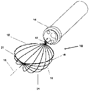
Referring now to the drawings in figures 1-14, wherein similar parts are
identified by like
reference numerals, there is seen in Figure 1 a perspective view of the device
10 in a generally
crescent shape, wherein the snare or cauterization loop 12 formed of non-
insulated conductive
material, is deployed from a lumen such as a sheath or catheter 14 in a
combination with the
radially deployable members 16 which radially positionable around the axis 15
to form a
collection or capture component 21 as in figure 12.
In all modes of the device 10 where a cauterization loop 12 is included, the
loop 12 will
be uninsulated and therefor reactive with tissue when electrified. Of course
the device 10 can be
employed with a loop 12 defining a mouth of the capture component 21 formed of
insulated
material just like the members 16 or of non electrifiable material if
cauterization is not desired.
Even in this mode without the cauterization, the biasing of the members 16 to
move to an
expanded position radially deployed provides great utility for a capture
component 21 that is
expandable at will to encircle material 19 such as tissue or a polyp for
removal.
A control wire 13 is engaged to or communicates motion to the trailing ends of
the loop
12 and members 16 to thereby translate and position the device 10 from the
catheter 14. During
deployment from the catheter 14 the members 16, which are formed of memory
material or
otherwise adapted to bias around the axis 15, are collapsed against or
adjacent to the loop 12 by
a means for restraint depicted restraint device 18. While depicted in figure 1
as a crescent shape,
the loop 12 and capture component 21 formed by the members 16 of the device 10
may be
formed in any geometric shape into which the loop 12 and members 16 can be
formed. Also, in
all modes of the device 10, while it may be shown as a 180 degree radial
deployment around the
axis 15 in a number of the drawings such as figure 3, the members 16 can be
deployed in any
8
CA 02744175 2011-05-18
WO 2009/158520
PCT/US2009/048696
radial deployment up to 360 degrees around the axis 15, and, can consist of a
plurality of any
number of members 16, and can be spaced at any distance from adjacently
situated members 16
in such a radial deployment.
Figure 2 depicts the restrain device 18 of figure 1 showing the means for
restraint being a
medical glue 25 which melts during heating of the cauterization loop 12. Such
a means of
restraint would result in an automatic deployment of the members 16 to form a
capture
component 21. The stacking of the underlying radially deployable members 16 is
held by one or
a plurality of the restraints 18 whereafter the members 16 which are biased
outwardly, will
radially deploy around the axis 15 from the loop 12 to which they were
constrained.
Figure 3 depicts the device of figure 1, wherein the restraint 18 which as
shown is
medical glue, has completely released whereafter the members 16 have deployed
to form a
capture component 21 in the form of a basket. As noted, any number of members
16 at any
spacing and any angular displacement around the axis 15 may be employed.
Figure 4 shows the device 10 in a different shape of six angular sides. As
noted the
members 16 forming the capture or collection component and the loop 12 can be
any shape.
When in the collapsed position as depicted, in figure 4 they will radially
deploy around the axis
15 to form a similarly shaped capture component 21 for collecting tissue or
foreign objects. The
number, and the spacing, of the members 16 to form gaps 23 therebetween is
infinitely variable
in that any number of members 16 can be used in any spacing around the axis 15
thereby making
the size of the gaps 23 infinitely variable.
Figure 5 depicts a perspective view of the device of figure 4 once the
restraint is released.
As shown in a 180 degree spaced radial deployment, it is envisioned as noted
that any spacing
and any radial deployment of the members 16 around the axis 15 can be
employed.
Figure 6 depicts the device herein wherein the restraint 18 employed for means
of
restraint of the members 16 is a knot in an encircling loop of surgical
thread, suture material or
similar flexible material. One or a plurality of knots may be employed as
needed. Translation of
a release wire 17 separate from the control wire 13, provides means for
controlled mechanical
release of the means for restraint 18. Figure 6a depicts a close up of the
knot and release
mechanism controlled by the release wire 17.
Figure 7 depicts a mode of the device having the deployable members 16 which
are
restrained by means of restraint in the form of medical tape 29 in one or a
plurality of positions.
A release wire 17 separate from the control wire 13, would provide a user
operable mechanical
means for controlled release of the release of the restraint 18 provided by
the tape or other
mechanically releasable means for restraining the members 16. Figure 7a
depicts a close up
view of the medical tape of figure 7 and the release wire 17.
Figure 8 depicts an embodiment of the device wherein the deployable members 16
of the
capture component expand radially around the axis to a 360 degree mode of the
capture
component 21 with gaps 23 between the deployed members 16. As noted, any
number of
9
CA 02744175 2011-05-18
WO 2009/158520
PCT/US2009/048696
members 16 in any spacing scheme be it equal or unequal, in any radial
deployment around an
axis 15 may be employed depending on the intended task and all are considered
included in this
application. The 180 degree deployment depicted in figure 3 is a particular
favorite having a
defined mouth, however all modes of the device 10 allow for the capture
component 21 to be
pulled into the deploying lumen or catheter 14 as shown in figure 12, to a
point where it will
collapse around and hold, a captured piece of material 19 for removal, but
other radial
deployments may be more favorable depending on the procedure intended. When
employed
with a 360 degree deployment the gaps 23 would be manipulated over the
material 19 to be
removed and the capture component 21 retracted into the lumen as in figure 12
to tightly hold it.
Figure 9 depicts a mode of the device 10 having members 16 and a loop 12
formed to
yield a substantially hexagonal shaped embodiment of the device 10 and also
showing a
deployment to a 360 degree capture component 21 with equidistant spacing and
gaps 23. Any
radial deployment around an axis may be employed and the plurality of members
16 and spacing
thereof is infinitely variable depending on the desired spacing in the
deployed position.
In Figure 9a the device 10 in the hexagonal shape of figure 9 is shown in the
collapsed
position substantially planar shape. As with the other modes and shapes of the
device 10, the
members 16 are engaged to either the loop 12 or the control wire 13 in a
fashion to cause a bias
of the members 16 around an axis once the means for restraint to hold the
members 16 in a
collapsed position adjacent to the loop 12 is released. Also as noted this
means for restraint on
any mode may be glue, tape, a knot, or any means for restrain that may
automatically release
with heat, or relapse with a release wire 17, or any means for restraint that
is adapted to the task
and would occur to those skilled in the art.
As with Figure 9, figure 10 depicts a 360 degree deployed capture component 21
formed
of members 16 yielding a substantially round shaped embodiment of the device
10. Figure 10a
depicts the device of figure 10 shown in a collapsed position having a
substantially planar round
shape with the deployable members 16 restrained adjacent to the centered loop
12 which can
double in the preferred mode as a cauterization loop and in that case would be
uninsulated.
In figure 11 there is shown a substantially oval shaped embodiment of the
device 10 with
the members 16 in a deployed position forming a capture component 21 which
encircles 360
degrees around an axis. As with the other embodiments, the members 16 are
shaped with
memory material to retain the shape induced and the members 16 are biased to
move to positions
around a center axis when released. This bias can be provided by forming the
members 16 using
shape memory material such as nitinol while they are in a radially deployed
position and then
restraining them to a collapsed position thereby providing a memory material
as the means for
radially biasing the members 16. Or a spring engaged to the connection points
of the members
16 to the control wire may be employed or other means to bias each member 16
to a radially
located position around the axis as would occur to those skilled in the art.
Figure lla depicts the device of figure 11 in a collapsed position
substantially planar
CA 02744175 2011-05-18
WO 2009/158520
PCT/US2009/048696
shape with the deployable members 16 forming the plurality or radially located
portions of the
capture component 21 when deployed, collapsed adjacent to a center loop 12
which can double
as a cauterization loop. As in other modes, the members 16 are formed to bias
against the
restraint to the collapsed position and return to a specific position of
radial deployment.
Figure 12 depicts the capture component formed of the radially deployed biased
members
16 which are translated partially into the lumen such as a catheter or sheath
by translating the
control wire thereby causing an encircling of a piece of tissue 19 or material
to be removed.
Another especially preferred mode of the device 10 is shown in figures 13-14.
This
mode of the device 10 provides the surgeon with an especially useful surgical
component in that
it will allow for the cutting and cauterizing of one or a plurality of pieces
of tissue prior to the
release of the restraints to initiate the deployment of the members 16 to form
a capture
component 21 to thereafter allow for capture and removal the tissue 19 as in
figure 12.
As shown in Figures 13-14 the device 10 in an especially preferred mode,
employs a
control wire 13 for translation of the loop 12 and deployable members 16 and
also a separate
release wire 17 which is engaged at a pair of distal ends, to a plurality of
restraints 18 which are
shown as ties such as in figure 13b. However, any means for restraint which
may be selectively
released by retracting the release wire 17 as would occur to those skilled in
the art is anticipated
within the scope of this invention.
As noted, in this mode of the device 10 the members 16 are maintained in their
collapsed
position with the loop 12 by the restraints 18 until the surgeon fully extends
the members 16 and
loop 12 from the insulating catheter 14 with the control wire 13 and
subsequently retracts the
release wire 17 to release the restraints 18. Control of the release wire 17
is shown accessible
under cap 33 however other means to control the release wire as would occur to
those skilled in
the art is anticipated.
This mode of the device10 is structured to yield an especially useful function
during a
surgery. On many occasions during surgery, the surgeon will need to remove and
cauterize
tissue 19 (figure 12) at a plurality of locations inside the body of the
patient. Since the loop 12
attached to the members 16 in their collapsed position, heats the tissue for
cutting and
cauterizing when placed in to contact with the tissue and the loop 12
energized with electrical
current to the grounded patient, it is important to maintain the members 16 in
their retracted
position for the duration of cutting and cauterizing. This mode of the device
10 provides that
function by maintaining the restraints 18, insulated within the lumen or
catheter 14 or other
instrument during the ongoing cauterization process wherein the loop 12 is
placed in contact
with tissue and then energized with electrical current to create heat at the
site of the tissue and
grounded patient.
Maintaining the restraints 18 in a first position shown in figure 14,
insulated within the
channel of the catheter 14, thereby provides a means to protect the restraints
18 from melting or
releasing due to heat generated in the loop 12 or members 16 adjacent to the
restraints. Since
11
CA 02744175 2016-04-14
WO 2009/158520 PCT/US2009/048696
the covered restraints 18 are maintained out of contact with tissue for the
duration of the cutting
and cauterization by the surgeon, until they are translated from the distal
end of the catheter 14,
there is no chance they may melt from being energized by tissue contact, or
that the restraint 18
will melt due to the loop 12 or members 16 become heated in the area of the
restraints 18.
So maintained inside the catheter 14, the restraints 18 will maintain the
members 16 in a
collapsed position against the loop 12 for the duration of cutting and
cauterization of tissue.
The surgeon can thus deploy the forward portion of the capture component 21 in
the
collapsed state formed by the members 16 and loop 12, from the distal end of
the passage of the
catheter 14. Once so deployed, the surgeon can energize the loop 12 to cut and
cauterized the
patient at a plurality of positions as needed for the procedure. All the
while, the restraints 18 are
safely maintained inside the catheter 14. This allows the surgeon to maintain
the device 10
positioned continually inside the patient for the duration of the procedure
without a release of the
restraints 18 which might require removal and insertion of a new capture
component 21 every
time the restraints 18 become released should the surgeon wish to cauterize
again.
A handle assembly 31 shown in figure 13 in a conventional fashion would have a
control
knob 33 wherein the control knob 33 would be removed to access and operate the
release wire
17. Upon completion of the cutting and cauterizing of tissue at one or a
plurality of different
positions, once the members 16 and loop 12 have been translated fully from the
distal end of the
catheter 14, the control knob 33 would be released to allow manipulation of
the control wire 17
to release the restraints 18 to thereby allow the members 16 to radially
deploy to form the
capture component 21. \While depicted as an oval loop, the device 10 in this
and all modes as
noted may be formed in any shape or configuration in which the members 16 may
be shaped and
which will adapt the ultimate formed capture component 21 to the task at hand.
The method and components shown in the drawings and described in detail herein
disclose arrangements of elements of particular construction, and
configuration for illustrating
preferred embodiments of structure of the present surgical device.
As such, while the present invention has been described herein with reference
to
particular embodiments thereof, a latitude of modifications, various changes
and substitutions
are intended in the foregoing disclosure, and will be appreciated that in some
instance some
features of the invention could be employed without a corresponding use of
other features,
12