Note: Descriptions are shown in the official language in which they were submitted.
CA 02744344 2011-05-20
WO 2010/060217 PCT/CA2009/001729
ANTIBODY-TARGETED CARRIER FOR CONTRAST AGENTS
Field of the Invention
The present invention is directed to a composition of self-assembled, lipidic
nanoparticles
targeted using single domain antibodies capable of treating and imaging
disease.
Background
Molecular imaging enables the simultaneous anatomical localization and
quantitative
evaluation of target biomolecules that can guide the selection of treatment
protocols, whose
efficacy can also be evaluated. The expected impact of these technologies in
shortening the
drug development cycle has been emphasized in the FDA's `Critical Path
Initiative' which
recommends "integration of molecular and imaging biomarkers into every stage
of the
regulatory review for drug, diagnostic, and biologic applications" (Woodcock &
Woosley,
2008).
Currently there are only a limited number of molecular imaging agents suitable
for clinical
applications. Most molecular imaging applications for central nervous system
(CNS) diseases
have been developed for radioactivity-dependent PET and SPECT modalities.
These imaging
compounds are typically small molecules with short circulation half-lives that
can readily
penetrate across the blood-brain barrier. However, similar compounds are
presently lacking
for the more accessible magnetic resonance imaging (MRI) modality, as well as
for the rapidly
developing and cheaper optical imaging modality. The clinical translation of
these imaging
agents will depend however, on advances in the development of new
targeting/delivery
moieties against disease-specific biomarkers which have been validated in
animal models and
the ability to scale up their production for commercialization at reasonable
cost and market
value.
1
CA 02744344 2011-05-20
WO 2010/060217 PCT/CA2009/001729
MRI is a non-invasive and powerful medical diagnostic technique that offers
high-resolution
anatomical information, and is frequently used for the non-invasive detection
of a variety of
diseases. MRI creates images of the body using the principles of nuclear
magnetic resonance.
Images are usually generated using gadolinium (Gd-DTPA) as contrast agent,
based on its free
distribution in the body. While these images provide good anatomical
information about the
disease (e.g., tumor) localization and spread, MRI does not deliver adequate
information about
molecular characteristics of the disease (e.g., expression of certain
receptors that could be
targeted by drugs or transporters that may cause resistance to certain drugs,
etc.), and therefore
biopsy of diseased tissue and molecular analyses ex vivo (e.g.,
histopathology,
immunochemistry, etc.) are still required.
Molecular imaging in MRI modality is currently not routinely used in clinical
applications
because of the lack of appropriate contrast agents that are targeted to
recognize specific
molecular targets. These contrast agents typically need to have very high
contrast properties
to provide measurable information on specific molecular target. Target
characteristics are also
important, including selectivity of the target for diseased tissues and the
high
expression/density of the target, to enable sufficient signal-to-noise ratio
for detection.
While, in principle, monoclonal antibodies could be used to target contrast
imaging agent or
drug delivery carrier to the antigen recognition site, these antibodies are
relatively large (150
kDa) proteins and can only be attached to nanoparticles in low numbers,
typically less than 25
proteins per nanoparticle. Moreover, repetitive display of large proteins on
the surface of
nanoparticles can also be immunogenic and in some instances further accelerate
biological
clearance. Peptides used as a targeting moiety suffer from low
affinity/specificity of binding
to the target and are often prone to degradation by proteases.
Industry needs to consider systems integration approaches in order to bring
together drug
delivery, imaging and activation technologies into one comprehensive product.
This is
especially true for the CNS diseases, where delivery across the BBB imposes
unique
challenges for the development of both therapeutic and imaging applications.
Integrated
platforms for targeted drug delivery and non-invasive monitoring and
quantification of drug
2
CA 02744344 2011-05-20
WO 2010/060217 PCT/CA2009/001729
distribution and accumulation/release at desired targets by means of imaging
are currently not
available.
Phospholipid bilayers forming spherical unilamellar vesicles (ULVs), or
liposomes, could be
used as biodegradable or biocompatible drug carriers to enhance the potency
and reduce the
toxicity of therapeutics. Typically, ULVs are produced by sonication or high-
pressure multi-
stage extrusion of multi-lamellar vesicles (MLV). The major drawbacks of these
methods
include degradation and modification of phospholipids (e.g. oxidation,
hydrolysis,
denaturation), difficulties in producing single size population liposomes, and
low throughput.
ULV produced by sonication and extrusion methods are inherently unstable (not
thermodynamically stable) and may, over time, revert to MLVs.
ULVs are also capable of entrapping and delivering contrast imaging agents.
There are two
categories of liposomal contrast agents: a) those that entrap paramagnetic
molecules in the
aqueous compartment and b) those that incorporate these molecules in the
liposomal
membrane, either by covalent attachment to the lipid acyl chains or by
chelation to a ligand
which is incorporated into the membrane. With regard to MRI, incorporating the
contrast
agent within the outer or inner membrane is preferred, as the bound
paramagnetic ions possess
a much longer rotational correlation time and therefore have a greater
relaxivity/mole than
those in solution (i.e. better contrast).
Gd-DTPA is an FDA approved imaging contrast agent for MRI. To achieve
sufficient signal-
to-noise ratio and detectable signal for molecular imaging applications, the
concentration of
Gd at diseased molecular recognition sites has to be very high; in other
words, a single Gd
molecule per targeting moiety is insufficient to achieve detectable signal.
To accelerate approval of molecular imaging agents for clinical use, it is
desirable to have the
ability to assess them using multi-modal imaging, for example optical in
animal studies and
MRI in animal and human studies.
Summary of the Invention
3
CA 02744344 2011-05-20
WO 2010/060217 PCT/CA2009/001729
The present invention comprises a composition of self-assembled, lipidic
nanoparticles
targeted using single domain antibodies, and capable of treating and imaging
disease.
The present invention comprises novel formulations of lipid-based
spontaneously forming
nanoparticles. These nanoparticles may comprise spontaneously forming
unilamellar vesicles
(ULVs) comprising phospholipids, and may be used for noninvasive molecular
imaging.
Such liposomal-based delivery systems may display efficacy and commercial
viability via: a)
vesicle stability i.e., extended shelf life; b) well-defined, monodisperse ULV
size; c) extended
plasma half-life in vivo, and d) potential for scale-up to industrial sized
production.
The present invention provides a nanoconjugate comprising:
(a) a self-assembled unilamellar vesicle (ULV);
(b) at least one contrast agent; and
(c) at least one antibody.
The ULV may be comprised of dimyristoyl phosphatidylcholine (DMPC); dihexanoyl
phosphatidylcholine (DHPC); dimyristoyl phosphatidylglycerol (DMPG); and
distearoyl
phosphoethanolamine-[maleimide(polyethylene glycol)-2000] (DSPE-PEG-
maleimide).
In the nanoconjugate described above, the contrast agent may be a MRI contrast
agent, a
radioisotope, a fluorophore, or a combinations thereof. In one embodiment, the
contrast agent
may be a MRI agent, and may be gadolinium-diethylene-triamine-pentaacetic acid
bis-oleate
(Gd-DTPA-BOA). In another embodiment, the fluorophore may be Cy5.5.
In the nanoconjugate described above, the antibody may specifically bind an
epitope present
in the brain endothelial cells or tumor cells. For example, the antibody may
selectively bind
Epidermal Growth Factor Receptor (EGFR). In another example, the antibody may
selectively bind Insulin-like Growth Factor Binding Protein 7 (IGFBP7). In one
specific
example, the antibody may comprise complementarity determining region (CDR)
sequences
RTSRRYAM or RTFSRLAM (CDR1; SEQ ID NOs:l and 2), GISRSGDGTHYAYSV
(CDR2; SEQ ID NO:3), and AAARTAFYYYGNDYNY (CDR3; SEQ ID NO:4).
Alternatively, the antibody may comprise the sequence:
4
CA 02744344 2011-05-20
WO 2010/060217 PCT/CA2009/001729
AIAIAVALAGFATVAQAQVKLEE SGGGLVQAGGS LRLSCAAS GRTSRRYAMGWF
RQAPGKEREFVAGISRSGDGTHYAYSVKGRFTISRDNAANTVELQMNSLKPEDT
AVYFCAAARTAFYYYGNDYNYWGQGTQVTVSS (SEQ ID NO:5),
or a sequence substantially identical thereto. In another alternative, the
antibody may comprise
the sequence:
AIAIAVALAGFATVAQAQVKLEESGGGSVQPGGSLRLSCAASGRTFSRL
AMGWFRQAPGKERELVAGISRSGDGTHYAYSVKGRFTISRDNAANTV
ELQMNS LKPEDTAVYFCAAARTAFYYYGNDYNYW GQGTQVTV S S
(SEQ ID NO:6),
or a sequence substantially identical thereto.
The present invention further provides a method of forming unilamellar
vesicles (ULV)
incorporating at least one contrast agent, the method comprising:
(a) mixing dimyristoyl phosphatidylcholine (DMPC); dihexanoyl
phosphatidylcholine
(DHPC); dimyristoyl phosphatidylglycerol (DMPG); distearoyl
phosphoethanolamine-
[maleimide(polyethylene glycol)-2000] (DSPE-PEG-maleimide) and gadolinium-
di ethyl ene-tri amine-pentaacetic acid bis-oleate (Gd-DTPA-BOA); and
(b) allowing the spontaneous formation of ULV.
In the method as described above, an antibody may be bioconjugated to DSPE-PEG-
maleimide prior to step (a), thus incorporating the antibody into the
nanoconjugate.
The present invention also provides a method for in vivo imaging of cells or
tissues in a
mammal, the method comprising the steps of:
(a) administering to the mammal a composition comprising the nanoconjugate
described
herein, wherein the antibody is specific for a selected receptor;
(b) waiting a time sufficient to allow the antibody to bind to the selected
receptor; and
(c) imaging the cells or tissues with a non-invasive imaging technique whose
resolution is
enhanced by the presence of the particles on or within the cells.
5
CA 02744344 2011-05-20
WO 2010/060217 PCT/CA2009/001729
The imaging technique used may be selected from the group consisting of
magnetic resonance
imaging, magnetic spectroscopy, X-ray, positron emission tomography, optical
imaging,
computed tomography, and ultrasonic imaging. The method as described may
allows for
imaging of one or more tumors, metastases, vascularized malignant cell
clusters, or individual
malignant cells selected from the group consisting of brain cancer, colon
cancer, breast
cancer, prostate cancer, lung cancer, pancreatic cancer, endometrial cancer,
oral cancer, liver
cancer, and renal cancer or any other cancer.
In one embodiment of the method as described above, the selected receptor may
be is
specifically expressed by tumor endothelial cells. The selected receptor may
be IGFBP7, and
the antibody may be as described above. In another embodiment, the selected
receptor may be
EGFR.
In one aspect, the invention comprises a method for detecting glioblastoma in
a patient,
comprising:
(a) contacting a tissue of interest with a nanoconjugate as described herein,
wherein the
antibody is specific for the IGFBP7 or EGFR, and may comprise the specific
antibodies
described herein; and
(b) measuring the level of binding of the nanoconjugate, wherein an elevated
level of
binding, relative to normal tissue, is indicative that the tissue is
neoplastic.
In yet another aspect, the present invention provides a method for detecting a
tissue expressing
IGFBP7, comprising:
(a) contacting a tissue of interest with a nanoconjugate as described herein,
wherein the
sdAb is specific for the IGFBP7, and may comprise the specific antibodies
described
herein; and
(b) measuring the level of binding of the nanoconjugate, wherein an elevated
level of
binding, relative to normal tissue is indicative of the presence of a tumor
expressing
IGFBP7.
6
CA 02744344 2011-05-20
WO 2010/060217 PCT/CA2009/001729
In yet another aspect, the present invention provides a method for detecting a
tissue expressing
EGFR, comprising:
(a) contacting a tissue of interest with a nanoconjugate as described herein,
wherein the
antibody is specific for the EGFR, and may comprise the specific antibodies
described
above; and
(b) measuring the level of binding of the nanoconjugate, wherein an elevated
level of
binding, relative to normal tissue is indicative of the presence of a tumor
expressing
EGFR.
In one embodiment, the step of measuring is performed by magnetic resonance
imaging. The
nanoconjugate used in the method as just described may further comprise a
fluorescent
imaging agent, and the step of measuring may be performed using fluorescence
imaging.
In another aspect, the invention comprises a method for determining the
location of
glioblastoma brain tumor cells in a patient pre-operatively, intra-
operatively, and/or post-
operatively, comprising administering a composition comprising a nanoconjugate
as described
herein, wherein the antibody is specific for the IGFBP7 or EGFR, and may
comprise the
specific antibodies described above, and a pharmaceutically acceptable carrier
to the patient,
wherein the composition is administered in an amount sufficient to image
glioblastoma cells
in vivo; and
(a) pre-operatively measuring the level of binding of nanoconjugate by
magnetic
resonance imaging to determine the location of glioblastoma cells, wherein an
elevated level of binding, relative to normal tissue, is indicative of the
presence of
glioblastoma cells;
(b) intra-operatively measuring the level of binding of the nanoconjugate by
fluorescence imaging to determine the location of residual glioblastoma cells,
wherein an elevated level of binding, relative to normal tissue, is indicative
of the
presence of residual glioblastoma cells;
7
CA 02744344 2011-05-20
WO 2010/060217 PCT/CA2009/001729
(c) post-operatively measuring the level of binding of the nanoconjugate by
magnetic
resonance imaging to determine the location of glioblastoma cells, wherein an
elevated level of binding, relative to normal tissue, is indicative of the
presence of
tumor cells; or
(d) a combination of (a), (b) or (c) .
In yet another aspect, the invention comprises a method for in vitro detection
or quantification
of biological or chemical molecule in a sample is also provided by the present
invention. The
method comprises the steps of:
(a) contacting the sample with a solution comprising the nanoconjugate of the
present
invention, so as to form a complex between the molecule and the nanoconjugate;
and
(b) detecting and/or quantifying said complex formed.
The step of detecting and/or quantifying may be performed by magnetic
resonance imaging,
fluorescence imaging, or a combination thereof.
The naonconjugates of the present invention may be used as a MRI contrast
agent, as they
contain a very high number of Gd molecules, up to 60,000 Gd molecules per ULV;
this may
result in increased sensitivity (i.e., high number of contrast agent molecules
at antigen
recognition sites) and increased signal-to-noise ratio. The ULVs may be self-
assembled using
components loaded with Gd-DTPA-BOA, thereby achieving high numbers of Gd
molecules
carried by each ULV.
The nanoconjugates of the present invention may also be used in bi-modal
imaging for MRI
and optical in vivo imaging. In one embodiment, the bi-modal capacity is
introduced by
attaching an optical probe such as the near-infrared probe, Cy5.5, applicable
to in vivo optical
imaging, to PEG moieties incorporated into self-assembled Gd-loaded ULVs
The present invention provides a process for formulating highly stable, self-
assembled
monodisperse, nanoscopic ULVs composed of commonly available low cost
phospholipids;
the ULVs can be tailored to suit a variety of biomedical needs. Self-assembled
ULVs have
8
CA 02744344 2011-05-20
WO 2010/060217 PCT/CA2009/001729
advantages over ULVs formed by sonication or extrusion in key areas important
for
development of commercially viable drug delivery formulations: namely a) both
their size and
entrapment efficiency can be controlled during the self assembly process; b)
they are very
stable (long shelf life and in vivo); and c) the process can be easily scaled
up for
manufacturing.
The present invention also produces targeted drug delivery/imaging
formulations.
Bioconjugation of antibodies, and particularly antibody fragments such as
sdAbs, to ULVs
may enhance sensitivity and specificity of targeting of imaging/drug delivery
formulation.
Compared to the conventional 150 kDa IgG molecules, a larger number of
antibody
fragments, such as sdAbs, can be incorporated into ULVs, lending polyvalency
and increasing
avidity. Moreover, antibody fragments/sdAbs may be more stable and soluble
compared to
conventional antibodies
The ability to perform these molecular analyses non-invasively by in vivo
imaging by MRI at
the time of diagnosis and during disease treatment may greatly improve
treatment efficacy by
a) obtaining early molecular information on disease, b) adjusting treatment to
fit `personal'
characteristics of disease, c) selecting appropriate patient populations for
clinical trials.
9
CA 02744344 2011-05-20
WO 2010/060217 PCT/CA2009/001729
Brief Description Of The Drawings
In the drawings, like elements are assigned like reference numerals. The
drawings are not
necessarily to scale, with the emphasis instead placed upon the principles of
the present
invention. Additionally, the embodiments depicted are but some of a number of
possible
arrangements utilizing the fundamental concepts of the present invention. The
drawings are
briefly described as follows:
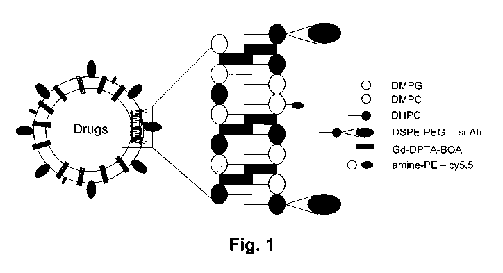
Figure 1 is a schematic representation of one embodiment of a unilamellar
vesicle (ULV)
functionalized with single domain antibody and loaded with gadolinium (Gd) and
optical
imaging contrast (Cy5.5) in the hydrophobic shell and with the drug in the
hydrophilic core.
DMPC = Dimyristoyl phosphatidylcholine; DMPG = Dimyristoyl
phosphatidylglycerol;
DHPC = Dihexanoyl phosphatidylcholine; Gd-DPTA BOA= Gadolinium
diethylenetriaminopentaacetic acid bisoleate; PEG-DSPE = Distearoyl
Phosphoethanolamine-
N-[Methoxy(Polyethylene glycol)-2000]; amine-PE = Dipalmitoyl
Phosphoethanolamine-N-
(dodecanylamine). Dode-PE = Dipalmitoyl Phosphoethanolamine-N-
(dodecanylamine).
Figure 2 graphically shows LC-Gd ULV nanoparticle size determined by dynamic
light
scattering.
Figure 3 graphically shows the size distribution of spontaneously formed ULVs
formulated
with HC-Gd as determined by dynamic light scattering.
Figure 4 shows evaluation of LC-Gd nanoparticles at various total lipid
concentrations: 10.0
(triangles; darkest grey), 5.0 (diamonds; lightest grey), 1.0 (squares;
black), 0.2 (circles; 2nd
lightest grey) wt%, using Small Angle Neutron Scattering (SANS). The peaks at -
0.055 and
-0.11 A-' correspond to the first and second order reflections from an MLV.
Figure 5 shows evaluation of HC-Gd nanoparticles using SANS. Total lipid
concentrations
are: 10.0 (triangles; darkest grey), 5.0 (diamonds; lightest grey), 1.0
(squares; black), 0.2
(circles; lightest grey) wt%. The peaks at - 0.055 and -0. 11 A` correspond to
the first and
CA 02744344 2011-05-20
WO 2010/060217 PCT/CA2009/001729
second order reflections from MLVs. The peak intensity shows that, compared to
the 20
mol% Gd sample, the 40 mol% sample results in more MLV being formed.
Figure 6 shows SANS data of the HC-Gd 20 nm mixture with total lipid
concentrations of 1.0
(black) and 0.5 (grey) wt% annealed at 50 C for 18 hours (inverted triangles)
and 3 days
(triangles). The nanoparticle mixtures were reformulated by replacing
PEGylated-DSPE-
maleimide with PEGylated-DSPE-amine and DMPC with DMPG (in Figure 4). The MLV
peaks disappeared indicating the absence of MLVs. The gray curves are the best
fits to the
data using the ellipsoidal shell model. The data and fits to the data are
rescaled for viewing
clarity.
Figure 7 is a schematic representation of an ellipsoidal shell model.
Figure 8 shows a table of the measurement of the Gd molecules (ng/ml) in ULV
nanoparticle
formulations (LC- and HC-Gd) determined using ICP-MS.
Figure 9 shows imaging of EGFR-expressing subcutaneous xenograft tumors in
nude mice
using Gd-Cy5.5-ULVs (40mol% Gd) (A) or Gd-Cy5.5-ULVs (HC-Gd) targeted with the
monoclonal IgG antibody C225 against EGFR (B). Images were taken 24h post-
injection.
Cy5.5 fluorescence was detected only in the tumor xenograft of animals
injected with targeted
ULV but not in animals injected with non-targeted ULVs.
Figure 10 shows imaging of time-dependent tumor accumulation of C225-targeted
(A) vs.
non-targeted (B) Gd-Cy5.5-ULVs (HC-Gd) in EGFR expressing subcutaneous flank
xenograft
tumors in nude mice. Cy5.5 fluorescence was detected only in the tumor
xenograft of animals
injected with C225 Ab targeted ULV but not in animals injected with non-
targeted ULVs.
Figure 11 shows quantitation of time-dependent in vivo accumulation of C225-
targeted vs.
non-targeted Gd-Cy5.5-ULVs (HC-Gd) in EGFR-expressing subcutaneous flank
xenograft
tumors in mice (from Figure 10).
Figure 12 shows imaging (whole body dorsal scan) of in vivo biodistribution
(24 h after
injection) of non-targeted (A) and C225-targeted (B) Gd-Cy5.5-ULVs vesicle (HC-
Gd) in
11
CA 02744344 2011-05-20
WO 2010/060217 PCT/CA2009/001729
tumor-bearing mice. Cy5.5 fluorescence was detected only in the tumor
xenograft of animals
injected with C225 Ab targeted ULV but not in animals injected with non-
targeted ULVs.
Figure 13 show imaging (whole body ventral scan) of in vivo biodistribution
(24 h after
injection) of non-targeted (A) and C225-targeted (B) Gd-Cy5.5-ULVs vesicle (HC-
Gd) in
tumor-bearing mice. Cy5.5 fluorescence was detected only in the tumor
xenograft of animals
injected with targeted ULV but not in animals injected with non-targeted ULVs.
Figure 14 shows ex vivo imaging of excised tumor and skeletal muscle 24h after
injection of
C225-targeted (A) or nontargeted (B) Gd-Cy5.5 ULVs (HC-Gd). Cy5.5 fluorescence
was
detected only in the excised tumor of animals injected with C225 Ab targeted
ULV but not in
excised muscle of similar size. Animals injected with non-targeted ULVs had
minimal
fluorescence in both excised tumor and muscle.
Figure 15 shows the presence of Cy5.5 fluorescence (red) in tumor section
immunostained for
EGFR (green) from mice injected with C225-targeted (A) or non-targeted (B) Gd-
Cy5.5-
ULVs (HC-Gd). Cy5.5 fluorescence was detected only in the tumor sections of
animals
injected with targeted ULV but not in animals injected with non-targeted ULVs.
Figure 16 shows gadolinium concentration measurement using laser ablation ICP-
MS in
tumors excised from mice injected with C225-targeted or non-targeted Gd-Cy5.5-
ULVs (HC-
Gd). N = 14 per group. High number of Gd was measured in C225 monoclonal
targeted ULV
in tumor compared to low content of Gd in non-targeted ULV.
Figure 17 shows measurement of the gadolinium content in organs using ICP-MS
in tumors
and organs excised from mice injected with C225-targeted Gd-Cy5.5-ULVs (HC-Gd)
in Table
(A) and graph (B and C) form. Figure 17C shows the concentration of Gd in
ng/mg or ppm,
and the measurement of Gd content in organs relative to dry weight of the
organ. High
number of Gd was measured in C225 monoclonal targeted ULV in tumor compared to
low
content of Gd in non-targeted ULV.
12
CA 02744344 2011-05-20
WO 2010/060217 PCT/CA2009/001729
Figure 18 shows optical in vivo imaging of the head of the mice bearing an
orthotopic brain
tumor at different time points after the injection of either non-targeted (A)
or IGFBP7 sdAb-
targeted (B) Gd-Cy5.5-ULVs (HC-Gd 20 nm). Cy5.5 fluorescence was detected only
in the
brain tumor of animals injected with IGFBP7 sdAb targeted ULV but not in
animals injected
with non-targeted ULVs.
Figure 19 shows depth-concentration analysis (A) and volumetric analysis (B)
of head
imaging after injection of non-targeted or IGFBP7 sdAb-targeted Gd-Cy5.5-ULVs
vesicle
HC-Gd 20 nm) in orthotopic brain tumor-bearing mice (from Figure 18). Two way
ANOVA
was run to test significance.
Figure 20 shows biodistribution of non-targeted (A) and IGFBP7 sdAb-targeted
(B)Gd-Cy5.5-
ULVs vesicles (HC-Gd 20 rim) 24 h after injection into orthotopic brain tumor-
bearing mice
by whole-body in vivo optical imaging (dorsal scan). Two examples of each are
shown. Cy5.5
fluorescence was detected only in the brain tumor of animals injected with
targeted ULV but
not in animals injected with non-targeted ULVs.
Figure 21 shows biodistribution of non-targeted (A) and IGFBP7 sdAb-targeted
(B) Gd-
Cy5.5-ULVs vesicles (HC-Gd 20 nm) 24 h after injection into orthotopic brain
tumor-bearing
mice by ex vivo optical imaging of excised organs. Cy5.5 fluorescence was
detected in the
brain tumor of animals injected with targeted ULV but not in animals injected
with non-
targeted ULVs. High signal was detected non-specifically in liver.
Figure 22 shows ex vivo imaging of brain tumors 24 h after injection of Gd-
Cy5.5-ULVs (HC-
Gd 20 nm) non-targeted (A) or targeted with the anti-IGFBP7 sdAb (B) that
recognize brain
tumor vasculature. Cy5.5 fluorescence was detected only in the implanted brain
tumor of
animals injected with targeted ULV but not in animals injected with non-
targeted ULVs.
Figure 23 shows the effect of Gd-DTPA-BOA ULVs (HC-Gd 20 nm) on Ti relaxation
measured by 9.4T MRI. The inset shows the location of different samples in the
phantom
apparatus.
13
CA 02744344 2011-05-20
WO 2010/060217 PCT/CA2009/001729
Figure 24A shows MRI in vivo imaging of the head in orthotopic brain tumor
bearing mice
injected with non-targeted Gd-Cy5.5-ULVs vesicle (HC-Gd 20 rim). Figure 24B
shows the
subtraction images of Figure 24A.
Figure 25A shows another example of MRI in vivo imaging of the head in
orthotopic brain
tumor-bearing mice injected with non-targeted Gd-Cy5.5-ULVs vesicle (HC-Gd 20
nm).
Figure 25B shows the subtraction images of Figure 25A.
Figure 26A shows MRI in vivo imaging of the head in orthotopic brain tumor
bearing mice
injected with IGFBP7 single domain antibody-targeted Gd-Cy5.5-ULVs vesicle (HC-
Gd 20
nm). Figure 26B shows the subtraction images of Figure 26A.
Figure 27A shows another example of MRI in vivo imaging of the head in
orthotopic brain
tumor bearing mice injected with IGFBP7 single domain antibody-targeted Gd-
Cy5.5-ULVs
vesicle (HC-Gd 20 nm). Figure 27B shows the subtraction images of Figure 27A.
Figure 28 shows pharmakinetics analysis of unilamellar vesicles labelled with
Cy5.5 (40mol%
Gd with 20 nm size) and injected intravenously in normal CD1 mice. Blood
samples were
taken at different time points and fluorescence was measured using a
fluorescence plate
reader. Analysis was undertaken using WinNonlin professional software using
one-
compartment, bolus injection modeling. R2=0.9932; Plasma half-life=96.8 3
minutes,
Vss(Apparent volume of distribution) = 1.004 ml; MRT (mean residence time) =
139.7 min,
CL (Clearance) = 0.00718 molecules/min.
Detailed Description Of Preferred Embodiments
The present invention is directed to a composition of self-assembled, lipidic
nanoconjugates
targeted using single domain antibodies capable of treating and imaging
disease.
When describing the present invention, all terms not defined herein have their
common art-
recognized meanings. To the extent that the following description is of a
specific embodiment
14
CA 02744344 2011-05-20
WO 2010/060217 PCT/CA2009/001729
or a particular use of the invention, it is intended to be illustrative only,
and not limiting of the
claimed invention. The following description is intended to cover all
alternatives,
modifications and equivalents that are included in the spirit and scope of the
invention, as
defined in the appended claims.
One embodiment of the present invention comprises antibody-modified ULVs
formulated to
incorporate both gadolinium ions and fluorescent dyes, and be capable of
selectively targeting
disease affected sites in the brain or in tumors, where the anatomical
localization and
molecular characteristics of diseased cells can be elucidated using MRI or
optical imaging, or
both simultaneously or consecutively. The ULVs also provide a vehicle by which
a
therapeutic can be delivered to these diseased sites in the same formulation,
enabling a non-
invasive monitoring of both therapeutic delivery and therapeutic efficacy.
In one embodiment, the present invention provides a nanoconjugate comprising:
(a) a self-assembled unilamellar vesicle (ULV);
(b) at least one contrast agent; and
(c) at least one antibody.
By the term "self-assembled unilamellar vesicle" or "spontaneously-formed
unilamellar
vesicle", it is meant spontaneously formed, homogenous monodisperse, and size-
controlled
ULVs. These ULVs may be tailored to suit a variety of biomedical and
nutraceutical needs, at
the same time being suitable for industrial scale production. The self-
assembled ULV may
have advantages over ULVs formed by sonication or extrusion. For example,
their size and
entrapment efficiency can be controlled during the self assembly process, they
are very stable,
and the process for producing them can be easily scaled up for manufacturing.
The ULV may comprise any suitable lipids known to form ULVs. For example, and
the ULV
may comprise lipids including, but not limited to dimyristoyl
phosphatidylcholine (DMPC);
dimyristoyl phosphatidylglycerol (DMPG); dihexanoyl phosphatidylcholine
(DHPC);
distearoyl Phosphoethanolamine-N-[Methoxy(Polyethylene glycol)-2000] (PEG-
DSPE);
CA 02744344 2011-05-20
WO 2010/060217 PCT/CA2009/001729
dipalmitoyl Phosphoethanolamine-N-(dodecanylamine) (amine-PE); and dipalmitoyl
phosphoethanolamine-N-(dodecanylamine) (Dode-PE).
As would be understood by those of skill in the art, the composition of the
ULV may vary
based on the type of contrast agent and/or sdAb to be included in the
nanoconjugate, as well
as the type of application for which the nanoconjugate is to be used. For
example, and
without wishing to be limiting in any manner, the amount of each lipid in a
composition of
ULVs may independently be between about 0 and 55 mol.% DMPC; between about 20
and 30
mol.% DHPC; between about 0 and 35 mol.% DMPG; between about 3 and 10 mol.%
DSPE-
PEG-maleimide; and between about 0 and 1 mol.% dode-PE. In specific, non-
limiting
examples, the amount of each lipid may independently be 0, 30.6, or 50.5 mol.%
DMPC; 23.8
or 23.9 mol.% DHPC; 0.4, 0.5, or 31.2 mol.% DMPG; 5 mol.% DSPE-PEG-maleimide;
0 or
0.1 mol.% Dode-PE.
Without wishing to be limiting, the ULV may comprise, for example:
(a) DMPC = 30.6 mol.%; DHPC = 23.9 mol.%; DMPG = 0.4 mol.%; DSPE-PEG-
maleimide = 5 mol.%; dode-PE = 0.1 mol.%;
(b) DMPC = 50.5 mol.%; DHPC = 23.9 mol.%; DMPG = 0.5 mol.%; DSPE-PEG-
maleimide = 5 mol.% ; dode-PE = 0.1 mol.%; or
(c) DHPC = 23.8 mol.%; DMPG = 31.2 mol.%; and DSPE-PEG-amine.
As would be understood by one of skill in the art, other combinations of
lipids are
encompassed by the present invention. Various physical parameters may also aid
in
determining the ULV composition. Such parameters include, but are not limited
to charge
density, chain length, temperature, and salt concentration. The skilled
artisan will be adept in
adapting the ULV composition to account for such parameters.
In addition to the amounts of lipids described above, the ULVs may be
characterized by ratios
of certain lipid components. For example, one embodiment of the ULVs may
possess a
constant long-to-short chain lipid ratio of 3.0-5.0; and/or a constant DSPE-
PEG2000-
Maleimide/total lipid of less than 0.05. In one embodiment, when Gd-DTPA-BOA
is used
(see below), the ULV may also comprises a DMPC/DMPG ratio of 100-0.
16
CA 02744344 2011-05-20
WO 2010/060217 PCT/CA2009/001729
It is noted that, in certain examples of ULVs described above, a portion of
the phospholipids
incorporate PEG molecules. Without wishing to be bound by theory, PEG
molecules
incorporated on the surface of the ULV nanoparticle may create a formulation
that is not
readily recognized or cleared by the reticuloendothelial system, therefore
improving their
plasma stability and plasma half-life. Such "stealth" formulations may
optimize the blood
circulation half-life. PEG molecules may be covalently attached to a
phospholipid (prior to
ULV assembly) by methods well-known to those of skill in the art.
While DSPE-PEG2000-Maleimide is mentioned above as a specific non-limiting
example,
any suitable size PEG may be used, and is encompassed by the present
invention. Without
wishing to be limiting, the PEG may be in the range of about 1000 to 5000 Da;
for example,
the PEG may be about 1000, 1250, 1500, 1750, 2000, 2250, 2500, 2750, 3000,
3250, 3500,
3750, 4000, 4250, 4500, 4750, or 5000 Da, or any size therebetween.
The ULVs may vary in size, based on their composition and/or other variables.
For example,
the ULV may generally be between about 30 and 150 nm; for example, the ULV may
be
about 30, 40, 50, 60, 70, 80, 90, 100, 110, 120, 130, 140, or 150 nm, or any
size therebetween.
The nanoconjugate of the present invention also comprises at least one
contrast agent. The
contrast agent may be a MRI contrast agent, an optical imaging agent, or a
combination
thereof.
The nanoconjugate of the present invention may comprise a MRI contrast agent.
The MRI
contrast agent may be any MRI contrast agent suitable for incorporation into
ULVs. The MRI
contrast agent should preferably be an agent that produces Ti enhancement
effect, and should
be preferably incorporated into the ULV with minimal effect of the morphology
of the vesicle.
For example, the MRI contrast agent may be, but is not limited to a chelated
paramagnetic ion.
For example, the paramagnetic ion may be gadolinium, manganese, ytterbium,
europium, or
the like. In a specific, non-limiting example, the paramagnetic ion may be a
gadolinium (Gd)
ion.
17
CA 02744344 2011-05-20
WO 2010/060217 PCT/CA2009/001729
Any paramagnetic ion-based lipid that may be incorporated into the lipid
bilayer of the ULV
would be suitable for use in the present invention. Different chelating agents
or alternatives
may also be used, such a, but not limited to EDTA, DTPA and DOTA and the like.
For
example, the chelating agent may be coupled directly to a lipid such as, but
not limited to
phosphatidyl ethanolamine, bis-oleate, and the like, or through linking
groups. In a specific,
non-limiting embodiment, the contrast agent incorporated into the ULV may
comprise
gadolinium-diethylene-triamine-pentaacetic acid bis-oleate (Gd-DTPA-BOA).
The MRI contrast agent may be incorporated into the ULV at a molar ratio in
the range of 15
to 40 mol% of MRI contrast agent to total lipid mixture. For example, and
without wishing to
be limiting, the molar ratio may be 15, 20, 25, 30, 35, or 40 mol%, or any
value therebetween.
In a specific, non-limiting example, the MRI contrast agent may be
incorporated into the ULV
at a molar ratio of 20 mol% or 40 mol%.
As the MRI contrast agent may be incorporated directly into the ULV, a high
number of the
contrast agent molecules may be incorporated into the nanoconjugate of the
present invention.
Without wishing to be bound by theory, the inclusion of a high number of
contrast agent
molecules into the nanoconjugate may result in increased sensitivity (i.e.,
high number of
contrast agent molecules at the site of interest) and increased signal-to-
noise ratio. For
example, the number of paramagnetic ions per ULV may be in the range of about
5,000 to
about 60,000; for example, the ULV may comprise about 5000, 10,000, 15,000,
20,000,
25,000, 30,000, 35,000, 40,000, 45,000, 50,000, 55,000, or 60,000, or any
amount
therebetween, paramagnetic ions per ULV. In a specific, non-limiting example,
the ULV may
comprise up to 60,000 Gd molecules per ULV. Without wishing to be bound by
theory,
ULVs exhibiting high Gd payload show enhanced Ti effect in 9.4T MRI phantoms,
comparable to the clinically used Gd-DTPA - Magnevist.
The contrast agent in the nanoconjugate may further comprise one or more than
one optical
imaging agent, thus creating a bimodal imaging agent. The optical imaging
agent may be, for
example, but not limited to, a radioisotope or a fluorophore. For example, the
optical imaging
agent may be, but is not limited to Cy5.5, Cy7, Cy7.5. Alexa 680, Alexa 750,
ICG, IR800, or
18
CA 02744344 2011-05-20
WO 2010/060217 PCT/CA2009/001729
any fluorophore that emits between 650 nm and 900 nm. Multiple copies of the
same or
different optical imaging agent may be present in the nanoconjugate. The
optical imaging
agent may be incorporated into the ULV by conjugation to a PEG molecule.
The nanoconjugate of the present invention further comprises at least one
antibody (Ab) as a
targeting moiety. By the term "antibody", it is meant any suitable antibody;
for example, but
not limited to antibodies (such as IgG) and antibody fragments, whether
naturally-occuring or
recombinantly-produced. The antibody may be engineered by molecular
techniques, and may
comprise associated sequences (such as signal peptides, purification tags,
etc). Antibody
fragments may comprise, but are not limited to Fab, Fab', Fv, scFv, and single-
domain
antibodies.
By the term "single-domain antibody" or "sdAb", it is meant an antibody
fragment comprising
a single protein domain. Single domain antibodies may comprise any variable
fragment,
including VL, VH, VHH, VNAR, and may be naturally-occurring or produced by
recombinant
technologies. For example VHS, VLS, VHHs, VNARS, may be generated by
techniques well
known in the art (Holt, et al., 2003; Jespers, et al., 2004a; Jespers, et al.,
2004b ; Tanha, et al.,
2001 ; Tanha, et al., 2002; Tanha, et al., 2006 ; Revets, et al., 2005 ;
Holliger, et al., 2005;
Harmsen, et al., 2007; Liu, et al., 2007; Dooley, et al., 2003; Nuttall, et
al., 2001; Nuttall, et
al., 2000; Hoogenboom, 2005; Arbabi-Ghahroudi et al., 2008). In the
recombinant DNA
technology approach, libraries of sdAbs may be constructed in a variety of
ways, "displayed"
in a variety of formats such as phage display, yeast display, ribosome
display, and subjected to
selection to isolate binders to the targets of interest (panning). Examples of
libraries include
immune libraries derived from llama, shark or human immunized with the target
antigen; non-
immune/naive libraries derived from non-immunized llama, shark or human; or
synthetic or
semi-synthetic librairies such as VH, VL, VHH or VNAR libraries.
The small size of the sdAbs allow their conjugation in nanoconjugates with a
much higher
binding site density compared to larger antibody fragments (> 5 fold compared
to IgGs and 2-
fold compared to scFvs) and do not promote nanoparticle aggregation associated
with scFvs
and IgGs, resulting in much more active nanoconjugates, and more robust signal
amplification
19
CA 02744344 2011-05-20
WO 2010/060217 PCT/CA2009/001729
strategy. Higher levels of imaging signal per unit level of target-probe
interaction lead to
higher sensitivity for any particular imaging modality. Additionally, the
highly stable nature
of sdAbs allows for flexibility in terms of choosing optimal conjugation
chemistry conditions
(Huang et al, 2007), leading to a more active end product.
In one embodiment, the antibody may be a sdAb that recognizes and binds to an
antigen
present in tumor endothelial cells. For example, and without wishing to be
limiting in any
manner, the single domain antibody may selectively bind Insulin-like Growth
Factor Binding
Protein 7 (IGFBP7), which is strongly upregulated in vessels of glioblastoma
tumors
undergoing neovascularization.. This target is less expressed in vessels of
low grade gliomas.
Without wishing to be limiting in any manner, the single domain antibody may
be an sdAb as
described in PCT/CA2009/001460 entitled "Formulations Targetting IGFBP7 for
Diagnosis
and Therapy of Cancer", the disclosure of which is incorporated herein by
reference where
permitted. In a specific, non-limiting example, the sdAb may comprise
complementarity
determining region (CDR) sequences RTSRRYAM [SEQ ID NO. 1] or RTFSRLAM [SEQ ID
NO. 2] (CDRI), GISRSGDGTHYAYSV [SEQ ID NO. 3] (CDR2), and
AAARTAFYYYGNDYNY [SEQ ID NO. 4] (CDR3). Alternatively, the single domain
antibody may comprise the sequence:
AIAIAVALAGFATVAQAQVKLEESGGGLVQAGGSLRLSCAASGRTSRR
YAM G W F RQAP GKERE F V AG I S R S GD GTHYAY S V KGRF TI S RDNAANT
VELQMNSLKPEDTAVYFCAAARTAFYYYGNDYNYWGQGTQVTVSS,
[SEQ ID NO. 5]
or a sequence substantially identical thereto. In another alternative, the
sdAb may comprise
the sequence:
AIAIAVALAGFATVAQAQVKLEESGGGSVQPGGSLRLSCAASGRTFSRL
AMGWFRQAPGKERELVAGISRSGDGTHYAYSVKGRFTISRDNAANTV
ELQMN S LKP EDTAVYFCAAARTAFYYYGNDYNYW GQ GTQ VTV S S,
[SEQ ID NO. 6]
or a sequence substantially identical thereto.
CA 02744344 2011-05-20
WO 2010/060217 PCT/CA2009/001729
In another embodiment, the antibody may be IgG C225 (Gridelli et al, 2009, the
contents of
which are hereby incorporated by reference where permitted), or an antibody
with a
substantially identical sequence thereto. The antibody may also be an antibody
fragment
based on or obtained from IgG C225, retaining the binding specificity of IgG
C225.
A sequence that is substantially identical to another sequence may comprise
one or more
conservative amino acid mutations. It is known in the art that one or more
conservative amino
acid mutations to a reference sequence may yield a mutant polypeptide with no
substantial
change in physiological, chemical, or functional properties compared to the
reference
sequence; in such a case, the reference and mutant sequences would be
considered
"substantially identical" polypeptides. Conservative amino acid mutation may
include
addition, deletion, or substitution of an amino acid; a conservative amino
acid substitution is
defined herein as the substitution of an amino acid residue for another amino
acid residue with
similar chemical properties (e.g. size, charge, or polarity).
In a non-limiting example, a conservative mutation may be an amino acid
substitution. Such a
conservative amino acid substitution may substitute a basic, neutral,
hydrophobic, or acidic
amino acid for another of the same group. By the term "basic amino acid" it is
meant
hydrophilic amino acids having a side chain pK value of greater than 7, which
are typically
positively charged at physiological pH. Basic amino acids include histidine
(His or H),
arginine (Arg or R), and lysine (Lys or K). By the term "neutral amino acid"
(also "polar
amino acid"), it is meant hydrophilic amino acids having a side chain that is
uncharged at
physiological pH, but which has at least one bond in which the pair of
electrons shared in
common by two atoms is held more closely by one of the atoms. Polar amino
acids include
serine (Ser or S), threonine (Thr or T), cysteine (Cys or C), tyrosine (Tyr or
Y), asparagine
(Asn or N), and glutamine (Gln or Q). The term "hydrophobic amino acid" (also
"non-polar
amino acid") is meant to include amino acids exhibiting a hydrophobicity of
greater than zero
according to the normalized consensus hydrophobicity scale of Eisenberg
(1984).
Hydrophobic amino acids include proline (Pro or P), isoleucine (Ile or I),
phenylalanine (Phe
or F), valine (Val or V), leucine (Leu or L), tryptophan (Trp or W),
methionine (Met or M),
alanine (Ala or A), and glycine (Gly or G). "Acidic amino acid" refers to
hydrophilic amino
21
CA 02744344 2011-05-20
WO 2010/060217 PCT/CA2009/001729
acids having a side chain pK value of less than 7, which are typically
negatively charged at
physiological pH. Acidic amino acids include glutamate (Glu or E), and
aspartate (Asp or D).
Sequence identity is used to evaluate the similarity of two sequences; it is
determined by
calculating the percent of residues that are the same when the two sequences
are aligned for
maximum correspondence between residue positions. Any known method may be used
to
calculate sequence identity; for example, computer software is available to
calculate sequence
identity. Without wishing to be limiting, sequence identity can be calculated
by software such
as NCBI BLAST2 service maintained by the Swiss Institute of Bioinformatics
(and as found
at http://ca.expasy.org/tools/blast/), BLAST-P, Blast-N, or FASTA-N, or any
other
appropriate software that is known in the art.
The substantially identical sequences of the present invention may be at least
75% identical; in
another example, the substantially identical sequences may be at least 70, 75,
80, 85, 90, 95,
or 100% identical at the amino acid level to sequences described herein.
Importantly, the
substantially identical sequences retain the activity and specificity of the
reference sequence.
As would be understood by a person of skill in the art, other antibodoes may
be used in the
nanoconjugate of the invention. The antibody may be chosen in accordance with
the desired
target for imaging.
The antibody may be bioconjugated (also referred to herein as "conjugated",
"linked" or
"coupled") to the ULV, using any suitable method known in the art. For
example, and
without wishing to be limiting, the single domain antibody may be linked to
the PEG-DPSE
moiety prior to formation of the ULV, through a functional group such as a
carboxylate, a
sulfonate, a phosphate, an amine, and any combination thereof.
If a PEG molecule is used, conjugation of antibody to the PEG molecule may be
accomplished using methods well known in the art (see for example Hermanson,
1996).
Antibodies and single domain antibodies in particular, have several exposed
lysine (primary
amine) residues, and thus one method of covalently anchoring the antibody to
the carboxylic
acid-modified nanoparticle surface is through bioconjugation chemistry.
Suitable coupling
22
CA 02744344 2011-05-20
WO 2010/060217 PCT/CA2009/001729
reagents include 1-ethyl-3 -(3 -dimethylaminopropyl) carbodiimide
hydrochloride (EDC) which
is often used in combination with N-hydroxysuccinimide (NHS). For example, the
antibody
as described above may have, or may be engineered to have, one or more lysine
residues
opposite or away from its antigen binding site, which is used in covalent
conjugation to the
nanoparticle surface. In one embodiment, the number of antibodies conjugated
to the surface
of the nanoparticle is controllable and controlled.
Alternatively, the antibody may be conjugated to the PEG molecule through an
amino acid
with a carboxylic acid (i.e., Glu or Asp) on the antibody and primary amines
on the PEG, or
through binding of the PEG (detecting entity) to a molecule that has binding
activity towards
the antibody and is already attached to the PEG molecule. For example, this
molecule could
be an antibody which binds to the antibody or to tags (C-Myc tag, His6 tag) on
the antibody
such as anti-C-Myc or anti-His6 antibodies, or through binding of a
biotinylated antibody to a
biotin binder on the surface of nanoparticles. Biotin binders are well known
and may include
streptavidin, neutravidin, avidin, or extravidin. The antibody could also be
coupled to the
nanoparticle by means of nickel-nitrilotri acetic acid chelation to a His6-
tag.
In another alternative, antibodies can also be engineered to have cysteines
opposite their
antigen binding sites. Conjugation via a maleimide cross-linking reaction
allows the
directional display of antibodies where all antibodies are optimally
positioned to bind to their
antigens. Amine-terminated PEG molecule is activated with maleimide in DMF
followed by
an incubation of cysteine-terminated single domain antibody to achieve
covalent binding
through the formation of sulfide bond formation.
The number of antibody molecules conjugated to the surface of the ULV may
vary, based on
various factors, such as the size of the ULV. The conjugate of the present
invention may
comprise at least 1 to 100 antibody molecules conjugated to the surface of the
ULV; for
example, the conjugate may carry at least 1, 2, 3, 4, 5, 6, 7, 8, 9, 10, 11,
12, 13, 14, 15, 16, 17,
18, 19, 20, 25, 30, 35, 40, 45, 50, 55, 60, 65, 70, 75, 80, 85, 90, 95, or 100
antibody moieties
linked to the ULV. As a person of skill in the art would recognize, it may be
possible to
23
CA 02744344 2011-05-20
WO 2010/060217 PCT/CA2009/001729
conjugate more antibody molecules to the surface of the nanoparticle,
depending on particle
size, antibody size and characteristics, and on immobilization efficiency.
It is to be noted that each of the antibody molecules linked to the
nanoconjugate may be the
same, or may differ from one another. Thus, the ULV may be conjugated to more
than one
antibody to detect multiple target molecules simultaneously. The ULV may be
conjugated to
different antibodies that recognize different parts (epitopes) on the same
pathogen, for
example, but not limited to different epitopes on the same toxin or different
epitopes on the
same bacterial cell surface molecules or different epitopes on different cell
surface molecules
of the same bacteria.
As will be recognized by those skilled in the art, the diameter of the
nanoconjugate may vary
depending on the lipid composition used and the type of antibody conjugated to
the ULV
suface. Without wishing to be limiting in any manner, the overall size of the
nanoconjugate of
the present invention may be between about 20 and 200 nm in diameter. For
example, and
without wishing to be limiting, the nanoconjugate may have a diameter of about
20, 30, 40,
50, 60, 70, 80, 90, 100, 110, 120, 130, 140, 150, 160, 170, 180, 190, or 200
nm, or any value
therebetween. In a specific, non-limiting example, the nanoconjugate diameter
may be about
40 to about 100 rim. Nanoconjugates of the present invention comprising larger
antibodies
(such as IgG) may have larger diameters.
An exemplary embodiment of the nanoconstruct of the present invention
comprising an ULV
functionalized with sdAb and an optical imaging agent molecule is shown in the
schematic of
Figure 1.
Formulations and compositions comprising the nanoconstruct of the present
invention are also
provided. In addition to the nanoconstruct of the present invention, such
formulations or
compositions may include pharmaceutically acceptable excipients or diluents,
buffers, and/or
water. The formulations may be powder, suspensions, or any other suitable
pharmaceutical
formulation.
24
CA 02744344 2011-05-20
WO 2010/060217 PCT/CA2009/001729
The present invention further provides a method of forming unilamellar
vesicles (ULV)
incorporating at least one contrast agent, the method comprising:
(a) mixing dimyristoyl phosphatidylcholine (DMPC); dihexanoyl
phosphatidylcholine
(DHPC); dimyristoyl phosphatidylglycerol (DMPG); distearoyl
phosphoethanolamine-
[maleimide(polyethylene glycol)-2000] (DSPE-PEG-maleimide) and gadolinium-
diethylene-triamine-pentaacetic acid bis-oleate (Gd-DTPA-BOA); and
(b) allowing the spontaneous formation of ULV.
In the method as described above, an antibody may be bioconjugated to DSPE-PEG-
maleimide prior to step (a), thus incorporating the antibody into the
nanoconjugate.
The present invention also provides a method for in vivo imaging of cells or
tissues in a
mammal, the method comprising the steps of:
(a) administering to the mammal a composition comprising a nanoconjugate as
described
herein, wherein the antibody is specific for a selected receptor;
(b) waiting a time sufficient to allow the antibody to bind to the selected
receptor; and
(c) imaging the cells or tissues with a non-invasive imaging technique whose
resolution is
enhanced by the presence of the particles on or within the cells.
The imaging technique used may be selected from the group consisting of
magnetic resonance
imaging, magnetic spectroscopy, X-ray, positron emission tomography, optical
imaging,
computed tomography, and ultrasonic imaging. The method as described may
allows for
imaging of one or more tumors, metastases, vascularized malignant cell
clusters, or individual
malignant cells selected from the group consisting of brain cancer, colon
cancer, breast
cancer, prostate cancer, lung cancer, pancreatic cancer, endometrial cancer,
oral cancer, liver
cancer, and renal cancer or any other cancer.
In the method as described above, the selected receptor may be is specifically
expressed by
tumor endothelial cells. The selected receptor may be IGFBP7 or EGFR, and the
antibody
may be as described above.
Also provided is a method for detecting glioblastoma in a patient, comprising:
CA 02744344 2011-05-20
WO 2010/060217 PCT/CA2009/001729
(a) contacting a tissue of interest with a nanoconjugate as described herein,
wherein the
antibody is specific for the IGFBP7 or EGFR, and may comprise the specific
antibodies
described above; and
(b) measuring the level of binding of the nanoconjugate, wherein an elevated
level of
binding, relative to normal tissue, is indicative that the tissue is
neoplastic.
In yet another aspect, the present invention provides a method for detecting a
tissue expressing
IGFBP7, comprising:
(a) contacting a tissue of interest with a nanoconjugate as described herein,
wherein the
antibody is specific for the IGFBP7, and may comprise the specific antibodies
described
above; and
(b) measuring the level of binding of the nanoconjugate, wherein an elevated
level of
binding, relative to normal tissue is indicative of the presence of a tumor
expressing
IGFBP7.
In yet another aspect, the present invention provides a method for detecting a
tissue expressing
EGFR, comprising:
(a) contacting a tissue of interest with a nanoconjugate as described herein,
wherein the
antibody is specific for the EGFR, and may comprise the specific antibodies
described
above; and
(b) measuring the level of binding of the nanoconjugate, wherein an elevated
level of
binding, relative to normal tissue is indicative of the presence of a tumor
expressing
EGFR.
In the methods as just described, the step of measuring is performed by
magnetic resonance
imaging. The nanoconjugate used in the method as just described may further
comprise a
fluorescent imaging agent, and the step of detecting may be performed using
fluorescence
imaging.
The present invention also provides a method for determining the location of
glioblastoma
brain tumor cells in a patient pre-operatively, intra-operatively, and/or post-
operatively,
26
CA 02744344 2011-05-20
WO 2010/060217 PCT/CA2009/001729
comprising the step of administering a composition comprising a nanoconjugate
as described
herein, wherein the antibody is specific for IGFBP7 or EGFR, and may comprise
the specific
antibodies described above, and a pharmaceutically acceptable carrier to the
patient, wherein
the composition is administered in an amount sufficient to image glioblastoma
cells in vivo;
and
(a) pre-operatively measuring the level of binding of nanoconstruct by
magnetic
resonance imaging to determine the location of glioblastoma cells, wherein an
elevated level of binding, relative to normal tissue, is indicative of the
presence of
glioblastoma cells;
(b) intra-operatively measuring the level of binding of the nanoconstruct by
fluorescence imaging to determine the location of residual glioblastoma cells,
wherein an elevated level of binding, relative to normal tissue, is indicative
of the
presence of residual glioblastoma cells;
(c) post-operatively measuring the level of binding of the nanoconstruct by
magnetic
resonance imaging to determine the location of glioblastoma cells, wherein an
elevated level of binding, relative to normal tissue, is indicative of the
presence of
tumor cells; or
(d) a combination of (a), (b) or (c) above.
A method for in vitro detection or quantification of biological or chemical
molecule in a
sample is also provided by the present invention. The method comprises the
steps of.
(a) contacting the sample with a solution comprising a nanoconjugate as
described herein,
so as to form a complex between the molecule and the nanoconjugate; and
(b) detecting or quantifying said complex formed.
The step of detecting or quantifying may be performed by magnetic resonance
imaging,
fluorescence imaging, or a combination thereof.
27
CA 02744344 2011-05-20
WO 2010/060217 PCT/CA2009/001729
In one embodiment, monoclonal antibodies against Epidermal Growth Factor (225
mAb) and
single domain antibodies against IGFBP7 may be conjugated to the ULV
nanoparticle, while
retaining their full activity. The IGFBP7 sdAb-targeted bi-modal ULVs are
selectively
targeted to orthotopic brain tumors in nude mice, which may be demonstrated
using optical in
vivo imaging modality and in vivo MRI imaging. The C225 (anti-EGFR IgG)-
targeted bi-
modal ULVs are selectively targeted to xenograft tumors expressing EGFR in
nude mice,
which may also be demonstrated using optical in vivo imaging modality. The
presence of
C225 targeted ULVs in the xenograft tumors may be confirmed by fluorescence
microscopy
based detection of Cy5.5 in tumor sections. Excised xenograft tumors after
injection of C225
(anti-EGFR IgG)-targeted bi-modal ULVs exhibit high Gd concentrations
(measured by ICP-
MS) The presence of C225 targeted ULVs in the xenograft tumors may also be
confirmed by
fluorescence microscopy based detection of Cy5.5 in tumor sections.
ULV nanoparticle formulations with high payload of Gadolinium-DTPA-BOA and
near-
infrared imaging contrast agent, Cy5.5, targeted using single domain antibody
against
IGFBP7 or monoclonal IgG antibody against EGFR have been synthesized and
tested in
xenograft and orthotopic brain tumor models in nude mice. The targeted ULVs of
the present
invention may be used in non-invasive (molecular) diagnosis/imaging (optical,
MRI) of brain
tumors and other tumors expressing IGFBP7 or EGFR and exhibiting a high-rate
of
angiogenesis (i.e., colon, breast). When a therapeutic agent is combined with
the ULVs,
either by encapsulation or surface incorporation, such tumors may be treated
by application of
the ULVs. Therapeutic agents may include pharmaceutical agents such as anti-
cancer drugs
or biological agents such as immunotherapeutic agents.
Examples
The following examples are intended to exemplify specific embodiments of the
invention, and
not to limit the claimed invention in any manner.
28
CA 02744344 2011-05-20
WO 2010/060217 PCT/CA2009/001729
Example 1 - Unilamellar vesicle production and their loading with gadolinium
Dimyristoyl phosphatidylcholine (DMPC), dimyristoyl phosphatidylglycerol
(DMPG),
dihexanoyl phosphatidylcholine (DHPC) and distearoyl phosphoethanolamine-N-
[Maleimide(Polyethylene Glycol)2000] (DSPE-PEG2000-Maleimide) were purchased
from
Avanti Polar Lipids (Alabaster AL). The Gd-DTPA-BOA was custom synthesized.
All
chemicals were used without further purification.
The structures of two lipid mixtures (high-concentration Gd mixture (HC-Gd)
and low-
concentration Gd mixtures (LC-Gd)) with the following molar ratios:
DMPC/DMPG/DHPC/DSPE-PEG2000-Maleimide/Gd-DTPA-BOA= 100:1:47:10:40 and
100:1:78:16:129 for LC-Gd and HC-Gd, respectively, were examined. The molar
ratios were
selected to yield 20 and 40 mol% of Gd-DTPA-BOA to total lipid mixture while
keeping a
constant long-to-short chain lipid ratio of 3.2, DMPG/DMPC ratio of 0.01 as
previously
published (Nieh et al, 2004, 2005) and a constant DSPE-PEG2000-Maleimide/total
lipid of
0.05. Specifically, the lipid mixtures comprised:
HC-Gd: Gd-DTPA-BOA = 40 mol.%; DMPC = 30.6 mol.%; DHPC = 23.9 mol.%;
DMPG = 0.4 mol.%; DSPE-PEG-maleimide = 5 mol.%; dode-PE = 0.1 mol.%.
LC-Gd: Gd-DTPA-BOA = 20 mol.%; DMPC = 50.5 mol.%; DHPC = 23.9 mol.%; DMPG
= 0.5 mol.%; DSPE-PEG-maleimide = 5 mol.% ; dode-PE = 0.1 mol.%.
Both LC-Gd and HC-Gd mixtures were first dissolved in chloroform (> 99.9% from
Aldrich)
at corresponding molar ratios and dried by continuously flowing N2 gas
followed by
evacuation with vacuum for 24 hours. The dried samples were then completely re-
dispersed
in D20 to form 10 wt% solutions by temperature cycling and vortex between 40
and 50 C
(4-5 cycles). The 10 wt% solutions are liquid-like at low temperature (the low-
temperature
viscosity of HC-Gd is higher than that of LC-Gd) but gel-like at high
temperature, illustrating
the same phenomenon as previously reported in the phospholipid systems of
spontaneously
forming vesicles (Nieh et al, 2004, 2005). All the samples were then diluted
with cold D20 at
4 C into total lipid concentrations, CLp of 5 wt%, 1 wt% and 0.2 wt%.
29
CA 02744344 2011-05-20
WO 2010/060217 PCT/CA2009/001729
A third formulation (HC-Gd 20 nm) was developed subsequent to SANS analysis
(see
Example 2) with the goal of forming smaller ULV. The new formula retained the
same
amounts of Gd-DTPA-BOA found in the original formula, except that DMPC was
completely
replaced by the charged DMPG lipid. Specifically, the third formulation
included 4
components, namely: 40 mol% of Gd-DTPA-BOA, 23.8 mol.% of DHPC, 31.2 mol.% of
DMPG and 5.0 mol.% DSPE-PEG-amine. The total lipid concentrations of samples
with this
formula in D20 were 0.5 wt.% and 1 wt.%. The formulations were prepared in a
manner
similar to that described above for HC-Gd and LC-Gd.
Example 2 - Small Angle Neutron Scattering (SANS)
For small angle neutron scattering (SANS) study, the lipid mixtures were
dissolved in
deuterium oxide with a purity > 99.9 % (Chalk River Laboratories, ON, Canada)
to enhance
the neutron scattering contrast. All the SANS measurements are done at 50 C
where the
lamellae are expected to form. SANS experiments were conducted at the 30m NG7
SANS
located at National Institute of Standards and Technology (NIST) Center for
Neutron
Researches (NCNR, Gaithersburg, Maryland, USA).
The wavelength (X) of the incident neutrons was 6A and three sample-to-
detector distances
(1m, 4m and 15.3m) were applied, covering a scattering vector (q) ranged from
0.003 A` to
0.3 A'. Data were collected using a 2-D position-sensitive detector as a
function of scattering
angle (0). The raw data were corrected for background (blocked beam),
normalized with the
monitored incident neutrons flux and sample transmission, and subtracted with
equally treated
empty cell scattering data. Finally, the reduced data were circularly averaged
with respect to
the beam center and put on an absolute intensity scale using the incident
neutron flux and the
sample thickness. The final scattering intensity, I, is presented as a
function of scattering
vector q, which is defined as sin 8
SANS data obtained from the original lipid mixtures, LC-Gd and HC-Gd, are
shown in
figures 4 and 5, respectively. The q-2 dependence at low- and mid-q indicated
a bilayered
structure, and the intensity oscillation at - q < 0.01 A' corresponded to a
characteristic length,
CA 02744344 2011-05-20
WO 2010/060217 PCT/CA2009/001729
mostly likely the size of the vesicles. At q - 0.06 A-1, a small, yet sharp
peak was observed,
indicating a well-defined distance originating from the interlamellar spacing
of a
multilamellar vesicle (MLV). In some cases, the second order peak (at q - 0.12
A') was also
observed. Compared to the LC-Gd sample, this feature was more pronounced in
the HC-Gd
sample, indicating that Gd-DTPA-BOA may induce the formation of MLVs.
SANS experiments were similarly conducted on the third formulation at the 30m
NG3 SANS
instrument located at the NIST (National Institute of Standards and
Technology) Center for
Neutron Research (NCNR, Gaithersburg, MD). In order to study the stability of
the resultant
structure as a function of annealing time, SANS measurements were taken from
both 0.5 and
1.0 wt.% samples incubated at 50 C for either 18 hours or 3 days. SANS results
indicated
small differences between the two samples (Fig. 6). Importantly, the data did
not exhibit any
peaks associated with the previously observed MLV structure.
The SANS data were best fit using an ellipsoidal shell model (schematic shown
in Fig. 7).
This model includes four structural parameters: a) the long core axis (aCOY,);
b) the short core
axis (bCOYe); c) shell thickness (t) and d) the polydispersity of bcore. The
best fit results are
listed in Table 1. Although the SANS data showed significant differences in
the low-q
regime, the structural parameters obtained from the fits to the data did not
differ dramatically,
exception being the value of aeon which is greater in the 1.0 wt.% sample. The
best fit results
of 18 hour and 3 day (0.5 wt.% and 1 wt.% samples) data showed that they
differed in contrast
(Table 1). This difference in contrast between the two time period samples may
be the result
of the PEG chain rearranging at high temperature. However, the reason for the
higher value
of best fit result for scattering length density of the nanoparticle core,
peore, compared to that
of D20, pD2o, is not understood. As would be understood by a person skilled in
the art, the
present model may be refined.
Table 1. Best-fit data from HC-Gd (20 nm) at 50 C for 18 hours and 3 days
31
CA 02744344 2011-05-20
WO 2010/060217 PCT/CA2009/001729
0.5 wt.% 1.0 wt.%
18 hours 3 days 18 hours 3 days
acore (A) 625 570 1070 1230
beore (A) 65 59 60 64
t() 38 38 39 38
polydispersity 0.48 0.5 0.49 0.46
(core - Pshell), (A 2) 6.6 x 10⁻⁶ 6.2 x 10⁻⁶ 6.7 x 10⁻⁶ 6.0 x 10⁻⁶
(PD20- Pshell), (A-2) 4.1 x 10⁻⁶ 4.6 x 10⁻⁶ 4.2 x 10⁻⁶ 4.7 x 10⁻⁶
Example 3 - Functionalization of Unilamellar vesicles with monoclonal
antibodies and single
domain antibodies
To enable targeting of the nanoconjugates to tumor-expressed antigens, ULVs
were
functionalized with antibodies attached to PEG-DSPE.
In one approach, ULVs were functionalized with a single-domain antibody
against IGFBP7,
newly discovered target selectively expressed in glioblastoma tumor vessels
(see PCT
Application No. PCT/CA2009/001460 and entitled Formulations of Targeting
IGFBP7 for
Diagnosis and Therapy of Cancer). Because the target is vascular, this
formulation is suitable
for imaging of intracranial tumors. Anti-IGFBP7 Single domain antibody was
produced in-
house. IGFBP7-sdAb was reconstituted in MES buffer (MES 0.1M, NaCI 0.5M, pH 6)
using
the aforementioned Amicon columns. To produce NHS-ester functionality on the
sdAb, Sulfo-
NHS and EDC were added to at 180- and 70-fold molar excess respectively and
reacted for 30
min at room temperature. Subsequently, EDC was removed by centrifugation using
Amicon
columns.
In another approach, ULVs were functionalized with the monoclonal IgG C225
against
Epidermal Growth Factor Receptor (EGFR) of glioblastoma cells (Gridelli et al,
2009).
Because this target is expressed in glioblatoma cells, xenograft (flank) tumor
model was used
to provide proof of targeting. C225 antibody was reconstituted in MES buffer
(MES 0.1 M,
32
CA 02744344 2011-05-20
WO 2010/060217 PCT/CA2009/001729
NaC1 0.5M, pH 6) using the aforementioned Amicon columns. To produce NHS-ester
functionality on the antibody, Sulfo-NHS and EDC were added to at 180- and 70-
fold molar
excess respectively and reacted for 30 min at room temperature. Subsequently,
EDC is
removed by centrifugation using Amicon columns.
Example 4 - Intracranial and xeongraft models of U87MG deltaEGFRvIII
glioblastoma in
nude mice
U87MG deltaEGFRvIII is a highly malignant glioblastoma cell line derived from
a human
brain tumor and has been engineered to overexpress both EGFR and the EGFRvIII
mutant
receptor (Dr W.K. Cavenee, Ludwig Institute for Cancer Research, San Diego,
CA, USA).
Cells were maintained in DME medium containing 10% fetal bovine serum (FBS),
penicillin/streptomycin and 200 g/ml of G418. Cells were grown at 37 C in a
humidified
atmosphere of 5% CO2. Before cell implantation, cells were harvested by
trypsinization in
EDTA/trypsin, washed in phosphate-buffered saline (PBS), and centrifuged at
200g three
times and cell density was determined. Cells were kept on ice until injection.
Animal
procedures were performed according to a protocol approved by Institution
Animal Care
Committee. Nude CD-1 mice, obtained from Charles River Laboratories, Inc.
(Cambridge,
MA) at 4-6 weeks of age. The animals were housed in cages, in groups of 3
maintained on a
12-h light/dark schedule with a temperature of 22 C and a relative humidity of
50 5%. Food
and water was available ad libitum. Mice were injected subcutaneously in the
left foreleg with
2 x 10⁶ U87MG glioblastoma cells suspended in 100 L of phosphate-buffered
saline (PBS).
The tumor bearing mice were subjected to in vivo imaging studies when the
tumors reached
0.4 cm in diameter (10 d after implant).
For intracerebral stereotactic implantation of U87MG cells, mice underwent
isofluorane deep
anesthesia and the scalp was swabbed with iodine and alcohol. The skin was
incised and a
l0 1 syringe was used to inoculate 5 l of 5 x 10⁴ U87MG deltaEGFRvIII cell
suspension into
the corpus striatum in the right hemisphere (3.0 mm deep; 1 mm anterior and 2
mm lateral to
the bregma). The skin was sutured with three knots, followed by application of
tissue glue.
The animals developed solid tumors for 10 days before experiment started.
33
CA 02744344 2011-05-20
WO 2010/060217 PCT/CA2009/001729
Example 5 - In vivo near-infrared fluorescence imaging
The mice prepared in Example 4 were anesthetized with 1.5% isoflurane
administered with a
face mask. Single domain antibodies or conventional antibodies (each at 80
nmol /kg)
bioconjugated to ULVs carrying 40% Gd-DTPA-BOA and labeled with the near-
infrared
fluorescent probe, Cy5.5, were administered via tail vein using a 0.5-m1
insulin syringe with a
27-gauge fixed needle (vehicle, 0.9% saline; injection volume, 120 ul). Mice
(n = 5-10 per
group) were imaged using small animal time-domain eXplore Optix pre-clinical
imager MX2
(Advanced Research Technologies, QC) prior to and at different time intervals
(4 h, 8h and 24
h) after nanoparticles injections. In all imaging experiments, a 670-nm pulsed
laser diode with
a repetition frequency of 80 MHz and a time resolution of 12 ps was used for
excitation. The
fluorescence emission at 700 nm was collected by a highly sensitive time-
correlated single
photon counting system and detected through a fast photomultiplier tube offset
by 3 mm for
diffuse optical topography reconstruction.
Each animal was positioned prone on a plate that was then placed on a heated
base (36 C) in
the imaging system. A two-dimensional scanning region (ROI) encompassing the
whole body
or the head was selected via a top-reviewing real-time digital camera. The
optimal elevation
of the animal was verified via a side viewing digital camera. The animal was
then
automatically moved into the imaging chamber where laser excitation beam
controlled by
galvomirrors was moved over the selected ROI. Laser power and counting time
per pixel
were optimized at 30 mW and 0.5 s, respectively and these values were
maintained constant
during the entire experiment. The raster scan interval of 1 mm was held
constant during the
acquisition of each frame; 1024 points were scanned for each ROI. The data
were recorded as
temporal point-spread functions (TPSF) and the images were reconstructed as
fluorescence
intensity, and fluorescence concentration maps. Following the last imaging
session, mice were
sacrificed by perfusion, organs were removed, placed into an imaging system
and imaged ex
vivo as described above.
eXplore Optix OptiView software program (Advanced Research Technologies, QC)
was used
to estimate fluorescence intensity; 3D reconstruction software by Advanced
Research
34
CA 02744344 2011-05-20
WO 2010/060217 PCT/CA2009/001729
Technologies (Montreal, QC) was used for reconstruction of topography and
optical
sectioning.
Results shown in Figures 9-14 demonstrate the targeting of C225 monoclonal
antibody
conjugated Gd-Cy5.5-ULV to EGFR-expressing subcutaneous xenograft tumors in
nude mice.
Cy5.5 fluorescence was only detected in C225 Ab-targeted ULV but not in non-
targeted
ULVs. Moreover, ex vivo tissue imaging of excised brain versus muscle showed
fluorescence
in brain tumor, but not in skeletal muscle tissue, only in animals injected
with C225 targeted
ULVs. In contrast, similar fluorescence was observed in excised brain tumor
and skeletal
muscle from animals injected with non-targeted ULVs.
Results shown in Figures 18-22 demonstrate the targeting of anti-IGFBP7 single
domain
antibody conjugated Gd-Cy5.5-ULV to U87MG glioblastoma cells in orthotopic
brain tumor
model in nude mice. Cy5.5 fluorescence was only detected in IGFBP7 sAb
targeted ULV but
not in non-targeted ULVs. Moreover, ex vivo tissue imaging of excised brain
showed
fluorescence in brain tumor only in animals injected with IGFBP7-targeted
ULVs, but not in
excised brain tumor of animals injected with non-targeted ULVs
Example 6 - Fluorescence microscopy and immunohistochemistry
After completion of the in vivo tumor imaging experiments of Example 5,
animals were
perfused with heparinized saline, organs and tumor were dissected and then
frozen on dry ice
and stored. Mouse tissues were embedded in Tissue-Tek freezing medium (Miles
Laboratories, Elkhart, IN) and sectioned on a cryostat (Jung CM3000; Leica,
Richmond Hill,
ON, Canada) at 10 m thickness, then mounted on Superfrost Plus microscope
slides (Fisher
Scientific, Nepean, ON, Canada). Slides were stored at -80 C until
immunohistochemical
studies. Frozen mouse brain tumor sections were thawed for a few seconds then
incubated in
methanol for 10 min at room temperature. Slides were rinsed with 0.2 M PBS (pH
7.3),
followed by incubation with 5% gpat serum in PBS for 1 hour with 0.1% triton-X
100 at room
temperature. After blocking, slides were incubated with Anti-Epidermal Growth
Factor
Receptor (EGFR) antibody as a tumor biomarker and then visualized using goat
anti-rabbit
CA 02744344 2011-05-20
WO 2010/060217 PCT/CA2009/001729
alexa 488 secondary antibody. Slides were then washed with PBS five times,
dried of excess
liquid and then coverslips were mounted using DAKO fluorescent mounting media.
Coverslips were allowed to harden at 4 C overnight and then visualized under
fluorescent
microscope.
Results shown in Figure 15 demonstrate the targeting of C225 Ab-Gd-ULV (Hc-
Gd). Cy5.5
fluorescence was detected only in the tumor sections of animals injected with
targeted ULV
but not in animals injected with non-targeted ULVs.
Example 7 - Sample preparation for determination of total Gadolinium in
tissues using
inductively coupled plasma mass spectrometry (ICP MS)
The ICP-MS instrument was an ELAN 6000 (PerkinElmer SCIEX, Thornhill, ON,
Canada).
The digested samples were introduced into the ICP via a cross-flow nebulizer
fitted in a Ryton
spray chamber. Nitric acid was purified in-house prior to use by sub-boiling
distillation of
reagent-grade feedstock in a quartz still. High-purity de-ionized water (DIW)
was obtained
from a NanoPure mixed bed ion-exchange system fed with reverse osmosis
domestic feed
water (Barnstead/Thermolyne Corp, Iowa, USA).
Samples up to 50 mg of the freeze dried mice organs and tumors were digested
in a PTFE
vessel heated at 90 C for 6 hours with 500 L nitric acid (69%) containing
Rhodium (10
g.L-1) as internal standard. The clear solution was diluted 15 times and
analyzed with ICP
MS. Concentrations of Gadolinium were determined by external calibration using
values
obtained after Rhodium normalisation.
Results shown in Figure 16-17 shows the quantitative measurements of
Gadolinium in tissues
from animals injected with C225-targeted or non-targeted Gd-Cy5.5-ULVs (HC-Gd)
. High
number of Gd was measured in C225 monoclonal targeted ULV in tumor compared to
low
content of Gd in non-targeted ULV.
Example 8 - MRI measurement
36
CA 02744344 2011-05-20
WO 2010/060217 PCT/CA2009/001729
The Ti relaxivity properties of samples containing the Gd loaded vesicles were
compared to
the Ti of samples containing unconjugated contrast agent or Gd-DTPA
(Magnevist, Berlex,
Canada). Solutions of 50 ug/mI or 200 ug/ml Gd concentration were prepared by
diluting
with distilled water the Gd-DTPA or Gd containing vesicles prepared with
either 20 mol% or
40 mol % Gd-DTPA-BOA. These solutions (270 l) were aliquoted into tubes which
were
embedded in agarose within a container. These samples were scanned using a
quadrature coil
and a Bruker Biospec Avance II MRI system with a 9.4T magnet and Paravision 4
software.
Ti maps of cross-sectional slices through the tubes were acquired using a RARE
inversion-
recovery sequence with variable repetition times. Three slices were acquired
using a matrix of
128x128, field of view of 3cm2, TE=10ms, flip angle of 180 and 10 different
times of 135ms,
375ms, 630ms, 950ms, 1300ms, 1750ms, 2300ms, 3100ms, 4400ms, 10000 ms. Ti for
each
sample was measured from the Ti maps calculated using the Paravision 4
software.
Results shown in figure 23 demonstrate the enhancement of Ti measurement by Gd-
DTPA-
BOA ULVs (HC-Gd 20 nm) in phantom using 9.4T MRI.
In vivo magnetic Resonance Imaging of IGFBP7 sdAb targeted and non-targeted
ULVs:
Tumor cells were implanted into CD-1 nude mice by injecting into the striatum
5 ul of cells
slowly over 2-3 min. 7-8 days following cell implantation animals were scanned
using
standard T2 imaging (see below) to confirm successful tumor implantation. Nine
to eleven
days following injection, animals were anesthetized with isoflurane for
contrast imaging using
magnetic resonance (MR) imaging. First, the femoral vein was isolated and a
catheter was
inserted into the vein for contrast administration. The mouse was then moved
into a cradle for
positioning in the centre of a 9.4T magnet equipped for MR imaging using a
Bruker Biospec
Avance II console. Animals were randomized to targeted or non-targeted
contrast injection
groups. In general prior to contrast injection T2, Ti weighted and Ti map
scans were
acquired. For the T2 map, a spin-echo sequence was acquired having 16 echoes
with 10 msec
echo spacing, a 2x2cm2 field of view, a 128x128 data matrix, a repetition time
of 5000 ms, for
each of 10 slices 1 mm thick. T1 maps were acquired using a single shot echo
planar sequence
with a 2x2 cm2 field of view, a 128xl28 data matrix, a repetition time of 8.5
s, an echo time
of 38 msec and 22 inversion time points every 400 ms for a 1 mm thick slice
through the
37
CA 02744344 2011-05-20
WO 2010/060217 PCT/CA2009/001729
tumor. T1 weighted images were acquired for 10 one mm thick slices with a RARE
sequence
using a 2x2cm2 field of view, a 128x128 data matrix, a repetition time of 750
ms, an echo
time of 7.56 ms and 7 averages. T2 and Ti maps were determined using local
imaging
software (Marevisi, National Research Council) and the Bruker Avance II
software,
respectively. After the pre-injection scans, either targeted or non-targeted
contrast were
injected intravenously (0.25 ml of HC-Gd 20 nm ULVs). In general, Ti maps were
acquired
repeatedly over the next 2 hours along with a T2 map and final Ti weighted
scan. Effect of
contrast was assessed using differences in intensity in the Ti weighted images
by subtracting
the pre-T1 weighted images from the final Ti weighted images. The effect of
contrast on Ti
values was also assessed by calculating Ti in tumor and contralateral
unaffected brain prior to
and following contrast injection.
Results shown in Figure 24-25 demonstrate the inability of non-targeted Gd-
Cy5.5-ULVs
vesicle (HC-Gd 20 nm) to image in vivo an implanted orthotopic brain tumor in
nude mice
using 9.4 T MRI. In contrast, Figure 25-27 demonstrate the capability of anti-
IGFBP7 sdAb
targeted Gd-Cy5.5-ULVs vesicle (HC-Gd 20 nm) to image and visualized in vivo
implanted
orthotopic brain tumor in nude mice using 9.4T MRI.
Example 9 - Pharmacokinetic analysis of unilamellar vesicles
Nanoparticles were injected via the tail vein in normal CD-1 mice. Blood
samples of 25 l
volume were collected by creating a small nick in the tail vein followed by
collection of blood
in a heparanized tube. Blood samples were collected at multiple time points at
5 min, 30 min,
lhr, 1.5hr, 2h, 4h and 24 h. Samples were analyzed for labeled nanoparticles
using a
fluorescent plate reader with excitation 670nm and emission 690 nm and
compared to a
standard curve of a range of known concentrations of the labeled nanoparticles
diluted in
whole blood. Pharmacokinetic parameters were calculated using the WinNonlin
pharmacokinetic software package (Pharsight Corporation, CA). A one-
compartment, IV-
Bolus model was selected for pharmacokinetic modeling, as it best represented
the actual data.
This model is described by the following equation: C(t) = A exp (- at) where
C(t) represents
the concentration of agent in serum. A represents the zero time intercept of
the alpha phase,
38
CA 02744344 2011-05-20
WO 2010/060217 PCT/CA2009/001729
a is the disposition rate constants. Total clearance was determined from the
equation Cl/F =
D/A UC o_~.
Results shown in Figure 28 demonstrate the relatively fast clearance of
unilamellar vesicles
labelled with Cy5.5 (40mol% Gd with 20 nm size) and injected intravenously in
normal CDI
mice. Half life of the formulation was 97 minutes. This is necessary to obtain
a high
signal/background ratio and have an optimum image.
References
The following references are indicative of the level of skilled of one skilled
in the art to which
this invention relates, and are incorporated herein by reference as if
reproduced in their
entirety (where permitted).
Arbabi-Ghahroudi, M., To, R., Gaudette, N., Hirama, T., Ding, W., MacKenzie
R., and
Tanha, J. (2008). Aggregation-resistant VHs selected by in vitro evolution
tend to have
disulfide-bonded loops and acidic isoelectric points. PEDS. In press.
Arruebo M, Fernandez-Pacheco R, Ibarra MR, Santamaria J. Magnetic
nanoparticles for drug
delivery Nano Today June 2007, Vol. 2,Issue3,pp 22-32
Dooley, H., Flajnik, M. F., and Porter, A. J. (2003). Selection and
characterization of naturally
occurring single-domain (IgNAR) antibody fragments from immunized sharks by
phage
display. Mol.Immunol. 40: 25-33.
Gole A, Stone JW, Gemmill WR, zur Loye HC, Murphy CJ. Iron oxide coated gold
nanorods:
synthesis, characterization, and magnetic manipulation. Langmuir. 2008,
17;24:6232-7
Harmsen, M. M. and de Haard, H. J. (2007). Properties, production, and
applications of
camelid single-domain antibody fragments. Appl.Microbiol.Biotechnol. 77: 13-
22.
Holliger, P. and Hudson, P. J. (2005). Engineered antibody fragments and the
rise of single
domains. Nat.Biotechnol. 23: 1126-1136.
Holt, L. J., Herring, C., Jespers, L. S., Woolven, B. P., and Tomlinson, I. M.
(2003). Domain
antibodies: proteins for therapy. Trends Biotechnol. 21: 484-490.
Hoogenboom, H. R. (2005). Selecting and screening recombinant antibody
libraries.
Nat.Biotechnol. 23: 1105-1116.
Huang, P., Tay, L., Tanha, J., Ryan, S. and Chau, L. Targeted detection of
pathogenic
bacteria, Staphylococcus aureus, with nano-aggregate embedded beads. In press.
39
CA 02744344 2011-05-20
WO 2010/060217 PCT/CA2009/001729
Jespers, L., Schon, 0., Famm, K., and Winter, G. (2004a). Aggregation-
resistant domain
antibodies selected on phage by heat denaturation. Nat.Biotechnol. 22: 1161-
1165.
Jespers, L., Schon, 0., James, L. C., Veprintsev, D., and Winter, G. (2004b).
Crystal Structure
of HEL4, a Soluble, Refoldable Human V(H) Single Domain with a Germ-line
Scaffold.
J.Mol.Biol. 337: 893-903.
Kell, K. Somaskandan, G. Stewart, M. G. Bergeron and B. Simard, Langmuir,
2008, 24, 3493-
3502
LaConte LE, Nitin N, Zurkiya 0, Caruntu D, O'Connor CJ, Hu X, Bao G. Coating
thickness
of magnetic iron oxide nanoparticles affects R2 relaxivity. J Magn Reson
Imaging.
2007;26:1634-41
Liu, J. L., Anderson, G. P., Delehanty, J. B., Baumann, R., Hayhurst, A., and
Goldman, E. R.
(2007). Selection of cholera toxin specific IgNAR single-domain antibodies
from a naive
shark library. Mol.Immunol. 44: 1775-1783.
Lu AH, Salabas EL, Schuth F: Magnetic NPs: synthesis, protection,
functionalization, and
application. Angew. Chem. Int. Ed. Engl.46(8),1222-1244 (2007)
Lu, Y. Yin, B. T. Mayers and Y. Xia, Nano. Lett., 2002, 2, 183 - 186.D. Ma, T.
Veres, L.
Clime, F. Normandin, J. Guan, D. Kingston and B. Simard, J. Phys. Chem. C.,
2007, 111,
1999-2007.
M.-P. Nieh, T. A. Harroun, V. A. Raghunathan, C. J. Glinka, J. Katsaras
"Spontaneously
formed monodispersed biomimetic unilamellar vesicles: the effect of charge,
dilution and
time" Biophys. J., 86, 2615-2629, (2004)
M.-P. Nieh, V. A. Raghunathan, S. R. Kline, T. A. Harroun, C.-Y. Huang, J.
Pencer, J.
Katsaras "Spontaneously formed unilamellar vesicles with path-dependent size
distribution"
Langmuir, 21, 6656-6661 (2005)
Magnetic resonance molecular imaging with nanoparticles. Lanza GM, Winter PM,
Caruthers
SD, Morawski AM, Schmieder AH, Crowder KC, Wickline SA. (2004) Magnetic
resonance
molecular imaging with nanoparticles. J Nucl Cardiol. 2004 Nov-Dec;,11(6):733-
43.
Meincke M, Schlorf T, Kossel E, Jansen 0, Glueer CC, Mentlein R. Iron oxide-
loaded
liposomes for MR imaging. Front Biosci. 2008,1;13 :4002-8
Multimodality molecular imaging of tumor angiogenesis. Cai W, Chen X. (2008)
Multimodality molecular imaging of tumor angiogenesis. J Nucl Med. 2008 Jun;49
Suppl
2:113S-28S.
CA 02744344 2011-05-20
WO 2010/060217 PCT/CA2009/001729
Nuttall, S. D., Irving, R. A., and Hudson, P. J. (2000). Immunoglobulin VH
domains and
beyond: design and selection of single-domain binding and targeting reagents.
Curr. Pharm. Bi otechnol. 1: 253-263.
Nuttall, S. D., Krishnan, U. V., Hattarki, M., De Gori, R., Irving, R. A., and
Hudson, P. J.
(2001). Isolation of the new antigen receptor from wobbegong sharks, and use
as a scaffold
for the display of protein loop libraries. Mol.Immunol. 38: 313-326.
Revets, H., De Baetselier, P., and Muyldermans, S. (2005). Nanobodies as novel
agents for
cancer therapy. Expert.Opin.Biol.Ther. 5: 111-124.
Tanha, J., Dubuc, G., Hirama, T., Narang, S. A., and MacKenzie, C. R. (2002).
Selection by
phage display of llama conventional V(H) fragments with heavy chain antibody
V(H)H
properties. J.Immunol.Methods 263: 97-109.
Tanha, J., Nguyen, T. D., Ng, A., Ryan, S., Ni, F., and MacKenzie, R. (2006).
Improving
solubility and refolding efficiency of human V(H)s by a novel mutational
approach. Protein
Eng Des Sel 19: 503-509.
Tanha, J., Xu, P., Chen, Z. G., Ni, F., Kaplan, H., Narang, S. A., and
MacKenzie, C. R.(2001).
Optimal design features of camelized human single-domain antibody libraries.
J.Biol.Chem
276: 24774-24780.
Wang L, Luo J, Fan Q, Suzuki M, Suzuki IS, Engelhard MH, Lin Y, Kim N, Wang
JQ, Zhong
CJ. Monodispersed core-shell Fe304@Au nanoparticles. J Phys Chem B.
2005;109:21593-
601
Wijaya A, Hamad-Schifferli K. High-density encapsulation of Fe304
nanoparticles in lipid
vesicles. Langmuir. 2007;23 :9546-50
Woodcock J, Woosley R. The FDA critical path initiative and its influence on
new drug
development. Annu Rev Med. 2008;59:1-12
PCT Application No. PCT/CA2009/001460 and entitled Formulations of Targeting
IGFBP7
for Diagnosis and Therapy of Cancer
41